Embed Size (px)
Citation preview

STUDIES ON PLASMID MAINTENANCE IN
ESOHERICHIA COLI
by
Anthony F. Morgan B.Sc. (Glasgow)
A Thesis presented for the
degree of
Doctor of Philosophy
NRC Molecular Genetics Unit, Department of Molecular Biology, University of Edinburgh.
C August 1972
r

ABSTRACT I'
The introduction to this thesis outlines some of the properties of
plasmias, and also the approaches that have been used to gain information on
the mechanisms of control by which plasmids are maintained. Two of the main
models of control are discussed.
The chapter on experimental studies describes a new method of obtaining
chromosomal mutants of E.coli that are temperature sensitive for the main-
tenance of various plasmids. The significance of the failure of this method
to isolate such mutants, with the exception of one F maintenance mutant of a
class already isolated, is discussed.
Utilising existing mutants, plus the mutant mentioned above, studies
were performed on the number of F-factors per cell under various conditions,
and also on the effect of alterations in the cell volume/F DNA ratio on F
replication. The results obtained from these studies did not agree with
predictions based upon the inhibitor-dilution model of plasmid replication
(Pritchard et al. 1969).
By means of genetic analysis using partial diploid strains, it has
been demonstrated that one of the chromosomal genes involved in F maintenance
is essential for cell growth, or is at least associated with essential genes
on a transcriptional unit. Evidence is presented that this gene, maf, is
associated with ribosomal structure and/or function. However, conclusive
proof of this is lacking, although methods.by which such proof might be
obtained are discussed. Possible mechanisms by which F maintenance could
be affected by changes inribosomal structure and/or function are discussed.

ACKNOWLEDGEMENTS
Enduring gratitude is due to: -
Willie Donachie for supervising my efforts and for many useful
criticisms during the preparation of this thesis.
Bill Hayes for accepting me as a student and for encouraging me
when -all seemed lost.
Ken Begg for taking and developing my photographs.
Nick Jones for helping me with the coulter counter.
Stuart Austin, who said "Do it", and for pestering me until I
did it.
My wife, Joyce, for not only showing remarkable patience, but also
for picking my colonies and preparing the Figures that appear in this
thesis.
Eric Moody, for teaching me most of the genetical techniques that I
know.
Gordon Peters, for. helping me with the sucrosegradients.
Almost everybody else in the Department of Molecular Biology, for
assisting me in some way at some time. -

LIST OF CONTENTS
CHAPTER I - INTRODUCTION
Section 1. General characteristics of plasmids ................... 1
Section 2. The nature of plasmids ................................ 3
A. The effect of chemical agents ......................... 3
B . Physicochemical studies ............................... 4
Section 3. Approaches to the problem of maintenance .............. 7
Genetic studies ....................................... 7
The timing of plasmid replication and the number of
copies per cell .................................. 11
Segregation .......................................... l 3a
Incompatibility ...................................... 14
Section 4. Models of replicationcontrOl ........................ 16
Section 5. Objectives of thesis research ........................ 21
CHAPTER II - MATERIALS AND METHODS
Section Media ............................................... 23
Section Bacterial strains .................................... 25
Section Plasmids ............................................. 28
Section Bacteriophage ........................................ 28
Section Methods .............................................. SO
CHAPTER III - EXPERIMENTAL STUDIES
Section ,- I. A search for chromosomal mutants temperature sensitive
for plasmid maintenance ........................... 3 6
A The plasmid P1 and its derivative P1Cm ............... .. 36
B. .The fit R factors •R192 and R100 ...................... 38

Section 1.
The fi R factor R64 141
The F prime F' £Elact ................................ 41
Discussion .............................................. #3
Section 2. - The rate of change of -galactosidase induction potential
as a measure of Ftlact replication ................. 46
Section 3. Kinetics of F factor replication in mutants ............. 52
Kinetics of loss of Fts62I lac at 142.5 0 from stationary
phase cells ........................................ 52
Kinetics of loss of Fts62 1 1act from exponential cells at
42.50 .............................................. 54
Kinetics of Fts62'lact replication monitored by changes in
B-gal I.P ........................................... 56
Segregation of F cells during replication of Fts621ac+ 58
Replication of F'laj traAl in TJ40 ..................... 60.
Rate of F replication at #2.50 relative to growth rate 63
Discussion ............................................... 63
Section 4. Studies on the Maf locus ................................ 70
A. H
tsWQ. and tsB17 are alleles of maf ...................... 70
B. The maf product has an essential cell function .......... 73
C. Maf not ENAG ............................................ 78
D. The effect of .2i' on the tsl12 phenotype ............... 79
E. tsB17 has no affect on F mediated bacteriophage T7
restriction ........................................ •, 81
F. Analysis of ribosomal subunits on sucrose gradients 83
G . Discussion ............................................... 87

REFERENCES 90
Additional references . 98
r

CHAPTER I
Introduction
r
'' ,•1:

1.
Section 1. General Characteristics of Plasmids
The non-replicating chromosome of Escherichia coil is a circular
DNA molecule (Cairns 1963) with a molecular weight of 2.5 x 10 daltons
(Coopr & Helmstetter 1968). It carries all the information necessary for
growth. However, there also exist a large number of extrachromosomai
genetic elements, composed of nucleic acid, which may sometimes be found
in the E.coli cytoplasm.
The nature of the interaction of the bacterial cell with these
genetic elements varies. The temporary association of a bacterium' with a
virulent bacteriophage results in cell lysis and the release of many progeny
e virus particles. Temprate bacteriophages may interact with the cell in
this way also, or they may enter into a semi-permanent relationship with
their host, known as lysogeny, in which they replicate only as fast as their
host divides, each daughter cell receiving a viral genome.
Bacteriophages were originally thought of only as intracellular
parasites (Burnett 1934), but they may perhaps be regarded as extreme
examples of the spectrum of extrachromosomal genetic elements. Lederberg
(1952) coined the generic term plasmid for those genetic elements which may
stably coexist with the cell in an extrachromosomal state. In E.coli
plasmids are found as circular DNA molecules which replicate autonomously
and which segregate into daughter cells upon division. (See Sections 2 and 3)
For convenience, plasmids have been classified according to the main
phenotypic characteristics they determine. ' Thus drug resistance factors
(R- factors) confer resistance bone or more antibiotics, and colicinogenic
factors (col factors) determine the synthesis of colicins, which are protein.
molecules with bacteriocidal properties (Fredericq 1957; Reeves 1965;
Nomura 1967). Cryptic plasmids have also been discoveredwhich result in

2.
no detectable phenotypic change, but which have beendetected in CsC1
gradients (Novick 1969). Many plasmids are capable of promoting conjugation,
allowing transfer of the plasmid from one cell to another (Hayes, 1953; Cavalli,
Leder3erg & Lederberg 1953; Watanabe & Fukatawa 1961a; Clowes 1951). Host
chromosomal material may be concomitantly transferred at a low frequency,
leading to the formation of recombinants in the recipient cell. Such trans-
missible plasmids are known as sex factors. The great majority may be
further classified as F- like or I-like by a variety of criteria, according
to whether they resemble F (Hayes 1953) or the sex factor of Col I (Meynell,
Meynell & Datta. 1968). Non-transthissible plasmids can be transferred
passively with a high frequency if a sex factor is present in the same cell
(Clowes 1963; Smith, Ozeki & Stocker 1963).
Under certain conditions some plasmids can become covalently linked
to the bacterial chromosome by means of a recorthinational event known as
integration (Campbell 1962). The term episome has been used for plasmids
with this additional property (Jacob & Wollman 1958). Association of the
sex factor F with the chromosome in this way can lead to the formation of F
primes upon the return of the sex factor to the autonomous state. Scaife
(1967) postulated two types of recombinational event leading to exision.
Type I gives rise to F primes lacking some F DNA and carrying chromosomal
DNA formerly.-.located on one side of the site of integration. Type II gives
rise to F primes containing all the F DNA and carrying chromosomal DNA
formerly located on both sides of the integration site. The F DNA presumed
lost by type I F primes is not known to affect any biological functions, and
it is generally assumed that all F primes are controlled in the same way as
the wild type F factor.
The distinction between those extrachromosomal elements whichare
3.
usually classified as plasmids and those whièh are not may be quite small.
Lacteriophage A with amber mutations in the N gene can. - persist indefinitely
in plasmid form, although cured cells are segregated out at a higher
frequency than is usual for other gsmids (Signer 1969; Takahashi &
Matsubara 1972). Ravin & Shulga (1970) showed that bacteriophage N15 has
an extrachromosoinal location in the prophage state, a property shared with
bacteriophage P1 (Ikeda & Tomizawa 1968). However, Ravin & Shulga (1970)
claithed that N15 and A were closely related on the basis of hybridisation
studies.
Section 2. The Nature of Plasmids
A. The effect of chemical agents
A number of chemicals are knpwn to have the effect of increasing
the frequency: of curing of various plasmids. Providing care is taken to
distinguish between selection of cured cells and inhibition of plasmid
replication (Novick 1969), such studies may throw light on the cell-plasmid
relationship.
Under conditions of high pH and in rich media the sex factor F is
efficiently elithinated by the intercalating:dye acridine orange (A0) (Hirota
1960), by preferentially inhibiting.F replication (Hohn & Korn 1969; Yamagata
& Uchida 1969). Other plasmids, however, are not efficiently cured by
AO. R factors are lost at a low frequency (Watanabe & Fukazawa 1961b), and
so are the Col factors V2 and V3 (MacFarren & Clowes 1967). Col El is
completely insensitive (MacFarren & Clowàs 1967).
The intercalating compound ethidium bromide (EthEr) causes curing of
many Col and R factors, as well as the P1 prophage (Chabbert, Baudens and
Eouanchaud 1969).
Thymine starvation results in the loss of F and various Col factors
(Clowes, Moody & Pritchard 1965), possibly due to single strand breaks in

MM
the plasmid DNA (Freifelder 1969).
Sodium dodecyl sulphate (SDS) treatment cures many plasmids (Tomoeda,
Inuzulca, Kubo & Nakamura 1968; Sonstein & Baldwin 1972). In the case of
F and at least some R factors this appears to be due to preferential lysis
of plasmid containing cells, and is dependent on sex pili production
(Adachi, Nakano, Inuzulca & Tomoeda 1972).
Sub-lethal concentrations of the RNA po1ymerase inhibitor, rifampicin,
also result in curing of F (Bazzica1po & Tocchini-Valentini 1972). This
may be due to the preferential inhibition of transcription of F genes
(Riva, Fietta & Silvestri 1972). The authors found that sub-lethal con-
iicLiz centrations of rifampcinin reduced F mediate4 T7 restriction, estimated
by measuring efficiency of plating (eop) and plaque size. However this
method does not exclude the possibility that the observed reduction in T7
restriction by rifampicin grown cultures was due to an increase in the
proportion of cells that were F a
B. Physicochemical Studies
The autonomy of plasmids can be demonstrated by physical means.
Plasmid DNA can be separated from chromosomal DNA by a number of methods,
allowing studies to be made- on the intercellular forms of the plasmid DNA.
Failure to achieve such a separation is taken as evidence that the plasmid
concerned has become integrated into the chromosome. $
One method exploits differences in the guanine and cytoine (GO)
content of different DNAs, allowing separation on a CsOl gradient. The GO
content of E.coli (50%) is too close to that of its plasmids to allow
separation, but many of the transmissible plasmids can be mated into other
genera, such as P'oteus mirabilis (40% GO) or Serratia marcescens (58% Go),
where separation is possible (Falkow, Wohlheiter, Citarella & Baron 196;
Falkow & Citarella 1965; Falkow, Citarella, Wohlheiter & Watanabe 1966;
5.
Rownd, Nakaya & Nakamura 1966; Hickson, Roth & Helinski 1967; • Bazaral &.
Helinski 1968a).
Where buoyant density differences do not exist naturally they may
sometimes be introduced by the addition of EthBr. This binds to linear or
nicked circular DNA molecules, and imparts a lower buoyant density to them.
Covalently-closed circular (CCC) molecules have a lower affinity and hence
their buoyant density is not lowered as much (Radloff, Bauer & Vinograd
1967).
Freifelder & Freifelderh described a method which allowed the specific
labelling of sex factor DNA. FTlact was labelled by mating, in the presence
of 311-thymidine, from a ssu' donor to an irradiated uvrA recipient. The
donor was unable to incorporate exogenous thymidine and so label only went
into replicating FTlact DNA in the recipient.
A substantial proportion of pismid DNA can be isolated in the CCC
form for F primes (Freifelder 1968 a and b; Freifelder, Folkrnanis &
Kirschner 1971), other sex factors (Hickson et al 1967), Col factors (Roth
& Helinski 1967), P1 prophage(Ikeda& Tomizawa 1968), and from a -htolysin
producing strain (Goebel & Schremph 1971). Freifelder et al. (1971) showed
that CCCs are not artifacts produced by lygase action during extraction:.
The biological role of these CCCs is hot known, but Freifelder (1968a)
suggested that they are molecules trapped between successive rounds of
replication by the process of isolation. - More recently it has been shown
• that some R factors may' exist in catenated form (Cohen, Silver & McCoubrey
1971).
Treatment of CCC DNA - molecules with low concentrations of DNAse converts
them to the nicked circular form, and finally to linear molecules. These
different forms can be readily distinguished in a neutral sucrose gradient

since they sediment with different velocities (Vinograd, Lebowitz, Radloff,
Watson & Lapais 1965; Bazaral & Helinski 1968a). This allows a determination
of the molecular weight of the plasmid DNA (Hershey, Burgi & Ingram 1963).
Both the molecular form and the molecular weight of the molecules may be
checked in the electron microscope (Lang & Nitani, 1970; Kontomichalou,
Mitani & Clowes 1970);
A number of plasmids have, been isolated as supercoiled circular
DNA-protein relaxation complexes, for example, Col. El (Clewell & Helinski
1969), Col E2 (Clewell & Helinski 1970b; Blair, Clewell, Sherratt &
Helinski 1971). Col E3 (Clewell & Helinski 1970a), Col lb (Cleweli a
Helinski 1970b) and the sex factor F (Kline 8 Helinski 1971). When treated
with proteases or SDS these complexes are converted into the nicked circular
r form, the nick always occurjng in the same strand. (Blair et al, 1971; KlYie
& Helinski 1971). Clewell & Helinski (1970c) suggested that the complex
could contain a strand-specific "nickase", the site of the nick being the
point of initiation of DNA replication. The linear, strand produced by the
relaxation of the complex is the same strand that is tra%ferred during
conjugation (Vapnek & Rupp 1970; Vapnek, Lipman & Rupp 1971). Thus the
complex might be involved in either vegetative replication or transfer
replication or both. However, a 'direct link between these complexes and DNA
replication has not yet been demonstrated.
Freifelder (1968b) estimated the molecular weight of F to be 45 x.10 °
daltons based upon the conversion of CCCs to nicked circles after X-ray
irradiation. However) more recent estimates based upon the contour length
of F DNA circles yield values of approximately 62 x 10 daltons (Kline and
Helinski 1971; Vapnek et al., 1971). As might be expected, the molecular
weights of several F primes are greater than F (Freifelder 1968b). The sizes

7.
of other transmissible plasmids are of the same order as that of F (Nisroka
et al. 1969, 1970; Silver & Falkow 1970 a and b; Cohen & Miller 1970a;
Haapala & Falkow 1971; Vapnek et al., 1971). The colicinogenic factors
Col El, Col E2 and Col E3 are much smaller, with molecular weights of
approximately 5 x 10 daltons (Bazaral & Helinski 1968a), although molecules
of dim'er and trimer length have been reported (Bazaral & Helinski 1968b).
R-factors are composed of two distinguishable units: a transfer
factor (RTF) and a unit which carries the drug resistance genes (r-determinant)
(Rownd et al. 1966; Falkow et al 1966; Cohen & Miller 1970 b; Haapala &
FalkotE 1971; Nisioka et al. 1970; Rownd & Mickel 1971). In E.coli these
two units are found as a single structure which is transferred as one unit
(Watanabe & Fukasawa 1961a), and which may be transduced as one unit
(Watanabe, Furuse 8 Sakaizami 1968). In P.mirabilis, however, R factors
may be found in three forms simultaneously, each of different size and
buoyant density. These correspond to the RTF and r-determinant units,
plus the composite unit (Falkow et al, 1966; Nisioka, et al. 1969;
Cohen 8 Miller 1969, 1970 a and b).
Section 3. Approaches to the problem of plasrnid maintenance
Plasmid maintenance has been studied in a number of ways, discussed
below. In view of the nature of the material presented in Chapter III
this Section is largely devoted to the sex factor F, with emphasis placed
on Sections 3A and 38.
(A) Genetic Studies
A number of plasmid, mutants which cause an elevated rate of curing at
high temperature have been reported It is relatively simple to distinguish
between segregation of cured cells and selection for a few spontaneously
Ftp
occuring cured cells, but as Novick (1969) pointed out, it has not always
been made clear whether the lesion is in the process of replication or in
the su,bsequent act of segregation. Defects in either process will result
in elevated curing. Also, transmissible plasmids have two replicative
processes, vegetative and transfer. A lesion in vegetative replication
might be partially compensated for by fresh infection of cured cells.
Stadler and AdeTherg (1972) found that many F primes show an elevated
rate of curing .at 420 if transfer is prevented, although the genetic
background of the host was important. Thus maintenance studies should
always be measured under conditions where transfer replication cannot
occur.
Maintenance mutations were first isolated for F'lac t
(Jacob, Brenner
& Cuzin 1963.). These mutants showed different degrees of replication
inhibition, but F ts62hla ct hardly replicated at all at 42, unless it
had become integrated into the chromosome (Cuzin & Jacob 1967a), allowing
passive replication of the F-factor. It has been shown that the
mutation carried by F ts62'lac t cannot be complemented by f1t orfi R-factors.
but can bc complemented by anttrer-f-fact . (Hirota & Nishimura - quoted by
Willetts & Broda 1969; Morrison & Malamy 1970).
Yamagata & Uchida (1972a) isolated a series of chromosomal mutations
that suppressed the F ts62 mutation. Some of them also reduced the
sensitivity of F replication to AO. They showed that one of these mutations
mapped very close to the Spc locus and was itself suppressed by introduction
of a Spc mutation. On this basis they claimed that their mutation was
itself a ribosomal mutation.
Mutants of Col El temperature sensitive for plasmid replication hve
also been isolated (I -ielinski 1970). A naturally occuring temperature

R-factor has been described (Terawaki, Kakizawa, Takayasu & Yoshikawa 1968),
but extensive attempts to isolate other temperature sensitive R-factors
have failed (Hirota; Watanabe - both quoted in Novick 1969).
Temperatur'e sensitive chromosomal mutations that affect plasmid
maintenance but apparently not chromosomal replication have also been
reported for, F (Hirota, Ryter & Jacob 1968; Hohn & Korn 1970) and for Col El
(Helinski 1970).
Hirota et al. (1968) isolated four such mutants, all of which were
mapped by conjugational crosses and found to lie between AspB and Str (see
Figure 1). They suggested that these mutations had an effect on the
bacterial membrane, because at 420 lipid-rich granules could be detected
in the cells. At least one of these mutations (tsl12) is specific for the
loss of F, and has no apparent effect on the maintenance of other plasmids
(Hirota et al. 1968; Alfaro 1971).;
Hohn & Korn (1970) isolated five mutants, all specific for the loss
of F, which could be divided into two classes on the basis of cell
morphology (one class filamented at 42?) and the ability to be suppressed
by a subsequently isolated mutant F prime. As a direct consequence of the
segregation of an established F-factor the suppressible class exhibited
a temporary depression in cell growth rate. Effects on cell growth rate
have been reported for other plasmids with impaired replication (Monk 1967;
Terawaki et al. 1968; and Pritchard 1969).
Helinski (1970) isolated chromosomal mutants that were temperature
sensitive for the maintenance of Col El. They could be divided into
three groups: group I mutants were unspecific since they were temperature
sensitive for the maintenance of all plasmids introduced (Col El, Col E2,

10.
Col V, Col Ia and F'lact); those in group II were temperature sensitive
for the maintenance of all plasmids introduced except CoJ. la, whilst those
in group III were specific for the loss of Col El. Kingsbury & Helinski
(1971) showed that Cal El replication was completely dependent on the
presence within the cell of a functional Kornberg DNA polymerase.
It is curious that neither Hirota et al (1968) - nor Hahn & Korn (1970)
found any non-specific mutations or F specific mutationsof the type found
by the other. All three groups of workers used similar isolation
procedures involving replica plating of NTG treated plasmid containing cells
and screening for temperature sensitive loss of the plasmid. This could
reflect strain differences, as Yamagata & Uchida (1972 have shown that
defects in plasmid replication can be suppressed by further mutational
events.
Tokano (1971) isolated chromosomal mutations that were unable to
lysogenise P1cm or AN, even although phage production was normal.
Characterisation of these mutants revealed that they were identical to
mutants of the ion locus, and that existing ion mutants only poorly
supported the lysogenisation of Fl Cm.
Evidence that the control of plasmid replication is independnt of
the control of chromosomal replication has come from the demonstration
that cells defective in the -initiation of DNA replication could be
rescued by integration of F into the chromosome (Nishimura, Caro, Berg
& Hirota 1970). These authors suggest that, chromosome replication is
under the control of the integrated F factor. Similar results have been
obtained with some of the fit R-factors, Col V2, Col VS IM and P1Cm -
(Moody - personal communication) and also possibly certain - mutants of phage A
(Brachet - quoted in Gross 1971). Mutants defective in the process of

11.
DNA replication itself could not be rescued in this manner (Nishimura et al.
1970).
(B) 'fhe Timing of Plasmid Replication and the Number of Copies per Cell
By estimating the number of plasmid copies per cell and by measuring
the time(s) in the cell cycle at which plasmid replication occurs, an
indication of the nature of the control processes that are operating may by
obtained.
(i) The sex factor F The timing of F replication has been measured
directly using synchroised cultures,by determining the time at which the
synthesis of an enzyme coded for by a gene carried on an F prime doubles in
rate, on the assumption that this time corresponds::to a doubling in the
number of genes, and hence of F primes. - Donachie & Masters (1966) showed
that a lac /F T lact partial diploidstrain had two such dontinuous
increases in each cell cycle, corresponding presumably to replication of
the chromosomal and episomal genes. By use of an F 2 lac control they
determined which increase corresponded to the presumed time of F'laJ
replication. Considerable drift in this time was found in consecutive
generations, and no precisé time of replication could be deduced. These
results showed, however, that F'lac t replication does not occur randomly
throughout the cell cycle. A similar conclusion was reached by Zeuthen &
Pato (1971) with a B/r strain carrying FTl act . In these experiments the
time of initiation and completion of DNA synthesis in the cell cycle was
also determined and correlated to the doubling of the rate of S-galactosidase
production. For generation times of between 35 and 90 minutes this time
of doubling almost coincided with the termination of chromosome replication.
- However, this sort of experiment does not allow a determination of the

12.
number of F primes per cell for which purpose a number of different
methods have been used.
,One such method is to follow the kinetics of segregation of F cells
after arresting F replication (Jacob et al. 1963; Cuzin& Jacob 1967a;
Hohn .& Korn 1969). If the proportion of cells that are F+ (ordinate)
is plotted against cell generations, then the result obtained can be
interpreted. 'in terms of classical target theory (see Norman & Atwood 1949),
and the number of factors per cell can be estimated from the intercept of
the linear curve with the ordinate. This method may be used even if the
F-factor continues to replicate under the "non-permissive" conditions,
providing that the residual replication rate is constant and is achieved
immediately the non-permissive conditions are imposed. A criticism of this
method is that one is actually measuring F segregation units and not F-factors
per se.
Cuzin & Jacob (1967a) used F ts62' lact to arrest F replication and
found that exponential cultures contained about 2 copies of F per cell.
This number fell to exactly one in stationary phase cells. When AO was
used (Hohn & Korn 1969) a figure of 1.35 FTt factors per exponential
phase cell was obtained.
A more direct approach is to determine the percentage of F DNA in
the cell. The equation of Helmstetter & Cooper (1968) can be used to predict .chromosomal
the mean number of genome equivalents of,,4DNA. per cell, and if the molecular
weights of the chromosome and of the F prime are known, then the number of
copies of F per genome equivalent can be calculated. If. this is done for
a number of growth rates then the results can be fitted to theoretical
predictions of the percentage of F DNA that different times of replication

13-.
would give. However, as molecular weight estimates of various F primes
differ by up to 30% (see Section 2B), this method cannot yet be expected to
realise its theoretical accuracy. Frame & Bishop (1971) calculated the
molecular weight of FtSt to be 76 x 106 daltons from renaturation
studies, and using a DNA-RNA hybridisation technique estimated the number
of copies of F'8C per replicating chromosome to be close to two during
exponential growth with a mean generation time of 50 minutes. Collins
(1971) used F ' lac in strain C600 and using a DNA-DNA hybridisation technique
estimated that for a number of growth rates there were two Ftlact factors
per chromosome terminus, and that F replication occured at approximately
the time of termination.
The percentage of F DNA in a cell may also be calculated from micro-
densitometer tracings of CsCl equilibrium gradients (Falkow & Citarella
1965; Hickson et al. 1967). However, this method is only reliable when
a natural difference in buoyant density exists, such as in P.mirabilis.
When a difference is introduced by means of EthBr, only CCCs can be
detected, and F DNA not in this form, as well as any CCCs that are nicked
during extraction will be lost. Using this method the above authors found
that both F and Col V were present in P.mirabilis at approximately one dopy
per chromosome.
(ii) Other plasmids Many plasmids other than F are present at
about one to two copies per chromosome. This has been found for Fl
(Ikeda & Tomizawa 1968) and for fit R-factors in E.coli and S.marcescens.
However, in P.mirabilis fit R-factors are present at about 10 copies per
exponential phase cell, and about 50 in stationary phase cells. A fit R
factor mutation has been described which, allows a 2-4 fold increase in-the
13a,
number of copies per cell in E.coli (Nordstrom, Ingram & Lundback 1972).
An fi R-factor, Rk6 (molecular weight 26 x 10 daltons), behaves in E.coli
as fit?R_factors do in P.mirabilis, with about 13 copies per chromosome in
exponential phase cells, rising to 38 copies in late stationary phase
(Kontomichalou, Mitani & Clowes 1970). The same authors showed that the
fi R-factor R28K (molecular weight 44 x 10
daltons) was present at between
1 and 2 copies per chromosome in E.coli. The small colicinogenic factors
Col El are present at between 9 and 15 copies per chromosome (Clewell &
;Helinski 1970c). This number may be increased up to 125-fold under synthesis
conditions of proteinXinhibition (Clewell 1972).
(C) Segregation
It has been suggested that DNA-membrane association might ensure
accurate distribution of newly-replicated DNA molecules into daughter cells
upon division (Jacob et al 1963). Newly synthesised chromosomal DNA can
be isolated in the form of a complex with the membrane (Smith & Hanawalt
1967), and a physical association of the chromosome and the membrane has been
demonstrated in the electron microscope (Ryter 1968). Several types of
mutants defective in the initiation of DNA replication (Hirota, Mordoh &
Jacob 1970) and in the process of DNA replication itself (Kohiyama, Cousin,
Ryter & Jacob 1966; Hirota et al. 1968) have been shown to exhibit membrane
alterations (Inouge & Guthrie 1969; Shapiro, Siccardi, Hirota & Jacob +970;
Siccardi, Shapiro, Hirota 8. Jacob 1971).
If F replication is arrested either by the use of Fts62 or by A0, then
the pre-existing F prime DNA molecules segregate strictly together with the
chromosomal DNA with which they were originally associated, and not with
subsequently synthesised DNA (Cuzin & Jacob 1967b; Hahn & Korn 1969).

14.
These results suggest that the chromosome and F are associated for purposes
of segregation, and consequently that F DNA might also be-membrane associated,
at least when its replication has been prevented
Under certain conditions plasmids may segregate independently of
the chromosome. This is the case if the host cell carries a mm
mutation, which causes the production of chromosome free minicells (Adler,
Fisher, Cohen & Hardigree 1966). Ft!t segregates with a low frequency
into minicells, however, (Kass & Yarmolinski 1970), although only linear
F prime molecules could be recovered. Other plasmids segregate into
minicells at a higher frequency, including Col El (Inselberg 1970) and
Col VB trp , Col B, R64-11 and R100-1 (Roozen, Fenwick & Curtis 1970).
Col VB zrp and R64-11 could be recovered from minicells as circles and could
be used in in vitro experiments forthe synthesis of RNA and protein
(Roozen, Fenwick & Curtis 1971).
(D) incompatibility
Although a cell may contain more than one type of plasmid certain
combinations of plasmids cannot occur. Such plasmids are said to be
incompatible. The phenomenon of incompatibility should not be confused -
with that of surface exclusion, whereby one plaâmid prevents the entry of
a second transmissible plasmid into the cell by means of changes in. the
cell surface.
On the basis of compatibility studies plasmids can be arranged into
a number of incompatibility groups (Kahn & Helinski 1964; Neynell et al.
1968; MacFarren & C1owes1967; Meynell 1969; Frydman & Neynell 1969;
Alfaro 1971; Dana & Hedges 1971; Hedges & Datta 1971). One Such group
comprises F, Col V2, Col V3 and R386 (Kahn & Helinski 1964; MacFarren & Clowes

15.
(Dennison 1972) 1967).,< Some } primes are retained preferentially to others (Echols 1963).
This may be due to the loss of F DNA proposed for type I F primes (Scaife
1967). Not all incompatibility groups behave in this way. Many incom-
patible R-factors segregate randomly such that 50% of the resulting clones.
retain one R-factor and 50% the other, irrespective of which was introduced
(Alfaro 1971).
Incompatibility occurs between two F-factors even if one of them
is integrated into the chromosome and hence not replicating autonomously
(Sèaife 8 Gross 1962; Maas 8 Maas 1962; Maas 1963).. The incoming F prime 01
fails to replicate and is diluted/nilinearly (Dubnau &Maas 1968) unless
it also integrates into the chromosome to give a double male (Maas &
Goldschmidt 1969). Such double males appear to be quite stable (Clark
1963). Selection for retention of a third F prime can lead to the
formation of triple males, in which three F-factors are integrated into the
chromosome (Bastarrachea & Clark 1968). Apparent exceptions to F
incompatibility (e.g. Cuzin;& Jacob 1967c) have been shown to be artifacts
caused by the high frequency of double males in the population (Maas &
Goldschmidt 1969). Even if a RecA host is used, escape from incom-
patibility may occw via fusion of to F primes (Press, Glansdorff, Miner,
DeVries, Kadner &.Maas 1971; Willetts & Bastarachea 1972). However, at
least some F DNA appears to be lost in the process (or in a subsequent
recoubinational event), presumably be two F-factors in tandem are unstable
(Willetts & Bastarrachea 1972).
Incompatibility between some R-factor.s is not as severe as that observed
between members of the F group. Cells carrying two incompatible R-factors
may be maintained under conditions of constant selection for both, even if
the cell is RecA (unpublished results - quoted in Hoar 1970).

16.
Palchoudury & Iyer (1971) reported that two F primes could replicate
autonomously at 300 in a cell carrying a temperature sensitive DNA mutation.
However, as this strain was not recA the possibility of chromosomal
integration cannot be ruled out.
Fmutants that will allow a second F-factor to replicate autonomously
have been sought using an integrated F (Maas & Goldschmidt 1969). However,
most of the mutants isolated appear to be deletions (DeVries & Maas - quoted
in Willetts 1972), and consequently it seems probable that more than one
gene is involved and that elimination of a single gene product is insufficient
to remove the incompatibility barrier.
Section 4. Models for the Regulation of Plasmid Replication
Models for the control of plasmid replication are based on the
replicon hypothesis (Jacob & Brenner 1963). This postulates that genetic
elements, such as bacterial and bacteriophage chromosomes or plasmids,
constitute units of replication, or replicons, which replicate, as a whole
and which set up specific systems of signals allowing, or preventing, their
own replication.
Jacob, Brenner & Cuzin (1963) proposed that replicons are attached to
specific sites onthe cell membrane; that the number of such sites is
limited, and that maintenance and replication of a replicon requires that
it be attached to such a site. Such a model of replication would mean
that plasmid replication was under host control, even if the functions that
mediated initiation and replication of the plasmid were specified by the
plasmid. This model also proposes that the control of replication is positive.
A general criticism of positive control mechanisms for closed systems is that
they generate an unstable situation. Thus.. if replication of the E.coli

17.
chromosome were regulated in this manner and if by statistical fluctuation
the chromosome produced its own site too early, then chromosome replication
woul&'take place too early. This would lead to a higher concentration of
attachment site genes per cell mass and consequently an even smaller
interval before the next round df replication, and so on. Thus such a
system lacks "negative feedback". This was recognised by Hirota, Mordoh
& Jacob (1970) when they restated the positive control model of Jacob et al.
(1963) with the addendum that "One should emphasize that this positive model
does not exclude a negative control operating on the synthesis of such an
initiationprotein." Thus the control of initiationi would be under
negative control.
However, a positive control system is possible for the control of
plasmid replication, provided that the production of maintenance sites is
under the control of the chromosome, which is itself controlled by a
mechanism that embodies negative feedback. The maintenance site model
has the attraction that it accounts for the small number per cell of many
plasmids and also the efficient segregation of plasmids into daughter
cells. Incompatibility between similar plasmids is explained by
competition for thaintenance sites and so a superinfecting. plasmid
either fails to replicate or it displaces the resident plasmid from the site.
Plasmids that can coexist in the same cell do so because they occupy
different attachment sites. This model predicts the existence of
mutants that are unable to support the maintenance of any of the plasmids
within one incompatibility group, but none of the maintenance mutants thus
far isolated exhibit this property. A more serious failure of the
maintenance site model is that it does not explain the exclusion of a --
Iff
superinfecting F-factor by a resident, integrated F-factor, unless one
supposes that the integrated F remains attached to its membrane site. r
However, if this were the case one would expect a chromosome that contained
an integrated F-factor to initiate replication from both its own and the
F-factor origins, which it does not (Berg & Caro 1967; Caro & Berg 1969;
Masters & Broda 1971). Furthermore, even although a superinfecting
F prime does not replicate in an Hfr strain (Dubnau & Maas 1968) it segregates
with pre-existing bacterial DNA (Korn 1971). This was taken to mean that a
superinfecting F-factor can still become associated with the bacterial -
segregation unit, and that an integrated F-factor prevented the replication
of a superinfecting F-factor by other means than occupying a specific membrane
site.
Pritchard, Barth & Collins (1969) proposed a negative control model
based upon dilution of an inhibitor of replication which could be applied
to both chromosomal and plasmid replication. This model proposed that a
fixed burst of inhibitor occurred when the gene concerned, which would, map
at or near the origin of 'replication, was replicated. This would inhibit
further initiation of replication until cell growth diluted the inhibitor
below a certain critical value, when initiation could once more occur.
This would ensure that initiation of replication occurred at a specific
cell volume, determined by the number of chromosome (or F-factor) origins
per cell. Thus the arrest of F replication when protein synthesis is
inhibited by amino acid starvation (Bazaral & Helinski 1970) would reflect
the fact that cessation 'of cell growth would result in no further dilution
of F inhibitor, rather than failure to synthesise an inducer. This model
does not disclaim an association between plasmids and the cell membrane, or

19.
even that such an association is an essential prerequisite for replication,
but that the association is not the mechanism of control.
The inhibitor-dilution model of Pritchard et al. e1969) makes the r
prediction that the replication of an autonomous F-factor would occur
late in the cell cycle. Integration into the chromosome would result in
its early, passive replication with the consequence that the concentration
of F inhibitbr would never fall below the critical value that would allow
replication from the F-factor origin. Superinfecting F-factors would
• therefore be free to become attached to the membrane segregation unit of
Jacob et al.(1963), but would be unable to replicate; If the resident
plasmid was in the autonomous state, and both resident and superinfecting
plasmids replicated at the same concentration of plasmid inhibitor, then
chance would determine which of the two replicated and which was segregated
out. However, if one of the plasmids replicated at a higher concentration
of inhibitor, i.e. earlier in the cell cycle, it would replicate preferentially
to the other. This could explain why Col V2 displaces F which displaces
Col V3. This model also predicts that integration of a plasmid that
replicates early in the cell cycle could be a lethal event because the
resulting structure of two replicons in tandem would be initiating replication
from two different origins.
Models for the control of fit R-factor maintenance must take into
account their behaviour in different hosts and the fact that they are
composed of two distinguishable units (see Sections 23 and 3C). In
P.mirabilis there are many copies of the RTF unit (Rnd 1966), which replicate
under relaxed control, such that throughout the cell cycle RTFs are selected
at random for replication from a multicopy pool during exponential growth
(Rownd 1969) and also when the pool size is increased due to replication in

20.
stationary phase or is decreased when a stationary phase culture is restored
to exponential growth (Kasamatsu & Rownd 1970). The number of rounds of
replication per cell generation remains constant (Rownd 1969). These
results have, been interpreted as being consonant with a positive system of
control (Kasamatsa & Rownd 1970), but the inhibitor-dilution hypothesis of
Pritchard et al. (1969) could also explain these results if one assumes
that (a) only one RTF per cell is replicated when the critical inhibitor con-
aen±r'atithi is reached and (b) the burst of inhibitor produced upon each
replication becomes progressively smaller as stationary phase is entered.
Rownd & Nickel (1971) proposed that the replication Of the r-determinant
in P.mirabilis is stringently controlled, with only 1-2 copies per
chromosome, but that the number of r-determinants could be increased by
association with the RTFs, when such extra copies would be passively replicated.
- - Cells containint these comp~site structures would be selected for by the
addition of drugs that the rdeterminants gave resistance to. Thus in
P.mirabilis the composite structure would be under the control of the RTF.
In E.coli only the composite structure is found. The fact that the R-
factor mutant of Nordstrom at al (1972) increases the number of R-factors
in E.coli, and of r-determinants in P.mirabilis may mean that the composite
structure is under the control of the r-determinant in E.coli.
However, working with the same R-factor in P.mirabilis, Punch & Kopecko
(1972) found that inhibitors of protein synthesis (including puromycin,a
drug for which this R-factor does not give resistance), increased the number
of copies per chromosome of both Rfactot units, which was taken tth imply
reduced synthesis of a repressor. In addition they claimed that the RTF
and the composite structure required protein synthesis for replication 4 and

21.
that therefore an inducer was also needed in this case. As pointed out •
above, however, failure to replicate in the absence of protein synthesis
could also reflect failure to dilute an inhibitor. Furthermore, experiments
with nalidixic acid or phenethanol led them to propose that membrane association
was necessary for RTF and composite structure replication, but not for
r-determinant replication. These authors speculate that the difference in
behaviour between different hosts could simply reflect the presence or absence
of a specific endonuclease which affects scission of the R-factor into its two
sub-units.
Col El continues to replicate in the absence of protein synthesis
(Bazaral & }-ielinski 1970) at an ever increasing rate (Clewell 1972) until
a 125 fold increase in Col El DNA has been achieved. This could reflect
instability of an inhibitor, or increased availability of cellular functions
that would otherwise be occupied with chromosomal replication. It has
been shown that this Col El replication is completely dependent upon
continued RNA synthesis (Clewell, Evenchik & Cranston 1972). In view of
the findings of Lark (1972) that RNA is directly involved in the initiation
of chromosomal replication in E.coli, this may reflect the nature of DNA
replication rather than a specific control of Col El replication.
Section 5. Objectives of Thesis Research
In the above sections an •attempt has been made to illustrate the
diffeijent approaches that have been employed in order to gain an understanding
of the mechanisms by which plasmids are replicated and segregated.
-- The aims of the research presented in this thesis were threefold.
Firstly, it was hoped that by isolating a series of chromosomal mutants
temperature sensitive for the maintenance of various plasmids ',.and then

22.
phenotypically grouping them according to specificity, an insight into
the relationships of replication mechanisms of various plasmids would be
obtained.
$econdly, a genetic analysis of an existing chromosomal mutant
temperature sensitive for the maintenance of F was undertaken. The
purpose of this study was to determine the nature of the cellular function
that is essential to F-factor maintenance, and thus to distinguish one of
the components of F replication.
Thirdly, by using F ts62 1 lac to arrest F-factor replication at
42.50 and measuring the kinetics of segregation off cells as outlined in
Section 3B it was hoped that information could be gained as to the number
of F-factoi's per cell under various conditions. An extension of this
approach using -galactosidase induction allowed F replication to be
monitored more directly. The purpose of this was primarily to check the
results obtained by measuring the rate of segregation of F cells after the
arrest of F replication. However, it also allowed studies on the effect of
changes in the F DNA/mass ratio, analogous to the use of thymine starvation
to alter the chromosomal DNA/mass ratio (Donachie, Hobbs & Masters 1968;
Donachie 1969).

CHAPTER II - MATERIALS AND METHODS
r
, 1

23.
A. Media
Broth: Oxoid No. 2 broth powder 25 g.; distilled water 1 1.
L-roth: (Lennox 1955) 10 g. Difco tryptone; 5 g. Difco yeast extract;
lOg. NaCl; distilled water 1 L.
Broth agar: Broth t 12.5 g./l of Davis New Zealand àgar.
M9 salts (X4); NaHPO4 28 g; K}i 2PO4 12 g.; NaCl 2.; NH4C1 14g;
distilled water 1 L.
VB salts (x20): MgSO 4 .7H 20 6 g; )C 2HPO4 300 g; Na NH4HPO 4 .4H 20 105 g;
citric acid .11120 60 g.; distilled water 1332 ml.
Sugars: Made as 20% solutions, and used at a final concentration of
0.2% unless otherwise stated.
Phage Buffer: (Htthacek and Glover 1970) KH 2P0'+ 3g.; Na21-IPO '+ 7 g.;
NaCl Sg; MgSO4 (0.1 N) 1 ml.; distilled water 1 L.
Buffer (bacterial): KH 2PO4 3 g.; Na2HPO '+ 7g.; NaCl 4g.;
MgSO4 .7H20 0.2 g.; distilled water 1 L.
P1 adsorption buffer: 0.01 N MgSO4 and 0.005 N Cafl 2 in distilled
water.
M63 buffer (x 10): k 211?04 70 3.; KH 2PO'+ 30 g.; NH4SO '+ 20 g.; FeSO '+
5 mg.; distilled water 1 L.; 2 mis; of 1 g/mi. MgSO 4 .7H20 added after auto-
claving. -
• Minimal medium: M9 salts C x 4) diluted 4-fold in distilled water,
plus MgSO4 to 0.001 N, plus appropriate sugar to 0.2% plus essential
amino acids.
Minimal agar: Davis New Zealand agar (2%) 300 ml.; VB salts (x 20);
appropriate sugar 1+ mis.; thiamine 1 ml.; essential amino acids 2 mis.;'
distilled water to 400 mis.
Top agar; 0.7 g. Difco agar; distilled water 100 ml.

24.
LC top agar: Difco tryptone 10 g.; Difco yeast extract 5 g.; NaCl 5 g.;
Difco agar 7 g.; distilled water 1 L.
LC bottom agar: As LC top agar, but with 10 g. Difco agar.
LC, Mix: 20 ml. 0.5 N CaCl 2 25 ml. 20% glucose; 20 ml. 0.25: thymidime.
Tetrazolium agar: 25 g. Difco Antibiotic medium No. 2; 50 mg. 2,3,5-
triphenyl-2H-tetrazolium chloride; distilled water to 950 ml.; 50 ml. 20%
sugar added after autoclaving.
MacConkey agar (lactose): 51.5 g. MacConkey agar No. 3; distilled water 1 L.
Macconkey agar (maltose): As above plus 50 ml. of 20% maltose.
Amino acids: Made as a solution of 50 mg./ml. and used at a final
concentration of 10/Ag/mi.
Thiamine hydrochloride: Used at a final concentration of 1 /cg/ml.
Thymine: Used at a final concentration of 20/cg/ml. unless tt&ted otherwise.
PM buffer: 5.77 g. NaH 2PO4 .2H20; 8.8 g. Na2HPO 4 ; 10 mi.s of 4.92 g.
MgSO 4 in 200 mls.; 2 mis. of 2.33 MnSO4 in 100 mls.; distilled water to
U.; 6.8 ml. mercaptoethanol after autoclaving.
ONPG (o-nitrophenyl- D-Gaiactopyranoside): Dissolved in 0.25 N
phosphate buffer (13.57 g NaH2PO4 .2H 2 0; 23.8 g. Na 2HPO4 ; distilled water
1 Ia.) at a concentration of M/75 and stored at _200.
IPTG (isopropyl- D-thiogalactopyranoside): Dissolved in distilled water
at 0.1 N and stored at -20 ° .
Trimethaprim (Burroughs Wellcome & Co., Tuckanoe N.Y.): Dissolved in
distilled water at a concentration of 1 mg./ml.
Streptomycin (Glaxo): 2 g. sterile powder: added to 100 ml. sterile water.
Used at a final concentration of 200,g/mi.
Spectinomycin sulphate:- 1.67 g., sterile powder ( 1g. spectinomycin,
- base) added to 100 mis. sterile water. Used at a final concentration of lOO/g/ml.

25.
Rifampicin (Lspettit S.p.A. Milan): Fresh solutions were made for. each
use. An appropriate quantity was dissolved in a few drops of formdimethylamide;
diluted with broth, and used at a final concentration of 200jig/ml.
Anpicillin .(Penbritin-Beecham): Fresh solutions made up weekly.
Dissolved in sterile distilled water and stored at -20 ° . Used at a final
concentration of 2 mg/mi. - Ii it-Ic
Naliici44 acid (Winthrop): Dissolved in 0.2N NaOH and used at a final
concentration of 40 g/ml.
Chloramphenicol: Dissolved in sterile distilled water and used at a
final concentration of 20/t/ml.
Tetracycline hydroghioide (Cyanamid, G. B. Dissolved in sterile
distilled water at 50 mg/ml. and stored at -20 ° . Used at a final con-
centration of 20/tg/ml.
:2-amino purine: Disolved in minimal medium at 350/,(g/ml. and used
immediately as described in Methods.
ICR-191e. 3-chloro-7-methoxy-9-(-3 - chloroethyl amino propylamino)
aridine dihydrochloride (provided by H. Creech): Dissolved in sterile
distilled water at 400/1/ml. and used as.in Methods.
NTG (N_methyl-N1nitro-N-flitrOsO-guanidine) Fresh solutions were
made for each use. Dissolved by whirlimixing in sterile distilled water
at a concentration of 2 mg./ml. Used within 15 minutes as in Methods.
B. Bacterial strains
The basic strains used are listed in Table 1. Where appropriate,
their properties are -described in the text. Figure 1- shows the map
positions of relevant genetic markers.
Other strains, used once or only , occasionally are mentioned in the -
text.

TABLE l
Bacterial Strains
Strain Relevant Chromosomal Markers Source - Reference
W1655 metB malA J. Scaife Lederberg & Lederberg (1953)
TLJ4 inetB malA. thyA drm From W1655 (see
AB1157 thy leu thi proA his argE strR lac N. Willetts Adelberg (1962)
3C1553 metB leb argG his lac malA strR recA ti " Clark & Margulies (1965)
TJ14 As JC1553 t NdlR From JC1553 (see Methods)
T45'9 As TJ14 + maf See Chapter III - Section
A lac J. Scaife
X5119 a (proiac) spcR tb
TJ40 'I " tsB17 malA From X5119 (Experi- mental section)
AJ1 metB argC lac recA S. Austin Austin (1971)
E441 -. lac ts112 C. Alfaro
TJ6 ts112 malA From E 1441 (See Methods)
0)

/ I I I I
I /
I. I I
/ /
/
/
/
/ 66
malA
• • 64 I 4
strAl I I I spc aro maf ram
rialA
63 • 62 61 60-... *
arG argR eryA
ma iA+
dia f+
arG KLF 41
maR
- - -- -
112
+ maf
arg1G L.> KLF 112
KLF 113 1 +
arg C

27.
Legend to Figure 1
Figure 1 shows the location of markers mentioned in the text on the
E.col,i, chromosome (Taylor 1970). The map positions of those, markers
with an asterisk are only approximate (Taylor 1970).
The map position of maf was determined on the basis of results
presented in Chapter III. - -
The construction and constitution of KLF112 and KLF113 are described
in Methods.
28.
Plasmids
The plasmids used in these studies are listed in Table 2. In the text
a,plasmid andits host are designated thus: W1655/PlCm. F
Bacteriophage
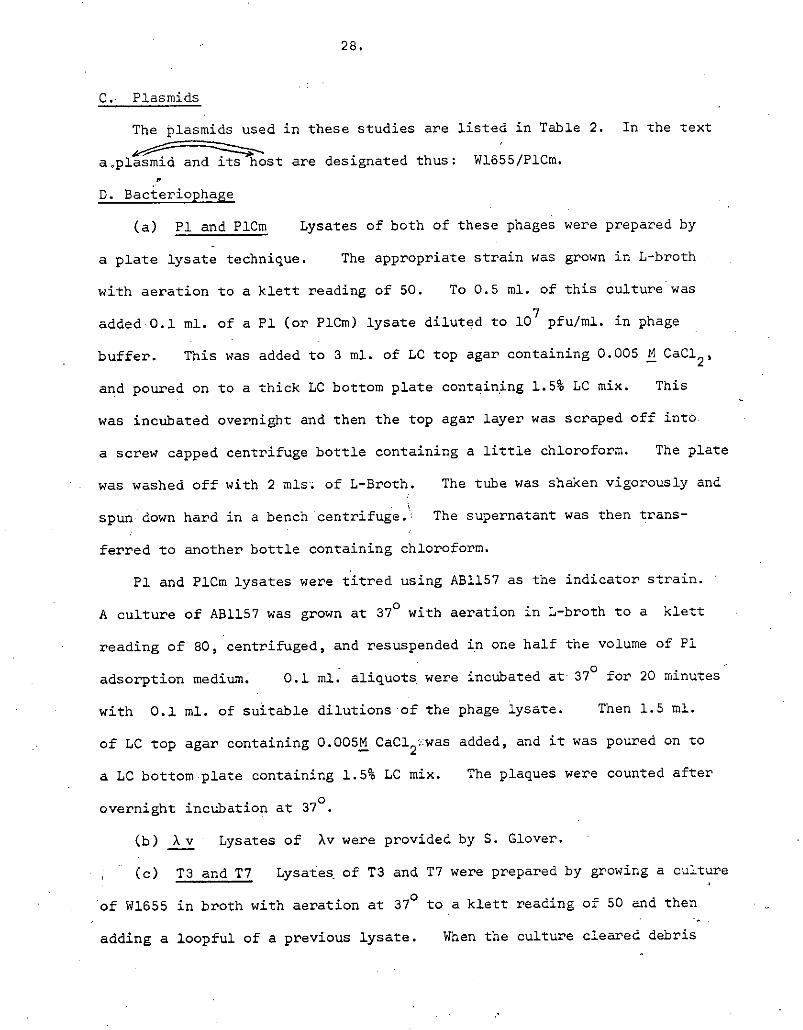
P1 and P1cm Lysates of both of these phages were prepared by
a plate lysate technique. The appropriate strain was grown in L-broth
with aeration to a klett reading of 50. To 0.5 ml. of this culture was
added 0.1 ml. of a P1 (or P1cm) lysate diluted to 10 pfu/ml. in phage
buffer. This was added to 3 ml. of LC top agar containing 0.005 N CaC1 2 ,
and poured on to a thick LC bottom plate containing 1.5% LC mix. This
was incubated overnight and then the top agar layer was scraped off into
a screw capped centrifuge bottle containing a little chloroform. The plate
was washed off with 2 mis; of L-Broth. The tube was shaken vigorously and
spun down hard in a bench centrifuge. The supernatant was then trans-
ferred to another bottle containing chloroform.
P1 and P1Cm lysates were titred using A51157 as the indicator strain.
A culture of AB1157 was grown at 37 ° with aeration in L-broth to a klett
reading of 80, centrifuged, and resuspended in one half the volume of P1
adsorption medium. 0.1 ml. aliquots: were incubated at 37 ° for 20 minutes
with 0.1 ml. of suitable dilutions of the phage iysate. Then 1.5 ml.
of LC top agar containing 0.005M CaCl2 :was added, and it was poured on to
a LC bottom plate containing 1.5% LC mix. The plaques were counted after
overnight incubation at 370
.
X Lysates of Xv were provided by S. Glover.
T3 and T7 Lysa-€es of T3 and T7 were prepared by growing a culture
of W1655 in broth with aeration at 37 ° to a klett reading of 50 and then
adding a loopful of a previous lysate. When the culture cleared debris

TABLE 2
Plasmids
Designation
Relevant Markers
Source
Reference
Fl - . W.J.Brammar --
P1Cm Crn' - J. Scott ;iKondo & Mitsuhashi (1964)
CIURTORStYSmR R192 E. Moody
Blob cmRTcRsu smR spcR
R100-1 As RlOO + drd-1 .):Egawa & Hirota (1962)
R64 - T0RSmR
R64-11 AsAs R64 + drd-11 . " It Meynell & Datta (1967)
1 + F lac + lac -- J. Scaife Scaife - & Gross (19o3)
F ts621 lact ts62 laot TI . it Cuzin & Jacob (1967)
F1 lac
tñaAl lact traAl "
1 t + F lac
+ + lacproB
KLF41 ArgOt Sm 5 Spc5 ma1maft N.- Willetts -
KLF112 ArgGt Su? Spc5 malAF t6112 See Methods
KLF113 Argot 5 R s malA+ f+ IT It
N) I-a

30.
was removed by centrifugation and the supernatant stored over chloroform.
Lysates were tltred in top agar poured on to broth plates using W1655 as
indicator.
MS2. Lysates of MS2 were prepared and titred as described in
Loels and Zinder (1961).
E. Methods
Growth and assay of bacterial cultures. Overnight cultures
were prepared by inoculating 5 ml. of either broth or the appropriate
minimal medium from •a single colony, followed by overnight (16-24 hrs.)
growth with aeration at the appropriate temperature.
Bacterial, growth was followed in a klett colourimeter, a klett reading
of 30 equalling approximately 1 x 10⁸ cells for a broth culture.
Viable counts (VC) were assayed by serial dilution in huffer, followed
by plating 0.1 ml. samples in 2.5 ml: of top agar spread over an appropriate
minimal agar plate. This was overlaid with a further 2.5 ml. of top agar.
Colonies were counted after incubation at 300. When colonies were to be
picked for further testing the bacteria were spread on the surface of
appropriate agar plates.
Conditioned Broth This was prepared by inoculating 3 litres of
broth with EC 1500/F1 lact and growing with aeration at 37° until a klett
reading of 55 was obtained. The majority of the cells were then centri-
fuged out and the remainder removed by filtration. The conditioned broth
was stored at -200C in 100 ml. portions. A fresh portion was used for
each experiment.
Steady state experiments. Where measurements were taken over many
cell generations, cultures were repeatedly diluted into prewarmed conditioned
broth, so as to maintain the cell density at a klett reading of between 10
and 50. The appropriate correction factor was made before plotting the

31.
results. Thus in these experiments the ordinate shows relative measurements,
and not the actual results obtained
-ga1actosidase assay Samples to be assayed were mixed with an
excess of chilled 1163 buffer containing a great excess of EC 1500 "carrier"
cells. They were centrifuged, washed with PM buffer, centrifuged again
and resuspended in 2 ml. of PM buffer. One drop of toluene was added,
the tithes whirlimixed and the toluene driven off by incubating at 370
.
The samples were then placed in a water-bath at 28 ° and 0.5 ml. of 11/75 ONPG
added with mixing. After a measured time the reaction was stopped by
the addition of 2 ml.: of M Na 2CO3 in 8 M urea. The samples were centrifuged 1111 ITA
and the Optical Density (OD) of the supernatant at 420/and 5504determined
in a Zeiss PMQ II spectrophotometer. J3 -galactosidase activity was calculated as OD units produced per minute of incubation using the
formula
0D420 - 1.75 x OD550
time of incubation
Mutagenesis. (i) 2-amino purine (2AP): TJ14/KLF112 was grown
overnight in minimal medium selecting for maintenance of the plasmid. This
culture was diluted 10 fold into fresh minimal medium containing 350fig/ml
2AP. This was immediately divided intQ 0.5 ml. aliquots. These were
incubated with aeration for 24 hrs. at 30 ° .• Each grew to a cell density
-
of about 5 x 10 8 cells/ml.
(ii)ICI-i91e:The Iroceduref same as for 2AP, except that l ,,ag/ml. ICR-191E
was used.
(iii) NTC: The method of Adenberg, Mandel and Chan (1965) was used,
except that 0.XM citrate buffer pH 5.5 was used. The final concentration

32.
of NTG was 100 ,,4g/mi. With X5119/R100 this procedure yielded 42%
survivors, of which 12.1% were auxotrophic.
(f) Isolation of spontaneous mutations. (1) Thymine requiring
mutant: ThyA mutations were isolated using trimethaprim as described
by Stacey and Simson (1965) and modified by Moody (1971).
Mutants with a low requirement for thymine were isolated from ThyA
derivatives by selection on plates containing 2,1'g/ml. thymine. Deoxy-
ribosemutase ( drm mutations were distinguishable from deoxyribose
aldolose (draY mutations by the degree of sensitivity to thytnidine
(Beecham, Eisenstark, Barth & Pritchard 1968).
t'alA mutations: Maltose requiring mutants were isolated from
the survivors of v infection. Ma1A mutations were distinguished
from Ma1B mutations by restoration of the mutant to mal+ by the introduction
of KLF'41.
NalR mutations: Broth + Nal plates were spread with about 108
sensitive bacteria. The colonies that grew up were restreaked on broth
t Nal plates.
mutations: 10 ml., of broth was inoculated from a single
colony and the culture incubated overnight with aeration. The culture -
was centrifuged and the cells resuspended in 0.5 ml. buffer. This was
added to 2.5 ml. top agar and poured over a broth agar plate containing
spectinomycin. After incubating for 2 days the resulting colonies were
restreaked on broth and spc plates. As often as not this procedure
yielded no mutants, and so it was always at least duplicated.
(g) Bacterial matings (i) Cross streaking: To introduce a
transmissible plasmid into a new host, exponential phase cultures of donor
and recipient were cross-streaked on a selective plate. Resulting colbnies
were restreaked on a similar plate.

33.
Flask matings: If the frequency of mating was too low for
cross-streaking, and for Hfr matings, exponexxtially growing recipient cells
were mixed with an excess of exponentially growing donor cells and incubated
as a thin layer in a large flask for 1 hr. at the appropriate temperature.
Dilutions were spread on selective plates, and resulting colonies purified
on similar plates.
Plate matings: (a) Fj.lact LEE - Recipients were replicated
on to selective plates spread with 5 x 10 8 exponential phase broth grown
A82463/F1 i AB2 1463 is a recA derivative of A31157 (Howard-
Flanders and Theriot (1966).
(b) R100-1 and R64V.11 - As for F 1 lac 2E2 .t except that the
donor strain was .36-2 (provided by F. Moody)..
The introduction of recA into a strain: To make a strain recA,.
a thyA mutation was first isolated.'. This was crossed with Hfr JC5088
(provided by N. Wilietts) which transfers thyC early. Selection was made
for thy recombinants, and purified thy +. recoirants were tested for
recA by replicating on to a freshly made broth plate containing three
parts per 10,000 of methyl methane sulphonate (MMS). RecA strains
re very sensitive to MMS. -
(h) Transduction. (i) A 10 ml. culture of recipient bacteria was
grown in L-Broth with aeration to a Klett reading of 40, centrifuged, and
resuspended in 1% Difco tryptone. 0.5 ml. of this was mixed with 0.5 ml.
of the PI lysate diluted to 5 x. 10 .pfu/ml. in LtBroth, and 0.5 ml. of
0 . 0 1 5 M:CaC12 and 0.03N MgS0 1 . The mixture was incubated at 370 for 20
minutes, centrifuged, washed with buffer, and finally resuspended in 0.1 ml.
buffer. This was then spread on a selective minimal plate, except for

34.
the case of markers which required growth to allow for expression of a
recessive allele, when the cells were diluted to about lO ml. in broth
and grown with aeration overnight, before spreading on selective plates.
In sudi cases, because of cloning effects, only one transductant per -
transduction could be scored for co-transduction.
(ii) Lysogenation by P1 and P1cm: P1 lysogens were isolated from
the survivors of a confluent lysis plate. Lysogeny was tested by spotting
about 10 T3 phage on to a patch of bacteria on a broth plate. P1Cm
lysogens were obtained by cross-streaking the recipient bacteria with a
P1Cm lysate on a broth and chloramphenicol plate.
Construction of KLF1I2 and KLF113 (i) KLF112: KLF41was
mated in to TJ6, and homogenotes for the tsl12 allele were selected by
replica plating for clones that were temperature sensitive malt
A number of such isolates were made, and the F-prime from each mated into
TJ14. To test that the F-prime now carried the ts112 mutation, it was
then mated into TJ17 (aleut transductant of the rect parent of JC1553).
ts112homogenotes were once again sought by replica plating for temperature
sensitive malt. These presumed homogenotes were cured of the F-prime
by growth at 42.50 and then infected with F1 lact traAl at 30
0 . The
majority of these clones were now temperature sensitive for the maintenance
of F1 lact trail. Thus tsl12 had been transferred from JJ6 to TJ17 via
an F-prime, which was designated 1CLF112.
KLF113: KLF41 was mated into TJ17, and homogenotes for the smR
allele selected by simultaneously selecting for malt, Sm'\ arg The
F-primes from the resulting strains were mated into AJ1. To test that these
F-primes carried smR and not a deletion orSm mutation, they were mated into
5i
R Sm strain (W1655) and n three cases out of eleven Sm colonies could be
35..
isolated at a much higher frequency than could be explained by mutation.
Thus smR had been transferred from TJ17 to W1655 via an F-prime, which was
designated KLF113. r
(j) Analysis of ribosomal subunits on sucrose gradients
The materials and methods iased were those given by Guthrie,
Nashimoto & Nomura (1969). and Nashimoto & Nomura (1970) except for the
following. Cells were lysed by sonication in an MSE 100 watt ultrasonic
disintegrator and cell debris removed by low speed centrifugation. 13 ml.
5-20% sucrose gradients were centrifuged at 32K th a SW36 rotor in a Beckman
L-250 tilt racentriuge for between S and 7 hours. Fractions were collected
directly into scintillation vials containing 10 ml s. of Bray's scintillant
(Bray 1960). Samples were counted in a Packard tri-carb scintillation
spectrophotometer • (model 3320). The 11 C labelled extract was kept frozen
° at -20. .

F
CHAPTER III EXPERIMENTAL STUDIES
Kim
SECTION I
A search for chromosomal mutants temperature
sensitive for plasmid maintenance
A. The plasmid P1 and its derivative PlQi
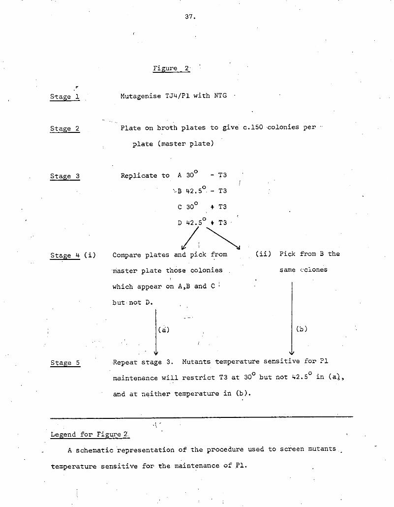
A cell that is lysogenic for P1 will restrict unmodified phage T3
infection by several orders of magnitude.(Bertani 1953; Lederberg 1957).
Figure 2 illustrates how this fact was utilised in a search for mutants
affecting P1 maintenance.
About 7 x lO mutagenised cells were screened in this way, but no
maintenance mutants were found. Seven clones were found in which T3
restriction was absent at 42.50, but regained after growth at 300.
As restriction was lost almost immediately at 42.50, these clones were
presumed to carry a P1 plasmid with a temperature-sensitive mutation
affecting the restriction enzyme.
P1CM is a derivative of phage P1 which confers chloramphenicol
resistance on cells that it lysogenises, but which is otherwise indistinguish-
able from the parent phage (Kondo& Mitsuhashi 1964). Mutants of TJ4/P1Ci
temperature sensitive for the maintenance of P1Cth were sought using the
procedure outlined in Fig. 2., except that chioramphenicol instead of phage Ta
was used.
About 5.x lO mutagenised cells were screened by this procedure, but no
maintenance mutants were found. Two mutants exhibiting temperature sensitive
chloramphenicol resistance were isolated (E. Reeve - personal communication).
In order to screen a much larger number of cells, a semi-selective
method was devised. This involved the use of ampicillin to select for cells
° which had lost the P1CM plasmid after growth at 42.5. Clones arising from
37.
Figure 2
r
Stage 1
Mutagenise TJ4/P1 with NTG
Stage 2 Plate on broth plates to give c. 150 -colonies per
plate (master plate)
Stage 3 Replicate to A 300 -
• 42.5° - T3
C30° +T3
D 42.5° 4T3
Stage 4 (i) Compare plates and pick from (ii) Pick from B the
master plate those colonies same colones
which appear on A.B and C
but not D.
(a) (b)
Stage 5 Repeat stage 3. Mutants temperature sensitive for Fl
maintenance will restrict T3 at 300 but not 42.50 in (al,
and at neither temperature in W.
Legend for Figure 2.•
A schematic representation of the procedure used to screen mutants •
temperature sensitive for the maintenance of fl.

these cells were then reinfected with P1Cth, and tested for loss of the newly
acquired plasmid at 42.50 .by replica plating. The full procedure is outlined
in figure 3.
A'total of 3315 Cth5 survivors of the ampicillin selection were reinfected
and tested, but none proved to be temperature sensitive for P1CM maintenance.
In view of the findings of Tokanb (1971), a lon strain was tested
for its ability to support P1CP'i in the plasmid state. Accordingly A31157
and A51157 ion (provided by W. Donachie). were grown overnight without
aeration in broth containing 0.05! Ca fl . Each culture was titred and then
infected with P1cm at a moi of 5, and incubated at 37 ° for 30 minutes.
Each culture was then r titred on broth plates containing 20 /ml chlor-
I/e?& cacfrehIa tde'ejt9/D'/rd yie4& 2.Zr/O 9
amphenicôl. J The ion overnight had a titre of 1.1 x 10 per ml. and
yielded 2.8 x 10 Cm* R tranductants per ml. (25.4)4 These results are
not consistent with the conclusion of Tokano (1971) that the ion locus is
essential, for PXCth plasmid formation.
B. The fit R-factors R-192 and'.Rl00
(a) R192 Mutants of TJ4/Rl92 temperature sensitive for the maintenance
of R192 were sought using a replica plating procedure similar to that
described in Section I A (b). About 7 x 10 rnutagenised cells were screened,
but no maintenance mutants were found. One temperature sensitive chlor-
amphenicol resistance mutant was isolated (E.. Reeve - personal communication).
(bE) RlOO Chromosomal mutants temperature sensitive for the maintenance
of R100 were sought using two different host strains, TJL and X5119. In
each case the procedure used was similar to that outlined in Fig 2, except
that Cth.5 cloneS resulting" from ampicillin selection were reinfected with
R100-1 ,from J6-2 as described in Methods. RlOO-i is a derivative of RlOO

39.
Figure a -
Stage 1. Mutagenise TJ4/P1CM with NTG. Cm added to cultures before and
after mutagenesis.
Stage, 12 Dilute 500-fold into broth and grow with aeration overnight at 42.5 °
Stage 3 Dilute 200-fold into broth t OM at 37 ° . After 1 hr. add ampicillin.
Incubate for 1 hr. and then plate on broth to give c.100 colonies
per plate (30 0 ).
Stage 4 Replicate to A Broth t Cw
B Broth - Qfff
Stage 5 Pick and patch CMS . Incubate at 30 ° (Master plate).
Stage 5 Replicate to broth + Cm, spread with PlC (30° ).
Stage 7 Replicate to broth + cm (30° ).
Stage S Replicate to broth (42.5 ° ).,
Stage 9 Replicate to A 42.5 ° + Cr
B42.5° -C
C30° - cm
Stage 10 Compare plates alter c.8 hr. Pick from master plate any
clones •which grow on plates B and C but not on A.
Stage 11 Reinfect with P1CxÜ at 300. Test by growing in broth at
42.5° and titring on broth 1- CM at 300.

Legend to Figure 3
A schematic representation of the semi-sensitive procedure used to
look for mutants temperature-sensitive for the maintenance of the plasmid
P1CM.
Stage 1 Chloramphenicol added to reduce the frequency of spontaneously
cured cells.
Stage 2 Stationary phase cells were used because this strain exhibits
loss of viability if chloramphenicol is added to a Cm5
exponential culture. Stationary phase cells, however, do not
lose viability.
Stage 3 The ampicillin treatment resulted in a 1000-fold reduction in
the viable count. About. 40% of the survivors were Cm 5 .
Stage S This step allows segregation of Cftl cells.
.142

41.
that differs only in its frequency of transfer (Egawa & Hirota 1962).
Using TJ4, 1207 CM clones were reinfectedand tested. Using X5119,
2982 Crñ5 clones were reinfected and tested. However, none of them proved
to be'tempevature sensitive for the maintenance of R100-1.
The fi R-factor R&4
R64 does not confer chloramphenicol resistance. - However, penicillin
can be used to select for loss of resistance to tetracycline (Bannister
1969). Accordingly, mutants of X5119/R64 temperature sensitive for the
maintenance of R64 were sought ubing a similar procedure to that outlined
in Fig. 2, except that tetracycline was used in place. of chloramphenicoL
TC5 clones were reinfected with R64-11 from J6-2 by plate mating as
described in Methods. R64-11 is a derivative of R64 that differs only in
its frequency of transfer (Meynell & Datta 1967).
A total of 2287 TC clones were tested, but none of them proved to be
temperature sensitive for R64 maintenance.
The F-prime F1 p ro t lac t
Ampicillin was used to select for cells, that had lost F1 2E57 lact
as outlined in Fig. 4. Four hundred such clones were reinfected, and one
was found that produced lac o. cells after growth at 42.50, but not at
0 ., . with + 0
30 . These cells could be reinfected with F lac at 30 , but again
yielded lac pro cells after growth at 42.5 ° . Sensitivity to the male
specific phage MS2 was concomitantly lost. Therefore it was concluded
that this strain now carried a mutation, ts B17, that rendered it temperature
sensitive for the maintenance of the plasmid F1 pro + lactL
42.
Figure 4
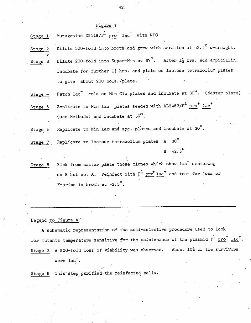
Stage 1 Mutagenisé X5119/F 1 j f lact with NIG
Stage 2 Dilute 500-fold into broth and grow with aeration at 42.50 overnight.
I'
Stage 3 Dilute 200-fold into Super-Min at 37 ° . After 1 12 hrs. add ampicillin.
Incubate for further l hrs. and plate on lactose tetrazoliurn plates
to give about 200 cols./plate. -
Stage 4 Patch lac- cols on Min Glu plates and incubate at 300. (Master plate)
Stage 5 Replicate to Min plates seeded with AB2463/F1 2 lac
(see Methods) and incubate at 300.
Stage 6 Replicate to Min lac and spc. plates and incubate at 300
.
Stage 7 Replicate to lactose tetrazolium plates A 30 0
B 42.5°
Stage S Pick from master plate those clones which show lac sectoring
on B but not A. Reinfect with F1 and test for loss of
F-prime in broth at 42.50
.
Legend to Figure 4
A schematic representation of the semi-selective procedure used to look
for mutants temperature sensitive for the maintenance of the plasinid F1pro lact.
Stage 3 A 500-fold loss of viability was observed. About 10% of the survivors
were lac.
Stage 6 This step purified-the reinfected cells.
43.
(E) Discussion
This section describes a method of obtaining chromosomal mutations
tempeature sensitive for the maintenance of the sex factor F, but which
was not successful in a search for similar mutations for P1Cm or for the
R—factors R192, R100 and R64. As spontaneously cured cells for each of
these plasmids occurred with a frequency of about S x 10- 4 , each cured
cell reinfected and tested theoretically represents the equivalent of : 2000
cells screened by replica plating. However, this upper limit of resolution
of the method could only be reached if there were only one plasmid per cell
and if the plasmid's replication were completely inhibited at 42.5 ° , so
that a 1000—fold increase in cell numbers at 42.58 produced 999 cured cells.
Furthermore, there would have to be no selection for wild-type cells, or
the segregation of cured cells would be offset by overgrowth of wild-type
cells. Thus the method would preferentially enrich for non-leaky
temperature sensitive mutants that did not affect the growth rate at 42.50.
Even if this method were only 10% efficient, it means that the
equivalent of over 3 x 10 P1cm containing cells, and about 5 x 10 R factor
containing cells, were screened. These figures do not include those cells
screened by replica plating.
One F maintenance mutant was found out of 400 cured cells, the
equivalent of 4 x lo replicated colonies allowing 10% efficiency. This
compares with 4 such mutants found by Jacob & Cuzin (1967a) from 10 replicated
colonies, also using NTC mutagenesis. Therefore either chromosomal mutations
temperature sensitive for the maintenance of P1Cm or of R factors occur with
a much lower frequency than for F, or this method does not enrich for them.
Tokano (1971) reported the isolation of chromosomal mutations that
would not allow P1Cm to enter the plasmid state, although the experiment

4 ,4.
described in Section 1A(c) does not support his claim that these were lon
mutations. His results would be compatible with those presented here if
for spme reason this locus could not yield temperature sensitive mutations,
or if either the introduction of the mutant allele into a cell already
lysogenic for P1Cm did not cause loss of the plasmid,or if such a mutation
was lethal to the cell in the presence of plasmid P1Cm because the phage
was induced- and entered the lytic cycle. Scott (1971) isolated temperature
sensitive clear plaque mutants of P1. If cells that are lysogenic for one
class of such mutants are raised to the non-permissive temperature then cured
cells are produced at an elevated rate, but only a small proportion of the
P1 plasmids are induced (R. Teather - personal communication).
The non-specific chromosomal mutants isolated by Helinski (1970) are
reported to be temperature sensitive for theimintenance of RI and R64
(Helinski et al. Abstracts of DNA meeting, Cold Spring Harbor, 1971). As
these mutations were isolated using a replica plating technique the frequency
with which they occur should not be beyond the limits of the method described
in this section. As R-factors are known to be composed étwo independent
replicons that are found in a composite structure in E.coli (Chapter I,
Section 2B) it seems plausible that if the replication system of one of the
replicons were to be impaired by mutation then the other replicon could take
over. This would explain the failure to find R-factor maintenance mutants
(Novick 1969) and also why the only chromosomal mutants to affect R-factor
maintenance are non specific, such that both R-factor replicons are
prevented from replicating.
As none of the F-maintenance mutants isolated by Hirota dt al.
or by Hohn & Korn (1970) show this lack of specificity, it is possiblethat
detection of such mutants is very dependnt on the genetic background of the

45.
host. This is consonant with the finding that strain differences affect
maintenance of F at high temperature (Stadler & AdeTherg 1972).
p.
/ . 1'

46.
Section 2. The Rate of Change of -galactosidase Induction Potential as a
Measure of F' lac Replication
If samples are taken from a culture and challenged to make an
enzyme under conditions of induction or derepression, then the rate at which
that enzyme is made is the inducibility or potential of the enzyme (Knempel,
Masters & Pardee 1965). In prokaryotes there is good evidence that in
exponentially growing cultures an increase in the potential of an enzyme
corresponds closely to an equal increase in the number of structurl genes
for that enzyme (Masters & Pardee 1965; Donachie & Masters 1966; Donachie
& Masters 1969; Helmstetter 1968; Pato & Glaser 1968). TheP•• if S-galacto-
sidase induction potential (B-gal I.?.) is measured in a strain in which the
lac region is deleted and which carries an FTlact factor, then the rate of
change of induction potential should correspond to the rate of change of the
number of copies of FTlact., This section desci?ibes. control experiments
to test this.
Rifampicin is known to inhibit completely transcription of the lac
region within 15 seconds of addition (Pato & von Meyenberg 1970). Figure 5
shows that if induction is stopped after 3 minutes by dilution into broth
and rifampicin, then -ga1actosidase synthesis stops completely within 20
minutes. As a result of this experiment, the s-gal I.P. of a culture in
all subsequent experiments was estimated by adding IPTG to an aliquot of the
culture at zero time, diluting into broth and rifampicin after three minutes,
and assaying for $-galactosidase as described in Methods after 20 minutes.
Figure 6 shows that if the s-gal I.?. of an exponential culture of
EC1500/F'lact is measured in thismanner, then anexponential increase is
observed.

5
75
50
25
47.
0 5 10 15 20 25 0 5 10
15
20 25
time:miflS
Legend to Figure S
An ECISOO/F'lac t overnight broth culture was diluted 100-fold into
conditioned broth and incub±èd with aeration at 300 until a klett reading of
30 was obtained. An aliquot of the culture was diluted 20 fold into con-
ditioned broth, and this second culture was incubated with aeration at 42.50.
IPTG was then added to the 300 culture. At the times indicated on the graph 1 ml.
samples were removed and assayed for 8-galactosidase as in Methods. When the
42.50 culture reached a klett reading of 30 it. was treated in an identical
manner, except that the temperature was 42.50 throughout.
Rsu1ts are plotted as CD units of colour produced/minute of incubation/
ml. of culture x 10, correction being made for the dilution at three minutes.
Controls showed that the colour produced in the absence of S-galactosidase was
negligible.
Conditioned broth was used for this and all experiments where S-gal I.P.
was measured because broth gave unreliable and unrepeatable results.

2 3 4 5 6 7 8 time: hrs

't8.
Legend to Figure 6
An EC1500/F' lact overnight broth culture was diluted 100-fold into
conditioned broth and incubated with aeration at 300. At the times
indicated the culture was shifted to 42.5 and then back to 3Q0•
Closed circles: Klett units
Closed triangles: lac + V.C.
Open circles: s-gal I.P.
H -- --. -
$ -

4 9
If the 5-gal I.P. of a culture is indeed a reflection of the number
of lac+ structural genes in that culture, and if the rate of F'lac t
• - replication is equal to the rate of increase of either optical density (OD) r
or cell number (VC), then the s-gal I. P. curve should be parallel to either
of the OD or VC curves However, this is not the case in Figure 6, where
the ratio of S-gal I.P. to OD or VC falls about 2-fold during. the course of
the experiment, and particularly while at 42.50. This could be due to a
progressive change in the 3-gal I.P. per lact gene, perhaps because of
changes in catabolite repression, or because the number of E"lac factors
per cell (or per OD unit) is falling. This could happen if Ftlact
replication is geared to some parameter of cell growth that is not measured
by either OD or VC.
The sharp discontinuities in the S-gal I.P. plot in Figure 6. suggest
that the higher final amount of enzyme produced in 42.50 cultures compared
to 3Q0 cultures of equal OD is due to an increased rate of transcription,
rather than a higher number of Fllact factors per unit OD. Such dis-
continuities were always found when temperature shifts were made.
Figure 7 shows the above experiment repeated using X5119/F'lac,
except that the shift back to 300 was omitted. Once again the lines are
not parallel. The VC to OD ratio - increases throughout the experiment,
suggesting that the cells are becoming smaller. The S-gal I.P. curve
is parallel to neither. At 300 it is approximately parallel to the 00
curve, whereas at 42.50 it more closely resembles the VC curve.
In view of the weight of evidence that induction potential is indeed
a measure of gene dosage the most likely explanation for the non-parallel
lines in Figures 6 and 7 is that the number of FTlact factors per cell or
per unit OD(ot both) is chaninj. ;1pehps,a..plot cfian 1iunmeauDéd

• 4 5. . 6 time:hrs
Leend to Figure 7
An X5119/Fhlac+ overnight broth culture was diluted 200 fold into
conditioned broth and incubated with aeration at 330• At the time
indicated the culture was shifted to 42.5.
- cloSed circles: Klett units
• closed triangles: lac V.C.
open circles: S-gal I.P.

51.
parameter of cell growth, such as membrane surface area, would give a
curve that was parallel to that S-gal I.P.
Oie curious difference between these two experiments was the finding
that an EC15007F'laJ culture produced about 3,-fold more 5-galactosidase
per unit OD than an X5119/Fllac+ culture. This could be due to differences
in catabolite repression in the two strains, or to the fact. that broth
conditioned by EC1500/Ftlact was used for both experiments. The levels
of metabolites excreted into the broth by EC1500/'F'lac t could effect
expression ofthe lac operon in another strain. The difference in 5-gal
I.P. is unlikely to reflect a 3-fold difference in the number of F lac
factors per cell, for reasons discussed in Section 3E.
Finally, it must be observed that implicit in studies of this sort is
the assumption that replication of the lac operon, and in particular the lac Z
gene, is synonomous with the replication of the F factor itself. Such an
assumption is based on the small size of most F primes, so that if F replication
proceeded at the same rate as for the chromosome, then the time between initiation
and termination of an F'lact replication event would be relatively short.
However, this has not yet been proved to be the case, particularly in mutants
that affect F maintenance. -
0

52.
Section 3. Kinetics of F-Factor Replication in Mutants
(A) Kinetics of loss of F ts62' lact at 42.5° from stationary
phase cells
Figure 8 shows the result of a typical experiment. When the proportion
of cells that are lact (N/No) is plotted against generations, extrapolation
back to zero time gives a figure of 14,3 F ts62' lac factors per cell.
This experiment was repeated several times, with a similar result on each
occasion. The number of F ts62' lact factors per lacF stationary phase
cell was estimated to be 14•9, 6.2 and 3.8 in these experiments, yielding
a mean value of 4.8. The wide variation between individual results is not
surprising in view of the method used to titre the lact population.
A number of attempts were made to alter this result by varying the
experimental conditions.
/ (a) The stationary phase culture was prepared in broth instead
of M9 lactose. This only resulted in a higher titre with up to 30%
of the cells lac.
The 4,eriment was conducted at 43.5 ° . This produced no
significant change, and 'a figure of 5.5 F ts62' lac factors per cell
was obtained. . -
The overnight culture was held at 42,50 for two hours before
dilution into broth at 42.50
. A fure of 5.8 F tsSQ' lact factors
per cell was obtained, compared to 6.2 for the control, which was not held
at 42.50. This result shows that the high number of F ts62' lact factors
consistantly obtained is not due to the presence of preformed, temperature
sensitive "F replication::factor(s)", unless they are in a temperature
resistant, presumably non-active, state in stationary phase cells.

[0] 53.
[bJ
101-.
Fts 62 +factors/ce
aI• Nol
1
10' I I I
0 30 60 90 120 150 180 0 1- 0 1 2 3 4 5
time:mins: generations
Legend to Figure 8
EC1500/F ts621act was grown into stationary phase overnight in minimal
lactose medium at 300
. A zero time this culture was diluted lO fold into
broth and incubated with aeration at42.5 0 . At intervals appropriate
dilutions were plated on tetrazolium lactose plates.
Fig. B (a) open triangles: total V.C.
closed triangles: lac + V.C.
t (N) Fig. 8 (b) open circles: proportion of cells that are lac
- . over the proportion that are lact at
- zero time (No). .

54.
The MY lactose stationary phase culture was incubated with
aeration at 3Q0 for two days, instead of overnight. No significant difference
was okserved, the estimated number Fts62' lac+ factors per cell being 4.9.
The experiment was repeated using strain PS200/Fts62' lac +' the
original strain used by Cuzin & Jacob (1967a). They claimed that there was
no replication of the Fts62' lact factor at 42.5 ° in broth, and that
there was only one copy per stationary phase cell. However, the result was
essentially similar -to those obtained with EC1500/F ts62' lac t An accurate estimate of the number of Fts62' lac factors per cellhwas not possible..
This was due to the small, proportion of the population which was thermostably
lac t , presumably due to integration of the Fts62' lac t into the lac region.
(B) Kinetics of loss of Fts62' lac '' from exponential cells at 42.5 ° .
Figure 9 shows the result of a typical experiment, in which a figure
of 2.7 Fts62' lact factors per exponential phase cell was obtained.
Two similarly conducted experiments yielded values of 3.6 and 4.5, giving
a mean of 3.6. Two experiments in which the cell titres were estimated
using tetrazolium lactose plates gave figures of 5.2 and 7.1 Fts62'lact
factors per cell, suggesting that this method tends to over estimate.
These results may be interpreted in a number of ways.
There are between three and four Fts62' lact factors per
exponential cell at 300, a number maintained when the cells enter stationary
phase.
There are less than this number of Fts62' lact factors per
cell, but replication continues for a short time at 42.50, before settling
to the non-permissive rate.
The result is being obscured by reinfection of cured c61ls

V.C.
io3
10,
101
r
2. . 3 4 5 6 7 time: hrs

55.
Legend to Figure 9
An EC15OO/F.ts621 lact minimal lactose overnight culture was diluted 10
fold into broth and incubated with aeration at 300. After 4 hours this
culture was diluted 10 fold into broth and incubated with aeration at 42.50.
Throughout, samples of appropriate diu.ltions were plated on minimal glucose
and minimal lactose plates.
• Fig. 9 (a) open triangles: total V.C.
closed triangles: lact V.C.
• Fig. 9 (b) open circles: proportion of cells that are lac t (N) over
the proportion that are .lac+ at the time of
101 r [b] temperature shift (No.).
27 F ts 62' lac factors/cell
F
N • N0
•1
generations

56.
This possibility may be discounted because • Fts62' lact transfers itself
with a very low frequency. This has been shown to be due to a second
mutati,pn, in the tra G gene (N. Willetts - personal communication).
In order to distinguish between (a) and (b) above, Fts62' lac
replication was followed more directly, by measuring rates of change of
5-gal I.P.
(C) Kinetics of Fta2' laQt replication monitored by changes in
S-gal I.P.
Figure 10 shows the result of an experiment in which s-gal I.P. was
measured. At the same time the experiment depicted in figure 9 was
extended to follow the recovery of Fts62lac+ replication upon shift-down
to 300.
Several observations may be made. Firstly, S-gal I.P. increases
exponentially after the shift to 42.5 ° , but at a much slower rate than:
that of cell growth. This rate is achieved within a matter of minutes,
if not immediately. Secondly, upon shift-down to 300 cell growth rate
immediately falls to a slower rate (doubling time about 52 mins.) whereas
s-gal I.P. increases at an exponential rate with a doubling time of 31
min s. After s-gal I.P. had increased 16-fold its rate of increase dropped
to approximately that of cell growth. During this time the lact titre
had risen just over 3-fold. This suggests that the number of Fts62t l act
per lact Cell rose to about 5 during this period. This agrees with a
figure of 4.8 Fts52!. lac factors per lac cell obtained from the kinetics
of segregation of F cells at 42.5 0, using the same method outlined
previously. (Not very accurate in this case, as segregation of F cells
was not followed for very long
During the period of rapid increase in 5-gal I.P., the lac. titre.
,. continued to rise at a slower rate than the total titre for about one

11 13
10
1
io
101
10.
time: hours

57.
Legend to Figure 10
An EC 1500/Fts62 lac+ minimal lactose overnight culture was diluted
500 fold into broth and grown overnight with aeration at 300. This was
then diluted 200-fold into conditioned broth and incubated with aeration at
300. When a klett reading of about 40 was reached the culture was diluted
6 fold into conditioned broth and this culture was grown with aeration at
42.5° , with dilution into fresh conditioned broth at intervals as indicated
in Methods. At the time indicated on the graph the culture was diluted
back to 300 .
Open triangles: total V.C. !
Closed triangles: lap + V.C.
Open circles: /3 -gal I.P. Closed circles: Klett units
The breaks which occur in all four curves just before seven hours
correspond in time to a dilution into fresh conditioned broth. Presumably
an error in the dilution factor is responsible.

W.
generation, and then rose until the two rates of increase were equal.
There are three possible explanations for this. First, there might be
a loss of viability of lac t cells upon plating. Secondly, the lactcelis
in the population might divide at a slower rate than the F cells upon shift-
down to 300.. Thirdly, cell division is normal, but F cells continue JttM'a'
to be segregated out. In order to distinsii4i between these possibilities
the experiment described in Section 3D was carried out.
(D) Segregation of F cells during replication of Fts62' lact H
Figure 11 illustrates an experiment that shows that the difference
in rates of increase of total titre and lact observed in figure 10 is not
due to loss of viability upon plating. The coulter counts and the viable
counts agree closely for all five secondary cultures. These cultures all
show constant growth rates. If division was temporarily inhibited in cells
containing an ,Fts62?lac+ factor upon shift back to 300, then coulter
counts should increase at a slow rate for a while, increasing to the normal
rate when the inhibition was1ifted.
Thus it seems that considerable segregation of F cells occurs after
shift back to 300, despite-the immediate recovery of F ts62' lact replication.
This accords with,the observation that colonies on lactose tetrazolium plates
arising from the small proportion of lact cells in a culture of EC1500/Fts62' lact
after many generations of growth at 1+2.50 are very deep pink, due to the many
lac cells present. - That these cells are spread throughout the colony,
rather than in idiscrete sectors suggests that the process of segregation
continues for many generations after shift back to 300. Colonies arising
from cells that have been cultured at 3Q0, or have been at 42.50 for only
- 1 or 2 generations do not show this effet. -r

F
[a] [b]
3 10
10
101
time:minS time: hrs

59.
Legend to Figure 11
An EC1500/F ts62' lact minimal lactose overnight culture was diluted
200—fold into conditioned broth and incubated with aeration at 30 ° . when
a klett reading of 40 was reached the culture was diluted into conditioned
broth and incubated with aeration at 42.50. At the times indicated in
Fig. 11 (a) samples were diluted back into conditioned broth at 30 ° . The
growth of these cultures was followed using a Coulter counter. (This part
of the experiment was performed by N. Jones.) After 3 hours at 42.50 the
primary culture was diluted back to 300•
Fig. 11 (a) open circles: total V.C.
closed triangles: lac t V.C.
closed circles: klett units
Fig. 11 (b) closed squares: Coulter counts plotted against time after
/ dilution back to 30 ° . (Corrected for
dilution so as to be comparable with the
primary culture.)
I '
I

60.
(E) Replication of Flact tra Al in ts B17
Figure 12 shows that FTlact tra Al replicates exponentially in
TJ40at 42.50, but at a slower rate than cell growth. As with F ts62' lact
this slow rate of replication is achieved very rapidly after raising the
temperature.. If these results are treated in the same way as before, then
a figure of 3.1 Ftlact traAl factors per TJ40 cell at 30 0 is obtained.
In connection with Figures 6 and 7 it was mentioned that an EC1500/
Ftlact culture produced about 3 fold more -ga1actosidase than an X5119/
F' lact culture of the same optical density. The same is true for the
amounts of -ga1actosidase produced by a culture of EC1500/F ts62' lac t
relative to a culture of TJ40/F'lact traAl. The result noted above makes
it unlikely that there is a 3-fold difference in the number of copies of F
between these two strains, unless one supposes that F ts62'lac t factors
segregate in units of 3.
The rate of F' lac traAl replication at 42.5 in TJ40 is about the
same as that of Fts62?lac+ in EC1500 (see . Section SF). This could mean that, .
the ts 62 and tsB17 are equally leaky. Another explanation is that a
second replication system,.not as efficient at 142.50 as the normal system,
is utilised. As the }Cornberg DNA polymerase is essential for Col El main-
tenance (Kingsburg & Helinski 1970) it seemed plausible that elimination of
this enzyme.from TJ40/F'lact traAl might abolish FlacttraAl replication
at 42.5° . Accordingly a mutation effecting this enzyme, pol Al. was
introduced into TJ40 /see Methods). The resulting strain (TJ69) was
- infected with F'laJ tra Al, and the experiment described in Figure 13
performed. This shows that F'lacttraAl replication is not abolished at
142.50 in TJ69, but continues at the same rate asin the p01 At.controL,
despite a slower growth rate.
Footnote - pol Al strains grow aboyt 10% more slowly than their pol At parents 04. Peacey - personal communication?. .

time:hours

61.
Legend to Figure 12
A TJ'+O/F'lact traAl minimal lactose overnight culture was diluted 500'
fold into broth and grown with aeration at-30 0 overnight. This culture was
then diluted 200 fold into conditioned broth and incubated with aeration at
300. When a klett reading of about 40 was reached the cuittire was diluted
into conditioned broth and incubated with aeration at 42.5.
open triangles: total V.C.
closed triangles: lack V.C.
open circles: p-gal I.P.
closed circles: klett units
/ /
\ fl
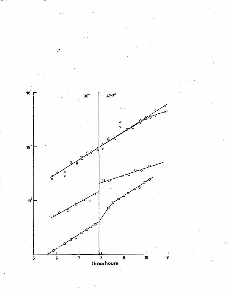
10
10
10'
5 6 7 8 9 10 11 time: hours

62.
Legend to Figure 13
A TJ69/F lac ttraAl minimal lactose overnight culture was diluted 500
fold into broth and grown with aeration at 300 overnight. This culture
was then diluted 200 fold into conditioned broth and incubated with aeration
at 3Q0• When a klett reading of about 40 was obtained the culture was
diluted into conditioned broth and incubated with aeration --at 42.50
open triangles: total V.C.
closed triangles: lact V.C.
open circles -gal I.P.
closed circles: klett units
H
\

63.
Rate of replication at 42.50 relative to growth rate (p.6?)
Table 3/gives measurements of growth rate and F replication rate at
42.5°,compiled from several previous figures and experiments mentioned in
the text.
These results show that the rate of F ts62 + lac replication falls
as the growth rate slows, but not in proportion. Introduction of po1 Al
into a strain carrying tsB17 does not have a marked effect on the rate of
Fllact tra Al replication, even though growth rate is retarded.
The slowing of growth rate observed when conditioned broth was used
is not due to a pH change. The most likely explanation is that more and
more nutrients become limiting as the broth is conditioned further. If
this is so then these results mean that the residual E replication at 42.50
does not respond to changes in growth rate in the same manner as the
bacterial chromosome (Cooper & Helmstetter 1968). It also explains why
there is less segregation of ,F- cells at 42.50 when less rich growth media
are used (see Table 4).
Discussion
This section describes experiments designed to estimate the number
of F factors per cell under different conditions, and to study the effect
of changes in the mass/plasmid DNA ratio on F replication.
However, the method used for estimating the number of copies per cell
is not a particularly good one, even when it can be demonstrated that residual
replication rate at 42.50 is constant. I suggest that a better way of
obtaining this information would be to use a non-mutant F-factor carrying
an integrated p phage. The number of copies of p phage, and hence the ,number
of F-factors, per cell could then be accurately estimated by titrating, the p
II

64.
DNA using hybridisation, in a similar way to that used to map the origin
of chromosome replication (Bird, Lauarn, Mart üscelli & Caro - in press).
This method could theoretically be applied to any plasmid.
Notwithstanding the fact that an indirect method was used, some
conclusions may be drawn from the results presented here. The estimate
of the number of F- factors per exponential phase cell agrees with earlier
findings that there is only a small number per cell, but differences over
a two-fold range were found from experiment to experiment. Coupled with
the finding that S-gal I.P. did not parallel either optical density or
-viable count (Figures 6 and 7) suggests that the number of F-factors per
cell may vary somewhat, even under apparently identical conditions. It is
not possible to conclude more from the results presented here.
The number of F-factors per cell appears to be maintained when
stationary phase is approached, although it was not possible to check the
rate of residual replication at 42.50 using S-gal I.P. because of the
difficulties of interpret/ing such results using stationary phase cultures.
Such a finding would be expected if F replication was coupled to the
physiological division of the cell (Clark 1968), as was suggested by Zeuthen
& Pato (1971). - If F replication were controlled by dilution of inhibitor
(Pritchard et al. 1969), then the number of F-factors per cell should fall
as stationary phase is approached owing tote smaller size of stationary
phase cells (Donachie - personal communication). -
The experiments in which F replication was followed by means of S-gal
I.P. demonstrate that Fts62 and tsB17 both affect the process of F replication,
and that the elevated rate of curing observed for both mutations is not due to
defects in the subsequent segregation process. This is evidenced by the fact-
that the rate of F replication immediately moves to a new rate upon raising

65.
the temperature to 42.50, and the observation that when the rate of
segregation of F cells had become constant the rate of F replication
paralleled the rate of increase in lac t viable count. The effect on cell
growth observed by Hohn & Korn (1970) as a direct consequence of the
segregation of an F-factor from their F-maintenance mutants was never
observed for either Fts62 or tsB17. It is possible that whilst Fts62
and tsB17 affect replication, the F-seg. mutations of Hohn & Korn (1970)
cause defects in the process of segregation.
When a 42 . 50 grown culture of EC15OOIFts62'lac t was returned to 300
F cells continued to be segregated out even though F replication immediately
rose to a rate faster than that of cell growth. This finding can partially
be explained by cell division before the first F replication event after
the shift to 30° . Indeed, the fact that it takes 30 mins. for the
4Jf -gal UP. to double means that some cells must take-heap a generation to
complete their first F replication. However, the continued segregation of
F cells at subsequent cell divisions, suggested by the appearance of the
resulting lac+ coloniesr ,on lactose indicatoi plates, ithpliCs that normal
patterns of segregation are disturbed for many generations. Novick & Brodsky
(1972) studied the events that followed the introduction of penicillinase
plasmids into Staphlococcus aureus and showed that there was a considerable
time lag between commencement of replication and the achievement of hereditary
stability. On the assumption that recovery of F replication from a state
in which there is presumably only one F-factor per cell is a similar event
to the introduction of a single plasrnid by transduction, the findings discussed
in this paragraph are ' ' in agreement with the conclusion of Novick & Brodsky (1972)
that plasmid establishment is not aunitary event subsuming both ability to
replicate, and hereditary stabilisation. If this is inde$the case for F,

66.
it is an at ument against the replicon-maintenance site model of Jacob et al.
(1963). However, aspointed out by Novick & Brodsky (1972) it is important
to determine the exact number of copies of plasmid per cell, in order to
distinguish between a 1-.*2-*l and a 2-?4--s2 plasmid replication cycle.
This information is of critical importance in interpreting the significance
of any disparity between commencement of replication and attainment of
hereditary stability. Indeed Collins (1971) claimed that there are 2
F' .lact factors per chromosomal terminus in E.coli, replicating at the
same time as termination. If this is the case, and if both segregate together,
then the method of estimating the number of F-factors per cell used here will
give a false answer.
The kinetics of recovery of F replication at 300 cannot easily be
interpreted on the basis of the dilution of inhibitor model of plasmid
replication (Pritchard et al. 1969) This model predicts that successive
rounds of initiation should occur in order to restore the volume/plasmid
origin ratio, such as occurs with the E.coli chromosome after inhibition of
DNA synthesis (Pritchard & Lark 1964). This does not appear to happen in
the case of F, however, some cells taking up to half a generation to complete
their first replication event (Figure 10). Similar kinetics of recovery of ?74)
F replication were found by Zeuthen & ?atojafter both chromosomal and episomal
replication had been arrested by means of a temperature sensitive DNA
initiation mutation.
Another aspect of the results presented in this section that is
difficult to reconcile with a simple inhibitor-dilution model of replication
control is the fact that ' a- constant rate of F replication ismaintained at
42.5° throughout the period when the number of plasmids per Ft cell is
falling. This means that the replication function(s) affected by Fts62 and

67.
tsB17 must be such that the rate of F replication is independent of the
number of copies of F pet cell at the time of F replication, and hence
independent of the number of F replication events per Ft cell per cell
generation. The dilution of inhibitor model predicts that a fall in the
number of F replcatiop events per cell cycle would mean fewer "bursts" of
inhibitor. This in turn would lead to a lowering of the concentration
of F inhibitor as cell growth proceeded, which should allow further F
replication. Modifications of the model such that at the non-permissive
temperature the "burst" of inhibitor produced per F replication was greater
or conversely that the critical concentration at which F replication occurred
was lowered, would result in only transient effects on the rate of F replication,
until a new, lower steady state of F factors per cell was achieved, after
which the rate of £ replication, would again parallel cell volume. Only
major alterations iniimhibitor burst size or sensitivity to inhibitor, such
that the degree of change was greater than the multiplicity of F-factors in
a "wild-type" cell, could result in a permanent alteration of the rate of
F replication relative to growth rate, because in such a situation a single
F replication event in a cell would more than double the concentration of
inhibitor. In such a case, however, one would expect no F replication at
the non-permissive temperature until sufficient cell growth had occurred to
dilute out existing inhibitor, and that the rate of F replication that then
occurred would be directly proportional to the growth rate. As neither
of these predictions hold true, the lesions caused by Fts62 and tsB17 cannot
be directly associated with the inhibitor or its target. However, if the
lesion in these mutants 'is- in the process of replication itself (either ,
directly or indirectly) rather than in the control circuit., then one would
expect the inherent self-regulating properties of the model to compensate for

the lesion as the number of F—factors per cell, and hence the concentration
of inhibitor, fell. Thus the dilution of inhibitor model of plasmid
replication (Pritchard et al. 1969) is insufficient to account for the.
kinetics of replication observed with Fts62 and F'lac +traAl in TJ40.
The finding that the residual rate of Fts62Tlact replication at
142.50 3s largely independent of the growth rate suggests that possibly
Fts62 affects F replication by lowering the affinity of F, or some F product,
for some cellular product that is diredtly or indirectly concerned with
replication. The similar insensitivity of residual F'lacttraAl replication
to growth rate in TJLO raises the interesting possibility that tsB17 acts -
by reducing the affinity of the same cellular product for F, or some F
product.
\

69.
Derived from Klett
TABLE 3
Mean doubling time
Total VC
in minutes at
: J3-gal I.P.
42.5°
lact VC
Fig. 9 - 26.5 - 69
(repeat. 24 75
(repeat 25.5 - 71.5
Fig. 10 144 • 5 43 ?0 90
it (repeat 1 67 68 102 105
Fig. 12 60 58 85 84
((repeat 1) 57 - 77.5 -
Fig. 13 70 72 85 -
- Legend to Table 3
lact VC doubling time is obtained from the portion of the graph after
segregation of F cells per generation is constant. In the cases where both
5-gal I.P. and lact VC doubling times were measured close agreement was
observed. Therefore the lact VC measurements taken from Figure 9 and the
two repeats may reasonably be taken as a measure of the rate of F replication.
Figure 10 (repeat 1) was performed in broth conditioned to a Xlett
reading of 100. This slowed the growth rate considerably.

70.
Section 4. Studies on the Maf locus
(A) tsl12 and tsBI7 are alleles of jj
Tab 4 shows the patterns of loss of various F primes from TJ40 r
and TJ6. Thus both tsl12 and tsB17 are complemented by the ts 1 allele
carried by K4'4l, but not by the ts112 allele carried by KLF112.
Both mutations are closely linked to spectinomycin resistance.
When JG108 (a W3110 derivative provided by J. Gross) was used as the
recipient, 8/19 •p transductants from TJ6 carried the ts112 mutation.
A co-tranEduction frequency of 7/10 was found for tsB17 and
These results indicate that ts112 and tsB17 are allelic. Henceforth
this locus will be referred to as Maf (MAintenance of F).
tsl12 and tsB17 appeared to differ in one respect. TJ40 does not
exhibit the lipid rich granules which appear in TJ6 after about 3 hours
growth at 142.5 ° (Hirota, Ryter & Jacob 1968) (see figure 14). These
inclusions are not observed in either strain at so° , nor in their respective
wild type parents. The JG108 ts112 transductants were examined micro-
scopically after growth at 42.50 but no granules could be detected. Thus
either the lipid-rich granules reported by Hirota et al. (1968) are not
associated with the ts112 mutation, or their detection is dependant upon
a particular genetic background. To distinguish between these possibilities
ts112 was co-transduced with SPC back into P8200 from the JG108 derivative.
No granules could be detected in the resulting transductant after growth at
42.50. Therefore it can be concluded that the lipid rich granules are not
associated with the ts112 mutation, and are no doubt caused by a second
mutation introduced at the-same time astsll2. This conclusion is in accord
with the finding that while TJ6 was slightly more sensitive to SDS at 42.5
than was P8200 (judged by growth on broth plates containing a range of SDS

71.
TABLE 4
Temperature sensitive loss of various F primes
StraipjPlasmid
Broth - % sugar+ &çerniin - % sugar t Mm; Glu - sugar
JC1553/KLF41 (
~ /,t) 97.2 . 99.5 No growth
TJI4'/KLF112 96.5 100 No growth
TJ40/KLF41 97.4 98.4 98.4
TJ40/aF112 (617111t) 4.1 18.5 29.2
TJ40/F'lacttraAl (617) 1.6 - 17.1 31.3
TJ6/F'lacttraAl (liz) 2.8 24.2 42.5
TJ6/KLF41 ('12./f) 95.0 97.4 96.1
(f/i/liz) 8.9 31.0 38.9
X5119/F'lacttraAl 98.5 100 9.9.6
PS200/FtlacttraAl .9.7 . . 98.8 100
EC15OO/F'lacttraAl 99.8 100 99.7
EC1500/Fts62 1 lact 0.5 . 9.2 23.4
. Legend to Table 4
Strain/Plasmid combinations to be tested were incubated overnight with
aeration at 300 in minimal media selecting for maintenance of the plasmid.
The cultures were diluted 10 fold into prewarmed broth, superrnin or minimal
glucose.. These cultures were incubated with aeration at 42.50 overnight,
and then titred on appropriate MacConkey indicator plates. Results were
expressed as the proportion of cells that were lac, malt.

72.
ftft
do
/ : 1
a I
I •1
WI
()
Legend to Figure l'-
Strains to be tested were aerated at 300 in broth until a Klett
reading of 30 was obtained. The cultures were diluted 0-fold into broth
and aeration continued at 142.50. When a Klett reading of 30 was obtained
a loopful of culture was examined under phase contrast using a Zeiss Photo-
miccpscope. The photographs (mag x 239) were taken and developed by
K. Begg.
Figure l+ () TJ6 at L12.5 °
Figure 14 (b) TJ40 at L2.5 °
Observations were also made of TJ6 at 30 ° , TJ40 at 30 ° , X5119 at 300 and
42.50,PS200 at 30° and 42.5 ° , and 3 of the JG108 ts112 transductants at 300 and
42.5° . In each case the result was essentially similar to that presented in
Figure 14 (b). PS200 is the strain from which 1-lirota et al (1968) isolated tsll2.

73.
concentrations), neither TJ40 nor the JG108 ts112 derivative could be
distinguished from their respective parental strains by this method.
I(B) The Maf product has an essential cell function
The Maf locus codes for a product that is essential for the maintenance
of the sex factor F. In this section evidence is presented that this
product is also essential for cell growth.
ts112 and tsE17 are mutations of Maf in which the gene product is
altered in such a way that F replication is normal, or very nearly so, at
the permissive temperature. If this gene product is not essential to
cell growth then the elimination of this product, such as in a maf* mutation,
would result in a strain totally unable to support the maintenance of any
autonomous F-factor, unless it carried the maft gene. If the maft gene
product is essential to cell growth; then a maf would be non-viable,
unless an F prime carrying the maf +'allele were present.
Figure 15 illustrates the procedure used for a search for a maf
mutation using TJ14/KL.F112. If the gene product is essential, and a mat
mutation is introduced into the chromosome, then the resulting phenotype
should be temperature -,:sensitive lethality, as the KLF112 should be lost
at 42.5 0, leaving a non-viable F7 cell. Furthermore, the strain should
be incurable at 3Q ° due to the depend/nce of the cell on the ts112 allele
carried by KLF112. This would be readily testable, as an F cell would
be mal- R Str R.
If the gene product is not essential, then t#e resulting maf strain
should be temperature sensitive for maltose utilisation, owing to the loss
of KLF112 at 42.5° .
When 2AP was used as the mutagen, about 5 x lO colonies were
replicatédi 18 Temperature sensitive lethals were found, but none of them

74.
FIGURE 15
Procedure used to search for maf
r
Stage 1 Mutagenisé:TJ14/KLF112 with 2AP or ICR191E
Stage 2 Plate on M9 Glu+His Met Leu to give about 100
colonies per plate and incubate at 3Q0 for
48 hrs. (master plate)
Stage 3 Replicate to (A) McConkey maltose plates at 42.5 °
(B) " at 3Q0
Stage 4 Compare (A) and (B) and pick from master plate
tslethals
(a) (including poor (b) tsmalt
growth at 42.5° )
NV Stage 5 Re-test ts lethality and re-test ts malt
ability to yield Sm' inaf
colonies. -

75.
proved incurable. One temperature sensitive mal+ was found, but this
'- - mutation had apparently occured in the Mal A region carried by KLF112.
,On the'., assumption that the gene product of maf is a protein,
the acridine half-mustard ICR 191 E was then used because of its ability to
produce frameshift mutations with a high frequency (Ames & Whitfield 1966;
Martin 1967; Berger, Brammar & Yanofsky 1968, and Newton 1970), an ability
not affected by the presence of a recA mutation (Bast.arrachea, Gonzalez
& Tam' in press). About 2 x 10 colonies were replicated and two
temperature sensitive lethals were obtained, one of which did not yield Tmal
Str colonies at 300. This clone (TJ59/KLF112) was investigated further
and the following information obtained. Firstly, when large numbers
(upwards of 10 10 ) of cells were spread on broth plates containing streptomycin,
a few strR colonies were obtained, 10 of which were tested flirther. All 10
were still temperature sensitive lethal, and 9 of them were mat argt.
Thermost likely explanation of this is that they carried F primes with a
deletion extending from beyond malA to between Str and tsll2. Deletions.:,
from one end of the ch%mosomal material carried on an F prime have been
found for other F primes (Yura & Igarashi 1968; Austin, Tittawella, Hayward
& Scaife 1971). This would place inaf anticlockwise to Str on the genetic co.-robora,tecl
map (see figure 1), which is ccl1ab.erattfrby the high co-transduction
frequency of maf and spc. The remaining atrR derivative was malt argt
and presumably carridd a spontaneous str or str' mutation on the KLF 112.
Secondly, growth did not cease completely at the non-permissive HZ
temperature. If TJ591(was streaked out on broth plates and incubated at
42.5° then small colonies appeared after 2-3 days', while figure 16 illustrates
the effect of a temperature shift on an exponentially growing culture.-.

time :hours

76.
Legend to Figure 16
TJ59/KLFI12 was grown into stationary phase overnight with aeration
at 300 in 1.3% tryptorie abroth. At time zerth this culture was diluted
200-fold into fresh 1.3% tryptone broth and incubated at 300 with aeration.
At the time indicated the culture was diluted 3 fold into prewarmed 1.3%
tryptone broth and incubated with aeration at'42.5 9 .- ---------------''- -
closed circles: Klett
closed, squares: coulter counts
open triangles: viable count.
At 10 hours the particle count was about 2—fold higher than viable
count. By 17 hours this difference had risen to 4—fold.
/ .
L

77.
1 Failure of growth to stop completely at 42.50 is to be expected, due to
the leakiness of the ts112 mutation (Alfaro 1971), allowing slow KLF112
replication which in turn allows slow growth. Two features of figure 16
that support the hypothesis that KLF112, and hence the ts112 allele, is
being segregated out leaving behind non-viable F cells are firstly that
growth rate falls slowly at 42.5 0 , which could reflect the dilution of tsll2
alleles, and secondly thatcoulter counts rise at a faster rate than viable
counts, indicating an increase in non-viable cells.
The phenotype described above could be the result of .a lethal
mutation, on the chromosome in the diploid region together with a leaky
temperature sensitive lethal mutation outwith this region. To test this /k&F//2.
possibility TJ59/was mated with AJX/KLF113, and T359/KLF113 selected by
selection for malstrR. 16 Such isolates were purified, and all 16 gave
normal colonies at 42 . 50 . However,: although several streaks of each
were made on MacConkey maltose plates, no mal colonies or sectors were
ever seen. 6 of these isolates were tested for curability by growth in
the presence of a range of concentrations of AO, followed by plating on
MacConkey maltose plates, but once again no mal colonies were seen. Thus
the substitution of KLF113 for }CLF112 relieves the temperature sensitivity
but not the incurability, and eliminates the possibility that there is a
temperature sensitive mutation outwith the diploid region.
The observed phenotype would also be expected if a maf mutation
was not lethal but was coupled to a second, lethal mutation on the chromosome
in the diploid region. If this were the case then temperature sensitive
revertants of TJ59/KLF112 should become temperature sensitive for maltose
- utilization because KLF112 would now be lost at 42.50. Three independant
revertants were isolated by picking the largest colonies from broth plates

78.
incubated at 142.50 and serially restreaking several times on broth plates
incubated at the high temperature. None of the 3 revertants exhibited
temperature sensitivity for maltose utilisation, although all 3 could now
be cured of KLFI12 as demonstrated by their ability to yield mal s.trR
arg colonies. Such cured cells that did arise yielded lact colonies when
infected with F'lac t traAl, and hence were not maf ..... It is possible, of
course, that these revertants were not "true" revertants, but carried a
suppressor mutation. However, if TJ59 did carry two independant mutations
then both would have to be suppressed to produce the observed phenotype.
Thus it would seem that TJ59 carries a maf mutation, and that maf
is an essential gene for cell growth.
However, one alternative explanation remains that cannot readily be
disproved. This is that the maf gene is not essential, but is part of .a
transcriptional unit that contains essential genes. A frameshift mutation
in a proximal essential gene, or in the maf gene distal to an essential gene,
could cause the phenotype described, if there were nonsense read through to
(or from) the maf gene. On the other hand, genes associated in transcript
tional units Usually have associated functions This point is discussed
in Section 14 G.
(C) Maf not DRAG
It seemed possible that Maf and DNAG might be one and the same, as
DNAG has an approximate map location very close to that of Maf (Gross 1971).
Accordingly three tsDNAG bearing strains (JG32, JG314 and JG150 - provided by
J. Gross) were made recA, lac and malA. In one experiment FTlac+traAl
was introduced into each strain, which was then tested for temperatur'e
sensitive loss of the Filact traAl factor at 300 and at •tl° intervals,.
until the mutants would not grow. No effect on F?l acttraAl maintenance
was observed.

79.
In a second experiment, KLF112 was introduced into each strain.
All three then yielded normal colonies at 1+2.50, but were incurable at
this temperature, as judged by their inability to yield mal colonies.
All three, gave mal colonies at 300. Thus in each case both the lesion
on the chromosome and on the F prime was compleJted. Therefore Maf and
DNAG do not appear to be in the same cistron.
(D) The Effect of spc' on the ts112 phenotype .
Both TJ6 and TJ40 are spectinomycin resistant. In view of the
findings of Yamagata and Uchida (1972a) discussed in Chapter I, the original
PS200 ts112 strain was obtained from N. Willetts. 11 independent
mutants were isolated at 3Q0, and infected with FTlacttraAl. They were
then tested for maintenance of FllacttraAl at 42.50. The results are
given in Table 5, together with theresults obtained from four similarly
tested thutan -ts isolated from PS200 ts
Thus the introduction of SpcR into PS200 tsll2 may either suppress
the temperature §ensitivity of FtlacttraAl maintenance, or it may render
the strain temperature sensitive lethal Two of the 11 Spc mutations had
no apparent effect Presumably the Spc R mutation in TJ6 was by chance
such a mutation.
The Spc gene is known to code for a single protein component, the
split protein P4 (Nomura, Mizushima, Ozaki, Traub & Lowry 1969), of the
30s ribosomal subunit (Bollen, Davies, Ozaki.& ilizushima 1969).
Many examples of interaction between ribosome mutations are known,
including restriction ofviable mutational changes in other ribosomal loci,
and modification of existing phenotype (Schlessinger & Ap'rion 1969;
Nomura 1970).
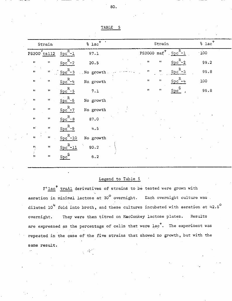
FM
TABLE 5
Strain % lac Strain % lact
PS200 tsl12 Spc R -1 97.1 PS2000 maf +
Spc
R-1 100
H 20.5 . Spc2 99.2
.No growth ......... - .. " - ii spcR_3 99.8
spc4 No. growth . " SDc R -4 100
7.1 Spc5 , 99.8 . .
". Spc-6 No growth
P spcR_7 No growth
87.0
Spc R -9 4.5 -
spcR_io No growth .
Spc - 11 90.2
Spc5 6.2
Legend to Table 5
Filact traAl derivatives of strains to be tested were grown with
aeration in minimal lactose at 3Q0
overnight. Each overnight culture was
diluted 10 fold into broth, and these cultures incubated with aeration at L42.5 °
overnight. They were then titred on MacConkey lactose plates. Results
are expressed as the percentage of cells that were lact. The experiment was
repeated in the case of the five strains that showed no growth, but with the
same result.

These facts strongly suggest that tsll2 is a mutation in a gene whose
function is affected by ribosomal structure. In view of the fact that all
known 30s ribosomal subunit genes, as well as some 50s subunit genes and r
some translation factor genes, map in the same region of the chromosome as
maf (Davies & Nomura - quoted in Nomura & Engae) 1972), it - Seems plausible
that rnaf itself codes for a.ribosomal protein. Indeed, •Nomura & Enaek
(1972) showed that at least four of the ribosomal genes in this region, one
of which was spc and which straddle the approximate map position of maf
(see figure 1), are transcribed asz;a single unit. -•
In view of the finding that some are themselves
temperature sensitive for the maintenance of Filact (Yamagata & Uchida
1972b), the possibility exists that maf and spc are one and the same, and
that mutations in different regions , of the gene can give different phenotypes.
However, in view of the fact that these authors used very high doses of NTG
(300,#g/ml for 40 minutes) the possibility of multiple mutation within a
• small region of the chromosome cannot be excluded. It has been shown that
lower doses of NTG (100 , .g/ml. for 30 minutes) can produce up to 20% unselected
mutation in genes less than 1 minute map distance away from the primary target
(Guerola, Ingraham & Cerada-Olmedo 1971). On the other hand, these authors
screened for total loss of function in the unselected marker. Mutations to
temperature sensitivity in an adjacent gene should be much less frequent.
The next two sub-sections describe experiments performed in an attempt
to demonstrate that máfis concerned with ribosomal structure and/or function
(E) tsB17 has no effect on F mediated phage T7 restriction
Morrison & Malamy (19 71) showed that F mediated restriction of the female
specific phages T7 and II operates at the level of translation, preventing
the synthesis of all but the very first phage proteins. They showed that at

82.
at least two F genes were involved, and speculated that these genes might
code for translation initiation factors. If this is so and if one further £
specuXates that tsB17 and ts112 affect F replication by imparing the
efficiency of one or more of these F initiation factors, then the presence
of such a mutation might reduce the restriction Of phage T7 by an Ft cell.
In order to test this it was necessary to use a tsB17 strain in which the
F prime had become integrated into the chromosome, in order to avoid
segregation ofF cells. Accordingly, T.340/F'lact was plated on tetra-
zolium lactose plates and incubated at 42 . 50 . Lact clones were picked
and tested for stability of the lact phenotype at 42.50
. This acquired
stability was presumed to be due to integration of the F prime into the
chromosome, because 5 such, clones readily gave recombinants for chromosomal
markers when cross-streaked with A31157. These 5 clones were tested for
retention of the tsB17 allele by screening for rare lac segregants at 42.50.
These segregants were then infected with FtlacttraAl and sl5own to be
temperature sensitive for maintenance of the F prime.
The 5 Hfr strains were then tested for their ability to restrict phage
T7 at 300 and 42.5 0 . At neither temperature did they show any difference
from the X5119/F'lact control, as judged by both eop or plaque morphology.
At both temperatures the 5 .Hfrs and the control gave an eop of about 30-fold
lower than X5119 alone, and the plaques were considerably smaller. This
result shows that chromosomal integration has no affect on F mediated T7
restriction. Restriction of another female specific phage W31, is reduced
by integration of the F factor (Watanabe & Okada 1964).
Thus, tsB17 has no apparent effect on F mediated phage T7 restriction.
- However, this does not disprove the hypothesis that competition for ribosomal
"sites" by F and T7 initiation factors is responsible for restriction.

83.
Firstly, even though the relevant F initiation factor(s) might only function
inefficiently in tsB17 at 42.5 ° , they may still be able to efficiently
exclude T7 initiation factors. Secondly, if only one F initiation factor
were'affected by tsB17 then restriction of T7 might still be expected.
Indeed, Morrison & Malamy (1971) isolated F mutants that exhibited reduced
restriction and concluded that mutationto complete lack of inhibition could
not be effected by a simple single step change. Thirdly, tsB174ould affect
a third F initiation factor, not involved in phage T7 restriction. The
reduced restriction mutants isolated by Morrison & Malamy (1971) did not
affect other F properties, such as transfer or autonomous replication.
However, one can argue that mutants that affected all F properties would
be a forbidden class, as such F-f actors would be lost.
(F) Analysis of ribosomal subunits on sucrose gradients
If the pattern of growth exhibited by TJ59/KLF112 in Figure 16
reflects the segregation of non viable F cells ) and it maf determines a
ribosomal structural protein, then after prolonged growth at 42.50 a culture
of TJ59/KLF112 would contain gells that were unable to synthesise one of
the many proteins required for ribosome assembly, although existing functional
ribosomes within such cells should allow synthesis of the remaining subunit
proteins. In vitro reconstruction studies on the 30s subunit (Nomura,
Mizushima, Ozaki, Traub & Lowry 1969) in which single ppotein components
were omitted revealed 3 classes of component. The first class (when omitted)
yieldedthat were indistinguishable from fully reconstructed subunits on
sucrose gradients. The second class yielded particles that sedimented at
27-295, whilst omission 6'f' - the third class resulted in particles which gave
a major sedimentation peak at 20-25s. If the lesion in TJ59 results in
failure to synthesise a 30s subunit structural protein that falls into either

8.
of the latter two classes, then F cells in a TJ59/KLF112 population might
be expected to assemblq incomplete particles, distinguishable on a sucrose
gradient. In view of the findings of Yamagata & Uchida (1972b), discussed
in Section LID, it is worth noting that Nomura et al. (1969) found that
omission of pro;4n N, coded for by the spc locus, yields 28s particles.
14 Figure 17 shows the ribosomal subunit pattern bbtained from a
labelled extract of strain MRE600, chosen because it is RMase I deficient
(Cammack & Wad9 1965), a property which is reputed to stabilise the subunits
during storage (J. Robertson - personal communication).
Figure 18 shows the result obtained when these 34C "reference" subunits
were mixed with H labelled extract from TJ59/KLF112. Several features
may be noted. Firstly the pattern obtained from the H extract is very
14 similar to that obtained for the .t:C extract in Figure 17. Allowing for
the longer time of centrifugation, both peaks have travelled the same
distance in the gradient. There is a hint of a shoulder to the slower
sedimenting peak but this does not look significant in view of the large
amount of material at the top of the gradient. The 14C labelled material
however, shows a distinctly different pattern from that observed in Figure 17.
This is presumably due to partial disintegration of the subunits , as the
proportion of the label found at the top of the gradient has increased.
There are a number of possible explanations for the failure of this
method to detect ribosomal subunit differences, apart from the possTh:ility
that maf does not code for a ribosomal protein.. Firstly, if maf coded for
a class I protein (see above), no difference would be detected. The same
result would be obtainedif maf coded for one of the many translation initiation
- or elongation factors that exist, but which are not considered to be structural -
proteins. Secondly, as many if not all of the ribosomal loci in this region
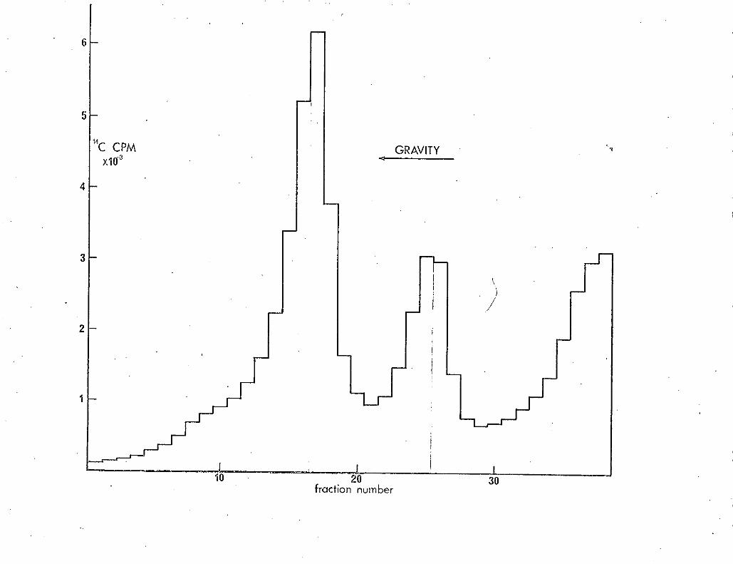
El
fraction number

85.
Legend to Figure 17
MRE600 (provided by R. Hayward) was grown overnight in 1.3% tryptone
broth into stationary phase at 37 .with aeration. This culture was diluted
200 fold into prewarined 1.3% tryptone broth and incubated with aeration at
42.50 izhen a Klett reading of 30 was obtained 14C -uracil(lO. jcCi; 62 mCi/rn mol)
was added. After 1 hour the cells were harvested and treated as in Methods.
50 /-11 of extract was layered on to the gradient, which was centrifuged for
5 hours. 12 drop fractions were collected and counted with a window of
50-1000 and a.20% gain...:
/

C PM 10 2
fraction number

Elm
Leaend to Fip.ure 18
TJ597KLFII2 was grown tiñto stationary phase overnight in 1.3%
trypt.6ne broth at 300 with aeration. This culture was 'dfluted 100-fold
into 1.3% tryptone broth and grown with aeration at 3Q0• When a Klett t
reading of 30 was reached the culture was diluted 12 fold into prewarmed
1.3% tryptone broth and aerated at 42.50. When the kClett reading again
reached 30 -uracil (50jtCi;29 mCi/mmol) was added. After one hour the13
cells were harvested and treated as in Methods. 50l of the extract was
mixed with 101 of 1 C labelled extract. This was layered on to the
gradient which was then centrifuged for 7 hours. 6 Drop.fractions were
collected and counted witha red channel setting of 50-370, gain 60% and
a green channel se-ting of 370-1000, gain 13%. These setting counted 3H
with an efficiency of 47.4% and +4C with an efficiency of 59.5%. 14 C
spillover into the red channel was 7.4%.
Results are plotted without correction for spillover, as the maximum
correction needea would only be in the order of 2%.
Continuous line - extract
Broken line - 14C extract
(Results not plotted as a histograrn1fQr the sake of clarity.)

87.
of the genetic map are transcribed as one unit (Nomura & Endbaek 1972).
The introduction of a frameshift mutation, whether located in maf or not,
might conceivably result in the elimination of many ribosomal proteins
simultaneously. In this case F cells in the population would probably
be unable to assembl.>/ even defective particles, and only ribosomal RNA would
-be labelled in extracts from these cells. Indeed, the proportion of
14 label ànincorporated into subunits is higher than is the case for the
a "reference" subunits, even after the presumed partial degrfdation of the
latter. This can hardly be taken as evidence, however, in view of the ,160sn,4s i,i ext fr&CtS .Jro'
apparent instability ofxeven/RNase I deficient strains.
Finally, it has not been proved that F cells are produced in this
situation, or that even if theyare that they are capable of making significant
• - - amounts of protein. The latter point could be demonstrated by autoradio-
• graphy following incorporation of, say, 3H-leucine, but the satisfactory
demonstration that non viable cells in a mixed population did not, contain
KLF112 would be difficult.
(G) Discussion
In this section evidence has been presented that tsll2 and tsB17
are alleles of the same gene, maf,and that the lipid-rich granules observed
in the original isolate of ts112 are due- to a second mutation. Maf was
found to be 85% co-transducible with !R2' although this figure may not
accurately reflect the map distance between these loci. This is because
of the long las between the primary recoinbinational event and the expr9ssion
of spectinomycin resistance, allowing distortion of the co-transduction
frequency by any selective-pressures that may be operative.
The maf product was shown genetically to have an essential cellular
function, or at least to be associated with essential genes in a single
MAN
transcriptional unit. In fact, the demonstration by Nomura & Endbaek (1972)
that 4 ribosomal loci, ery, str, !Ea.and fus, which straddle the approximate
map pçsition of maf, are transcribed as a single transcriptional unit makes
it quite possible that the putative frameshift mutation in TJ59 is in fact
in a proximal gene to maf. Such a possibility, however, serves to strengthen
the hypothesis that maf codes for a protein connected with ribosomal structure
and/or function, suggested by the finding that the tsll2 phenotype can be
altered by the: introduction of a spc mutation. Direct attempts to demon-
strate a connection between maf and ribosomes failed (Sections 4(E) and 4(F)).
This point might be resolved by an analysis of ribosomal components extracted
from cells carrying ts112 or tsB17 by means of polyacrilamide gels, or
indeed any technique designed to detect small changes in the primary
• structure of proteins.
/ Yamagata & Uchida (1972)a isolated a mutation (76) that suppressed.
the Fts62 phenotype and which was itself suppressed by spc 1' to which it
was very closely linked. Furthermore, the same authors (1972)b showed that
mutation to alone could affect F maintenance at 420. Are maf, eps
• and spc one and the same gene, mutations of which, perhaps in different
regions of the gene, can give widely differing phenotypes? The line of
approach mentioned in the preceding; paragraph might also solve this problem.
A genetic analysis of the whole region is theoretically another way in which
the problem might be tackled, but such an approach is beset by technical
• .•. difficulties, such as the very poor growth rate of some ribosomal mutations
and the unpredictable phenotype of strains carrying different ribosomal
mutations. Interactive relationships can drastically change ratios of
recombinant classes, making conclusion;about genetic mapping extremely
• ifficu1t(Schlessiflger & Apirion 1969). . .

99
One possible way in which a ribosomal alteration could affect F
replication, namely by loss of recognition of F translation factor(s),
has already been discussed (Section 4E). If this is indeed the mechanism
of action of ts112 and tsB17, then the primary lesion in the ribosome would
lead to secondary effects by reducing the amount of F specified protein(s).
Thus physiologically the mutation would be observed -as an F mutation, and
could be the cause of the similarity of phenotype observed for Fts62 and
tsB17. If this is the case, and if the suggestion of Yamagata & Uchida
(1972)a that in Fts62 the F translation factor(s) are altered, then neither
Fts62 nor maf are directly concerned with replication, but act by affecting -
the production of essential F replication protein(s). The efficiency
of translation of F mRNA would not be affected by growth rate if this is
the mechanism of Fts62 and tsB17, because the critical parameter, namely
the degree of efficiency of the F translation factor, would be determined
by physical constants, in the same way as is the rate of peptide addition.
This has been shown to be constant at growth rates faster than one generation
per hour (Forchhammer & Lindahl 1971). This is consonant with the finding
that the residual rate of replication in Fts62 and tsB17 is largely independent
of growth rate (see Section 3).
An alternative model to explain the involvement of the ribosome in F
maintenance is to assume that the ribosome or ribosomal protein interacts
'with the membrane,. and in some way impairs the efficiency of F replication,
perhaps by means of conformational changes. Some support for this idea
comes from the finding that 2ps76 affects the sensitivity of wild type FTlact
to AO (Yamagata & Uchidal972a)..nsitiVity to this class df compound may
also be altered by means of changes in the membrane (Nakamura & Suganuma 1972).-

P

90.
Adachi, H., Nakano, M., Inuzuka, H. & Tomoeda, H. (1972) J. Bact. 109, 1114-1124.
Adenberg, E.A., Handel, M. & Chen. G.C.C., BBRC (1965), 18, 788.
Ad1er"H.I., Fisher, W.D., Cohen, A. & Hardigree, A.A. (1966) Proc..Nat.
Acad. Sd. USA, 57 5 321-326.
Alfaro, G. (1971) Ph.D. Thesis (University of Edinburgh)
Ames, B. & Whitfield, H. Jr. (1966) CSHSQB 31, 221-225
Atwood, K. & Norman, A. (1949) PNAS (USA), 35, 696-709
Austin, S. (1911) Ph.D. Thesis (University of Edinburgh)
Austin, S., Tittawella, I.., Hayward, R. &Scaife, J. (1971) Nature New
Biology, 232, 133-136
- Bannister, D. (1969) Ph.D. Thesis (Univerthity ;of Edinburgh)
Bastarrachea, F. & Clark, A.J. (1968) Genetics, 60, 641-660
Bazaral, M. & I-ielinski, D. (1968)a J Mol.Biol. 36, 185-194
Bazaral, H. & Helinski (1968)b Biochemistry, 7, 3513-3519
Bazzicalupo, P. & Tocchini-Valantifli, G.P. (1972) PNAS, 69, 298-300
Beacham, I.R., Eisenstark, A., Barth, PT. & Pritchard, R.H. (1968)
Mol.Gen.Genetics, 102, 112-127.
Berg, C. &Caro, L. (1967) J.Mol.Biol., 29, 419-431.
Berger, H. Brammar, W. & Yanofsky, C. (1968) J.Bact. 96, 1672-1679.
Bertani, G. Ann Inst.Pasteur, 84, 273-280 (1953)
Blair, D.G., Clewell, D.B., Sherratt, D.J. & Helinski, D.R. (1971) PNAS
68 9 1 9 210-214.
Bollen, A., Davies, J., Ozaki, H. & Mizushima, S. (1969), Science, 165, 85.
Bray, N. (19.60). Analyt. Biochem., 1, 279.
Burnett, F.M. (1934) Bid. Rev., 9, 332-350.
Cairns, J. (1963) CSHSQB, 28, 43-49.
Cammack, K. & Wade, H. (1965) Biochem.J., 96, 671-680.

91.
Campbell, A. (196 Advances in Genetics, II, 101-146.
Caralli, L.L. , L.ederberg, J. & Lederberg, E.M. (1953), J.gen.Microbiol. .
89-aO3.
Caro, L. &.Berg, C. (1969). J.Mol.Biol., 45, 325-336.
Chabbert, Y.A., Baunders, .3.G. & Bouanchaud, D. H. (1969) Ciba Foundation
Symposium on Bacterial Episomes & Plasinids, 227, 239.
Clark, A. (1963) Genetics, 48, 115-120.
Clark, A. J. & Margulies, A.D. (1965),PNAS, USA, 53, 451
Clark, A.J., Maas, W.K. & Low, B. (1969), Mol.Gen,.Genetics, 105, 1-15.
Clark, D. (1968), CSHSQB, 33, 823-836.
Clewell, D. & Helinski, D. (1969), PNAS, USA, 62, 1159-1166.
Clewell, D. & Helinski, D. (1970)a BBRC, 40, 608-613.
Clewell, D. & Helinski, D. (1970)b,l }3BRC, 41, 150-156.
•
Clewell, D. &Helinski, D. (1970)c,Biochemistry, 9, 4428-4440.
Clewell, D. (1972) J.Bact. , 110, 667-676.
• Clowes, R. C, & Rowley, D. (1954) J. gen. Microbiol., II, 250
• Clowes, R.C.(1961), Nature, 190, 988-989.
Clowes, R.C. (1963), Genet.Res., 4, 162-165.
Clowes, R.C., Moody, E.E.M., Pritchard, R.H. (1965), Genet.Res., 6, 147-152
Cohen, S. & Miller, C. (1969), Nature, 224, 1273-1277.
Cohen, S. & Miller, C. • (1970)a, J.Mo1.Bio1. 50, 671-687.
Cohen, S. N. & Miller, C.A.(l970)b, PNAS, 67, 2, 510-516.
Cohen, S.N.,Silver, R.P. & McCoubrey, A.E. (1971), Nature New Biology,
231, 249-252.
Collins, J. (1971) Ph.D. Thesis (University of Leicester);
Cooper, S. &Helmstetter, C.E. (1968), J.Mol.Biol., 31, 519-540.
Cuzin, F. & Jacob, F. (1967)a, Ann.Inst. Pasteur, 112, 397-418

92.
Cuzin, F, & Jacob, F. (1967)b Ann.Inst.Pasteur, 112, 529-545.
Cuzin, F. & Jacob, F. (1967)c Ann.Inst.Pasteur, 113, 145-155.
Datti, N. & Hedges, A. (1971), Nature, 234, 222-223.
Dennison, S. (1972), J.Bact., 109, 416-422.
Donachie, W. (1969), J.Bact., 100, 260-268. -
Donachie, Si., Hobbs, D. & Masters, N. (1968), Nature, 219, 1079-1080.
Donachie, Si. & Masters, H. (1966), Genet.Res. j 119-124.
Donachié, W...& Masters, N. (1969), The Cell Cycle (Ed. Padilla, G.M.,
Whitson, G.L. & Cameron, I.L.) Academic Press, pp. : 37_76
Dubnau, E. Maas, Si. (1968), J.Bact. 95, 531-539
Echolls, H. (1963), J.Bact., 95, 531-539.
Egawa, R. & Hirota, Y. (1962), Jap.J.Genet., 37, 66
Falkiow, S., Wbhlheiter, J., Citarella, R. & Baron, L. (1964), J.Sact.,
87, 209-219
Falklow, S. & Citarella, R. (1965), J.Mol.Biol. 12, 138-151.
Falklow, S., Citarella, R, Wohiheiter, J. & Watanabe, T.(1966), J.Mol.Biol.
17, 102-116.
Frame, R. & Bishop, J. (1971), Biochem. J., 121, 93-103.
Fredericq, P. (1957), Ann.Rev.Microbiol., 11, 7-22.
Freifelder, D. (1968)a, J.Mol.Biol. , 34, 31-38
Freifelder, D., (1968)b, J.Mol.Biol., 35, 95.
Freifelder, D. & Freifelder, D. (1968)a, JJlol.Biol., 32, 15-23
Freifelder,D. (1969), J.Mol.Biol., 45, 1-7 -
'reife1der,D.,, Folkmanis, A. & Kirschner, I.. (1971), J.Bact., 105, 3, 722
- Frydman, A. & Meynell, E. (1969) Genet.Res., 14,315-332.
Goebel, Si. & Schrempf,H. (1971) J.Bact., 106, 311-317.

93.
Gross, J. (1971) Current Topics in Microbial. & Immunology, 57, 39-74
Guerola, N., Ingraham, J. & Cerda-Olmedo, E. (1911), Nature New Biology,
230, 122-125
GuthrIe, C.. Nashimoto, H. & Nomura, M. (1969), PNAS (USA), 63, 384-391.
Haapala, P. K. & Falkiow, S. (1971),J.._Bact.,?10..,J1, 294-295.
Hayes, W. (1953), J.gen.Microbiol., !' 72-88.
Hedges, R. & Datta, N. (1971), Nature, 234, 220-221
Helinski, D. (1970) Abstracts Proc. 10th Int.Cong. Microbiol. Mexico. p. 103
• Helmstetter, C. (1968) J.Bact., 95, 1634-1641.
Helmstetter, C. & Cooper, S. (1968), J.Mol.Biol., 31, 507-518.
Hershey, A. , Burgi, &Ingram, L. (1963), PNAS (USA) 49, 748-755.
Hickson, F. Roth, T., & Helinski, D. (1967), PNAS (USA) 58, 1731-1738
Hirota, Y. (1960), Proc. Nat. Acad. Sci. USA, 46, 57-64
Hirbta, Y,, Ryter, A. & Jacob, F. (1968) Cold Spring Harbor Symp, 33, 677-693.
Hirota, Y., Mordob, J. & Jacob, F. (1970) "On the process of Cellular
Division in E.coli III Thermosensitive mutants of E.coli altered in the
process of DNA initiation. JMo1.Biol., 53, 369.
Hoar, D. (1970) J. Bact. 101, 9167920.
Hohn, B. & Korn, D. (1969) J.Mol.Biàl., 45, 385-395 • • . -,
Horn, B. & Korn, D. (1970) Abstracts Proc. 10th Int. Cong. Microbiol.
Mexico. p. 103.
Howard-Flanders, P. & Theriot, L. (1966) Genetics, 53, 1137
Hubacek, J. & Glover, S.W. (1970) J.Mol.Biol. 50, 111
Ikeda, H. & Tomizawa, J., Cold Spring Harbor (1968) 33, p. 791
Inoue, M. & Guthrie, P. (1969) PNAS (USA) 64, 957-961. .
Insemurg, 3, J.Bact., (1971)2105, 620.
Iyer, V. N. & Palchoudhury, S.R. (1971) 57. 319

94.
Jacob, F., & Woliman, E.L. (1958) Comp.Rend., 247, 154-156.
Jacob, F. & Brenner, S. (1963) Cornp.Rend., 256, 298-300.
Jacob, F.,, Brenner, S. & Cuzin, F. (1963) CSHSQB, 28, 329-343. r
Kahn, P. & Helinski, D. (1964)':88, 1573-1579
Kass, L.R. & Yarmolinsky, M.B.,(197Q), PN4S,,66:r3815822
Kasumatsu, H. &Rownd,R.(1970) J.14o1.Bio1.,51,473489
Kuempel, P., Masters, M. & Pardee, A. (1965). BBRC, 18, 858-867
Kingsbury, D. & Helinski, IL (1970), BBRC, 41, 1538-1544
Kline, B. & Helmski, D. (1971). Biochemistry, 10, 4975-4980
Kohiyama, M., Cousin, D. Ryter, A. & Jacob, F. (1966) Ann. Inst. Pasteur, .
110, 465"486.
Kondo, E. & Mitstthashi, S. (1964) J.Bact., 88, 1266-1276.
Kontomichalou, P., Mitani, M. & Clowes, R. (1970) J.Bact., 104, 34-44.
Kohn H. (1971) Royal Society Symp. on 'Bacterial PlasTnids'.
Kuempel, P.L., Masters, M. & Pardee, A.B. (1965), BBRC, 18, 858-867.
Lang, D. & Mitani, M. (1970), Biopolymers, 9, 373-379
Lark, K.G. (1972), J.Mol.Biol., 64, 47-60.
Lederberg, J. (1952), Physiological Reviews, 32, 403-430.
Lederberg, E. & Lederberg, J. (1953) Genetics, 38, 51
Lederberg, S. (1957), Virology, 3, 496-513
Lennox, E. (1955), Virology, 1, 190
Loels, & Zinder. (1961)PNAS, 47,'1135-
Maas, R. & Maas, W. (1962) PNAS, 48, 1887-1893.
Maas, R. (1963), PNAS, 50, 1051-1055.
Maas, W. & Goldschmidt, A.'(1969), PNAS, 62, 873-880.
MacFarren, A.C. & Clowes, R.C. (1967) J.Bact., 94, 365-377 . ... -
Martin,R. (1967), J.Môl.Biol., 26, 311-328

Li
95.
Masters, M. & Broda, P. (1971) Nature New Biology, 232, 137-140.
Masters, N. & Pardee, A.B. (1965) PNAS(USA), 54, 64-70.
Meynefl, E. & Datta, N. (1967) Nature, 214, 885-887
Meynell, E.,. Meynell, G.G. & Datta, N. (1968) Bact.Rev. 32, 55-83
Meynell, G. (1969) Genet.Res. 13, 113-115
Monk, M. (1967) Mol.Gen.Genetics, 100, 264fr274.
Moody, E. (1971) Ph.D. Thesis (University of Edinburgh)
Morrison, T.& Malamy, N. (1970) J.Bact. 103, 81-88
Morrison, T. G. & Malamy, M. H. (1971) Nature New Biology, 231, 37-41
Nashimoto, H. & Nomura, N. (1970) PNAS (USA), 67, 14140-1447
Newton, A. (1970) J.Mol.Biol., 49, 589-601
Nishimura, Y., Caro, La., Berg, C.M. & Hirota, Y. (1971) J.Nol.Biol. 55, 441-456
Nisioka, T., Mitani, M. & Clowes, R. (1969) J.Bact., 97, 376-385
Nisioka, T., Mitani, N. & Clowes, R.C. (1970) J.Bact. 103, 166-177
Nomura, M. (1967) Ann.Rev.Microbiol., 21, 257 - 284
Nomura, M. (1970) Bact.Rev., 34, 228-277
Nomura, M. & Engack, F. (1972) PNAS(USA) 69,. 1526-1530.
Nomura, H., Mizushima, S., Ozaki, M., Traub, P. & Lowry, C.V. (1969)
CSHSQB, 34, 49-61 /
Nordstrom, K., Ingram, L. & Lundback, A; (1972) J.Bact. , 110, 562-559
Novick, R. (1969) Bact.Rev. 33, 210263
Novick, R. & Brodsky, R. (1972) J.Mol.Biol. , 68, 285-302
Palchoudury, S. & Iyer, V. (1971) J.Mol.Biol., 57, 319-333
Pato, M. L. & Glaser, D.A. (1968) PNAS(USA) 60, 1268-74
Pato, M. L. & von Meyenberg, K. (1970) CSHSQB, 35, 497-504.
Pritchard. -R. (1969) "Bacterial Episomes & Plasmids" Ciba Foundation -
Pritchard, R., Barth, P. & Collins, J. (1969) Symp. Soc.Gen.Microbiol., 19,
26 3-29 7

Pritchard, R. & Lark, K. (1964) J.Mol.Biol., 9, 288
Pritchard, R.H. & Zaritsky, A. (1970) Nature, 226, 126-131
Press, R. Glansdorff, N., Miner, P. de Vries, J., Kadner, R. & Maas, W.
(1971) PNAS(USA) 68, 795-798
Punch, J.D. & Kopecko, D.J. (1972) J.Bact., 109, 336-349
Radloff, R., Bauer, W. & Vinograd, J. (1967) PNAS(USA) 57, 1514-1521. ((9 70) -
Ravin, V. & Shu1ga,4 Virology, 40, 800-807
Reeves, P. (1965) Bact.Rev., 29, 24-45
Riva, S., Fietta, A.M. &Silvestri, L.G. (1972) Nature New Biology, 235, 78-80
Roozen, K.,Fenwick, R. & Curtis, R. III, (1970) Proc. of the NATO Syrnp.
on "Uptake of Informative Molecules by Living Cells"
Roozen, K., Fenwick, R. & Curtis; R. III (1971) J.Bact., 107, 21-23
Roth, T. & Helinski,D. (1967) PNAS(USA) 58, 650-657.
Rownd, R., Nakaya, R. & Nakamura, A. (1966) J.Mol.Biol., 17, 376-393
Rownd, R. (1969) J.Mol.Biol., 44, 387-402
Rownd, R. & Mickel, S. (1971) Nature New Biology, 234, 40-43
Ryker, A. (1968) Bact.Rev., 32, 39-54
Scaife, J. & Gross, J. (1962) BBRC, 7, 403-407
Scaife, J. & Gross, J.D. (1963) Genet.Res., 4, 328
Scaife, J. (1967) Ann.Rev.Microbiol., 21, 601-638
Schlessinger, D. & Apirion, D. (1969) Ann.Rev.Microbiol., 23, 387-426
Scott, J.R. (1970), Virology, 41, 66-71
Shapiro, B.M., Siccardi, A.G., I-Tirota,Y. & Jacob, F. (1970) J.Mol.Biol.,
52, 75-89 - F
Siccardi, A.G., Shapiro, B.M., Hirota, Y. & Jacob, ,}i'. (1971) J.Mol.Biol.',
56, 475-490

97
Signer, E.R. (1969), Nature, 223, 158-160
Silver, R. P. & Falklow, S. (1970)aJ.Bact., 104, 331-339
Silvef, R. & Falkiow, S. (1970)b, J.Bact., 104, 340-344
Smith, S. M., Ozeki, H. & Stocker, B.A.D., (1963) J.gen.Microbiol., 33,
231-242.
Smith, & Hanawalt (1967), Biochirn. Biophys. Acta, 149, 519-531.
Sonstein, S.A. & Baldwin, J.N. (1972) J.Bact. , 109, 262-265
Stacey K.A. & Simson, E. (1965) J.Bact., 90, 554
Stadler, J. & AdeTherg, E. (1972) J.Bact., 109, 447-449
Takahashi, S. & Matsubara, K. (1972) Mol. Gen. Genetics, 114, 281-289
Taylor, A. (1970), Bact.Rev., 34, 155-175
Terawalci, Y., Kalcizawa, Y., Takayasa,Y. & Yohimawa, M. (1968) Nature,
219, 284-285
Tokano, T. (1971), PNAS(USA), 68, 1469
Tomoeda, l'1., Inuzuka, M., Kubo, N. & Nakamura, S. (1968), J.Bact., 95,
1078-1089
Vapnek, D. & Rupp, W. (1970), J.Mol.Biol., 53, 287-303.
Vapnek, D., Lipman, M. &.Rupp, W. (1971), J.Bact., 108, 508-514
Vinograd, 3., Lebowitz, .3., Radoff, k., Watson, R. & Laipis, P. (1965)
PNAS(USA), 53, 1104-1111.
Watanabe, T. & Fakasawa, T. (1961)a, J. Bact., 81, 669-678
Watanabe, T. & Fuicasawà, T. (1961)b, J.Bact., 81, 679-683
Watanabe, T., Furuse, S.&Sakaizum,S. (1968) J.Bact., 96, 1791-1795
Watanabe, T., Nishida, H. ; Ogata, C., Arai, T. & Sato, S. (1964) J.Bact./,
88, 716-726
Watanabe, R. & Okada, M. (1964)J.Bact., 87, 727-736

98.
Willetts, N.S. & Broda, P. (1969) Ciba Foundation Symposium on "Bacterial.
Episomes and Plasmids", p. 32-48
Willetts, N.S. S Bastarrachea, F. (1972) PNAS(USA) 69, 1481-1485
Willetts, N. (1972) Ann.Rev.Genetics, 6, in preparation.
Yamagata, H. & Uchida, H. (1969) .3.Mol.Biol., 46, 73-84
Yamagata, H. & Uchida,.H. (1972)a, J.Mol.Biol., L3, -281
Yamagata, H. & Uchida, H. (1972)b, J.Nol.Biol., 67, 533-536
Yura, T. 8 Igarashi: K. (1968) PNAS(USA), 61, 1313-1319
Zeuthen, J. & Pato, M.L. (1971) Mol.Gen.Genetics, 111, 242-255
ADDITIONAL REFERENCES
Forchhammer, J. & Lindafli, L. (1971) .J.Nol.Biol., 55, 563-568
Nakamura, H. & Suganuma, A. (1972) 3.Bact., 110, 329-335
\


![1,§, Markus Alahuhta2 , Robert L. Hettich (Merck KGaA, Darmstadt, Germany). The pRARE2 plasmid from E. coli Rosetta2[DE3] was transferred to E. coli B834[DE3] for](https://img.pdfslide.net/doc/110x75/5ae75dd37f8b9a9e5d8f0040/1-markus-alahuhta2-robert-l-hettich-merck-kgaa-darmstadt-germany-the-prare2.jpg)


![Construction of Dihydrofolate Reductase-Deficient Mutant ... · [e.g., ColEl plasmid in apolA(Ts) E. coli strain] such that plasmid integration and segregation into the chromosome](https://img.pdfslide.net/doc/110x75/5ebd88488cc25a61143bb9bc/construction-of-dihydrofolate-reductase-deficient-mutant-eg-colel-plasmid.jpg)