Embed Size (px)
Citation preview
Case Report
Articles © The authors | Journal compilation © Gastroenterol Res and Elmer Press™ | www.gastrores.org
Gastroenterology Research • 2010;3(5):216-218
PressElmer
Subcutaneous Emphysema, Pneumothorax and Pneumomediastinum Following
Endoscopic Sphincterotomy
Leonardo L. Schiavona, b, Rodrigo A. Rodriguesa, Frank S. Nakaoa, Veruska O. Di Senaa, Angelo P. Ferraria, Ermelindo D. Libera Jra
Abstract
Retroperitoneal perforation during therapeutic endoscopic ret-rograde cholangiopancreatography (ERCP) is uncommon and is usually manifested by abdominal pain, fever and leukocytosis. We report the case of a patient with post-ERCP subcutaneous emphy-sema, pneumomediastinum and pneumothorax treated conserva-tively. A 79-year-old woman with a diagnosis of choledocholitiasis was referred to our institution for an elective outpatient therapeu-tic ERCP. At the end of the procedure, subcutaneous emphysema was observed, and a thoracic computed tomography revealed a right pneumothorax and pneumomediastinum. Supportive care was instituted and she was discharged asymptomatic after 10 days of hospitalization. Subcutaneous emphysema, pneumothorax and pneumomediastinum are potencial complications of ERCP and sphincterotomy. We review the other cases previously reported and discuss the management.
Keywords: Subcutaneous emphysema; Pneumothorax; Pneumo-mediastinum; Sphincterotomy; Endoscopic retrograde cholangio-pancreatography
Introduction
Therapeutic endoscopic retrograde cholangiopancreatogra-phy (ERCP) is a well established method for treating recur-rent or retained common bile duct stones. The reported rate of complications related to this procedure ranges from 0.8% to 45%, including pancreatitis, hemorrhage, perforation and cholangitis. The main risk factors for complication are dif-
ficult bile duct cannulation, cirrhosis, suspected sphincter of Oddi dysfunction, pre-cut technique and previous gastroin-testinal surgery [1, 2]. Perforation during ERCP occurs in 2.1% of cases [3, 4]. Peritoneal perforation most frequently results from gastrointestinal wall injury during the procedure [2]. Retroperitoneal perforation is usually related to exten-sive sphincterotomy beyond the intramural portion of bile and pancreatic ducts. The classic features of retroperitoneal perforation include abdominal pain, fever and leukocytosis. However, most cases turn out well with minor clinical mani-festations if an early diagnosis is made [5]. Subcutaneous emphysema, pneumomediastinum and pneumothorax as a result of retroperitoneal perforation after sphincterotomy are rarely reported complications [6-11]. We report the case of a patient with post-ERCP subcutaneous emphysema, pneumo-mediastinum and pneumothorax treated conservatively.
Case Report
A 79-year-old woman had a recent episode of jaundice and abdominal pain that resolved spontaneously. A transabdomi-nal ultrasound revealed a dilated common hepatic duct with stone inside and she was referred to an elective outpatient ERCP because of choledocholitiasis. Her past medical histo-ry included diabetes and a cholecystectomy for cholelithiasis 16 years before. Examination was unremarkable. Aspartate aminotransferase, alanine aminotransferase, alkaline phos-phatase, and bilirubin were within the normal range.
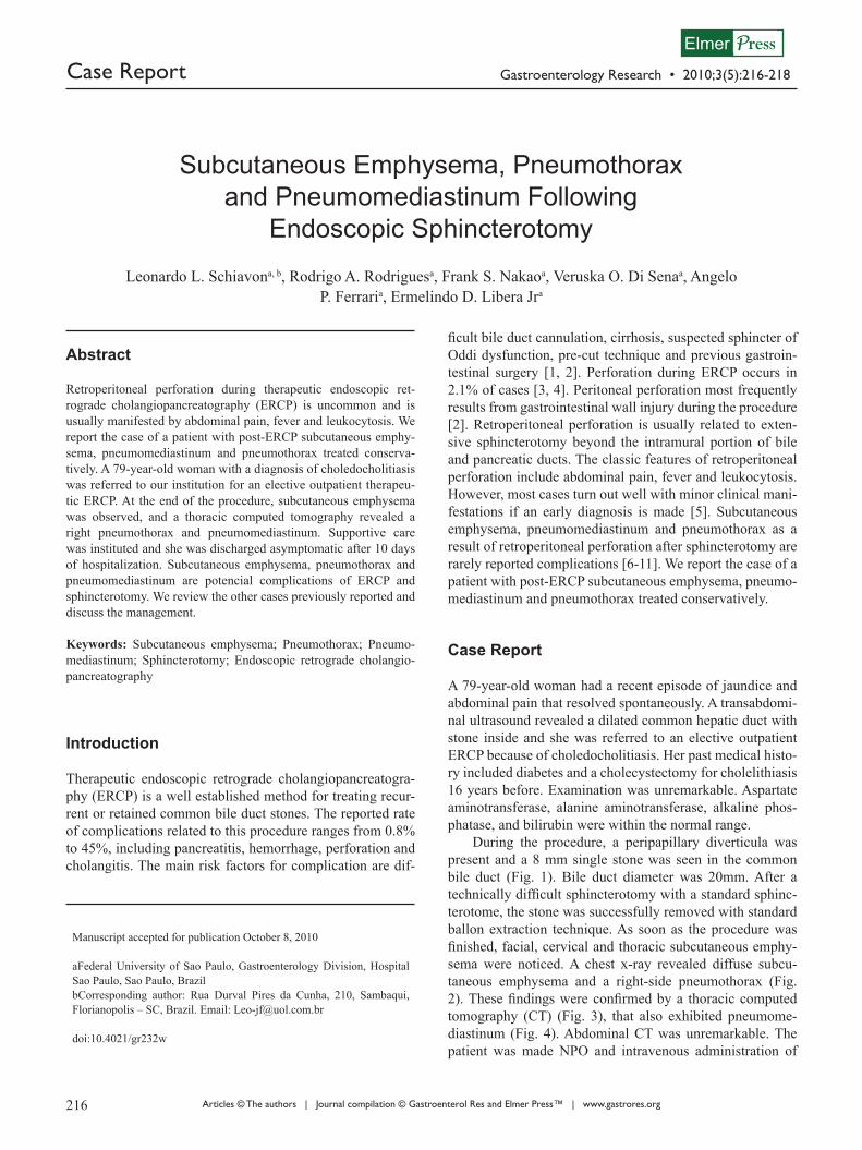
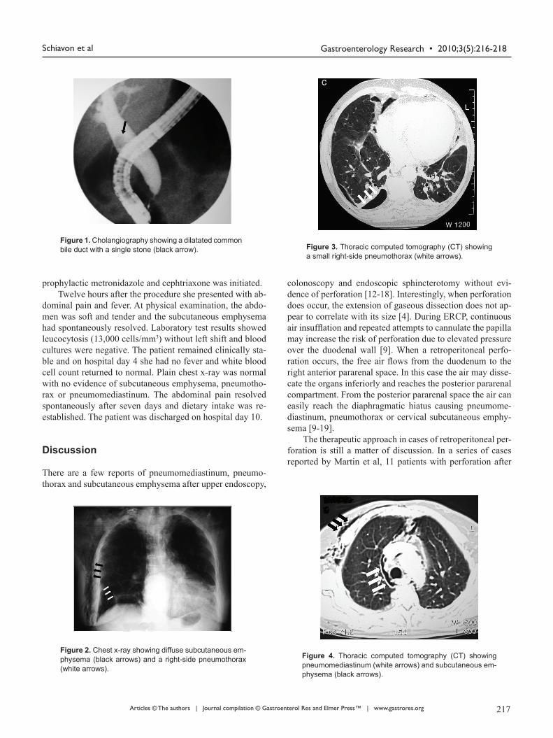
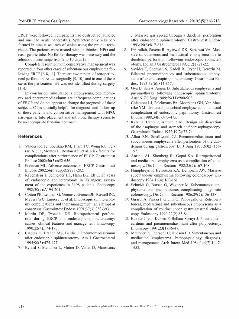
During the procedure, a peripapillary diverticula was present and a 8 mm single stone was seen in the common bile duct (Fig. 1). Bile duct diameter was 20mm. After a technically difficult sphincterotomy with a standard sphinc-terotome, the stone was successfully removed with standard ballon extraction technique. As soon as the procedure was finished, facial, cervical and thoracic subcutaneous emphy-sema were noticed. A chest x-ray revealed diffuse subcu-taneous emphysema and a right-side pneumothorax (Fig. 2). These findings were confirmed by a thoracic computed tomography (CT) (Fig. 3), that also exhibited pneumome-diastinum (Fig. 4). Abdominal CT was unremarkable. The patient was made NPO and intravenous administration of
Manuscript accepted for publication October 8, 2010
aFederal University of Sao Paulo, Gastroenterology Division, Hospital Sao Paulo, Sao Paulo, BrazilbCorresponding author: Rua Durval Pires da Cunha, 210, Sambaqui, Florianopolis – SC, Brazil. Email: [email protected]
doi:10.4021/gr232w
216 217
Gastroenterology Research • 2010;3(5):216-218Schiavon et al
Articles © The authors | Journal compilation © Gastroenterol Res and Elmer Press™ | www.gastrores.org
prophylactic metronidazole and cephtriaxone was initiated.Twelve hours after the procedure she presented with ab-
dominal pain and fever. At physical examination, the abdo-men was soft and tender and the subcutaneous emphysema had spontaneously resolved. Laboratory test results showed leucocytosis (13,000 cells/mm3) without left shift and blood cultures were negative. The patient remained clinically sta-ble and on hospital day 4 she had no fever and white blood cell count returned to normal. Plain chest x-ray was normal with no evidence of subcutaneous emphysema, pneumotho-rax or pneumomediastinum. The abdominal pain resolved spontaneously after seven days and dietary intake was re-established. The patient was discharged on hospital day 10.
Discussion There are a few reports of pneumomediastinum, pneumo-thorax and subcutaneous emphysema after upper endoscopy,
colonoscopy and endoscopic sphincterotomy without evi-dence of perforation [12-18]. Interestingly, when perforation does occur, the extension of gaseous dissection does not ap-pear to correlate with its size [4]. During ERCP, continuous air insufflation and repeated attempts to cannulate the papilla may increase the risk of perforation due to elevated pressure over the duodenal wall [9]. When a retroperitoneal perfo-ration occurs, the free air flows from the duodenum to the right anterior pararenal space. In this case the air may disse-cate the organs inferiorly and reaches the posterior pararenal compartment. From the posterior pararenal space the air can easily reach the diaphragmatic hiatus causing pneumome-diastinum, pneumothorax or cervical subcutaneous emphy-sema [9-19].
The therapeutic approach in cases of retroperitoneal per-foration is still a matter of discussion. In a series of cases reported by Martin et al, 11 patients with perforation after
Figure 1. Cholangiography showing a dilatated common bile duct with a single stone (black arrow).
Figure 2. Chest x-ray showing diffuse subcutaneous em-physema (black arrows) and a right-side pneumothorax (white arrows).
Figure 3. Thoracic computed tomography (CT) showing a small right-side pneumothorax (white arrows).
Figure 4. Thoracic computed tomography (CT) showing pneumomediastinum (white arrows) and subcutaneous em-physema (black arrows).
216 217
Gastroenterology Research • 2010;3(5):216-218 Post-ERCP Massive Gas Spread
Articles © The authors | Journal compilation © Gastroenterol Res and Elmer Press™ | www.gastrores.org
ERCP were followed. Ten patients had obstructive jaundice and one had acute pancreatitis. Sphincterotomy was per-formed in nine cases, two of which using the pre-cut tech-nique. The patients were treated with antibiotics, NPO and naso-gastric tube. No further therapy was necessary and the admission time range from 2 to 10 days [5].
Complete resolution with conservative management was reported in four other cases of subcutaneous emphysema fol-lowing ERCP [6-8, 11]. There are two reports of retroperito-neal perforation treated surgically [9, 10], and in one of these cases the perforation site was not identified during surgery [10].
In conclusion, subcutaneous emphysema, pneumotho-rax and pneumomediastinum are infrequent complications of ERCP and do not appear to change the prognosis of these subjects. CT is specially helpful for diagnosis and follow-up of these patients and conservative management with NPO, naso-gastric tube placement and antibiotic therapy seems to be an appropriate first-line approach.
References
1. Vandervoort J, Soetikno RM, Tham TC, Wong RC, Fer-rari AP, Jr., Montes H, Roston AD, et al. Risk factors for complications after performance of ERCP. Gastrointest Endosc 2002;56(5):652-656.
2. Freeman ML. Adverse outcomes of ERCP. Gastrointest Endosc 2002;56(6 Suppl):S273-282.
3. Rabenstein T, Schneider HT, Hahn EG, Ell C. 25 years of endoscopic sphincterotomy in Erlangen: assess-ment of the experience in 3498 patients. Endoscopy 1998;30(9):A194-201.
4. Cotton PB, Lehman G, Vennes J, Geenen JE, Russell RC, Meyers WC, Liguory C, et al. Endoscopic sphincteroto-my complications and their management: an attempt at consensus. Gastrointest Endosc 1991;37(3):383-393.
5. Martin DF, Tweedle DE. Retroperitoneal perfora-tion during ERCP and endoscopic sphincterotomy: causes, clinical features and management. Endoscopy 1990;22(4):174-175.
6. Ciaccia D, Branch MS, Baillie J. Pneumomediastinum after endoscopic sphincterotomy. Am J Gastroenterol 1995;90(3):475-477.
7. Evrard S, Mendoza L, Mutter D, Vetter D, Marescaux
J. Massive gas spread through a duodenal perforation after endoscopic sphincterotomy. Gastrointest Endosc 1993;39(6):817-818.
8. Ibrarullah, Saxena R, Agarwal DK, Saraswat VA. Mas-sive subcutaneous and mediastinal emphysema due to duodenal perforation following endoscopic sphincter-otomy. Indian J Gastroenterol 1993;12(1):21-22.
9. Savides T, Sherman S, Kadell B, Cryer H, Derezin M. Bilateral pneumothoraces and subcutaneous emphy-sema after endoscopic sphincterotomy. Gastrointest En-dosc 1993;39(6):814-817.
10. Gya D, Sali A, Angus D. Subcutaneous emphysema and pneumothorax following endoscopic sphincterotomy. Aust N Z J Surg 1989;59(11):900-902.
11. Colemont LJ, Pelckmans PA, Moorkens GH, Van Mae-rcke YM. Unilateral periorbital emphysema: an unusual complication of endoscopic papillotomy. Gastrointest Endosc 1988;34(6):473-475.
12. Katz D, Cano R, Antonelle M. Benign air dissection of the esophagus and stomach at fiberesophagoscopy. Gastrointest Endosc 1972;19(2):72-74.
13. Allan RN, Smallwood CJ. Pneumomediastinum and subcutaneous emphysema after perforation of the duo-denum during gastroscopy. Br J Surg 1973;60(2):156-157.
14. Amshel AL, Shonberg IL, Gopal KA. Retroperitoneal and mediastinal emphysema as a complication of colo-noscopy. Dis Colon Rectum 1982;25(2):167-168.
15. Humphreys F, Hewetson KA, Dellipiani AW. Massive subcutaneous emphysema following colonoscopy. En-doscopy 1984;16(4):160-161.
16. Schmidt G, Borsch G, Wegener M. Subcutaneous em-physema and pneumothorax complicating diagnostic colonoscopy. Dis Colon Rectum 1986;29(2):136-138.
17. Girardi A, Piazza I, Giunta G, Pappagallo G. Retroperi-toneal, mediastinal and subcutaneous emphysema as a complication of routine upper gastrointestinal endos-copy. Endoscopy 1990;22(2):83-84.
18. Bakker J, van Kersen F, Bellaar Spruyt J. Pneumoperi-cardium and pneumomediastinum after polypectomy. Endoscopy 1991;23(1):46-47.
19. Maunder RJ, Pierson DJ, Hudson LD. Subcutaneous and mediastinal emphysema. Pathophysiology, diagnosis, and management. Arch Intern Med 1984;144(7):1447-1453.
218

![Case Report Subcutaneous Emphysema, …downloads.hindawi.com/journals/criem/2015/134816.pdfpneumothorax, pneumomediastinum, pneumopericardium, or subcutaneous emphysema [ ]. Diagnosis](https://img.pdfslide.net/doc/110x75/5f4072ff5627821a5534fd08/case-report-subcutaneous-emphysema-pneumothorax-pneumomediastinum-pneumopericardium.jpg)