Embed Size (px)
Citation preview
226https://e-jcvi.org
A 26-year-old male presented with history of progressively worsening dyspnoea for last six months and history of paroxysmal nocturnal dyspnoea and orthopnea for 3 days. There was no history of chest pain, palpitation, presyncope or syncope. There was no previous history suggestive of rheumatic fever. Besides this there was no history of any other chronic disease.
On examination, his blood pressure was 110/70 mmHg and pulse rate was 126 beats/min and regular. His jugular venous pressure was raised with prominent ‘V’ wave and ‘Y’ descent. There was presence of bilateral pedal edema up to the level of ankle joint. On cardiovascular examination there was cardiomegaly with apex shifted downwards and outwards. Fist heart sound (S1) was soft, pulmonary component of second heart sound (P2) was loud and grade IV/VI pan systolic murmur was heard at apex and left sternal border. Chest auscultation revealed bilateral basal crepitations. Abdominal examination revealed tender hepatomegaly.
His electrocardiogram showed sinus tachycardia. A two-dimensional transthoracic echocardiogram was performed in the usual manner with a Vivid S5 General Electric (Milwaukee, WI, USA) ultrasound system and a 3 MHz transducer. The result showed situs solitus and normal arrangement of atria and ventricles with no atrioventricular or ventricular-arterial discordance. Two-dimensional transthoracic echocardiography done at our centre which revealed large aneurysmal cavity behind the posterior mitral valve leaflet freely communicating with left ventricle (Figures 1, 2, Movies 1, 2). The patient also had
J Cardiovasc Imaging. 2020 Jul;28(3):226-229https://doi.org/10.4250/jcvi.2019.0131pISSN 2586-7210·eISSN 2586-7296
Images in Cardiovascular Disease
Received: Dec 24, 2019Revised: Feb 3, 2020Accepted: Feb 5, 2020
Address for Correspondence: Deepak Agrawal, MDDepartment of Cardiology, Jaipur Heart Institute, Lal Kothi, Near S. M. S. Stadium, Tonk Road, Jaipur 302015, India.E-mail: [email protected]
Copyright © 2020 Korean Society of EchocardiographyThis is an Open Access article distributed under the terms of the Creative Commons Attribution Non-Commercial License (https://creativecommons.org/licenses/by-nc/4.0/) which permits unrestricted non-commercial use, distribution, and reproduction in any medium, provided the original work is properly cited.
ORCID iDsAshok Garg https://orcid.org/0000-0001-9991-0538Deepak Agrawal https://orcid.org/0000-0002-2448-2687G L Sharma https://orcid.org/0000-0002-3710-4511
Conflict of InterestThe authors have no financial conflicts of interest.
Ashok Garg , MD1, Deepak Agrawal , MD2, and G L Sharma , MD2
1Department of Preventive and Non Invasive Cardiology, Jaipur Heart Institute, Jaipur, India2Department of Cardiology, Jaipur Heart Institute, Jaipur, India
Submitral Aneurysm: A Rare Cause of Severe Mitral Regurgitation
Submitralaneurysm
Figure 1. Parasternal long axis view showing submitral aneurysm behind posterior mitral valve leaflet.
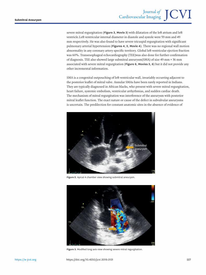
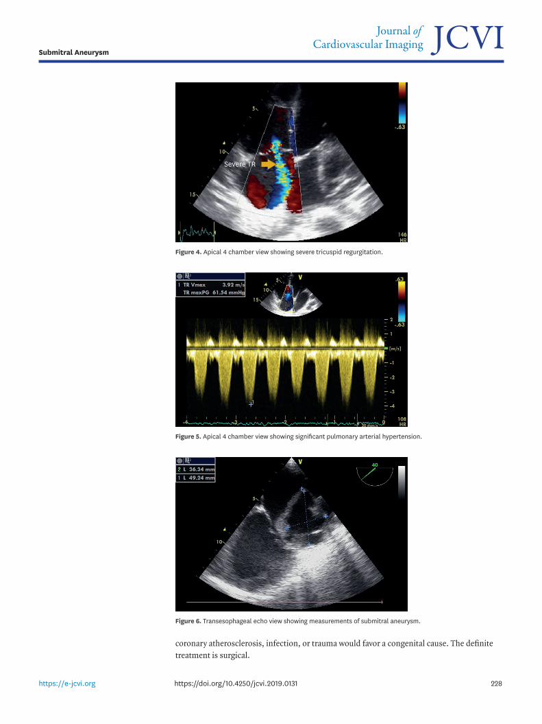
severe mitral regurgitation (Figure 3, Movie 3) with dilatation of the left atrium and left ventricle.Left ventricular internal diameter in diastole and systole were 59 mm and 49 mm respectively. He was also found to have severe tricuspid regurgitation with significant pulmonary arterial hypertension (Figures 4, 5, Movie 4). There was no regional wall motion abnormality in any coronary artery specific territory. Global left ventricular ejection fraction was 60%. Transesophageal echocardiography (TEE)was also done for further confirmation of diagnosis. TEE also showed large submitral aneurysm(SMA) of size 49 mm × 36 mm associated with severe mitral regurgitation (Figure 6, Movies 5, 6) but it did not provide any other incremental information.
SMA is a congenital outpouching of left ventricular wall, invariably occurring adjacent to the posterior leaflet of mitral valve. Annular SMAs have been rarely reported in Indians. They are typically diagnosed in African blacks, who present with severe mitral regurgitation, heart failure, systemic embolism, ventricular arrhythmias, and sudden cardiac death. The mechanism of mitral regurgitation was interference of the aneurysm with posterior mitral leaflet function. The exact nature or cause of the defect in subvalvular aneurysms is uncertain. The predilection for constant anatomic sites in the absence of evidence of
227https://e-jcvi.org https://doi.org/10.4250/jcvi.2019.0131
Submitral Aneurysm
Submitralaneurysm
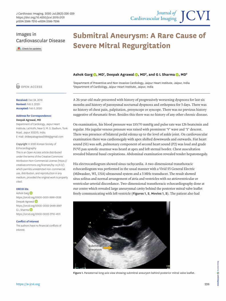
Figure 2. Apical 4 chamber view showing submitral aneurysm.
Severe MR
Figure 3. Modified long axis view showing severe mitral regurgitation.
coronary atherosclerosis, infection, or trauma would favor a congenital cause. The definite treatment is surgical.
228https://e-jcvi.org https://doi.org/10.4250/jcvi.2019.0131
Submitral Aneurysm
Severe TR
Figure 4. Apical 4 chamber view showing severe tricuspid regurgitation.
Figure 5. Apical 4 chamber view showing significant pulmonary arterial hypertension.
Figure 6. Transesophageal echo view showing measurements of submitral aneurysm.
SUPPLEMENTARY MATERIALS
Movie 1Apical five-chamber view showing submitral aneurysm behind posterior mitral valve leaflet.
Click here to view
Movie 2Apical 4 chamber view showing submitral aneurysm.
Click here to view
Movie 3Modified long axis view showing severe mitral regurgitation.
Click here to view
Movie 4Apical 4 chamber view showing severe tricuspid regurgitation.
Click here to view
Movie 5Transesophageal echo view showing submitral aneurysm.
Click here to view
Movie 6Transesophageal echo view showing severe mitral regurgitation.
Click here to view
229https://e-jcvi.org https://doi.org/10.4250/jcvi.2019.0131
Submitral Aneurysm