Embed Size (px)
Citation preview
Central Annals of Community Medicine and Practice
Cite this article: El-Bahnasawy MM (2015) A Case of Crimean-Congo Hemorrhagic Fever and a General Review. Ann Community Med Pract 1(2): 1010.
*Corresponding author
Mamdouh M. El-Bahnasawy, Department of Tropical Medicine, Military Medical Academy, 2, Ahmed KhaleelStree, El Zawya El Hamra, Cairo, 11291, Egypt, Tel: 201223777352; Email:
Submitted: 27 November 2015
Accepted: 18 December 2015
Published: 21 December 2015
Copyright© 2015 El-Bahnasawy
OPEN ACCESS
Keywords•Crimean-Congo hemorrhagic fever•Tick-borne virus•Treatment
Case Report
A Case of Crimean-Congo Hemorrhagic Fever and a General ReviewMamdouh M. El-Bahnasawy*Department of Tropical Medicine, Military Medical Academy, Egypt
Abstract
Crimean-Congo hemorrhagic fever (CCHF) is a tick-borne disease caused by an arbovirus, a member of the Nairovirus genera of Bunyaviridae family, one of the viral hemorrhagic fever causes and can be transmitted to humans by Hyalomma tick-bite, by exposure to infected blood and fomites of patient with CCHF or contact with animal tissue in viremic phase, which was first recognized during a large outbreak among agricultural workers in the mid-1940s in the Crimean Peninsula.
Crimean-Congo hemorrhagic fever is reported from many countries in Africa, Asia, South-East Europe, and the Middle East. The majority of human cases are workers in agriculture, working in endemic areas, slaughterhouses, and veterinary practice. Nosocomial transmission is also well documented. Clinical manifestations are nonspecific and symptoms typically include high fever, headache, malaise, arthralgia, myalgia, nausea, abdominal pain, and non-bloody diarrhea. Patients may show signs of progressive hemorrhagic diathesis. Laboratory abnormalities may include anemia, leukopenia, thrombocytopenia, increased AST/ALT levels, and prolonged prothrombin, bleeding, and activated partial thromboplastin times. Diagnostic methods include antibody detection by enzyme-linked immunosorbent assay, virus isolation, antigen detection, and polymerase chain reaction. The mainstay of treatment of Crimean-Congo hemorrhagic fever is supportive, with careful maintenance of fluid and electrolyte balance, circulatory volume, and blood pressure and giving antiviral drug ribavirin as the Crimean-Congo hemorrhagic fever virus is susceptible to ribavirin in vitro.
Humans become infected through the bites of ticks, by contact with hemorrhage from nose, mouth, gums, vagina, and injection sites of a CCHF patient during the acute phase or follow-up as a nosocomial infection, or by contact with blood or tissues from viremic livestock.
This paper reported a Crimean-Congo hemorrhagic fever man from Egypt, referred to Almaza fever hospital, as an F.U.O case for more than one month, with fever comes and go without history of tick bite or exposure to infected fomites, even not coming from endemic areas. No doubt, distribution of tick-vector (Hyalomma spp.) worldwide including Egypt and presence of CCHF in regional countries must be considered by the Health and Veterinary Authorities.
INTRODUCTIONCrimean-Congo hemorrhagic fever virus (CCHFV) is one of
at least 30 known viruses capable of causing viral hemorrhagic fever syndrome. All agents that cause viral hemorrhagic fever syndrome are RNA viruses with a lipid envelope, all are considered zoonoses, all damage the microvasculature, resulting in increased vascular permeability and ultimately bleeding diathesis, and all are members of one of four families: Arenaviridae, Bunyaviridae, Flaviviridae, and Filoviridae [1].
Crimean-Congo hemorrhagic fever (CCHF) was described in
the Crimea in 1944 during an outbreak, which involved more than 200 cases and was called Crimean hemorrhagic fever due to its peculiar picture of bleeding tendency. A later in time, the virus was also isolated from Congo and was noted to be the same pathogen, resulting in the name (CCHFV) [2].
CCHFV can infect a wide range of domestic and wild animals, including sheep and cattle. Animals are infected with CCHFV by the bite of infected ticks (Hyalommatick); seroprevalence of CCHFV is about 13 – 36% in animals [3,4].
A number of tick genera can be infected with CCHFV, but the
Central
El-Bahnasawy (2015)Email:
Ann Community Med Pract 1(2): 1010 (2015) 2/4
most efficient and common vectors for CCHFV are the members of the genus Hyalomma. The most important source of virus transmission is immature Hyalomma tick, which feeds small vertebrates’ blood. Once infected, the tick remains infected throughout its life [4].
A sero-epidemiological study of CCHF in local and imported sheep in Isfahan Province of Iran revealed the endemic spreading of the virus in sheep and the need for special attention to prevent the infection in the community and during occupational exposures [3].
Like other tick-borne zoonotic agents, CCHFV generally circulates in nature in an enzootic tick- vertebrate-tick cycle. Although many domestic and wild vertebrates are infected with CCHFV, as evidenced by development of viremia and/or antibody response, birds, in general, appear to be resistant to this infection [7,8].
Community-acquired CCHF happens through transmission of the virus by direct contact with blood or other infected tissues of livestock or from an infected tick bite. Most of the human cases are workers in livestock and agriculture industry, slaughterhouses, and veterinary practice. Humans can be infected incidentally by the bite of an infected arthropod or via aerosol generated from infected rodents’ excreta [15]. Infected humans can spread the disease via close contacts which may result in community outbreaks and nosocomial infections. Possible transmission of CCHFV from a mother to her child indicates the importance of preventive measures for in-house outbreaks of CCHF [5].
Distribution of CCHF largely mirrors that of its Ixodid tick hosts, particularly those of the genus Hyalomma. Persons become infected when bitten by virus-infected ticks or after contact with blood or tissue from viremic livestock or other persons. Outbreaks often involve persons in rural communities, such as shepherds, slaughterhouse workers, or medical staff. Suspected CCHF outbreaks and sporadic cases in the Kordufan region of Sudan have been reported [12].
From a public health perspective, confirming CCHF in Sudan and determining which virus lineages may be present in this region will provide a more detailed understanding of the movement of virus strains and identification of areas at risk for CCHFV including a nosocomial chain of transmission in a rural hospital in Sudan in 2008 [16].
The viraemia or viral antigen is concentrated in RECs of liver and spleen the main sites of its replication, vascular damage end into leakage of erythrocytes and plasma, with clinical pictures ranging from unapparent infection to fatal bleeding outcome with high case fatality rates [6].
Laboratory abnormalities may include anemia, leucopenia, thrombocytopenia, increased AST/ ALT, prolonged bleeding time, prothrombin time and activated partial thromboplastin, elevated fibrin degradation products and decreased fibrinogen. Urinalysis may reveal proteinuria and hematuria, and patients may develop oliguria and azotemia [7-11].
Ribavirin is effective if give to suspected patient and/or contact or given as a prophylaxis for health care workers managing CCHF case (it is a guanosine analogue that has an incomplete purine
ring rather than an acyclic ribose moiety [12]. After intracellular phosphorylation, ribavirin triphosphate interferes with early events in viral transcription, such as capping and elongation of messenger RNA, and inhibits ribonucleoprotein synthesis. It has a broad spectrum of activity in vitro against RNA viruses [13].
The Study
The patient was a 40-year-old man who had worked at Egyptian Sudanese border, at Camel trading area. He was suffered from fever of unknown origin with generalized weakness and sever body aches, headache and diarrhea .He took a treatment in the form of tablets and capsules as antibiotic (he did not mentioned its name) and antipyretics for 5 days with some improvement. Then he stopped the drugs when he felt well after 5 days fever returned again with sever e body aches, rash and epistaxis. From the patient history, he was working at Camel trading area (at Egyptian Sudanese border) where mosquitoes most abundant there, he said that he was sharing some persons from Sudan, crossing the Egyptian Sudanese border at Camel trading area, slaughtering a Camel and ate with them.
Also, there was no precaution taken for mosquito bite prevention in that area, as there was no repellants or mosquito nets and at the same time no malaria prophylaxis was taken by him. Thin and thick blood film was taken and stained with Giemsa stain and examined for malaria, the slides was negative and in spite of that he took a course of antimalarial drugs with no improvement.
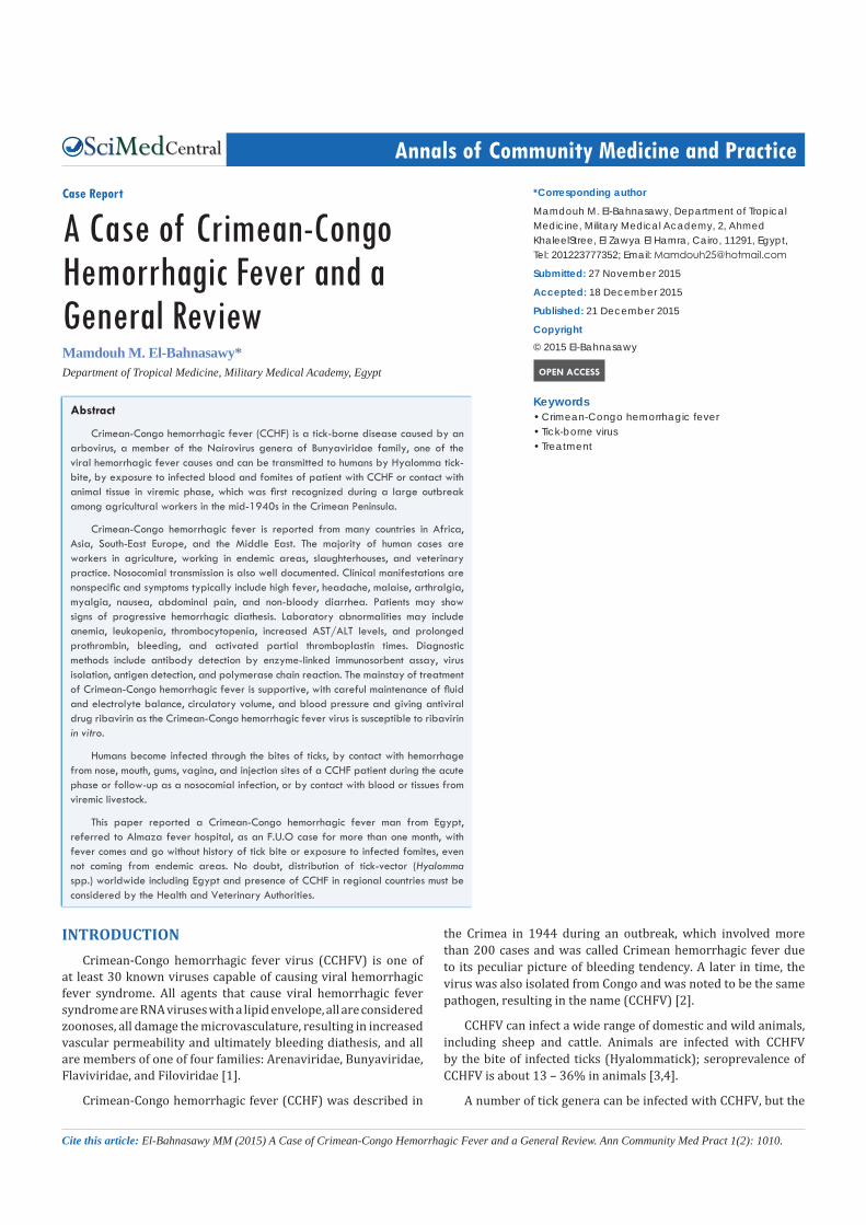
The patient referred to Almaza fever hospital as a case of fever for investigation, we examined him and found that he suffered from high fever (40 °C) and rash in the form of peticheal rash over the abdomen (it was difficult to be seen because of his dark colored skin (except in a good illumination) and maculopapular eruption over hands and feet (Figure 1-4).
Laboratory investigations showed anemia (hemoglobin level 10.7 g/dL), leucopenia, WBCs= 2500/mm3 (Normal range of leukocytes, 4,000–11,000/mm3) and thrombocytopenia 60,000/mm3 (N: platelets, 150,000–450,000/mm3), increased AST= 240 and ALT= 287 (Normally AST, <50 IU/L; ALT, <50 IU/L respectively), prolonged bleeding time= 13 (Normally = 1-9 minutes), prothrombin time= INR 2.1 (Normally INR of 1.1 or below), and activated partial thromboplastin time 80 (APTT, N 2: 4–36 sec ), elevated fibrin degradation products 25mg/mL (FDPs Normally less than 10 mcg/mL), and decreased fibrinogen
Figure 1 A patient with CCHF showing peticheal hemorrhage on his abdomen and Maculopapular eruption on both hands and feet.
Central
El-Bahnasawy (2015)Email:
Ann Community Med Pract 1(2): 1010 (2015) 3/4
180 mg/dL ( Normal value 200–400 mg/dL). Urinalysis revealed proteinuria and hematuria.
We suspect this case as CCHF and the source of his infection was suspected to be tissues and/or blood from infected Camel during slaughtering, preparing and/ or eating camel meat although follow-up investigation was unable to precisely determine the source. Serum samples were taken, and referred for ELISA CCHFV and it was positive.Laboratory confirmation of this case of CCHF demonstrates the presence of this disease which transferred to him through butchering infected camel (blood or animal tissues), or infected during preparing food with them.
Treatment
The patient was given supportive treatment (fluids and electrolyte balance), antipyretics in the form of paracetamol
tablet (1 tablets three times a day) and ribavirin 30 mg/kg as an initial loading dose, then 15 mg/kg every 6 h (4 × 1 g) for 4 days, and 7.5 mg/kg every 8 h for 6 days. The patient was improved and discharged after 10 days.
DISCUSSIONSero-survey in Egypt for Crimean-Congo hemorrhagic fever
virus was carried out by Mohamed, et al (2008) revealed that imported camels with previous CCHF viral infection did not show evidence of transmission to Egyptian animals. The study was conducted among 1,022 sera from 313 cattle, 264 water-buffalo, 270 sheep and 175 goats. Of 1,022 samples examined, 32 (3.13%) were positive to IgG ELISA, of 270 sheep, 17 (6.30%) were confirmed to have anti-CCHFV IgG with the highest titer recorded at 1:800. CCHFV-specific IgG-positive cases among cattle, buffalo and goats were 3.83%, 0.38% and 1.14%, respectively. They concluded that Belbis City had the highest number of positive cases compared to all other localities (p<0.001) [8].
An outbreak of suspected viral hemorrhagic fever involving 7 patients occurred in Mecca, Western Saudi Arabia. CCHF not previously present in Saudi Arabia was incriminated. El-Azazy and Scrimgeour (1997) carried out a study in Mecca, and in nearby Jeddah and Taif in 1991-1993; 13 species of ixodid ticks (5 Hyalomma spp., 5 Rhipicephalus spp., 2 Amblyomma spp., 1 Boophilus sp.) collected from livestock (camels, cattle, sheep, goats), and of these 10 were capable of transmitting CCHF and (97%) of Camels were highly infested [14].
Also, Memish, et al. (2011) gave evidence that Alkhurma, CCHF, RVF and dengue viruses to be endemic to western, if not all Saudi Arabia provinces [15].
In Sudan Sporadic cases and multiple CCHF outbreaks, associated with nosocomial chain of transmission, have been reported in the Kordufan region [16]. Antibody captured ELISA, reverse transcription PCR, partial S segment sequences of the virus and subsequent phylogenetic analysis were used to confirm the CCHFV infection and to determine the virus genetic lineages [7-10].
Clinical features usually include a rapid progression characterized by hemorrhage, myalgia and fever, with high mortality rate [17]. The first symptoms of our patient were fever, sever body aches, headache and diarrhea. The most common symptoms reported in the literature have been fever, fatigue, headache, loss of appetite, and myalgia [18]. Typically, 5-6 days after exposure to infected blood or tissues, the flu-like symptoms occur that can last up to a week. However, in 75% of cases, hemorrhagic syndrome appears within 3-5 days after onset of illness with gingival bleeding, epistaxis, hematuria and melena. In our case hemorrhagic syndrome appears in 10th day of illness. Laboratory tests showed thrombocytopenia, high liver enzymes and prolonged prothrombin time, indicating damage of the liver cells. Treatment with ribavirin may be useful if given within the early stage of disease [12,13,16,18]. Diagnosis of CCHF is important to prevent the spread of CCHF virus among the health-care workers and relatives of patients.
CONCLUSIONFrom these data’s we conclude that early diagnosis of the
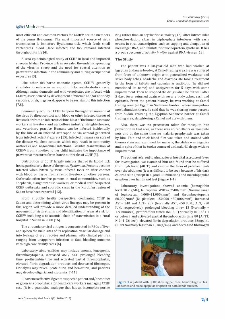
Figure 2 Peticheal hemorrhage on patient’s abdomen but his dark coloured skin difficult to recognize except under good illumination.
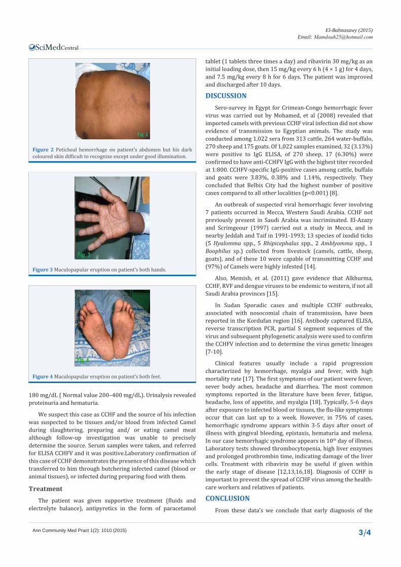
Figure 3 Maculopapular eruption on patient’s both hands.
Figure 4 Maculopapular eruption on patient’s both feet.
Central
El-Bahnasawy (2015)Email:
Ann Community Med Pract 1(2): 1010 (2015) 4/4
El-Bahnasawy MM (2015) A Case of Crimean-Congo Hemorrhagic Fever and a General Review. Ann Community Med Pract 1(2): 1010.
Cite this article
CCHF is a very important factor in the disease outcome and efficacy of therapy. Diagnosis of the disease, in prehemorrhagic
Phase is difficult but essential for the prognosis of the disease and prevention of spreading the infection among contact persons. Absence of history of the tick bite does not exclude the CCHF. No doubt, CCHF virus may be introduced to Egypt by infected ticks on imported animals. CCHF is now endemic in the Sub-Sahara and in Western Saudi Arabia. Besides, many Egyptian animals (farm, domestic, pets) and carnivores are infested with soft and hard ticks.
REFERENCES1. McGovern TW, Christopher GW, Eitzen EM. Cutaneous manifestations
of biological warfare and related threat agents. Arch Dermatol. 1999; 135: 311-322.
2. Wallace MR, Hale BR, Utz GC, Olson PE, Earhart KC, Thornton SA, et al. Endemic infectious diseases of Afghanistan. Clin Infect Dis. 2002; 34: 171-207.
3. Darvishi M, Ataee B, Chinikar S, Jalali N, Mardani M, Mirkhani M. Seroepidemiology of Crimean-Congo hemorrhagic fever in local and imported sheep in Isfahan Province of Iran. Clin Microbiol Infect. 2005; 11: 649.
4. Athar MN, Baqai HZ, Ahmad M, Khalid MA, Bashir N, Ahmad AM, et al. Short report: Crimean-Congo hemorrhagic fever outbreak in Rawalpindi, Pakistan. Am J Trop Med Hyg. 2003; 69: 284–287.
5. Saijo M, Tang Q, Shimayi B, Han L, Zhang Y, Asiguma M, Tianshu D. Possible horizontal transmission of crimean-congo hemorrhagic Fever virus from a mother to her child. Jpn J Infect Dis. 2004; 57: 55-57.
6. Wilson ME. Prevention of tick-borne diseases. Med Clin North Am. 2002; 86: 219-238.
7. Shepherd AJ, Swanepoel R, Leman PA. Antibody response in Crimean-Congo hemorrhagic fever. Rev Infect Dis. 1989; 11: 801-806.
8. Mohamed M, Said AR, Murad A, Graham R. A serological survey of Crimean-Congo haemorrhagic fever in animals in the Sharkia
Governorate of Egypt. Vet Ital. 2008; 44: 513-517.
9. Morrill JC, Soliman AK, Imam IZ, Botros BA, Moussa MI, Watts DM. Serological evidence of Crimean-Congo haemorrhagic fever viral infection among camels imported into Egypt. J Trop Med Hyg. 1990; 93: 201-204.
10. Schwarz TF, Nsanze H, Longson M, Nitschko H, Gilch S, Shurie H, et al. Polymerase chain reaction for diagnosis and identification of distinct variants of Crimean-Congo hemorrhagic fever virus in the United Arab Emirates. Am J Trop Med Hyg. 1996; 55: 190-196.
11. Mohamed M, Said AR, Murad A, Graham R. A serological survey of Crimean-Congo haemorrhagic fever in animals in the Sharkia Governorate of Egypt. Vet Ital. 2008; 44: 513-517.
12. Fisher-Hoch SP, Khan JA, Rehman S, Mirza S, Khurshid M, McCormick JB,. Crimean Congo-haemorrhagic fever treated with oral ribavirin. Lancet. 1995; 346: 472-475.
13. Ergonul O. Treatment of Crimean-Congo hemorrhagic fever. Antiviral Res. 2008; 78: 125-131.
14. el-Azazy OM, Scrimgeour EM. Crimean-Congo haemorrhagic fever virus infection in the western province of Saudi Arabia. Trans R Soc Trop Med Hyg. 1997; 91: 275-278.
15. Memish ZA, Albarrak A, Almazroa MA, Al-Omar I, Alhakeem R, Assiri A, et al. Seroprevalence of Alkhurma and other hemorrhagic fever viruses, Saudi Arabia. Emerg Infect Dis. 2011; 17: 2316-2318.
16. McCarthy MC, Haberberger RL, Salib AW, Soliman BA, El-Tigani A, Khalid IO, et al. Evaluation of arthropod-borne viruses and other infectious disease pathogens as the causes of febrile illnesses in the Khartoum Province of Sudan. J Med Virol. 1996; 48: 141-146.
17. LA Berisha, S Namani, E Q Buçaj, B Halili. Crimen-Congo Hemorrhagic Fever without History of Tick Bite. The Internet Journal of Infectious Diseases. 2014; 13: 1.
18. Ergönül O1, Celikbaş A, Dokuzoguz B, Eren S, Baykam N, Esener H. Characteristics of patients with Crimean-Congo hemorrhagic fever in a recent outbreak in Turkey and impact of oral ribavirin therapy. Clin Infect Dis. 2004; 39: 284-287.