Embed Size (px)
Citation preview
NATURE CHEMISTRY | www.nature.com/naturechemistry 1
SUPPLEMENTARY INFORMATIONDOI: 10.1038/NCHEM.2505
1
Supplementary Information Covalent Functionalization and Passivation of Exfoliated Black Phosphorus via Aryl Diazonium Chemistry
Christopher R. Ryder1, Joshua D. Wood1, Spencer A. Wells1, Yang Yang2, Deep Jariwala1, Tobin
J. Marks1,2, George C. Schatz2 and Mark C. Hersam1,2* 1Dept. of Materials Science and Engineering, Northwestern University, Evanston, IL 60208 2Dept. of Chemistry, Northwestern University, Evanston, IL 60208 *E-mail: [email protected]. Table of Contents Density functional theory calculation methods 2
Atomic force microscopy (AFM) characterization for increasing aryl diazonium functionalization 2
AFM of flake before and after being submerged in electrolyte and solvent for 30 min 3
X-ray photoelectron spectroscopy (XPS) study of the thermal stability of aryl diazonium modification 4
Raman spectra of aryl diazonium functionalized BP 5
AFM characterization of BP after ambient exposure 7
Angle-resolved XPS of functionalized BP flakes after 15 days of ambient exposure 10
Contact angle measurements of aryl diazonium modified BP 11
Control studies of BP field-effect transistors 12
References 12
© 2016 Macmillan Publishers Limited. All rights reserved.
NATURE CHEMISTRY | www.nature.com/naturechemistry 2
SUPPLEMENTARY INFORMATIONDOI: 10.1038/NCHEM.2505
2
Density functional theory calculation methods Calculations were performed using the VASP package.1,2 The exchange-correlation potential was
described by the generalized gradient approximation (GGA) using the PBE functional.3,4 An
energy cutoff of 400 eV was used for the plane-wave basis set. The conjugate gradient method was
used to optimize the ion positions.5 Valence electrons included in the calculations were P 3s23p3,
C 2s22p2 and H 1s1, and the interaction between ions and electrons was described by the projector
augmented wave (PAW) method.6,7
Atomic force microscopy (AFM) characterization for increasing aryl diazonium functionalization
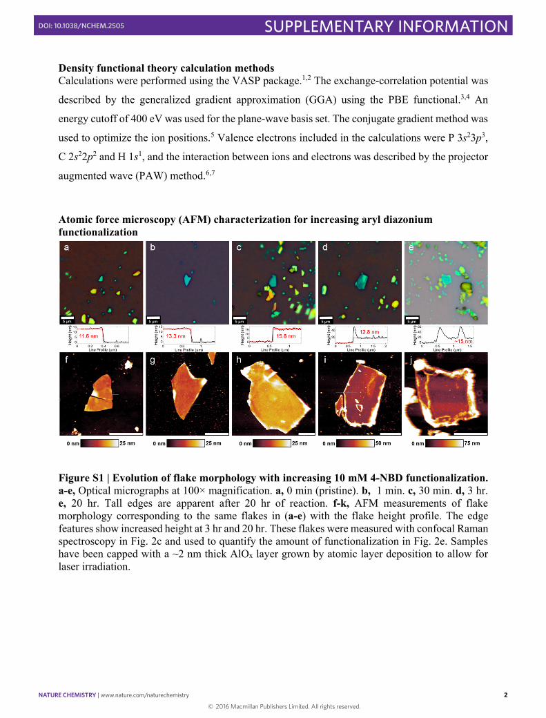
Figure S1 | Evolution of flake morphology with increasing 10 mM 4-NBD functionalization. a-e, Optical micrographs at 100× magnification. a, 0 min (pristine). b, 1 min. c, 30 min. d, 3 hr. e, 20 hr. Tall edges are apparent after 20 hr of reaction. f-k, AFM measurements of flake morphology corresponding to the same flakes in (a-e) with the flake height profile. The edge features show increased height at 3 hr and 20 hr. These flakes were measured with confocal Raman spectroscopy in Fig. 2c and used to quantify the amount of functionalization in Fig. 2e. Samples have been capped with a ~2 nm thick AlOx layer grown by atomic layer deposition to allow for laser irradiation.
© 2016 Macmillan Publishers Limited. All rights reserved.
NATURE CHEMISTRY | www.nature.com/naturechemistry 3
SUPPLEMENTARY INFORMATIONDOI: 10.1038/NCHEM.2505
3
AFM of flake before and after being submerged in electrolyte and solvent for 30 min
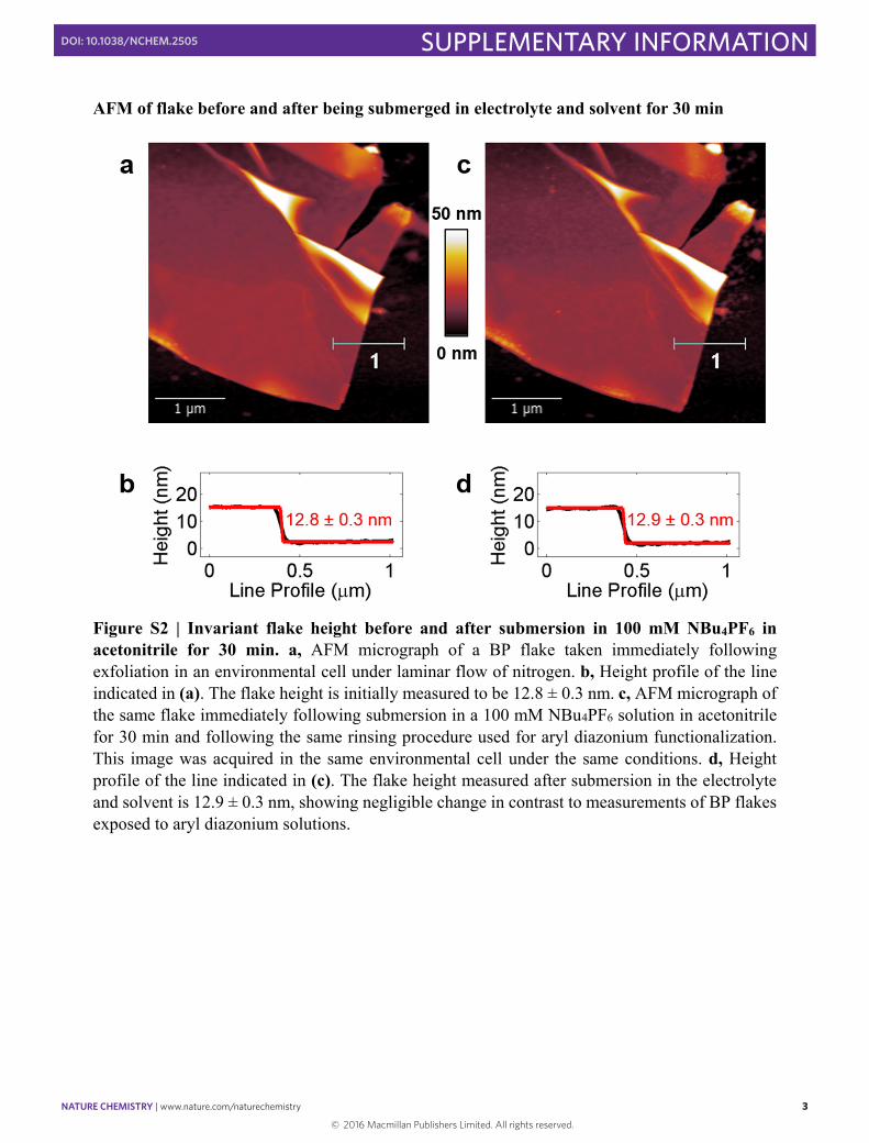
Figure S2 | Invariant flake height before and after submersion in 100 mM NBu4PF6 in acetonitrile for 30 min. a, AFM micrograph of a BP flake taken immediately following exfoliation in an environmental cell under laminar flow of nitrogen. b, Height profile of the line indicated in (a). The flake height is initially measured to be 12.8 ± 0.3 nm. c, AFM micrograph of the same flake immediately following submersion in a 100 mM NBu4PF6 solution in acetonitrile for 30 min and following the same rinsing procedure used for aryl diazonium functionalization. This image was acquired in the same environmental cell under the same conditions. d, Height profile of the line indicated in (c). The flake height measured after submersion in the electrolyte and solvent is 12.9 ± 0.3 nm, showing negligible change in contrast to measurements of BP flakes exposed to aryl diazonium solutions.
© 2016 Macmillan Publishers Limited. All rights reserved.
NATURE CHEMISTRY | www.nature.com/naturechemistry 4
SUPPLEMENTARY INFORMATIONDOI: 10.1038/NCHEM.2505
4
X-ray photoelectron spectroscopy (XPS) study of the thermal stability of aryl diazonium modification
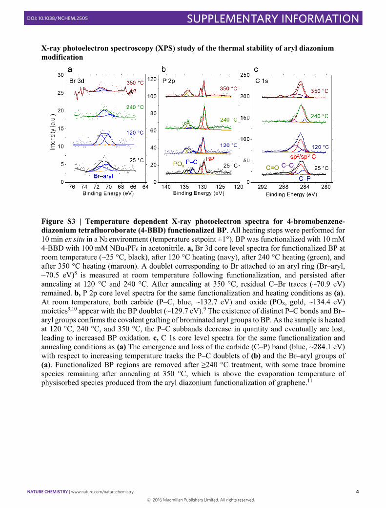
Figure S3 | Temperature dependent X-ray photoelectron spectra for 4-bromobenzene-diazonium tetrafluoroborate (4-BBD) functionalized BP. All heating steps were performed for 10 min ex situ in a N2 environment (temperature setpoint ±1°). BP was functionalized with 10 mM 4-BBD with 100 mM NBu4PF6 in acetonitrile. a, Br 3d core level spectra for functionalized BP at room temperature (~25 °C, black), after 120 °C heating (navy), after 240 °C heating (green), and after 350 °C heating (maroon). A doublet corresponding to Br attached to an aryl ring (Br–aryl, ~70.5 eV)8 is measured at room temperature following functionalization, and persisted after annealing at 120 °C and 240 °C. After annealing at 350 °C, residual C–Br traces (~70.9 eV) remained. b, P 2p core level spectra for the same functionalization and heating conditions as (a). At room temperature, both carbide (P–C, blue, ~132.7 eV) and oxide (POx, gold, ~134.4 eV) moieties9,10 appear with the BP doublet (~129.7 eV).9 The existence of distinct P–C bonds and Br–aryl groups confirms the covalent grafting of brominated aryl groups to BP. As the sample is heated at 120 °C, 240 °C, and 350 °C, the P–C subbands decrease in quantity and eventually are lost, leading to increased BP oxidation. c, C 1s core level spectra for the same functionalization and annealing conditions as (a) The emergence and loss of the carbide (C–P) band (blue, ~284.1 eV) with respect to increasing temperature tracks the P–C doublets of (b) and the Br–aryl groups of (a). Functionalized BP regions are removed after ≥240 °C treatment, with some trace bromine species remaining after annealing at 350 °C, which is above the evaporation temperature of physisorbed species produced from the aryl diazonium functionalization of graphene.11
© 2016 Macmillan Publishers Limited. All rights reserved.

NATURE CHEMISTRY | www.nature.com/naturechemistry 5
SUPPLEMENTARY INFORMATIONDOI: 10.1038/NCHEM.2505
5
Raman spectra of aryl diazonium functionalized BP
Figure S4 | Raman spectra of large lateral size 18 nm flake before and after 180 min functionalization in 10 mM 4-NBD. a, Optical image of a flake immediately following exfoliation taken in a nitrogen environment. b, Optical image of the same flake following 180 min functionalization in 10 mM 4-NBD showing the same flake in nearly identical orientation. c, AFM of the flake used to determine its thickness, taken after functionalization. d, Normalized Raman spectra immediately following exfoliation (blue) and immediately following aryl diazonium functionalization (red). The spectra were acquired in a nitrogen environment in a Linkam N2 environmental cell to prevent ambient degradation and photooxidation during measurements using a 50× long-working distance objective (NA = 0.5). Three spectra are depicted for each case to show the reproducibility of the measurements. Spectra are normalized to the Si TO phonon mode.
© 2016 Macmillan Publishers Limited. All rights reserved.
NATURE CHEMISTRY | www.nature.com/naturechemistry 6
SUPPLEMENTARY INFORMATIONDOI: 10.1038/NCHEM.2505
6
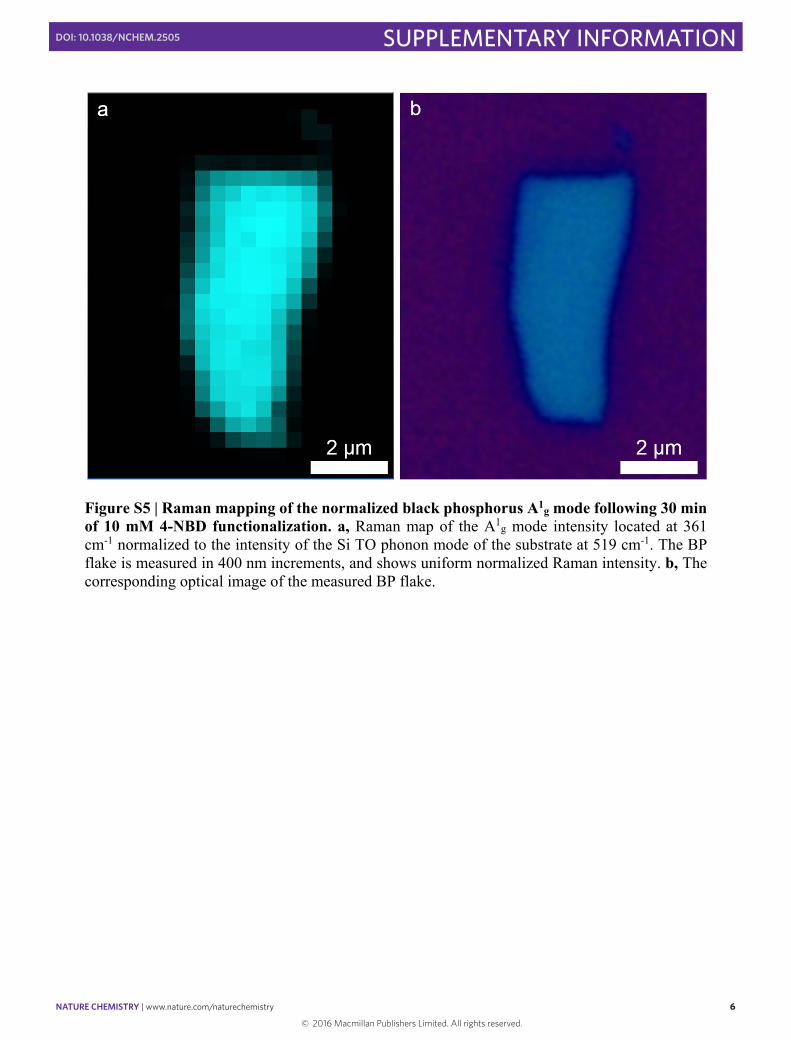
Figure S5 | Raman mapping of the normalized black phosphorus A1g mode following 30 min of 10 mM 4-NBD functionalization. a, Raman map of the A1g mode intensity located at 361 cm-1 normalized to the intensity of the Si TO phonon mode of the substrate at 519 cm-1. The BP flake is measured in 400 nm increments, and shows uniform normalized Raman intensity. b, The corresponding optical image of the measured BP flake.
© 2016 Macmillan Publishers Limited. All rights reserved.
NATURE CHEMISTRY | www.nature.com/naturechemistry 7
SUPPLEMENTARY INFORMATIONDOI: 10.1038/NCHEM.2505
7
AFM characterization of BP after ambient exposure
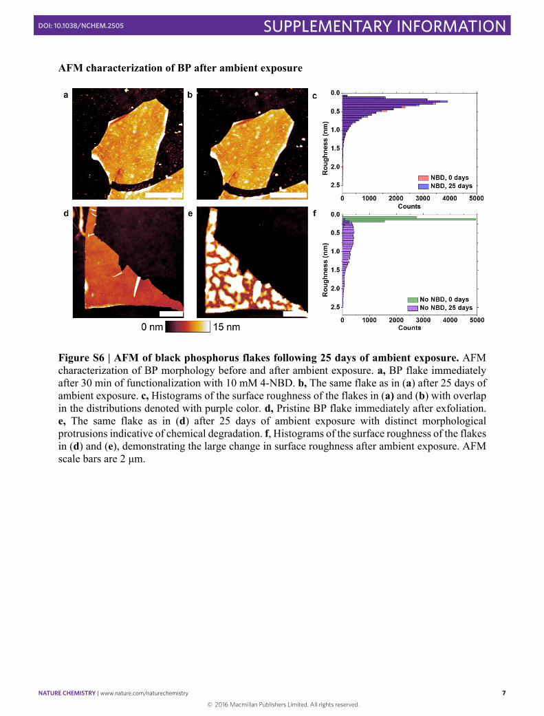
Figure S6 | AFM of black phosphorus flakes following 25 days of ambient exposure. AFM characterization of BP morphology before and after ambient exposure. a, BP flake immediately after 30 min of functionalization with 10 mM 4-NBD. b, The same flake as in (a) after 25 days of ambient exposure. c, Histograms of the surface roughness of the flakes in (a) and (b) with overlap in the distributions denoted with purple color. d, Pristine BP flake immediately after exfoliation. e, The same flake as in (d) after 25 days of ambient exposure with distinct morphological protrusions indicative of chemical degradation. f, Histograms of the surface roughness of the flakes in (d) and (e), demonstrating the large change in surface roughness after ambient exposure. AFM scale bars are 2 μm.
© 2016 Macmillan Publishers Limited. All rights reserved.
NATURE CHEMISTRY | www.nature.com/naturechemistry 8
SUPPLEMENTARY INFORMATIONDOI: 10.1038/NCHEM.2505
8
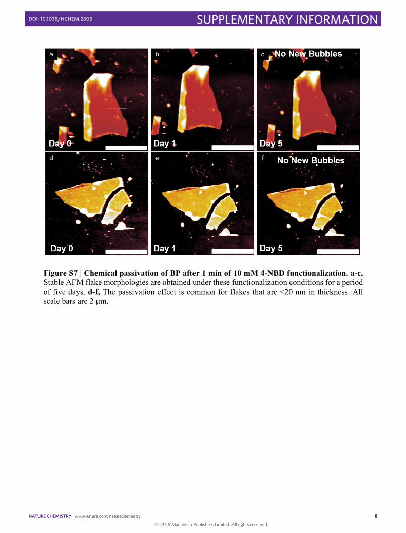
Figure S7 | Chemical passivation of BP after 1 min of 10 mM 4-NBD functionalization. a-c, Stable AFM flake morphologies are obtained under these functionalization conditions for a period of five days. d-f, The passivation effect is common for flakes that are <20 nm in thickness. All scale bars are 2 μm.
© 2016 Macmillan Publishers Limited. All rights reserved.
NATURE CHEMISTRY | www.nature.com/naturechemistry 9
SUPPLEMENTARY INFORMATIONDOI: 10.1038/NCHEM.2505
9
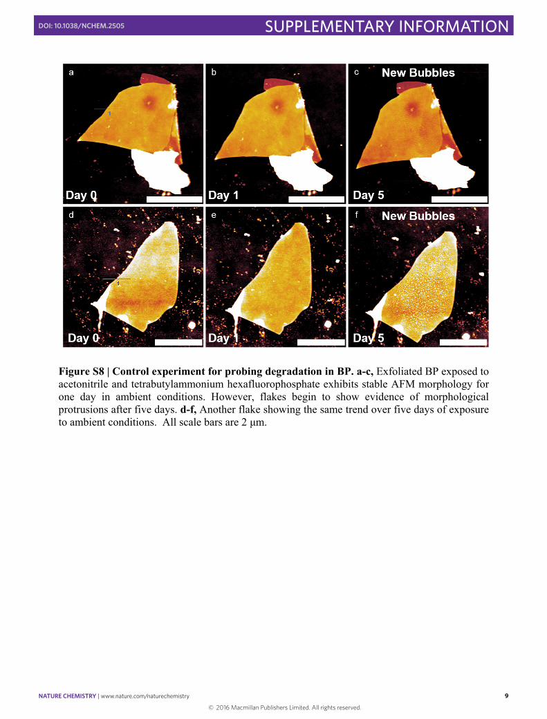
Figure S8 | Control experiment for probing degradation in BP. a-c, Exfoliated BP exposed to acetonitrile and tetrabutylammonium hexafluorophosphate exhibits stable AFM morphology for one day in ambient conditions. However, flakes begin to show evidence of morphological protrusions after five days. d-f, Another flake showing the same trend over five days of exposure to ambient conditions. All scale bars are 2 μm.
© 2016 Macmillan Publishers Limited. All rights reserved.
NATURE CHEMISTRY | www.nature.com/naturechemistry 10
SUPPLEMENTARY INFORMATIONDOI: 10.1038/NCHEM.2505
10
Angle-resolved XPS of functionalized BP flakes after 15 days of ambient exposure
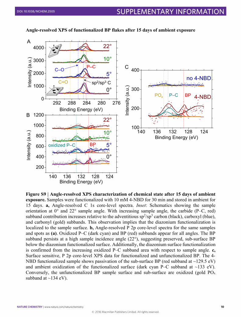
Figure S9 | Angle-resolved XPS characterization of chemical state after 15 days of ambient exposure. Samples were functionalized with 10 mM 4-NBD for 30 min and stored in ambient for 15 days. a, Angle-resolved C 1s core-level spectra. Inset: Schematics showing the sample orientation at 0° and 22° sample angle. With increasing sample angle, the carbide (P–C, red) subband contribution increases relative to the adventitious sp2/sp3 carbon (black), carboxyl (blue), and carbonyl (gold) subbands. This observation implies that the diazonium functionalization is localized to the sample surface. b, Angle-resolved P 2p core-level spectra for the same samples and spots as (a). Oxidized P–C (dark cyan) and BP (red) subbands appear for all angles. The BP subband persists at a high sample incidence angle (22°), suggesting preserved, sub-surface BP below the diazonium functionalized surface. Additionally, the diazonium surface functionalization is confirmed from the increasing oxidized P–C subband area with respect to sample angle. c, Surface sensitive, P 2p core-level XPS data for functionalized and unfunctionalized BP. The 4-NBD functionalized sample shows passivation of the sub-surface BP (red subband at ~129.5 eV) and ambient oxidization of the functionalized surface (dark cyan P–C subband at ~133 eV). Conversely, the unfunctionalized BP sample surface and sub-surface are oxidized (gold POx subband at ~134 eV).
292 288 284 280 2760
1000
2000
3000
4000
10°
22°
Inte
nsity
(a.u
.)
Binding Energy (eV)
5°
0°BP
A
P–C
sp2/sp3 C
C–O
C=O
140 136 132 128 124
200
400
600
800
1000
1200
Inte
nsity
(a.u
.)
Binding Energy (eV)
22°
10°
5°
0°
B
oxidized P–C BP
140 136 132 128 124100
200
300
400
4-NBD
no 4-NBD
Inte
nsity
(a.u
.)
Binding Energy (eV)
POx P–C BP
C
© 2016 Macmillan Publishers Limited. All rights reserved.
NATURE CHEMISTRY | www.nature.com/naturechemistry 11
SUPPLEMENTARY INFORMATIONDOI: 10.1038/NCHEM.2505
11
Contact angle measurements of aryl diazonium modified BP
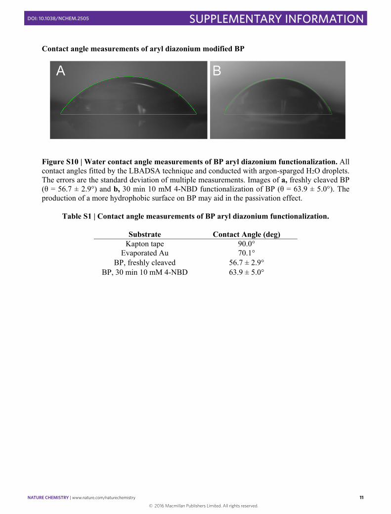
Figure S10 | Water contact angle measurements of BP aryl diazonium functionalization. All contact angles fitted by the LBADSA technique and conducted with argon-sparged H2O droplets. The errors are the standard deviation of multiple measurements. Images of a, freshly cleaved BP (θ = 56.7 ± 2.9°) and b, 30 min 10 mM 4-NBD functionalization of BP (θ = 63.9 ± 5.0°). The production of a more hydrophobic surface on BP may aid in the passivation effect.
Table S1 | Contact angle measurements of BP aryl diazonium functionalization.
Substrate Contact Angle (deg) Kapton tape 90.0°
Evaporated Au 70.1° BP, freshly cleaved 56.7 ± 2.9°
BP, 30 min 10 mM 4-NBD 63.9 ± 5.0°
© 2016 Macmillan Publishers Limited. All rights reserved.
NATURE CHEMISTRY | www.nature.com/naturechemistry 12
SUPPLEMENTARY INFORMATIONDOI: 10.1038/NCHEM.2505
12
Control studies of BP field-effect transistors
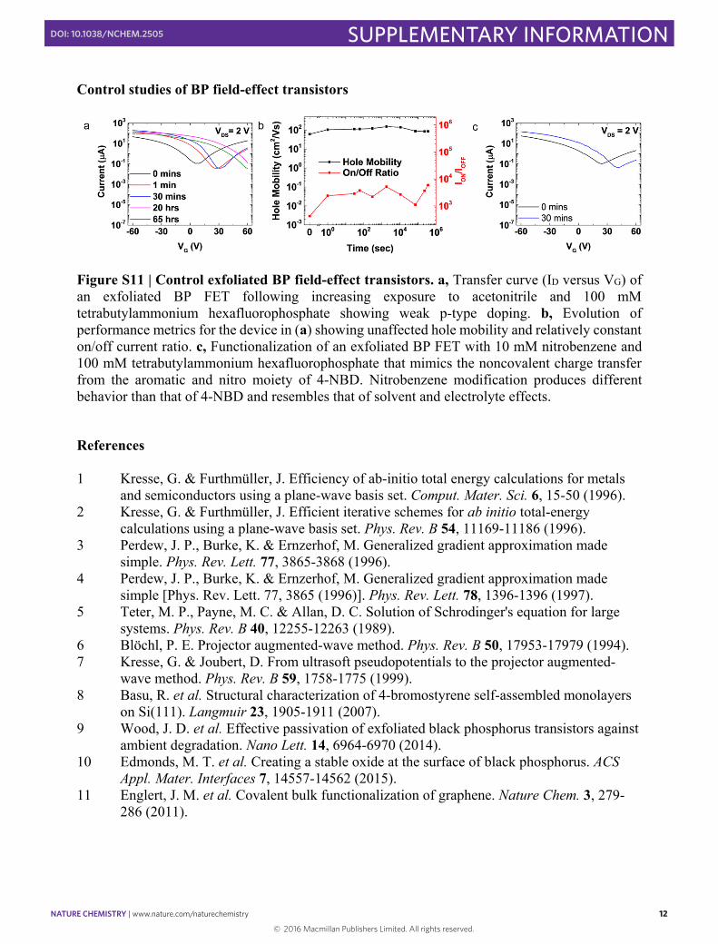
Figure S11 | Control exfoliated BP field-effect transistors. a, Transfer curve (ID versus VG) of an exfoliated BP FET following increasing exposure to acetonitrile and 100 mM tetrabutylammonium hexafluorophosphate showing weak p-type doping. b, Evolution of performance metrics for the device in (a) showing unaffected hole mobility and relatively constant on/off current ratio. c, Functionalization of an exfoliated BP FET with 10 mM nitrobenzene and 100 mM tetrabutylammonium hexafluorophosphate that mimics the noncovalent charge transfer from the aromatic and nitro moiety of 4-NBD. Nitrobenzene modification produces different behavior than that of 4-NBD and resembles that of solvent and electrolyte effects.
References 1 Kresse, G. & Furthmüller, J. Efficiency of ab-initio total energy calculations for metals
and semiconductors using a plane-wave basis set. Comput. Mater. Sci. 6, 15-50 (1996). 2 Kresse, G. & Furthmüller, J. Efficient iterative schemes for ab initio total-energy
calculations using a plane-wave basis set. Phys. Rev. B 54, 11169-11186 (1996). 3 Perdew, J. P., Burke, K. & Ernzerhof, M. Generalized gradient approximation made
simple. Phys. Rev. Lett. 77, 3865-3868 (1996). 4 Perdew, J. P., Burke, K. & Ernzerhof, M. Generalized gradient approximation made
simple [Phys. Rev. Lett. 77, 3865 (1996)]. Phys. Rev. Lett. 78, 1396-1396 (1997). 5 Teter, M. P., Payne, M. C. & Allan, D. C. Solution of Schrodinger's equation for large
systems. Phys. Rev. B 40, 12255-12263 (1989). 6 Blöchl, P. E. Projector augmented-wave method. Phys. Rev. B 50, 17953-17979 (1994). 7 Kresse, G. & Joubert, D. From ultrasoft pseudopotentials to the projector augmented-
wave method. Phys. Rev. B 59, 1758-1775 (1999). 8 Basu, R. et al. Structural characterization of 4-bromostyrene self-assembled monolayers
on Si(111). Langmuir 23, 1905-1911 (2007). 9 Wood, J. D. et al. Effective passivation of exfoliated black phosphorus transistors against
ambient degradation. Nano Lett. 14, 6964-6970 (2014). 10 Edmonds, M. T. et al. Creating a stable oxide at the surface of black phosphorus. ACS
Appl. Mater. Interfaces 7, 14557-14562 (2015). 11 Englert, J. M. et al. Covalent bulk functionalization of graphene. Nature Chem. 3, 279-
286 (2011).
© 2016 Macmillan Publishers Limited. All rights reserved.


![[PPT]GARAM DIAZONIUM & SENYAWA AZO - abc | xyz · Web viewGARAM DIAZONIUM & SENYAWA AZO GARAM DIAZONIUM Ar – N2+X- X Cl-, Br-, HSO4-, NO3-, BF4- (fluoroborat), ClO4- Pembuatan :](https://img.pdfslide.net/doc/110x75/5b015f6a7f8b9a84338e0ff4/pptgaram-diazonium-senyawa-azo-abc-xyz-viewgaram-diazonium-senyawa-azo-garam.jpg)