Embed Size (px)
Citation preview

Superhydrophilic and solvent resistant coatings on polypropylene
fabrics by a simple deposition process
Jason Fang, Antonios Kelarakis, Luis Estevez, Yue Wang, Robert Rodriguez, Emmanuel P
Giannelis*
Department of Materials Science and Engineering, Cornell University, Bard Hall, Ithaca, New
York, 14853
* To whom correspondence should be addressed. Email: [email protected]
Keywords: multilayer deposition, plasma treatment, superhydrophilicity, modified nanosilica
Abstract
A simple yet general coating method to plasma treated polymeric substrates is presented. The
method is based on electrostatic interactions between the surface functionalized nanoparticles
and the charged substrate and leads to stable and solvent resistant multilayer coatings. The
coatings render polypropylene (PP) hydrophilic and in the case of PP fabric superhydrophilic.
The superhydrophilicity is attributed to the topography and increased roughness of the fabric
compared to a planar, smooth substrate.
1

Coating technologies are continuously being developed in an attempt to meet a diverse range of
very specific requirements and applications 1. Full or partial coatings are applied to surfaces for a
number of different reasons including aesthetic or functional finishes and protective layers.
Current trends in this field have focused on introducing nanoparticles to coating formulations 2,3.
In this report, we present a simple deposition process using functionalized SiO2 nanoparticles on
plasma treated polypropylene (PP) fabrics. As a control experiment, deposition on planar PP
substrates is also demonstrated. The electrostatic attraction between the functionalized
nanoparticles and the charged plastic substrates imparts stability and durability to the coatings.
Judicious selection of the functional groups grafted to the nanoparticles and optimization of their
charge density leads to coatings exhibiting hydrophilicity (superhydrophilicity in the case of the
PP fabric). To the best of our knowledge this is the first demonstration of a superhydrophilic PP
fabric. While, we focus here on a very specific system, the approach is general and applicable to
a wide range of substrate-particle combinations.
Plasma treated polypropylene, PP, was chosen as a model substrate, since plasma treatment leads
conveniently to the formation of various surface groups4,5 without sacrificing any of its bulk
properties. Silica nanoparticles were used because their surface chemistry and charge density can
be fine-tuned, enabling exquisite control of the electrostatic interactions between the coating and
the substrate6,7. The silica nanoparticles used here were treated with N-Trimethoxysilylpropyl-
N,N,N-trimethylammonium chloride. The presence of ammonium groups on the surface renders
the functionalized nanoparticles positively charged. They remain well dispersed in water within a
wide pH range without any tendency for agglomeration as measured by dynamic light scattering.
The ζ potential of the nanoparticles was 36.4 and 21.5 mV for pH 4 and 7, respectively.
2
SiO2 nanoparticles can be readily deposited onto plasma treated PP (fabric or planar substrate).
In contrast, SiO2 nanoparticles regardless of their surface functionalization or charge adhere
weakly to untreated PP (S. I. Figure 1). Systematic variation of the plasma treatment protocol
revealed that it plays only a minor role in the process which is governed instead by the charge
density of the nanoparticles.
Deposition of nanoparticles with ζ= 36.4 mV to either fabric or planar substrate (Figure 1 a and
b, respectively) results in a monolayer coating with a patchy coverage. On the other hand, a
multilayer coating on both PP substrates is obtained when nanoparticles with ζ=21.5 mV are
used. The TEM picture shown in Figure 2a suggests a complete and uniform coverage along the
periphery of the fiber after a single coating cycle (immersion to the aqueous nanoparticle
suspension, solvent evaporation followed by repeated rinsings in water). Moreover, cross-
sectional TEM imaging indicates the formation of a multilayer coating with a mean value of four
particle layers in thickness after a single coating, as shown in Figures 2b and 2c for fabric and
planar substrates, respectively. Given that the functionalized nanoparticles do not show any
tendency for agglomeration in solution, we believe that the multilayer coating consists of discrete
nanoparticles and is not due to packing of larger aggregates. The moderate magnitude of the zeta
potential (ζ=21.5mV) of the nanoparticles is critical for the simultaneous formation of a multi
layer (rather than a monolayer) and the good coating quality. Uncharged nanoparticles tend to
aggregate; low charge density nanoparticles result in weak adhesion, while high ζ values induce
strong particle-particle repulsive forces, ultimately inhibiting efficient coating. In an attempt to
evaluate their stability and durability, the multi layer coatings were subjected to ultrasonic field
3

while being suspended in four different solvents (water, acetone, ethanol and THF). As shown in
S.I. Figure 2 even after 1 hour of sonication while immersed in the solvents, the coatings resist
detachment and are virtually unchanged.
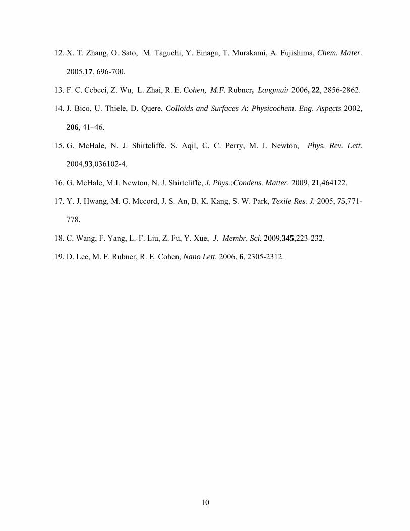
The series of images shown in Figure 3i depict the profiles of an impinging water droplet onto
various plasma treated PP surfaces. In the case of the planar, smooth substrate deposition of SiO2
nanoparticles leads to a hydrophilic surface with a water advancing contact angle of 28o
compared to 71o for the bare surface. The water contact angle is 62o for the coating obtained
using higher ζ potential nanoparticles. There already exists extensive literature on altering the
wetting characteristics of planar substrates. Various approaches include deposition of thin films
of titania8, hollow silica spheres9, silica nanoparticles10,11 or titania-silica alternating
multilayers12 based on layer-by-layer assembly. It has been already shown that a minimum film
thickness is required for obtaining superhydrophilic coatings necessitating multiple deposition
cycles13.
In contrast to the planar substrate, the coated fabric surface exhibits superhydrophilic
characteristics showing virtually 0o advancing water contact angle in less than 0.07s (S.I. Figure
3). The corresponding contact angles for the monolayer coated and the bare fabric are 45o and
120o , respectively (Figure 3). Wetting of a textured surface from a given solvent critically
depends upon the surface-solvent chemical affinity as well as certain topological characteristics
of the surface such as roughness and porosity14,15. The observed difference in wetting behavior
between the planar substrate and the fabric is a direct consequence of the topography of the latter
that gives rise to fundamental differences in the roughness between the two substrates 16. We
4

note that in the case of PP fabrics, approaches such as plasma treatment 17 or grafting of
vinylpyridine chains 18 do not confer superhydrophicity. We believe our approach is the first
demonstration of a superhydrophilic PP fabric.
In order to further explore the coating mechanism of our system we followed an identical
deposition protocol using instead an acidified colloidal dispersion (pH=4) of the unmodified
silica nanoparticles. After a single coating cycle the unmodified nanoparticles form an
incomplete, patchy monolayer (S.I. Figure 4), which does not improve hydrophilicity. This result
underlines the key role of the functional groups on the nanoparticles to the coating quality in
determining the charge density, the layer thickness and topography and ultimately altering the
intrinsic wetting characteristics of the coating. Note that the water contact angle of unmodified
planar silica surface is 20o 19, e.g. substantially higher than the values for superhydrophilic silica
based coating described here.
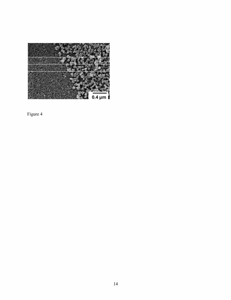
Lastly, we note that the cationically modified silica nanoparticles can effectively support
sequential deposition of opposite charged nanoparticles. To demonstrate this opportunity,
negatively charged carbon black particles were deposited on silica coated fabric as clearly shown
in Figure 4. Current work is focusing on multilayer (ABAB…) deposition of opposite charged
nanoparticles to create highly tuned, functional surfaces.
In summary, we report on a simple coating approach that allows the formation of a multilayer of
nanoparticles on charged substrates after a single-step deposition. The method relies on
electrostatic substrate-particle and particle-particle interactions that largely depend upon the
5

charge density of nanoparticles. The resulting multilayer coatings show remarkable stability in
water and other solvents. In addition the coatings render the substrate hydrophilic
(superhydrophilic in the case of the PP fabric due to the topography and increased roughness of
that substrate).
Acknowledgements
This publication was based on work supported by Award No KUS-C1-018-02, made by King
Abdullah University of Science and Technology (KAUST).
Experimental Section
Cationically modified Silica nanoparticles. Colloidal silica Ludox HS-30 with a mean diameter
of 18 nm was purchased from Sigma Aldrich. 3 gram of colloidal silica was diluted with
deionized water (30 mL) and sonicated for 30 min. A concentrated solution of HCl (1N) was
added to the dispersion followed by the addition of 3.2 g of N-Trimethoxysilylpropyl-N,N,N-
trimethylammonium chloride (50 wt %, Gelest). The mixture was stirred at 60 oC for 10 min.
NaOH (0.1 M) was added to adjust the pH to ~5 and the mixture was stirred continuously at 60
oC for 24 h to complete the reaction. Subsequently, the suspension was dialyzed in deionized
water using SnakeSkin tubing (3.5k MWCO, Pierce) for 48 h.
Plasma treated Polypropylene : Industrial spun-bounded polypropylene nonwoven fabric (0.9
g/cm3, 240 ± 20 μm thick) with a melt flow index of 36.0 g/600 s supplied by Kimberly-Clark
Company, was used as substrate. The substrate was treated with Ar/O2 (50/50) mixed gas plasma
under 234 W for 2 min. Based on X-ray photoelectron spectroscopy the oxygen/carbon ratio of
6

the surface after plasma treatment was 0.12. The planar PP substrate was provided by Bamberger
Polymers (product Bapolene 4082) and was plasma treated under 75 W for 1 min using a Glen
1000 Resist Strip apparatus, (oxygen/carbon ratio of the plasma treated surface was 0.12).
Surface Contact Angle: Static and dynamic advancing contact angle measurements were carried
out by means of a VCA Optima XE apparatus. The water droplets (deionized water from
Millipore purification system, specific conductance 0.05 μS/cm, pH 5.5, droplet volume 0.5μL)
were monitored by a CCD camera and analyzed by standard drop-shape analysis methods.
Zeta potential: Electrophoretic measurements were made using a Malvern Zetasizer Nano-ZS
(Malvern Instruments, England) package which includes a 4 mW He-Ne laser operating at λ =
633 nm. Dust-free solutions were obtained more than 12 h before measurement by filtration
through Nylon membrane filters with a pore size of (0.2 ~ 0.45 µm) (GE Nylon ® Syringe
Filter).
Scanning Electronic Microscopy (SEM): SEM measurements were performed on a Keck Field
Emission Scanning Electron Microscope (FE-SEM), LEO 1550 model.
Transmission electron microscopy (TEM): TEM imaging was performed on FEI Tecnai T12
using microtomed epoxy-embedded ultrathin samples.
7

Figures
Figure 1. TEM images of the plasma treated PP surfaces coated with a monolayer of cationically
modified silica nanoparticles (ζ=36.4 mV) after deposition on a fabric (a) and planar (b)
substrate.
Figure 2. TEM images of the plasma treated PP surfaces coated with a multilayer of cationically
modified silica nanoparticles (ζ= 21.5 mV) after a one-step deposition on a fabric (a and b) and
planar (b) substrate.
Figure 3. i) Profiles of advancing water contact angle of various plasma treated polypropylene
substrates, both fabric and planar: a) uncoated substrates; b and c) coated with silica
nanoparticles with ζ= 36.4 mV and 21.5 mV, respectively. ii) Structure- properties relationships
in plasma treated polypropylene fabrics.
Figure 4. SEM image of the plasma treated polypropylene fabric after the first layer deposition
of cationically modified SiO2 nanoparticles (left side of Figure 4), followed by sequential
deposition of negatively charged carbon black nanoparticles (right side of Figure 4).
8

References
1. “Coating Technology Handbook” edited by D. Satas, A. A. Tracton, Marcel Dekker Inc.,
New York, 2001.
2. “Nanotechnology Applications in Coatings” edited by R. H. Fernando, L.-P. Sung, ACS
Symposium Series, vol. 1008, 2009.
3. “Smart Coatings II” edited by T. Provder, J. Baghdachi, ACS Symposium Series, vol.
1002, 2009.
4. E. M. Liston, L. Martinu, M. R. Wertheimer, J. Adhension Sci. Technol. 1993,7,1091-1127.
5. M. Strobel, M. J. Walzak, J. M. Hill, A. Lin, E. Karbashewski, C. S. Lyons, J. Adhension
Sci. Technol. 1995,9,365-383.
6. E. F. Vansant, P. van der Voort, K. C. Vrancken, “Characterization and Chemical
Modification of the Silica Surface” Elsevier, Amsterdam, 1995.
7. “Colloidal Silica: Fundamentals and applications” edited by H. E. Bergna, W.O. Roberts,
Taylor and Francis Group, U.S.A., 2006.
8. R. Wang, K. R. Wang, K. Hashimoto, A. Fujishima, M. Chikuni, E. Kojima, A. Kitamura,
M. Shimohigoshi, T. Watanabe, Nature 1997, 388, 431-432.
9. X. Liu, X. Du, J. He, Chem. Phys. Chem. 2008,9,305-309.
10. F. Wang, S. Peters, J. Cuzda, R. H. Blunk, A. P. Angelopoulos, Langmuir 2009,25,4384-
4392.
11. X. Liu, J. He, J. Phys. Chem. C 2009 113,148-152.
9
12. X. T. Zhang, O. Sato, M. Taguchi, Y. Einaga, T. Murakami, A. Fujishima, Chem. Mater.
2005,17, 696-700.
13. F. C. Cebeci, Z. Wu, L. Zhai, R. E. Cohen, M.F. Rubner, Langmuir 2006, 22, 2856-2862.
14. J. Bico, U. Thiele, D. Quere, Colloids and Surfaces A: Physicochem. Eng. Aspects 2002,
206, 41–46.
15. G. McHale, N. J. Shirtcliffe, S. Aqil, C. C. Perry, M. I. Newton, Phys. Rev. Lett.
2004,93,036102-4.
16. G. McHale, M.I. Newton, N. J. Shirtcliffe, J. Phys.:Condens. Matter. 2009, 21,464122.
17. Y. J. Hwang, M. G. Mccord, J. S. An, B. K. Kang, S. W. Park, Texile Res. J. 2005, 75,771-
778.
18. C. Wang, F. Yang, L.-F. Liu, Z. Fu, Y. Xue, J. Membr. Sci. 2009,345,223-232.
19. D. Lee, M. F. Rubner, R. E. Cohen, Nano Lett. 2006, 6, 2305-2312.
10

0.2 μm
(b)(a)
0.2 μm
Figure 1
11

(b)(a)
1 μm 0.2 μm
(c)
0.1 μm
Figure 2
12
i
13
ii
Figure 3
14
0.4 μm
Figure 4


![Surface & Coatings Technologyantifouling surfaces [18,19], antibacterial coatings [2,13], renewable energy [20] and epoxy-based powder coatings as a promising alter-native to solvent-borne](https://img.pdfslide.net/doc/110x75/5edb6a70ad6a402d6665a00c/surface-coatings-antifouling-surfaces-1819-antibacterial-coatings-213.jpg)