Embed Size (px)
Citation preview

1
LIVER TRANSCRIPTOMICS HIGHLIGTHS INTERLEUKIN-32 AS NOVEL NAFLD-RELATED
CYTOKINE AND CANDIDATE BIOMARKER
Guido A Baselli, Paola Dongiovanni, Raffaela Rametta, Marica Meroni, Serena Pelusi, Marco
Maggioni, Sara Badiali, Piero Pingitore, Tiziana Montalcini, Alice E Taliento, Daniele Prati, Giorgio
Rossi, Anna L Fracanzani, Rosellina M Mancina, Stefano Romeo, Luca VC Valenti
SUPPLEMENTARY DATA
Supplementary material Gut
doi: 10.1136/gutjnl-2019-319226–12.:10 2020;Gut, et al. Baselli GA

2
SUPPLEMENTARY METHODS
Transcriptomic analysis
Total RNA was isolated using miRNeasy mini-kit (Qiagen, Hulsterweg, Germany). RNA quality
was assessed through Agilent 2100 Bioanalyzer and samples with RNA integrity numbers greater
than or equal to 7 were used for library preparation. RNA sequencing was performed in paired-end
mode with a read length of 150nt using the Illumina HiSeq 4000 (Novogene, Hong Kong, China). Raw
reads were mapped against the Human Genome [1] using a custom pipeline based on the standard
primary analysis procedure. The pipeline performed the primary analysis step including reads
quality check (FastQC software, Babraham Bioinformatics, Cambridge, UK), low-quality reads
trimming and the mapping on GRCh37 reference genome using STAR mapper [2]. Samples
characterized by low coverage (<10 million mapped reads) or insufficient mapping specificity
(uniquely mapped < 60% mapped reads) were excluded from the analysis. Reads count (ENSEMBL
human transcript reference assembly version 75) was performed using RSEM package [3]. Counts
were normalized using DESeq2 package [4]. To quantify isoform expression, per transcript reads
count and normalization in transcript per million was performed exploiting RSEM package.
Interleukin-32 (IL32) transcripts were grouped and reconducted to isoforms described in the
literature [5], by evaluation of the exon structure and the coding sequence. As no transcripts
corresponding to IL32ζ and IL32sm are described in ENSEMBL, their expression levels were not
assessed.
Bioinformatic analysis
For principal components analysis (PCA), gene-level expression data were normalized under
the null model through DESeq2 standard pipeline, and variance stabilizing transformation function
was applied. To identify the major clinical determinants of the transcriptome variability, the first 5
principal components (explaining 30.4% of the variance) were correlated to a set of clinical and
genetic features (listed in the results section). Due to differences in the clinical variables type and
presence of ties, non-parametric Kendall correlation was employed. Differential expression of genes
was investigated following the standard DESeq2 data analysis pipeline by corrected Wald test. Batch
factor, sex, and age were included in the analysis models as confounding factors. For gene
expression correlation, variance stabilized normalized counts of IL32 gene were Pearson correlated
with those of all the other genes. For both the analyses, after multiplicity correction low expressed
genes (mean count ≤ 5) were excluded.
After differential expression analysis, Euclidean distances from case to controls were
calculated considering the variance stabilized expression values of the differential genes. For each
case, mean of distances to each control sample was considered. To identify differentially expressed
pathways, pre-ranked gene set enrichment analysis (GSEA) was performed on differentially
expressed or significantly correlated genes [6]. Respectively, log2(fold change) or the correlation
estimate was used as ranking metrics. The “hallmark” gene sets from the Molecular Signature
Database (MSigDB) version 6.1 were employed [7]. IL32 promoter in HepG2 cells was defined
exploiting chromatin state segmentation data from the ENCODE project. Chromatin immune
precipitation sequencing (ChIP-seq) analysis performed by the ENCODE consortium (Version 3) [8]
evaluated the transcription factors which bind to IL32 promoter in HepG2 cells.
Cell isolation, culture and fatty acids treatment
Primary human hepatocytes (HH) and hepatic stellate cells (HSCs) were isolated from human
tissues resulting from surgical resections and transplants by enzymatic digestion and density
Supplementary material Gut
doi: 10.1136/gutjnl-2019-319226–12.:10 2020;Gut, et al. Baselli GA

3
gradient centrifugation [9]. Stellate cells were cultured on plastic in in DMEM (ThermoFisher,
Waltham, US) for three passages before RNA extraction; hepatocytes were lysed immediately after
isolation. Primary human umbilical vein endothelial cells (HUVEC) were a kind gift of L. Lazzari
(Fondazione IRCCS Ca’ Granda), and were cultured for three passages in MEM 199 (Thermofisher,
Waltham, US) supplemented with Endocult (Stemcell, Vancouver, Canada). Lymphocytes and
monocytes were isolated from healthy donors (N=16) whole blood and cultured for 24h before lysis
[10]. Human M1 and M2 like macrophage derived from monocytes treated for 14 days with 10 µM
granulocyte-macrophages or macrophage colony stimulating factors [10].
HepG2 cells (ATCC, Washington DC, USA) were cultured on plastic in DMEM medium
supplemented with 10% fetal bovine serum (FBS), 2mM L-Glutamine, and 100 U/ml
Penicillin/Streptomycin (all from ThermoFisher, Waltham, US). Growth medium was replaced every
2 days and cells were sub-cultured once a week. To model steatosis in vitro HepG2 cells where
cultured without FBS for 24h and then exposed for 24h to a mixture of palmitic acid (PA) and oleic
acid (OA) at 1:2 molar ratio to a total concentration of 0.5mM or 1mM.
RNA extraction and RT-qPCR.
Cell lysis and RNA extraction was performed using Trizol reagent. RNA was reverse
transcribed using SuperScript VILO cDNA Synthesis Kit. Gene expression levels were measured by
Real time quantitative polymerase chain reaction (RT-qPCR) exploiting the SYBR green chemistry
(Fast SYBR Green Master Mix) on ABI 7500 fast thermocycler (all from ThermoFisher, Waltham, US).
All reactions were performed in triplicate. Gene expression levels were normalized using the 2-ΔCt
method; β-Actin was used as housekeeping gene. For human IL32 expression quantification, 5’-
AATCAGGACGTGGACAGGTG-3’ forward and 5’-TCACAAAAGCTCTCCCCAGG-3’ reverse primers were
employed.
Supplementary material Gut
doi: 10.1136/gutjnl-2019-319226–12.:10 2020;Gut, et al. Baselli GA

4
SUPPLEMENTARY RESULTS
Correlation of principal components of gene expression with GSEA pathways
We next explored gene pathways defining the principal components of transcriptome
variability (PC1 and PC2) by GSEA. PC1 was correlated with apoptosis, inflammation, and
fibrogenesis (Figure S1A). Conversely, it showed a negative correlation with metabolic pathways and
especially with those involved in lipid metabolism (e.g. oxidative phosphorylation, fatty acids
metabolism, peroxisome metabolism). Overall, PC1 seemed to reflect activation of liver
inflammation and damage response secondary to hepatocellular fat retention. PC2 was negatively
correlated with genes involved in cell metabolism (e.g. oxidative phosphorylation, adipogenesis,
fatty acids metabolism, PI3K-AKT-mTOR pathway, myc targets), response to cell damage (e.g.
unfolded protein response, UV response up, DNA repair) and cell proliferation (e.g. G2-M
checkpoint) (Figure S1B).
Supplementary material Gut
doi: 10.1136/gutjnl-2019-319226–12.:10 2020;Gut, et al. Baselli GA

5
SUPPLEMENTARY TABLES
Table S1 – Supplementary clinical and genetic features
A - Transcriptomic cohort stratified by presence of PNPLA3 I148M variant
PNPLA3 I148M
II (N=60)
PNPLA3 I148M
IM (N=56)
PNPLA3 I148M
IM (N=9)
PNPLA3 I148M
carriers (N=65)
p value
dominant
p value
additive
Age 43.42±10.2 43.84±10.8 44.11±11.7 43.88±10.8 0.81 0.80
Sex, F 51 (85) 48 (86) 8 (89) 56 (86) 0.85 0.79
BMI, Kg/m2 40.80±6.1 40.56±8.5 40.24±6.1 40.51±8.2 0.83 0.81
Severe NAFLD,
Yes 12 (20) 16 (29) 3 (33) 19 (29) 0.97 0.83
NAFLD, Yes 50 (83.3) 47 (84) 7 (78) 54 (83) 0.97 0.83
Diabetes, Yes 6 (10) 7 (13) 2 (25) 9 (15) 0.45 0.29
ALT, U/L 20 {15-26} 21 {16-30.00} 20 {18-28} 20 {16-30} 0.52 0.70
AST, U/L 18 {15-22} 17 {15-22} 19 {16-24} 17 {15-22} 0.41 0.38
Steatosis grade
0/1/2/3
10/29/11/10
(17/48/18/17)
9/18/16/13
(16/32/29/23)
2/1/1/5
(22/11/11/56)
11/19/17/18
(17/29/26/28) 0.10 0.047
Inflammation
grade 0/1/2/3
32/26/2
(53/43/3)
28/20/8
(50/36/14)
3/5/1
(33/56/11)
31/25/9
(48/38/14) 0.27 0.20
Ballooning
grade 0/1/2
55/5/0
(92/8/0)
48/7/1
(86/12/2)
7/2/0
(78/22/0)
55/9/1
(85/14/2) 0.23 0.17
Fibrosis stage
0/1/2/3/4
42/18/0/0/0
(70/30/0/0/0)
35/14/4/1/1
(64/26/7/2/2)
5/3/1/0/0
(56/33/11/0/0)
40/17/5/1/1
(62/27/8/2/2) 0.21 0.19
Cholesterol,
mg/dl 212.56±43.6 208.76 (44.20) 179.33 (75.44) 205.08±49.1 0.45 0.20
LDL, mg/dl 130.52±36.6 132.60±38.2 147.17±36.7 134.42±38 0.62 0.42
HDL, mg/dl 56.88±14.1 52.05±14.2 57±8.5 52.68±13.7 0.15 0.32
Triglycerides,
mg/dl* 110 {83-140} 121 {89-179} 139 {126-176} 124 {90-180} 0.39 0.34
B – Metabolic unit cohort
CAP < median
(244 db/m2, N=21)
CAP ≥ median
(244 db/m2, N=23) p value
Age 51±11 54±9 0.25
Sex, F 14 (67) 11 (48) 0.21
BMI, Kg/m2 25±3 29±3 0.001
Diabetes, Yes 1 (4.8) 1 (4.3) 0.95
ALT, U/L 17 {15 – 21} 18 {16 – 26} 0.08
AST, U/L 19 {16 – 22} 19 {16 – 21} 0.42
Steatosis, CAP 214 {198-226} 278 {255-294} -
Fibrosis, LSM 4 {4-5} 6 {5-6} 0.01
Cholesterol, mg/dl 216±23 222±32 0.45
LDL, mg/dl 125±31 147±28 0.04
Supplementary material Gut
doi: 10.1136/gutjnl-2019-319226–12.:10 2020;Gut, et al. Baselli GA

6
HDL, mg/dl 62±11 50±10 0.003
Triglycerides, mg/dl* 84 {72 – 124} 128 {87 – 149} 0.14
PNPLA3 I148M
CC/CG/GG
11/9/1
(52/43/5)
13/8/1
(59/36/1) 0.70
A) Clinical features of the transcriptomic cohort patients stratified PNPLA3 I148M genotype. B)
Clinical features of the Metabolic unit cohort stratified by CAP. Data are presented as mean±sd, ():
% values, {}: interquartile range. Comparison were performed by generalized linear models, and
non-normally distributed variables were log-transformed before the analysis, Effect of the PNPLA3
variant was evaluated either under a dominant or an additive model. BMI: body max index; NAFLD
non-alcoholic fatty liver disease; LDL low-density lipoprotein; HDL high-density lipoprotein; PNPLA3:
Patatin-like phospholipase domain-containing protein 3.
Supplementary material Gut
doi: 10.1136/gutjnl-2019-319226–12.:10 2020;Gut, et al. Baselli GA

7
Table S2 – Genes differentially expressed in severe NAFLD in both PNPLA3 I148M
carriers and non carriers
Severe NAFLD vs not severe NAFLD – PNPLA3 I148M non carriers (N=12 vs 48)
Gene
Symbol Description
Mean
count
Log2(FC) ±
SE adjusted p
IL32 interleukin 32 1368 1.45±0.35 0.027
COL1A1 collagen type I, alpha 1 chain 422 1.01±0.24 0.027
PLP1 proteolipid protein 1 14 -2.46±0.65 0.047
DUSP8 dual specificity phosphatase 8 30 1.79±0.43 0.027
Severe NAFLD vs not severe NAFLD – PNPLA3 I148M carriers (N=19 vs 46)
Gene
Symbol Description
Mean
count
Log2(FC) ±
SE adjusted p
IL32 interleukin 32 1673 1.32±0.28 0.006
COL1A1 collagen type I, alpha 1 chain 603 0.75±0.22 0.074
PLP1 proteolipid protein 1 20 -1.65±0.47 0.069
DUSP8 dual specificity phosphatase 8 55 1.77±0.47 0.040
Upper panel: Severe NAFLD vs NAFLD and normal liver, wild types. Lower panel: Severe NAFLD vs
NAFLD and normal liver; PNPLA3 I148M carriers. FC: Fold change; SE: standard error; NAFLD: non-
alcoholic fatty liver disease; PNPLA3: patatin-like phospholipase domain-containing protein 3.
Supplementary material Gut
doi: 10.1136/gutjnl-2019-319226–12.:10 2020;Gut, et al. Baselli GA

8
Table S3 – Genes co-regulated with IL32 in the liver of severely obese individuals
Strongest correlation
Gene
symbol Description
Mean
count
estimate
(95% CI) adjusted p
KRT8 keratin 8 3261 0.61
(0.49 - 0.71) 5.18*10-10
HLA-A major histocompatibility complex, class I, A 3821 0.59
(0.46 - 0.7) 3.66*10-9
JUND jun D proto-oncogene 1742 0.57
(0.43 - 0.67) 3.21*10-8
GPX2 glutathione peroxidase 2 532 0.57
(0.43 - 0.67) 3.21*10-8
TYMP thymidine phosphorylase 448 0.56
(0.42 - 0.67) 5.90*10-8
LGALS3BP lectin, galactoside-binding, soluble, 3 binding protein 1298 0.54
(0.4 - 0.65) 3.53*10-7
TAGLN2 transgelin 2 875 0.52
(0.38 - 0.64) 8.80*10-7
C9orf16 chromosome 9 open reading frame 16 190 0.52
(0.38 - 0.64) 8.80*10-7
PKD2 polycystic kidney disease 2 295 -0.52
(-0.26) 1.06*10-6
GAS6 growth arrest-specific 6 678 0.52
(0.38 - 0.64) 1.06*10-6
DUSP8 dual specificity phosphatase 8 30 1.79±0.43 0.027
Cytokines and Growth Factors
Gene
symbol Description
Mean
count
estimate
(95% CI) adjusted p
CCL20 chemokine (C-C motif) ligand 20 1238 0.42
(0.27 - 0.56) 1.64*10-4
CCL14 chemokine (C-C motif) ligand 14 18 -0.37
(-0.51 - -0.2) 1.77*10-3
CXCL9 chemokine (C-X-C motif)
ligand 9 136
0.36
(0.2 - 0.5) 2.26*10-3
CXCL10 chemokine (C-X-C motif)
ligand 10 2663
0.35
(0.19 - 0.5) 3.11*10-3
IL8 interleukin 8 1545 0.34
(0.17 - 0.48) 4.71*10-3
CCL5 chemokine (C-C motif) ligand 5 599 0.31
(0.14 - 0.46) 1.15*10-2
PDGFA platelet-derived growth factor alpha polypeptide 169 0.31
(0.14 - 0.46) 1.18*10-2
CCL19 chemokine (C-C motif) ligand 19 233 0.29
(0.12 - 0.44) 1.93*10-2
GFER growth factor, augmenter of liver regeneration 4755 0.28
(0.11 - 0.43) 2.69*10-2
CXCL1 chemokine (C-X-C motif) ligand 1 533 0.28
(0.1 - 0.43) 2.81*10-2
Upper panel: Most significantly IL32 correlated genes. Lower panel: Most significantly IL32
correlated cytokines and growth factors. IL32: Interleukin 32; CI: confidence interval
Supplementary material Gut
doi: 10.1136/gutjnl-2019-319226–12.:10 2020;Gut, et al. Baselli GA

9
Table S4 – Severe NAFLD/NAFLD correlation with IL32 levels
A, B, C: IL32 association with either NAFLD (upper panels) or severe NAFLD (lower panels)
independently of ALT and AST levels as assessed by logistic regression in patients of the Bariatric
surgery cohort (A), the Hepatology service (B), and the overall (C) cohorts. CI: confidence interval;
SE: standard error; IL32: Interleukin 32; ALT: alanine aminotransferase; AST: aspartate
aminotransferase; NAFLD: non-alcoholic fatty liver disease.
Outcome Predictor β
coefficient 95% CI SE p
A
-
Bariatric
surgery
Cohort
NAFLD
intercept -34.72 -61.35 - -15.85 11.52 0.003
log2(IL32, pg/ml) 2.17 0.81 - 4.07 0.83 0.009
log2(ALT, mg/dl) 0.96 -0.66 - 2.74 0.85 0.26
log2(AST, mg/dl) 2.37 0.14 - 4.94 1.2 0.049
Severe
NAFLD
intercept -28.93 -46.26 - -16.1 7.6 0.0001
log2(IL32, pg/ml) 1.01 0.4 - 1.77 0.34 0.003
log2(ALT, mg/dl) 1.13 -0.37 - 2.77 0.79 0.15
log2(AST, mg/dl) 2.95 0.36 - 6.08 1.44 0.04
B
-
Hepatology
service
Cohort
NAFLD
intercept -21.95 -37.68 - -11.04 6.63 0.0009
log2(IL32, pg/ml) 1.3 0.53 - 2.8 0.56 0.02
log2(ALT, mg/dl) 2.26 0.96 - 3.91 0.74 0.002
log2(AST, mg/dl) 0.03 -3.69 0.92 0.98
Severe
NAFLD
intercept -6.61 -6.76 1.72 0.0001
log2(IL32, pg/ml) 0.15 0.02 - 0.3 0.07 0.03
log2(ALT, mg/dl) 0.2 -1.16 0.29 0.51
log2(AST, mg/dl) 0.7 -1.74 0.44 0.11
C
-
overall
cohort
NAFLD
intercept -24.61 -21.47 5.47 6*10-6
log2(IL32, pg/ml) 1.53 0.85 - 2.39 0.39 0.0001
log2(ALT, mg/dl) 1.56 0.62 - 2.61 0.5 0.002
log2(AST, mg/dl) 0.85 -2.71 0.69 0.21
Severe
NAFLD
intercept -7.03 -5.37 1.36 3*10-7
log2(IL32, pg/ml) 0.16 0.03 - 0.3 0.07 0.01
log2(ALT, mg/dl) 0.18 -1.01 0.26 0.47
log2(AST, mg/dl) 0.81 0.02 - 1.62 0.41 0.05
Supplementary material Gut
doi: 10.1136/gutjnl-2019-319226–12.:10 2020;Gut, et al. Baselli GA

10
Table S5 – Severe NAFLD correlation with IL32 levels
IL32 levels were associated with severe NAFLD independently of FIB4 score as assessed by binary
logistic regression. CI: confidence interval; SE: standard error; IL32: Interleukin 32; NAFLD: non-
alcoholic fatty liver disease.
Predictor β
coefficient 95% CI SE p
Severe
NAFLD
intercept -12.29 -20.91 - -5.75 3.79 0.001
log2(IL32, pg/ml) 1.00 0.42 - 1.78 0.34 0.003
FIB4 1.74 -0.15 - 3.75 0.98 0.076
Supplementary material Gut
doi: 10.1136/gutjnl-2019-319226–12.:10 2020;Gut, et al. Baselli GA

11
Table S6 – Diagnostic scores formulas
Upper and middle panels: NAFLD and severe NAFLD predictive models estimates and best AUROC
thresholds. Data are presented as value (95% CI). Lower panel: Formula applied to calculate disease
probability given by models for each sample. IL32: Interleukin 32; ALT: alanine aminotransferase;
AST: aspartate aminotransferase; NAFLD: non-alcoholic fatty liver disease.
NAFLD/High CAP diagnostic scores coefficients
β Intercept β IL32
log2(pg/ml)
β ALT
log2(mg/dl)
β AST
log2(mg/dl)
IL32-ALT-AST -34.72
(-61.35 - -15.85)
2.17
(0.81 - 4.07)
0.96
(-0.66 - 2.74)
2.37
(0.14 - 4.94)
ALT-AST -7.85
(-15.06 - -1.71) -
0.4
(-0.89 - 1.76)
1.73
(-0.26 - 3.89)
Severe NAFLD diagnostic scores coefficients
β Intercept β IL32
log2(pg/ml)
β ALT
log2(mg/dl)
β AST
log2(mg/dl)
IL32-ALT-AST -28.93
(-46.26 - -16.1)
1.01
(0.4 - 1.77)
1.13
(-0.37 - 2.77)
2.95
(0.36 - 6.08)
ALT-AST -14.44
(-23.56 - -7.25) -
0.61
(-0.7 - 1.99)
2.57
(0.35 - 5.21)
Formula
x"#$%&'#(&')( =β-./01203/ + β"#$% ∗ log%(IL32) + β'#( ∗ log%(ALT) + β')( ∗ log%(AST)
x'#(&')( =β-./01203/ + β'#( ∗ log%(ALT) + β')( ∗ log%(AST)
predictedprobability = eL
1 + eL
Supplementary material Gut
doi: 10.1136/gutjnl-2019-319226–12.:10 2020;Gut, et al. Baselli GA

12
Table S7 – IL32 diagnostic accuracy for NAFLD
A, B, C: IL32-ALT-AST diagnostic accuracy parameters for NAFLD diagnosis as assessed by ROC
analysis in the Bariatric surgery (A), the Hepatology service (B), and the overall(C) cohorts. D)
Diagnostic accuracy for “high-CAP” detection assessed in the Metabolic unit cohort. Data are
presented as value (95% CI). ROC: Receiver operating characteristic, AUROC: area under the ROC
curve. CI: confidence interval, NPV negative predictive value, PPV positive predictive value, IL32:
Interleukin 32; ALT: alanine aminotransferase; AST: aspartate aminotransferase; NAFLD: non-
alcoholic fatty liver disease.
A - NAFLD diagnostic accuracy, Bariatric surgery cohort
AUROC Threshold
(probability) specificity sensitivity NPV PPV
IL32-ALT-AST 0.85
(0.76-0.94)
0.78
(0.58-0.8)
0.94
(0.67-1)
0.68
(0.58-0.92)
0.50
(0.44-0.77)
0.97
(0.89-1)
ALT-AST 0.72
(0.58-0.86)
0.6
(0.6-0.88)
0.5
(0.39-1)
0.92
(0.32-0.98)
0.69
(0.33-0.91)
0.84
(0.82-1)
B - NAFLD diagnostic accuracy, Hepatology service cohort
AUROC Threshold
(probability) specificity sensitivity NPV PPV
IL32-ALT-AST 0.95
(0.91-0.99)
0.91
(0.66-0.96)
0.93
(0.87-1)
0.86
(0.74-0.97)
0.41
(0.28-0.72)
0.99
(0.98-1)
ALT-AST 0.81
(0.71-0.92)
0.87
(0.85-0.95)
0.73
(0.6-1)
0.83
(0.52-0.9)
0.31
(0.17-0.44)
0.97
(0.95-1)
C - NAFLD diagnostic accuracy, overall cohort
AUROC Threshold
(probability) specificity sensitivity NPV PPV
IL32-ALT-AST 0.92
(0.88-0.95)
0.93
(0.69-0.96)
0.97
(0.82-1)
0.74
(0.66-0.91)
0.39
(0.33-0.63)
0.99
(0.97-1)
ALT-AST 0.81
(0.73-0.88)
0.87
(0.83-0.92)
0.85
(0.7-0.97)
0.69
(0.52-0.81)
0.31
(0.24-0.4)
0.96
(0.94-0.99)
D - CAP, high vs. low diagnostic accuracy, Metabolic unit cohort
AUROC Threshold
(probability) specificity sensitivity NPV PPV
IL32-ALT-AST 0.72
(0.57-0.88)
0.96
(0.58-0.99)
0.9
(0.48-1)
0.57
(0.39-0.96)
0.66
(0.58-0.9)
0.87
(0.65-1)
ALT-AST 0.55
(0.38-0.73)
0.91
(0.62-0.96)
0.95
(0.24-1)
0.22
(0.09-0.96)
0.53
(0.5-0.89)
0.83
(0.56-1)
Supplementary material Gut
doi: 10.1136/gutjnl-2019-319226–12.:10 2020;Gut, et al. Baselli GA

13
Table S8 – IL32 diagnostic accuracy for severe NAFLD
A, B, C: IL32-ALT-AST diagnostic accuracy parameters for severe NAFLD diagnosis as assessed by
ROC analysis in the Bariatric surgery (A), the Hepatology service (B), and the overall (C) cohorts. Data
are presented as value (95% CI). ROC: Receiver operating characteristic, AUROC: area under the ROC
curve. CI: confidence interval, NPV negative predictive value, PPV positive predictive value, IL32:
Interleukin 32; ALT: alanine aminotransferase; AST: aspartate aminotransferase; NAFLD: non-
alcoholic fatty liver disease.
A - Severe NAFLD diagnostic accuracy, Biomarker cohort
AUROC Threshold
(probability) specificity sensitivity NPV PPV
IL32-ALT-AST 0.88
(0.8-0.96)
0.21
(0.16-0.69)
0.21
(0.16-0.69)
0.65
(0.54-1)
0.96
(0.6-1)
0.97
(0.82-1)
ALT-AST 0.8
(0.7-0.91)
0.42
(0.17-0.63)
0.42
(0.17-0.59)
0.85
(0.46-0.96)
0.64
(0.52-1)
0.81
(0.77-1)
B - Severe NAFLD diagnostic accuracy, Hepatology service cohort
AUROC Threshold
(probability) specificity sensitivity NPV PPV
IL32-ALT-AST 0.69
(0.61-0.77)
0.9
(0.85-1)
0.5
(0.42-0.85)
0.82
(0.47-0.92)
0.77
(0.65-0.88)
0.57
(0.54-0.76)
ALT-AST 0.68
(0.6-0.76)
0.61
(0.56-0.89)
0.44
(0.36-0.8)
0.89
(0.54-0.96)
0.83
(0.66-0.93)
0.57
(0.53-0.71)
C - Severe NAFLD diagnostic accuracy, overall cohort
AUROC Threshold
(probability) specificity sensitivity NPV PPV
IL32-ALT-AST 0.72
(0.66-0.79)
0.67
(0.27-0.99)
0.51
(0.37-0.86)
0.85
(0.51-0.96)
0.82
(0.7-0.93)
0.55
(0.52-0.73)
ALT-AST 0.71
(0.64-0.78)
0.61
(0.46-0.87)
0.58
(0.47-0.84)
0.78
(0.54-0.89)
0.79
(0.7-0.88)
0.58
(0.53-0.71)
Supplementary material Gut
doi: 10.1136/gutjnl-2019-319226–12.:10 2020;Gut, et al. Baselli GA

14
REFERENCES
[1] Cunningham F, Amode MR, Barrell D, Beal K, Billis K, Brent S, et al. Ensembl 2015. Nucleic
Acids Res 2015;43:D662-669.
[2] Dobin A, Davis CA, Schlesinger F, Drenkow J, Zaleski C, Jha S, et al. STAR: ultrafast universal
RNA-seq aligner. Bioinformatics 2013;29:15-21.
[3] Li B, Dewey CN. RSEM: accurate transcript quantification from RNA-Seq data with or
without a reference genome. BMC Bioinformatics 2011;12:323.
[4] Love MI, Huber W, Anders S. Moderated estimation of fold change and dispersion for RNA-
seq data with DESeq2. Genome Biol 2014;15:550.
[5] Hong JT, Son DJ, Lee CK, Yoon DY, Lee DH, Park MH. Interleukin 32, inflammation and
cancer. Pharmacol Ther 2017;174:127-137.
[6] Subramanian A, Tamayo P, Mootha VK, Mukherjee S, Ebert BL, Gillette MA, et al. Gene set
enrichment analysis: a knowledge-based approach for interpreting genome-wide expression
profiles. Proc Natl Acad Sci U S A 2005;102:15545-15550.
[7] Liberzon A, Birger C, Thorvaldsdottir H, Ghandi M, Mesirov JP, Tamayo P. The Molecular
Signatures Database (MSigDB) hallmark gene set collection. Cell Syst 2015;1:417-425.
[8] Consortium EP. An integrated encyclopedia of DNA elements in the human genome.
Nature 2012;489:57-74.
[9] Dongiovanni P, Meroni M, Baselli GA, Bassani GA, Rametta R, Pietrelli A, et al. Insulin
resistance promotes Lysyl Oxidase Like 2 induction and fibrosis accumulation in non-alcoholic fatty
liver disease. Clinical science 2017;131:1301-1315.
[10] Milano M, Dongiovanni P, Artoni A, Gatti S, Rosso L, Colombo F, et al. Particulate matter
phagocytosis induces tissue factor in differentiating macrophages. Journal of Applied Toxicology
2016;36:151-160.
Supplementary material Gut
doi: 10.1136/gutjnl-2019-319226–12.:10 2020;Gut, et al. Baselli GA

15
SUPPLEMENTARY FIGURE LEGENDS
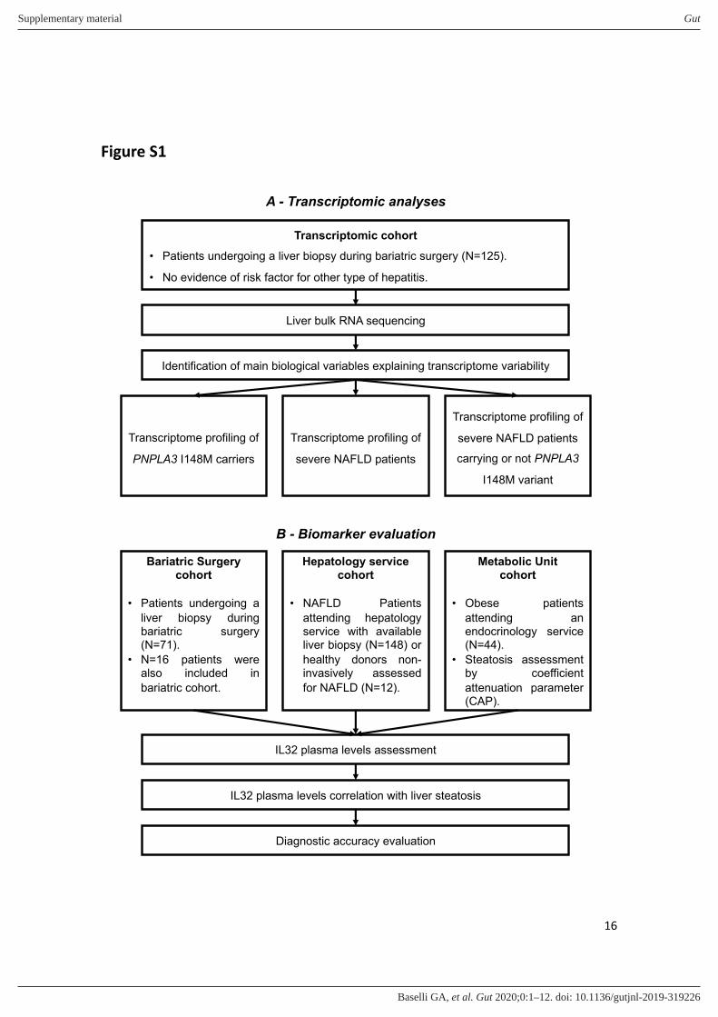
Figure S1 – Study flow chart
A – Transcriptomic analysis workflow. B – Biomarker study workflow.
Supplementary material Gut
doi: 10.1136/gutjnl-2019-319226–12.:10 2020;Gut, et al. Baselli GA
16
Figure S1
Transcriptomic cohort
• Patients undergoing a liver biopsy during bariatric surgery (N=125).
• No evidence of risk factor for other type of hepatitis.
Liver bulk RNA sequencing
Identification of main biological variables explaining transcriptome variability
Transcriptome profiling of
PNPLA3 I148M carriers
Transcriptome profiling of
severe NAFLD patients
Transcriptome profiling of
severe NAFLD patients
carrying or not PNPLA3
I148M variant
Bariatric Surgery cohort
• Patients undergoing a
liver biopsy duringbariatric surgery(N=71).
• N=16 patients werealso included in
bariatric cohort.
IL32 plasma levels assessment
IL32 plasma levels correlation with liver steatosis
Diagnostic accuracy evaluation
Hepatology service cohort
• NAFLD Patients
attending hepatologyservice with availableliver biopsy (N=148) or
healthy donors non-invasively assessed
for NAFLD (N=12).
Metabolic Unit cohort
• Obese patients
attending anendocrinology service(N=44).
• Steatosis assessmentby coefficient
attenuation parameter(CAP).
B - Biomarker evaluation
A - Transcriptomic analyses
Supplementary material Gut
doi: 10.1136/gutjnl-2019-319226–12.:10 2020;Gut, et al. Baselli GA

17
Figure S2 – Pathways correlated with principal components of variability.
A - Pathways enriched in principal component 1 correlated genes (adjusted p<0.1, gene set
enrichment analysis). B - Pathway enriched in principal component 2 correlated genes (adjusted
p<0.1, gene set enrichment analysis). NES: Normalized Enrichment Score; IFN: interferon; ROS:
reactive oxygen species; EMT: epithelial to mesenchymal transition.
Figure S2
Supplementary material Gut
doi: 10.1136/gutjnl-2019-319226–12.:10 2020;Gut, et al. Baselli GA

18
Figure S3 – Class comparison volcano plots and Venn diagrams.
A, B – Genes effect size vs significance level, as assessed by multiplicity adjusted negative binomial
regression for: PNPLA3 I148M carriers vs non-carriers (A), severe NAFLD vs NAFLD and healthy
individuals (B). C – Overlap of genes differentially expressed in PNPLA3 I148M carriers vs non-
carriers and in severe NAFLD vs NAFLD and healthy individuals. D, E – Genes effect size vs
significance level, as assessed by multiplicity adjusted negative binomial regression for: severe vs.
non-severe in PNPLA3 I148M non-carriers (D), severe vs. non-severe NAFLD among PNPLA3 I148M
carriers (E). NAFLD: nonalcoholic fatty liver disease; PNPLA3: Patatin-like phospholipase domain-
containing protein 3.
Supplementary material Gut
doi: 10.1136/gutjnl-2019-319226–12.:10 2020;Gut, et al. Baselli GA

19
Figure S3
Supplementary material Gut
doi: 10.1136/gutjnl-2019-319226–12.:10 2020;Gut, et al. Baselli GA

20
Figure S4 – Distances from NAFLD and healthy individuals.
Mean Euclidean distances from severe NAFLD samples to each NAFLD or Healthy individuals
according to PNPLA3 rs738409 presence. ** p<0.01 Student’s T test. NAFLD: non-alcoholic fatty liver
disease; PNPLA3: patatin-like phospholipase domain-containing protein 3.
Figure S4
Supplementary material Gut
doi: 10.1136/gutjnl-2019-319226–12.:10 2020;Gut, et al. Baselli GA

21
Figure S5 – IL32 promoter region and IL32 expression in response to fatty acids.
A: IL32 promoter region analysis. B: IL32 expression levels in HepG2 cells exposed to a mixture of
palmitic and oleic acids. Data are expressed as fold increase ± SD. ** p<0.01 vs. Untreated, ***
p<0.001 vs Untreated, $ p<0.05 vs 0.5mM PAOA post-hoc Tukey’s honestly significant difference
test. POLR2A: RNA Polymerase II Subunit A; MAX: MYC Associated Factor X; PPARGC1A: Peroxisome
Proliferator Activated Receptor Gamma Coactivator 1 Alpha; ESRRA: Estrogen Related Receptor
Alpha; MAFK: MAF BZIP Transcription Factor K; HSF1: Heat Shock Transcription Factor 1; TBP: TATA-
Box Binding Protein; HNF4A: Hepatocytes Nuclear Factor 4 Alpha; EP300: E1A Binding Protein P300;
MYBL2: MYB Proto-Oncogene Like 2; NFIC: Nuclear Factor I C; RXRA Retinoid X Receptor Alpha; JUN:
Jun Proto-Oncogene; SP1: Sp1 Transcription Factor; HDAC2: Histone Deacetylase 2; MXI1: MAX
Interactor 1, Dimerization Protein; FOSL2: FOS Like 2, AP-1 Transcription Factor Subunit; HNF4G:
Hepatocytes Nuclear Factor 4 Gamma; JUND: JunD Proto-Oncogene, AP-1 Transcription Factor
Subunit; CEBPB: CCAAT/Enhancer Binding Protein Beta; ZBTB7A: Zinc Finger and BTB Domain
Containing 7A; MAZ: MYC Associated Zinc Finger Protein; SIN3AK20: SIN3 Transcription Regulator
Family Member A; PA: palmitic acid; OA: oleic acid; mM: millimolar; IL32: interleukin 32.
Figure S5
A B
Supplementary material Gut
doi: 10.1136/gutjnl-2019-319226–12.:10 2020;Gut, et al. Baselli GA

22
Figure S6 – Metabolic unit cohort data
A) Spearman correlation between IL32 plasma levels and CAP. B) IL32 differential expression in
patients stratified by CAP as assessed by Wilcoxon test. C) Diagnostic accuracy for “high-CAP”
detection of IL32-ALT-AST model compared as compared with ALT-AST model by Delong test. CAP:
Controlled Attenuation Parameter; IL32: interleukin-32; ALT: alanine aminotransferase; AST
aspartate aminotransferase.
Figure S6
Supplementary material Gut
doi: 10.1136/gutjnl-2019-319226–12.:10 2020;Gut, et al. Baselli GA