Embed Size (px)
Citation preview
Supporting Information
for
Advanced Materials, adma.200701125
© Wiley-VCH 200869451 Weinheim, Germany
1
Light-Induced Charge Transfer in Pyrene/CdSe-SWNT Hybrids
Liangbing Hu,†,§ Yan-Li Zhao,‡,§ Koungmin Ryu,¶ Chongwu Zhou,¶
J. Fraser Stoddart,*,‡ and George Grüner*,†
†Department of Physics and Astronomy, ‡California NanoSystems Institute and Department of
Chemistry and Biochemistry, University of California, Los Angeles, 405 Hilgard Avenue, Los Angeles,
California 90095, ¶Department of Electrical Engineering, University Park Campus, University of
Southern California, Los Angeles, California 90089
§These authors contributed equally to this work and both should be considered as first authors.
Supporting Information
Correspondence Addresses Professor George Grüner
Department of Physics and Astronomy University of California, Los Angeles
405 Hilgard Avenue Los Angeles, CA 90095-1547 (USA)
Tel: (+1)-310-825-8782 Fax: (+1)-310-825-5734
Email: [email protected]
Professor J Fraser Stoddart California NanoSystems Institute and
Department of Chemistry and Biochemistry University of California, Los Angeles
405 Hilgard Avenue Los Angeles, CA 90095-1569 (USA)
Tel: (+1)-310-206-7078 Fax: (+1)-310-206-5621
Email: [email protected]
2
Materials. All reagents, including 1-pyrenebutanol (1), (±)- -lipoic acid (2), N,N -
dicyclohexylcarbodiimide (DCC), 4-di(methylamino)pyridine (DMAP), sodium borohydride (NaBH4),
sodium citrate, cadmium perchlorate, and 1,1-dimethyl-2-selenourea are commercially available and
were used without further purification. Single-walled carbon nanotubes (SWNTs) were purchased
from Carbon Solutions Inc and used without further purification.
Instruments. UV-vis spectra were recorded at 25 C on a Varian 100 Bio instrument. Fluorescence
spectra were recorded on LPS-220B fluorescence spectrometer (Photon Technology International)
with Lamp Power Supply at 25 C. Nuclear magnetic resonance (NMR) spectra were recorded on a
Bruker Avance 500 Spectrometer. Chemical shifts were reported in parts per million (ppm) downfield
from the Me4Si resonance which was used as the internal standard when recording 1H NMR spectra.
High-resolution matrix-assisted laser desorption/ionization spectra (HR-MALDI) were measured on an
AppliedBiosystems DE-STR MALDI time-of-flight mass spectrometer. The reported molecular mass
(m/z) values were the most abundant monoisotopic mass. Scanning electron microscopy (SEM)
experiments were carried out using Hitachi S4700 Field Emission SEM. Transmission electron
microscopy (TEM) experiments were performed using a JEOL JEM-2000FX microscope. Atomic
force microscopy (AFM) imaging was taken using Veeco Nanoscope multimode AFM.
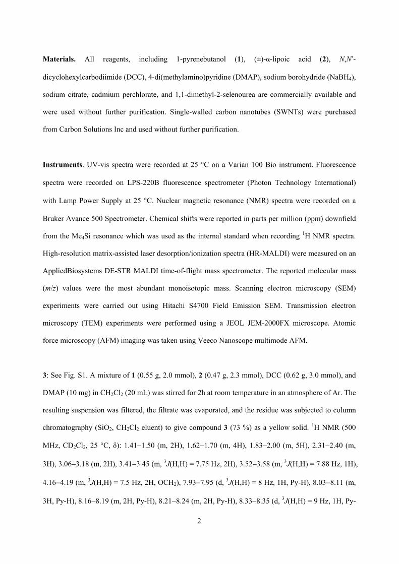
3: See Fig. S1. A mixture of 1 (0.55 g, 2.0 mmol), 2 (0.47 g, 2.3 mmol), DCC (0.62 g, 3.0 mmol), and
DMAP (10 mg) in CH2Cl2 (20 mL) was stirred for 2h at room temperature in an atmosphere of Ar. The
resulting suspension was filtered, the filtrate was evaporated, and the residue was subjected to column
chromatography (SiO2, CH2Cl2 eluent) to give compound 3 (73 %) as a yellow solid. 1H NMR (500
MHz, CD2Cl2, 25 C, ): 1.41 1.50 (m, 2H), 1.62 1.70 (m, 4H), 1.83 2.00 (m, 5H), 2.31 2.40 (m,
3H), 3.06 3.18 (m, 2H), 3.41 3.45 (m, 3J(H,H) = 7.75 Hz, 2H), 3.52 3.58 (m, 3J(H,H) = 7.88 Hz, 1H),
4.16 4.19 (m, 3J(H,H) = 7.5 Hz, 2H, OCH2), 7.93 7.95 (d, 3J(H,H) = 8 Hz, 1H, Py-H), 8.03 8.11 (m,
3H, Py-H), 8.16 8.19 (m, 2H, Py-H), 8.21 8.24 (m, 2H, Py-H), 8.33 8.35 (d, 3J(H,H) = 9 Hz, 1H, Py-
3
H). 13C NMR (125 MHz, CD2Cl2, 25 C, ): 24.8, 25.2, 28.1, 28.5, 32.3, 33.9, 34.4, 35.1, 40.0, 56.2,
63.9, 123.3, 124.5, 124.6, 124.7, 124.8, 125.7, 126.5, 127.0, 127.1, 127.3, 128.4, 129.6, 130.8, 131.4,
136.6, 173.2. MS (HR-MALDI): Calcd for C28H30O2S2 m/z = 462.1687, found m/z = 462.1654.
OH
S SHO
O DCC / DMAP
OS S
O NaBH4
OSH SH
O
OS
SO
OS
SO
Nanotube-coated Silicon WafersO
SS
O
O
SS
OO
SS
O
O
SS
O
O
SS
OO
SS
O
O
SS
O
73 %
or Nanotube Bandles
Cd(ClO4)2 / Me2NC(Se)NH2
+CH2Cl2 Me2CO / H2O
DMF / H2O
1 2 3 4
5
Pyrene/CdSe-SWNT
87 %
63 %
CdSe
CdSe CdSe
Figure S1. Schematic representation of the formation of pyrene/CdSe-coated SWNT hybrids.
4: Compound 3 (0.56 g, 1.17 mmol) was dissolved in 100 mL of 4:1 Me2CO/H2O with stirring in an
atmosphere of Ar. NaBH4 (0.057 g, 1.5 mmol) was added and stirred for 2 h. The reaction mixture was
diluted with H2O (100 mL) and extracted with CH2Cl2 (3 75 mL). The combined organic phases
were dried (MgSO4), filtered, and evaporated. The residue was purified by column chromatography
(SiO2, 97:3 CH2Cl2/MeOH eluent) and evaporated to give compound 4 (87%) as a yellow solid. 1H
4
NMR (500 MHz, CD2Cl2, 25 C, ): 1.41 1.51 (m, 2H), 1.61 1.70 (m, 4H), 1.84 2.00 (m, 4H), 2.18
(d, 1H), 2.31 2.34 (m, 3H), 3.06 3.17 (m, 2H), 3.31 3.44 (m, 3J(H,H) = 7.5 Hz, 2H), 3.52 3.57 (m,
3J(H,H) = 6.75 Hz, 1H), 4.16 4.19 (m, 3J(H,H) = 6.5 Hz, 2H, OCH2), 7.93 7.94 (d, 3J(H,H) = 7.5 Hz,
1H, Py-H), 8.03 8.10 (m, 3H, Py-H), 8.16 8.18 (m, 2H, Py-H), 8.21 8.24 (m, 2H, Py-H), 8.33 8.35
(d, 3J(H,H) = 9 Hz, 1H, Py-H). 13C NMR (125 MHz, CD2Cl2, 25 C, ): 21.3, 24.6, 25.3, 28.0, 28.5,
32.8, 33.9, 34.4, 34.8, 41.8, 63.9, 123.2, 124.5, 124.6, 124.7, 124.8, 125.7, 126.4, 127.0, 127.1, 127.3,
128.4, 129.6, 130.8, 131.3, 136.6, 173.2. MS (HR-MALDI): Calcd for C28H32O2S2 m/z = 464.1844,
found m/z = 464.1738.
8 6 4 2 0ppm
OS S
O
OSH SH
O
OS
SO
OS
SO
OS
SO
CdSe
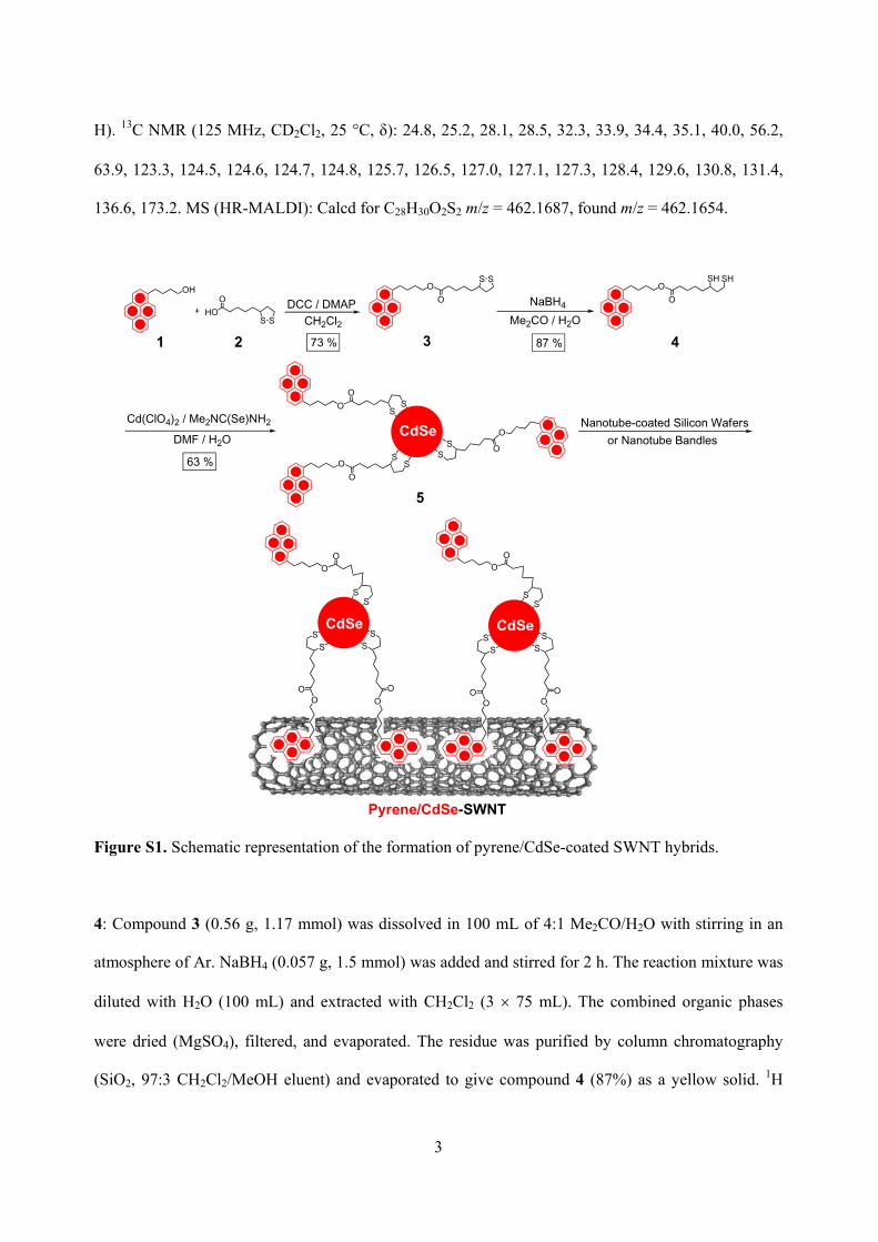
Figure S2. 1H NMR spectra of a) 3 in CD2Cl2, b) 4 in CD2Cl2, and c) pyrene/CdSe 5 in CD3SOCD3 at
25 C.
5
5: The pyrene/CdSe nanoparticles 5 were prepared using a procedure similar to that reported in
reference S1. Sodium citrate (50 mg) was added to deionized H2O (25 mL) containing cadmium
perchlorate (0.025 g, 0.08 mmol) at pH 9. This mixture was purged with Ar for 10 min, and 1,1-
dimethyl-2-selenourea (0.003 g, 0.02 mmol) was then added. The reaction was conducted at 85 C in
an atmosphere of Ar. After 30 min, a DMF solution (30 mL) of 3 (0.021 g, 0.043 mmol) was added,
and the reaction was carried out for another 6h. During this course, the solution became a clear orange
color, indicating the formation of pyrene/CdSe nanoparticles. The reaction was left to cool down to
room temperature, before being reduced to about 10 mL under reduced pressure. The precipitate which
formed was collected by centrifugation and was washed with H2O (2 5 mL) and DMF (2 5 mL) to
remove free salt and 3. The isolated pyrene/CdSe nanoparticles 5 (63 %) were stored in DMF (50 mL)
until ready for use. Figure S2 shows the 1H NMR spectra of 3, 4, and pyrene/CdSe nanoparticles 5.
Pyrene/CdSe-SWNT Hybrids: The pyrene/CdSe-SWNT hybrids were prepared using two methods.
(1) The DMF solution (10 mL) of pyrene/CdSe nanoparticles 5 (2.0 mg) was added dropwise to a
DMF suspension (10 mL) of SWNTs (1.0 mg). The resulting mixture was then sonicated at room
temperature for 8 h. The solution was filtered to remove precipitated material, affording a filtrate
which was the pyrene/CdSe-SWNT hybrid solution. (2) The SWNT-coated silicon wafers were
immersed in a DMF solution (0.2 mg mL 1) of pyrene/CdSe 5 overnight, before being thoroughly
washed briefly with H2O and finally blow dried.
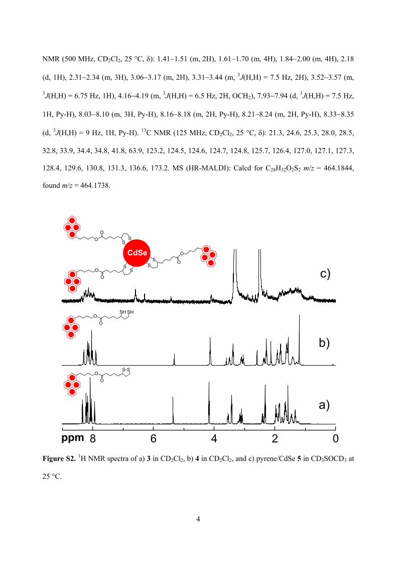
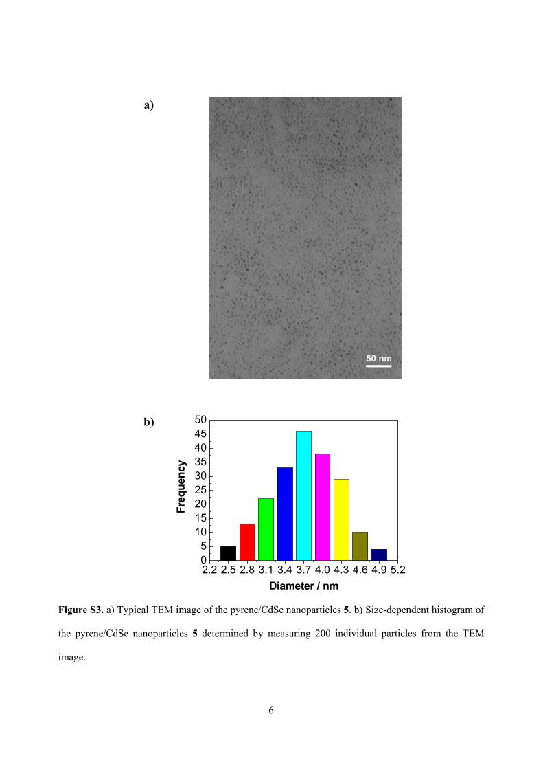
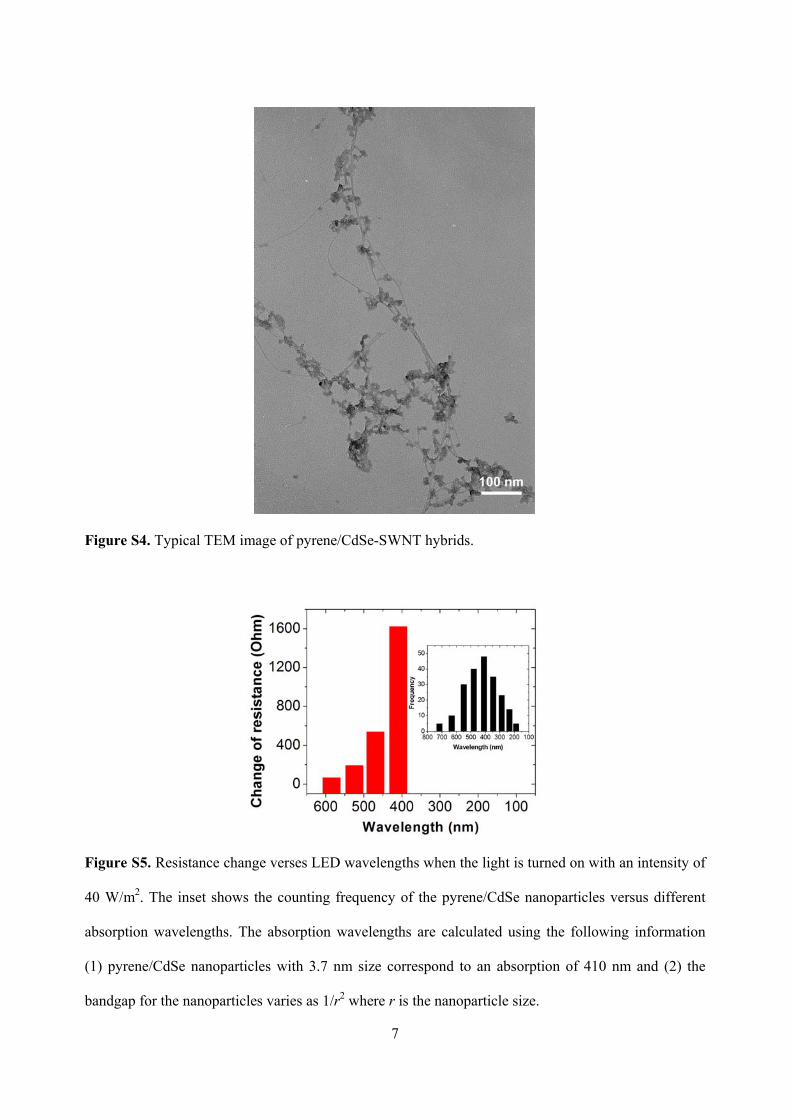
TEM of Nanoparticles 5 and Hybrids: TEM provides the direct information about the shape, size,
and size distribution of the pyrene/CdSe nanoparticles 5 and pyrene/CdSe-SWNT hybrids. A typical
TEM image of 5 shows (Fig. S3) numerous discrete pyrene/CdSe nanoparticles with an average
diameter of 3.7 1.2 nm by measuring 200 individual nanoparticles. The TEM image of pyrene/CdSe-
SWNT hybrids is recorded in Fig. S4. It shows clearly that the pyrene/CdSe nanoparticles can be
attached onto the surfaces of the SWNTs, indicating the formation of pyrene/CdSe-SWNT hybrids.
6
a)
b)
2.2 2.5 2.8 3.1 3.4 3.7 4.0 4.3 4.6 4.9 5.205
101520253035404550
Freq
uenc
y
Diameter / nm
Figure S3. a) Typical TEM image of the pyrene/CdSe nanoparticles 5. b) Size-dependent histogram of
the pyrene/CdSe nanoparticles 5 determined by measuring 200 individual particles from the TEM
image.
7
Figure S4. Typical TEM image of pyrene/CdSe-SWNT hybrids.
Figure S5. Resistance change verses LED wavelengths when the light is turned on with an intensity of
40 W/m2. The inset shows the counting frequency of the pyrene/CdSe nanoparticles versus different
absorption wavelengths. The absorption wavelengths are calculated using the following information
(1) pyrene/CdSe nanoparticles with 3.7 nm size correspond to an absorption of 410 nm and (2) the
bandgap for the nanoparticles varies as 1/r2 where r is the nanoparticle size.
8
a)
250 350 450 550 650 7500.0
0.1
0.2
0.3
0.4
0.5
0.6
0.7
k
k
a
a
[nm]
A
b)
290 310 330 350 370 390 410 430 4500.0
0.1
0.2
0.3
0.4
0.5
0.6
a
a
[nm]
A
k
k
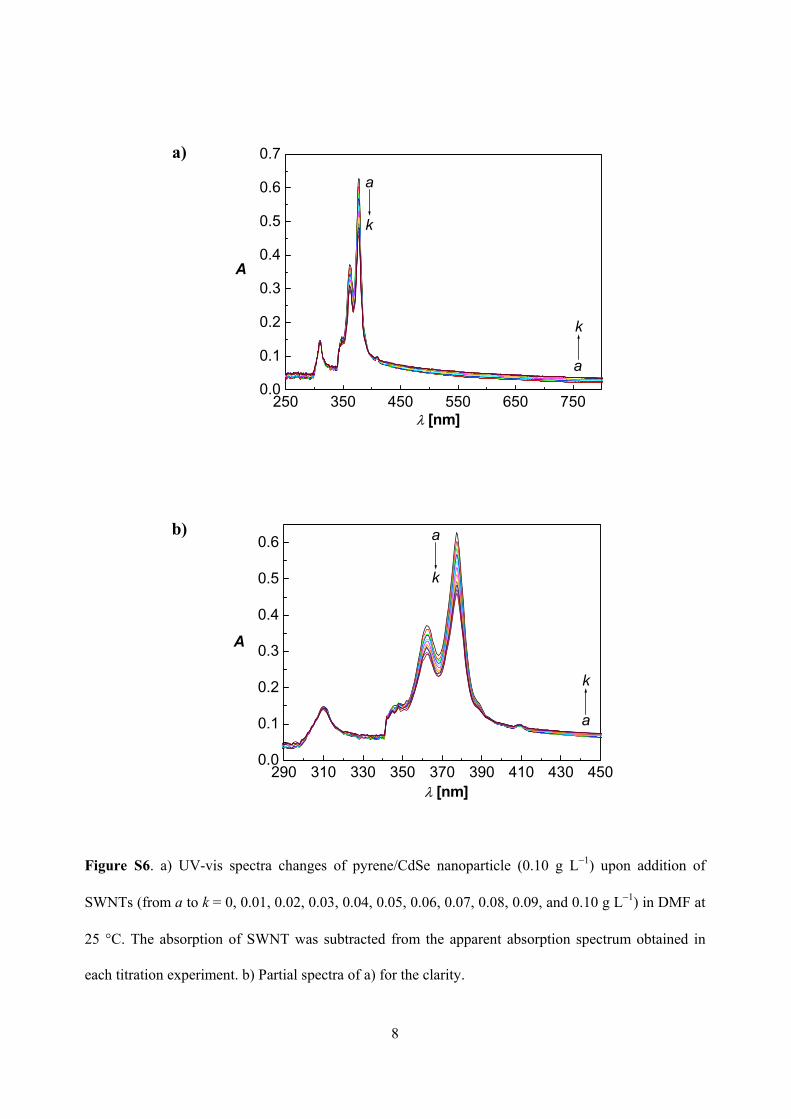
Figure S6. a) UV-vis spectra changes of pyrene/CdSe nanoparticle (0.10 g L 1) upon addition of
SWNTs (from a to k = 0, 0.01, 0.02, 0.03, 0.04, 0.05, 0.06, 0.07, 0.08, 0.09, and 0.10 g L 1) in DMF at
25 C. The absorption of SWNT was subtracted from the apparent absorption spectrum obtained in
each titration experiment. b) Partial spectra of a) for the clarity.
9
250 350 450 550 650 7500.0
0.1
0.2
0.3
0.4
0.5
0.6
0.7
A
[nm]
SWNTs Pyrene 4
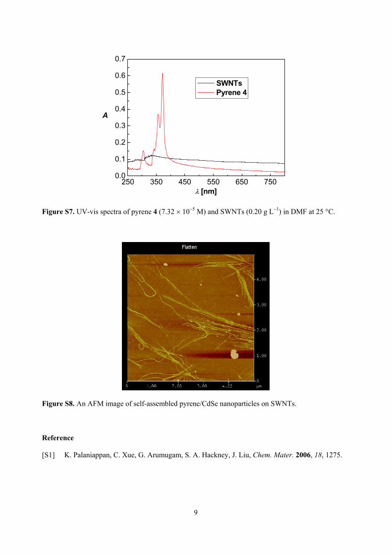
Figure S7. UV-vis spectra of pyrene 4 (7.32 10 5 M) and SWNTs (0.20 g L 1) in DMF at 25 C.
Figure S8. An AFM image of self-assembled pyrene/CdSe nanoparticles on SWNTs.
Reference
[S1] K. Palaniappan, C. Xue, G. Arumugam, S. A. Hackney, J. Liu, Chem. Mater. 2006, 18, 1275.