Embed Size (px)
Citation preview
International Immunology 2010; 1 of 12doi:10.1093/intimm/dxq010
ª The Japanese Society for Immunology. 2010. All rights reserved.For permissions, please e-mail: [email protected]
Suppressed induction of mycobacterial antigen-specific Th1-type CD41 T cells in the lung afterpulmonary mycobacterial infection
Ayano Yahagi1,2, Masayuki Umemura1,2, Toshiki Tamura3,4, Ai Kariyone4,5, M. Dilara Begum1,Kazuyoshi Kawakami6, Yuko Okamoto1,2, Satoru Hamada1,7, Kiyotetsu Oshiro1,8,Hideyasu Kohama1, Takeshi Arakawa1,2, Naoya Ohara9, Kiyoshi Takatsu4,5 and Goro Matsuzaki1,2
1Molecular Microbiology Group, Department of Tropical Infectious Diseases, Center of Molecular Biosciences, TropicalBiosphere Research Center, University of the Ryukyus, Nishihara, Okinawa 903-0213, Japan2Division of Host Defense and Vaccinology, Graduate School of Medicine, University of the Ryukyus, Nishihara, Okinawa,903-0215, Japan3Department of Microbiology, Leprosy Research Center, National Institute of Infectious Diseases, Tokyo 189-0002, Japan4Department of Microbiology and Immunology, Division of Immunology, The Institute of Medical Science, The University of Tokyo,Tokyo 108-8639, Japan5Department of Immunobiology and Pharmacological Genetics, Graduate School of Medicine and Pharmaceutical Science,University of Toyama, Toyama, Toyama 930-0194, Japan6Department of Medical Microbiology, Mycology and Immunology, Tohoku University Graduate School of Medicine, Sendai,Miyagi 980-8575, Japan7Division of Child Health and Welfare and 8Division of Digestive and General Surgery, Graduate School of Medicine, University ofthe Ryukyus, Nishihara, Okinawa, 903-0215, Japan9Department of Immunology, National Institute of Infectious Diseases, Tokyo 162-8640, Japan
Correspondence to: G. Matsuzaki; E-mail: [email protected]
Transmitting editor: S. Koyasu
Received 8 May 2009, accepted 21 January 2010
Abstract
Although the importance of Th1-type immune response in protection against mycobacterial infectionis well recognized, its regulatory mechanism in theMycobacterium tuberculosis (Mtb)-infected lung isnot well characterized. To address this issue, we analyzed kinetics of induction of mycobacterialantigen-specific CD41 Th1 T cells after mycobacterial infection in P25 TCR-transgenic (Tg) mice whichexpress TCR a and b chains from a mycobacterial Ag85B-specific MHC class II Ab-restricted CD41
T-cell clone. To supply normal regulatory T-cell repertoire, we transferred normal spleen T cells intothe P25 TCR-Tg mice before infection. High dose subcutaneous infection with Mtb or Mycobacteriumbovis bacillus Calmette–Guerin (BCG) induced P25 TCR-Tg CD41 Th1 cells within a week. In contrast,high-dose Mtb or BCG infection into the lung failed to induce P25 TCR-Tg CD41 Th1 cells at the earlystage of the infection. Furthermore, low-dose Mtb infection into the lung induced P25 TCR-Tg CD41
Th1 cells on day 21 in the mediastinal lymph node but not in the lung. IL-10 was partially involved inthe suppression of Th1 induction in the lung because pretreatment of mice with anti-IL-10 antibodyresulted in increase of P25 TCR-Tg CD41 Th1 cells in the Mtb-infected lung on day 21 of the infection,whereas neutralization of transforming growth factor-b, another important suppressive cytokine inthe lung, showed no effects on the Th1 induction. Our data suggest that induction of anti-mycobacterial CD41 Th1 cells is suppressed in the mycobacteria-infected lung partially by IL-10.
Keywords: infection, lung, Mycobacterium, TCR transgenic mouse, Th1
Introduction
Tuberculosis caused by Mycobacterium tuberculosis (Mtb)infection is a leading cause of morbidity and mortality re-sponsible for 1.7 million deaths annually (1). Mycobacterial
antigen-specific IFN-c-producing CD4+ (Th1) and CD8+
(Tc1) T cells have pivotal roles in the protective immune re-sponse against tuberculosis (2–4). The T cells also
International Immunology Advance Access published February 18, 2010
contribute to the generation of granulomas in which aggre-gated macrophages engulfed Mtb are surrounded by a cuffof lymphocytes, including the CD4+ and CD8+ T cells (5, 6).Mycobacterium bovis bacillus Calmette–Guerin (BCG) is an
attenuated strain of M. bovis established by Calmette andGuerin for vaccination against Mtb. BCG vaccination to chil-dren has been reported to be efficacious against infant tuber-culosis, especially miliary tuberculosis and tuberculousmeningitis (7–9). However, prophylactic effect of BCG againstadult pulmonary tuberculosis is variable in that the efficacyhas ranged from 0 to 80% (10–12). Therefore, a new vaccineeffective against adult pulmonary tuberculosis is required.Since mucosal tissues of respiratory tract show unique im-
munoregulatory mechanism (13), a new vaccine strategywould be required to induce efficient protective immunity inthe Mtb-infected lung. However, it is not well understoodhow induction of Th1 and Tc1 responses is regulated in thelung after Mtb infection. In tissues such as skin and musclethat are usually used to inoculate vaccines, dendritic cells(DC) capture the inoculated pathogens or antigens and mi-grate into draining lymph node (LN) to present the antigento T cells to initiate immune response. In contrast, pulmonaryimmune response was initiated not only in the lung-drainingLN but also in bronchus-associated lymphoid tissue (14).Although it has been reported that pulmonary immune re-sponse is regulated by alveolar macrophages and cytokinessuch as IL-10 and transforming growth factor (TGF)-b (13),their involvement in regulation of anti-mycobacterial Th1 cellsin mycobacteria-infected lung is not clearly understood.Mtb Ag85B is known as a major Mtb antigen that induces
Th1 response. A 15-mer peptide (Peptide-25, P25) coveringamino acid residues 240–254 of the Ag85B has beenreported to be recognized by CD4+ T cells in MHC class IIH-2Ab-restricted manner and induced Th1 T cells producingIFN-c and IL-2 (15–17). A transgenic (Tg) mouse line thatexpresses functional TCR specific for the P25 epitope ofAg85B in an Ab-restricted manner (P25 TCR-Tg mice) hasbeen established (18). The P25 TCR-Tg CD4+ T cells were ac-tivated to proliferate in the lung-draining mediastinal lymphnode (MLN) from day 14 of pulmonary Mtb infection (19).Kinetics analysis of mycobacterial antigen-specific T cells us-ing another Tg mice expressing TCR specific for Mtb-derivedESAT6 antigen showed that the T cells are activated between7 and 10 days after pulmonary Mtb infection, whereas Th1 re-sponse was demonstrated in the lung 15 days after the infec-tion (20). For all the results, kinetics of Th1 induction was notcompared between vaccine strain BCG and virulent Mtb andbetween subcutaneous (s.c.) and pulmonary infection routs.By using the P25 TCR-Tg mice, we analyzed kinetics of in-
duction of mycobacterial antigen-specific CD4+ Th1 T cells afterMtb or BCG infection in the lung and compared the kineticswith that induced by s.c. infection. Furthermore, regulatorymechanism of the mycobacterial antigen-specific Th1 T-cell in-duction in the lung was analyzed in the Tg T-cell system.
Methods
Animals
The P25 TCR-Tg mice express Ag85B P25 epitope-specificH-2Ab-restricted TCR from CD4+ T-cell clone BP1 under
C57BL/6 background (18). B6 Ly5.1 mice were a kind gift ofDr Yasunobu Yoshikai (Kyushu University, Fukuoka, Japan).Mice were used at 8–12 weeks of age. To supply normalT-cell repertoire to the P25 TCR-Tg mice, each P25 TCR-Tgmouse was transferred with 5 3 107 spleen cells from naiveB6 Ly5.1 mice 1 day before infection (Normal T-cellrepertoire-supplied (N)-P25 TCR-Tg mice). Experimentswere conducted according to the Institutional Ethical Guide-lines for Animal Experiments and the Safety Guideline forLiving Modified Organism Experiments of the University ofthe Ryukyus under approval of the Animal ExperimentsSafety and Ethics Committee and the Living Modified Organ-ism Experiments Safety Committee of the University of theRyukyus, respectively.
P25 TCR-Tg T cell-specific anti-idiotype mAb
B cells from rats immunized with BP1-derived TCR-expressingrat TG40 cells were fused with SP2/0 myeloma cells, anda B-cell hybridoma that react with CD4+ T cells of the P25TCR-Tg mice but not to other T cells was established as theproducer of anti-idiotype mAb KN7.
Microorganisms
Mtb H37Rv strain was grown in Middlebrook 7H9 broth (BD,Sparks, MD, USA) with albumin-dextrose-catalase enrich-ment (BD) at 37�C for 3 weeks. Viable bacterial number wasdetermined on 7H10 agar plates (BD) with oleic acid-albumin-dextrose-catalase (OADC) enrichment (BD). BCGTokyo strain was purchased from Japan BCG (Kiyose, Ja-pan). The bacteria were re-suspended in PBS before use.Mtb was infected s.c. into footpad or i.t. with 1 3 103 or5 3 106 colony-forming unit (CFU) in 50 ll of PBS. BCGwere infected s.c. into footpad or i.t. with 5 3 106 CFU in 50ll of PBS. Bacterial number in infected organs was deter-mined by plating serially diluted organ homogenates onto7H10 agar plates with OADC enrichment.
Cell preparation
Single-cell suspensions were prepared from the lungs asdescribed (21). Footpad-draining popliteal and inguinallymph node (DLN), lung-draining MLN and spleens weresuspended by passing through 30-mm stainless steelmesh. Adherent splenocytes of B6 Ly5.1 mice were col-lected after 90 min culture and used as antigen presentingcells (APC).
Bacterial burden and cytokine assay
Mice were infected with s.c. into footpad or i.t. with 5 3 106
CFU of Mtb or BCG, and the DLN, MLN and lung were re-moved 1, 2 and 4 weeks later, homogenized in distilled waterand plated on 7H10 agar (BD) to determine bacterial number.The lung homogenates were also used to determine IFN-clevels by ELISA (R&D systems, Minneapolis, MN, USA).
Cell culture
Cells were suspended in RPMI 1640 medium (Wako,Osaka, Japan) supplemented with 10% FBS (Equitech Bio,Kerrville, TX, USA), 100 U ml�1 of penicillin (Meiji,
2 Kinetics of Th1 in mycobacteria-infected lung
Yokohama Japan) and 100 lg ml�1 of streptomycin (Meiji)at 5 3 106 cells per ml and cultured with 5 lg ml�1 purifiedprotein derivative (PPD) of Mtb (Japan BCG), recombinant(r) Ag85B of BCG or P25 peptide (FQDAYNAAGGHNAVF)(Invitrogen, Carlsbad, CA, USA). rAg85B was producedfrom yeast Pichia pastoris as secretory protein by usingpPIC9K expression vector (Invitrogen) containing the full-length Ag85B gene cloned from the genomic DNA of BCGTokyo strain.
FACS analysis
To analyze surface molecules on freshly isolated cells, cellswere stained with biotin-conjugated anti-P25 TCR idiotype(KN7), anti-CD4 (Caltag, Burlingame, CA, USA), anti-CD69(Caltag), anti-CD3 (PharMingen, San Jose, CA, USA) andanti-TCRb (PharMingen) mAb. Before surface staining, cellswere treated with HBSS containing 5% of 2.4G2 anti-FcRchybridoma supernatant to block non-specific binding.To detect IFN-c+ T cells, cells were cultured in the pres-
ence or absence of PPD for 24 h, at 37�C in 5% of CO2 withbreferdin A (Golgi Plug, BD) for the last 6 h. The cells werepretreated with anti-FcRc mAb, surface stained withallophycocyanin-conjugated anti-CD4 and biotin-conjugatedanti-P25 TCR idiotype KN7 mAb followed by streptavidin–PE, then permeabilized and fixed with Cytofix/Cytoperm Re-agent (BD) according to the manufacturer’s instructions. Thefixed/permeabilized cells were stained with FITC-conjugatedanti-IFN-c mAb (eBioscience, San Diego, CA, USA).To label proliferating cells in vivo, mice were given 5-
bromo-2-deoxyuridine (BrdU; Sigma–Aldrich, St Louis, MI,USA) at 0.8 mg ml�1 in drinking water during the last 3 daysbefore analysis (22, 23). To label proliferating cells in vitro,naive splenocytes from the P25 TCR-Tg mice werestimulated with antigen for 7 days at 37�C 5% CO2, thenre-stimulated for 48 h with the antigen and APC in the pres-ence of 10 lM BrdU. The BrdU-labeled cells were detectedusing a BrdU Flow kit (BD) according to the manufacturer’sinstructions.The stained cells were analyzed with the FACSCalibur
flowcytometer (BD) and CellQuest software (BD).
Reverse transcription–PCR
Total RNA was extracted, reverse transcribed and amplifiedby PCR as described (21). The PCR product was electro-phoresed on a 1.8% agarose gel and then was observedby Gel Documentation system (Bio-Rad, Hercules, CA,USA). To perform real-time PCR assay, the cDNA wasamplified using the iCycler iQ and the amplification datawere analyzed by the 2�DDCT method using Real-TimePCR Optical System Software Version 3.0 (Bio-Rad) asdescribed (21). The PCR primers used were as follows:TGF-b sense (5#-ATTCCTGGCGTTACCTTGG-3#), Tgfb anti-sense (5#-CCTGTATTCCGTCTCCTTGG-3#); Il10 sense (5#-AGGGAGATTATATTATATGATGGG-3#), Il10 antisense (5#-TTTCTCACCTCTCTTAGG-3#); Il4 sense (5#-CGAAGAACA-CCACAGAGACTGAGCT-3#), Il4 antisense (5#-GACTCATT-CATGGTGCAGCTTATCG-3#) and Actb sense (5#-TGGAA-TCCTGTGGCATCCATGAAA-C-3#), Actb antisense (5#-TAA-AACGCAGCTCAGTAACAGTCCG-3#).
Neutralizing mAb
Anti-IL-10 (SXC-1) mAb and anti-TGF-b (1D11) mAb werepurified from hybridoma culture supernatants and 400 lg ofthe mAb was i.v. injected as indicated.
Statistical analysis
To compare more than three groups, a Kruskal–Wallis testwas used to analyze difference of mean ranks of the groups,followed by a post-hoc test when significant result wasobtained using the Kruskal–Wallis test. All the statistical anal-yses were carried out using Statcel2 software (OMS,Tokorozawa, Japan). A P value of <0.05 was considered toindicate significant difference.
Results
Experimental design to detect mycobacterial antigen-specificTh1 T cells in the lung
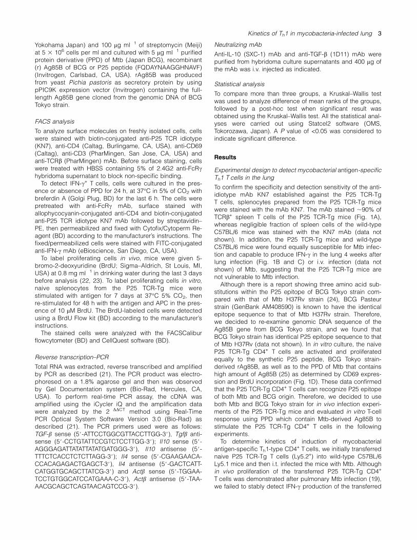
To confirm the specificity and detection sensitivity of the anti-idiotype mAb KN7 established against the P25 TCR-TgT cells, splenocytes prepared from the P25 TCR-Tg micewere stained with the mAb KN7. The mAb stained ;90% ofTCRb+ spleen T cells of the P25 TCR-Tg mice (Fig. 1A),whereas negligible fraction of spleen cells of the wild-typeC57BL/6 mice was stained with the KN7 mAb (data notshown). In addition, the P25 TCR-Tg mice and wild-typeC57BL/6 mice were found equally susceptible for Mtb infec-tion and capable to produce IFN-c in the lung 4 weeks afterlung infection (Fig. 1B and C) or i.v. infection (data notshown) of Mtb, suggesting that the P25 TCR-Tg mice arenot vulnerable to Mtb infection.Although there is a report showing three amino acid sub-
stitutions within the P25 epitope of BCG Tokyo strain com-pared with that of Mtb H37Rv strain (24), BCG Pasteurstrain (GenBank AM408590) is known to have the identicalepitope sequence to that of Mtb H37Rv strain. Therefore,we decided to re-examine genomic DNA sequence of theAg85B gene from BCG Tokyo strain, and we found thatBCG Tokyo strain has identical P25 epitope sequence to thatof Mtb H37Rv (data not shown). In in vitro culture, the naiveP25 TCR-Tg CD4+ T cells are activated and proliferatedequally to the synthetic P25 peptide, BCG Tokyo strain-derived rAg85B, as well as to the PPD of Mtb that containshigh amount of Ag85B (25) as determined by CD69 expres-sion and BrdU incorporation (Fig. 1D). These data confirmedthat the P25 TCR-Tg CD4+ T cells can recognize P25 epitopeof both Mtb and BCG origin. Therefore, we decided to useboth Mtb and BCG Tokyo strain for in vivo infection experi-ments of the P25 TCR-Tg mice and evaluated in vitro T-cellresponse using PPD which contain Mtb-derived Ag85B tostimulate the P25 TCR-Tg CD4+ T cells in the followingexperiments.To determine kinetics of induction of mycobacterial
antigen-specific Th1-type CD4+ T cells, we initially transferrednaive P25 TCR-Tg T cells (Ly5.2+) into wild-type C57BL/6Ly5.1 mice and then i.t. infected the mice with Mtb. Althoughin vivo proliferation of the transferred P25 TCR-Tg CD4+
T cells was demonstrated after pulmonary Mtb infection (19),we failed to stably detect IFN-c production of the transferred
Kinetics of Th1 in mycobacteria-infected lung 3
P25 TCR-Tg CD4+ T cells using KN7 mAb or anti-CD45.2(Ly5.2) mAb in the mycobacteria-infected lung because theP25 TCR-Tg CD4+ T cells are <1% of the total lung T cellseven when 5 3 106 T cells were transferred, and IFN-c+
T cells represent <10% of the Tg CD4+ T cells on day 28 af-ter the infection (data not shown). Therefore, we concludedthat a system which contains higher number of the P25TCR-Tg CD4+ T cells with normal T-cell regulatory system isrequired to analyze kinetics of mycobacterial antigen-specific Th1-type T cells using the Tg mice. To overcome thisproblem, the P25 TCR-Tg mice were transferred with the nor-mal T-cell repertoire of C57BL/6 Ly5.1 mice to construct thenormal T cell-supplied (N)-P25 TCR-Tg mice. Twenty-fourhours after the construction of the N-P25 TCR-Tg mice, thedonor C57BL/6-Ly5.1-derived T cells were detectable withanti-CD45.1 mAb (Ly5.1) in the lung, (Fig. 1E). The N-P25TCR-Tg mice were used to analyze the kinetics of activationand Th1 differentiation of the P25 TCR-Tg CD4+ T cells undernormal immunoregulation after mycobacterial infection.
The Ag85B-specific P25 TCR-Tg CD4+ T cells were activatedand differentiated to Th1 cells rapidly after BCG s.c. infection
Using the N-P25 TCR-Tg mice, we examined kinetics of bac-terial number and activation, proliferation and Th1 differentia-tion of the mycobacterial Ag85B-specific P25 TCR-Tg CD4+
T cells after mycobacterial infection. We first analyzed the re-sponse of the P25 TCR-Tg CD4+ T cells after s.c. infection ofBCG into the footpads. After s.c. infection of 5 3 106 CFU ofBCG, the bacteria was stably detected in the DLN until day28 of the infection (Fig. 2A). The number of P25 TCR-TgCD4+ T cells in the DLN increased from day 3 and peakedon day 28 after the infection (Fig. 2B). The s.c. BCG infec-tion induced expression of early T-cell activation markerCD69, proliferation detected by BrdU incorporation andIFN-c expression of the P25 TCR-Tg CD4+ T cells that aredetected by FACS analysis (Fig. 2B). The IFN-c expressionwas induced by antigen recognition of the P25 TCR-TgCD4+ T cells because IFN-c production was not induced inthe absence of in vitro antigen stimulation of the T cells(Fig. 2C). The number of the CD69+ (Fig. 2D) and IFN-c+
(Fig. 2F) P25 TCR-Tg CD4+ T cells peaked on day 3, andBrdU uptake of the T cells peaked on day 7 (Fig. 2E) afterthe s.c. BCG infection. Although the P25 TCR-Tg CD4+
T cells showed the highest number on day 28 of the s.c. in-fection and IFN-c+ Tg CD4+ T cell number also increasedslightly on day 28, the increase was not statistically signifi-cant. Inoculation of low doses (1 3 103 to 1 3 104 CFU) ofBCG failed to induce detectable activation and Th1 induction
Fig. 1. Phenotype and anti-mycobacterial response of the P25 TCR-Tg mice. (A) Splenocytes from the naive P25 TCR-Tg mice werestained with anti-idiotypic mAb KN7, with anti-TCRb or with anti-CD4mAb. The cells were analyzed by FACS with analysis gate onlymphocytes. (B) Bacterial burden in the lungs of the wild-type or P25TCR-Tg mice was analyzed 4 weeks after lung infection with 200 CFUof Mtb. The lungs were homogenized, plated on 7H10 agar andincubated to count the number of colonies. (C) IFN-c levels in the lunghomogenates were measured by ELISA 4 weeks after Mtb lunginfection. (D) Activation and proliferation of the naive P25 TCR-Tgsplenocytes were analyzed in vitro. Naive splenocytes from P25 TCR-Tg mice were cultured for 7 days with Mtb-derived PPD (open bars),BCG-derived rAg85B (gray bars), P25 peptide (striped bars) orwithout antigen (closed bars). Cells were re-stimulated for 48 h withrelevant antigen, and the number of CD69-expressing and BrdU-incorporated P25 TCR-Tg CD4+ T cells were measured by the FACS.
The data shown are representatives of two independent experiments.(E) Detection of donor-derived CD45.1 cells in the lung of N-P25 TCR-Tg mice. Splenocytes of C57BL/6 Ly5.1 mice were intraperitonealinjection into P25 TCR-Tg mice. The next day, lymphocytes preparedfrom the lung were stained with anti-CD45.1 (Ly5.1), anti-CD4 andanti-CD8, and donor-derived CD45.1+ Tcells were detected by FACS.The data also demonstrate CD4 and CD8 expression of the donor-derived and the recipient P25 TCR-Tg mouse-derived T cells. Therecipient-derived T cells are consisted of higher percentage of CD4+
T cells which express Tg TCR, whereas donor-derived T cells showednormal CD4:CD8 ratio (;2:1).
4 Kinetics of Th1 in mycobacteria-infected lung

of the P25 TCR-Tg CD4+ T cells at early stage of the infec-tion (data not shown). These results suggest that theAg85B-specific P25 TCR-Tg CD4+ T cells in the DLN are ac-tivated and differentiated into Th1 cells at early stage afters.c. BCG infection such as on day 3–7 when high dose(1–5 3 106 CFU) of BCG was infected.
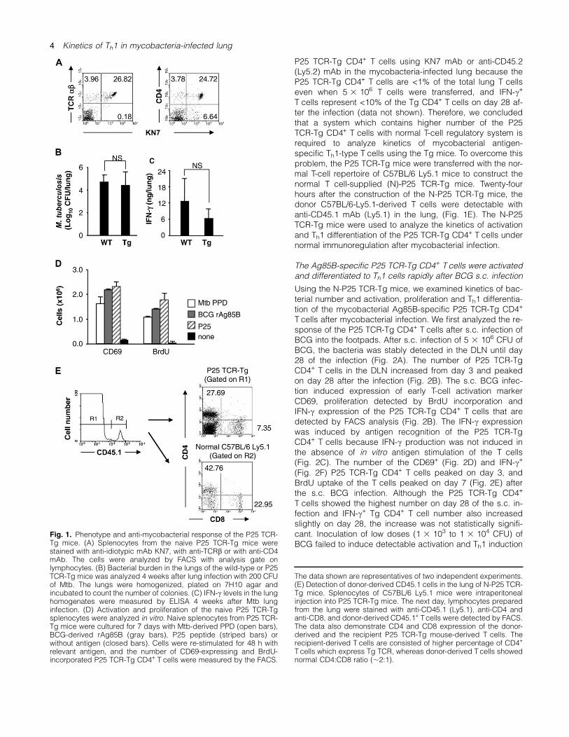
The Ag85B-specific P25 TCR-Tg CD4+ T cells were activatedrapidly but differentiated to Th1 cells at later stages in the lungand MLN after pulmonary BCG infection
We next analyzed kinetics of mycobacterial Ag85B-specificCD4+ T-cell response after pulmonary BCG infection in theN-P25 TCR-Tg mouse system. We used high dose (5 3 106
CFU) of BCG in the pulmonary infection because the doseof BCG induced strong T-cell response while mice remainedhealthy after both s.c. and i.t. infection and maintained highbacterial burden in the lung and MLN until day 28 after thei.t. infection (Fig. 3A). After i.t. BCG infection, the P25 TCR-Tg CD4+ T-cell number peaked on day 3 in the lung (Fig. 3B).
In the MLN, the TCR-Tg T cell slightly increased on day3 although the increase was not statistically significant(Fig. 3B). Representative FACS profiles of CD69 and IFN-cexpression of the P25 TCR-Tg CD4+ T cells in the lung andMLN before and 28 days after BCG i.t. infection are shownin Fig. 3(C), and the kinetics of the CD69+, BrdU+ andIFN-c+ P25 TCR-Tg CD4+ T cells are demonstrated in theFig. 3(D–F). The data indicated that the CD69+ and BrdU+
P25 TCR-Tg CD4+ T cells were vigorously increased (up to270-folds) in the lung and MLN on day 3–7 of i.t. BCG infec-tion followed by decrease of the number (Fig. 3D and E). Incontrast, the Th1-type IFN-c+ P25 TCR-Tg CD4+ T cells werehardly detected on day 3 (Fig. 3F). Although low level of in-crease in the number of IFN-c+ P25 TCR-Tg CD4+ T cells(<10-folds) was observed on day 7–21, there was no statis-tical significant increase when compared with that beforei.t. BCG infection. Robust increase (20- to 30-folds) ofthe IFN-c+ P25 TCR-Tg CD4+ T cells was seen on day 28in the lung and MLN (Fig. 3F). These data indicate that
Fig. 2. Activation and Th1 response of the P25 TCR-Tg CD4+ T cells after BCG s.c. infection. The N-P25 TCR-Tg mice were s.c. infected with5 3 106 CFU of BCG, and bacterial burden and the P25 TCR-Tg CD4+ T cells in the DLN were analyzed. (A) Bacterial number in the DLN wasanalyzed on day 7, 14 and 28 after BCG s.c. infection. (B) The absolute number of the P25 TCR-Tg CD4+ Tcells detected as KN7+ CD4+ cells wasmeasured on day 0, 3 and 7 after s.c. infection of BCG at the indicated doses. (C) FACS profile on the CD69 expression, BrdU incorporation orIFN-c production of the P25 TCR-Tg CD4+ T cells was analyzed in the DLN. Representative data on day 0, and 3 after s.c. infection of BCG areshown. Analysis gate was set on the P25 TCR-Tg CD4+ Tcells. (D–F) The absolute number of the CD69+ (D), BrdU+ (E) or IFN-c+ (F) P25 TCR-TgCD4+ Tcells was measured on day 0, 3, 7, 14 and 28 after s.c. infection BCG. For data given in the panel (D–F), results are shown as mean6 SD,and the asterisks indicate significance of difference (P < 0.05) compared with the cell numbers on day 0. The data shown are as therepresentatives of two to five independent experiments with 3–5 mice used and individually analyzed for each experiment.
Kinetics of Th1 in mycobacteria-infected lung 5
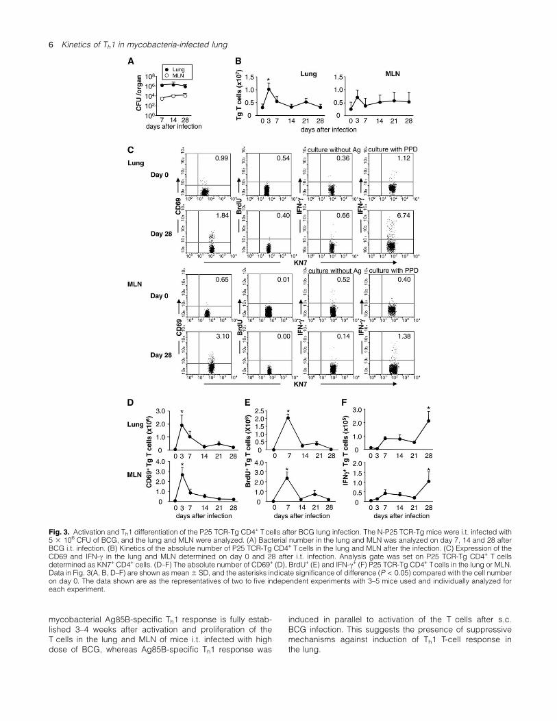
mycobacterial Ag85B-specific Th1 response is fully estab-lished 3–4 weeks after activation and proliferation of theT cells in the lung and MLN of mice i.t. infected with highdose of BCG, whereas Ag85B-specific Th1 response was
induced in parallel to activation of the T cells after s.c.BCG infection. This suggests the presence of suppressivemechanisms against induction of Th1 T-cell response inthe lung.
Fig. 3. Activation and Th1 differentiation of the P25 TCR-Tg CD4+ Tcells after BCG lung infection. The N-P25 TCR-Tg mice were i.t. infected with5 3 106 CFU of BCG, and the lung and MLN were analyzed. (A) Bacterial number in the lung and MLN was analyzed on day 7, 14 and 28 afterBCG i.t. infection. (B) Kinetics of the absolute number of P25 TCR-Tg CD4+ T cells in the lung and MLN after the infection. (C) Expression of theCD69 and IFN-c in the lung and MLN determined on day 0 and 28 after i.t. infection. Analysis gate was set on P25 TCR-Tg CD4+ T cellsdetermined as KN7+ CD4+ cells. (D–F) The absolute number of CD69+ (D), BrdU+ (E) and IFN-c+ (F) P25 TCR-Tg CD4+ Tcells in the lung or MLN.Data in Fig. 3(A, B, D–F) are shown as mean6 SD, and the asterisks indicate significance of difference (P < 0.05) compared with the cell numberon day 0. The data shown are as the representatives of two to five independent experiments with 3–5 mice used and individually analyzed foreach experiment.
6 Kinetics of Th1 in mycobacteria-infected lung
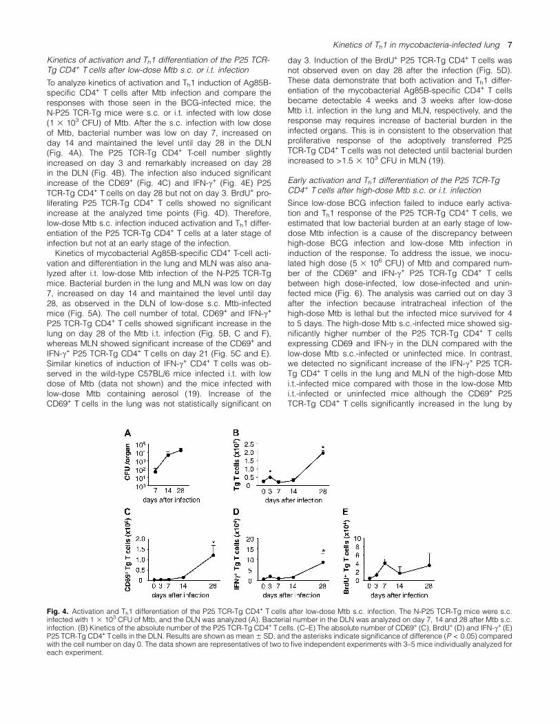
Kinetics of activation and Th1 differentiation of the P25 TCR-Tg CD4+ T cells after low-dose Mtb s.c. or i.t. infection
To analyze kinetics of activation and Th1 induction of Ag85B-specific CD4+ T cells after Mtb infection and compare theresponses with those seen in the BCG-infected mice, theN-P25 TCR-Tg mice were s.c. or i.t. infected with low dose(1 3 103 CFU) of Mtb. After the s.c. infection with low doseof Mtb, bacterial number was low on day 7, increased onday 14 and maintained the level until day 28 in the DLN(Fig. 4A). The P25 TCR-Tg CD4+ T-cell number slightlyincreased on day 3 and remarkably increased on day 28in the DLN (Fig. 4B). The infection also induced significantincrease of the CD69+ (Fig. 4C) and IFN-c+ (Fig. 4E) P25TCR-Tg CD4+ T cells on day 28 but not on day 3. BrdU+ pro-liferating P25 TCR-Tg CD4+ T cells showed no significantincrease at the analyzed time points (Fig. 4D). Therefore,low-dose Mtb s.c. infection induced activation and Th1 differ-entiation of the P25 TCR-Tg CD4+ T cells at a later stage ofinfection but not at an early stage of the infection.Kinetics of mycobacterial Ag85B-specific CD4+ T-cell acti-
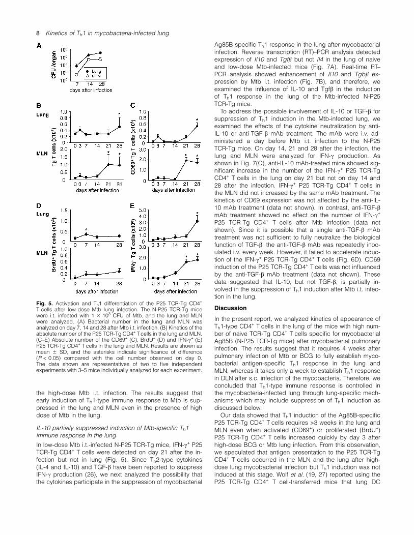
vation and differentiation in the lung and MLN was also ana-lyzed after i.t. low-dose Mtb infection of the N-P25 TCR-Tgmice. Bacterial burden in the lung and MLN was low on day7, increased on day 14 and maintained the level until day28, as observed in the DLN of low-dose s.c. Mtb-infectedmice (Fig. 5A). The cell number of total, CD69+ and IFN-c+
P25 TCR-Tg CD4+ T cells showed significant increase in thelung on day 28 of the Mtb i.t. infection (Fig. 5B, C and F),whereas MLN showed significant increase of the CD69+ andIFN-c+ P25 TCR-Tg CD4+ T cells on day 21 (Fig. 5C and E).Similar kinetics of induction of IFN-c+ CD4+ T cells was ob-served in the wild-type C57BL/6 mice infected i.t. with lowdose of Mtb (data not shown) and the mice infected withlow-dose Mtb containing aerosol (19). Increase of theCD69+ T cells in the lung was not statistically significant on
day 3. Induction of the BrdU+ P25 TCR-Tg CD4+ T cells wasnot observed even on day 28 after the infection (Fig. 5D).These data demonstrate that both activation and Th1 differ-entiation of the mycobacterial Ag85B-specific CD4+ T cellsbecame detectable 4 weeks and 3 weeks after low-doseMtb i.t. infection in the lung and MLN, respectively, and theresponse may requires increase of bacterial burden in theinfected organs. This is in consistent to the observation thatproliferative response of the adoptively transferred P25TCR-Tg CD4+ T cells was not detected until bacterial burdenincreased to >1.5 3 103 CFU in MLN (19).
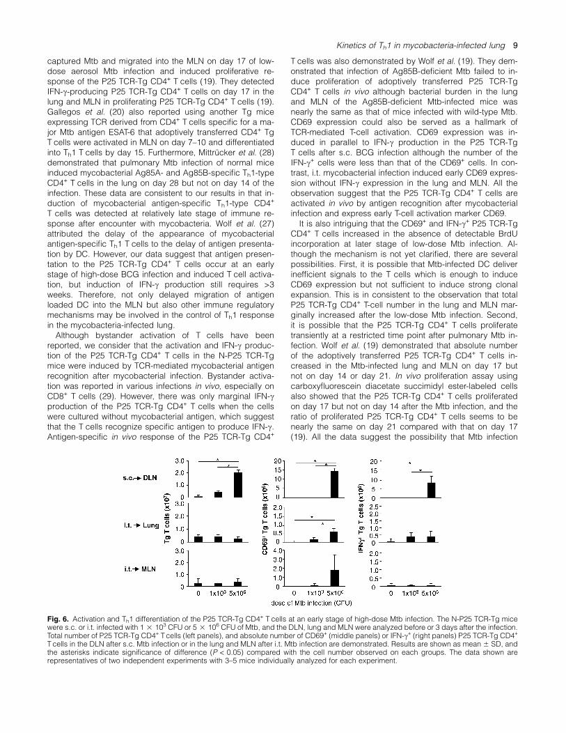
Early activation and Th1 differentiation of the P25 TCR-TgCD4+ T cells after high-dose Mtb s.c. or i.t. infection
Since low-dose BCG infection failed to induce early activa-tion and Th1 response of the P25 TCR-Tg CD4+ T cells, weestimated that low bacterial burden at an early stage of low-dose Mtb infection is a cause of the discrepancy betweenhigh-dose BCG infection and low-dose Mtb infection ininduction of the response. To address the issue, we inocu-lated high dose (5 3 106 CFU) of Mtb and compared num-ber of the CD69+ and IFN-c+ P25 TCR-Tg CD4+ T cellsbetween high dose-infected, low dose-infected and unin-fected mice (Fig. 6). The analysis was carried out on day 3after the infection because intratracheal infection of thehigh-dose Mtb is lethal but the infected mice survived for 4to 5 days. The high-dose Mtb s.c.-infected mice showed sig-nificantly higher number of the P25 TCR-Tg CD4+ T cellsexpressing CD69 and IFN-c in the DLN compared with thelow-dose Mtb s.c.-infected or uninfected mice. In contrast,we detected no significant increase of the IFN-c+ P25 TCR-Tg CD4+ T cells in the lung and MLN of the high-dose Mtbi.t.-infected mice compared with those in the low-dose Mtbi.t.-infected or uninfected mice although the CD69+ P25TCR-Tg CD4+ T cells significantly increased in the lung by
Fig. 4. Activation and Th1 differentiation of the P25 TCR-Tg CD4+ T cells after low-dose Mtb s.c. infection. The N-P25 TCR-Tg mice were s.c.infected with 1 3 103 CFU of Mtb, and the DLN was analyzed (A). Bacterial number in the DLN was analyzed on day 7, 14 and 28 after Mtb s.c.infection. (B) Kinetics of the absolute number of the P25 TCR-Tg CD4+ Tcells. (C–E) The absolute number of CD69+ (C), BrdU+ (D) and IFN-c+ (E)P25 TCR-Tg CD4+ Tcells in the DLN. Results are shown as mean6 SD, and the asterisks indicate significance of difference (P < 0.05) comparedwith the cell number on day 0. The data shown are representatives of two to five independent experiments with 3–5 mice individually analyzed foreach experiment.
Kinetics of Th1 in mycobacteria-infected lung 7
the high-dose Mtb i.t. infection. The results suggest thatearly induction of Th1-type immune response to Mtb is sup-pressed in the lung and MLN even in the presence of highdose of Mtb in the lung.
IL-10 partially suppressed induction of Mtb-specific Th1immune response in the lung
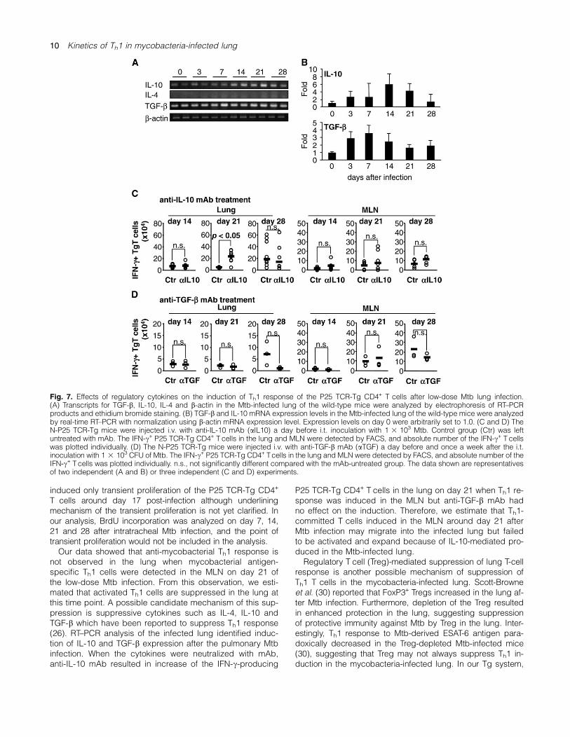
In low-dose Mtb i.t.-infected N-P25 TCR-Tg mice, IFN-c+ P25TCR-Tg CD4+ T cells were detected on day 21 after the in-fection but not in lung (Fig. 5). Since Th2-type cytokines(IL-4 and IL-10) and TGF-b have been reported to suppressIFN-c production (26), we next analyzed the possibility thatthe cytokines participate in the suppression of mycobacterial
Ag85B-specific Th1 response in the lung after mycobacterialinfection. Reverse transcription (RT)–PCR analysis detectedexpression of Il10 and Tgfb but not Il4 in the lung of naiveand low-dose Mtb-infected mice (Fig. 7A). Real-time RT–PCR analysis showed enhancement of Il10 and Tgbb ex-pression by Mtb i.t. infection (Fig. 7B), and therefore, weexamined the influence of IL-10 and Tgfb in the inductionof Th1 response in the lung of the Mtb-infected N-P25TCR-Tg mice.To address the possible involvement of IL-10 or TGF-b for
suppression of Th1 induction in the Mtb-infected lung, weexamined the effects of the cytokine neutralization by anti-IL-10 or anti-TGF-b mAb treatment. The mAb were i.v. ad-ministered a day before Mtb i.t. infection to the N-P25TCR-Tg mice. On day 14, 21 and 28 after the infection, thelung and MLN were analyzed for IFN-c production. Asshown in Fig. 7(C), anti-IL-10 mAb-treated mice showed sig-nificant increase in the number of the IFN-c+ P25 TCR-TgCD4+ T cells in the lung on day 21 but not on day 14 and28 after the infection. IFN-c+ P25 TCR-Tg CD4+ T cells inthe MLN did not increased by the same mAb treatment. Thekinetics of CD69 expression was not affected by the anti-IL-10 mAb treatment (data not shown). In contrast, anti-TGF-bmAb treatment showed no effect on the number of IFN-c+
P25 TCR-Tg CD4+ T cells after Mtb infection (data notshown). Since it is possible that a single anti-TGF-b mAbtreatment was not sufficient to fully neutralize the biologicalfunction of TGF-b, the anti-TGF-b mAb was repeatedly inoc-ulated i.v. every week. However, it failed to accelerate induc-tion of the IFN-c+ P25 TCR-Tg CD4+ T cells (Fig. 6D). CD69induction of the P25 TCR-Tg CD4+ T cells was not influencedby the anti-TGF-b mAb treatment (data not shown). Thesedata suggested that IL-10, but not TGF-b, is partially in-volved in the suppression of Th1 induction after Mtb i.t. infec-tion in the lung.
Discussion
In the present report, we analyzed kinetics of appearance ofTh1-type CD4+ T cells in the lung of the mice with high num-ber of naive TCR-Tg CD4+ T cells specific for mycobacterialAg85B (N-P25 TCR-Tg mice) after mycobacterial pulmonaryinfection. The results suggest that it requires 4 weeks afterpulmonary infection of Mtb or BCG to fully establish myco-bacterial antigen-specific Th1 response in the lung andMLN, whereas it takes only a week to establish Th1 responsein DLN after s.c. infection of the mycobacteria. Therefore, weconcluded that Th1-type immune response is controlled inthe mycobacteria-infected lung through lung-specific mech-anisms which may include suppression of Th1 induction asdiscussed below.Our data showed that Th1 induction of the Ag85B-specific
P25 TCR-Tg CD4+ T cells requires >3 weeks in the lung andMLN even when activated (CD69+) or proliferated (BrdU+)P25 TCR-Tg CD4+ T cells increased quickly by day 3 afterhigh-dose BCG or Mtb lung infection. From this observation,we speculated that antigen presentation to the P25 TCR-TgCD4+ T cells occurred in the MLN and the lung after high-dose lung mycobacterial infection but Th1 induction was notinduced at this stage. Wolf et al. (19, 27) reported using theP25 TCR-Tg CD4+ T cell-transferred mice that lung DC
Fig. 5. Activation and Th1 differentiation of the P25 TCR-Tg CD4+
T cells after low-dose Mtb lung infection. The N-P25 TCR-Tg micewere i.t. infected with 1 3 103 CFU of Mtb, and the lung and MLNwere analyzed. (A) Bacterial number in the lung and MLN wasanalyzed on day 7, 14 and 28 after Mtb i.t. infection. (B) Kinetics of theabsolute number of the P25 TCR-Tg CD4+ Tcells in the lung and MLN.(C–E) Absolute number of the CD69+ (C), BrdU+ (D) and IFN-c+ (E)P25 TCR-Tg CD4+ T cells in the lung and MLN. Results are shown asmean 6 SD, and the asterisks indicate significance of difference(P < 0.05) compared with the cell number observed on day 0.The data shown are representatives of two to five independentexperiments with 3–5 mice individually analyzed for each experiment.
8 Kinetics of Th1 in mycobacteria-infected lung
captured Mtb and migrated into the MLN on day 17 of low-dose aerosol Mtb infection and induced proliferative re-sponse of the P25 TCR-Tg CD4+ T cells (19). They detectedIFN-c-producing P25 TCR-Tg CD4+ T cells on day 17 in thelung and MLN in proliferating P25 TCR-Tg CD4+ T cells (19).Gallegos et al. (20) also reported using another Tg miceexpressing TCR derived from CD4+ T cells specific for a ma-jor Mtb antigen ESAT-6 that adoptively transferred CD4+ TgT cells were activated in MLN on day 7–10 and differentiatedinto Th1 T cells by day 15. Furthermore, Mittrucker et al. (28)demonstrated that pulmonary Mtb infection of normal miceinduced mycobacterial Ag85A- and Ag85B-specific Th1-typeCD4+ T cells in the lung on day 28 but not on day 14 of theinfection. These data are consistent to our results in that in-duction of mycobacterial antigen-specific Th1-type CD4+
T cells was detected at relatively late stage of immune re-sponse after encounter with mycobacteria. Wolf et al. (27)attributed the delay of the appearance of mycobacterialantigen-specific Th1 T cells to the delay of antigen presenta-tion by DC. However, our data suggest that antigen presen-tation to the P25 TCR-Tg CD4+ T cells occur at an earlystage of high-dose BCG infection and induced T cell activa-tion, but induction of IFN-c production still requires >3weeks. Therefore, not only delayed migration of antigenloaded DC into the MLN but also other immune regulatorymechanisms may be involved in the control of Th1 responsein the mycobacteria-infected lung.Although bystander activation of T cells have been
reported, we consider that the activation and IFN-c produc-tion of the P25 TCR-Tg CD4+ T cells in the N-P25 TCR-Tgmice were induced by TCR-mediated mycobacterial antigenrecognition after mycobacterial infection. Bystander activa-tion was reported in various infections in vivo, especially onCD8+ T cells (29). However, there was only marginal IFN-cproduction of the P25 TCR-Tg CD4+ T cells when the cellswere cultured without mycobacterial antigen, which suggestthat the T cells recognize specific antigen to produce IFN-c.Antigen-specific in vivo response of the P25 TCR-Tg CD4+
T cells was also demonstrated by Wolf et al. (19). They dem-onstrated that infection of Ag85B-deficient Mtb failed to in-duce proliferation of adoptively transferred P25 TCR-TgCD4+ T cells in vivo although bacterial burden in the lungand MLN of the Ag85B-deficient Mtb-infected mice wasnearly the same as that of mice infected with wild-type Mtb.CD69 expression could also be served as a hallmark ofTCR-mediated T-cell activation. CD69 expression was in-duced in parallel to IFN-c production in the P25 TCR-TgT cells after s.c. BCG infection although the number of theIFN-c+ cells were less than that of the CD69+ cells. In con-trast, i.t. mycobacterial infection induced early CD69 expres-sion without IFN-c expression in the lung and MLN. All theobservation suggest that the P25 TCR-Tg CD4+ T cells areactivated in vivo by antigen recognition after mycobacterialinfection and express early T-cell activation marker CD69.It is also intriguing that the CD69+ and IFN-c+ P25 TCR-Tg
CD4+ T cells increased in the absence of detectable BrdUincorporation at later stage of low-dose Mtb infection. Al-though the mechanism is not yet clarified, there are severalpossibilities. First, it is possible that Mtb-infected DC deliverinefficient signals to the T cells which is enough to induceCD69 expression but not sufficient to induce strong clonalexpansion. This is in consistent to the observation that totalP25 TCR-Tg CD4+ T-cell number in the lung and MLN mar-ginally increased after the low-dose Mtb infection. Second,it is possible that the P25 TCR-Tg CD4+ T cells proliferatetransiently at a restricted time point after pulmonary Mtb in-fection. Wolf et al. (19) demonstrated that absolute numberof the adoptively transferred P25 TCR-Tg CD4+ T cells in-creased in the Mtb-infected lung and MLN on day 17 butnot on day 14 or day 21. In vivo proliferation assay usingcarboxyfluorescein diacetate succimidyl ester-labeled cellsalso showed that the P25 TCR-Tg CD4+ T cells proliferatedon day 17 but not on day 14 after the Mtb infection, and theratio of proliferated P25 TCR-Tg CD4+ T cells seems to benearly the same on day 21 compared with that on day 17(19). All the data suggest the possibility that Mtb infection
Fig. 6. Activation and Th1 differentiation of the P25 TCR-Tg CD4+ T cells at an early stage of high-dose Mtb infection. The N-P25 TCR-Tg micewere s.c. or i.t. infected with 1 3 103 CFU or 5 3 106 CFU of Mtb, and the DLN, lung and MLN were analyzed before or 3 days after the infection.Total number of P25 TCR-Tg CD4+ Tcells (left panels), and absolute number of CD69+ (middle panels) or IFN-c+ (right panels) P25 TCR-Tg CD4+
T cells in the DLN after s.c. Mtb infection or in the lung and MLN after i.t. Mtb infection are demonstrated. Results are shown as mean 6 SD, andthe asterisks indicate significance of difference (P < 0.05) compared with the cell number observed on each groups. The data shown arerepresentatives of two independent experiments with 3–5 mice individually analyzed for each experiment.
Kinetics of Th1 in mycobacteria-infected lung 9
induced only transient proliferation of the P25 TCR-Tg CD4+
T cells around day 17 post-infection although underliningmechanism of the transient proliferation is not yet clarified. Inour analysis, BrdU incorporation was analyzed on day 7, 14,21 and 28 after intratracheal Mtb infection, and the point oftransient proliferation would not be included in the analysis.Our data showed that anti-mycobacterial Th1 response is
not observed in the lung when mycobacterial antigen-specific Th1 cells were detected in the MLN on day 21 ofthe low-dose Mtb infection. From this observation, we esti-mated that activated Th1 cells are suppressed in the lung atthis time point. A possible candidate mechanism of this sup-pression is suppressive cytokines such as IL-4, IL-10 andTGF-b which have been reported to suppress Th1 response(26). RT–PCR analysis of the infected lung identified induc-tion of IL-10 and TGF-b expression after the pulmonary Mtbinfection. When the cytokines were neutralized with mAb,anti-IL-10 mAb resulted in increase of the IFN-c-producing
P25 TCR-Tg CD4+ T cells in the lung on day 21 when Th1 re-sponse was induced in the MLN but anti-TGF-b mAb hadno effect on the induction. Therefore, we estimate that Th1-committed T cells induced in the MLN around day 21 afterMtb infection may migrate into the infected lung but failedto be activated and expand because of IL-10-mediated pro-duced in the Mtb-infected lung.Regulatory T cell (Treg)-mediated suppression of lung T-cell
response is another possible mechanism of suppression ofTh1 T cells in the mycobacteria-infected lung. Scott-Browneet al. (30) reported that FoxP3+ Tregs increased in the lung af-ter Mtb infection. Furthermore, depletion of the Treg resultedin enhanced protection in the lung, suggesting suppressionof protective immunity against Mtb by Treg in the lung. Inter-estingly, Th1 response to Mtb-derived ESAT-6 antigen para-doxically decreased in the Treg-depleted Mtb-infected mice(30), suggesting that Treg may not always suppress Th1 in-duction in the mycobacteria-infected lung. In our Tg system,
Fig. 7. Effects of regulatory cytokines on the induction of Th1 response of the P25 TCR-Tg CD4+ T cells after low-dose Mtb lung infection.(A) Transcripts for TGF-b, IL-10, IL-4 and b-actin in the Mtb-infected lung of the wild-type mice were analyzed by electrophoresis of RT–PCRproducts and ethidium bromide staining. (B) TGF-b and IL-10 mRNA expression levels in the Mtb-infected lung of the wild-type mice were analyzedby real-time RT–PCR with normalization using b-actin mRNA expression level. Expression levels on day 0 were arbitrarily set to 1.0. (C and D) TheN-P25 TCR-Tg mice were injected i.v. with anti-IL-10 mAb (aIL10) a day before i.t. inoculation with 1 3 103 Mtb. Control group (Ctr) was leftuntreated with mAb. The IFN-c+ P25 TCR-Tg CD4+ T cells in the lung and MLN were detected by FACS, and absolute number of the IFN-c+ T cellswas plotted individually. (D) The N-P25 TCR-Tg mice were injected i.v. with anti-TGF-b mAb (aTGF) a day before and once a week after the i.t.inoculation with 1 3 103 CFU of Mtb. The IFN-c+ P25 TCR-Tg CD4+ Tcells in the lung and MLN were detected by FACS, and absolute number of theIFN-c+ T cells was plotted individually. n.s., not significantly different compared with the mAb-untreated group. The data shown are representativesof two independent (A and B) or three independent (C and D) experiments.
10 Kinetics of Th1 in mycobacteria-infected lung
naive P25 TCR-Tg mice without T-cell transfer contained verylow level of FoxP3+ T cells (0.3% of the lung T cells) andtransfer of normal T-cell repertoire to prepare the N-P25 TCR-Tg mice resulted in >2-fold increase in the Treg in the lung.However, the P25 TCR-Tg mice and N-P25 TCR-Tg mice con-tained nearly the same number of IFN-c-producing P25 TCR-Tg CD4+ T cells on day 21 after Mtb lung infection (data notshown). The experiment did not support Treg-mediated sup-pression as a major suppressive factor of Th1 development inthe lung after mycobacterial infection.Alternatively, it is possible that innately programmed
pulmonary microenvironment suppress induction of Th1 re-sponse, especially at an early stage of mycobacterial lunginfection. It was reported that alveolar macrophages se-creted nitric oxide, TGF-b and prostaglandin E2 that controlthe function of DC (14, 31, 32). Furthermore, it has beenreported that alveolar macrophages suppress maturation oflung DC to express MHC class II (31, 33), which may resultin suppression of Th1 response in the lung. Lung plasmacy-toid DC were reported as immunomodulatory cells whichshift immune response to Th2 type (34). Pulmonary DC sub-population was reported to suppress Th1 response via IL-10production (35). Interestingly, adoptive transfer of bonemarrow-derived DC into the lung rapidly induced T-cell pro-liferative response and cytokine production including IFN-c(36). The innately programmed suppressive mechanismsmay suppress early stage of Th1 immune response after my-cobacterial lung infection. To address the hypothesis thatpulmonary microenvironment constructed by alveolar macro-phages suppress function of lung DC to induce Th1 re-sponse, a preliminary experiment was carried out usingmice depleted of pulmonary macrophages and DC andtransferred systemically or intratracheally with bone marrow-derived DC before pulmonary BCG infection. Unexpectedly,the mice showed no accelerated induction of mycobacterialantigen-specific T-cell response in the lung. Therefore, alveo-lar macrophage-mediated suppression would not be an im-portant mechanism of the delay in induction of Th1response in the lung. Further analysis on immunoregulatoryfunction of pulmonary DC is on going.
Funding
Program of Founding Research Centers for Emerging andReemerging Infectious Diseases, Ministry of Education,Culture, Sports, Science and Technology (MEXT) of Japan;Grant-in-Aids for Scientific Research, Japan Society forPromotion of Science.
Acknowledgements
We thank Dr Y. Yoshikai for providing B6 Ly5.1 mice.
Disclosures
The authors have no financial conflict of interest.
References
1 Dye, C., Floyd, K. and Uplekar, M. 2008. Global TuberculosisControl: Surveillance, Planning, Financing: WHO Report 2008.World Health Organization, Geneva, Switzerland.
2 Cooper, A. M., Dalton, D. K., Stewart, T. A., Griffin, J. P., Russell, D. G.and Orme, I. M. 1993. Disseminated tuberculosis in interferon cgene-disrupted mice. J. Exp. Med. 178:2243.
3 North, R. J. and Jung, Y. J. 2004. Immunity to tuberculosis. Annu.Rev. Immunol. 22:599.
4 Flynn, J. L. and Chan, J. 2001. Immunology of tuberculosis. Annu.Rev. Immunol. 19:93.
5 Kobayashi, K. and Yoshida, T. 1996. The immunopathogenesis ofgranulomatous inflammation induced by Mycobacterium tubercu-losis. Methods 9:204.
6 Saunders, B. M. and Britton, W. J. 2007. Life and death in thegranuloma: immunopathology of tuberculosis. Immunol. Cell Biol.85:103.
7 Roche, P. W., Triccas, J. A. and Winter, N. 1995. BCG vaccinationagainst tuberculosis: past disappointments and future hopes.Trends Microbiol. 3:397.
8 Hussey, G., Hawkridge, T. and Hanekom, W. 2007. Childhoodtuberculosis: old and new vaccines. Paediatr. Respir. Rev. 8:148.
9 Walker, V., Selby, G. and Wacogne, I. 2006. Does neonatal BCGvaccination protect against tuberculosis meningitis? Arch. Dis.Child. 91:789.
10 Bloom, B. R. and Fine, P. E. M. 1994. The BCG experience:implications for future vaccines against tuberculosis. In Bloom, B. R.,ed., Tuberculosis: Pathogenesis, Protection and Control, p. 531.ASM press, Washington, DC.
11 Editorial. 1980. BCG: bad news from India. Lancet 315:73.12 Kristensen, I. and Jensen, H. A. P. 2000. Routine vaccinations and
child survival: follow up study in Guinea-Bissau, West Africa. BMJ321:1435.
13 Holt, P. G., Strickland, D. H., Wikstrom, M. E. and Jahnsen, F. L.2008. Regulation of immunological homeostasis in the respiratorytract. Nat. Rev. Immunol. 8:142.
14 Moyron-Quiroz, J. E., Rangel-Moreno, J., Hartson, L. et al. 2004.Role of inducible bronchus associated lymphoid tissue (iBALT) inrespiratory immunity. Nat. Med. 10:927.
15 Yanagisawa, S., Koike, M., Kariyone, A., Nagai, S. and Takatsu, K.1997. Mapping of Vb11+ helper T cell epitopes on mycobacterialantigen in mouse primed with Mycobacterium tuberculosis. Int.Immunol. 9:227.
16 Kariyone, A., Higuchi, K., Yamamoto, S. et al. 1999. Identificationof amino acid residues of the T-cell epitope of Mycobacteriumtuberculosis a antigen critical for Vb11+ Th1 cells. Infect. Immun.67:4312.
17 Kariyone, A., Tamura, T., Kano, H. et al. 2003. Immunogenicity ofpeptide-25 of Ag85B in Th1 development: role of IFN-c. Int.Immunol. 15:1183.
18 Tamura, T., Ariga, H., Kinashi, T. et al. 2004. The role of antigenicpeptide in CD4+ T helper phenotype development in a T cellreceptor transgenic model. Int. Immunol. 16:1691.
19 Wolf, A. J., Desvignes, L., Linas, B. et al. 2008. Initiation of theadaptive immune response to Mycobacterium tuberculosisdepends on antigen production in the local lymph node, not thelungs. J. Exp. Med. 205:105.
20 Gallegos, A. M., Pamer, E. G. and Glickman, M. S. 2008. Delayedprotection by ESAT-6-specific effector CD4+ T cells after airborneM. tuberculosis infection. J. Exp. Med. 205:2359.
21 Umemura, M., Yahagi, A., Hamada, S. et al. 2007. IL-17-mediatedregulation of innate and acquired immune response againstpulmonary Mycobacterium bovis bacille Calmette-Guerin infec-tion. J. Immunol. 178:3786.
22 Mannering, S. I., Zhong, J. and Cheers, C. 2002. T-cell activation,proliferation and apoptosis in primary Listeria monocytogenesinfection. Immunology 106:87.
23 Grujic, M., Christensen, J. P., Sørensen, M. R., Abrink, M., Pejler, G.and Thomsen, A. R. 2008. Delayed contraction of the CD8+ T cellresponse toward lymphocytic choriomeningitis virus infection inmice lacking serglycin. J. Immunol. 181:1043.
24 Matsuo, K., Yamaguchi, R., Yamazaki, A., Tasaka, H. and Yamada,T. 1988. Cloning and expression of the Mycobacterium bovis BCGgene for extracellular alpha antigen. J. Bacteriol. 170:3847.
25 Wiker, H. G. and Harboe, M. 1992. The antigen 85 complex:a major secretion product of Mycobacterium tuberculosis. Micro-biol. Rev. 56:648.
Kinetics of Th1 in mycobacteria-infected lung 11

26 Sher, A., Gazzinelli, R. T., Oswald, I. P. et al. 1992. Role of T-cellderived cytokines in the downregulation of immune responses inparasitic and retroviral infection. Immunol. Rev. 127:183.
27 Wolf, A. J., Linas, B., Trevejo-Nunez, G. J. et al. 2007.Mycobacterium tuberculosis infects dendritic cells with highfrequency and impairs their function in vivo. J. Immunol.179:2509.
28 Mittrucker, H.-W., Steinhoff, U., Koler, A. et al. 2007. Poorcorrelation between BCG vaccination-induced T cell responsesand protection against tuberculosis. Proc. Natl Acad. Sci. USA104:12434.
29 Gilbertson, B., Germano, S., Steele, P., Turner, S., Fazekas, G. B.and Cheers, C. 2004. Bystander activation of CD8+ T lymphocytesduring experimental mycobacterial infection. Infect. Immun.72:6884.
30 Scott-Browne, J. P., Shafiani, S., Tucker-Heard, G. et al. 2007.Expansion and function of Foxp3-expressing T regulatory cellsduring tuberculosis. J. Exp. Med. 204:2159.
31 Holt, P. G., Oliver, J., Bilyk, N. et al. 1993. Downregulation of theantigen presenting cell function(s) of pulmonary dendritic cells invivo by resident alveolar macrophages. J. Exp. Med. 177:397.
32 Lipscomb, M. F., Pollard, A. M. and Yates, J. L. 1993. A role for TGF-beta in the suppression by murine bronchoalveolar cells of lungdendritic cell initiated immune responses. Reg. Immunol. 5:151.
33 Vermaelen, K. and Pauwels, R. 2005. Pulmonary dendritic cells.Am. J. Respir. Crit. Care Med. 172:530.
34 de Heer, H. J., Hammad, H., Soullie, T. et al. 2004. Essential role oflung plasmacytoid dendritic cells in preventing asthmatic reac-tions to harmless inhaled antigen. J. Exp. Med. 200:89.
35 Akbari, O., DeKruyff, R. H. and Umetsu, D. T. 2001. Pulmonarydendritic cells producing IL-10 mediate tolerance induced byrespiratory exposure to antigen. Nat. Immunol. 2:725.
36 Lambrecht, B. N., Pauwels, R. A. and St Groth, B. F. 2000.Induction of rapid T cell activation, division, and recirculation byintratracheal injection of dendritic cells in a TCR transgenic model.J. Immunol. 164:2937.
12 Kinetics of Th1 in mycobacteria-infected lung