Embed Size (px)
Citation preview

RESEARCH Open Access
Suppression of adipocyte differentiationand lipid accumulation by stearidonic acid(SDA) in 3T3-L1 cellsYueru Li1†, Yinghui Rong1†, Lisui Bao1, Ben Nie2, Guang Ren1, Chen Zheng1, Rajesh Amin2,3, Robert D. Arnold2,3,Ramesh B. Jeganathan1,3 and Kevin W. Huggins1,3*
Abstract
Background: Increased consumption of omega-3 (ω-3) fatty acids found in cold-water fish and fish oil has beenreported to protect against obesity. A potential mechanism may be through reduction in adipocyte differentiation.Stearidonic acid (SDA), a plant-based ω-3 fatty acid, has been targeted as a potential surrogate for fish-based fattyacids; however, its role in adipocyte differentiation is unknown. This study was designed to evaluate the effects ofSDA on adipocyte differentiation in 3T3-L1 cells.
Methods: 3T3-L1 preadipocytes were differentiated in the presence of SDA or vehicle-control. Cell viability assaywas conducted to determine potential toxicity of SDA. Lipid accumulation was measured by Oil Red O staining andtriglyceride (TG) quantification in differentiated 3T3-L1 adipocytes. Adipocyte differentiation was evaluated by adipogenictranscription factors and lipid accumulation gene expression by quantitative real-time polymerase chain reaction (qRT-PCR).Fatty acid analysis was conducted by liquid chromatography-mass spectrometry/mass spectrometry (LC-MS/MS).
Results: 3T3-L1 cells treated with SDA were viable at concentrations used for all studies. SDA treatment reduced lipidaccumulation in 3T3-L1 adipocytes. This anti-adipogenic effect by SDA was a result of down-regulation of mRNA levels ofthe adipogenic transcription factors CCAAT/enhancer-binding proteins alpha and beta (C/EBPα, C/EBPβ), peroxisomeproliferator-activated receptor gamma (PPARγ), and sterol-regulatory element binding protein-1c (SREBP-1c). SDAtreatment resulted in decreased expression of the lipid accumulation genes adipocyte fatty-acid binding protein (AP2),fatty acid synthase (FAS), stearoyl-CoA desaturase (SCD-1), lipoprotein lipase (LPL), glucose transporter 4 (GLUT4) andphosphoenolpyruvate carboxykinase (PEPCK). The transcriptional activity of PPARγ was found to be decreased with SDAtreatment. SDA treatment led to significant EPA enrichment in 3T3-L1 adipocytes compared to vehicle-control.
Conclusion: These results demonstrated that SDA can suppress adipocyte differentiation and lipid accumulation in 3T3-L1 cells through down-regulation of adipogenic transcription factors and genes associated with lipid accumulation. Thisstudy suggests the use of SDA as a dietary treatment for obesity.
Keywords: Obesity, Stearidonic acid, Omega-3 fatty acids, 3T3-L1, Adipocyte differentiation
* Correspondence: [email protected]†Equal contributors1Department of Nutrition, Dietetics and Hospitality Management, AuburnUniversity, Auburn, AL, USA3Boshell Diabetes and Metabolic Diseases Research Program, AuburnUniversity, Auburn, AL, USAFull list of author information is available at the end of the article
© The Author(s). 2017 Open Access This article is distributed under the terms of the Creative Commons Attribution 4.0International License (http://creativecommons.org/licenses/by/4.0/), which permits unrestricted use, distribution, andreproduction in any medium, provided you give appropriate credit to the original author(s) and the source, provide a link tothe Creative Commons license, and indicate if changes were made. The Creative Commons Public Domain Dedication waiver(http://creativecommons.org/publicdomain/zero/1.0/) applies to the data made available in this article, unless otherwise stated.
Li et al. Lipids in Health and Disease (2017) 16:181 DOI 10.1186/s12944-017-0574-7
BackgroundObesity is a complex disorder involving an excessiveamount of body fat and has become one of the most ser-ious health problems globally [1]. Although pharmaceut-ical interventions and surgical options have beenadopted to address obesity, the usual primary strategy isthrough dietary modifications. Increased attention hasbeen focused on dietary ω-3 polyunsaturated fatty acids(PUFAs), mainly EPA (20:5; ω-3) and DHA (22:6; ω-3),which have been reported to protect against the develop-ment of obesity and reduce body fat in humans [2, 3].Cold water fish and fish oils are the most direct sourceof EPA and DHA to the body. However, due to concernsregarding fish palatability, sustainability, and food safety,there is a need to identify and develop alternativesources of EPA and DHA that have similar biologicalproperties [4, 5].Alpha-linolenic acid (ALA; 18:3; ω-3) is the main ω-3
fatty acid available in vegetable oils. Dietary ALA is cap-able of going through a series of desaturation and elong-ation reactions leading to its conversion to EPA andDHA (Fig. 1). However, due to limited activity of therate-limiting enzyme-Δ6 desaturase for ALA substrate,the conversion to EPA and DHA is poor thus question-ing the potential health benefits of ALA as a source ofEPA and DHA [6]. Fortunately, consumption of steari-donic acid (SDA; 18:4; ω-3), the metabolic intermediatebetween ALA and EPA (Fig. 1), was found to result insignificant EPA enrichment due to bypassing the Δ6desaturase enzyme in ω-3 fatty acid metabolism [7].Dietary SDA increased red blood cell EPA by approxi-mately 17%, whereas the efficiency of ALA was about0.1% [8].SDA is found in the seeds and leaves of the boragenase
plant family, such as echium, borage, evening primrose,and blackcurrant. Oils extracted from these plants arenatural sources of SDA. Genetically modified SDA-enriched soybean oil with an improved SDA content isnow available for research and commercial use [9]. In-take of SDA has been shown to significantly increaseconcentrations of long-chain ω-3 PUFAs in many tissues[10–12]. In addition, SDA has been observed to displaysimilar biological functions to EPA and DHA. In thestudy by Kuhnt and colleges [12], healthy humans whoconsumed SDA (2 g/d) for 8 weeks, had improved lipidprofile as evidenced by decreased serum levels of TG,cholesterol, low-density lipoprotein (LDL)-cholesterol,and oxidized LDL. Similar results were shown in a studywith mild hypertriglyceridemia subjects, in whichechium oil supplementation decreased plasma TG by anaverage of 21% compared to the baseline [13]. Additionalstudies have established the beneficial roles of SDA indyslipidemia [14], inflammation [15], atherosclerosis[16], hepatic steatosis [10], cardiovascular disease [12],
and cancer [17], suggesting SDA could be a new supple-mental source of long-chain ω-3 PUFAs in health pro-motion and disease prevention.Obesity is characterized at the cellular level by an in-
crease in adipogenesis [18]. 3 T3-L1 cells have been usedextensively as a cell culture model to study the molecu-lar control of adipogenesis [19]. During 3 T3-L1 differ-entiation, a cascade of transcription factors is activatedto modulate the expression of genes that are responsiblefor adipocyte development. Upon stimulation, C/EBPβ isfirst activated and directly induces the expression of C/EBPα and PPARγ, two key transcriptional regulators ofadipocyte differentiation [20]. C/EBPα and PPARγ initi-ate a positive feedback loop to induce their own expres-sion and playing pivotal roles by activating a largenumber of downstream target genes whose expressiondetermines the phenotype of mature adipocytes [21].These target adipogenic genes are mainly associated with
Fig. 1 Metabolism of essential ω-3 fatty acids
Li et al. Lipids in Health and Disease (2017) 16:181 Page 2 of 10
cellular uptake of glucose and fatty acids, as well as TGhydrolysis and lipogenesis.Long-chain ω-3 PUFAs, EPA and DHA, are known to in-
hibit adipocyte differentiation and decrease lipid accumula-tion by down-regulating the expression of certaintranscriptional factors or lipolytic genes, such as C/EBP,PPARγ, SREBP-1c, AP2, FAS, SCD-1, and GLUT4 [22–26].However, the effect of SDA on adipogenesis is unknown.Therefore, the present study hypothesizes that SDA willsuppress adipocyte differentiation and reduce fat depos-ition in 3T3-L1 cells.
MethodsCell culture3T3-L1 mouse embryo fibroblasts were purchased fromAmerican Type Culture Collection (ATCC Manassas,VA) and cultured in humidified atmosphere of 5% CO2,95% air at 37 °C. The cells were differentiated into adi-pocytes as previously described [27]. Briefly, 3 T3-L1cells were maintained in a growth medium containingthe following components: Dulbecco’s modified Eagle’smedium (DMEM) with high glucose, 10% fetal calfserum, and 1% penicillin-streptomycin. Two days afterthe cells reached confluence, differentiation was initiatedby addition of differentiation medium (DMEM with highglucose, 10% fetal bovine serum, and 1% penicillin-streptomycin) along with the following components:0.5 mM isobutylmethylxanthine, 1 μM dexamethasone,and 10 μg/mL bovine insulin (Sigma, MO). After an-other 3 days (Day 3), fresh differentiation medium con-taining only insulin was added for further 3 days untilsample collection. All cell culture components were pur-chased from Invitrogen (Carlsbad, CA).
Fatty acid treatmentFatty acids (ALA, SDA, EPA, and DHA) were purchasedfrom Matreya LLC, (State College, PA). Stock solutionsof fatty acids were in ethanol and further diluted inDMEM containing 1.5% of fatty acid-free bovine serumalbumin (BSA). After incubation at 37 °C for 1 h withconstant shaking, fatty acid-supplemented medium andethanol vehicle-control was applied to 3 T3-L1 adi-pocytes on Day 0. Cells were harvested on Day 3and Day 6.
Cell viability assay3T3-L1 preadipocytes were treated with SDA (200 and400 μM) or ethanol vehicle-control for 24 or 72 h fromDay 0. Cell viability was determined by calcein-AM/pro-pidium iodide staining with a commercial kit (DojindoMolecular Technologies). Briefly, cells were suspendedin 200 μl phosphate buffer saline (PBS) containing1 mM calcein-AM and 1.5 mM propidium iodide. Afterincubation at 37 °C for 45 min, cells were pictured
immediately under a Canon PowerShot S31S-attachedNikon TS100-F inverted microscope to assess the spatialdistribution of the living cells in green (calcein staining)and dead cells in red (propidium iodide staining). Ineach well, at least three different random fields were ex-amined. Live and dead cells in each field were quantifiedwith NIS Elements Basic Research imaging software, andthe percentage of cells with exclusively green fluores-cence (interpreted as viable cells) was calculated.
Oil red O staining3T3-L1 cells differentiated for 6 days in the absence orpresence of fatty acids were washed with PBS and thenapplied to Oil Red O staining assay according to theprotocol of a commercial kit (Abcam, #133102). Briefly,cells were first washed with PBS and fixed with formalinsolution for 15 min. The fixed lipid droplets were thenstained with Oil Red O solution for 30 min at roomtemperature. Microscope images were taken to visualizered oil droplets staining in differentiated cells.
Triglyceride accumulation assay3T3-L1 cells differentiated for 6 days in the absence orpresence of fatty acids were used to determine TG con-centrations with a commercial kit (Abcam, #102513).Briefly, total cellular lipids were extracted with lipid ex-traction solution under heating. The TG content wasthen determined by adding lipase, which converted TGto glycerol. Glycerol was subsequently reacted to convertthe probe to generate color, which can be measuredspectrophotometrically at 570 nm in a plate reader. TGconcentrations were calculated based upon a standardcurve made from TG standards and normalized to totalcellular protein content.
Total RNA isolation and quantitative real-time PCRanalysisTotal RNA was extracted using RNeasy Mini Kit (Qiagen;Valencia, CA) according to manufacturer’s instructionsfrom 3T3-L1 cells differentiated for three or 6 days in theabsence or presence of SDA. The quality and concentra-tion of total RNA was determined spectrophotometricallyusing NanoDrop (Thermo Scientific). ComplementaryDNA (cDNA) was synthesized from 1 μg of RNA usingiScript™ cDNA Synthesis Kit (Bio-Rad) according to themanufacturer’s protocol. Quantitative real-time PCR (qRT-PCR) was performed in the MyiQ single-color real-timePCR detection thermocycler (Bio-Rad) using iQTM SYBR®Green Supermix (Bio-Rad) to evaluate gene expression.Mouse gene specific primers were designed from PrimerBank and constructed by Integrated DNA Technologies,Inc. (IDT, Inc., Coralville, IA). Oligonucleotide sequencesof the primers used for amplification are presented inAdditional file 1: Table S1. The cycle threshold (ΔCT)
Li et al. Lipids in Health and Disease (2017) 16:181 Page 3 of 10
method was used to measure relative quantification of thetarget gene, where values were normalized to the refer-ence gene, 36B4. Fold changes of gene expression werecalculated by the 2-ΔΔCT method [28]. The statistical ana-lysis was based on ΔCT values.
PPARγ transcriptional activity assayThe transcriptional activity of PPARγ was determinedusing the PPARγ ELISA kit (Cayman) according to themanufacturer’s protocol. Briefly, 3T3-L1 preadipocyteswere treated with or without SDA for 24, 48, or 72 h,after which cells were washed with PBS and harvestedwith buffer A (20 mM HEPES, 10 mM KCl, 0.1 mMEDTA, 0.1 mM EGTA) containing protease inhibitorcocktail and PMSF (10 μL/mL). The cell lysate was cen-trifuged at 2500 × g for 5 min, and nuclear fractionatewas then suspended in buffer B (0.4 M NaCl, 25% gly-cerol) containing protease inhibitor cocktail and PMSF(10 μL/mL). After incubation at 4 °C for 30 min, lysateswere spun at 20,000 g for 30 min. The supernatant wasthen collected as nuclear protein fraction. The nuclearextracts were then incubated in wells coated with specificPPRE oligonucleotide sequences and further exposed tothe primary anti- PPARγ antibody. Subsequently, theHRP-conjugated secondary antibody was added and theabsorbance was quantified at 450 nm using a spectropho-tometer. The result was normalized to total cellular pro-tein content.
Fatty acid analysis3T3-L1 cells differentiated for 6 days in the presence offatty acids or vehicle-control were used for fatty acidanalysis. Lipid extracts from 3T3-L1 adipocytes wereprepared using chloroform/methanol (C/M, 2/1, v/v).The organic phase was collected, dried under N2 gas,and dissolved in C/M (1/1, v/v). Saponification and for-mation of fatty acid methyl esters made from cellularlipids were then performed and measured by LC-MS/MS. Agilent 1290 UHPLC coupled Agilent 6460 QQQtriple quadrupole mass spectrometer was utilized toquantify the content of EPA and DHA within adipocytes.Palmitic acid-d31 (Sigma, purity >99%) was added as in-ternal standard. Fatty acid content was normalized tototal cellular protein content. Protein quantification wasperformed using the Bio-Rad DC Protein Assay Kit (Bio-Rad, Hercules, CA). BSA standard curve and samplepreparation and analysis were realized according tomanufacturer’s instructions.
Statistical analysisAll data are presented as mean ± SD. The statistical sig-nificance of differences between groups was determinedby one-way analysis of variance (One-way ANOVA) andStudent’s t-test (two-tailed). The results were considered
to be significant when the value of P was <0.05. Figureswere produced by GraphPad Prism™ version 6.01(GraphPad software, San Diego, CA).
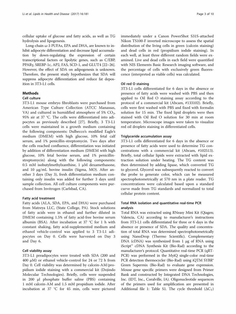
ResultsEffect of SDA on 3T3-L1 cell viabilityThe concentration of 200 μM of ω-3 fatty acids wereused in our experiments. Based on previous studies,200 μM is a safe concentration for ALA, EPA, andDHA treatment in 3T3-L1 cells [29–31]. To assessthat 200 μM of SDA was also safe for 3T3-L1 cells,cell viability assay was conducted. As shown in Fig. 2,the number of living cells (green) and dead cells (red)showed no statistically significant difference betweencontrols and cells treated with 200 μM of SDA, nomatter for 24 or 72 h. In contrast, 400 μM of SDAsignificantly decreased the cell viability to 8% (24 h)and to 5% (72 h) when compared to the controlgroups. These results indicate that the viability of3T3-L1 cells was not affected by 200 μM of SDA.Therefore, the effect of SDA on adipocyte differenti-ation and lipid accumulation found in this study wasindependent of non-specific cell toxicity.
Fig. 2 Effect of SDA on 3T3-L1 adipocyte viability. Two-day post-confluency preadipocytes were incubated with differentiationmedium in the presence of SDA (0, 200, and 400 μM) for 24 or72 h. Cell viability was detected by calcein-AM/propidium iodidestaining. a A representative image of cell staining photographedusing a microscope (X200). Living cells were stained with green(calcein staining) and dead cells were stained with red (propidiumiodide staining). b Quantification of cell viability using spectrophotom-etry. Three images for each treatment were captured and analyzed.Values were obtained from three independent experiments and areexpressed as the means ± SD; different from control cells: *P < 0.05
Li et al. Lipids in Health and Disease (2017) 16:181 Page 4 of 10
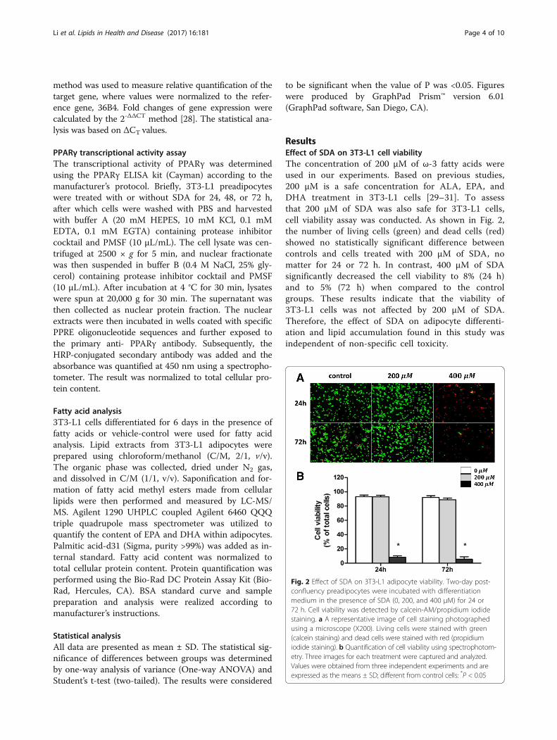
SDA reduced lipid accumulation in 3T3-L1 adipocytesThe effect of SDA and other ω-3 fatty acids on the storageof intracellular lipid in mature 3T3-L1 adipocytes was vi-sualized by Oil Red O staining as shown in Fig. 3a. SDA,EPA, and DHA decreased the amount of lipid in 3T3-L1adipocytes. This observed reduction in lipid accumulationwas confirmed by TG quantification assay. As shown inFig. 3b, compared to the differentiated control cells,50 μM treatment of SDA, EPA, and DHA significantly de-creased the TG content in 3T3-L1 adipocytes by 15, 30,and 31%, respectively. 200 μM treatment of SDA, EPA,and DHA significantly decreased the TG content by 47,54, and 59%, respectively, compared to control cells. Therewas no significant effect on lipid accumulation in cellstreated with ALA versus control cells.
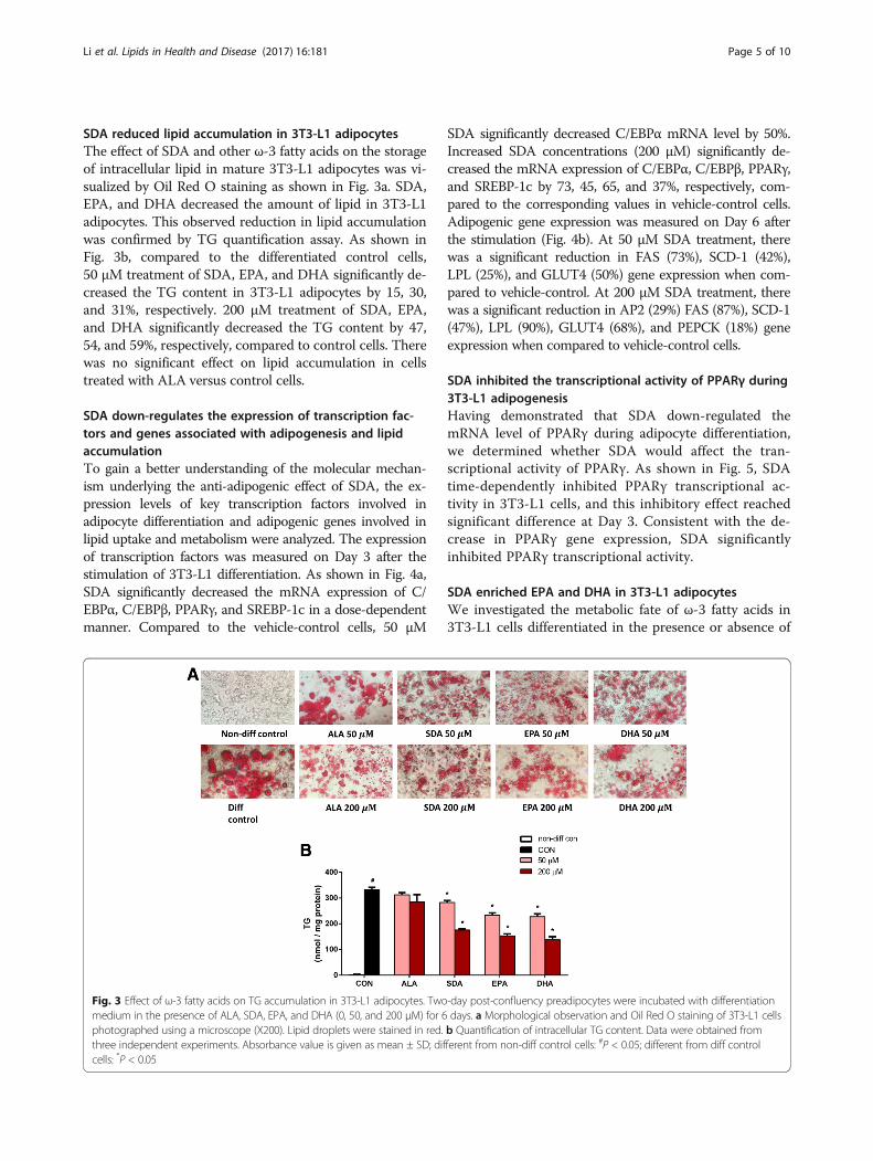
SDA down-regulates the expression of transcription fac-tors and genes associated with adipogenesis and lipidaccumulationTo gain a better understanding of the molecular mechan-ism underlying the anti-adipogenic effect of SDA, the ex-pression levels of key transcription factors involved inadipocyte differentiation and adipogenic genes involved inlipid uptake and metabolism were analyzed. The expressionof transcription factors was measured on Day 3 after thestimulation of 3T3-L1 differentiation. As shown in Fig. 4a,SDA significantly decreased the mRNA expression of C/EBPα, C/EBPβ, PPARγ, and SREBP-1c in a dose-dependentmanner. Compared to the vehicle-control cells, 50 μM
SDA significantly decreased C/EBPα mRNA level by 50%.Increased SDA concentrations (200 μM) significantly de-creased the mRNA expression of C/EBPα, C/EBPβ, PPARγ,and SREBP-1c by 73, 45, 65, and 37%, respectively, com-pared to the corresponding values in vehicle-control cells.Adipogenic gene expression was measured on Day 6 afterthe stimulation (Fig. 4b). At 50 μM SDA treatment, therewas a significant reduction in FAS (73%), SCD-1 (42%),LPL (25%), and GLUT4 (50%) gene expression when com-pared to vehicle-control. At 200 μM SDA treatment, therewas a significant reduction in AP2 (29%) FAS (87%), SCD-1(47%), LPL (90%), GLUT4 (68%), and PEPCK (18%) geneexpression when compared to vehicle-control cells.
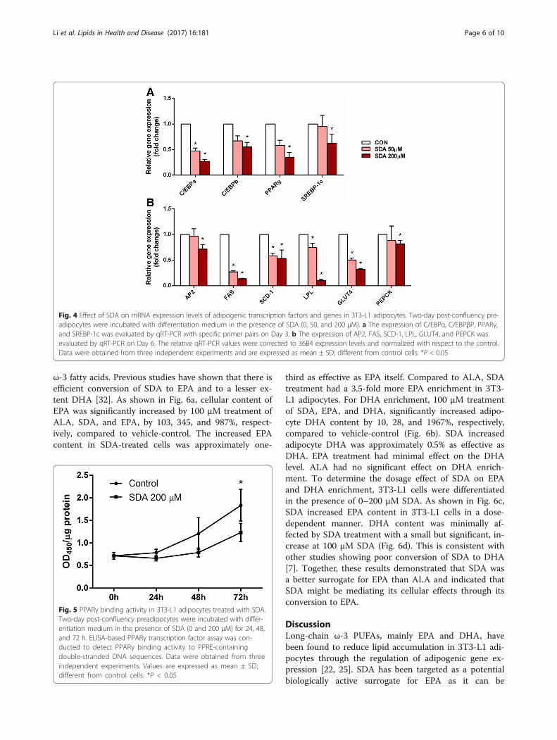
SDA inhibited the transcriptional activity of PPARγ during3T3-L1 adipogenesisHaving demonstrated that SDA down-regulated themRNA level of PPARγ during adipocyte differentiation,we determined whether SDA would affect the tran-scriptional activity of PPARγ. As shown in Fig. 5, SDAtime-dependently inhibited PPARγ transcriptional ac-tivity in 3T3-L1 cells, and this inhibitory effect reachedsignificant difference at Day 3. Consistent with the de-crease in PPARγ gene expression, SDA significantlyinhibited PPARγ transcriptional activity.
SDA enriched EPA and DHA in 3T3-L1 adipocytesWe investigated the metabolic fate of ω-3 fatty acids in3T3-L1 cells differentiated in the presence or absence of
Fig. 3 Effect of ω-3 fatty acids on TG accumulation in 3T3-L1 adipocytes. Two-day post-confluency preadipocytes were incubated with differentiationmedium in the presence of ALA, SDA, EPA, and DHA (0, 50, and 200 μM) for 6 days. a Morphological observation and Oil Red O staining of 3T3-L1 cellsphotographed using a microscope (X200). Lipid droplets were stained in red. b Quantification of intracellular TG content. Data were obtained fromthree independent experiments. Absorbance value is given as mean ± SD; different from non-diff control cells: #P < 0.05; different from diff controlcells: *P < 0.05
Li et al. Lipids in Health and Disease (2017) 16:181 Page 5 of 10
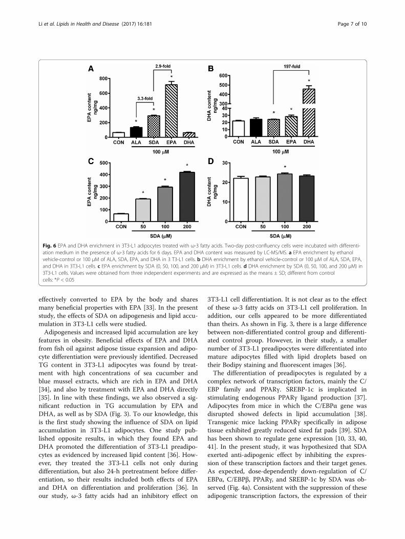
ω-3 fatty acids. Previous studies have shown that there isefficient conversion of SDA to EPA and to a lesser ex-tent DHA [32]. As shown in Fig. 6a, cellular content ofEPA was significantly increased by 100 μM treatment ofALA, SDA, and EPA, by 103, 345, and 987%, respect-ively, compared to vehicle-control. The increased EPAcontent in SDA-treated cells was approximately one-
third as effective as EPA itself. Compared to ALA, SDAtreatment had a 3.5-fold more EPA enrichment in 3T3-L1 adipocytes. For DHA enrichment, 100 μM treatmentof SDA, EPA, and DHA, significantly increased adipo-cyte DHA content by 10, 28, and 1967%, respectively,compared to vehicle-control (Fig. 6b). SDA increasedadipocyte DHA was approximately 0.5% as effective asDHA. EPA treatment had minimal effect on the DHAlevel. ALA had no significant effect on DHA enrich-ment. To determine the dosage effect of SDA on EPAand DHA enrichment, 3T3-L1 cells were differentiatedin the presence of 0–200 μM SDA. As shown in Fig. 6c,SDA increased EPA content in 3T3-L1 cells in a dose-dependent manner. DHA content was minimally af-fected by SDA treatment with a small but significant, in-crease at 100 μM SDA (Fig. 6d). This is consistent withother studies showing poor conversion of SDA to DHA[7]. Together, these results demonstrated that SDA wasa better surrogate for EPA than ALA and indicated thatSDA might be mediating its cellular effects through itsconversion to EPA.
DiscussionLong-chain ω-3 PUFAs, mainly EPA and DHA, havebeen found to reduce lipid accumulation in 3T3-L1 adi-pocytes through the regulation of adipogenic gene ex-pression [22, 25]. SDA has been targeted as a potentialbiologically active surrogate for EPA as it can be
Fig. 4 Effect of SDA on mRNA expression levels of adipogenic transcription factors and genes in 3T3-L1 adipocytes. Two-day post-confluency pre-adipocytes were incubated with differentiation medium in the presence of SDA (0, 50, and 200 μM). a The expression of C/EBPα, C/EBPβP, PPARγ,and SREBP-1c was evaluated by qRT-PCR with specific primer pairs on Day 3. b The expression of AP2, FAS, SCD-1, LPL, GLUT4, and PEPCK wasevaluated by qRT-PCR on Day 6. The relative qRT-PCR values were corrected to 36B4 expression levels and normalized with respect to the control.Data were obtained from three independent experiments and are expressed as mean ± SD; different from control cells: *P < 0.05
Fig. 5 PPARγ binding activity in 3T3-L1 adipocytes treated with SDA.Two-day post-confluency preadipocytes were incubated with differ-entiation medium in the presence of SDA (0 and 200 μM) for 24, 48,and 72 h. ELISA-based PPARγ transcription factor assay was con-ducted to detect PPARγ binding activity to PPRE-containingdouble-stranded DNA sequences. Data were obtained from threeindependent experiments. Values are expressed as mean ± SD;different from control cells: *P < 0.05
Li et al. Lipids in Health and Disease (2017) 16:181 Page 6 of 10
effectively converted to EPA by the body and sharesmany beneficial properties with EPA [33]. In the presentstudy, the effects of SDA on adipogenesis and lipid accu-mulation in 3T3-L1 cells were studied.Adipogenesis and increased lipid accumulation are key
features in obesity. Beneficial effects of EPA and DHAfrom fish oil against adipose tissue expansion and adipo-cyte differentiation were previously identified. DecreasedTG content in 3T3-L1 adipocytes was found by treat-ment with high concentrations of sea cucumber andblue mussel extracts, which are rich in EPA and DHA[34], and also by treatment with EPA and DHA directly[35]. In line with these findings, we also observed a sig-nificant reduction in TG accumulation by EPA andDHA, as well as by SDA (Fig. 3). To our knowledge, thisis the first study showing the influence of SDA on lipidaccumulation in 3T3-L1 adipocytes. One study pub-lished opposite results, in which they found EPA andDHA promoted the differentiation of 3T3-L1 preadipo-cytes as evidenced by increased lipid content [36]. How-ever, they treated the 3T3-L1 cells not only duringdifferentiation, but also 24-h pretreatment before differ-entiation, so their results included both effects of EPAand DHA on differentiation and proliferation [36]. Inour study, ω-3 fatty acids had an inhibitory effect on
3T3-L1 cell differentiation. It is not clear as to the effectof these ω-3 fatty acids on 3T3-L1 cell proliferation. Inaddition, our cells appeared to be more differentiatedthan theirs. As shown in Fig. 3, there is a large differencebetween non-differentiated control group and differenti-ated control group. However, in their study, a smallernumber of 3T3-L1 preadipocytes were differentiated intomature adipocytes filled with lipid droplets based ontheir Bodipy staining and fluorescent images [36].The differentiation of preadipocytes is regulated by a
complex network of transcription factors, mainly the C/EBP family and PPARγ. SREBP-1c is implicated instimulating endogenous PPARγ ligand production [37].Adipocytes from mice in which the C/EBPα gene wasdisrupted showed defects in lipid accumulation [38].Transgenic mice lacking PPARγ specifically in adiposetissue exhibited greatly reduced sized fat pads [39]. SDAhas been shown to regulate gene expression [10, 33, 40,41]. In the present study, it was hypothesized that SDAexerted anti-adipogenic effect by inhibiting the expres-sion of these transcription factors and their target genes.As expected, dose-dependently down-regulation of C/EBPα, C/EBPβ, PPARγ, and SREBP-1c by SDA was ob-served (Fig. 4a). Consistent with the suppression of theseadipogenic transcription factors, the expression of their
Fig. 6 EPA and DHA enrichment in 3T3-L1 adipocytes treated with ω-3 fatty acids. Two-day post-confluency cells were incubated with differenti-ation medium in the presence of ω-3 fatty acids for 6 days. EPA and DHA content was measured by LC-MS/MS. a EPA enrichment by ethanolvehicle-control or 100 μM of ALA, SDA, EPA, and DHA in 3 T3-L1 cells. b DHA enrichment by ethanol vehicle-control or 100 μM of ALA, SDA, EPA,and DHA in 3T3-L1 cells. c EPA enrichment by SDA (0, 50, 100, and 200 μM) in 3T3-L1 cells. d DHA enrichment by SDA (0, 50, 100, and 200 μM) in3T3-L1 cells. Values were obtained from three independent experiments and are expressed as the means ± SD; different from controlcells: *P < 0.05
Li et al. Lipids in Health and Disease (2017) 16:181 Page 7 of 10
target genes, AP2, FAS, SCD-1, LPL, GLUT4, and PEPCKwere also significantly decreased dose-dependently bySDA treatment (Fig. 4b). The adipogenic genes are oftenconnected with insulin sensitivity and inflammatory cyto-kines. AP2 has been demonstrated to act as an adipokinefor the development of insulin resistance in liver [42].Adipose-specific deletion of SCD-1 was found to induceGLUT1 up-regulation and was associated with decreasedadiponectin and increased TNFα production [43].Adipocyte-derived LPL was shown to induce macrophageactivation and monocyte adhesion [44]. Inflammation hasbeen shown to inhibit the expression of GLUT4 andPEPCK in 3T3-L1 adipocytes [45, 46]. EPA and DHA havebeen reported to have preventive properties in the devel-opment of insulin resistance and inflammation [47].Therefore, it is necessary to further investigate the effectof SDA on adipokine secretion and its role in insulin re-sistance and inflammatory response for future study.SDA is the Δ6 desaturase product of ALA in the biocon-
version of ALA to EPA. In humans, the conversion of ALAto EPA is in low amounts (less than 7%) and in even loweramounts to DHA (less than 1%) due to the rate-limiting en-zyme [48]. Nutritional supplementation with ALA was notable to induce the accumulation of long-chain ω-3 PUFAs.In the present study, it was hypothesized that by skippingthe rate-limiting step, the conversion of SDA to EPA wouldbe more efficient than that of ALA to EPA. By LC-MS/MSanalysis, it was shown that SDA increased EPA content in3T3-L1 cells by 345% compared to the control group(Fig. 6a). The efficacy of EPA enrichment by SDA wasabout 3.5-fold greater than the comparable level of ALAand was about one-third of the EPA enrichment by EPA.For DHA enrichment, both ALA and SDA, even EPA wasnot able to increase DHA content extensively in 3T3-L1cells, although statistical significance was observed in SDAand EPA groups. This was probably due to another rate
limiting step which converts docosapentaenoic acid toDHA [49]. Human studies have reported that consumptionof SDA, as ethyl esters, echium oil, or SDA-soybean oil, sig-nificantly increased the EPA level in red blood cells [8], per-ipheral blood mononuclear cell [11], plasma [15], andneutrophils [13]. In animal studies, feeding with SDA wasshown to increase the EPA content in many tissues, includ-ing red blood cells, plasma, liver, muscle, heart, brain, andileum of dogs [50], sows and piglets [51], lambs [52], androdents [53, 54]. In addition, SDA can lead to EPA enrich-ment in animal products, such as egg yolk, chicken meat[55], and milk of dairy cows [56]. Based on human studies,the efficacy of SDA on EPA enrichment in different tissuesrange from 17 to 85% as much as the efficacy of EPA treat-ment on EPA enrichment [8, 12, 57–60]. When comparedto ALA, SDA was about 1.9 to 4.3-fold as effective as thatof ALA on EPA enrichment [57, 61]. Most studies did notfind significant change in DHA enrichment with SDA sup-plementation. These results indicate that SDA consumptionwill be expected to confer the health benefits associatedwith the consumption of EPA, but not DHA. While theexact molecular mechanisms by which SDA decreases genetranscription is not apparent from this study, the down-stream biosynthesis of lipid-derived eicosanoids via its con-version to EPA may be a potential mechanism throughwhich gene transcription effects could be modulated [25,62, 63].
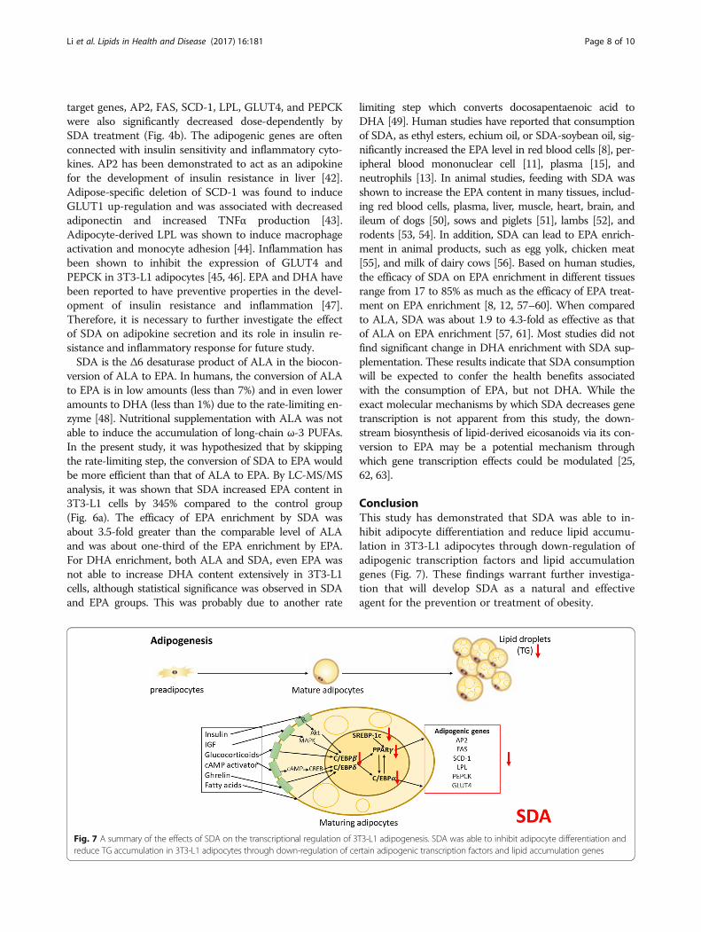
ConclusionThis study has demonstrated that SDA was able to in-hibit adipocyte differentiation and reduce lipid accumu-lation in 3T3-L1 adipocytes through down-regulation ofadipogenic transcription factors and lipid accumulationgenes (Fig. 7). These findings warrant further investiga-tion that will develop SDA as a natural and effectiveagent for the prevention or treatment of obesity.
Fig. 7 A summary of the effects of SDA on the transcriptional regulation of 3T3-L1 adipogenesis. SDA was able to inhibit adipocyte differentiation andreduce TG accumulation in 3T3-L1 adipocytes through down-regulation of certain adipogenic transcription factors and lipid accumulation genes
Li et al. Lipids in Health and Disease (2017) 16:181 Page 8 of 10
Additional file
Additional file 1: Table S1. Oligonucleotide primer sequences used inreal-time PCR for adipogenic transcriptional factors and lipid accumulationgenes. (DOCX 15 kb)
AbbreviationsALA: Alpha-linolenic acid; AP2: Adipocyte fatty acid-binding protein; C/EBP: CCAAT-enhancer-binding protein; FAS: Fatty acid synthase;GLUT: Glucose transporter; LPL: Lipoprotein lipase;PEPCK: Phosphoenolpyruvate carboxykinase; PPARγ: Peroxisome proliferator-activated receptor gamma; SCD: Stearoyl-CoA desaturase; SDA: Stearidonicacid; SREBP: Sterol regulatory element-binding protein
AcknowledgementsWe thank Bulbul Ahmed and Dr. Michael W. Green for the technical assistance.We would also like to thank Dr. B. Douglas White and Dr. Robert L. Judd forfruitful discussion.
FundingThis study was supported by grants from the Diabetes Action Research andEducation Foundation (KWH) and Alabama Agricultural Experiment StationHatch Award (KWH).
Availability of data and materialsNot applicable.
Authors’ contributionsKWH and YR designed and developed the study. YL conceived the studyand wrote the manuscript. YL and YR were responsible for laboratory workincluding cell culture, treatment, Oil Red O staining, and triglyceridequantification. YR and GR performed the cell viability assay. YL and BNcarried out the LC-MS/MS. YL and LB conducted the real-time PCR assays. YL,YR, and CZ was responsible for data analysis and statistical calculations. Allauthors interpreted the results, and read and approved the final manuscript.
Ethics approval and consent to participateNot applicable.
Consent for publicationNot applicable.
Competing interestsThe authors declare that they have no competing interests.
Publisher’s NoteSpringer Nature remains neutral with regard to jurisdictional claims inpublished maps and institutional affiliations.
Author details1Department of Nutrition, Dietetics and Hospitality Management, AuburnUniversity, Auburn, AL, USA. 2Department of Drug Discovery andDevelopment, Harrison School of Pharmacy, Auburn University, Auburn, AL,USA. 3Boshell Diabetes and Metabolic Diseases Research Program, AuburnUniversity, Auburn, AL, USA.
Received: 16 June 2017 Accepted: 20 September 2017
References1. Hurt RT, Kulisek C, Buchanan LA, McClave SA. The obesity epidemic:
challenges, health initiatives, and implications for gastroenterologists.Gastroenterol Hepatol (N Y). 2010;6:780–92.
2. Logan SL, Spriet LL. Omega-3 Fatty Acid Supplementation for 12 WeeksIncreases Resting and Exercise Metabolic Rate in Healthy Community-Dwelling Older Females. PLoS One. 2015;10:e0144828.
3. Noreen EE, Sass MJ, Crowe ML, Pabon VA, Brandauer J, Averill LK. Effects ofsupplemental fish oil on resting metabolic rate, body composition, andsalivary cortisol in healthy adults. J Int Soc Sports Nutr. 2010;7:31.
4. Peinado I, Miles W, Koutsidis G. Odour characteristics of seafood flavourformulations produced with fish by-products incorporating EPA, DHA andfish oil. Food Chem. 2016;212:612–9.
5. Sioen I, De Henauw S, Van Camp J. Evaluation of benefits and risks relatedto seafood consumption. Verh K Acad Geneeskd Belg. 2007;69:249–89.
6. Wang C, Harris WS, Chung M, Lichtenstein AH, Balk EM, Kupelnick B, et al. n-3 Fatty acids from fish or fish-oil supplements, but not alpha-linolenic acid,benefit cardiovascular disease outcomes in primary- and secondary-prevention studies: a systematic review. Am J Clin Nutr. 2006;84:5–17.
7. Baker EJ, Miles EA, Burdge GC, Yaqoob P, Calder PC. Metabolism andfunctional effects of plant-derived omega-3 fatty acids in humans. ProgLipid Res. 2016;64:30–56.
8. Harris WS, Lemke SL, Hansen SN, Goldstein DA, DiRienzo MA, Su H, et al.Stearidonic acid-enriched soybean oil increased the omega-3 index, anemerging cardiovascular risk marker. Lipids. 2008;43:805–11.
9. Harris WS. Stearidonic acid-enhanced soybean oil: a plant-based source of(n-3) fatty acids for foods. J Nutr. 2012;142:600S–4S.
10. Botelho PB, Mariano Kda R, Rogero MM, de Castro IA. Effect of Echium oilcompared with marine oils on lipid profile and inhibition of hepaticsteatosis in LDLr knockout mice. Lipids Health Dis. 2013;12:38.
11. Casey JM, Banz WJ, Krul ES, Butteiger DN, Goldstein DA, Davis JE. Effect ofstearidonic acid-enriched soybean oil on fatty acid profile and metabolicparameters in lean and obese Zucker rats. Lipids Health Dis. 2013;12:147.
12. Kuhnt K, Fuhrmann C, Kohler M, Kiehntopf M, Jahreis G. Dietary echium oilincreases long-chain n-3 PUFAs, including docosapentaenoic acid, in bloodfractions and alters biochemical markers for cardiovascular diseaseindependently of age, sex, and metabolic syndrome. J Nutr. 2014;144:447–60.
13. Surette ME, Edens M, Chilton FH, Tramposch KM. Dietary echium oil increasesplasma and neutrophil long-chain (n-3) fatty acids and lowers serumtriacylglycerols in hypertriglyceridemic humans. J Nutr. 2004;134:1406–11.
14. Kawabata T, Shimoda K, Horiguchi S, Domon M, Hagiwara C, Takiyama M, et al.Influences of stearidonic acid-enriched soybean oil on the blood and organbiochemical parameters in rats. Prostaglandins Leukot Essent Fatty Acids. 2013;88:179–84.
15. Arm JP, Boyce JA, Wang L, Chhay H, Zahid M, Patil V, et al. Impact ofbotanical oils on polyunsaturated fatty acid metabolism and leukotrienegeneration in mild asthmatics. Lipids Health Dis. 2013;12:141.
16. Forrest LM, Boudyguina E, Wilson MD, Parks JS. Echium oil reducesatherosclerosis in apoB100-only LDLrKO mice. Atherosclerosis. 2012;220:118–21.
17. Subedi K, Yu HM, Newell M, Weselake RJ, Meesapyodsuk D, Qiu X, et al.Stearidonic acid-enriched flax oil reduces the growth of human breastcancer in vitro and in vivo. Breast Cancer Res Treat. 2015;149:17–29.
18. Stephens JM. The fat controller: adipocyte development. PLoS Biol. 2012;10:e1001436.
19. Ruiz-Ojeda FJ, Ruperez AI, Gomez-Llorente C, Gil A, Aguilera CM. CellModels and Their Application for Studying Adipogenic Differentiation inRelation to Obesity: A Review. Int J Mol Sci. 2016;17
20. Tang QQ, Otto TC, Lane MD. CCAAT/enhancer-binding protein beta isrequired for mitotic clonal expansion during adipogenesis. Proc Natl AcadSci U S A. 2003;100:850–5.
21. Moseti D, Regassa A, Kim WK. Molecular Regulation of Adipogenesis andPotential Anti-Adipogenic Bioactive Molecules. Int J Mol Sci. 2016;17
22. Barber E, Sinclair AJ, Cameron-Smith D. Comparative actions of omega-3fatty acids on in-vitro lipid droplet formation. Prostaglandins Leukot EssentFatty Acids. 2013;89:359–66.
23. Furuhashi M, Hiramitsu S, Mita T, Omori A, Fuseya T, Ishimura S, et al.Reduction of circulating FABP4 level by treatment with omega-3 fatty acidethyl esters. Lipids Health Dis. 2016;15:5.
24. Madsen L, Petersen RK, Kristiansen K. Regulation of adipocytedifferentiation and function by polyunsaturated fatty acids. BiochimBiophys Acta. 2005;1740:266–86.
25. Prostek A, Gajewska M, Balasinska B. The influence of eicosapentaenoic acidand docosahexaenoic acid on expression of genes connected withmetabolism and secretory functions of ageing 3T3-L1 adipocytes.Prostaglandins Other Lipid Mediat. 2016;125:48–56.
26. Wojcik C, Lohe K, Kuang C, Xiao Y, Jouni Z, Poels E. Modulation of adipocytedifferentiation by omega-3 polyunsaturated fatty acids involves theubiquitin-proteasome system. J Cell Mol Med. 2014;18:590–9.
27. Hemati N, Ross SE, Erickson RL, Groblewski GE, MacDougald OA. Signalingpathways through which insulin regulates CCAAT/enhancer binding proteinalpha (C/EBPalpha) phosphorylation and gene expression in 3T3-L1
Li et al. Lipids in Health and Disease (2017) 16:181 Page 9 of 10
adipocytes. Correlation with GLUT4 gene expression. J Biol Chem. 1997;272:25913–9.
28. Livak KJ, Schmittgen TD. Analysis of relative gene expression data usingreal-time quantitative PCR and the 2(−Delta Delta C(T)) Method. Methods.2001;25:402–8.
29. Fukumitsu S, Villareal MO, Onaga S, Aida K, Han J, Isoda H. alpha-Linolenic acidsuppresses cholesterol and triacylglycerol biosynthesis pathway by suppressingSREBP-2, SREBP-1a and -1c expression. Cytotechnology. 2013;65:899–907.
30. Lee MS, Kwun IS, Kim Y. Eicosapentaenoic acid increases lipolysis through up-regulation of the lipolytic gene expression and down-regulation of theadipogenic gene expression in 3T3-L1 adipocytes. Genes Nutr. 2008;2:327–30.
31. Wang X, Huang M, Wang Y. The effect of insulin, TNFalpha and DHA on theproliferation, differentiation and lipolysis of preadipocytes isolated fromlarge yellow croaker (Pseudosciaena Crocea R.). PLoS One. 2012;7:e48069.
32. Abeywardena MY, Adams M, Dallimore J, Kitessa SM. Rise in DPA FollowingSDA-Rich Dietary Echium Oil Less Effective in Affording Anti-ArrhythmicActions Compared to High DHA Levels Achieved with Fish Oil in Sprague-Dawley Rats. Nutrients. 2016;8
33. Whelan J, Gouffon J, Zhao Y. Effects of dietary stearidonic acid onbiomarkers of lipid metabolism. J Nutr. 2012;142:630S–4S.
34. Vaidya H, Cheema SK. Sea cucumber and blue mussel: new sources ofphospholipid enriched omega-3 fatty acids with a potential role in 3T3-L1adipocyte metabolism. Food Funct. 2014;5:3287–95.
35. Yeop Han C, Kargi AY, Omer M, Chan CK, Wabitsch M, O'Brien KD, et al.Differential effect of saturated and unsaturated free fatty acids on thegeneration of monocyte adhesion and chemotactic factors by adipocytes:dissociation of adipocyte hypertrophy from inflammation. Diabetes. 2010;59:386–96.
36. Murali G, Desouza CV, Clevenger ME, Ramalingam R, Saraswathi V.Differential effects of eicosapentaenoic acid and docosahexaenoic acid inpromoting the differentiation of 3T3-L1 preadipocytes. ProstaglandinsLeukot Essent Fatty Acids. 2014;90:13–21.
37. Kim JB, Wright HM, Wright M, Spiegelman BM. ADD1/SREBP1 activatesPPARgamma through the production of endogenous ligand. Proc Natl AcadSci U S A. 1998;95:4333–7.
38. Wang ND, Finegold MJ, Bradley A, Ou CN, Abdelsayed SV, Wilde MD, et al.Impaired energy homeostasis in C/EBP alpha knockout mice. Science. 1995;269:1108–12.
39. He W, Barak Y, Hevener A, Olson P, Liao D, Le J, et al. Adipose-specificperoxisome proliferator-activated receptor gamma knockout causes insulinresistance in fat and liver but not in muscle. Proc Natl Acad Sci U S A. 2003;100:15712–7.
40. Hsueh HW, Zhou Z, Whelan J, Allen KG, Moustaid-Moussa N, Kim H, et al.Stearidonic and eicosapentaenoic acids inhibit interleukin-6 expression inob/ob mouse adipose stem cells via Toll-like receptor-2-mediated pathways.J Nutr. 2011;141:1260–6.
41. Jaya-Ram A, Shu-Chien AC, Kuah MK. Echium oil increased the expression ofa Delta4 Fads2 fatty acyl desaturase and the deposition of n-3 long-chainpolyunsaturated fatty acid in comparison with linseed oil in stripedsnakehead (Channa striata) muscle. Fish Physiol Biochem. 2016;42:1107–22.
42. Cao H, Sekiya M, Ertunc ME, Burak MF, Mayers JR, White A, et al. Adipocytelipid chaperone AP2 is a secreted adipokine regulating hepatic glucoseproduction. Cell Metab. 2013;17:768–78.
43. Hyun CK, Kim ED, Flowers MT, Liu X, Kim E, Strable M, et al. Adipose-specificdeletion of stearoyl-CoA desaturase 1 up-regulates the glucose transporterGLUT1 in adipose tissue. Biochem Biophys Res Commun. 2010;399:480–6.
44. Li L, Renier G. Adipocyte-derived lipoprotein lipase induces macrophageactivation and monocyte adhesion: role of fatty acids. Obesity (SilverSpring). 2007;15:2595–604.
45. Feingold KR, Moser A, Shigenaga JK, Grunfeld C. Inflammation inhibits theexpression of phosphoenolpyruvate carboxykinase in liver and adiposetissue. Innate Immun. 2012;18:231–40.
46. Lagathu C, Bastard JP, Auclair M, Maachi M, Capeau J, Caron M. Chronicinterleukin-6 (IL-6) treatment increased IL-6 secretion and induced insulinresistance in adipocyte: prevention by rosiglitazone. Biochem Biophys ResCommun. 2003;311:372–9.
47. Bashir S, Sharma Y, Elahi A, Khan F. Amelioration of obesity-associatedinflammation and insulin resistance in c57bl/6 mice via macrophagepolarization by fish oil supplementation. J Nutr Biochem. 2016;33:82–90.
48. Harris WS. Stearidonic acid as a ‘pro-eicosapentaenoic acid’. Curr Opin Lipidol.2012;23:30–4.
49. Arterburn LM, Hall EB, Oken H. Distribution, interconversion, and doseresponse of n-3 fatty acids in humans. Am J Clin Nutr. 2006;83:1467S–76S.
50. Harris WS, DiRienzo MA, Sands SA, George C, Jones PG, Eapen AK.Stearidonic acid increases the red blood cell and heart eicosapentaenoicacid content in dogs. Lipids. 2007;42:325–33.
51. Tanghe S, Millet S, De Smet S. Echium oil and linseed oil as alternatives forfish oil in the maternal diet: Blood fatty acid profiles and oxidative status ofsows and piglets. J Anim Sci. 2013;91:3253–64.
52. Kitessa SM, Young P, Nattrass G, Gardner G, Pearce K, Pethick DW. Whenbalanced for precursor fatty acid supply echium oil is not superior to linseed oilin enriching lamb tissues with long-chain n-3 PUFA. Br J Nutr. 2012;108:71–9.
53. Surette ME. Dietary omega-3 PUFA and health: stearidonic acid-containingseed oils as effective and sustainable alternatives to traditional marine oils.Mol Nutr Food Res. 2013;57:748–59.
54. Yang Q, O'Shea TM. Dietary Echium oil increases tissue (n-3) long-chainpolyunsaturated fatty acids without elevating hepatic lipid concentrations inpremature neonatal rats. J Nutr. 2009;139:1353–9.
55. Elkin RG, Ying Y, Harvatine KJ. Feeding laying hens stearidonic acid-enrichedsoybean oil, as compared to flaxseed oil, more efficiently enriches eggswith very long-chain n-3 polyunsaturated fatty acids. J Agric Food Chem.2015;63:2789–97.
56. Bernal-Santos G, O'Donnell AM, Vicini JL, Hartnell GF, Bauman DE. Hot topic:Enhancing omega-3 fatty acids in milk fat of dairy cows by usingstearidonic acid-enriched soybean oil from genetically modified soybeans. JDairy Sci. 2010;93:32–7.
57. James MJ, Ursin VM, Cleland LG. Metabolism of stearidonic acid in humansubjects: comparison with the metabolism of other n-3 fatty acids. Am JClin Nutr. 2003;77:1140–5.
58. Krul ES, Lemke SL, Mukherjea R, Taylor ML, Goldstein DA, Su H, et al. Effectsof duration of treatment and dosage of eicosapentaenoic acid andstearidonic acid on red blood cell eicosapentaenoic acid content.Prostaglandins Leukot Essent Fatty Acids. 2012;86:51–9.
59. Lemke SL, Maki KC, Hughes G, Taylor ML, Krul ES, Goldstein DA, et al.Consumption of stearidonic acid-rich oil in foods increases red blood celleicosapentaenoic acid. J Acad Nutr Diet. 2013;113:1044–56.
60. Lemke SL, Vicini JL, Su H, Goldstein DA, Nemeth MA, Krul ES, et al. Dietaryintake of stearidonic acid-enriched soybean oil increases the omega-3index: randomized, double-blind clinical study of efficacy and safety. Am JClin Nutr. 2010;92:766–75.
61. Dittrich M, Jahreis G, Bothor K, Drechsel C, Kiehntopf M, Bluher M, et al.Benefits of foods supplemented with vegetable oils rich in alpha-linolenic,stearidonic or docosahexaenoic acid in hypertriglyceridemic subjects: adouble-blind, randomized, controlled trail. Eur J Nutr. 2015;54:881–93.
62. Annamalai D, Clipstone NA. Prostaglandin F2alpha inhibits adipogenesis viaan autocrine-mediated interleukin-11/glycoprotein 130/STAT1-dependentsignaling cascade. J Cell Biochem. 2014;115:1308–21.
63. Miller CW, Casimir DA, Ntambi JM. The mechanism of inhibition of 3T3-L1preadipocyte differentiation by prostaglandin F2alpha. Endocrinology. 1996;137:5641–50.
• We accept pre-submission inquiries
• Our selector tool helps you to find the most relevant journal
• We provide round the clock customer support
• Convenient online submission
• Thorough peer review
• Inclusion in PubMed and all major indexing services
• Maximum visibility for your research
Submit your manuscript atwww.biomedcentral.com/submit
Submit your next manuscript to BioMed Central and we will help you at every step:
Li et al. Lipids in Health and Disease (2017) 16:181 Page 10 of 10


![Adipocyte lipid synthesis coupled to neuronal control of ... · (DNL), was markedly reduced in the insulin resistant state [19,20] in spite of near normal glucose uptake [21]. These](https://img.pdfslide.net/doc/110x75/5cd5a4de88c993ea4e8bd5da/adipocyte-lipid-synthesis-coupled-to-neuronal-control-of-dnl-was-markedly.jpg)