Embed Size (px)
Citation preview
Sensors 2015, 15, 10481-10510; doi:10.3390/s150510481
sensors ISSN 1424-8220
www.mdpi.com/journal/sensors
Review
Surface Plasmon Resonance: A Versatile Technique for Biosensor Applications
Hoang Hiep Nguyen 1,2,†, Jeho Park 1,2,†, Sebyung Kang 3,* and Moonil Kim 1,2,4,*
1 BioNanotechnology Research Center, Korea Research Institute of Bioscience and
Biotechnology (KRIBB), Daejeon 305-806, Korea; E-Mails: [email protected] (H.H.N.);
[email protected] (J.P.) 2 Department of Nanobiotechnology, Korea University of Science and Technology (UST),
Daejeon 305-350, Korea 3 Department of Biological Sciences, School of Life Sciences, Ulsan National Institute of Science and
Technology (UNIST), Ulsan 689-798, Korea 4 Department of Pathobiology, College of Veterinary Medicine Nursing & Allied Health (CVMNAH),
Tuskegee University, Tuskegee, AL 36088, USA
† These authors contributed equally to this work.
* Authors to whom correspondence should be addressed; E-Mails: [email protected] (S.K.);
[email protected] (M.K.); Tel.: +82-52-217-5325 (S.K.); +82-42-879-8447 (M.K.);
Fax: +82-52-217-5309 (S.K.); +82-42-879-8594 (M.K.).
Academic Editor: Yeshaiahu Fainman
Received: 12 March 2015 / Accepted: 28 April 2015 / Published: 5 May 2015
Abstract: Surface plasmon resonance (SPR) is a label-free detection method which has
emerged during the last two decades as a suitable and reliable platform in clinical analysis
for biomolecular interactions. The technique makes it possible to measure interactions in
real-time with high sensitivity and without the need of labels. This review article discusses
a wide range of applications in optical-based sensors using either surface plasmon resonance
(SPR) or surface plasmon resonance imaging (SPRI). Here we summarize the principles,
provide examples, and illustrate the utility of SPR and SPRI through example applications
from the biomedical, proteomics, genomics and bioengineering fields. In addition, SPR
signal amplification strategies and surface functionalization are covered in the review.
Keywords: surface plasmon resonance; SPR; biosensor; SPR imaging; applications
OPEN ACCESS
Sensors 2015, 15 10482
1. Introduction
Numerous strategies for protein labeling have been developed that allow the characterization of
proteins regarding their structure, folding or interaction with other proteins [1,2]. Labeling strategies are
used to bring about the covalent attachment of reporter tags, such as biotin, radioisotopes, fluorophores,
or enzymes, to the target biomolecules (i.e., proteins and nucleotides) to quantitatively assess binding
among biomolecules [3,4]. In addition, the use of molecular labels can cause steric hindrance or change
structural configurations, affecting the labelled molecules’ affinities for their target biomolecules, which
is a major challenge. Label-free detection eliminates the need for specialized tags or dyes, thereby
allowing the sensitive measurement of target analytes and enabling the use of native biomolecules
suitable for biologically relevant approaches. In the past few decades, a variety of optical biosensor
methods have been developed, including surface plasmon resonance (SPR) [5], quartz crystal
microbalance (QCM) [6], and ellipsometry [7]. Among the various optical sensing methodologies, the
SPR-based system is a representative type of label-free technique for monitoring biomolecular
interactions in real-time.
Since it was first introduced in the early 1990s, SPR has been proven to be one of the most
powerful technologies to determine specificity, affinity and kinetic parameters during the binding
of macromolecules in many bonds types, including protein-protein [8,9], protein-DNA [10,11],
enzyme-substrate or inhibitor [4,12], receptor-drug [13,14], lipid membrane-protein [15,16],
protein-polysaccharide [17], cell or virus-protein [18–20], among others. This optical technique measures
the refractive index changes in the vicinity of thin metal layers (i.e., gold, silver, or aluminum films) in
response to biomolecular interactions. Before a sample solution flows across the SPR surface, capturing
agents, such as antibodies, enzymes, peptides and DNAs are immobilized on the surface. The changes in
the SPR angle, which is the angle of minimum reflectivity, can be determined by varying the incidence
angle and recording the reflected light intensity during the biological binding reactions between various
biomolecules. So far, numerous studies have advanced the potential of SPR sensors by increasing the
effectiveness of the technique [21–23]. Accordingly, the possible fields of application of SPR technology
have expanded to biomedical, environmental and industrial areas. As has been extensively and intensively
documented in the literature, SPR is an acceptable method for disease diagnosis, drug discovery, foodborne
pathogen detection, and so on [24,25]. Above all, the application of SPR for biomedical purposes is
remarkable. So far, various types of SPR measurement systems have been developed for the monitoring
of chemical and biological species via the basic theory of SPR detection [26–28]. Here we review recent
advances in SPR biosensing, with a particular focus on practical applications of SPR-type biosensors,
describing their usefulness and challenges for bioassays.
2. Operating Principle of SPR Biosensors
2.1. General Principle of SPR
Surface plasmon resonance occurs when a photon of incident light hits a metal surface (typically a
gold surface). At a certain angle of incidence, a portion of the light energy couples through the metal
coating with the electrons in the metal surface layer, which then move due to excitation. The electron
movements are now called plasmon, and they propagate parallel to the metal surface. The plasmon
Sensors 2015, 15 10483
oscillation in turn generates an electric field whose range is around 300 nm from the boundary between
the metal surface and sample solution [29]. In a commercial SPR biosensor configuration, incident light
is employed by using a high-reflective index glass prism in the Kretschmann geometry of the attenuated
total reflection (ATR) method (Figure 1). The defined SPR angle, at which resonance occurs, on the
conditions of the constant light source wavelength and metal thin surface, is dependent on the refractive
index of the material near the metal surface. Consequently, when there is a small change in the reflective
index of the sensing medium (e.g., through biomolecule attachment), plasmon cannot be formed.
Detection is thus accomplished by measuring the changes in the reflected light obtained on a detector.
In addition, the amount of surface concentration can be quantified by monitoring the reflected light
intensity or tracking the resonance angle shifts. Typically, an SPR biosensor has a detection limit on the
order of 10 pg/mL.
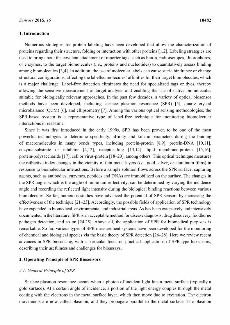
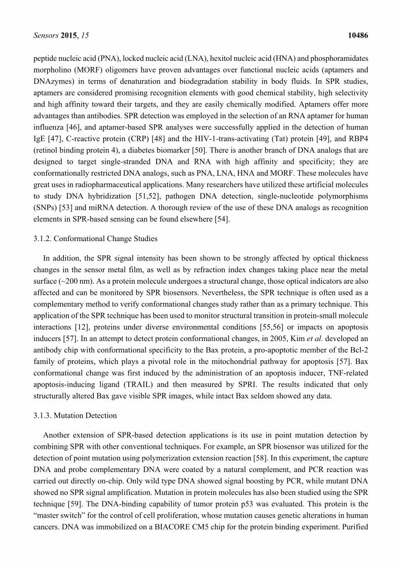
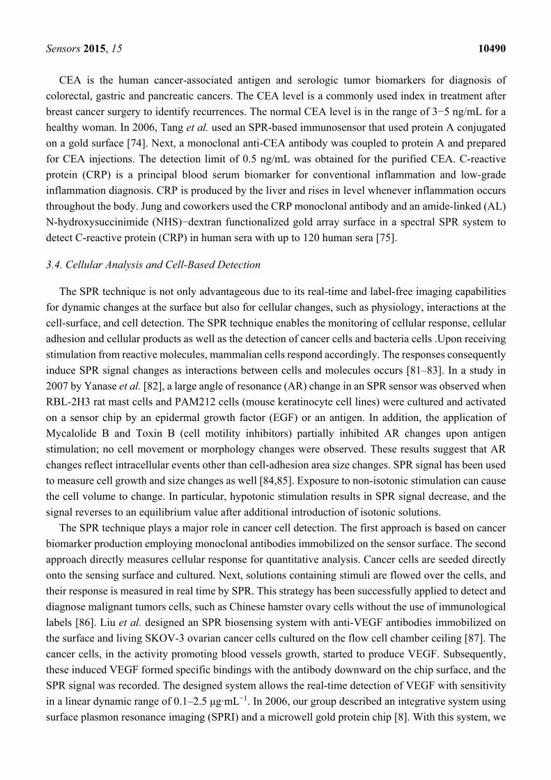
Figure 1. Concept of a surface plasmon resonance (SPR) biosensor: (A) Kretschmann
geometry of the ATR method; (B) spectrum of reflected light before and after refractive
index change; (C) analyte-biorecognition elements binding on SPR sensor surface and
(D) refractive index changes caused by the molecular interactions in the reaction medium.
Adapted from [30].
In SPR biosensors, probe molecules are firstly immobilized on to the sensor surface. When the
solution of target molecules is flown into contact with the surface, a probe-target binding via affinity
interaction occurs, which consequently induces an increase in the refractive index at the SPR sensor
surface (Figure 1D). In SPR experiments, resonance or response units (RU) are used to describe the
signal change, where 1 RU is equivalent to a critical angle shift of 10−4 degree. At the start of the
experiment whereas probe target interactions have not occurred, the initial RU value corresponds to the
starting critical angle. The change in refractive index Δnd arisen within a layer of thickness h can be
calculated as
Sensors 2015, 15 10484
Δnd = (dn/dc)vol ΔΓ/h (1)
where (dn/dc)vol is the increase of refractive index n with the volume concentration of analyte c, and ΔΓ
is the concentration of the bound target on the surface [31]. The change in the refractive index is tracked
by the coupling of incident light into a propagating surface plasmon (PSP) on a gold surface in real time.
Accordingly, the rate of association (kon) during association phase, the rate of disassociation (koff) when
target molecules is removed from the continuous flow by buffer washing, and the association rate
constant (ka) and dissociation rate constant (kd) can be determined by SPR evaluation of binding kinetics.
The parameter related to the refractive index can also be used to detect and quantify the target molecules
bound to a known probe immobilized on the sensor surface. The limit of detection (LOD) in the SPR
experiment depends on a number of factors including the molecular weight, optical property and binding
affinity of target-probe molecules as well as the surface coverage of the probe molecule.
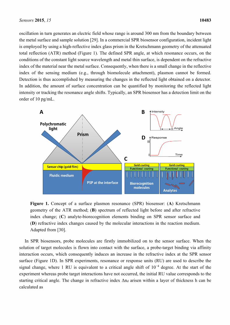
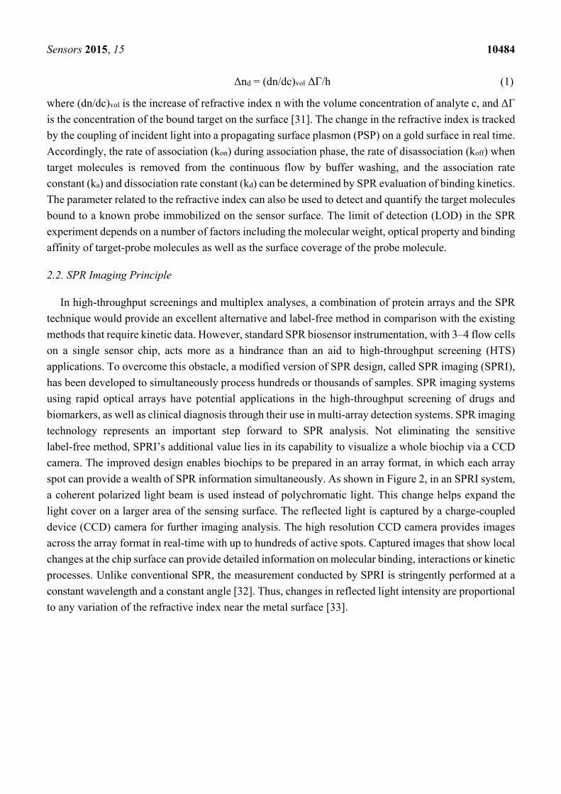
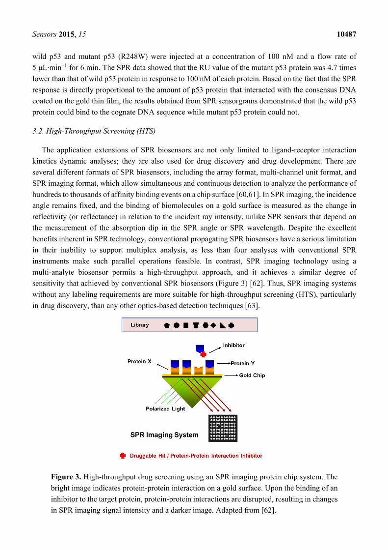
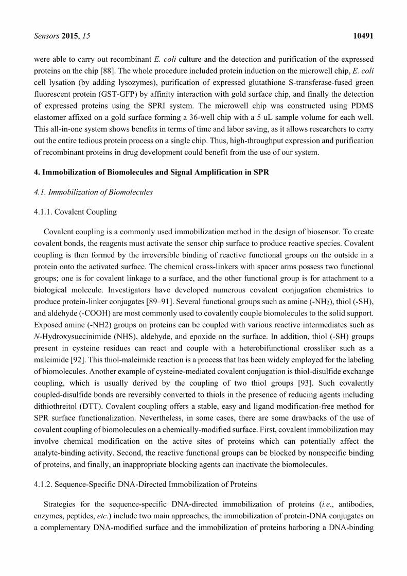
2.2. SPR Imaging Principle
In high-throughput screenings and multiplex analyses, a combination of protein arrays and the SPR
technique would provide an excellent alternative and label-free method in comparison with the existing
methods that require kinetic data. However, standard SPR biosensor instrumentation, with 3–4 flow cells
on a single sensor chip, acts more as a hindrance than an aid to high-throughput screening (HTS)
applications. To overcome this obstacle, a modified version of SPR design, called SPR imaging (SPRI),
has been developed to simultaneously process hundreds or thousands of samples. SPR imaging systems
using rapid optical arrays have potential applications in the high-throughput screening of drugs and
biomarkers, as well as clinical diagnosis through their use in multi-array detection systems. SPR imaging
technology represents an important step forward to SPR analysis. Not eliminating the sensitive
label-free method, SPRI’s additional value lies in its capability to visualize a whole biochip via a CCD
camera. The improved design enables biochips to be prepared in an array format, in which each array
spot can provide a wealth of SPR information simultaneously. As shown in Figure 2, in an SPRI system,
a coherent polarized light beam is used instead of polychromatic light. This change helps expand the
light cover on a larger area of the sensing surface. The reflected light is captured by a charge-coupled
device (CCD) camera for further imaging analysis. The high resolution CCD camera provides images
across the array format in real-time with up to hundreds of active spots. Captured images that show local
changes at the chip surface can provide detailed information on molecular binding, interactions or kinetic
processes. Unlike conventional SPR, the measurement conducted by SPRI is stringently performed at a
constant wavelength and a constant angle [32]. Thus, changes in reflected light intensity are proportional
to any variation of the refractive index near the metal surface [33].
Sensors 2015, 15 10485
Figure 2. General principle of surface plasmon resonance imaging (SPRI). (Left) The
instrumentation of an SPR imaging system: The light source is a quartz tungsten-halogen
lamp; the light is delivered through a liquid light guide to a goniometer arm, collimated by
lenses, and passed through a narrow interference filter and a polarizer. A p-polarized and
monochromatic light beam is then focused directly onto a prism coupler. The reflected light
from the gold surface is captured by a monochromatic CCD camera. L2, L3 are lenses
positioned in front of CCD for higher quality images. The images could be digitally stored
using a B/W frame grabber and further analyzed using photography software; (Right) The
analyte-ligand interaction shifts the SPR curve towards a higher angle (red to orange).
Due to the measurement confinements (fixed wavelength and angle of incidence θ), changes
in the reflectivity (Δ%R) at a single spot of the array can be simultaneously detected.
Adapted from [32].
3. Applications of SPR-Based Biosensors
3.1. Biomedical Applications
SPR biosensing appears to be one of the most powerful approach for monitoring of affinity binding
of biomolecules, and primary screening of druggable molecules. SPR-type sensors are increasingly used
to study a variety of biological entities, such as DNAs, RNAs, proteins, carbohydrates, lipids, and cells
in the field of biomedical research. In this subsection, several examples of biomedical applications of
SPR technology including interaction analyses, conformational change studies, and mutation detection
are described.
3.1.1. Interaction Analyses
A wide range of applications has been developed for the use of SPR biosensors in the biomedical
field. First of all, SPR has been used as a powerful tool to study interactions between biomolecules based
on affinity binding analysis of a variety of bonds, including antibody-antigen [34], ligand-receptor
kinetics [35–42], enzyme-substrate reaction [4,43] and epitope mapping [44]. Real-time monitoring of
DNA manipulation such as hybridization kinetics, enzymatic modifications, and DNA strand separation
using SPR biosensors was earlier introduced in 1995 by Nilsson et al. [45]. The advent of click chemistry
has allowed scientists to design DNA analogs with novel properties unseen in nature as well as improved
stability, functionality and binding characteristics that can be utilized to develop innovative therapeutic
agents or new tools for diagnostics. Artificial nucleosides with unusual structural features, such as
Sensors 2015, 15 10486
peptide nucleic acid (PNA), locked nucleic acid (LNA), hexitol nucleic acid (HNA) and phosphoramidates
morpholino (MORF) oligomers have proven advantages over functional nucleic acids (aptamers and
DNAzymes) in terms of denaturation and biodegradation stability in body fluids. In SPR studies,
aptamers are considered promising recognition elements with good chemical stability, high selectivity
and high affinity toward their targets, and they are easily chemically modified. Aptamers offer more
advantages than antibodies. SPR detection was employed in the selection of an RNA aptamer for human
influenza [46], and aptamer-based SPR analyses were successfully applied in the detection of human
IgE [47], C-reactive protein (CRP) [48] and the HIV-1-trans-activating (Tat) protein [49], and RBP4
(retinol binding protein 4), a diabetes biomarker [50]. There is another branch of DNA analogs that are
designed to target single-stranded DNA and RNA with high affinity and specificity; they are
conformationally restricted DNA analogs, such as PNA, LNA, HNA and MORF. These molecules have
great uses in radiopharmaceutical applications. Many researchers have utilized these artificial molecules
to study DNA hybridization [51,52], pathogen DNA detection, single-nucleotide polymorphisms
(SNPs) [53] and miRNA detection. A thorough review of the use of these DNA analogs as recognition
elements in SPR-based sensing can be found elsewhere [54].
3.1.2. Conformational Change Studies
In addition, the SPR signal intensity has been shown to be strongly affected by optical thickness
changes in the sensor metal film, as well as by refraction index changes taking place near the metal
surface (~200 nm). As a protein molecule undergoes a structural change, those optical indicators are also
affected and can be monitored by SPR biosensors. Nevertheless, the SPR technique is often used as a
complementary method to verify conformational changes study rather than as a primary technique. This
application of the SPR technique has been used to monitor structural transition in protein-small molecule
interactions [12], proteins under diverse environmental conditions [55,56] or impacts on apoptosis
inducers [57]. In an attempt to detect protein conformational changes, in 2005, Kim et al. developed an
antibody chip with conformational specificity to the Bax protein, a pro-apoptotic member of the Bcl-2
family of proteins, which plays a pivotal role in the mitochondrial pathway for apoptosis [57]. Bax
conformational change was first induced by the administration of an apoptosis inducer, TNF-related
apoptosis-inducing ligand (TRAIL) and then measured by SPRI. The results indicated that only
structurally altered Bax gave visible SPR images, while intact Bax seldom showed any data.
3.1.3. Mutation Detection
Another extension of SPR-based detection applications is its use in point mutation detection by
combining SPR with other conventional techniques. For example, an SPR biosensor was utilized for the
detection of point mutation using polymerization extension reaction [58]. In this experiment, the capture
DNA and probe complementary DNA were coated by a natural complement, and PCR reaction was
carried out directly on-chip. Only wild type DNA showed signal boosting by PCR, while mutant DNA
showed no SPR signal amplification. Mutation in protein molecules has also been studied using the SPR
technique [59]. The DNA-binding capability of tumor protein p53 was evaluated. This protein is the
“master switch” for the control of cell proliferation, whose mutation causes genetic alterations in human
cancers. DNA was immobilized on a BIACORE CM5 chip for the protein binding experiment. Purified
Sensors 2015, 15 10487
wild p53 and mutant p53 (R248W) were injected at a concentration of 100 nM and a flow rate of
5 μL·min−1 for 6 min. The SPR data showed that the RU value of the mutant p53 protein was 4.7 times
lower than that of wild p53 protein in response to 100 nM of each protein. Based on the fact that the SPR
response is directly proportional to the amount of p53 protein that interacted with the consensus DNA
coated on the gold thin film, the results obtained from SPR sensorgrams demonstrated that the wild p53
protein could bind to the cognate DNA sequence while mutant p53 protein could not.
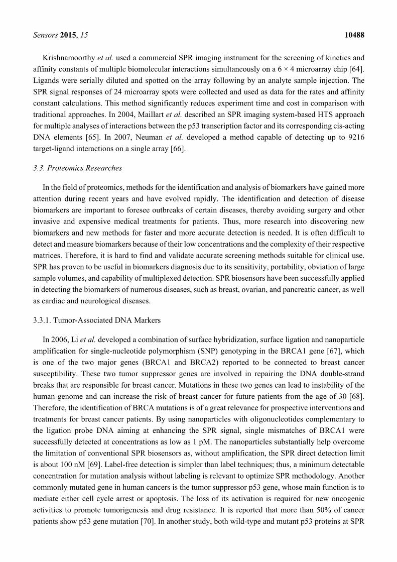
3.2. High-Throughput Screening (HTS)
The application extensions of SPR biosensors are not only limited to ligand-receptor interaction
kinetics dynamic analyses; they are also used for drug discovery and drug development. There are
several different formats of SPR biosensors, including the array format, multi-channel unit format, and
SPR imaging format, which allow simultaneous and continuous detection to analyze the performance of
hundreds to thousands of affinity binding events on a chip surface [60,61]. In SPR imaging, the incidence
angle remains fixed, and the binding of biomolecules on a gold surface is measured as the change in
reflectivity (or reflectance) in relation to the incident ray intensity, unlike SPR sensors that depend on
the measurement of the absorption dip in the SPR angle or SPR wavelength. Despite the excellent
benefits inherent in SPR technology, conventional propagating SPR biosensors have a serious limitation
in their inability to support multiplex analysis, as less than four analyses with conventional SPR
instruments make such parallel operations feasible. In contrast, SPR imaging technology using a
multi-analyte biosensor permits a high-throughput approach, and it achieves a similar degree of
sensitivity that achieved by conventional SPR biosensors (Figure 3) [62]. Thus, SPR imaging systems
without any labeling requirements are more suitable for high-throughput screening (HTS), particularly
in drug discovery, than any other optics-based detection techniques [63].
Figure 3. High-throughput drug screening using an SPR imaging protein chip system. The
bright image indicates protein-protein interaction on a gold surface. Upon the binding of an
inhibitor to the target protein, protein-protein interactions are disrupted, resulting in changes
in SPR imaging signal intensity and a darker image. Adapted from [62].
Sensors 2015, 15 10488
Krishnamoorthy et al. used a commercial SPR imaging instrument for the screening of kinetics and
affinity constants of multiple biomolecular interactions simultaneously on a 6 × 4 microarray chip [64].
Ligands were serially diluted and spotted on the array following by an analyte sample injection. The
SPR signal responses of 24 microarray spots were collected and used as data for the rates and affinity
constant calculations. This method significantly reduces experiment time and cost in comparison with
traditional approaches. In 2004, Maillart et al. described an SPR imaging system-based HTS approach
for multiple analyses of interactions between the p53 transcription factor and its corresponding cis-acting
DNA elements [65]. In 2007, Neuman et al. developed a method capable of detecting up to 9216
target-ligand interactions on a single array [66].
3.3. Proteomics Researches
In the field of proteomics, methods for the identification and analysis of biomarkers have gained more
attention during recent years and have evolved rapidly. The identification and detection of disease
biomarkers are important to foresee outbreaks of certain diseases, thereby avoiding surgery and other
invasive and expensive medical treatments for patients. Thus, more research into discovering new
biomarkers and new methods for faster and more accurate detection is needed. It is often difficult to
detect and measure biomarkers because of their low concentrations and the complexity of their respective
matrices. Therefore, it is hard to find and validate accurate screening methods suitable for clinical use.
SPR has proven to be useful in biomarkers diagnosis due to its sensitivity, portability, obviation of large
sample volumes, and capability of multiplexed detection. SPR biosensors have been successfully applied
in detecting the biomarkers of numerous diseases, such as breast, ovarian, and pancreatic cancer, as well
as cardiac and neurological diseases.
3.3.1. Tumor-Associated DNA Markers
In 2006, Li et al. developed a combination of surface hybridization, surface ligation and nanoparticle
amplification for single-nucleotide polymorphism (SNP) genotyping in the BRCA1 gene [67], which
is one of the two major genes (BRCA1 and BRCA2) reported to be connected to breast cancer
susceptibility. These two tumor suppressor genes are involved in repairing the DNA double-strand
breaks that are responsible for breast cancer. Mutations in these two genes can lead to instability of the
human genome and can increase the risk of breast cancer for future patients from the age of 30 [68].
Therefore, the identification of BRCA mutations is of a great relevance for prospective interventions and
treatments for breast cancer patients. By using nanoparticles with oligonucleotides complementary to
the ligation probe DNA aiming at enhancing the SPR signal, single mismatches of BRCA1 were
successfully detected at concentrations as low as 1 pM. The nanoparticles substantially help overcome
the limitation of conventional SPR biosensors as, without amplification, the SPR direct detection limit
is about 100 nM [69]. Label-free detection is simpler than label techniques; thus, a minimum detectable
concentration for mutation analysis without labeling is relevant to optimize SPR methodology. Another
commonly mutated gene in human cancers is the tumor suppressor p53 gene, whose main function is to
mediate either cell cycle arrest or apoptosis. The loss of its activation is required for new oncogenic
activities to promote tumorigenesis and drug resistance. It is reported that more than 50% of cancer
patients show p53 gene mutation [70]. In another study, both wild-type and mutant p53 proteins at SPR
Sensors 2015, 15 10489
chip preimmobilized with consensus DNA and monoclonal antibody were detected [71]. The normal
cell samples displayed significantly higher levels of wild-type p53; therefore, they showed higher
affinity to the immobilized consensus double-stranded DNA. Meanwhile, the pre-immobilized
monoclonal antibodies showed a specific affinity for the total p53. The difference between the SPR
signals revealed the extent of p53 mutation. Low detection levels of wild-type p53 (10.6 pM) and total
p53 (1.06 pM) were obtained due to the strong affinity of the consensus ds-DNA to the wild-type p53
and that of the antibody to total p53.
3.3.2. Disease-Related Protein Biomarkers
In contrast to genetic markers, protein markers enable rigorous evaluation for clinical applications;
therefore, protein-based assays are being developed. Because biomarkers in real blood samples exist in
small concentration, this hinders the use of SPR biosensors. To overcome this, the aid of nanoparticles
is needed for signal amplification. With the assistance of nanoparticles, researchers have been able to
employ SPR in the detection of important biomarkers, such as total prostate-specific antigen (tPSA) [72],
carbohydrate antigen 15-3 (CA15-3) [73], carcinoembryonic antigen (CEA) [74], C-reactive protein
(CRP) [75], human epidermal growth factor receptor 2 (HER2) [76], estrogen receptor (ER) [14,77,78],
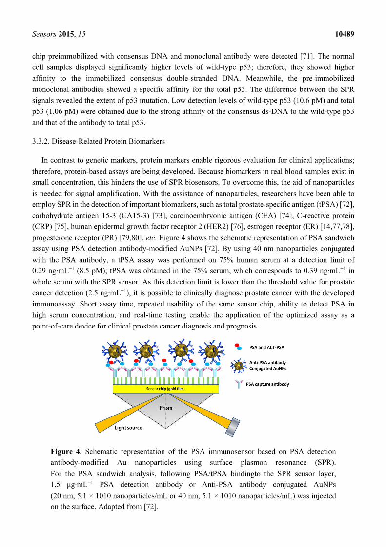
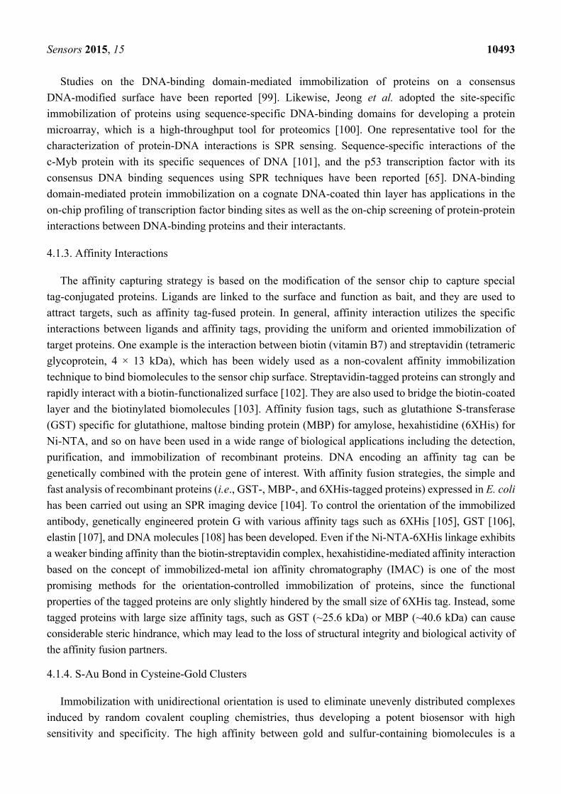
progesterone receptor (PR) [79,80], etc. Figure 4 shows the schematic representation of PSA sandwich
assay using PSA detection antibody-modified AuNPs [72]. By using 40 nm nanoparticles conjugated
with the PSA antibody, a tPSA assay was performed on 75% human serum at a detection limit of
0.29 ng·mL−1 (8.5 pM); tPSA was obtained in the 75% serum, which corresponds to 0.39 ng·mL−1 in
whole serum with the SPR sensor. As this detection limit is lower than the threshold value for prostate
cancer detection (2.5 ng·mL−1), it is possible to clinically diagnose prostate cancer with the developed
immunoassay. Short assay time, repeated usability of the same sensor chip, ability to detect PSA in
high serum concentration, and real-time testing enable the application of the optimized assay as a
point-of-care device for clinical prostate cancer diagnosis and prognosis.
Figure 4. Schematic representation of the PSA immunosensor based on PSA detection
antibody-modified Au nanoparticles using surface plasmon resonance (SPR).
For the PSA sandwich analysis, following PSA/tPSA bindingto the SPR sensor layer,
1.5 μg·mL−1 PSA detection antibody or Anti-PSA antibody conjugated AuNPs
(20 nm, 5.1 × 10¹⁰ nanoparticles/mL or 40 nm, 5.1 × 10¹⁰ nanoparticles/mL) was injected
on the surface. Adapted from [72].
Sensors 2015, 15 10490
CEA is the human cancer-associated antigen and serologic tumor biomarkers for diagnosis of
colorectal, gastric and pancreatic cancers. The CEA level is a commonly used index in treatment after
breast cancer surgery to identify recurrences. The normal CEA level is in the range of 3−5 ng/mL for a
healthy woman. In 2006, Tang et al. used an SPR-based immunosensor that used protein A conjugated
on a gold surface [74]. Next, a monoclonal anti-CEA antibody was coupled to protein A and prepared
for CEA injections. The detection limit of 0.5 ng/mL was obtained for the purified CEA. C-reactive
protein (CRP) is a principal blood serum biomarker for conventional inflammation and low-grade
inflammation diagnosis. CRP is produced by the liver and rises in level whenever inflammation occurs
throughout the body. Jung and coworkers used the CRP monoclonal antibody and an amide-linked (AL)
N-hydroxysuccinimide (NHS)−dextran functionalized gold array surface in a spectral SPR system to
detect C-reactive protein (CRP) in human sera with up to 120 human sera [75].
3.4. Cellular Analysis and Cell-Based Detection
The SPR technique is not only advantageous due to its real-time and label-free imaging capabilities
for dynamic changes at the surface but also for cellular changes, such as physiology, interactions at the
cell-surface, and cell detection. The SPR technique enables the monitoring of cellular response, cellular
adhesion and cellular products as well as the detection of cancer cells and bacteria cells .Upon receiving
stimulation from reactive molecules, mammalian cells respond accordingly. The responses consequently
induce SPR signal changes as interactions between cells and molecules occurs [81–83]. In a study in
2007 by Yanase et al. [82], a large angle of resonance (AR) change in an SPR sensor was observed when
RBL-2H3 rat mast cells and PAM212 cells (mouse keratinocyte cell lines) were cultured and activated
on a sensor chip by an epidermal growth factor (EGF) or an antigen. In addition, the application of
Mycalolide B and Toxin B (cell motility inhibitors) partially inhibited AR changes upon antigen
stimulation; no cell movement or morphology changes were observed. These results suggest that AR
changes reflect intracellular events other than cell-adhesion area size changes. SPR signal has been used
to measure cell growth and size changes as well [84,85]. Exposure to non-isotonic stimulation can cause
the cell volume to change. In particular, hypotonic stimulation results in SPR signal decrease, and the
signal reverses to an equilibrium value after additional introduction of isotonic solutions.
The SPR technique plays a major role in cancer cell detection. The first approach is based on cancer
biomarker production employing monoclonal antibodies immobilized on the sensor surface. The second
approach directly measures cellular response for quantitative analysis. Cancer cells are seeded directly
onto the sensing surface and cultured. Next, solutions containing stimuli are flowed over the cells, and
their response is measured in real time by SPR. This strategy has been successfully applied to detect and
diagnose malignant tumors cells, such as Chinese hamster ovary cells without the use of immunological
labels [86]. Liu et al. designed an SPR biosensing system with anti-VEGF antibodies immobilized on
the surface and living SKOV-3 ovarian cancer cells cultured on the flow cell chamber ceiling [87]. The
cancer cells, in the activity promoting blood vessels growth, started to produce VEGF. Subsequently,
these induced VEGF formed specific bindings with the antibody downward on the chip surface, and the
SPR signal was recorded. The designed system allows the real-time detection of VEGF with sensitivity
in a linear dynamic range of 0.1–2.5 μg·mL−1. In 2006, our group described an integrative system using
surface plasmon resonance imaging (SPRI) and a microwell gold protein chip [8]. With this system, we
Sensors 2015, 15 10491
were able to carry out recombinant E. coli culture and the detection and purification of the expressed
proteins on the chip [88]. The whole procedure included protein induction on the microwell chip, E. coli
cell lysation (by adding lysozymes), purification of expressed glutathione S-transferase-fused green
fluorescent protein (GST-GFP) by affinity interaction with gold surface chip, and finally the detection
of expressed proteins using the SPRI system. The microwell chip was constructed using PDMS
elastomer affixed on a gold surface forming a 36-well chip with a 5 uL sample volume for each well.
This all-in-one system shows benefits in terms of time and labor saving, as it allows researchers to carry
out the entire tedious protein process on a single chip. Thus, high-throughput expression and purification
of recombinant proteins in drug development could benefit from the use of our system.
4. Immobilization of Biomolecules and Signal Amplification in SPR
4.1. Immobilization of Biomolecules
4.1.1. Covalent Coupling
Covalent coupling is a commonly used immobilization method in the design of biosensor. To create
covalent bonds, the reagents must activate the sensor chip surface to produce reactive species. Covalent
coupling is then formed by the irreversible binding of reactive functional groups on the outside in a
protein onto the activated surface. The chemical cross-linkers with spacer arms possess two functional
groups; one is for covalent linkage to a surface, and the other functional group is for attachment to a
biological molecule. Investigators have developed numerous covalent conjugation chemistries to
produce protein-linker conjugates [89–91]. Several functional groups such as amine (-NH2), thiol (-SH),
and aldehyde (-COOH) are most commonly used to covalently couple biomolecules to the solid support.
Exposed amine (-NH2) groups on proteins can be coupled with various reactive intermediates such as
N-Hydroxysuccinimide (NHS), aldehyde, and epoxide on the surface. In addition, thiol (-SH) groups
present in cysteine residues can react and couple with a heterobifunctional crossliker such as a
maleimide [92]. This thiol-maleimide reaction is a process that has been widely employed for the labeling
of biomolecules. Another example of cysteine-mediated covalent conjugation is thiol-disulfide exchange
coupling, which is usually derived by the coupling of two thiol groups [93]. Such covalently
coupled-disulfide bonds are reversibly converted to thiols in the presence of reducing agents including
dithiothreitol (DTT). Covalent coupling offers a stable, easy and ligand modification-free method for
SPR surface functionalization. Nevertheless, in some cases, there are some drawbacks of the use of
covalent coupling of biomolecules on a chemically-modified surface. First, covalent immobilization may
involve chemical modification on the active sites of proteins which can potentially affect the
analyte-binding activity. Second, the reactive functional groups can be blocked by nonspecific binding
of proteins, and finally, an inappropriate blocking agents can inactivate the biomolecules.
4.1.2. Sequence-Specific DNA-Directed Immobilization of Proteins
Strategies for the sequence-specific DNA-directed immobilization of proteins (i.e., antibodies,
enzymes, peptides, etc.) include two main approaches, the immobilization of protein-DNA conjugates on
a complementary DNA-modified surface and the immobilization of proteins harboring a DNA-binding
Sensors 2015, 15 10492
domain on the cognate DNA-coated sensing layer. These DNA-directed immobilization techniques
can orient proteins on a DNA chip surface in a manner that would allow the high binding affinity and
specificity of proteins to their targets. Addressable biosensor chips using the site-direct immobilization of
protein-DNA conjugates on a DNA-functionalized sensing layer have been developed [94–96].
Streptavidin-DNA conjugates have been used as molecular linkers in the DNA-directed immobilization of
biotinylated antibodies on the chip surface (Figure 5A,B) [97]. In addition, DNA-linked antibodies have
been applied onto a gold surface modified with complementary DNA (Figure 5C) [98]. The drawback of
using antibody-DNA conjugates for DNA-directed immobilization is that labeling of antibodies with DNA
molecules by covalent modification may have significant effects on their functional properties, that is, their
abilities to bind antigens.
Figure 5. Schematic representation of DNA-directed immobilization. (A) Conjugation of
four covalent conjugates (HA-HD) with the biotinylated antibodies anti-carcinoembryonic
antigen (RAC), anti-ceruloplasmin (SAC), anti-complement-1-inactivator (SCI), and
anti-lectin (GAL) to generate capture reagents A-RAC, B-SAC, C-SCI, and D-GAL.
Adapted from [97]; (B) Conjugation of Streptavidin-Cy5 with biotinylated RAC, SAC, SCI
and GAL for the use of a microscaled fluorescence immunoassay. Adapted from [97];
(C) DNA-directed immobilization of DNA-linked antibodies. c-A (Target sequence
conjugated to anti-hCG): 5'-AGC GGA TAA CAA TTT CAC ACA GGA-3'; c-B (Target
sequence conjugated to anti-hLH): 5'-AAC AGC TAT GAC CAT GAT TAC-3'; OEG, oligo
(ethylene glycol). Adapted from [98].
Sensors 2015, 15 10493
Studies on the DNA-binding domain-mediated immobilization of proteins on a consensus
DNA-modified surface have been reported [99]. Likewise, Jeong et al. adopted the site-specific
immobilization of proteins using sequence-specific DNA-binding domains for developing a protein
microarray, which is a high-throughput tool for proteomics [100]. One representative tool for the
characterization of protein-DNA interactions is SPR sensing. Sequence-specific interactions of the
c-Myb protein with its specific sequences of DNA [101], and the p53 transcription factor with its
consensus DNA binding sequences using SPR techniques have been reported [65]. DNA-binding
domain-mediated protein immobilization on a cognate DNA-coated thin layer has applications in the
on-chip profiling of transcription factor binding sites as well as the on-chip screening of protein-protein
interactions between DNA-binding proteins and their interactants.
4.1.3. Affinity Interactions
The affinity capturing strategy is based on the modification of the sensor chip to capture special
tag-conjugated proteins. Ligands are linked to the surface and function as bait, and they are used to
attract targets, such as affinity tag-fused protein. In general, affinity interaction utilizes the specific
interactions between ligands and affinity tags, providing the uniform and oriented immobilization of
target proteins. One example is the interaction between biotin (vitamin B7) and streptavidin (tetrameric
glycoprotein, 4 × 13 kDa), which has been widely used as a non-covalent affinity immobilization
technique to bind biomolecules to the sensor chip surface. Streptavidin-tagged proteins can strongly and
rapidly interact with a biotin-functionalized surface [102]. They are also used to bridge the biotin-coated
layer and the biotinylated biomolecules [103]. Affinity fusion tags, such as glutathione S-transferase
(GST) specific for glutathione, maltose binding protein (MBP) for amylose, hexahistidine (6ХHis) for
Ni-NTA, and so on have been used in a wide range of biological applications including the detection,
purification, and immobilization of recombinant proteins. DNA encoding an affinity tag can be
genetically combined with the protein gene of interest. With affinity fusion strategies, the simple and
fast analysis of recombinant proteins (i.e., GST-, MBP-, and 6ХHis-tagged proteins) expressed in E. coli
has been carried out using an SPR imaging device [104]. To control the orientation of the immobilized
antibody, genetically engineered protein G with various affinity tags such as 6ХHis [105], GST [106],
elastin [107], and DNA molecules [108] has been developed. Even if the Ni-NTA-6ХHis linkage exhibits
a weaker binding affinity than the biotin-streptavidin complex, hexahistidine-mediated affinity interaction
based on the concept of immobilized-metal ion affinity chromatography (IMAC) is one of the most
promising methods for the orientation-controlled immobilization of proteins, since the functional
properties of the tagged proteins are only slightly hindered by the small size of 6ХHis tag. Instead, some
tagged proteins with large size affinity tags, such as GST (~25.6 kDa) or MBP (~40.6 kDa) can cause
considerable steric hindrance, which may lead to the loss of structural integrity and biological activity of
the affinity fusion partners.
4.1.4. S-Au Bond in Cysteine-Gold Clusters
Immobilization with unidirectional orientation is used to eliminate unevenly distributed complexes
induced by random covalent coupling chemistries, thus developing a potent biosensor with high
sensitivity and specificity. The high affinity between gold and sulfur-containing biomolecules is a
Sensors 2015, 15 10494
well-known interaction concept for unidirectional immobilization. The orderliness in the SAM and
molecular orientation can be achieved through S-Au binding, also referred to as the thiolated gold bond,
involving the thiol (-SH) functional group. Cysteine residues have been widely used to immobilize
proteins or peptides on an SPR gold surface for the purpose of controlled immobilization. With the
advent of recombinant protein techniques, it is possible to express and purify a fusion protein containing
cysteine amino acids with the ease of preparation of the cysteine-modified protein. [109]. The direct
immobilization method has some important benefits such as speed, simplicity, favorable orientation, and
increased functionality. Recently, the antibody-binding protein (ABP)-mediated immobilization of
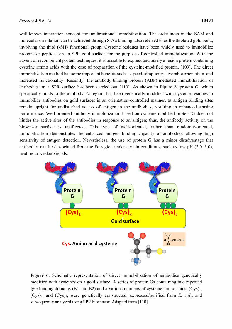
antibodies on a SPR surface has been carried out [110]. As shown in Figure 6, protein G, which
specifically binds to the antibody Fc region, has been genetically modified with cysteine residues to
immobilize antibodies on gold surfaces in an orientation-controlled manner, as antigen binding sites
remain upright for undisturbed access of antigen to the antibodies, resulting in enhanced sensing
performance. Well-oriented antibody immobilization based on cysteine-modified protein G does not
hinder the active sites of the antibodies in response to an antigen; thus, the antibody activity on the
biosensor surface is unaffected. This type of well-oriented, rather than randomly-oriented,
immobilization demonstrates the enhanced antigen binding capacity of antibodies, allowing high
sensitivity of antigen detection. Nevertheless, the use of protein G has a minor disadvantage that
antibodies can be dissociated from the Fc region under certain conditions, such as low pH (2.0~3.0),
leading to weaker signals.
Figure 6. Schematic representation of direct immobilization of antibodies genetically
modified with cysteines on a gold surface. A series of protein Gs containing two repeated
IgG binding domains (B1 and B2) and a various numbers of cysteine amino acids, (Cys)1,
(Cys)2, and (Cys)3, were genetically constructed, expressed/purified from E. coli, and
subsequently analyzed using SPR biosensor. Adapted from [110].
Sensors 2015, 15 10495
4.2. SPR Signal Amplification
4.2.1. Metal Nanoparticles
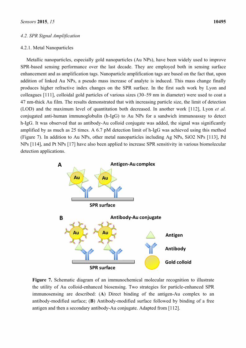
Metallic nanoparticles, especially gold nanoparticles (Au NPs), have been widely used to improve
SPR-based sensing performance over the last decade. They are employed both in sensing surface
enhancement and as amplification tags. Nanoparticle amplification tags are based on the fact that, upon
addition of linked Au NPs, a pseudo mass increase of analyte is induced. This mass change finally
produces higher refractive index changes on the SPR surface. In the first such work by Lyon and
colleagues [111], colloidal gold particles of various sizes (30–59 nm in diameter) were used to coat a
47 nm-thick Au film. The results demonstrated that with increasing particle size, the limit of detection
(LOD) and the maximum level of quantitation both decreased. In another work [112], Lyon et al.
conjugated anti-human immunoglobulin (h-IgG) to Au NPs for a sandwich immunoassay to detect
h-IgG. It was observed that as antibody-Au colloid conjugate was added, the signal was significantly
amplified by as much as 25 times. A 6.7 pM detection limit of h-IgG was achieved using this method
(Figure 7). In addition to Au NPs, other metal nanoparticles including Ag NPs, SiO2 NPs [113], Pd
NPs [114], and Pt NPs [17] have also been applied to increase SPR sensitivity in various biomolecular
detection applications.
Figure 7. Schematic diagram of an immunochemical molecular recognition to illustrate
the utility of Au colloid-enhanced biosensing. Two strategies for particle-enhanced SPR
immunosensing are described: (A) Direct binding of the antigen-Au complex to an
antibody-modified surface; (B) Antibody-modified surface followed by binding of a free
antigen and then a secondary antibody-Au conjugate. Adapted from [112].
Sensors 2015, 15 10496
4.2.2. Magnetic Nanoparticles
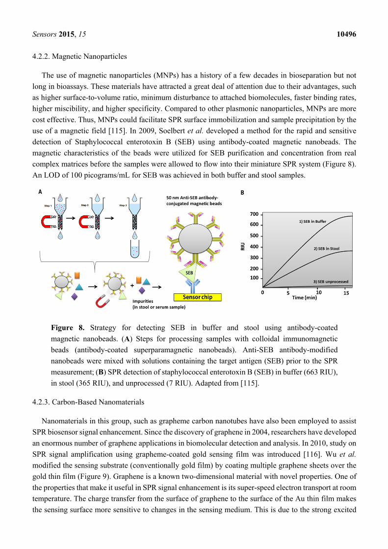
The use of magnetic nanoparticles (MNPs) has a history of a few decades in bioseparation but not
long in bioassays. These materials have attracted a great deal of attention due to their advantages, such
as higher surface-to-volume ratio, minimum disturbance to attached biomolecules, faster binding rates,
higher miscibility, and higher specificity. Compared to other plasmonic nanoparticles, MNPs are more
cost effective. Thus, MNPs could facilitate SPR surface immobilization and sample precipitation by the
use of a magnetic field [115]. In 2009, Soelbert et al. developed a method for the rapid and sensitive
detection of Staphylococcal enterotoxin B (SEB) using antibody-coated magnetic nanobeads. The
magnetic characteristics of the beads were utilized for SEB purification and concentration from real
complex matrices before the samples were allowed to flow into their miniature SPR system (Figure 8).
An LOD of 100 picograms/mL for SEB was achieved in both buffer and stool samples.
Figure 8. Strategy for detecting SEB in buffer and stool using antibody-coated
magnetic nanobeads. (A) Steps for processing samples with colloidal immunomagnetic
beads (antibody-coated superparamagnetic nanobeads). Anti-SEB antibody-modified
nanobeads were mixed with solutions containing the target antigen (SEB) prior to the SPR
measurement; (B) SPR detection of staphylococcal enterotoxin B (SEB) in buffer (663 RIU),
in stool (365 RIU), and unprocessed (7 RIU). Adapted from [115].
4.2.3. Carbon-Based Nanomaterials
Nanomaterials in this group, such as grapheme carbon nanotubes have also been employed to assist
SPR biosensor signal enhancement. Since the discovery of graphene in 2004, researchers have developed
an enormous number of graphene applications in biomolecular detection and analysis. In 2010, study on
SPR signal amplification using grapheme-coated gold sensing film was introduced [116]. Wu et al.
modified the sensing substrate (conventionally gold film) by coating multiple graphene sheets over the
gold thin film (Figure 9). Graphene is a known two-dimensional material with novel properties. One of
the properties that make it useful in SPR signal enhancement is its super-speed electron transport at room
temperature. The charge transfer from the surface of graphene to the surface of the Au thin film makes
the sensing surface more sensitive to changes in the sensing medium. This is due to the strong excited

Sensors 2015, 15 10497
electric field that exists on the surface of the Au thin film due to the graphene. The SPR signal was
enhanced by as much as 25% when there were 10 graphene layers.
Figure 9. The configuration of the proposed graphene-on-gold surface plasmon resonance
biosensor based on generalized N-Layer model, where the gold film is deposited on top of a
SF10 glass prism. A polychromatic light wave passes through the prism and is internally
reflected on the prism-gold interface, creating an evanescent wave which penetrate the metal
film (50 nm) and propagate along the x direction with propagation constant. The light
propagation constant matches the surface plasmon polariton (SPP) propagation constant
across the interface by controlling the incident angle θ. Plots of totally reflected intensity
versus incident angle yield a peak, which is known as SPR angle. The graphene-on-gold
surface plasmon resonance: prism | Au (50 nm) | graphene (L × 0.34 nm) | sensing medium,
where L is the number of graphene layers, and Z0 = 100 nm is the thickness of the
biomolecule layer. Adapted from [116].
For optimal amplification, many authors have coupled multiple types of nanomaterials in their design
so as to benefit from both sensing substrate enhancement and amplification tags. Examples include
graphene oxide (GO) and gold nanorod (AuNR)–antibody conjugates [117], graphene oxide coupled with
gold nanoparticles [118], multiple layers of graphene, and silicon on gold thin film [119]. In a publication
by Lee [120], carbon nanotubes (CNTs) conjugated with a poly-clonal antibody were employed for the
detection of human erythropoietin (EPO) and human granulocyte macrophage colony-stimulating factor
(GM–CSF) in a sandwich assay format. The results showed that the signal obtained by this CNTs
amplification tags were 30 times higher than those of a direct antibody-antigen scheme.
4.2.4. Other Approaches
Despite the benefits of nanoparticles, there are also drawbacks of using them; for example, the
synthesis and tuning steps are time and reagent-consuming. As SPR-based detection is heavily dependent
on the mass change of the analytes in the sensing medium, researchers have tried to achieve signal
Sensors 2015, 15 10498
amplification using biochemical approaches in combination with nanoparticles. Examples include
rolling circle amplification (RCA) [121,122], hybridization chain reaction (HCR) [123–125], DNA
manipulation driven [126–128], strand displacement amplification (SDA) [129,130], bio barcode
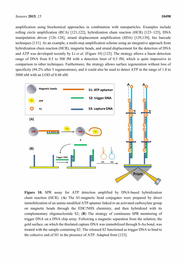
techniques [131]. As an example, a multi-step amplification scheme using an integrative approach from
hybridization chain reaction (HCR), magnetic beads, and strand displacement for the detection of DNA
and ATP was developed recently by Li et al. (Figure 10) [123]. The strategy allows a linear detection
range of DNA from 0.5 to 500 fM with a detection limit of 0.3 fM, which is quite impressive in
comparison to other techniques. Furthermore, the strategy allows surface regeneration without loss of
specificity (94.2% after 5 regenerations), and it could also be used to detect ATP in the range of 1.0 to
5000 nM with an LOD of 0.48 nM.
Figure 10. SPR assay for ATP detection amplified by DNA-based hybridization
chain reaction (HCR). (A) The S1-magnetic bead conjugates were prepared by direct
immobilization of an amine-modified ATP aptamer linked to an activated carboxylate group
on magnetic beads through the EDC/NHS chemistry, and then hybridized with its
complementary oligonucleotide S2; (B) The strategy of continuous SPR monitoring of
trigger DNA on a DNA chip array. Following a magnetic separation from the solution, the
gold surface, on which the thiolated capture DNA was immobilized through S-Au bond, was
treated with the sample containing S2. The released S2 functioned as trigger DNA to bind to
the cohesive end of H1 in the presence of ATP. Adapted from [123].
Sensors 2015, 15 10499
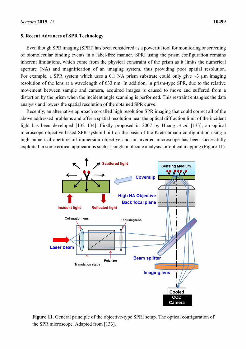
5. Recent Advances of SPR Technology
Even though SPR imaging (SPRI) has been considered as a powerful tool for monitoring or screening
of biomolecular binding events in a label-free manner, SPRI using the prism configuration remains
inherent limitations, which come from the physical constraint of the prism as it limits the numerical
aperture (NA) and magnification of an imaging system, thus providing poor spatial resolution.
For example, a SPR system which uses a 0.1 NA prism substrate could only give ~3 µm imaging
resolution of the lens at a wavelength of 633 nm. In addition, in prism-type SPR, due to the relative
movement between sample and camera, acquired images is caused to move and suffered from a
distortion by the prism when the incident angle scanning is performed. This restraint entangles the data
analysis and lowers the spatial resolution of the obtained SPR curve.
Recently, an alternative approach so-called high resolution SPR imaging that could correct all of the
above addressed problems and offer a spatial resolution near the optical diffraction limit of the incident
light has been developed [132–134]. Firstly proposed in 2007 by Huang et al. [133], an optical
microscope objective-based SPR system built on the basis of the Kretschmann configuration using a
high numerical aperture oil immersion objective and an inverted microscope has been successfully
exploited in some critical applications such as single molecule analysis, or optical mapping (Figure 11).
Figure 11. General principle of the objective-type SPRI setup. The optical configuration of
the SPR microscope. Adapted from [133].
Sensors 2015, 15 10500
A major superiority of objective-type SPRI is that the sample and imaging optical paths are fixed
throughout incident angle scanning. Thus, it permits a pixel-by-pixel tracking of the reflectivity in the
SPR images. Each of these pixels accordingly produces a SPR curve and the image is framed using the
SPR minimum angle information. Following this approach, variations such as inhomogeneity and
unwanted interferences from the laser intensity could be significantly removed by using a homogeneous
referencing surface. More importantly, the divergence in incident angles at different spots of the image
field caused by the abnormality of the objective lens could be corrected as well. Firstly, the use of a high
NA and high magnification imaging system could provide a diffraction limit resolution (~300 nm) for
the imaging optics [133]. Providing that SPR occurs at a larger angle than the critical angle, the
immersion objective is chosen to have a NA (such as an Olympus 1.65 NA objective) which is larger
than the refractive index of the medium. Consequently, when the incident light is transferred to the
objective back aperture edge, it will arrive at the sample surface at an angle that is greater than the critical
angle. Additionally, the angle coverage is also increased by this high NA objective. Secondly, compared
to the angle scanning mode in a conventional SPR measurement, the use of an objective lens could
simplify the system design by converting the rotational motion of the incident light at the sample into
linear motion of the stage. Hence, it helps improve the overall mechanical performance of the system.
Taking advantage of the advanced SPRI new capabilities, researchers have successfully employed
this high-spatial resolution SPR in imaging and detection of single DNA molecules [135], virus [136],
cells [137]. Plus, single cell-substrate interactions mapping [137] and direct binding kinetics of
proteins-cell membrane proteins mapping [138] were also made possible using this innovative system.
The recent advances in plasmonic imaging technique using high-resolution surface plasmon resonance
microscopy (SPRM) approach would undoubtedly have extensive impacts on the quantitative analysis
of intracellular dynamics in live cells, single molecule analysis and the studies of the biological activities
of membrane proteins as well as the discovery of new drugs that target membrane proteins.
6. Conclusions
SPR-based biosensor applications extend beyond the scope of this review paper. The SPR technique
has been proven to be one of the most versatile frameworks for biosensors applications in the most
concerning medical, biology, environment, and food safety areas. SPR biosensors provide excellent
analytical performance in terms of high sensitivity, fast response, LOD and reproducibility with its
label-free, real-time approach. However, one challenge for SPR technology, which was raised in 2008
by Schasfoort and Schuck about its suitability for point-of-care (POC) diagnostics [139], has not been
completely solved. The inherent drawback of SPR lies in the interference of non-specific bindings to the
outcome signals. Additionally, the kinetic rate and equilibrium constants data for biomolecular
interactions obtained by SPR biosensors have low significance in POC analysis. Given the growing trend
of applying biosensors in point-of-care testing (POCT), which is often performed by using portable and
hand-held devices, SPR technology is one of the most promising tools because it is continuously
progressing and evolving to be more suitable for user-friendly hand-held devices [69,140,141]. POCT
is an important issue in settings where timing is critical (i.e., emergency care or disease outbreaks). To
meet this growing need for SPR devices that can be deployed in the field, every effort is being made to
build more affordable, more accessible, and more applicable SPR sensor products. Clearly, a portable
Sensors 2015, 15 10501
SPR system offers s strong potential for POCT applications related to security and defense against
bioterrorism by detecting pathogenic species and identifying toxins in water in areas with no access to
laboratory facilities. However, current SPR instruments are still bulky and costly, and this remains as an
obstacle to the commercialization of SPR technology. In the coming years, more research on the
development of innovative chip chemistry and antifouling strategies in combination with amplification
schemes and miniaturization are needed to make SPR an irreplaceable tool for routine clinical analysis
and POC diagnostics.
Acknowledgments
This work was supported by the R&D Convergence Program of the Korea Research Council
of Fundamental Science and Technology (KRCF), and the KRIBB Initiative Research Program
(KRIBB, Korea).
Conflicts of Interest
The authors declare no conflict of interest.
References
1. Stephanopoulos, N.; Francis, M.B. Choosing an effective protein bioconjugation strategy.
Nat. Chem. Biol. 2011, 7, 876–884.
2. Tugarinov, V.; Kanelis, V.; Kay, L.E. Isotope labeling strategies for the study of
high-molecular-weight proteins by solution NMR spectroscopy. Nat. Protoc. 2006, 1, 749–754.
3. Phelan, M.L.; Nock, S. Generation of bioreagents for protein chips. Proteomics 2003, 3,
2123–2134.
4. Fong, C.-C.; Lai, W.-P.; Leung, Y.-C.; Lo, S.C.-L.; Wong, M.-S.; Yang, M. Study of
substrate-enzyme interaction between immobilized pyridoxamine and recombinant porcine
pyridoxal kinase using surface plasmon resonance biosensor. Biochim. Biophys. Acta 2002, 1596,
95–107.
5. Nelson, B.P.; Grimsrud, T.E.; Liles, M.R.; Goodman, R.M.; Corn, R.M. Surface plasmon
resonance imaging measurements of DNA and RNA hybridization adsorption onto DNA
microarrays. Anal. Chem. 2001, 73, 1–7.
6. Caruso, F.; Rodda, E.; Furlong, D.N.; Niikura, K.; Okahata, Y. Quartz crystal microbalance study
of DNA immobilization and hybridization for nucleic Acid sensor development. Anal. Chem. 1997,
69, 2043–2049.
7. Arwin, H.; Poksinski, M.; Johansen, K. Total internal reflection ellipsometry: Principles and
applications. Appl. Opt. 2004, 43, 3028.
8. Kim, M.; Park, K.; Jeong, E.-J.; Shin, Y.-B.; Chung, B.H. Surface plasmon resonance imaging
analysis of protein-protein interactions using on-chip-expressed capture protein. Anal. Biochem.
2006, 351, 298–304.
Sensors 2015, 15 10502
9. Madeira, A.; Vikeved, E.; Nilsson, A.; Sjögren, B.; Andrén, P.E.; Svenningsson, P. Identification
of protein-protein interactions by surface plasmon resonance followed by mass spectrometry.
Curr. Protoc. Protein Sci. 2011, 65, 19.21.1–19.21.9.
10. Majka, J.; Speck, C. Analysis of protein-DNA interactions using surface plasmon resonance.
Adv. Biochem. Eng. Biotechnol. 2007, 104, 13–36.
11. Teh, H.F.; Peh, W.Y.X.; Su, X.; Thomsen, J.S. Characterization of protein—DNA interactions
using surface plasmon resonance spectroscopy with various assay schemes. Biochemistry 2007, 46,
2127–2135.
12. Geitmann, M.; Danielson, U.H. Studies of substrate-induced conformational changes in human
cytomegalovirus protease using optical biosensor technology. Anal. Biochem. 2004, 332, 203–214.
13. Salamon, Z.; Cowell, S.; Varga, E.; Yamamura, H.I.; Hruby, V.J.; Tollin, G. Plasmon resonance
studies of agonist/antagonist binding to the human delta-opioid receptor: New structural insights
into receptor-ligand interactions. Biophys. J. 2000, 79, 2463–2474.
14. Rich, R.L.; Hoth, L.R.; Geoghegan, K.F.; Brown, T.A.; LeMotte, P.K.; Simons, S.P.; Hensley, P.;
Myszka, D.G. Kinetic analysis of estrogen receptor/ligand interactions. Proc. Natl. Acad. Sci. USA
2002, 99, 8562–8567.
15. Baron, O.L.; Pauron, D.; Antipolis, S. Protein-lipid interaction analysis by surface plasmon
resonance (SPR). Bio-Protocol 2014, 4, 1–8.
16. Erb, E.M.; Chen, X.; Allen, S.; Roberts, C.J.; Tendler, S.J.; Davies, M.C.; Forsén, S.
Characterization of the surfaces generated by liposome binding to the modified dextran matrix of
a surface plasmon resonance sensor chip. Anal. Biochem. 2000, 280, 29–35.
17. Beccati, D.; Halkes, K.M.; Batema, G.D.; Guillena, G.; Carvalho de Souza, A.; van Koten, G.;
Kamerling, J.P. SPR studies of carbohydrate-protein interactions: Signal enhancement of
low-molecular-mass analytes by organoplatinum(II)-labeling. Chembiochem 2005, 6, 1196–1203.
18. Zhang, H.; Yang, L.; Zhou, B.; Wang, X.; Liu, G.; Liu, W.; Wang, P. Investigation of biological
cell-protein interactions using SPR sensor through laser scanning confocal imaging-surface
plasmon resonance system. Spectrochim. Acta. A. Mol. Biomol. Spectrosc. 2014, 121, 381–386.
19. Besenicar, M.; Macek, P.; Lakey, J.H.; Anderluh, G. Surface plasmon resonance in
protein-membrane interactions. Chem. Phys. Lipids 2006, 141, 169–178.
20. Miyoshi, H.; Suehiro, N.; Tomoo, K.; Muto, S.; Takahashi, T.; Tsukamoto, T.; Ohmori, T.;
Natsuaki, T. Binding analyses for the interaction between plant virus genome-linked protein (VPg)
and plant translational initiation factors. Biochimie 2006, 88, 329–340.
21. Buijs, J.; Franklin, G.C. SPR-MS in functional proteomics. Brief. Funct. Genomic. Proteomic.
2005, 4, 39–47.
22. Nedelkov, D.; Nelson, R.W. Analysis of native proteins from biological fluids by biomolecular
interaction analysis mass spectrometry (BIA/MS): Exploring the limit of detection, identification
of non-specific binding and detection of multi-protein complexes. Biosens. Bioelectron. 2001, 16,
1071–1078.
23. Grasso, G.; D’Agata, R.; Rizzarelli, E.; Spoto, G.; D’Andrea, L.; Pedone, C.; Picardi, A.;
Romanelli, A.; Fragai, M.; Yeo, K.J. Activity of anchored human matrix metalloproteinase-1
catalytic domain on Au (111) surfaces monitored by ESI-MS. J. Mass Spectrom. 2005, 40,
1565–1571.
Sensors 2015, 15 10503
24. Homola, J. Present and future of surface plasmon resonance biosensors. Anal. Bioanal. Chem. 2003,
377, 528–539.
25. Cooper, M.A. Label-free screening of bio-molecular interactions. Anal. Bioanal. Chem. 2003, 377,
834–842.
26. Mullett, W.M.; Lai, E.P.; Yeung, J.M. Surface plasmon resonance-based immunoassays. Methods
2000, 22, 77–91.
27. Kukanskis, K.; Elkind, J.; Melendez, J.; Murphy, T.; Miller, G.; Garner, H. Detection of DNA
hybridization using the TISPR-1 surface plasmon resonance biosensor. Anal. Biochem. 1999, 274,
7–17.
28. Lowe, P.A.; Clark, T.J.; Davies, R.J.; Edwards, P.R.; Kinning, T.; Yeung, D. New approaches for
the analysis of molecular recognition using the IAsys evanescent wave biosensor. J. Mol. Recognit.
1998, 11, 194–199.
29. Homola, J.; Yee, S.S.; Gauglitz, G. Surface plasmon resonance sensors: Review. Sens. Actuators
B Chem. 1999, 54, 3–15.
30. Šípová, H.; Homola, J. Surface plasmon resonance sensing of nucleic acids: A review. Anal. Chim.
Acta 2013, 773, 9–23.
31. De Feijter, J.A.; Benjamins, J.; Veer, F.A. Ellipsometry as a tool to study the adsorption behavior
of synthetic and biopolymers at the air-water interface. Biopolymers 1978, 17, 1759–1772.
32. Yu, X.; Xu, D.; Cheng, Q. Label-free detection methods for protein microarrays. Proteomics 2006,
6, 5493–5503.
33. Shumaker-Parry, J.S.; Campbell, C.T. Quantitative methods for spatially resolved
adsorption/desorption measurements in real time by surface plasmon resonance microscopy.
Anal. Chem. 2004, 76, 907–917.
34. Zeder-Lutz, G.; Zuber, E.; Witz, J.; van Regenmortel, M.H. Thermodynamic analysis of
antigen-antibody binding using biosensor measurements at different temperatures. Anal. Biochem.
1997, 246, 123–132.
35. Evans, S.V.; Roger MacKenzie, C. Characterization of protein-glycolipid recognition at the
membrane bilayer. J. Mol. Recognit. 1999, 12, 155–168.
36. Cooper, M.A.; Hansson, A.; Löfås, S.; Williams, D.H. A vesicle capture sensor chip for kinetic
analysis of interactions with membrane-bound receptors. Anal. Biochem. 2000, 277, 196–205.
37. Cooper, M.A.; Williams, D.H. Kinetic analysis of antibody-antigen interactions at a supported lipid
monolayer. Anal. Biochem. 1999, 276, 36–47.
38. Cooper, M.A.; Try, A.C.; Carroll, J.; Ellar, D.J.; Williams, D.H. Surface plasmon resonance
analysis at a supported lipid monolayer. Biochim. Biophys. Acta—Biomembr. 1998, 1373, 101–111.
39. Saenko, E.; Sarafanov, A.; Greco, N.; Shima, M.; Loster, K.; Schwinn, H.; Josic, D. Use of surface
plasmon resonance for studies of protein-protein and protein-phospholipid membrane interactions.
Application to the binding of factor VIII to von Willebrand factor and to phosphatidylserine-containing
membranes. J. Chromatogr. A 1999, 852, 59–71.
40. Baird, C.L.; Courtenay, E.S.; Myszka, D.G. Surface plasmon resonance characterization of
drug/liposome interactions. Anal. Biochem. 2002, 310, 93–99.
Sensors 2015, 15 10504
41. Hubbard, J.B.; Silin, V.; Plant, A.L. Self assembly driven by hydrophobic interactions at
alkanethiol monolayers: Mechanisms of formation of hybrid bilayer membranes. Biophys. Chem.
1998, 75, 163–176.
42. Pattnaik, P. Surface plasmon resonance: Applications in understanding receptor-ligand interaction.
Appl. Biochem. Biotechnol. 2005, 126, 79–92.
43. Wegner, G.J.; Wark, A.W.; Lee, H.J.; Codner, E.; Saeki, T.; Fang, S.; Corn, R.M. Real-time
surface plasmon resonance imaging measurements for the multiplexed determination of protein
adsorption/desorption kinetics and surface enzymatic reactions on peptide microarrays. Anal. Chem.
2004, 76, 5677–5684.
44. Sibille, P.; Strosberg, A.D. A FIV epitope defined by a phage peptide library screened with a
monoclonal anti-FIV antibody. Immunol. Lett. 1997, 59, 133–137.
45. Nilsson, P.; Persson, B.; Uhlén, M.; Nygren, P.A. Real-time monitoring of DNA manipulations
using biosensor technology. Anal. Biochem. 1995, 224, 400–408.
46. Misono, T.S.; Kumar, P.K.R. Selection of RNA aptamers against human influenza virus
hemagglutinin using surface plasmon resonance. Anal. Biochem. 2005, 342, 312–317.
47. Kim, Y.H.; Kim, J.P.; Han, S.J.; Sim, S.J. Aptamer biosensor for lable-free detection of human
immunoglobulin E based on surface plasmon resonance. Sens. Actuators B Chem. 2009, 139,
471–475.
48. Bini, A.; Centi, S.; Tombelli, S.; Minunni, M.; Mascini, M. Development of an optical RNA-based
aptasensor for C-reactive protein. Anal. Bioanal. Chem. 2008, 390, 1077–1086.
49. Tombelli, S.; Minunni, M.; Luzi, E.; Mascini, M. Aptamer-based biosensors for the detection of
HIV-1 Tat protein. Bioelectrochemistry 2005, 67, 135–141.
50. Lee, S.J.; Youn, B.-S.; Park, J.W.; Niazi, J.H.; Kim, Y.S.; Gu, M.B. ssDNA aptamer-based surface
plasmon resonance biosensor for the detection of retinol binding protein 4 for the early diagnosis
of type 2 diabetes. Anal. Chem. 2008, 80, 2867–2873.
51. Lao, A.I.K.; Su, X.; Aung, K.M.M. SPR study of DNA hybridization with DNA and PNA probes
under stringent conditions. Biosens. Bioelectron. 2009, 24, 1717–1722.
52. Ratilainen, T.; Holmén, A.; Tuite, E.; Nielsen, P.E.; Nordén, B. Thermodynamics of
sequence-specific binding of PNA to DNA. Biochemistry 2000, 39, 7781–7791.
53. Wang, J.; Rivas, G.; Cai, X.; Chicharro, M.; Parrado, C.; Dontha, N.; Begleiter, A.; Mowat, M.;
Palecek, E.; Nielsen, P.E. Detection of point mutation in the p53 gene using a peptide nucleic acid
biosensor. Anal. Chim. Acta 1997, 344, 111–118.
54. D’Agata, R.; Spoto, G. Artificial DNA and surface plasmon resonance. Artif. DNA. PNA XNA 2012,
3, 45–52.
55. Sota, H.; Hasegawa, Y.; Iwakura, M. Detection of conformational changes in an immobilized
protein using surface plasmon resonance. Anal. Chem. 1998, 70, 2019–2024.
56. Mannen, T.; Yamaguchi, S.; Honda, J.; Sugimoto, S.; Kitayama, A.; Nagamune, T. Observation of
charge state and conformational change in immobilized protein using surface plasmon resonance
sensor. Anal. Biochem. 2001, 293, 185–193.
57. Kim, M.; Jung, S.O.; Park, K.; Jeong, E.-J.; Joung, H.-A.; Kim, T.-H.; Seol, D.-W.; Chung, B.H.
Detection of Bax protein conformational change using a surface plasmon resonance imaging-based
antibody chip. Biochem. Biophys. Res. Commun. 2005, 338, 1834–1838.
Sensors 2015, 15 10505
58. Li, Y.; Yan, Y.; Lei, Y.; Zhao, D.; Yuan, T.; Zhang, D.; Cheng, W.; Ding, S. Surface plasmon
resonance biosensor for label-free and highly sensitive detection of point mutation using
polymerization extension reaction. Colloids Surf. B. Biointerfaces 2014, 120, 15–20.
59. Han, S.H.; Kim, S.K.; Park, K.; Yi, S.Y.; Park, H.-J.; Lyu, H.-K.; Kim, M.; Chung, B.H. Detection
of mutant p53 using field-effect transistor biosensor. Anal. Chim. Acta 2010, 665, 79–83.
60. Smith, E.A.; Corn, R.M. Surface plasmon resonance imaging as a tool to monitor biomolecular
interactions in an array based format. Appl. Spectrosc. 2003, 57, 320A–332A.
61. Steiner, G. Surface plasmon resonance imaging. Anal. Bioanal. Chem. 2004, 379, 328–331.
62. Jung, S.O.; Ro, H.S.; Kho, B.H.; Shin, Y.B.; Kim, M.G.; Chung, B.H. Surface plasmon resonance
imaging-based protein arrays for high-throughput screening of protein-protein interaction inhibitors.
Proteomics 2005, 5, 4427–4431.
63. Kim, M.; Han, S.H.; Shin, Y. Surface plasmon resonance biosensor chips. Biochip J. 2007, 1,
81–89.
64. Krishnamoorthy, G.; Bianca Beusink, J.; Schasfoort, R.B.M. High-throughput surface plasmon
resonance imaging-based biomolecular kinetic screening analysis. Anal. Methods 2010, 2, 1020.
65. Maillart, E.; Brengel-Pesce, K.; Capela, D.; Roget, A.; Livache, T.; Canva, M.; Levy, Y.;
Soussi, T. Versatile analysis of multiple macromolecular interactions by SPR imaging: Application
to p53 and DNA interaction. Oncogene 2004, 23, 5543–5550.
66. Neumann, T.; Junker, H.-D.; Schmidt, K.; Sekul, R. SPR-based fragment screening: Advantages
and applications. Curr. Top. Med. Chem. 2007, 7, 1630–1642.
67. Li, Y.; Wark, A.W.; Lee, H.J.; Corn, R.M. Single-nucleotide polymorphism genotyping by
nanoparticle-enhanced surface plasmon resonance imaging measurements of surface ligation
reactions. Anal. Chem. 2006, 78, 3158–3164.
68. Narod, S.A.; Foulkes, W.D. BRCA1 and BRCA2: 1994 and beyond. Nat. Rev. Cancer 2004, 4,
665–676.
69. Jiang, T.; Minunni, M.; Wilson, P.; Zhang, J.; Turner, A.P.F.; Mascini, M. Detection of TP53
mutation using a portable surface plasmon resonance DNA-based biosensor. Biosens. Bioelectron.
2005, 20, 1939–1945.
70. Levine, A.J. p53, the cellular gatekeeper for growth and division. Cell 1997, 88, 323–331.
71. Wang, Y.; Zhu, X.; Wu, M.; Xia, N.; Wang, J.; Zhou, F. Simultaneous and label-free determination
of wild-type and mutant p53 at a single surface plasmon resonance chip preimmobilized with
consensus DNA and monoclonal antibody. Anal. Chem. 2009, 81, 8441–8446.
72. Uludag, Y.; Tothill, I.E. Cancer biomarker detection in serum samples using surface plasmon
resonance and quartz crystal microbalance sensors with nanoparticle signal amplification.
Anal. Chem. 2012, 84, 5898–5904.
73. Chang, C.-C.; Chiu, N.-F.; Lin, D.S.; Chu-Su, Y.; Liang, Y.-H.; Lin, C.-W. High-sensitivity
detection of carbohydrate antigen 15-3 using a gold/zinc oxide thin film surface plasmon
resonance-based biosensor. Anal. Chem. 2010, 82, 1207–1212.
74. Tang, D.-P.; Yuan, R.; Chai, Y.-Q. Novel immunoassay for carcinoembryonic antigen based on
protein A-conjugated immunosensor chip by surface plasmon resonance and cyclic voltammetry.
Bioprocess Biosyst. Eng. 2006, 28, 315–321.
Sensors 2015, 15 10506
75. Jung, S.-H.; Jung, J.-W.; Suh, I.-B.; Yuk, J. S.; Kim, W.-J.; Choi, E.Y.; Kim, Y.-M.; Ha, K.-S.
Analysis of C-reactive protein on amide-linked N-hydroxysuccinimide-dextran arrays with a
spectral surface plasmon resonance biosensor for serodiagnosis. Anal. Chem. 2007, 79, 5703–5710.
76. Martin, V.S.; Sullivan, B.A.; Walker, K.; Hawk, H.; Sullivan, B.P.; Noe, L.J. Surface plasmon
resonance investigations of human epidermal growth factor receptor 2. Appl. Spectrosc. 2006, 60,
994–1003.
77. Yang, N.; Su, X.; Tjong, V.; Knoll, W. Evaluation of two- and three-dimensional streptavidin
binding platforms for surface plasmon resonance spectroscopy studies of DNA hybridization and
protein-DNA binding. Biosens. Bioelectron. 2007, 22, 2700–2706.
78. Neo, S.J.; Su, X.; Thomsen, J.S. Surface plasmon resonance study of cooperative interactions of
estrogen receptor alpha and transcriptional factor Sp1 with composite DNA elements. Anal. Chem.
2009, 81, 3344–3349.
79. Yuan, J.; Oliver, R.; Li, J.; Lee, J.; Aguilar, M.; Wu, Y. Sensitivity enhancement of SPR
assay of progesterone based on mixed self-assembled monolayers using nanogold particles.
Biosens. Bioelectron. 2007, 23, 144–148.
80. Gillis, E.H.; Gosling, J.P.; Sreenan, J.M.; Kane, M. Development and validation of a
biosensor-based immunoassay for progesterone in bovine milk. J. Immunol. Methods 2002, 267,
131–138.
81. Hide, M.; Tsutsui, T.; Sato, H.; Nishimura, T.; Morimoto, K.; Yamamoto, S.; Yoshizato, K.
Real-time analysis of ligand-induced cell surface and intracellular reactions of living mast cells
using a surface plasmon resonance-based biosensor. Anal. Biochem. 2002, 302, 28–37.
82. Yanase, Y.; Suzuki, H.; Tsutsui, T.; Hiragun, T.; Kameyoshi, Y.; Hide, M. The SPR signal in living
cells reflects changes other than the area of adhesion and the formation of cell constructions.
Biosens. Bioelectron. 2007, 22, 1081–1086.
83. Tanaka, M.; Hiragun, T.; Tsutsui, T.; Yanase, Y.; Suzuki, H.; Hide, M. Surface plasmon resonance
biosensor detects the downstream events of active PKCbeta in antigen-stimulated mast cells.
Biosens. Bioelectron. 2008, 23, 1652–1658.
84. Baumgarten, S.; Robelek, R. Surface plasmon resonance (SPR) sensors for the rapid, sensitive
detection of the cellular response to osmotic stress. Sens. Actuators B Chem. 2011, 156, 798–804.
85. Robelek, R.; Wegener, J. Label-free and time-resolved measurements of cell volume changes by
surface plasmon resonance (SPR) spectroscopy. Biosens. Bioelectron. 2010, 25, 1221–1224.
86. Hiragun, T.; Yanase, Y.; Kose, K.; Kawaguchi, T.; Uchida, K.; Tanaka, S.; Hide, M. Surface
plasmon resonance-biosensor detects the diversity of responses against epidermal growth factor in
various carcinoma cell lines. Biosens. Bioelectron. 2012, 32, 202–207.
87. Liu, C.; Lei, T.; Ino, K.; Matsue, T.; Tao, N.; Li, C.-Z. Real-time monitoring biomarker expression
of carcinoma cells by surface plasmon resonance biosensors. Chem. Commun. (Camb). 2012, 48,
10389–10391.
88. Kim, M.; Lee, S.-Y.; Choi, H.; Shin, Y.-B.; Jung, S.O.; Kim, M.-G.; Chung, B.H. On-chip
Escherichia coli culture, purification, and detection of expressed proteins. Eur. Biophys. J. 2006,
35, 655–662.

Sensors 2015, 15 10507
89. Gao, Y.; Kyratzis, I. Covalent immobilization of proteins on carbon nanotubes using the
cross-linker 1-ethyl-3-(3-dimethylaminopropyl)carbodiimide—A critical assessment. Bioconjug.
Chem. 2008, 19, 1945–1950.
90. Roberts, M.J.; Bentley, M.D.; Harris, J.M. Chemistry for peptide and protein PEGylation.
Adv. Drug Deliv. Rev. 2002, 54, 459–476.
91. Mädler, S.; Bich, C.; Touboul, D.; Zenobi, R. Chemical cross-linking with NHS esters: A
systematic study on amino acid reactivities. J. Mass Spectrom. 2009, 44, 694–706.
92. Kim, Y.; Ho, S.O.; Gassman, N.R.; Korlann, Y.; Landorf, E.V; Collart, F.R.; Weiss, S. Efficient
site-specific labeling of proteins via cysteines. Bioconjug. Chem. 2008, 19, 786–791.
93. Fernandes, P.A.; Ramos, M.J. Theoretical insights into the mechanism for thiol/disulfide exchange.
Chemistry 2004, 10, 257–266.
94. Niemeyer, C.M. The developments of semisynthetic DNA-protein conjugates. Trends Biotechnol.
2002, 20, 395–401.
95. Niemeyer, C.M. Semi-synthetic DNA-protein conjugates: Novel tools in analytics and
nanobiotechnology. Biochem. Soc. Trans. 2004, 32, 51–53.
96. Takeda, S.; Tsukiji, S.; Nagamune, T. Site-specific conjugation of oligonucleotides to the
C-terminus of recombinant protein by expressed protein ligation. Bioorg. Med. Chem. Lett. 2004,
14, 2407–2410.
97. Wacker, R.; Niemeyer, C.M. DDI-microFIA—A readily configurable microarray-fluorescence
immunoassay based on DNA-directed immobilization of proteins. Chembiochem 2004, 5, 453–459.
98. Boozer, C.; Ladd, J.; Chen, S.; Jiang, S. DNA-directed protein immobilization for simultaneous
detection of multiple analytes by surface plasmon resonance biosensor. Anal. Chem. 2006, 78,
1515–1519.
99. Choi, Y.S.; Pack, S.P.; Yoo, Y.J. Development of a protein microarray using sequence-specific
DNA binding domain on DNA chip surface. Biochem. Biophys. Res. Commun. 2005, 329,
1315–1319.
100. Jeong, E.-J.; Jeong, Y.S.; Park, K.; Yi, S.Y.; Ahn, J.; Chung, S.J.; Kim, M.; Chung, B.H. Directed
immobilization of DNA-binding proteins on a cognate DNA-modified chip surface. J. Biotechnol.
2008, 135, 16–21.
101. Oda, M.; Furukawa, K.; Sarai, A.; Nakamura, H. Kinetic analysis of DNA binding by the c-Myb
DNA-binding domain using surface plasmon resonance. FEBS Lett. 1999, 454, 288–292.
102. Cho, I.-H.; Paek, E.-H.; Lee, H.; Kang, J.Y.; Kim, T.S.; Paek, S.-H. Site-directed biotinylation of
antibodies for controlled immobilization on solid surfaces. Anal. Biochem. 2007, 365, 14–23.
103. Diamandis, E.P.; Christopoulos, T.K. The biotin-(strept)avidin system: Principles and applications
in biotechnology. Clin. Chem. 1991, 37, 625–636.
104. Jung, J.-M.; Shin, Y.-B.; Kim, M.-G.; Ro, H.-S.; Jung, H.-T.; Chung, B.H. A fusion protein
expression analysis using surface plasmon resonance imaging. Anal. Biochem. 2004, 330, 251–256.
105. Patrie, S.M.; Mrksich, M. Self-assembled monolayers for MALDI-TOF mass spectrometry for
immunoassays of human protein antigens. Anal. Chem. 2007, 79, 5878–5887.
Sensors 2015, 15 10508
106. Ha, T.H.; Jung, S.O.; Lee, J.M.; Lee, K.Y.; Lee, Y.; Park, J.S.; Chung, B.H. Oriented
immobilization of antibodies with GST-fused multiple Fc-specific B-domains on a gold surface.
Anal. Chem. 2007, 79, 546–556.
107. Gao, D.; McBean, N.; Schultz, J.S.; Yan, Y.; Mulchandani, A.; Chen, W. Fabrication of antibody
arrays using thermally responsive elastin fusion proteins. J. Am. Chem. Soc. 2006, 128, 676–677.
108. Jung, Y.; Lee, J.M.; Jung, H.; Chung, B.H. Self-directed and self-oriented immobilization of
antibody by protein G-DNA conjugate. Anal. Chem. 2007, 79, 6534–6541.
109. Park, K.; Lee, J.M.; Jung, Y.; Habtemariam, T.; Salah, A.W.; Fermin, C.D.; Kim, M. Combination
of cysteine- and oligomerization domain-mediated protein immobilization on a surface plasmon
resonance (SPR) gold chip surface. Analyst 2011, 136, 2506–2511.
110. Lee, J.M.; Park, H.K.; Jung, Y.; Kim, J.K.; Jung, S.O.; Chung, B.H. Direct immobilization of
protein g variants with various numbers of cysteine residues on a gold surface. Anal. Chem. 2007,
79, 2680–2687.
111. Lyon, L.A.; Peña, D.J.; Natan, M.J. Surface plasmon resonance of Au colloid-modified Au films:
Particle size dependence. J. Phys. Chem. B 1999, 103, 5826–5831.
112. Lyon, L.A.; Musick, M.D.; Natan, M.J. Colloidal Au-enhanced surface plasmon resonance
immunosensing. Anal. Chem. 1998, 70, 5177–5183.
113. Luckarift, H.R.; Balasubramanian, S.; Paliwal, S.; Johnson, G.R.; Simonian, A.L.
Enzyme-encapsulated silica monolayers for rapid functionalization of a gold surface. Colloids Surf.
B Biointerfaces 2007, 58, 28–33.
114. Lin, K.; Lu, Y.; Chen, J.; Zheng, R.; Wang, P.; Ming, H. Surface plasmon resonance hydrogen
sensor based on metallic grating with high sensitivity. Opt. Express 2008, 16, 18599–18604.
115. Soelberg, S.D.; Stevens, R.C.; Limaye, A.P.; Furlong, C.E. Surface plasmon resonance detection
using antibody-linked magnetic nanoparticles for analyte capture, purification, concentration, and
signal amplification. Anal. Chem. 2009, 81, 2357–2363.
116. Wu, L.; Chu, H.S.; Koh, W.S.; Li, E.P. Highly sensitive graphene biosensors based on surface
plasmon resonance. Opt. Express 2010, 18, 14395–14400.
117. Zhang, J.; Sun, Y.; Xu, B.; Zhang, H.; Gao, Y.; Zhang, H.; Song, D. A novel surface plasmon
resonance biosensor based on graphene oxide decorated with gold nanorod-antibody conjugates for
determination of transferrin. Biosens. Bioelectron. 2013, 45, 230–236.
118. Cittadini, M.; Bersani, M.; Perrozzi, F.; Ottaviano, L.; Wlodarski, W.; Martucci, A. Graphene oxide
coupled with gold nanoparticles for localized surface plasmon resonance based gas sensor.
Carbon N. Y. 2014, 69, 452–459.
119. Verma, R.; Gupta, B.D.; Jha, R. Sensitivity enhancement of a surface plasmon resonance based
biomolecules sensor using graphene and silicon layers. Sens. Actuators B Chem. 2011, 160,
623–631.
120. Lee, E.G.; Park, K.M.; Jeong, J.Y.; Lee, S.H.; Baek, J.E.; Lee, H.W.; Jung, J.K. Carbon
nanotube-assisted enhancement of surface plasmon resonance signal. Anal. Biochem. 2011, 408,
206–211.
121. Shi, D.; Huang, J.; Chuai, Z.; Chen, D.; Zhu, X.; Wang, H.; Peng, J.; Wu, H.; Huang, Q.; Fu, W.
Isothermal and rapid detection of pathogenic microorganisms using a nano-rolling circle
amplification-surface plasmon resonance biosensor. Biosens. Bioelectron. 2014, 62, 280–287.
Sensors 2015, 15 10509
122. Chen, H.; Hou, Y.; Qi, F.; Zhang, J.; Koh, K.; Shen, Z.; Li, G. Detection of vascular endothelial
growth factor based on rolling circle amplification as a means of signal enhancement in surface
plasmon resonance. Biosens. Bioelectron. 2014, 61, 83–87.
123. Li, X.; Wang, Y.; Wang, L.; Wei, Q. A surface plasmon resonance assay coupled with a
hybridization chain reaction for amplified detection of DNA and small molecules. Chem. Commun.
(Camb). 2014, 50, 5049–5052.
124. Yao, G.-H.; Liang, R.-P.; Yu, X.-D.; Huang, C.-F.; Zhang, L.; Qiu, J.-D. Target-triggering
multiple-cycle amplification strategy for ultrasensitive detection of adenosine based on surface
plasma resonance techniques. Anal. Chem. 2015, 87, 929–936.
125. Yang, X.; Yu, Y.; Gao, Z. A highly sensitive plasmonic DNA assay based on triangular silver
nanoprism etching. ACS Nano 2014, 8, 4902–4907.
126. Lee, H.J.; Li, Y.; Wark, A.W.; Corn, R.M. Enzymatically amplified surface plasmon resonance
imaging detection of DNA by exonuclease III digestion of DNA microarrays. Anal. Chem. 2005,
77, 5096–5100.
127. Chang, C.-C.; Lin, S.; Wei, S.-C.; Chu-Su, Y.; Lin, C.-W. Surface plasmon resonance detection of
silver ions and cysteine using DNA intercalator-based amplification. Anal. Bioanal. Chem. 2012,
402, 2827–2835.
128. Goodrich, T.T.; Lee, H.J.; Corn, R.M. Direct detection of genomic DNA by enzymatically
amplified SPR imaging measurements of RNA microarrays. J. Am. Chem. Soc. 2004, 126,
4086–4087.
129. Guo, Q.; Yang, X.; Wang, K.; Tan, W.; Li, W.; Tang, H.; Li, H. Sensitive fluorescence detection
of nucleic acids based on isothermal circular strand-displacement polymerization reaction.
Nucleic Acids Res. 2009, 37, e20.
130. Yao, G.-H.; Liang, R.-P.; Huang, C.-F.; Zhang, L.; Qiu, J.-D. Enzyme-free surface plasmon
resonance aptasensor for amplified detection of adenosine via target-triggering strand displacement
cycle and Au nanoparticles. Anal. Chim. Acta 2015, 871, 28–34.
131. He, P.; Liu, L.; Qiao, W.; Zhang, S. Ultrasensitive detection of thrombin using surface plasmon
resonance and quartz crystal microbalance sensors by aptamer-based rolling circle amplification
and nanoparticle signal enhancement. Chem. Commun. (Camb). 2014, 50, 1481–1484.
132. Jamil, M.M.A.; Denyer, M.C.T.; Youseffi, M.; Britland, S.T.; Liu, S.; See, C.W.; Somekh, M.G.;
Zhang, J. Imaging of the cell surface interface using objective coupled widefield surface plasmon
microscopy. J. Struct. Biol. 2008, 164, 75–80.
133. Huang, B.; Yu, F.; Zare, R.N. Surface plasmon resonance imaging using a high numerical aperture
microscope objective. Anal. Chem. 2007, 79, 2979–2983.
134. Sefat, F.; Denyer, M.C.T.; Youseffi, M. Imaging via widefield surface plasmon resonance
microscope for studying bone cell interactions with micropatterned ECM proteins. J. Microsc. 2011,
241, 282–290.
135. Yu, H.; Shan, X.; Wang, S.; Chen, H.; Tao, N. Plasmonic imaging and detection of single DNA
molecules. ACS Nano 2014, 8, 3427–3433.
136. Wang, S.; Shan, X.; Patel, U.; Huang, X.; Lu, J.; Li, J.; Tao, N. Label-free imaging, detection, and
mass measurement of single viruses by surface plasmon resonance. Proc. Natl. Acad. Sci. USA
2010, 107, 16028–16032.
Sensors 2015, 15 10510
137. Wang, W.; Wang, S.; Liu, Q.; Wu, J.; Tao, N. Mapping single-cell-substrate interactions by surface
plasmon resonance microscopy. Langmuir 2012, 28, 13373–13379.
138. Wang, W.; Yang, Y.; Wang, S.; Nagaraj, V.J.; Liu, Q.; Wu, J.; Tao, N. Label-free measuring and
mapping of binding kinetics of membrane proteins in single living cells. Nat. Chem. 2012, 4,
846–853.
139. Schasfoort, R.; Schuck, P. Future trends in SPR technology. Handb. Surf. Plasmon. 2008, 354–394.
140. Soelberg, S.D.; Chinowsky, T.; Geiss, G.; Spinelli, C.B.; Stevens, R.; Near, S.; Kauffman, P.;
Yee, S.; Furlong, C.E. A portable surface plasmon resonance sensor system for real-time
monitoring of small to large analytes. J. Ind. Microbiol. Biotechnol. 2005, 32, 669–674.
141. Shin, Y.-B.; Kim, H. M.; Jung, Y.; Chung, B.H. A new palm-sized surface plasmon resonance
(SPR) biosensor based on modulation of a light source by a rotating mirror. Sens. Actuators B Chem.
2010, 150, 1–6.
© 2015 by the authors; licensee MDPI, Basel, Switzerland. This article is an open access article
distributed under the terms and conditions of the Creative Commons Attribution license
(http://creativecommons.org/licenses/by/4.0/).