Embed Size (px)
Citation preview

185I. Shergill et al. (eds.), Surgical Emergencies in Clinical Practice,DOI 10.1007/978-1-4471-2876-2_10, © Springer-Verlag London 2013
Introduction
There are very few dedicated neurosurgical departments in the country, and as such, referrals from a wide geographical area are common. Initial management, therefore, of such emergen-cies is often done in general hospitals, often under the care of general surgical or trauma and orthopedic specialties, which may not have any specialist knowledge of neurosurgical condi-tions. These facts may result in sub-optimum care for patients who are at risk of life-threatening conditions.
Importantly, it should be remembered that the skull is essentially a closed space, and, as such, there is no room for expansion in this space. Those patients who initially appear to have a simple head injury may have a sudden change in clinical
Chapter 10 Neurosurgery Emergencies Alan W. Hewitt and David Choi
A. W. Hewitt , M.A., FRCS (SN) () Department of Spine Surgery , James Cook University Hospital , Middlesbrough, Teeside , UK e-mail: [email protected]
D. Choi , M.A., MBChB, FRCS, Ph.D. Department of Neurosurgery, Institute of Neurology and Neurosurgery, University College London , London , UK
Department of Neurosurgery , The National Hospital for Neurology and Neurosurgery , London , UK

186 A.W. Hewitt and D. Choi
condition, especially if underlying brain conditions are not recognized or inappropriately treated.
This chapter outlines the main neurosurgical emergencies which junior trainees should be fully aware of how to diag-nose, initially manage, and then appropriately transfer to regional neurosurgical center for optimal patient outcome. The mere fact that the brain and spinal cord are made of cells which are generally unable to regenerate, should they become irreversibly injured, should highlight the trainee to the importance of early diagnosis and early treatment, with expeditious and appropriate transfer to specialist neurosur-gical center.
Clinical Case Scenario 1: Diffuse Brain Injury
Case Presentation
A 26-year-old man is riding his motorcycle on a main road when he skids, colliding with a tree. On arrival, the ambulance crew fi nds he has a diminished conscious level with signs of head injury. Oxygen and cervical spine immobilization are administered, and he is transferred to the nearby accident and emergency department (A&E) with en route call to alert the trauma team. On arrival in A&E, the trauma team performs ATLS assessment and resuscitation. There is no evidence of noisy breathing or shock but his conscious level is such that his best responses are fl exion (decorticate) to painful stimulus, utters incomprehensible sounds, with no eye opening. Pupils are equal and reactive with symmetrical limb responses. He is intubated immediately by the anesthetist and ventilated with oxygen, and a primary survey is completed including chest and pelvic X-ray. CT of the brain and cervical spine is obtained without delay (Fig. 10.1 ) and the leader of the trauma team contacts the regional neurosurgical on call registrar by tele-phone. The images show no focal hematoma, and the diagno-sis is severe diffuse brain injury. No emergency surgery is required and transfer is arranged to the neurosurgical ITU.

187Chapter 10. Neurosurgery Emergencies
Figure 10.1 Diffuse brain injury Axial CT. Note that even in this severe brain injury, the image on the left is not signi fi cantly abnormal. The higher slice shows small petechial hemorrhages in the right frontal lobe which are commonly seen in this type of injury
Key Features of History and Examination
Initial assessment uses the ATLS approach common to all trauma patients. Certain points are particularly important in brain injury.
Mechanism – brain injury causes approximately a quarter • of all trauma deaths in Britain, and around half are caused by road accidents. The energy level involved informs as to the likely severity and type of brain injury. A high-energy mechanism is likely to result in more severe primary brain injury, that is, when the brain is injured severely at the moment of impact. Immediate priority is recognition of the unsafe or occluded • airway, recognition of hypoventilation or shock, and asso-ciated injuries threatening breathing or circulation as these are potentially treatable factors which will cause exacerbation of the injury, that is, secondary brain injury. It is essential to accurately assess and record conscious level, • using the Glasgow Coma Scale (GCS – see Table 10.1 ), and any asymmetry of limb responses and papillary responses as

188 A.W. Hewitt and D. Choi
part of the initial assessment and prior to intubation. The conscious level is the most important indicator of the sever-ity of brain injury. Pupillary dilatation may indicate brain herniation with compression of the oculomotor nerve which may occur in severe injuries. A unilateral dilated pupil is a reliable indicator of which side of the brain is affected when it is caused by a focal lesion such as a hematoma. Each assessment area of the GCS must be recorded indi-• vidually – motor response, verbal response, and eye open-ing – rather than the total score. The motor response is the most important guide to the severity of brain injury in
Table 10.1 Glasgow Coma Scale (GCS) Assessment area Score
Best motor response ( M )
Obeys commands 6
Localizes pain 5
Normal fl exion (withdrawal) 4
Abnormal fl exion (decorticate) 3
Extension (decerebrate) 2
None ( fl accid) 1
Verbal response ( V )
Orientated 5
Confused conversation 4
Inappropriate single words 3
Incomprehensible sounds 2
None 1
Eye opening ( E )
Spontaneous 4
To speech 3
To pain 2
None 1
GCS score = ( E + M + V ); best possible score = 15; worst possible score = 3

189Chapter 10. Neurosurgery Emergencies
most cases, and this essential information must not be embedded within a total score. History of conscious level on the scene and subsequent dete-• rioration or improvement will indicate information as to the severity and type of injury. A lucid interval is indicative of a less severe primary brain injury which, if followed by dete-rioration, may become a severe secondary brain injury. Assessment of the safety of the airway includes the conscious • level. An adequate sensorium is required to protect the airway; therefore, an apparently clear airway is not safe if the conscious level is severely reduced and intubation is necessary.
Principles of Acute Management
Brain injury may be classi fi ed as primary – that which occurs at the moment of impact – and secondary – that which compli-cates the initial injury. The main principle of immediate man-agement is to minimize the secondary brain injury. Primary brain injury may be prevented or minimized, for example, by road safety initiatives or vehicle design, but is not treatable.
Good ATLS management is key, as hypoventilation, • obstructed breathing, and shock contributes rapidly to secondary brain injury, morbidity, and death. The conscious level and particularly motor response is the • most important indicator of severity, and this may be classi fi ed as mild, moderate, or severe. Severe brain injury, when GCS is 8 or less, is associated • with inadequate consciousness to protect the airway, so intubation is mandatory for all patients with potentially survivable injuries. Following initial resuscitation, CT of the head and C-spine • should be performed without delay. This will indicate whether there is a focal injury, for example, acute subdural hematoma, extradural hematoma, or intracerebral hema-toma (contusion), which might require surgical resection, or a diffuse injury as in this case. All patients with a signi fi cant brain injury should be • discussed with the regional neurosurgery team.

190 A.W. Hewitt and D. Choi
Discussion
In the “golden hour” following trauma, it is coordination of good ATLS care which is key. This may be provided by para-medics, A&E staff, and dedicated trauma teams. Systems must be developed to allow the A, B, C, D, and E of trauma care to be delivered in a consistent and coordinated manner. In a modern hospital, this will involve an ATLS-trained team performing simultaneous assessment and management of each of these aspects of care, but if that is not possible, then they must be worked through in that order of priority.
Following resuscitation, CT will allow the neurosurgeon to identify those patients who require urgent surgery for traumatic lesions causing mass effect. Many patients who have severe dif-fuse brain injury with no speci fi c surgical lesion will also require transfer to the neurosurgical ITU. This will allow invasive moni-toring of intracranial pressure by a transducing probe which is placed in the parenchyma or ventricle of the brain, and protocol-guided, coordinated specialist ITU care which aims to minimize secondary brain injury. There is evidence that those patients with severe diffuse injuries who are managed in a dedicated neurosciences center have better eventual outcome than those managed in general ITU. It is as yet unknown which aspects of management produce this difference in outcome [ 1 ] .
Key Points
1. Secondary brain injury is potentially treatable, and effective ATLS trauma care is the most important factor during the “golden hour.”
2. The key aspects of emergency neurological assessment are the GCS with assessment of the pupils and asymmetry of limb responses. This indicates the severity of the injury and may indicate if there is a mass lesion in one side of the brain. It is essential to record the motor, verbal, and eye opening components of the GCS separately rather than just the total score.

191Chapter 10. Neurosurgery Emergencies
3. When the GCS is 8 or less, immediate intubation is usually required.
4. CT of the brain and cervical spine should be obtained without delay which will allow the neurosurgeon to iden-tify those patients requiring emergency surgery.
5. Severely brain-injured patients who do not require emer-gency surgery also often require transfer to the neurosur-gical center for specialist care in ITU.
Clinical Case Scenario 2: Subarachnoid Hemorrhage
Case Presentation
A 55-year-old woman presents to accident and emergency department with a sudden onset of headache. It is the worst headache she has ever experienced and felt as though she had been struck on the back of the head. It occurred while she was in the classroom working as a school teacher. She reports no loss of consciousness but she vomited repeatedly. One hour later, the headache is continuing and she is very nauseated. She now complains of double vision and prefers dim light. Her past medical history includes hypertension and migraine, and she is an ex-smoker. On examination, she has photophobia and neck stiffness but is afebrile. The GCS is 14/15 (obeying commands, orientated, eye opening to speech). Examination of cranial nerves reveals that the right eye is not abducting fully and the diplopia occurs on looking to the right. CT of the brain con fi rms subarachnoid hemorrhage (Fig. 10.2 ).
Key Features of History and Examination
The symptom of sudden onset of headache always raises suspicion of subarachnoid hemorrhage.
The headache typically occurs without warning but may • occasionally be preceded by a headache of lesser severity
192 A.W. Hewitt and D. Choi
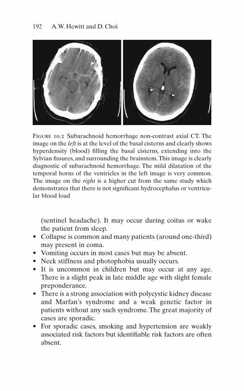
(sentinel headache). It may occur during coitus or wake the patient from sleep. Collapse is common and many patients (around one-third) • may present in coma. Vomiting occurs in most cases but may be absent. • Neck stiffness and photophobia usually occurs. • It is uncommon in children but may occur at any age. • There is a slight peak in late middle age with slight female preponderance. There is a strong association with polycystic kidney disease • and Marfan’s syndrome and a weak genetic factor in patients without any such syndrome. The great majority of cases are sporadic. For sporadic cases, smoking and hypertension are weakly • associated risk factors but identi fi able risk factors are often absent.
Figure 10.2 Subarachnoid hemorrhage non-contrast axial CT. The image on the left is at the level of the basal cisterns and clearly shows hyperdensity (blood) fi lling the basal cisterns, extending into the Sylvian fi ssures, and surrounding the brainstem. This image is clearly diagnostic of subarachnoid hemorrhage. The mild dilatation of the temporal horns of the ventricles in the left image is very common. The image on the right is a higher cut from the same study which demonstrates that there is not signi fi cant hydrocephalus or ventricu-lar blood load

193Chapter 10. Neurosurgery Emergencies
A past history of migraine or other headache should never • distract from the suspicion of subarachnoid hemorrhage. Neurological examination must establish the GCS, and • each aspect – motor, verbal, and eye opening responses – must be recorded separately. Cranial nerve examination should include careful assessment • of eye movements. Oculomotor nerve palsy may occur as a result of direct contact with an aneurysm arising close to that nerve (commonly posterior communicating artery aneurysm) or may occur as a result of brain herniation for those patients with impaired conscious level. Abducent nerve palsy may occur without any direct structural contact, as it is a long, deli-cate nerve which is susceptible to stretching. This may occur particularly when hydrocephalus develops. Examination of the limbs may reveal hemiparesis or • monoparesis (weakness of a single limb).
Principles of Acute Management
It is essential to establish the diagnosis without delay and refer to neurosurgery. This enables treatment to prevent rebleeding as well as detection and treatment of complications, especially hydrocephalus and vasospasm with delayed ischemia.
CT should be obtained promptly for all patients with clini-• cal suspicion of subarachnoid hemorrhage. When obtained within 48 h of the hemorrhage, it has 95% sensitivity in con fi rming the diagnosis. When CT is negative, all patients require lumbar puncture • (unless contraindicated) if there is clinical suspicion of subarachnoid hemorrhage. Lumbar puncture with analysis of CSF by spectrophotom-• etry is the gold standard. Spectrophotometry for the pres-ence of bilirubin is essential to differentiate between subarachnoid hemorrhage and blood from the trauma of the lumbar puncture. Bilirubin is formed in vivo in CSF by enzyme-dependent breakdown of blood in approximately

194 A.W. Hewitt and D. Choi
6 h, but is not formed following traumatic lumbar punc-ture. It should be con fi rmed with the lab that spectropho-tometry can be performed before performing the lumbar puncture. Older methods of attempting to differentiate between subarachnoid hemorrhage and traumatic tap such as comparison of red cell counts in three CSF samples are unreliable and should not be used. Lumbar puncture should be performed between 12 h and 2 • weeks of the onset of symptoms. When performed in this interval, the reported sensitivity is 100%. Many surgeons consider that delay of greater than 1 week could risk false negative error. History of headache of other cause, for example, migraine, • must not distract from appropriate investigation if current symptoms raise suspicion of subarachnoid hemorrhage, nor should investigation results such as ECG changes which may occur in subarachnoid hemorrhage. If diagnosis is missed because lumbar puncture is not per-• formed when it is required, a likely consequence is that the patient will suffer a further preventable hemorrhage in the subsequent 6 months with a high risk of mortality. Once diagnosed, all cases should be referred to neuro-• surgery by telephone without delay. The clinical details must be accurately explained. For patients in good grade (conscious and without disabling neurological de fi cit), usual management will include bed rest, oxygen, intrave-nous infusion with 3 l of 0.9% saline per day with potas-sium replacement, nimodipine 60 mg 4 hourly by mouth, laxatives, and analgesia with paracetamol and codeine. Those patients in poor grade require admission to criti-cal care. Nimodipine is a calcium channel antagonist which has been • shown to improve rates of vasospasm and delayed ischemia which may complicate subarachnoid hemorrhage. Daily U&E is necessary as hypokalemia and hyponatremia • are very common in the metabolic aftermath of subarach-noid hemorrhage. Hypokalemia results from the profound stress response whereas sodium is usually lost as a result of cerebral salt wasting. Both are treated by intravenous

195Chapter 10. Neurosurgery Emergencies
replacement with fl uid; fl uid restriction is generally con-traindicated in subarachnoid hemorrhage. All patients in good grade will be transferred to the neuro-• surgical unit. Imaging (CT angiography) will be used to con fi rm that a cerebral aneurysm has caused the hemor-rhage, and treatment (endovascular coiling or craniotomy and clipping) to occlude the aneurysm will be offered on the next available list with the aim of prevention of rebleeding. In the event of deterioration of conscious level, CT is • obtained, and if hydrocephalus is present, this may be treated by surgical placement of an external ventricular drain.
Discussion
Spontaneous subarachnoid hemorrhage is due to ruptured cerebral aneurysm in approximately 80% of cases. This may be fatal in approximately one-third of cases. Those who survive without treatment would face the likelihood of rebleeding with a risk of approximately 50% in 6 months with high mortality rates. Once diagnosed, however, the aneurysm can usually be occluded without complication, and a majority of patients admitted to the neurosurgical unit in good grade have a good outcome. A potentially potent cause of avoidable adverse out-come is therefore missed diagnosis. The key to emergency man-agement is early diagnosis and discussion with neurosurgery. The diagnosis is often easy, but problems arise when CT is negative and there are distracting factors in the presentation. It is essential that lumbar puncture is performed with spectropho-tometry analysis of the CSF in these cases. In case of any doubt, neurosurgery should be consulted.
Key Points
1. The typical symptoms of subarachnoid hemorrhage are sudden severe headache with vomiting, neck stiffness, and photophobia, but remember that the key feature is the onset. Any sudden onset headache raises suspicion.

196 A.W. Hewitt and D. Choi
2. All patients with clinical suspicion of subarachnoid hemorrhage must have CT. If CT is negative, this should be followed by lumbar puncture at least 12 h and less than 1 week following the onset of symptoms. If there is delayed presentation, contraindication to lumbar puncture or any other concerned neurosurgery should be consulted.
3. Clinical fi ndings of conscious level and focal neurological de fi cit allow the hemorrhage to be graded by the neurosur-geon, which will in fl uence management, so this information must be accurately recorded and clearly communicated in the referral.
4. Management following diagnosis will be guided by neuro-surgery and will consist of oxygen, intravenous saline, oral nimodipine, bed rest, analgesia and daily electrolyte test-ing, or admission to critical care if necessary.
5. Early transfer to the neurosurgical unit will usually be appropriate. Imaging for cerebral aneurysm will then be performed, and, if present, it will then be occluded by coil-ing or clipping to prevent rebleeding.
Clinical Case Scenario 3: Cauda Equina Syndrome
Case Presentation
A 32-year-old plumber has a chronic history of back pain. One week before admission, he develops sudden sharp back pain on moving which develops over hours into an agonizing elec-tric pain which extends down his left leg and into the lateral part of his foot. He has suffered from sciatica in the past and self-medicates in the expectation of spontaneous improve-ment. The day before admission, the pain spreads to involve both buttocks but is no more severe. The morning of admission he notices that the pain is actually less severe, but he has numbness affecting his buttocks, anal region, and penis. In the afternoon, he becomes incontinent of urine and attends the accident and emergency department. On examination, he has
197Chapter 10. Neurosurgery Emergencies
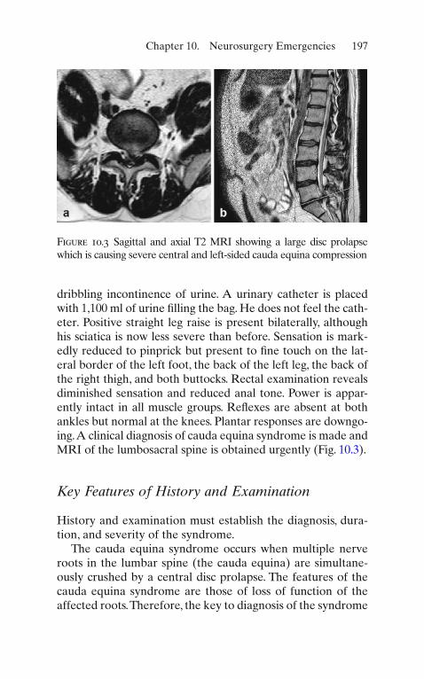
dribbling incontinence of urine. A urinary catheter is placed with 1,100 ml of urine fi lling the bag. He does not feel the cath-eter. Positive straight leg raise is present bilaterally, although his sciatica is now less severe than before. Sensation is mark-edly reduced to pinprick but present to fi ne touch on the lat-eral border of the left foot, the back of the left leg, the back of the right thigh, and both buttocks. Rectal examination reveals diminished sensation and reduced anal tone. Power is appar-ently intact in all muscle groups. Re fl exes are absent at both ankles but normal at the knees. Plantar responses are downgo-ing. A clinical diagnosis of cauda equina syndrome is made and MRI of the lumbosacral spine is obtained urgently (Fig. 10.3 ).
Key Features of History and Examination
History and examination must establish the diagnosis, dura-tion, and severity of the syndrome.
The cauda equina syndrome occurs when multiple nerve roots in the lumbar spine (the cauda equina) are simultane-ously crushed by a central disc prolapse. The features of the cauda equina syndrome are those of loss of function of the affected roots. Therefore, the key to diagnosis of the syndrome
Figure 10.3 Sagittal and axial T2 MRI showing a large disc prolapse which is causing severe central and left-sided cauda equina compression
198 A.W. Hewitt and D. Choi
is to have a working knowledge of the function of the nerve roots potentially involved and knowledge of how to recognize when these functions are lost.
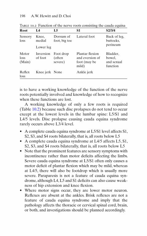
A working knowledge of only a few roots is required (Table 10.2 ) because such disc prolapses do not tend to occur except at the lowest levels in the lumbar spine: L5/S1 and L4/5 levels. Disc prolapse causing cauda equina syndrome rarely occurs above L3/4 level.
A complete cauda equina syndrome at L5/S1 level affects S1, • S2, S3, and S4 roots bilaterally, that is, all roots below L5 A complete cauda equina syndrome at L4/5 affects L5, S1, • S2, S3, and S4 roots bilaterally, that is, all roots below L5 Note that the prominent features are sensory symptoms with • incontinence rather than motor de fi cits affecting the limbs. Severe cauda equina syndrome at L5/S1 often only causes a motor de fi cit of plantar fl exion which may be mild, whereas at L4/5, there will also be footdrop which is usually more severe. Paraparesis is not a feature of cauda equina syn-drome, although L4, L5 and S1 deficits can also cause weak-ness of hip extension and knee fl exion. Where motor signs occur, they are lower motor neuron. • Re fl exes are absent at the ankles. Brisk re fl exes are not a feature of cauda equina syndrome and imply that the pathology affects the thoracic or cervical spinal cord, brain, or both, and investigations should be planned accordingly.
Table 10.2 Function of the nerve roots consisting the cauda equina Root L4 L5 S1 S2/3/4
Sensory loss
Knee, medial
Dorsum of foot, big toe
Lateral foot Back of leg, buttocks, perineum
Lower leg
Motor loss (Main)
Inversion of foot
Foot drop (often severe)
Plantar fl exion and eversion of foot (may be mild)
Bladder, bowel, and sexual function
Re fl ex loss
Knee jerk None Ankle jerk

199Chapter 10. Neurosurgery Emergencies
Typically, nerve roots affected by disc prolapse fi rst produce • pain in the dermatomal distribution, followed by paresthe-sia and fi nally numbness. This is understood as a progression from abnormal function to loss of function. The duration of symptoms must be noted as well as rate of • any deterioration. Incontinence with impaired sensation is a cardinal feature. • This is important because urinary retention is not uncom-mon during an attack of simple sciatica due to pain inhibi-tion of bladder emptying rather than a deficit of the cauda equina. If there is numbness of buttocks or perineum, painless urinary retention, or inability to feel the catheter, occurring with or after sciatica, this is very suggestive of cauda equina syndrome. Often, the cauda equina syndrome may be incomplete, • that is, some roots are spared or the syndrome is unilateral. This means that all features are not necessarily present but a potentially disabling disc prolapse may still have occurred. This implies that an index of suspicion is required in all patients with either urinary symptoms or saddle sensory symptoms, and this must be investigated urgently if symp-toms are acute or progressive.
Principles of Acute Management
MRI of the lumbosacral spine is the investigation of choice • and should be obtained urgently when there are clinical features of cauda equina syndrome. If there is paraparesis or upper motor neuron signs, this is • not consistent with cauda equina syndrome. Alternative pathology affecting spinal cord or brain must be consid-ered and investigations planned accordingly. Where there is bladder involvement, a urinary catheter • should be placed immediately as insensate urinary reten-tion leads to stretching of the bladder and detrusor muscle which worsens function further. If MRI is unavailable or contraindicated but clinically • required, consult neurosurgery. Consideration can be

200 A.W. Hewitt and D. Choi
given to transferring the patient for MRI or alternative investigation. If MRI excludes cauda equina compression, it is essential • to examine the patient again and consider an alternative diagnosis. Are there upper motor neuron signs implying that the cervical or thoracic spinal cord is involved? It may be necessary to consult a neurologist. If disc prolapse is con fi rmed, referral to a neurosurgeon • should be made urgently. Clinical features of duration and severity of the syndrome is key information for the referral. The patient can be transferred and offered urgent decom-pressive lumbar disc surgery with the aim of preventing further deterioration, relieving pain, and maximizing reha-bilitation potential of bladder and bowel.
Discussion
Even following decompressive surgery, it is common for de fi cits to remain, and rehabilitation care is often necessary. In severe cauda equina syndrome, there is often permanent disability with incontinence of bladder and bowel, numbness, and loss of sexual function. Although it is as yet unproven whether surgery overnight offers any advantage over surgery on the next available list, it is widely accepted that once detected early decompression should be offered to minimize the resulting de fi cit.
Early diagnosis is therefore important but may sometimes be dif fi cult owing to the fact that a potentially disabling disc prolapse may not produce all of the features of the cauda equina syndrome. There may also be dif fi culty if the patient cannot have MRI, for example, in cases of severe obesity, or if there are problems with availability of MRI. It is advisable that all patients with sensory symptoms affecting thighs or buttocks, or bilateral sensory symptoms, should be investi-gated for possible cauda equina compression. The diagnosis should also be considered for all patients with urinary reten-tion or incontinence. In case of any dif fi culty, the case should be discussed with neurosurgery.

201Chapter 10. Neurosurgery Emergencies
Key Points
1. Cauda equina syndrome involves the nerve roots in the lumbar spinal canal ( not the spinal cord which ends at L1). As such, it is predominantly a syndrome of sensory changes and impaired continence. Motor de fi cits where they occur are of lower motor neuron type.
2. Painless urinary retention with over fl ow incontinence is a cardinal feature of severe cauda equina syndrome. A catheter should be placed early to prevent further stretching of the bladder.
3. If cauda equina syndrome is suspected, urgent MRI is nec-essary. If symptoms are acute or deteriorating, this may need to be obtained out of hours even if this requires the patient to be transferred.
4. Diagnostic dif fi culty may occur because the syndrome is often incomplete. If doubt arises and MRI is available, it is advisable to investigate. If MRI is not available, it is advis-able to discuss the case with neurosurgery.
5. Cauda equina syndrome may result in severe permanent dis-ability even where minimal motor de fi cit in the limbs exists because of impairment of continence and sexual function.
References
1. Patel H, Bouamra O, Woodford M, King A, Yates D, Lecky F, Trauma Audit and Research Network. Trends in head injury outcome from 1989 to 2003 and the effect of neurosurgical care: an observational study. Lancet. 2005;366:1538–44.