Embed Size (px)
Citation preview

UvA-DARE is a service provided by the library of the University of Amsterdam (http://dare.uva.nl)
UvA-DARE (Digital Academic Repository)
Surgical treatment of perianal and rectal fistula
van Koperen, P.J.
Link to publication
Citation for published version (APA):van Koperen, P. J. (2010). Surgical treatment of perianal and rectal fistula s.l
General rightsIt is not permitted to download or to forward/distribute the text or part of it without the consent of the author(s) and/or copyright holder(s),other than for strictly personal, individual use, unless the work is under an open content license (like Creative Commons).
Disclaimer/Complaints regulationsIf you believe that digital publication of certain material infringes any of your rights or (privacy) interests, please let the Library know, statingyour reasons. In case of a legitimate complaint, the Library will make the material inaccessible and/or remove it from the website. Please Askthe Library: http://uba.uva.nl/en/contact, or a letter to: Library of the University of Amsterdam, Secretariat, Singel 425, 1012 WP Amsterdam,The Netherlands. You will be contacted as soon as possible.
Download date: 07 Oct 2018

Chapter 1
Perianal fistulas: developments in the
classification and diagnostic techniques,
and a new treatment strategy
P.J. van Koperen, K. Horsthuis, W.A. Bemelman, J. Stoker, J.F.M. Slors
Translated from Nederlands Tijdschrift voor Geneeskunde, 2009
19

ABSTRACT
The aim of surgical treatment of perianal fistulas is to eradicate the perianal fistula,
with low recurrence rates and risk of incontinence. In recent years there were devel-
opments regarding imaging and diagnostics of perianal fistulas.
Magnetic resonance is the most appropriate diagnostic tool. In the hands of an
experienced operator anal endosonography is a suitable, less expensive and readily-
available technique.
As a result of developments in fistula surgery it is now recommended to divide
perianal fistulas into low or high fistulas, as this has implications for the surgical
treatment. Low perianal fistulas are defined as fistulas located in the lower third
of the external anal sphincter. High fistulas are fistulas in which the fistula tract
is located in the upper two-thirds of the external sphincter muscle. Low perianal
fistulas can be treated safely by fistulotomy. Presently, the mucosal advancement
flap is the gold standard for the surgical treatment of high transsphincteric perianal
fistulas.
The anal fistula plug might be an alternative for the treatment of high transsphinc-
teric perianal fistulas.

Developments in the classification and diagnostic techniques
INTRODUCTION
A perianal fistula is one of the most frequently encountered anorectal disease in
today’s surgical practice. The incidence in females is 5.6 out of 100.000 and 12.3 out
of 100.000 in males. The incidence is highest between 30 and 50 years of age.1 When
patients experience minor complaints surgical treatment is not necessary and a wait
and see policy can be chosen. The aim of surgical treatment of perianal fistulas is
to eradicate the fistula with a surgical treatment that leads to the lowest possible
recurrence percentage, without endangering continence.
Historically, all perianal fistulas were treated by fistulotomy or by radical fistulec-
tomy. This resulted in high success percentages, however incontinence was frequently
encountered.2 In 2001, Schouten et al. described the classification and the imaging
options for perianal fistulas.3 In the present article the changes in fistula classifica-
tion, imaging and surgical treatment options are reviewed. Furthermore, a surgical
treatment strategy will be presented.
ETIOLOGY
The majority of the fistulas are of cryptoglandular origin (approximately 90%).
These non-specific fistulas originate from infection and abscess development in the
intersphincteric anal glands.4 Alternative causes are for instance Crohn’s disease and
HIV. In the present article only perianal fistulas of cryptoglandular origin will be
reviewed.
CLASSIFICATION
In 1976 the Parks’ classification of perianal fistulas was introduced. It is an anatom-
ical classification of perianal fistulas based on the relation of the fistula tract and the
external sphincter muscle.3;5 As a result of developments in the surgical treatment
of perianal fistulas it is currently advised to divide perianal fistula into low and high
perianal fistulas (Figure 1.1). Division of more than 30% of the external sphincter
muscle is associated with significantly more incontinence.6 In low perianal fistulas
the fistula tract is submucosal, intersphincteric, or located in the lower third of the
21

Chapter 1
external anal sphincter. In high perianal fistulas the fistula tract is located in the
upper two-thirds of the external sphincter.
M. levator ani
M. puborectalis
M. sphincterani internus
M. sphincterani externus
2/3
1/3
M. levator ani
M. puborectalis
M. sphincterani internus
M. sphincterani externus
2/3
1/3
Figure 1.1 – Low perianal fistulas are fistulas where the fistula tract transverses thelower 1/3 of the external sphincter complex. High fistulas transverse the upper 2/3of the external sphincter complex.
DIAGNOSTICS AND IMAGING
It is important to obtain information on the exact location of the internal opening,
the route of the fistula tract, the relation of the fistula and anal sphincter muscles,
and the presence of abscesses and multiple fistula tracts.
Fistulography en Computerized Tomography (CT)
Fistulography is considered obsolete as no information is obtained on the route
of the fistula tract in relation to the external sphincter muscle. Secondary fistula
tracts are often not filled with contrast, which leads to inaccurate information of
the fistulacomplex.7
There is currently no role for the CT as result of low contrast resolution. It is
also difficult to differentiate between scar tissue and active perianal fistulas. In a
prospective study anal endosonography was superior compared to the CT.8
22

Developments in the classification and diagnostic techniques
Anal endosonography
Anal endosonography is cheap, quick and easily accessible compared to other kinds
of imaging.9 The initial results were promising, however in later studies in which the
endosonography was compared to the Magnetic Resonance (MR) scan the results
were less promising.10−13 This discrepancy in the results may be explained by the
experience of the radiologist performing the examination.
For the identification of the internal opening the anal endosonography is suitable
as the internal opening is located close to the transducer. From earlier studies it
became clear that the endosonography is capable to successfully locate the internal
opening in around 70%.14 In a more recent study involving 151 patients in 93%
the localization of the internal opening corresponded with the examination under
anesthesia.15 Furthermore, by injecting hydrogenperoxide into the fistula tract the
accuracy was increased in some studies.13;16−18 Three-dimensional images can be
produced, however the value in fistula imaging should be studied.19 With anal en-
dosonography it is possible to assess pre- and postoperatively the presence and the
extent of damage to the anal sphincters. A drawback from anal endosonography is
the inadequate penetration of the transducer in the perianal fossa and the supral-
evatoric area.7;20 Secondary extensions of the fistula can be missed for this reason.
Furthermore, it is difficult to differentiate between fibrosis and active infection. Hy-
drogenperoxide can be helpful to differentiate between these two. This makes the
anal endosonography less suitable for patients with a history of fistula surgery.
Magnetic Resonance Imaging (MR)
The diagnostic value of the MR became clear in the nineties. Advantages were the
correct visualization of secondary fistula tracts, presence of abscesses, and the abil-
ity to differentiate between fibrosis and an active fistula.21−24 There are two ways
to visualize perianal fistulas by MR. The first option is to use an endoanal coil (an
internal MR coil) (Figure 1.2b) or by using a body coil (Figure 1.2a.). With an
endoanal coil it is possible to achieve higher spatial resolution at the level of the
anal sphincter compared to the body coil. This results in an anatomically supe-
rior image (Figure 1.2b).25 The internal fistula opening and small secondary fistula
tracts should theoretically be better visible compared to the body coil. There are
23
Chapter 1
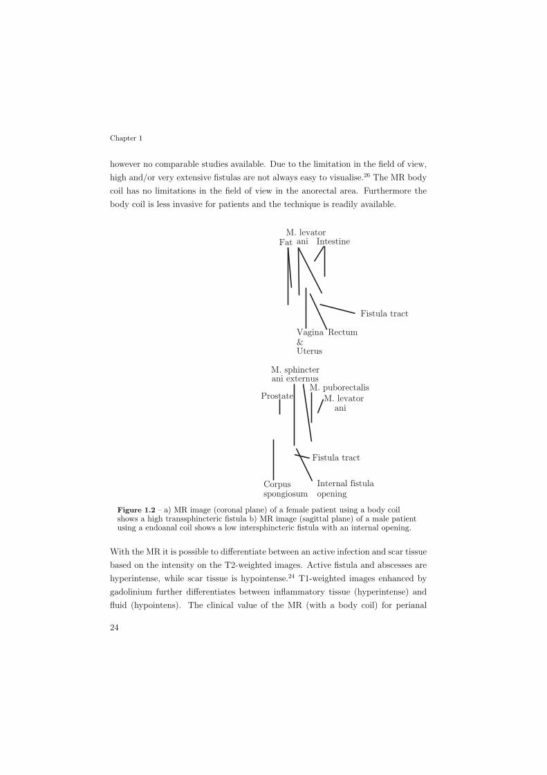
however no comparable studies available. Due to the limitation in the field of view,
high and/or very extensive fistulas are not always easy to visualise.26 The MR body
coil has no limitations in the field of view in the anorectal area. Furthermore the
body coil is less invasive for patients and the technique is readily available.
FatM. levator
ani Intestine
Vagina &Uterus
Rectum
Fistula tract
ProstateM. puborectalis
M. levatorani
Corpusspongiosum
Fistula tract
Internal fistulaopening
M. sphincterani externus
Figure 1.2 – a) MR image (coronal plane) of a female patient using a body coilshows a high transsphincteric fistula b) MR image (sagittal plane) of a male patientusing a endoanal coil shows a low intersphincteric fistula with an internal opening.
With the MR it is possible to differentiate between an active infection and scar tissue
based on the intensity on the T2-weighted images. Active fistula and abscesses are
hyperintense, while scar tissue is hypointense.24 T1-weighted images enhanced by
gadolinium further differentiates between inflammatory tissue (hyperintense) and
fluid (hypointens). The clinical value of the MR (with a body coil) for perianal
24

Developments in the classification and diagnostic techniques
fistulas is confirmed by two studies. In a study reporting on 104 patients with the
MR lead to the correct diagnosis in 90%. This was significantly higher than exam-
ination under anesthesia (61%) and also better than anal endosonography (81%).9
The diagnosis was related to a reference standard built up from examination under
anesthesia, MR and outcome. In the second study of 71 patients with recurrent pe-
rianal fistulas the result of the pre-operative MR scan was used to guide the surgical
treatment. This reduced the postoperative recurrent fistulas with 75%.27
TREATMENT
Low fistula
Submucosal, intersphincteric, and low transsphincteric fistulas, located in the lower
one-third of the external sphincter complex can be treated by fistulotomy, with favor-
able success rates and relatively little impact on fecal continence (Figure 1.3). The
recurrence rates of these fistulas are low, ranging from 2-9%.6 In a recently published
study reporting on 109 patients with cryptoglandular fistulas treated by fistulotomy
a recurrence rate of 7% at a follow-up duration of 76 months was found.28 In 40% of
these patients soiling was reported. In the literature the reported incontinence fol-
lowing fistulotomy ranges from 0-70%.6;29 In a retrospective series consisting of 624
patients, the factors female sex and a ventral fistula location were associated with
incontinence.30 This is probably the result from obstetric damage of the sphincter-
complex. Only in selected patients in this group a fistulotomy should be performed.
High fistula
This group consists of patients with perianal fistulas where the fistula tract is located
in the upper two-thirds of the external sphincter. The surgical treatment options
are the mucosal advancement flap, fibrin glue, seton drainage, and the anal fistula
plug.
Mucosal advancement flap
The mucosal advancement flap is currently the gold standard for high transsphinc-
teric fistulas (Figure 1.4). The rationale behind the advancement flap is that the
open internal opening is the cause of the persisting fistula tract. By advancing tis-
25

Chapter 1
Figure 1.3 – Fistulotomy; a) the fistula is a superficial fistula; b) the fistula isdivided by coagulation.
sue over the internal opening, it is impossible for fecal material to be forced into
the fistula tract during defecation. The advancement flap is done according to the
following technique. The internal opening is excised followed by mobilization of the
mucosa, submucosa, and a small amount of muscular fibers from the internal sphinc-
ter complex. A rectal flap with a two to three centimeters broad base is mobilized.
The rectal flap is mobilized sufficiently to cover the internal opening with overlap.
Hemostasis is performed to prevent a hematoma under the flap. The fistula tract is
curetted and the internal opening is closed after advancing the flap over the internal
opening. Finally, the flap is sutured in the distal anal canal with interrupted Vicryl
2-0 sutures (Ethicon Endo-Surgery, Cincinnati, OH). Possible complications of the
mucosal flap advancement are retraction, hematoma and necrosis of the flap. In case
of acute sepsis, patients can be treated with three months of seton drainage before
performing the advancement flap. The recurrence rates for the mucosal advance-
ment flap reported in literature vary and are reported ranging from 0-69%.31−34 Van
Koperen et al. reported a series of 70 patients with high transsphincteric fistulas
with a recurrence rate of 21%.28 Soiling was reported in 43% of the patients. In the
literature problems with continence are reported between zero and 40%.29
Fibrin glue
By injecting the fibrin glue the fistula tract and the internal opening are temporary
closed. When the glue resolves after a few weeks, fibroblasts activated by the fibrin
glue matrix, achieve closure of the fistula tract.35 Although the first results were
26

Developments in the classification and diagnostic techniques
Figure 1.4 – Mucosal advancement flap; a) seton in situ; b) the internal opening isexcised; c) sutures are fixed to the mucosal advancement flap.
good, later studies were disappointing. In a recent systematic review, the success
percentages of the 19 included studies varied from 0-100%.35 This large variety
is possibly the result of different etiologies, operation technique and perioperative
policy.
Seton drainage
The seton can be used as cutting or non-cutting (loose) seton. The loose seton is
lead through the fistula tract. The seton can serve as a bridge for the definitive
procedure. The cutting seton is designed to cut through the sphincter and leads to
muscle division. It is comparable to the fistulotomy, but the seton migrates slowly
through the sphincter. The rationale is that the muscle is divided very slowly and
has the time to heal. The seton is nowadays primarily used for the temporary or
long term drainage of the perianal fistula tracts.
Anal fistula plug
Recently there are reports on the anal fistula plug, a bioabsorbable xenograft made
of lyophilized porcine intestinal submucosa which resolves in time (Surgisis, Cook
Surgical). Through tissue remodelling the plug closes the fistula tract. The material
is fashioned into a conical plug and secured into the primary opening of the fistula
tract. The internal end of the plug is sutured in place with two sutures. The external
opening is left open to allow for drainage of the tract. In a series of 46 patients a
success percentage of 83% was found at a follow-up duration of 12 months.36 A
27

Chapter 1
comparable result was found in a series of 18 patients with a follow-up duration
of six months.37 Recently, the results of a small series of 17 patients with therapy
resistent complex high transsphincteric fistulas was published. A recurrence rate of
41% was found (follow-up 15 weeks).38 An advantage of the plug is the minimally
invasive character of the plug. The procedure is repeatable and possibly there is less
incontinence and anal scarring.
patient historyphysical examination
e xploration
suspicion Crohn's disease
endoscopy
normalMR
drainageseton
recurrent fistulacomplex fistula
MR
high transsphinctericlow transsphincteric
intersphinctericsubmucosal
mucosal advancementfistulotomy
acute sepsis
seton (3 months)
Medication
Crohn's disease
Figure 1.5 – Treatment strategy perianal fistulas.
Conclusion
Due to the impact on the chosen treatment it is advisable to divide patients with
perianal fistulas in low (lower 1/3) and high (upper 2/3) fistulas.
The MR is the treatment of choice for imaging of perianal fistulas. The anal en-
dosonography is a cheap, easy and suitable alternative readily available. The anal
endosonography is less useful in patients that have a history of fistula surgery.
Low perianal fistulas, situated in the lower 1/3 of the external sphincter muscle can
be treated with low recurrence rates by fistulotomy. The mucosal advancement flap
is the treatment of choice for high perianal fistulas (Figure 1.5). The anal fistula
plug is a potential alternative for high perianal fistulas.
28

Developments in the classification and diagnostic techniques
REFERENCES
1. Sainio P. Fistula-in-ano in a defined population. Incidence and epidemiological aspects.
Ann Chir Gynaecol 1984; 73:219-224.
2. Kronborg O. To lay open or excise a fistula-in-ano: a randomized trial. Br J Surg 1985;
72:970.
3. Schouten WR, Zimmerman DD, Meuwissen SG. Gastro-intestinale chirurgie en gastro-
enterologie. XIII. Classificatie en diagnostiek van perianale fistels. Ned Tijdschr Geneeskd
2001; 145:1398-1402.
4. Parks AG. Pathogenesis and treatment of fistula-in-ano. Br Med J 1961; 1:463-469.
5. Parks AG, Gordon PH, Hardcastle JD. A classification of fistula-in-ano. Br J Surg 1976;
63:1-12.
6. Whiteford MH, Kilkenny J, III, Hyman N, Buie WD, Cohen J, Orsay C et al. Practice
parameters for the treatment of perianal abscess and fistula-in-ano (revised). Dis Colon
Rectum 2005; 48:1337-1342.
7. Halligan S, Stoker J. Imaging of fistula in ano. Radiology 2006; 239:18-33.
8. Schratter-Sehn AU, Lochs H, Vogelsang H, Schurawitzki H, Herold C, Schratter M. Endo-
scopic ultrasonography versus computed tomography in the differential diagnosis of peri-
anorectal complications in Crohn’s disease. Endoscopy 1993; 25:582-586.
9. Buchanan GN, Halligan S, Bartram CI, Williams AB, Tarroni D, Cohen CR. Clinical ex-
amination, endosonography, and MR imaging in preoperative assessment of fistula in ano:
comparison with outcome-based reference standard. Radiology 2004; 233:674-681.
10. Orsoni P, Barthet M, Portier F, Panuel M, Desjeux A, Grimaud JC. Prospective comparison
of endosonography, magnetic resonance imaging and surgical findings in anorectal fistula and
abscess complicating Crohn’s disease. Br J Surg 1999; 86:360-364.
11. Gustafsson UM, Kahvecioglu B, Astrom G, Ahlstrom H, Graf W. Endoanal ultrasound
or magnetic resonance imaging for preoperative assessment of anal fistula: a comparative
study. Colorectal Dis 2001; 3:189-197.
12. Schwartz DA, Wiersema MJ, Dudiak KM, Fletcher JG, Clain JE, Tremaine WJ et al. A
comparison of endoscopic ultrasound, magnetic resonance imaging, and exam under anes-
thesia for evaluation of Crohn’s perianal fistulas. Gastroenterology 2001; 121:1064-1072.
13. Kruskal JB, Kane RA, Morrin MM. Peroxide-enhanced anal endosonography: technique,
image interpretation, and clinical applications. Radiographics 2001; 21 Spec No:S173-S189.
14. Deen KI, Williams JG, Hutchinson R, Keighley MR, Kumar D. Fistulas in ano: endoanal
ultrasonographic assessment assists decision making for surgery. Gut 1994; 35:391-394.
29

Chapter 1
15. Lengyel AJ, Hurst NG, Williams JG. Pre-operative assessment of anal fistulas using en-
doanal ultrasound. Colorectal Dis 2002; 4:436-440.
16. Ratto C, Gentile E, Merico M, Spinazzola C, Mangini G, Sofo L et al. How can the assess-
ment of fistula-inano be improved? Dis Colon Rectum 2000; 43:1375-1382.
17. Sloots CE, Felt-Bersma RJ, Poen AC, Cuesta MA, Meuwissen SG. Assessment and classi-
fication of fistula-in-ano in patients with Crohn’s disease by hydrogen peroxide enhanced
transanal ultrasound. Int J Colorectal Dis 2001; 16:292-297.
18. Poen AC, Felt-Bersma RJ, Eijsbouts QA, Cuesta MA, Meuwissen SG. Hydrogen peroxide-
enhanced transanal ultrasound in the assessment of fistula-in-ano. Dis Colon Rectum 1998;
41:1147-1152.
19. Buchanan GN, Bartram CI, Williams AB, Halligan S, Cohen CR. Value of hydrogen peroxide
enhancement of three-dimensional endoanal ultrasound in fistula-in-ano. Dis Colon Rectum
2005; 48:141-147.
20. Choen S, Burnett S, Bartram CI, Nicholls RJ. Comparison between anal endosonography
and digital examination in the evaluation of anal fistulae. Br J Surg 1991; 78:445-447.
21. Barker PG, Lunniss PJ, Armstrong P, Reznek RH, Cottam K, Phillips RK. Magnetic res-
onance imaging of fistula-in-ano: technique, interpretation and accuracy. Clin Radiol 1994;
49:7-13.
22. Hussain SM, Stoker J, Schouten WR, Hop WC, Lameris JS. Fistula in ano: endoanal
sonography versus endoanal MR imaging in classification. Radiology 1996; 200:475-481.
23. Stoker J, Hussain SM, van KD, Elevelt AJ, Lameris JS. Endoanal coil in MR imaging of
anal fistulas. AJR Am J Roentgenol 1996; 166:360-362.
24. Beets-Tan RG, Beets GL, van der Hoop AG, Kessels AG, Vliegen RF, Baeten CG et al.
Preoperative MR imaging of anal fistulas: Does it really help the surgeon? Radiology 2001;
218:75-84.
25. Stoker J, Hussain SM, Lameris JS. Endoanal magnetic resonance imaging versus endosonog-
raphy. Radiol Med (Torino) 1996; 92:738-741.
26. Halligan S, Bartram CI. MR imaging of fistula in ano: are endoanal coils the gold standard?
AJR Am J Roentgenol 1998; 171:407-412.
27. Buchanan G, Halligan S, Williams A, Cohen CR, Tarroni D, Phillips RK et al. Effect of
MRI on clinical outcome of recurrent fistula-in-ano. Lancet 2002; 360:1661-1662.
28. van Koperen PJ, Wind J, Bemelman WA, Bakx R, Reitsma JB, Slors JF. Long-term func-
tional outcome and risk factors for recurrence after surgical treatment for low and high
perianal fistulas of cryptoglandular origin. Dis Colon Rectum 2008; 51(10):1475-1481.
30

Developments in the classification and diagnostic techniques
29. Williams JG, Farrands PA, Williams AB, Taylor BA, Lunniss PJ, Sagar PM et al. The
treatment of anal fistula: ACPGBI position statement. Colorectal Dis 2007; 9 Suppl 4:18-
50.
30. Garcia-Aguilar J, Belmonte C, Wong WD, Goldberg SM, Madoff RD. Anal fistula surgery.
Factors associated with recurrence and incontinence. Dis Colon Rectum 1996; 39:723-729.
31. Zimmerman DD, Briel JW, Schouten WR. Endoanal advancement flap repair for complex
anorectal fistulas. Am J Surg 2001; 181:576-577.
32. Ortiz H, Marzo J. Endorectal flap advancement repair and fistulectomy for high trans-
sphincteric and suprasphincteric fistulas. Br J Surg 2000; 87:1680-1683.
33. Sonoda T, Hull T, Piedmonte MR, Fazio VW. Outcomes of primary repair of anorectal
and rectovaginal fistulas using the endorectal advancement flap. Dis Colon Rectum 2002;
45:1622-1628.
34. van der Hagen SJ, Baeten CG, Soeters PB, van Gemert WG. Long-term outcome following
mucosal advancement flap for high perianal fistulas and fistulotomy for low perianal fistu-
las : Recurrent perianal fistulas: failure of treatment or recurrent patient disease? Int J
Colorectal Dis 2006; 21:784-790.
35. Hammond TM, Grahn MF, Lunniss PJ. Fibrin glue in the management of anal fistulae.
Colorectal Dis 2004; 6:308-319.
36. Champagne BJ, O’Connor LM, Ferguson M, Orangio GR, Schertzer ME, Armstrong DN.
Efficacy of anal fistula plug in closure of cryptoglandular fistulas: long-term follow-up. Dis
Colon Rectum 2006; 49:1817-1821.
37. Ellis CN. Bioprosthetic plugs for complex anal fistulas: an early experience. J Surg Educ
2007; 64:36-40.
38. van Koperen PJ, D’Hoore A, Wolthuis AM, Bemelman WA, Slors JF. Anal fistula plug for
closure of difficult anorectal fistula: a prospective study. Dis Colon Rectum 2007; 50:2168-
2172.
31