Embed Size (px)
Citation preview
HAL Id: hal-00608987https://hal.archives-ouvertes.fr/hal-00608987
Submitted on 17 Jul 2011
HAL is a multi-disciplinary open accessarchive for the deposit and dissemination of sci-entific research documents, whether they are pub-lished or not. The documents may come fromteaching and research institutions in France orabroad, or from public or private research centers.
L’archive ouverte pluridisciplinaire HAL, estdestinée au dépôt et à la diffusion de documentsscientifiques de niveau recherche, publiés ou non,émanant des établissements d’enseignement et derecherche français ou étrangers, des laboratoirespublics ou privés.
Surgical treatment of Spheno-Orbital MeningiomasPeerooz Saeed, Wouter van Furth, Michael Tanck, Nicole Freling, Jan Willem
Berkelbach van Der Berkelbach van der Sprenkel, Lucas Stalpers, JakobusOverbeeke, Maarten Mourits
To cite this version:Peerooz Saeed, Wouter van Furth, Michael Tanck, Nicole Freling, Jan Willem Berkelbach van DerBerkelbach van der Sprenkel, et al.. Surgical treatment of Spheno-Orbital Meningiomas. BritishJournal of Ophthalmology, BMJ Publishing Group, 2011, 95 (7), pp.996. �10.1136/bjo.2010.189050�.�hal-00608987�
1
Surgical treatment of sphenoorbital meningioma
Competing interest: None to declare.
Abstract
Purpose: To evaluate the outcome of surgery and radiotherapy in the treatment of
sphenoorbital meningioma (SOM).
Method: A retrospective study of 66 consecutive cases treated with surgery for SOM
with a minimum follow-up of 4 years. Clinical and radiological information were
compared before and after the following surgical approaches: frontotemporal
craniotomy, frontotemporal craniotomy combined with orbitozygomatic resection, and
extended lateral orbitotomy alone.
Results: The median age at presentation was 46 (range, 26–68) years and the median
follow-up after surgery was 102 (48–288) months. In total, 48 (73%) patients showed
preoperative visual deterioration, with visual field defects. All patients had proptosis at
presentation (mean±SD = 6.4±3.0 mm). Surgery for patients with SOM arrested visual
deterioration in 61% and improved vision in 30% of cases. Furthermore, a substantial
reduction of proptosis was achieved in 85% of patients. The proptosis in our group was
reduced by 2.6±2.6 mm. There was no correlation between surgical approach and
proptosis reduction (P = 0.125). The recurrence rate was 17%. Only one of 15 patients
who underwent radiotherapy showed signs of recurrence.
2
Conclusions: The surgical aims in the treatment of SOM should be the restoration of
visual acuity and reduction of proptosis, rather than complete tumour removal. The
surgical approach can be tailored to individual cases. We recommend radiotherapy in
cases of subtotally removed SOM.
Introduction
Sphenoorbital meningiomas (SOM) originate from the dura of the sphenoid wing,
involve the orbit and cause visual deterioration and proptosis.1–12 Given their extensive
dural and orbital involvement, adequate resection of SOM is difficult, leading to a
recurrence rate of up to 50%.13–15 The main aim of surgery is to restore the deteriorated
vision and treat proptosis.1–12 Adequate treatment strategies and surgical approaches
remain controversial. Some authors have reported that longstanding tumour-related
proptosis cannot be treated surgically and that it should therefore not be attempted.16
Others advocate early, aggressive surgical therapy as the initial treatment.17
Postoperative radiotherapy is recommended for subtotally removed or recurrent
SOMs,18 although little information is available regarding the efficacy and side effects
of this treatment.
This study was performed to evaluate different surgical approaches used in two
centres over a period of two decades. We studied long-term surgical outcomes and the
effects and complications associated with postoperative radiotherapy in SOM.
Patients and Methods
We examined the records of 90 patients treated between 1980 and 2007 at the Orbital
Centre of the Academic Medical Centre, University of Amsterdam (n = 61), and at the
3
University Medical Centre in Utrecht (n = 29) with a diagnosis of SOM.
Sixty-six patients in this group underwent surgery at the Orbital Centre of the Academic
Medical Centre, University of Amsterdam (n = 40), and the University Medical Centre
in Utrecht (n = 26). Fifty-one patients with optic neuropathy combined with disfiguring
proptosis and 15 with disfiguring proptosis were indicated for surgery. Twenty-three
patients underwent no surgery due to stable visual acuity and the absence of optic
neuropathy. Despite visual deterioration in the amblyopic eye, one patient declined
surgical intervention. Only patients who underwent surgery were included in this study.
The clinical history, surgical approach, and outcome were obtained
retrospectively from a review of patient charts and imaging. Our analysis considered
neurological symptoms and their duration, ophthalmological examinations, consisting
of testing the patient’s visual acuity (Snellen notation) and pupil responses, funduscopy,
tonometry, and Goldmann or Humphrey perimetry for visual field defects. Ocular
motility was also evaluated.
Diplopia was considered present when the patient experienced double vision (in
any gaze direction) that disappeared with one eye closed. The degree of proptosis was
determined using a Hertel exophthalmometer. In Amsterdam, an Oculus Hertel (Oculus
Optikgeräte GmbH, Wetzlar, Germany) was used and in Utrecht, a Zeiss Hertel (Carl
Zeiss, Jena, Germany) was used.
All patients underwent preoperative computed tomography (CT) or magnetic
resonance imaging (MRI; n = 51) with contrast. Imaging was scheduled immediately
after surgery, at 3 and 12 months postoperatively, and annually thereafter for 5 years,
and then at longer intervals, depending on the presence or absence of residual or
recurrent tumours.

4
In patients who underwent radiotherapy, tear production was assessed using the
Schirmer test and corneal dryness was classified as no corneal change, corneal stippling,
limited to the inferior periphery, more extended stippling, ulceration and clouding
postradiation cataract. When necessary, fluorescein angiography was performed to
evaluate any postradiation retinopathy. After radiotherapy, all patients underwent
detailed endocrinological testing.
Surgical procedures
Frontotemporal (pterional) craniotomy was performed as described.19 The greater wing
of the sphenoid ridge was removed to the lateral limit of the lesser wing. When
hyperostotic, the anterior clinoid was removed extradurally and the superior orbital
fissure (SOF) was unroofed. The optic canal was unroofed, either intra- or extradurally,
and the inferior optic strut was removed. When intracranial or intraorbital soft tissue
was present, it was resected.
Orbitozygomatic craniotomy (OZ)20 is an extension of the frontotemporal
approach. The superior and lateral orbital rims were mobilised with additional removal
of part of the lateral wall along the zygoma and orbital roof, which provides access to
the floor of the anterior and middle cranial fossae. This was performed in either one or
two pieces, with the superior and lateral rim.
Extended lateral orbitotomy was performed through either a coronal or lid crease
incision. In this approach, the classical lateral orbitotomy bone incision described by
Berke was enlarged from the deep sphenoid wing up to the SOF.21,22
5
Reconstruction
Periorbital reconstruction was not performed. The superior and lateral orbital rim bone
was reattached. The roof and lateral wall of the orbit were routinely reconstructed with a
split cranial bone graft and secured to the orbital rim by titanium miniplates. In two
cases, this was performed using titanium mesh. Dural defects were reconstructed with
autologous galea grafts, which included convexity and basal defects. If there was a large
cavity between the orbital and dural reconstruction and the cranioplasty, an autologous
fat graft, harvested from the abdomen, was used to fill this and prevent a postoperative
pseudomeningocoele. The temporal muscle was resuspended and sutured meticulously.
Radiotherapy
Postoperatively, 15 patients underwent radiotherapy of 54 Gy in 1.8-Gy fractions. The
inclusion criteria for radiotherapy were large rest tumour and involvement of the
cavernous sinus, while the exclusion criteria were the presence of diabetes mellitus and
hypertension.
Statistical Analyses
Visual acuity and pre- and postoperative proptosis are described as means±SD.
Differences between the three different surgical approaches were compared using the
Kruskal–Wallis test. Proptosis reduction was compared between the three approaches
with correction for preoperative proptosis (nonparametric ANOVA). All analyses were
carried out using the SPSS software (ver. 16 for Windows; SPSS, Chicago, IL, USA). In
all analyses, P < 0.05 was deemed to indicate statistical significance.
6
Results
The median age at presentation was 46 (range, 26–68) years. The study population
consisted of 61 (92%) females and five (8%) males. The left and right orbits were
involved in 35 and 31 patients, respectively. The median follow-up after surgery was
102 months (48–288 months) and the median period between presentation and surgery
was 24 months (1–119 months). In total, 51 (77%) patients had preoperative progressive
visual deterioration with visual field defects. Three patients had no light perception
(NLP) preoperatively. All patients had proptosis at presentation, with a mean of
5.89±2.89 mm (mean±SD). At presentation, seven (11%) patients had diplopia and 19
(29%) had headaches or retrobulbar pain.
Surgery
The surgical approach was frontotemporal craniotomy in 45 patients and this approach
was combined with orbitozygomatic resection in 10 patients. Forty-six patients in this
group had optic neuropathy due to compression of the optic nerve, five had disfiguring
proptosis with hyperostosis of the optic canal and four had proptosis combined with
retrobulbar pain. As all of these 55 patients had disfiguring proptosis, this was also
addressed at the time of surgery.
An extended lateral orbitotomy was performed in 11 patients. Patients in this group had
only disfiguring proptosis and two had severe proptosis with stretching of the optic
nerve.
7
Craniotomies
Fifty-five patients underwent a large unilateral frontotemporal craniotomy, followed by
removal of additional tumour-infiltrated bone via an extradural approach in all cases.
Ten procedures also included OZ resection. The orbital roof was resected in 49 cases
and the lateral orbital wall was removed in 35. The optic canal and SOF were opened
and decompressed in 51 and 29 cases, respectively. After opening the orbit, periorbital
resection or stripping was necessary in 32 procedures. Extensive en plaque dural
involvement was found in 45 cases; resection of the convexity-dura was necessary in 39
cases.
Reconstruction was performed in 9 of 10 patients treated via the frontotemporal
approach in combination with OZ. The reconstruction used calvarial bone in seven
patients and titanium mesh in two; both the lateral and orbital roof were reconstructed in
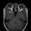
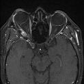
all nine patients (Fig. 1).
Orbitotomy
In 11 patients who underwent lateral orbitotomy, the lateral wall of the orbit until the
SOF was removed; in five cases, the orbital roof was also partially removed. In six
procedures, periorbital resection was also performed due to tumour infiltration. In five
cases in which there was infiltration of the ocular muscles, only the exophytic tumour
was removed. None of these patients underwent reconstruction. All lateral orbitotomies
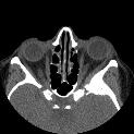
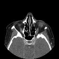
were performed by orbital surgeons (Fig. 1).
Radiotherapy
Fifteen patients underwent secondary radiotherapy after surgical excision. All 15 had
8
significant residual tumours, which also involved the cavernous sinus in four cases. Two
patients underwent radiotherapy after debulking of a recurrent lesion. Only one of these
patients showed further recurrence after follow-up for 38 months. In four patients, the
tumour volume was reduced to less than 10%. In all four, there was significant soft
tissue involvement.
Outcome
Visual acuity
Forty (61%) patients showed either stabilisation of their visual acuity or an
improvement of less than two lines on the Snellen chart. Three eyes from this group
maintained a visual acuity of NLP (no light perception). Fifteen had a visual acuity of
more than 0.8 preoperatively, which remained stable postoperatively. Twenty (30%) had
improved visual acuity. Eight improved by more than four lines on the Snellen chart and
12 others by more than two lines.
There was no significant difference (P = 0.195) in postoperative vision between
the different approaches. All except two patients with visual deterioration underwent
decompression of the optic canal through a craniotomy. The two exceptions had
proptosis of more than 10 mm with stretching of the optic nerve and underwent only
extended lateral orbitotomy with total removal of the lateral orbital wall.
Proptosis
The mean proptosis, measured with a Hertel exophthalmometer, in this group was
6.4±3.0 mm preoperatively and 3.8±2.3 mm postoperatively. The mean reduction in
proptosis was 2.6±2.6 mm. Seventeen orbits showed a proptosis reduction of more than
9
5 mm, while ten showed no change in proptosis compared with preoperative values.
One patient who underwent no reconstruction of the orbit developed enophthalmos of
2 mm. Fifty patients had a residual proptosis exceeding 2 mm. Patients who underwent
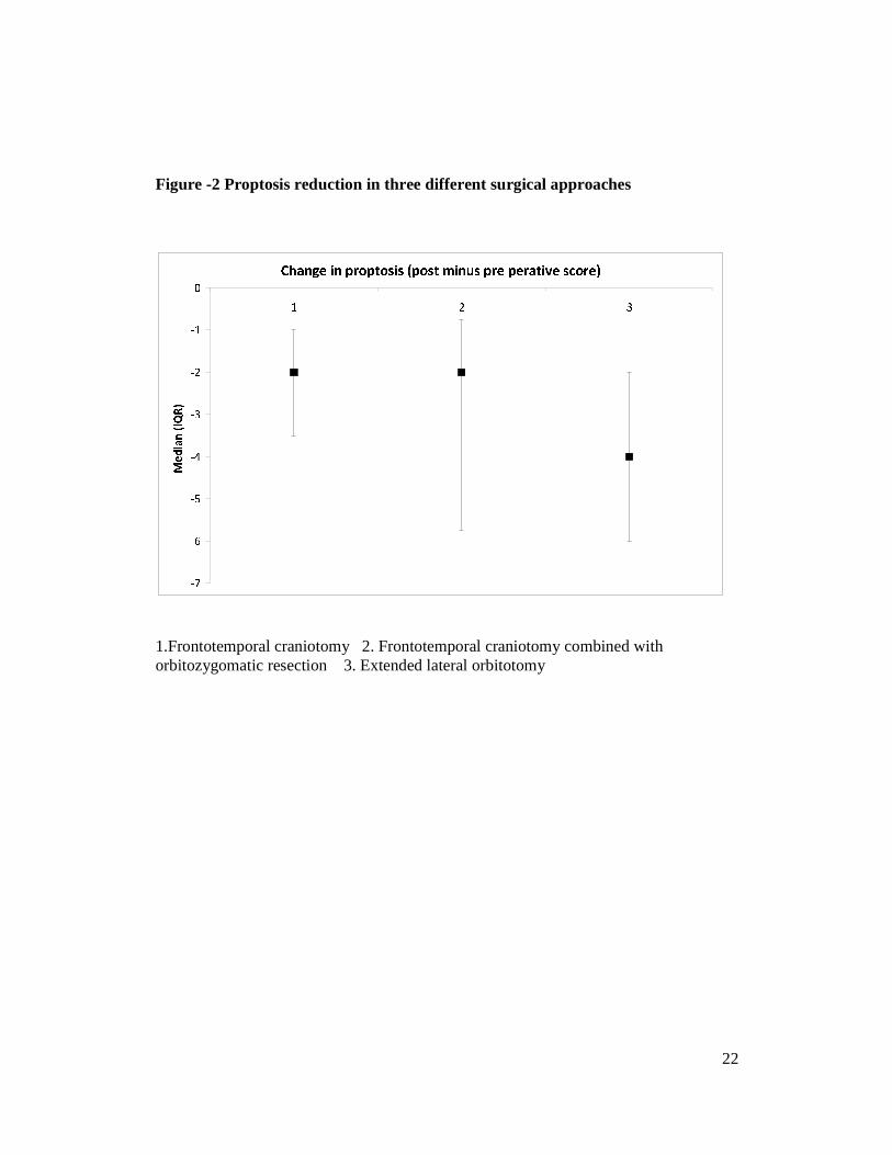
lateral orbitotomy and combined craniotomy and OZ had the greatest mean preoperative
proptosis of 7.9±3.4 and 7.7±2.9 mm, respectively. The mean proptosis with
frontotemporal craniotomy only was 5.9±2.9 mm. However, there was no significant (P
= 0.125) difference in the postoperative reduction in proptosis among the approaches
(Fig. 2).
Diplopia
New diplopia was observed in 40 patients; it was transient in 32 cases. Two patients
showed no eye movement in the first weeks after surgery; in both patients, the lateral
wall was removed completely. Within 6 months, eye movement recovered completely,
with no diplopia. Eight patients (12%) still showed diplopia; three of these underwent
strabismus surgery with satisfactory results. In six patients after a frontotemporal
craniotomy, the cause of the diplopia was third nerve palsy; thus, no strabismus surgery
was performed. Ptosis surgery was performed in one patient with partial third nerve
palsy and no diplopia. Nine patients with diplopia in the primary position preoperatively
still had diplopia postoperatively and underwent surgical correction.
Pain
Of 19 patients with retrobulbar pain after surgery, 16 had no further pain. All of these
patients underwent craniotomies.
10
Complications
Surgery
Five patients developed subgaleal cerebrospinal fluid (CSF) accumulation, which
regressed spontaneously in four cases; one patient required surgical revision of the dural
leak and placement of a lumboperitoneal shunt. After craniotomies, permanent third
nerve palsies were seen in six cases, and fourth and sixth nerve palsies occurred in one
patient each. Permanent trigeminal and facial nerve palsies were present in six and three
patients, respectively.
In two patients who underwent OZ, the lateral excursion of the mandible was reduced
by approximately 50%.
Radiotherapy
Three patients developed severe punctate keratopathy necessitating the daily use of
lubricants. One patient also developed radiation retinopathy, but had stable visual acuity
of 0.5 after laser treatment. During follow-up, one patient developed pituitary function
impairment. There was no death due to treatment or SOM in this study population.
Recurrence
Eleven (17%) patients showed recurrence (regrowth). The median time between surgery
and recurrence was 46 months (10–108 months). Macroscopic radical excision was
achieved in only six patients. Of 61 patients with rest tumour, 15 underwent
radiotherapy. Despite the larger size of the rest tumour in this group, only one of these
15 patients (7%) showed recurrence.
11
The signs of recurrence in four patients were a combination of visual
deterioration and increased proptosis, while four patients showed only a mild increase in
proptosis (< 3 mm). Four patients with visual deterioration underwent second
craniotomy—two had improved visual acuity and two other patients with NLP
underwent two other debulking surgeries and the affected eye was ultimately enucleated
because of corneal ulceration caused by proptosis.
Discussion
Symptom-oriented surgery for SOM is directed primarily at optic nerve decompression
when there is decreased visual acuity. In our series, the visual acuity improved in 30%
of cases and remained the same in 61% of cases. The cause of optic neuropathy was
addressed and treated in all of the patients with optic neuropathy. This was probably
related to the degree and period of optic nerve compression. In our series, the proptosis
was reduced by a mean of 2.6 mm from preoperative values, ranging from 2 to 14 mm.
Seventeen (26%) patients showed a marked reduction in proptosis by more than 5 mm
and up to 11 mm. Excluding one case of enophthalmos, residual proptosis was present
in 50 (76%) of the remaining patients. Nevertheless, 85% showed an improvement of
more than 2 mm. All patients in our series underwent periodical proptosis measurements
with a Hertel exophthalmometer. There have been only a few previous attempts to
quantify postoperative proptosis in SOM.2,5,12 Ringel et al. reported improvement of
proptosis in 43 of 56 patients (77%) with no change in proptosis in the remaining 13
patients (23%).2 A recent study quantified the proptosis by MRI and reported significant
residual proptosis in 53% of cases.5 We believe that the cause of this significant residual
proptosis was long-standing congestion and fibrosis, resulting in less mobile orbital
12
contents. Despite this residual proptosis in the surgical treatment of SOM, the proptosis
improves in most cases. Surgery also seemed to be effective at relieving pain associated
with SOM; 16 of the 19 patients with reported pain showed no sign of pain
postoperatively. We believe that pain is an underreported symptom in these series.
Our recurrence rate of 17% was consistent with other recently published series
with long-term follow-up.1–12 These recurrence rates are much lower than those in
earlier studies, which reported recurrence rates of 30–50%.13–14 Maroon et al. listed the
following reasons for the high recurrence rate in SOM: failure to diagnose the tumour
early because the symptoms were confused with those of other clinical entities, such as
fibrous dysplasia and Graves’ disease; inadequate resection, due to the involvement of
important neurovascular structures; and the surgeon’s concern of iatrogenic death and
serious complications in radical resections.18
New imaging techniques appear to have improved the early diagnosis and
effective surgical resection. To resect SOM, different surgical approaches are used,
including transzygomatic, pterional, frontotemporal, and combined transcranial-
transmalar and cranioorbital approaches. All of these approaches allow sufficient access
to the orbit and middle fossa base for bony and soft-tissue tumour resection and
decompression of the SOF and optic canal.19,20,23,24 Extended lateral orbitotomy or total
lateral orbitotomy has been used routinely for severe cases of Graves’ orbitopathy and
tumours in the orbital apex. This approach is effective in patients with SOM in which
the main symptom is disfiguring proptosis, without involving the risks of a
craniotomy.21,22,25,26 Recently, Lund reported 12 patients with SOM who underwent
endoscopic endonasal medial orbital wall decompression and decompression of the
optic canal in eight patients with opticopathy and visual deterioration.27 In these
13
patients, the opticopathy improved and the visual acuity improved by 1–4 lines on the
Snellen chart.
Depending on the extent of the orbital wall and roof resection, most authors
recommend firm reconstruction of the orbital walls to avoid pulsating enophthalmos and
diplopia.
Maroon et al. reported 200 cases of orbital wall and roof resection without
reconstruction, in which no cases showed permanent pulsating enophthalmos.18
DeMonte et al. concluded that partial or complete orbital roof resection, isolated or
combined with lateral or medial orbital wall defects, did not require routine
reconstruction when the periorbita was not resected.28 We advocate reconstruction of
the orbital roof and the lateral orbital rim. We consider reconstruction of the lateral
orbital wall to be unnecessary in most cases. Studies of orbital decompression in
Graves’ orbitopathy have shown that lateral wall decompression can reduce the
proptosis by up to 2 mm.29,30 As significant residual proptosis was present with all
surgical approaches in SOM in these series, no further reconstruction of the lateral
orbital wall may contribute to further proptosis reduction, while reconstruction of the
orbit roof is sufficient to avoid pulsating enophthalmos. Techniques have improved
since 1952, when Castellano et al. reported a high surgical mortality rate (20%)
associated with SOM and concluded that SOM should be used only as a last resort.16
Although the mortality rate has decreased, some recent studies still reported mortality
rates of 3–6%.2,3,11,26
Other complications, such as vision loss and transient (6–84%) or permanent (7–30%)
cranial nerve deficits, are frequent.1–12 Although life-threatening complications were not
seen in this series, postoperative visual deterioration occurred in 6 of 66 cases (9%),
14
oculomotor nerve palsy in 5 of 66 cases (7%) patients, and facial nerve palsy in 3 of 66
cases (5%).
Radiotherapy is often used to treat SOM. Peele et al. delivered a total dose of
45 Gy to subtotally or recurrent SOMs and none of the 42 patients in the radiation
treatment group experienced recurrence during an observation period of 4.2 years,
whereas 21 of the 44 patients in the non-radiation treatment group later presented with
recurrent tumour growth.31
Another study indicated a decrease in size of skull base meningiomas in 51% of
patients, stabilisation in 47% of cases and increased tumour size in 2% of cases.32 In our
series, postoperative radiotherapy in subtotally removed SOM resulted in a reduction in
tumour size and stabilisation in 27% and 67% of cases, respectively.
In conclusion, the aim of surgery in the treatment of SOM should be restoration
of visual acuity and reduction of proptosis, rather than complete bony tumour removal.
The surgical approach can be tailored to individual cases. Decompression of the optic
canal can be achieved either through a combined frontotemporal approach and OZ or a
frontotemporal approach alone. However, the frontotemporal approach combined with
OZ in these series was not associated with better visual or proptosis outcome.
When the major symptom is proptosis without optic canal stenosis, extended
lateral orbitotomy alone can be preformed to avoid the complications of a craniotomy.
As the majority of SOMs are resected subtotally, postoperative radiotherapy may
provide better control of tumour growth. As retrospective studies, by their nature, have
potential bias, these case series should be followed in future by prospective studies.

15
16
References
1. Shrivastava RK, Sen C, Costantino PD, Della-Rocca R: Sphenoorbital
meningiomas: Surgical limitations and lessons learned in their long-term
management. J Neurosurg 103:491–497, 2005.
2. Ringel F, Cedzich C, Schramm J: Microsurgical technique and results of a series of
63 spheno-orbital meningiomas. Neurosurgery 60 [Suppl 2]: 2007.
3. Carrizo A, Basso A: Current surgical treatment for sphenoorbital meningiomas. Surg
Neurol 50:574–578, 1998
4. Sandalcioglu IE, Gasser T, Mohr C, Stolke D, Wiedemayer H: Spheno-orbital
meningiomas: interdisciplinary surgical approach, resectability and long-term
results. J Craniomaxillofac Surg 33:260–266, 2005.
5. Scarone P, Leclerq D, Héran F, Robert G: Long-term results with exophthalmos in a
surgical series of 30 sphenoorbital meningiomas. Clinical article. J Neurosurg
111(5):1069–77, 2009.
6. Honeybul S, Neil-Dwyer G, Lang DA, Evans BT, Ellison DW: Sphenoid wing
meningioma en plaque: a clinical review. Acta Neurochir (Wien) 143:749–758,
2001.
7. Roser F, Nakamura M, Jacobs C, Vorkapic P, Samii M: Sphenoid wing meningiomas
with osseous involvement. Surg Neurol 64:37–43, 2005.
8. De Jesús O, Toledo MM: Surgical management of meningioma en plaque of the
sphenoid ridge. Surg Neurol 55:265–69, 2001.
9. Schick U, Bleyen J, Bani A, Hassler W: Management of meningiomas en plaque of
the sphenoid wing. J Neurosurg 104: 208–214, 2006.
10. Mourits MP, van der Sprenkel JW: Orbital meningiomas, the Utrecht experience.
17
Orbit 20:25–33, 2001.
11. Pompili A, Derome PJ, Visot A, Guiot G: Hyperostosing meningiomas of the
sphenoid ridge-clinical features, surgical therapy, and long-term observations:
review of 49 cases. Surg Neurol 17:411–416, 1982.
12. Heufelder MJ, Sterker I, Trantakis C, et al.: Reconstructive and ophthalmologic
outcomes following resection of spheno-orbital meningiomas. Ophthal Plast
Reconstr Surg 5(3):223–6, 2009.
13. Abbott KH, Glass B: Pterional meningioma en plaque; report of a case of thirty-six
years’ duration. J Neurosurg 12:50–52, 1955.
14. Adegbite AB, Khan MI, Paine KW, Tan LK: The recurrence of intracranial
meningiomas after surgical treatment. J Neurosurg 58: 51–56, 1983.
15. Cophignon J, Lucena J, Clay C, Marchac D: Limits to radical treatment of spheno-
orbital meningiomas. Acta Neurochir Suppl (Wien) 28:375–380, 1979
16. Castellano F, Guidetti B, Olivecrona H: Pterional meningiomas en plaque. J
Neurosurg 9:188–196, 1952.
17 Bikmaz K, Mrak R, Al-Mefty O: Management of bone-invasive, hyperostotic
sphenoid wing meningiomas. J Neurosurg 107:905–912, 2007.
18. Maroon JC, Kennerdell JS, Vidovich DV, Abla A, Sternau L: Recurrent spheno-
orbital meningioma. J Neurosurg 80:202–208, 1994
19. Yasargil MG: Microneurosurgery, Vol. 1. Stuttgart: Georg Thieme Verlag, 1984, pp
208–271.
20. Al-Mefty O, Anand VK: Zygomatic approach to skull-base lesions. J Neurosurg
73:668–673, 1990.
21. Rootman J, Stewart B, Goldberg RA: Orbital Surgery, Lippincott-Raven,
18
Philadelphia (1995), pp. 303–333.
22. Kennerdell JS, Maroon JC, Malton ML: Surgical approaches to orbital tumors. Clin
Plast Surg 15(2):273–282, 1988.
23. Al-Mefty O: Supraorbital-pterional approach to skull base lesions. Neurosurgery
21:474–477, 1987.
24. Hassler WE, Eggert H: Extradural and intradural microsurgical approaches to
lesions of the optic canal and the superior orbital fissure. Acta Neurochir (Wien)
74:87–93, 1985.
25. Kim JW, Yates BS, Goldberg RA: Total lateral orbitotomy. Orbit 28(6):320–7, 2009.
26. Mariniello G, Maiuri F, Strianese D, Donzelli R, Iuliano A, Tranfa F, de Divitiis E,
Bonavolontà G: Spheno-orbital meningiomas: surgical approaches and outcome
according to the intraorbital tumor extent. Zentralbl Neurochir 69(4):175–81, 2008.
27. Lund VJ, Rose GE: Endoscopic transnasal orbital decompression for visual failure
due to sphenoid wing meningioma. Eye (Lond). 20(10):1213-9, 2006
28. DeMonte F, Tabrizi P, Culpepper SA, Suki D, Soparkar CN, Patrinely JR:
Ophthalmological outcome after orbital entry during anterior and anterolateral skull
base surgery. J Neurosurg 97:851–856, 2002.
29. Goldberg RA, Kim AJ, Kerivan KM: The lacrimal keyhole, orbital door jamb and
basin of the inferior orbital fissure. Three areas of deep bone in the lateral orbit.
Arch Ophthalmol 116:1618–1624, 1998.
30. Baldeschi L, MacAndie K, Hintschich C, Wakelkamp IM, Prummel MF, Wiersinga
WM: The removal of the deep lateral wall in orbital decompression: its contribution
to exophthalmos reduction and influence on consecutive diplopia. Am J Ophthalmol
140(4):642–7, 2005.
19
31. Peele KA, Kennerdell JS, Maroon JC, Kalnicki S, Kazim M, Gardner T, et al.: The
role of postoperative irradiation in the management of sphenoid wing meningiomas.
A preliminary report. Ophthalmology 103:1761–1767, 1996.
32. Goldsmith BJ, Wara WM, Wilson CB, Larson DA: Postoperative irradiation for
subtotally resected meningiomas. A retrospective analysis of 140 patients treated
from 1967 to 1990. J Neurosurg 80:195–201, 1994.
Licence for Publication
“The Corresponding Author has the right to grant on behalf of all authors and
does grant on behalf of all authors, an exclusive licence (or non exclusive
for government employees) on a worldwide basis to the BMJ Publishing Group Ltd.
and its Licensees to permit this article (if accepted) to be published in BJO
editions and any other BMJPGL products to exploit all subsidiary rights, as
set out in our licence(http://group.bmj.com/products/journals/instructions-for-
authors/licence-forms.”
20
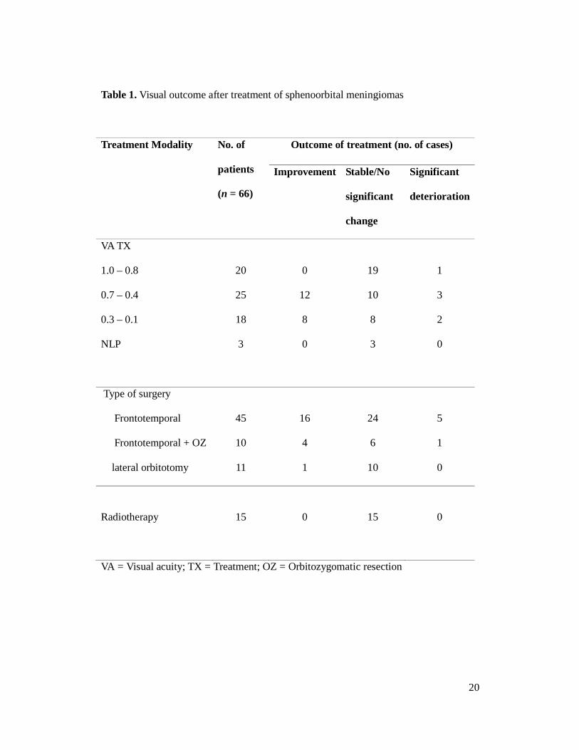
Table 1. Visual outcome after treatment of sphenoorbital meningiomas
Treatment Modality No. of
patients
(n = 66)
Outcome of treatment (no. of cases)
Improvement Stable/No
significant
change
Significant
deterioration
VA TX
1.0 – 0.8
0.7 – 0.4
0.3 – 0.1
NLP
20
25
18
3
0
12
8
0
19
10
8
3
1
3
2
0
Type of surgery
Frontotemporal
Frontotemporal + OZ
lateral orbitotomy
45
10
11
16
4
1
24
6
10
5
1
0
Radiotherapy
15
0
15
0
VA = Visual acuity; TX = Treatment; OZ = Orbitozygomatic resection
21
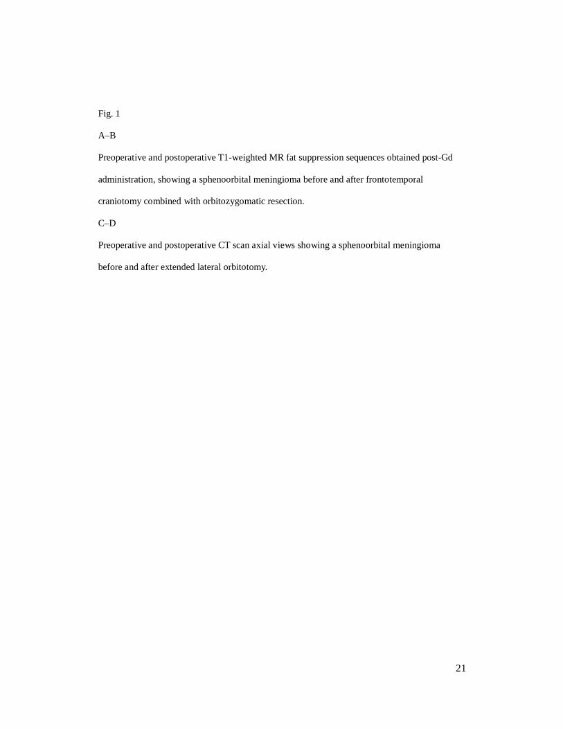
Fig. 1
A–B
Preoperative and postoperative T1-weighted MR fat suppression sequences obtained post-Gd
administration, showing a sphenoorbital meningioma before and after frontotemporal
craniotomy combined with orbitozygomatic resection.
C–D
Preoperative and postoperative CT scan axial views showing a sphenoorbital meningioma
before and after extended lateral orbitotomy.
22
Figure -2 Proptosis reduction in three different surgical approaches
Change in proptosis (post minus pre perative score)
-7
-6
-5
-4
-3
-2
-1
0
1 2 3
Median (IQR)
1.Frontotemporal craniotomy 2. Frontotemporal craniotomy combined with orbitozygomatic resection 3. Extended lateral orbitotomy

23