Embed Size (px)
Citation preview
Synaptic and Nuclear Localization of Brain-Enriched GuanylateKinase-Associated Protein
Ikuko Yao, Junko Iida, Wataru Nishimura, and Yutaka Hata
Department of Medical Biochemistry, Graduate School of Medicine, Tokyo Medical and Dental University, Bunkyo-ku,Tokyo 113–8519, Japan
Brain-enriched guanylate kinase-associated protein (BEGAIN)interacts with postsynaptic density (PSD)-95/synapse-associated protein (SAP) 90. In immunohistochemistry and im-munocytochemistry, BEGAIN was detected in nuclei and atsynapses in neurons. Nuclear localization was also confirmedthrough subcellular fractionation. BEGAIN was localized exclu-sively in nuclei when expressed in epithelial cells. These find-ings led us to analyze the mechanism to determine the subcel-lular localization of BEGAIN in neurons. Green fluorescentprotein (GFP)-tagged BEGAIN appeared first in nuclei and sub-sequently accumulated at dendrites. Approximately 75 and90% of GFP-BEGAIN clusters were colocalized with synapto-physin and PSD-95/SAP90, respectively. GFP-protein contain-ing only the N-terminal region also formed foci in nuclei and
clusters at dendrites. The N-terminal BEGAIN was not preciselytargeted to synapses, although it was partially localized atsynapses, possibly through dimer formation with endogenousBEGAIN. The truncated form of PSD-95/SAP90 containing theguanylate kinase domain blocked synaptic targeting of BEGAINbut did not affect cluster formation at dendrites. NMDA receptorantagonists blocked localization of GFP-BEGAIN at synapsesbut did not affect recruitment to dendrites. These results sug-gest that BEGAIN is recruited to dendrites by the N-terminalregion independently of NMDA receptor activity and that syn-aptic targeting of BEGAIN depends on NMDA receptor activityand may be mediated by interaction with PSD-95/SAP90.
Key words: BEGAIN; PSD-95/SAP90; NMDA receptor; syn-apse; nucleus; dendrite
NMDA receptors are ionotropic glutamate receptors that playessential roles for synaptic plasticity. Recent studies have revealedthat various proteins interact either directly or indirectly withNMDA receptors and are involved in the accumulation of thereceptors at postsynaptic sites and in signals triggered by neuro-transmission. To clarify the molecular mechanism of synaptogen-esis and synaptic remodeling of excitatory synapses, it is impor-tant to understand the biochemical and biophysical characters ofproteins associated with NMDA receptors. Postsynaptic density(PSD)-95/synapse-associated protein (SAP) 90 is the prototypicsynaptic membrane-associated protein that induces clustering ofNMDA receptors and binds signaling molecules, including ty-rosine kinase and regulators for small GTP-binding proteins(Cho et al., 1992; Kistner et al., 1993; Kornau et al., 1995; Chenet al., 1998; Kim et al., 1998; Hata and Takai, 1999; Tezuka et al.,1999). PSD-95/SAP90 also binds a cytoskeleton adapter protein,guanylate kinase-associated protein (GKAP) (also called synapse-associated protein 90/postsynaptic density-95-associated proteinand discs large tumor suppressor protein-associated protein), andthe complex of PSD-95/SAP90 and GKAP links NMDA recep-tors to the cytoskeleton (Kim et al., 1996; Naisbitt et al., 1997;
Satoh et al., 1997; Takeuchi et al., 1997; Hirao et al., 2000a). Weidentified BEGAIN as a PSD-95/SAP90-binding protein (Degu-chi et al., 1998). BEGAIN has two isoforms, BEGAIN1 andBEGAIN2, that are different in the N terminus. BEGAIN1 startswith MGSDQQSSQ and BEGAIN2 starts with MWTG-GRRPGRLRRA (single letters indicate amino acids). After theseamino acids, BEGAIN1 and BEGAIN2 have an identical se-quence. BEGAIN forms a complex with PSD-95/SAP90 andGKAP and is a member of NMDA receptor-associated proteins.These synaptic components are present in mature synapses; how-ever, during synaptogenesis, some components should come tosynapses first and others later. We are doing a series of studies onsynaptic membrane-associated proteins to determine which regionof each molecule is involved in interacting with other components,which region is necessary and sufficient for synaptic localization,and which molecular interaction depends on synaptic activity.These studies will shed light on which proteins play key roles in theassembly of synaptic components.
In this paper, we first report that BEGAIN is localized not onlyat synapses but also in nuclei in neurons through immunohisto-chemistry and subcellular fractionation. BEGAIN is localizedonly in nuclei in epithelial cells, when expressed exogenously. Wesuppose that neurons have a mechanism to determine the ex-tranuclear localization of BEGAIN. To clarify the mechanism,we have examined which regions of BEGAIN are involved innuclear and extranuclear localization and whether extranuclearlocalization of BEGAIN depends on NMDA receptor activity.BEGAIN has two putative nuclear localization signals in theN-terminal region. Consistently, the N-terminal region is in-volved in nuclear localization. The N-terminal region also medi-ates recruitment from nuclei to dendrites, but the N-terminalregion is not sufficient for synaptic localization. The truncated
Received March 13, 2002; revised March 13, 2002; accepted March 22, 2002.This study was supported by grants-in-aids for Scientific Research and Special
Coordination Funds for Promoting Science and Technology from the Ministry ofEducation, Culture, Sports, Science, and Technology, a grant from NOVARTISFoundation (Japan) for the Promotion of Science, and a grant from YamanouchiFoundation for Research on Metabolic Disorders. We thank Prof. Y. Takai (OsakaUniversity, Osaka, Japan) for critically reading this manuscript, Prof. A. Lamond(University of Dundee, Dundee, UK) for valuable advice, and Dr. Gary Nolan(Stanford University) for phoenix ampho cells.
Correspondence should be addressed to Yutaka Hata, Department of MedicalBiochemistry, Graduate School of Medicine, Tokyo Medical and Dental University,1-5-45 Yushima, Bunkyo-ku, Tokyo 113-8519, Japan. E-mail: [email protected] © 2002 Society for Neuroscience 0270-6474/02/225354-11$15.00/0
The Journal of Neuroscience, July 1, 2002, 22(13):5354–5364
form of PSD-95/SAP90 containing the guanylate kinase (GK)domain blocks synaptic targeting of BEGAIN. These findingssuggest that interaction with PSD-95/SAP90 is involved insynaptic targeting of BEGAIN. Furthermore, recruitment ofBEGAIN from nuclei to dendrites does not depend on NMDAreceptor activity, but targeting to synapses does depend onNMDA receptor activity.
MATERIALS AND METHODSPlasmid construction. Various expression vectors were constructed byconventional molecular biology techniques and the PCR method usingpLGFPC (Clontech, Palo Alto, CA), pSindRep5 (Invitrogen, Carlsbad,CA), pGex5X-3 (Amersham Biosciences, Piscataway, NJ), and pClneoMyc. pClneo Myc BEGAIN1, BEGAIN2, and pcDNA BEGAIN havebeen described previously (Deguchi et al., 1998). A linker was ligated toHindIII /BamHI sites of pLGFPC to generate pLGFPC-2 with additionalcloning sites. The PCR product (sense primer, actagttttggcaccaaaatcaacg;antisense primer, gcatgcacgcgtgacgtctctagacttgtacagctcgtcca; and tem-plate, pLGFPC) was digested in SpeI /SphI and ligated into XbaI /SphIsites of pSindRep5 to generate pSind GFP. pSind GFP BEGAIN-1, -2,-3, and -4 were constructed from pSind GFP and contain the amino acidresidues 1–611, 1–226, 216–415, and 407–611 of BEGAIN2 (GenBankaccession number NM024163), respectively. pGex 5X-3 BEGAIN-8 con-tained the amino acid residues 20–226, which are common for BEGAIN1and BEGAIN2. pLGFPC BEGAIN-2 and -4 were constructed frompLGFPC-2 and contain the same amino acid residues as pSind GFPBEGAIN-2 and -4. Oligonucleotides (ctagccccccaacatggagcagaaacttat-cagcgaggaggacctgacgcgtctagag and catgctctagacgcgtcaggtcctcctcgctgataagtttctgctccatgttgggggg) were phosphorylated, annealed, and ligated toXbaI /SphI sites of pSindRep5 to generate pSind Myc. pSind Myc PSD-95–1 and -2 contain the amino acid residues 1–724 and 294–724 ofPSD-95/SAP90, respectively. pCMV Myc PSD-95–1 and pClneo MycPSD-95–4 contain the amino acid residues 1–724 and 435–724 of PSD-95/SAP90, respectively.
Antibodies. The antibody against the C terminus of BEGAIN (anti-BEGAIN-C) has been described previously (Deguchi et al., 1998). Therabbit anti-BEGAIN-N antibody was raised against the product ofpGex5X-3 BEGAIN-8. Sheep polyclonal anti-Myc antibody was raisedagainst the synthetic peptide, Glu-Gln-Lys-I le-Ser-Glu-Glu-Asp-Leu-Asn-Ser-Ala-Val-Asp. Mouse monoclonal anti-synaptophysin and anti-GFP antibodies were obtained from Roche Molecular Biochemicals(Mannheim, Germany) and Clontech (Palo Alto, CA), respectively.Mouse monoclonal anti-PSD-95/SAP90 (K28/86.2) antibodies were pur-chased from Upstate Biotechnology (Lake Placid, NY). The secondaryantibodies for dual labeling were obtained from Chemicon International(Temecula, CA).
Cell cultures and stable transformants. Phoenix ampho, Madin-Darbycanine kidney, HeLa, and COS cells were cultured in DMEM supple-mented with 10% FBS, 100 U/ml penicillin, and 100 �g/ml streptomycin.For normal rat kidney cells, calf serum was used instead of FBS, andnonessential amino acids were added. Baby hamster kidney cells werecultured in MEM supplemented with 5% FBS, 100 U/ml penicillin, and100 �g/ml streptomycin. For the glycerol density gradient experiment,we used stable transformants of HeLa cells expressing GFP-BEGAIN-2and -4. Phoenix ampho cells were transfected with pLGFP BEGAIN-2and -4 using the Mammalian Transfection Kit (Stratagene, La Jolla, CA).The medium was collected 2 d after transfection and used to infect HeLacells. To generate stable cell lines, the infected cells were cultured in themedium containing 1 mg/ml Geneticin (Sigma-Aldrich, St. Louis, MO).
Hippocampal neuron culture and hippocampal slice culture. All proce-dures related to the care and treatment of animals were in accordancewith institutional and National Institutes of Health guidelines. Hip-pocampal neuron cultures were performed from embryonic day 18 em-bryos as described previously (Takeuchi et al., 1997; Goslin et al., 1998).Hippocampal slice was obtained from postnatal day (P) 6 or P8 rats andcultured on Millicell CM culture plate inserts (Millipore, Bedford, MA)in MEM containing 25% (v/v) HBSS, 6.5 gm/l glucose, 100 U/ml peni-cillin, 100 �g/ml streptomycin, and 25% (v/v) horse serum at 32°C under5% CO2. To transfect primary cultured hippocampal neurons,endotoxin-free plasmids were prepared with the EndoFree Plasmid Kit(Qiagen, Hilden, Germany), and 0.2 �g of DNA was transfected usingEffectene Transfection Reagent (Qiagen) to neurons 3 d after plating.Seven days after transfection, neurons were fixed and immunostainedwith the appropriate antibodies.
Immunofluorescence and immunohistochemistry. Hippocampal neuronswere fixed with 4% (w/v) paraformaldehyde for 15 min, blocked with 50mM glycine in PBS for 30 min, and permeabilized with 0.25 (w/v) %Triton X-100 in PBS for 5 min. Alternatively, hippocampal neurons werefixed in ice-cold methanol for 20 min at �20°C as described (Allison etal., 2000). After cells were blocked with PBS containing 10% (w/v) BSA,they were incubated with the first antibody in PBS containing 3% (w/v)BSA overnight, washed with PBS, and incubated with the second anti-body in PBS containing 3% (w/v) BSA for 1 hr. After the samples werewashed with PBS, they were embedded in 95% (w/v) glycerol in PBS.Immunohistochemical studies were performed as described (Lee et al.,1998). Wistar rats (4 weeks old) were deeply anesthetized with sodiumpentobarbital (60 mg/kg, i.p.) and perfused with 4% (w/v) paraformal-dehyde in 0.1 M phosphate buffer (PB), pH 7.4. Brains were removed andpostfixed in the same fixative, immersed with 10% (w/v), 20% (w/v), and30% (w/v) sucrose in 0.1 M PB, pH 7.4, sequentially, and frozen at �80°C.Then, 5 �m sections were prepared, washed in 0.1 M PB, blocked with 0.1M PB containing 5% goat serum and 0.2% (w/v) Triton X-100 for 2 hr,and incubated with the first antibodies at 4°C overnight. After they werewashed with 0.1 M PB four times, bound antibodies were detected with thesecond antibodies at room temperature for 3 hr. Then, the samples werecounterstained with 0.1 �g/ml Hoechst 33342 for 10 min when indicated,washed with 0.1 M PB, and embedded in 50% (w/v) glycerol in 0.1 M PB.For methanol fixation, brains were quickly removed from decapitated ratsand frozen in powdered dry ice. Sections were cut at 10 �m thickness on acryostat (Leica CM1800), mounted on aminopropyltriethoxysilane-coatedglass slides (Matsunami Glass, Osaka, Japan), fixed in ice-cold methanol for20 min at �20°C, and dried with a stream of cold air. The images wereobtained by confocal microscopies (Olympus FV300-BX and ZeissLSM510).
Isolation of nuclei f rom rat brains. Isolation of nuclei from rat brainsand immunostaining of the nuclear fraction were performed according tothe previously reported protocols with modifications (Wu et al., 1995;Rickwood et al., 1997) (see Fig. 2 A). Rat brains were removed andhomogenized in 9 vol of homogenizing buffer [0.25 M sucrose, 5 mMMgCl2, 10 mM Tris/HCl, pH 7.4, 1 mM phenylmethylsulfonyl fluoride(PMSF)] in a Potter-Elvehjem homogenizer using eight to nine strokesdriven at 1000 rpm. After the homogenate was filtered through fourlayers of gauze, it was centrifuged at 600 � g for 10 min at 5°C. The pellet(P1) was resuspended in half the original volume of the homogenizingbuffer and centrifuged again. The pellet of crude nuclei was resuspendedand homogenized in 9 vol of 2.2 M sucrose, 1 mM MgCl2, 10 mMTris/HCl, pH 7.4, 1 mM PMSF in a Potter-Elvehjem homogenizer usingfive to six strokes driven at 1000 rpm. The suspension of nuclei wascentrifuged at 80,000 � g for 80 min at 5°C in a swing-bucket rotor. Theresulting mixed membrane (M.M.) fraction occurring as a top layer wasremoved, and the pellet was collected as isolated nuclei (P2). Then,freshly isolated nuclei were fixed in ice-cold 4% (w/v) paraformaldehydein PBS for 30 min at 4°C and were applied to poly-L-lysine-coated coverglasses for 30 min at room temperature. After they were washed oncewith 50 mM glycine in PBS, the nuclei were incubated with 5% (w/v) goatserum, 1.5% (w/v) BSA, and 1.5% (w/v) Triton X-100 in PBS for 20 minand were incubated with the first antibodies at 4°C overnight. After thenuclei were washed with PBS, they were incubated with second antibod-ies for 2 hr at room temperature, stained with 0.1 �g/ml Hoechst 33342for 10 min, and embedded in 95% glycerol (w/v) in PBS.
Subcellular f ractionation of HeLa cells. HeLa cells were treated with0.25% (w/v) trypsin and 1 mM EDTA for 5 min at 37°C and collected.The cells were washed with ice-cold PBS twice and resuspended in a 0.5ml/10 cm dish of TM-2 buffer (10 mM Tris/HCl, pH 7.4, with 2 mMMgCl2 and 0.5 mM PMSF). The resuspended cells stood at room tem-perature for 1 min and were incubated in ice-water for 5 min. TritonX-100 was added to a final concentration of 0.5% (w/v). The cells wereincubated in ice-water for an additional 5 min and sheared by threepassages through a 22 gauge needle. The nuclei were examined in aphase-contrast microscope and isolated from the cytosol by centrifuga-tion at 980 � g for 10 min at 4°C. The pellet was rinsed with 0.5 ml ofTM-2 buffer twice and designated as the nuclear fraction.
Sindbis virus production and infection. Capped in vitro transcripts andhelper RNA were synthesized from various linearized pSind GFP con-structs and DH(26S) template (Invitrogen) using a RiboMAX LargeScale RNA production system (Promega, Madison, WI) and transfectedinto baby hamster kidney cells by electroporation with a GenePulser(Bio-Rad, Hercules, CA). Two days later, the medium was collected andcentrifuged at 400 � g for 5 min; the supernatant was centrifuged at
Yao et al. • Synaptic and Nuclear Localization of BEGAIN J. Neurosci., July 1, 2002, 22(13):5354–5364 5355
113,000 � g for 90 min. The pellet was collected, resuspended in 200 �lof the medium, and stocked at �80°C. Primary cultured hippocampalneurons were infected using 3–5 �l of the virus stock per 500 �l of theculture medium 10 d after plating. For double infection, 3–5 �l of eachvirus stock was added to 500 �l of the culture medium at the same time.Hippocampal slices were infected using 1 �l of the virus stock per eachslice 7 d after plating. All experiments using primary cultured hippocam-pal neurons were repeated independently at least three times.
Image acquisition and quantification. For quantification of cluster num-ber and size, primary cultured hippocampal neurons were observed 24 hrafter the infection, and confocal images were obtained using an OlympusFV300-BX 40� objective with sequential acquisition at 1024 � 1024pixels resolution and then converted to 512 � 512 pixels resolution. Eachimage was averaged four times and taken with the confocal aperture setat 3. Neurons expressing GFP-BEGAIN-1 or -2 were chosen at randomfrom two to three cover glasses from three independent preparations.Measurements were performed using NIH image 1.61. We definedsignals as clusters if they had the following properties: (1) peak fluores-cence levels 50% greater than the maximal fluorescence levels of diffusedendritic signals in the vicinity; (2) 3–30 pixels in size; and (3) locationbetween 10 and 100 �m from the soma. We counted the number from 700�m of the dendrites for each neuron. Diameters of clusters were calcu-lated from the areas of clusters.
Glycerol density gradient. HeLa cells expressing GFP-BEGAIN-2 or -4of three 10 cm plates were homogenized in 0.4 ml of buffer A [20 mMHEPES/KOH, pH 7.4, 100 mM NaCl, 0.5% (w/v) Triton X-100, and 2%(v/v) glycerol], and centrifuged at 100,000 � g for 30 min. The superna-tant was charged on 4.4 ml of 5–25% (v/v) glycerol density gradient in
buffer A, overlaid with 0.1 ml of buffer A, and centrifuged at 4°C at100,000 � g for 17 hr. Eighteen fractions were collected from the gradientand analyzed by immunoblotting with the anti-GFP antibody.
Coimmunoprecipitation of BEGAIN-1 and –2. COS cells were trans-fected with pClneo Myc BEGAIN-1 and -2 using the DEAE-dextranmethod. Forty-eight hours later, the cells of two 10 cm plates werecollected and homogenized in 1 ml of 20 mM HEPES/NaOH, pH 7.4,containing 100 mM NaCl and 1% (w/v) Triton X-100. After centrifuga-tion at 100,000 � g for 15 min at 4°C, the supernatant was collected, and0.45 ml of the supernatant was incubated with 10 �l of the anti-BEGAIN-C serum or the preimmune serum and precipitated with 7.5 �lof protein-G Sepharose 4 Fast Flow (Amersham Biosciences). Theprecipitates were immunoblotted with the anti-Myc antibody.
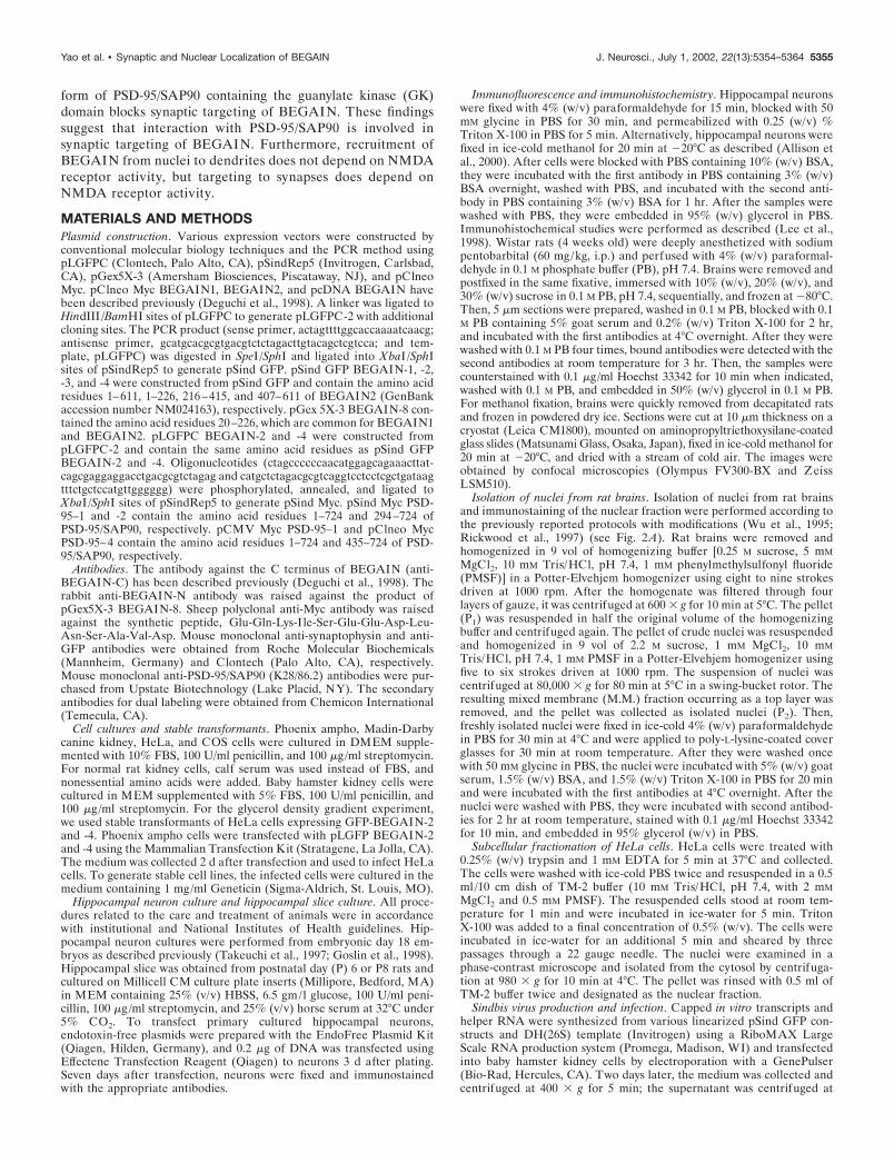
RESULTSSynaptic and nuclear localization of BEGAIN inrat brainsFirst, we detected BEGAIN in rat hippocampus with the affinity-purified anti-BEGAIN-N antibody using immunofluorescencemicroscopy. In paraformaldehyde-fixed sections, BEGAIN wasdetected around pyramidal neurons and colocalized with synap-tophysin (Fig. 1A). In methanol-fixed sections, BEGAIN showedfoci in pyramidal neuron nuclei (Fig. 1B). Immunohistochemistrywith the anti-BEGAIN-C antibody likewise showed synaptic lo-calization in paraformaldehyde-fixed sections and nuclear local-
Figure 1. Immunohistochemistry of BEGAINin rat hippocampus. A, Laser confocal image ofparaformaldehyde-fixed rat hippocampus CA1region. Rats were perfused with paraformalde-hyde, and brains were further postfixed. Sec-tions were stained with anti-BEGAIN-N andthe anti-synaptophysin antibodies. a, BEGAIN(arrows); b, synaptophysin (arrowheads); c, su-perimposed image. a2, b2, and c2 show thedemarcated areas in a1, b1, and c1 at highermagnification. Scale bars, 10 �m. B, Laser con-focal image of methanol-fixed rat hippocampusCA1 region. Rat brains were frozen in pow-dered dry ice. Sections were fixed with ice-coldmethanol and stained with anti-BEGAIN-Nand anti-synaptophysin antibodies and withHoechst 33342. a, BEGAIN (arrows); b, synap-tophysin (arrowheads); c, Hoechst 33342; d, su-perimposed image. a2, b2, c2, and d2 show thedemarcated areas in a1, b1, c1, and d1 at highermagnification. Scale bar, 10 �m.
5356 J. Neurosci., July 1, 2002, 22(13):5354–5364 Yao et al. • Synaptic and Nuclear Localization of BEGAIN
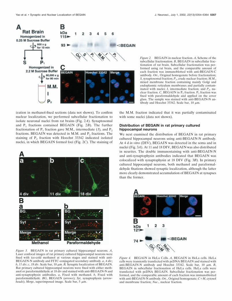
ization in methanol-fixed sections (data not shown). To confirmnuclear localization, we performed subcellular fractionation toisolate neuronal nuclei from rat brains (Fig. 2A). Synaptosomaland P1 fractions contained BEGAIN (Fig. 2B). The furtherfractionation of P1 fraction gave M.M., intermediate ( I), and P2
fractions. BEGAIN was detected in M.M. and P2 fractions. Thestaining of P2 fraction with Hoechst 33342 indicated isolatednuclei, in which BEGAIN formed foci (Fig. 2C). The staining of
the M.M. fraction indicated that it was partially contaminatedwith some nuclei (data not shown).
Distribution of BEGAIN in rat primary culturedhippocampal neuronsWe next examined the distribution of BEGAIN in rat primarycultured hippocampal neurons using anti-BEGAIN-N antibody.At 4 d in vitro (DIV), BEGAIN was detected in the soma and innuclei (Fig. 3A). At 11 and 18 DIV, BEGAIN was also distributedin neurites. The double immunostaining with anti-BEGAIN-Nand anti-synaptophysin antibodies indicated that BEGAIN wascolocalized with synaptophysin at 18 DIV (Fig. 3B). In primarycultured hippocampal neurons, both methanol and paraformal-dehyde fixations showed synaptic localization, although the lattermore clearly demonstrated accumulation of BEGAIN at synapsesthan the former.
Figure 3. BEGAIN in rat primary cultured hippocampal neurons. A,Laser confocal images of rat primary cultured hippocampal neurons werefixed with ice-cold methanol at various stages and stained with anti-BEGAIN-N antibody and FITC-conjugated secondary antibody. a, 4 div;b, 11 div; c, 18 div. Scale bar, 10 �m. B, Synaptic localization of BEGAIN.Rat primary cultured hippocampal neurons were fixed with either meth-anol or paraformaldehyde at 18 div and stained with anti-BEGAIN-N andanti-synaptophysin antibodies. a, Fixed with methanol. b, Fixed withparaformaldehyde. BG, BEGAIN (arrows); Syt, synaptophysin (arrow-heads); Merge, superimposed image. Scale bar, 5 �m.
Figure 4. BEGAIN in HeLa Cells. A, BEGAIN in HeLa cells. HeLacells were transiently transfected with pcDNA BEGAIN and stained withanti-BEGAIN-N antibody and Hoechst 33342. Scale bar, 10 �m. B,BEGAIN in subcellular fractionation of HeLa cells. HeLa cells weretransfected with pcDNA BEGAIN. Subcellular fractionation was per-formed, and the comparable amount of each fraction was immunoblottedwith anti-BEGAIN-N antibody. Ori., Original homogenate; C�M, cytosoland membrane fraction; Nuc., nuclear fraction.
Figure 2. BEGAIN in nuclear fraction. A, Scheme of thesubcellular fractionation. B, BEGAIN in subcellular frac-tionation of rat brain. Subcellular fractionation was per-formed using rat brain, and the comparable amount ofeach fraction was immunoblotted with anti-BEGAIN-Cantibody. Ori., Original homogenate before fractionation;S, synaptosomal fraction; P1, crude nuclear fraction; M.M.,mixed membrane fraction containing mainly Golgi andendoplasmic reticulum membranes and partially contam-inated with nuclei; I, intermediate fraction; and P2, nu-clear fraction. C, BEGAIN in P2 fraction. P2 fraction wasfixed with paraformaldehyde and applied on the coverglass. The sample was stained with anti-BEGAIN-N an-tibody and Hoechst 33342. Scale bar, 10 �m.
Yao et al. • Synaptic and Nuclear Localization of BEGAIN J. Neurosci., July 1, 2002, 22(13):5354–5364 5357
Distribution of BEGAIN in non-neuronal cellsOn the basis of the findings described above, we concluded thatBEGAIN was localized at synapses and in nuclei in neurons.BEGAIN was detected only in nuclei, however, when it was ex-pressed in HeLa cells (Fig. 4A). We examined the nuclear local-ization of BEGAIN through subcellular fractionation. BEGAINwas recovered in the nuclear fraction (Fig. 4B). To further confirmthat BEGAIN was not detected outside nuclei, we also expressedBEGAIN in other non-neuronal cells, including COS, Madin-Darby canine kidney, and normal rat kidney cells. In all these cells,BEGAIN was localized only in nuclei (data not shown). Thesefindings suggest that neurons have some mechanism to recruitBEGAIN outside nuclei.
GFP-BEGAIN-1 accumulates first in nuclei in neuronsand then at dendrites
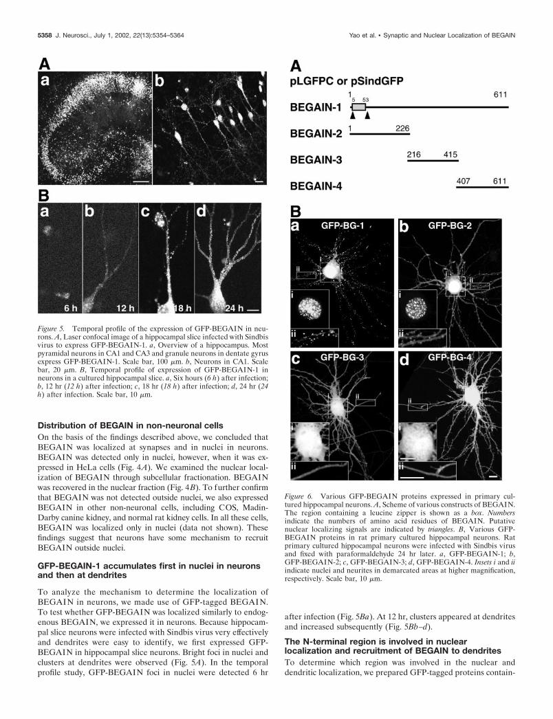
To analyze the mechanism to determine the localization ofBEGAIN in neurons, we made use of GFP-tagged BEGAIN.To test whether GFP-BEGAIN was localized similarly to endog-enous BEGAIN, we expressed it in neurons. Because hippocam-pal slice neurons were infected with Sindbis virus very effectivelyand dendrites were easy to identify, we first expressed GFP-BEGAIN in hippocampal slice neurons. Bright foci in nuclei andclusters at dendrites were observed (Fig. 5A). In the temporalprofile study, GFP-BEGAIN foci in nuclei were detected 6 hr
after infection (Fig. 5Ba). At 12 hr, clusters appeared at dendritesand increased subsequently (Fig. 5Bb–d).
The N-terminal region is involved in nuclearlocalization and recruitment of BEGAIN to dendritesTo determine which region was involved in the nuclear anddendritic localization, we prepared GFP-tagged proteins contain-
Figure 6. Various GFP-BEGAIN proteins expressed in primary cul-tured hippocampal neurons. A, Scheme of various constructs of BEGAIN.The region containing a leucine zipper is shown as a box. Numbersindicate the numbers of amino acid residues of BEGAIN. Putativenuclear localizing signals are indicated by triangles. B, Various GFP-BEGAIN proteins in rat primary cultured hippocampal neurons. Ratprimary cultured hippocampal neurons were infected with Sindbis virusand fixed with paraformaldehyde 24 hr later. a, GFP-BEGAIN-1; b,GFP-BEGAIN-2; c, GFP-BEGAIN-3; d, GFP-BEGAIN-4. Insets i and iiindicate nuclei and neurites in demarcated areas at higher magnification,respectively. Scale bar, 10 �m.
Figure 5. Temporal profile of the expression of GFP-BEGAIN in neu-rons. A, Laser confocal image of a hippocampal slice infected with Sindbisvirus to express GFP-BEGAIN-1. a, Overview of a hippocampus. Mostpyramidal neurons in CA1 and CA3 and granule neurons in dentate gyrusexpress GFP-BEGAIN-1. Scale bar, 100 �m. b, Neurons in CA1. Scalebar, 20 �m. B, Temporal profile of expression of GFP-BEGAIN-1 inneurons in a cultured hippocampal slice. a, Six hours (6 h) after infection;b, 12 hr (12 h) after infection; c, 18 hr (18 h) after infection; d, 24 hr (24h) after infection. Scale bar, 10 �m.
5358 J. Neurosci., July 1, 2002, 22(13):5354–5364 Yao et al. • Synaptic and Nuclear Localization of BEGAIN
ing various regions of BEGAIN (Fig. 6A). We expressed theseGFP-constructs in rat primary cultured hippocampal neurons.GFP-BEGAIN-2 containing the N-terminal region was accumu-lated in nuclei and at dendrites like GFP-BEGAIN-1 (Fig.6Ba,b). In contrast, GFP-BEGAIN-3 and -4 were distributeddiffusely in the soma and neurites and did not form clusters atdendrites (Fig. 6 Bc,d). These data indicate that the N-terminalregion of BEGAIN is involved in nuclear localization andrecruitment to dendrites in neurons. In HeLa and Madin-Darby canine kidney cells, the N-terminal region of BEGAINwas localized in nuclei and formed foci, suggesting that thenuclear localizing signal in this region determines the subcel-
lular localization of BEGAIN in non-neuronal cells (data notshown).
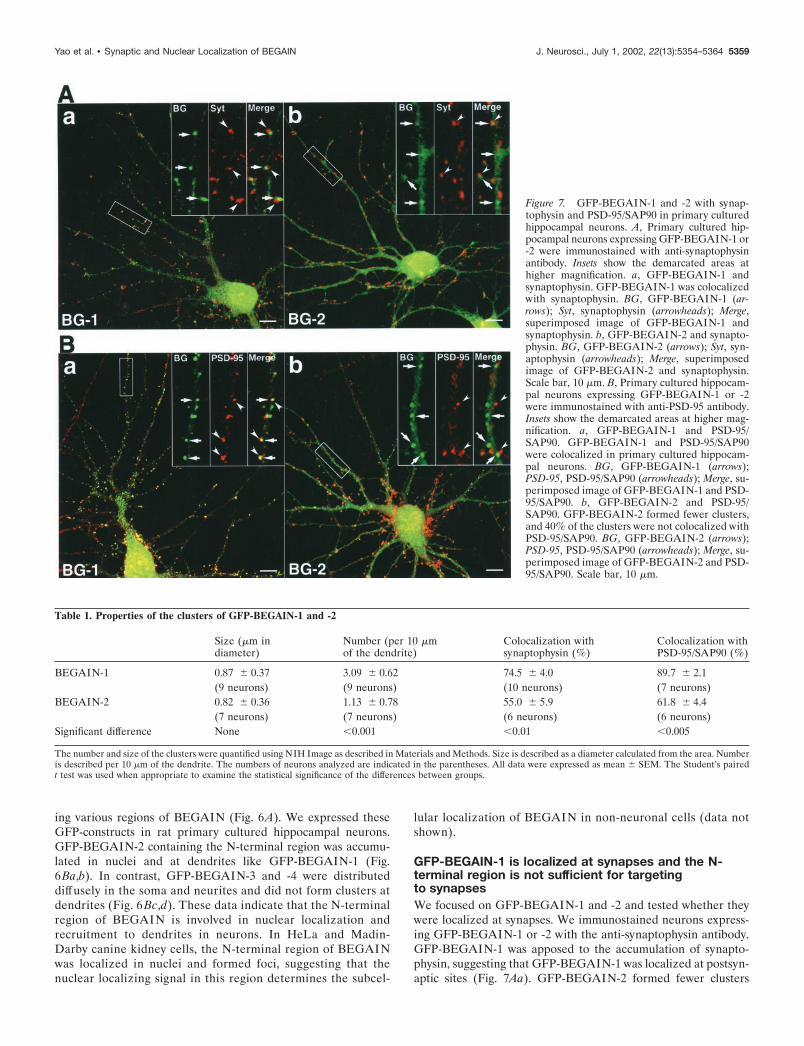
GFP-BEGAIN-1 is localized at synapses and the N-terminal region is not sufficient for targetingto synapsesWe focused on GFP-BEGAIN-1 and -2 and tested whether theywere localized at synapses. We immunostained neurons express-ing GFP-BEGAIN-1 or -2 with the anti-synaptophysin antibody.GFP-BEGAIN-1 was apposed to the accumulation of synapto-physin, suggesting that GFP-BEGAIN-1 was localized at postsyn-aptic sites (Fig. 7Aa). GFP-BEGAIN-2 formed fewer clusters
Figure 7. GFP-BEGAIN-1 and -2 with synap-tophysin and PSD-95/SAP90 in primary culturedhippocampal neurons. A, Primary cultured hip-pocampal neurons expressing GFP-BEGAIN-1 or-2 were immunostained with anti-synaptophysinantibody. Insets show the demarcated areas athigher magnification. a, GFP-BEGAIN-1 andsynaptophysin. GFP-BEGAIN-1 was colocalizedwith synaptophysin. BG, GFP-BEGAIN-1 (ar-rows); Syt, synaptophysin (arrowheads); Merge,superimposed image of GFP-BEGAIN-1 andsynaptophysin. b, GFP-BEGAIN-2 and synapto-physin. BG, GFP-BEGAIN-2 (arrows); Syt, syn-aptophysin (arrowheads); Merge, superimposedimage of GFP-BEGAIN-2 and synaptophysin.Scale bar, 10 �m. B, Primary cultured hippocam-pal neurons expressing GFP-BEGAIN-1 or -2were immunostained with anti-PSD-95 antibody.Insets show the demarcated areas at higher mag-nification. a, GFP-BEGAIN-1 and PSD-95/SAP90. GFP-BEGAIN-1 and PSD-95/SAP90were colocalized in primary cultured hippocam-pal neurons. BG, GFP-BEGAIN-1 (arrows);PSD-95, PSD-95/SAP90 (arrowheads); Merge, su-perimposed image of GFP-BEGAIN-1 and PSD-95/SAP90. b, GFP-BEGAIN-2 and PSD-95/SAP90. GFP-BEGAIN-2 formed fewer clusters,and 40% of the clusters were not colocalized withPSD-95/SAP90. BG, GFP-BEGAIN-2 (arrows);PSD-95, PSD-95/SAP90 (arrowheads); Merge, su-perimposed image of GFP-BEGAIN-2 and PSD-95/SAP90. Scale bar, 10 �m.
Table 1. Properties of the clusters of GFP-BEGAIN-1 and -2
Size (�m indiameter)
Number (per 10 �mof the dendrite)
Colocalization withsynaptophysin (%)
Colocalization withPSD-95/SAP90 (%)
BEGAIN-1 0.87 � 0.37 3.09 � 0.62 74.5 � 4.0 89.7 � 2.1(9 neurons) (9 neurons) (10 neurons) (7 neurons)
BEGAIN-2 0.82 � 0.36 1.13 � 0.78 55.0 � 5.9 61.8 � 4.4(7 neurons) (7 neurons) (6 neurons) (6 neurons)
Significant difference None �0.001 �0.01 �0.005
The number and size of the clusters were quantified using NIH Image as described in Materials and Methods. Size is described as a diameter calculated from the area. Numberis described per 10 �m of the dendrite. The numbers of neurons analyzed are indicated in the parentheses. All data were expressed as mean � SEM. The Student’s pairedt test was used when appropriate to examine the statistical significance of the differences between groups.
Yao et al. • Synaptic and Nuclear Localization of BEGAIN J. Neurosci., July 1, 2002, 22(13):5354–5364 5359
than GFP-BEGAIN-1 (Fig. 7Ab). The numbers and sizes ofclusters formed by GFP-BEGAIN-1 and -2 are summarized inTable 1. Forty-five percent of GFP-BEGAIN-2 clusters were notcolocalized with synaptophysin. We also determined what popu-lation of GFP-BEGAIN-1 or -2 clusters was associated withPSD-95/SAP90. Almost 90% of GFP-BEGAIN-1 clusters werecolocalized with PSD-95/SAP90 (Fig. 7Ba). In contrast, 40% ofGFP-BEGAIN-2 clusters were not associated with PSD-95/SAP90 (Fig. 7Bb). These findings suggest that the N-terminalregion of BEGAIN is not sufficient for targeting to synapses.
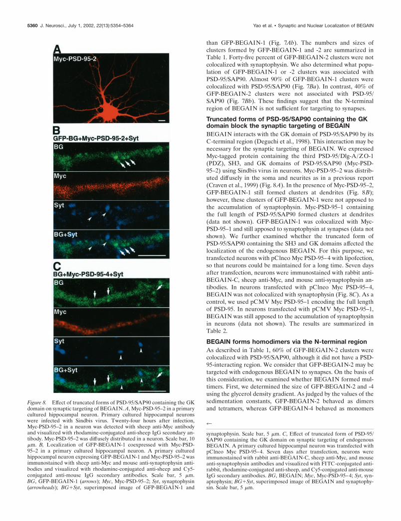
Truncated forms of PSD-95/SAP90 containing the GKdomain block the synaptic targeting of BEGAINBEGAIN interacts with the GK domain of PSD-95/SAP90 by itsC-terminal region (Deguchi et al., 1998). This interaction may benecessary for the synaptic targeting of BEGAIN. We expressedMyc-tagged protein containing the third PSD-95/Dlg-A/ZO-1(PDZ), SH3, and GK domains of PSD-95/SAP90 (Myc-PSD-95–2) using Sindbis virus in neurons. Myc-PSD-95–2 was distrib-uted diffusely in the soma and neurites as in a previous report(Craven et al., 1999) (Fig. 8A). In the presence of Myc-PSD-95–2,GFP-BEGAIN-1 still formed clusters at dendrites (Fig. 8B);however, these clusters of GFP-BEGAIN-1 were not apposed tothe accumulation of synaptophysin. Myc-PSD-95–1 containingthe full length of PSD-95/SAP90 formed clusters at dendrites(data not shown). GFP-BEGAIN-1 was colocalized with Myc-PSD-95–1 and still apposed to synaptophysin at synapses (data notshown). We further examined whether the truncated form ofPSD-95/SAP90 containing the SH3 and GK domains affected thelocalization of the endogenous BEGAIN. For this purpose, wetransfected neurons with pClneo Myc PSD-95–4 with lipofection,so that neurons could be maintained for a long time. Seven daysafter transfection, neurons were immunostained with rabbit anti-BEGAIN-C, sheep anti-Myc, and mouse anti-synaptophysin an-tibodies. In neurons transfected with pClneo Myc PSD-95–4,BEGAIN was not colocalized with synaptophysin (Fig. 8C). As acontrol, we used pCMV Myc PSD-95–1 encoding the full lengthof PSD-95. In neurons transfected with pCMV Myc PSD-95–1,BEGAIN was still apposed to the accumulation of synaptophysinin neurons (data not shown). The results are summarized inTable 2.
BEGAIN forms homodimers via the N-terminal regionAs described in Table 1, 60% of GFP-BEGAIN-2 clusters werecolocalized with PSD-95/SAP90, although it did not have a PSD-95-interacting region. We consider that GFP-BEGAIN-2 may betargeted with endogenous BEGAIN to synapses. On the basis ofthis consideration, we examined whether BEGAIN formed mul-timers. First, we determined the size of GFP-BEGAIN-2 and -4using the glycerol density gradient. As judged by the values of thesedimentation constants, GFP-BEGAIN-2 behaved as dimersand tetramers, whereas GFP-BEGAIN-4 behaved as monomers
Figure 8. Effect of truncated forms of PSD-95/SAP90 containing the GKdomain on synaptic targeting of BEGAIN. A, Myc-PSD-95–2 in a primarycultured hippocampal neuron. Primary cultured hippocampal neuronswere infected with Sindbis virus. Twenty-four hours after infection,Myc-PSD-95–2 in a neuron was detected with sheep anti-Myc antibodyand visualized with rhodamine-conjugated anti-sheep IgG secondary an-tibody. Myc-PSD-95–2 was diffusely distributed in a neuron. Scale bar, 10�m. B, Localization of GFP-BEGAIN-1 coexpressed with Myc-PSD-95–2 in a primary cultured hippocampal neuron. A primary culturedhippocampal neuron expressing GFP-BEGAIN-1 and Myc-PSD-95–2 wasimmunostained with sheep anti-Myc and mouse anti-synaptophysin anti-bodies and visualized with rhodamine-conjugated anti-sheep and Cy5-conjugated anti-mouse IgG secondary antibodies. Scale bar, 5 �m.BG, GFP-BEGAIN-1 (arrows); Myc, Myc-PSD-95–2; Syt, synaptophysin(arrowheads); BG�Syt, superimposed image of GFP-BEGAIN-1 and
4
synaptophysin. Scale bar, 5 �m. C, Effect of truncated form of PSD-95/SAP90 containing the GK domain on synaptic targeting of endogenousBEGAIN. A primary cultured hippocampal neuron was transfected withpClneo Myc PSD-95–4. Seven days after transfection, neurons wereimmunostained with rabbit anti-BEGAIN-C, sheep anti-Myc, and mouseanti-synaptophysin antibodies and visualized with FITC-conjugated anti-rabbit, rhodamine-conjugated anti-sheep, and Cy5-conjugated anti-mouseIgG secondary antibodies. BG, BEGAIN; Myc, Myc-PSD-95–4; Syt, syn-aptophysin; BG�Syt, superimposed image of BEGAIN and synaptophy-sin. Scale bar, 5 �m.
5360 J. Neurosci., July 1, 2002, 22(13):5354–5364 Yao et al. • Synaptic and Nuclear Localization of BEGAIN
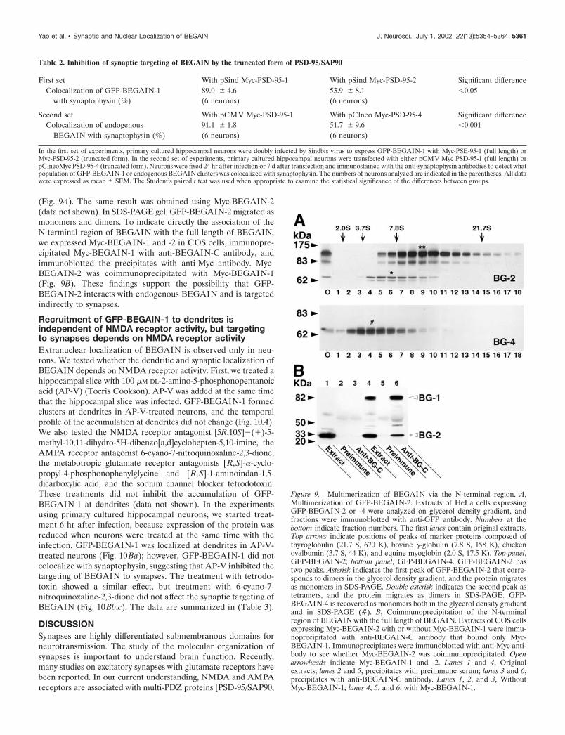
(Fig. 9A). The same result was obtained using Myc-BEGAIN-2(data not shown). In SDS-PAGE gel, GFP-BEGAIN-2 migrated asmonomers and dimers. To indicate directly the association of theN-terminal region of BEGAIN with the full length of BEGAIN,we expressed Myc-BEGAIN-1 and -2 in COS cells, immunopre-cipitated Myc-BEGAIN-1 with anti-BEGAIN-C antibody, andimmunoblotted the precipitates with anti-Myc antibody. Myc-BEGAIN-2 was coimmunoprecipitated with Myc-BEGAIN-1(Fig. 9B). These findings support the possibility that GFP-BEGAIN-2 interacts with endogenous BEGAIN and is targetedindirectly to synapses.
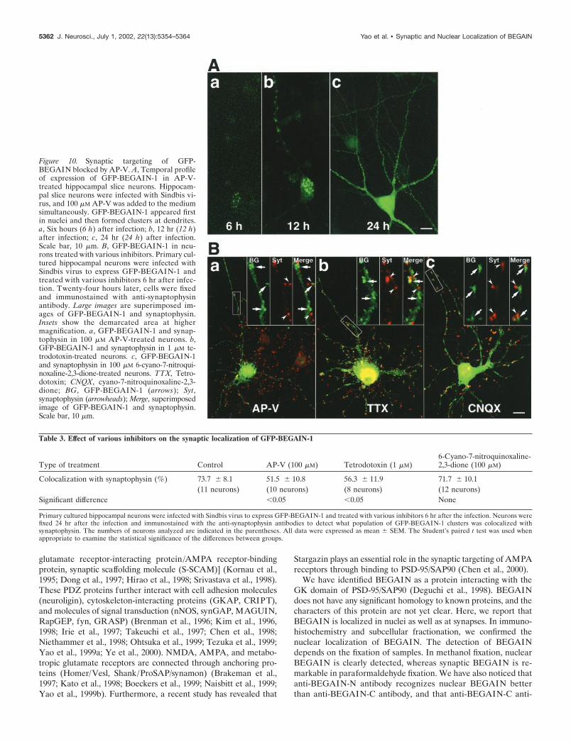
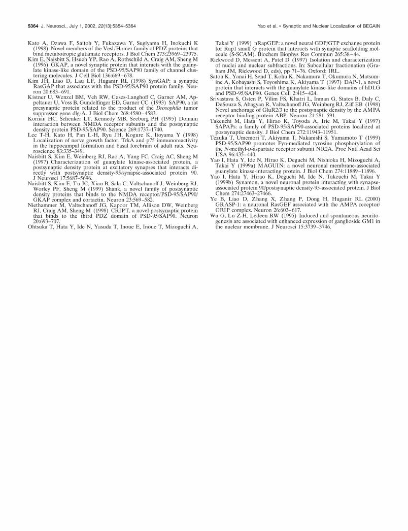
Recruitment of GFP-BEGAIN-1 to dendrites isindependent of NMDA receptor activity, but targetingto synapses depends on NMDA receptor activityExtranuclear localization of BEGAIN is observed only in neu-rons. We tested whether the dendritic and synaptic localization ofBEGAIN depends on NMDA receptor activity. First, we treated ahippocampal slice with 100 �M DL-2-amino-5-phosphonopentanoicacid (AP-V) (Tocris Cookson). AP-V was added at the same timethat the hippocampal slice was infected. GFP-BEGAIN-1 formedclusters at dendrites in AP-V-treated neurons, and the temporalprofile of the accumulation at dendrites did not change (Fig. 10A).We also tested the NMDA receptor antagonist [5R,10S]�(�)-5-methyl-10,11-dihydro-5H-dibenzo[a,d]cyclohepten-5,10-imine, theAMPA receptor antagonist 6-cyano-7-nitroquinoxaline-2,3-dione,the metabotropic glutamate receptor antagonists [R,S]-�-cyclo-propyl-4-phosphonophenylglycine and [R,S]-1-aminoindan-1,5-dicarboxylic acid, and the sodium channel blocker tetrodotoxin.These treatments did not inhibit the accumulation of GFP-BEGAIN-1 at dendrites (data not shown). In the experimentsusing primary cultured hippocampal neurons, we started treat-ment 6 hr after infection, because expression of the protein wasreduced when neurons were treated at the same time with theinfection. GFP-BEGAIN-1 was localized at dendrites in AP-V-treated neurons (Fig. 10Ba); however, GFP-BEGAIN-1 did notcolocalize with synaptophysin, suggesting that AP-V inhibited thetargeting of BEGAIN to synapses. The treatment with tetrodo-toxin showed a similar effect, but treatment with 6-cyano-7-nitroquinoxaline-2,3-dione did not affect the synaptic targeting ofBEGAIN (Fig. 10Bb,c). The data are summarized in (Table 3).
DISCUSSIONSynapses are highly differentiated submembranous domains forneurotransmission. The study of the molecular organization ofsynapses is important to understand brain function. Recently,many studies on excitatory synapses with glutamate receptors havebeen reported. In our current understanding, NMDA and AMPAreceptors are associated with multi-PDZ proteins [PSD-95/SAP90,
Figure 9. Multimerization of BEGAIN via the N-terminal region. A,Multimerization of GFP-BEGAIN-2. Extracts of HeLa cells expressingGFP-BEGAIN-2 or -4 were analyzed on glycerol density gradient, andfractions were immunoblotted with anti-GFP antibody. Numbers at thebottom indicate fraction numbers. The first lanes contain original extracts.Top arrows indicate positions of peaks of marker proteins composed ofthyroglobulin (21.7 S, 670 K), bovine �-globulin (7.8 S, 158 K), chickenovalbumin (3.7 S, 44 K), and equine myoglobin (2.0 S, 17.5 K). Top panel,GFP-BEGAIN-2; bottom panel, GFP-BEGAIN-4. GFP-BEGAIN-2 hastwo peaks. Asterisk indicates the first peak of GFP-BEGAIN-2 that corre-sponds to dimers in the glycerol density gradient, and the protein migratesas monomers in SDS-PAGE. Double asterisk indicates the second peak astetramers, and the protein migrates as dimers in SDS-PAGE. GFP-BEGAIN-4 is recovered as monomers both in the glycerol density gradientand in SDS-PAGE (#). B, Coimmunoprecipitation of the N-terminalregion of BEGAIN with the full length of BEGAIN. Extracts of COS cellsexpressing Myc-BEGAIN-2 with or without Myc-BEGAIN-1 were immu-noprecipitated with anti-BEGAIN-C antibody that bound only Myc-BEGAIN-1. Immunoprecipitates were immunoblotted with anti-Myc anti-body to see whether Myc-BEGAIN-2 was coimmunoprecipitated. Openarrowheads indicate Myc-BEGAIN-1 and -2. Lanes 1 and 4, Originalextracts; lanes 2 and 5, precipitates with preimmune serum; lanes 3 and 6,precipitates with anti-BEGAIN-C antibody. Lanes 1, 2, and 3, WithoutMyc-BEGAIN-1; lanes 4, 5, and 6, with Myc-BEGAIN-1.
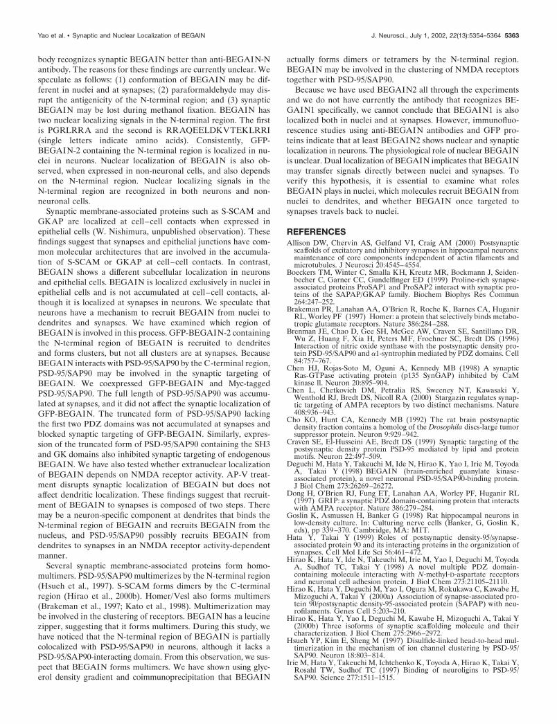
Table 2. Inhibition of synaptic targeting of BEGAIN by the truncated form of PSD-95/SAP90
First set With pSind Myc-PSD-95-1 With pSind Myc-PSD-95-2 Significant differenceColocalization of GFP-BEGAIN-1 89.0 � 4.6 53.9 � 8.1 �0.05
with synaptophysin (%) (6 neurons) (6 neurons)
Second set With pCMV Myc-PSD-95-1 With pClneo Myc-PSD-95-4 Significant differenceColocalization of endogenous 91.1 � 1.8 51.7 � 9.6 �0.001
BEGAIN with synaptophysin (%) (6 neurons) (6 neurons)
In the first set of experiments, primary cultured hippocampal neurons were doubly infected by Sindbis virus to express GFP-BEGAIN-1 with Myc-PSE-95-1 (full length) orMyc-PSD-95-2 (truncated form). In the second set of experiments, primary cultured hippocampal neurons were transfected with either pCMV Myc PSD-95-1 (full length) orpClneoMyc PSD-95-4 (truncated form). Neurons were fixed 24 hr after infection or 7 d after transfection and immunostained with the anti-synaptophysin antibodies to detect whatpopulation of GFP-BEGAIN-1 or endogenous BEGAIN clusters was colocalized with synaptophysin. The numbers of neurons analyzed are indicated in the parentheses. All datawere expressed as mean � SEM. The Student’s paired t test was used when appropriate to examine the statistical significance of the differences between groups.
Yao et al. • Synaptic and Nuclear Localization of BEGAIN J. Neurosci., July 1, 2002, 22(13):5354–5364 5361
glutamate receptor-interacting protein/AMPA receptor-bindingprotein, synaptic scaffolding molecule (S-SCAM)] (Kornau et al.,1995; Dong et al., 1997; Hirao et al., 1998; Srivastava et al., 1998).These PDZ proteins further interact with cell adhesion molecules(neuroligin), cytoskeleton-interacting proteins (GKAP, CRIPT),and molecules of signal transduction (nNOS, synGAP, MAGUIN,RapGEP, fyn, GRASP) (Brenman et al., 1996; Kim et al., 1996,1998; Irie et al., 1997; Takeuchi et al., 1997; Chen et al., 1998;Niethammer et al., 1998; Ohtsuka et al., 1999; Tezuka et al., 1999;Yao et al., 1999a; Ye et al., 2000). NMDA, AMPA, and metabo-tropic glutamate receptors are connected through anchoring pro-teins (Homer/Vesl, Shank/ProSAP/synamon) (Brakeman et al.,1997; Kato et al., 1998; Boeckers et al., 1999; Naisbitt et al., 1999;Yao et al., 1999b). Furthermore, a recent study has revealed that
Stargazin plays an essential role in the synaptic targeting of AMPAreceptors through binding to PSD-95/SAP90 (Chen et al., 2000).
We have identified BEGAIN as a protein interacting with theGK domain of PSD-95/SAP90 (Deguchi et al., 1998). BEGAINdoes not have any significant homology to known proteins, and thecharacters of this protein are not yet clear. Here, we report thatBEGAIN is localized in nuclei as well as at synapses. In immuno-histochemistry and subcellular fractionation, we confirmed thenuclear localization of BEGAIN. The detection of BEGAINdepends on the fixation of samples. In methanol fixation, nuclearBEGAIN is clearly detected, whereas synaptic BEGAIN is re-markable in paraformaldehyde fixation. We have also noticed thatanti-BEGAIN-N antibody recognizes nuclear BEGAIN betterthan anti-BEGAIN-C antibody, and that anti-BEGAIN-C anti-
Table 3. Effect of various inhibitors on the synaptic localization of GFP-BEGAIN-1
Type of treatment Control AP-V (100 �M) Tetrodotoxin (1 �M)6-Cyano-7-nitroquinoxaline-2,3-dione (100 �M)
Colocalization with synaptophysin (%) 73.7 � 8.1 51.5 � 10.8 56.3 � 11.9 71.7 � 10.1(11 neurons) (10 neurons) (8 neurons) (12 neurons)
Significant difference �0.05 �0.05 None
Primary cultured hippocampal neurons were infected with Sindbis virus to express GFP-BEGAIN-1 and treated with various inhibitors 6 hr after the infection. Neurons werefixed 24 hr after the infection and immunostained with the anti-synaptophysin antibodies to detect what population of GFP-BEGAIN-1 clusters was colocalized withsynaptophysin. The numbers of neurons analyzed are indicated in the parentheses. All data were expressed as mean � SEM. The Student’s paired t test was used whenappropriate to examine the statistical significance of the differences between groups.
Figure 10. Synaptic targeting of GFP-BEGAIN blocked by AP-V. A, Temporal profileof expression of GFP-BEGAIN-1 in AP-V-treated hippocampal slice neurons. Hippocam-pal slice neurons were infected with Sindbis vi-rus, and 100 �M AP-V was added to the mediumsimultaneously. GFP-BEGAIN-1 appeared firstin nuclei and then formed clusters at dendrites.a, Six hours (6 h) after infection; b, 12 hr (12 h)after infection; c, 24 hr (24 h) after infection.Scale bar, 10 �m. B, GFP-BEGAIN-1 in neu-rons treated with various inhibitors. Primary cul-tured hippocampal neurons were infected withSindbis virus to express GFP-BEGAIN-1 andtreated with various inhibitors 6 hr after infec-tion. Twenty-four hours later, cells were fixedand immunostained with anti-synaptophysinantibody. Large images are superimposed im-ages of GFP-BEGAIN-1 and synaptophysin.Insets show the demarcated area at highermagnification. a, GFP-BEGAIN-1 and synap-tophysin in 100 �M AP-V-treated neurons. b,GFP-BEGAIN-1 and synaptophysin in 1 �M te-trodotoxin-treated neurons. c, GFP-BEGAIN-1and synaptophysin in 100 �M 6-cyano-7-nitroqui-noxaline-2,3-dione-treated neurons. TTX, Tetro-dotoxin; CNQX, cyano-7-nitroquinoxaline-2,3-dione; BG, GFP-BEGAI N-1 (arrows); Syt,synaptophysin (arrowheads); Merge, superimposedimage of GFP-BEGAIN-1 and synaptophysin.Scale bar, 10 �m.
5362 J. Neurosci., July 1, 2002, 22(13):5354–5364 Yao et al. • Synaptic and Nuclear Localization of BEGAIN
body recognizes synaptic BEGAIN better than anti-BEGAIN-Nantibody. The reasons for these findings are currently unclear. Wespeculate as follows: (1) conformation of BEGAIN may be dif-ferent in nuclei and at synapses; (2) paraformaldehyde may dis-rupt the antigenicity of the N-terminal region; and (3) synapticBEGAIN may be lost during methanol fixation. BEGAIN hastwo nuclear localizing signals in the N-terminal region. The firstis PGRLRRA and the second is RRAQEELDKVTEKLRRI(single letters indicate amino acids). Consistently, GFP-BEGAIN-2 containing the N-terminal region is localized in nu-clei in neurons. Nuclear localization of BEGAIN is also ob-served, when expressed in non-neuronal cells, and also dependson the N-terminal region. Nuclear localizing signals in theN-terminal region are recognized in both neurons and non-neuronal cells.
Synaptic membrane-associated proteins such as S-SCAM andGKAP are localized at cell–cell contacts when expressed inepithelial cells (W. Nishimura, unpublished observation). Thesefindings suggest that synapses and epithelial junctions have com-mon molecular architectures that are involved in the accumula-tion of S-SCAM or GKAP at cell–cell contacts. In contrast,BEGAIN shows a different subcellular localization in neuronsand epithelial cells. BEGAIN is localized exclusively in nuclei inepithelial cells and is not accumulated at cell–cell contacts, al-though it is localized at synapses in neurons. We speculate thatneurons have a mechanism to recruit BEGAIN from nuclei todendrites and synapses. We have examined which region ofBEGAIN is involved in this process. GFP-BEGAIN-2 containingthe N-terminal region of BEGAIN is recruited to dendritesand forms clusters, but not all clusters are at synapses. BecauseBEGAIN interacts with PSD-95/SAP90 by the C-terminal region,PSD-95/SAP90 may be involved in the synaptic targeting ofBEGAIN. We coexpressed GFP-BEGAIN and Myc-taggedPSD-95/SAP90. The full length of PSD-95/SAP90 was accumu-lated at synapses, and it did not affect the synaptic localization ofGFP-BEGAIN. The truncated form of PSD-95/SAP90 lackingthe first two PDZ domains was not accumulated at synapses andblocked synaptic targeting of GFP-BEGAIN. Similarly, expres-sion of the truncated form of PSD-95/SAP90 containing the SH3and GK domains also inhibited synaptic targeting of endogenousBEGAIN. We have also tested whether extranuclear localizationof BEGAIN depends on NMDA receptor activity. AP-V treat-ment disrupts synaptic localization of BEGAIN but does notaffect dendritic localization. These findings suggest that recruit-ment of BEGAIN to synapses is composed of two steps. Theremay be a neuron-specific component at dendrites that binds theN-terminal region of BEGAIN and recruits BEGAIN from thenucleus, and PSD-95/SAP90 possibly recruits BEGAIN fromdendrites to synapses in an NMDA receptor activity-dependentmanner.
Several synaptic membrane-associated proteins form homo-multimers. PSD-95/SAP90 multimerizes by the N-terminal region(Hsueh et al., 1997). S-SCAM forms dimers by the C-terminalregion (Hirao et al., 2000b). Homer/Vesl also forms multimers(Brakeman et al., 1997; Kato et al., 1998). Multimerization maybe involved in the clustering of receptors. BEGAIN has a leucinezipper, suggesting that it forms multimers. During this study, wehave noticed that the N-terminal region of BEGAIN is partiallycolocalized with PSD-95/SAP90 in neurons, although it lacks aPSD-95/SAP90-interacting domain. From this observation, we sus-pect that BEGAIN forms multimers. We have shown using glyc-erol density gradient and coimmunoprecipitation that BEGAIN
actually forms dimers or tetramers by the N-terminal region.BEGAIN may be involved in the clustering of NMDA receptorstogether with PSD-95/SAP90.
Because we have used BEGAIN2 all through the experimentsand we do not have currently the antibody that recognizes BE-GAIN1 specifically, we cannot conclude that BEGAIN1 is alsolocalized both in nuclei and at synapses. However, immunofluo-rescence studies using anti-BEGAIN antibodies and GFP pro-teins indicate that at least BEGAIN2 shows nuclear and synapticlocalization in neurons. The physiological role of nuclear BEGAINis unclear. Dual localization of BEGAIN implicates that BEGAINmay transfer signals directly between nuclei and synapses. Toverify this hypothesis, it is essential to examine what rolesBEGAIN plays in nuclei, which molecules recruit BEGAIN fromnuclei to dendrites, and whether BEGAIN once targeted tosynapses travels back to nuclei.
REFERENCESAllison DW, Chervin AS, Gelfand VI, Craig AM (2000) Postsynaptic
scaffolds of excitatory and inhibitory synapses in hippocampal neurons:maintenance of core components independent of actin filaments andmicrotubules. J Neurosci 20:4545–4554.
Boeckers TM, Winter C, Smalla KH, Kreutz MR, Bockmann J, Seiden-becher C, Garner CC, Gundelfinger ED (1999) Proline-rich synapse-associated proteins ProSAP1 and ProSAP2 interact with synaptic pro-teins of the SAPAP/GKAP family. Biochem Biophys Res Commun264:247–252.
Brakeman PR, Lanahan AA, O’Brien R, Roche K, Barnes CA, HuganirRL, Worley PF (1997) Homer: a protein that selectively binds metabo-tropic glutamate receptors. Nature 386:284–288.
Brenman JE, Chao D, Gee SH, McGee AW, Craven SE, Santillano DR,Wu Z, Huang F, Xia H, Peters MF, Froehner SC, Bredt DS (1996)Interaction of nitric oxide synthase with the postsynaptic density pro-tein PSD-95/SAP90 and �1-syntrophin mediated by PDZ domains. Cell84:757–767.
Chen HJ, Rojas-Soto M, Oguni A, Kennedy MB (1998) A synapticRas-GTPase activating protein (p135 SynGAP) inhibited by CaMkinase ll. Neuron 20:895–904.
Chen L, Chetkovich DM, Petralia RS, Sweeney NT, Kawasaki Y,Wenthold RJ, Bredt DS, Nicoll RA (2000) Stargazin regulates synap-tic targeting of AMPA receptors by two distinct mechanisms. Nature408:936–943.
Cho KO, Hunt CA, Kennedy MB (1992) The rat brain postsynapticdensity fraction contains a homolog of the Drosophila discs-large tumorsuppressor protein. Neuron 9:929–942.
Craven SE, El-Husseini AE, Bredt DS (1999) Synaptic targeting of thepostsynaptic density protein PSD-95 mediated by lipid and proteinmotifs. Neuron 22:497–509.
Deguchi M, Hata Y, Takeuchi M, Ide N, Hirao K, Yao I, Irie M, ToyodaA, Takai Y (1998) BEGAIN (brain-enriched guanylate kinase-associated protein), a novel neuronal PSD-95/SAP90-binding protein.J Biol Chem 273:26269–26272.
Dong H, O’Brien RJ, Fung ET, Lanahan AA, Worley PF, Huganir RL(1997) GRIP: a synaptic PDZ domain-containing protein that interactswith AMPA receptor. Nature 386:279–284.
Goslin K, Asmussen H, Banker G (1998) Rat hippocampal neurons inlow-density culture. In: Culturing nerve cells (Banker, G, Goslin K,eds), pp 339–370. Cambridge, MA: MIT.
Hata Y, Takai Y (1999) Roles of postsynaptic density-95/synapse-associated protein 90 and its interacting proteins in the organization ofsynapses. Cell Mol Life Sci 56:461–472.
Hirao K, Hata Y, Ide N, Takeuchi M, Irie M, Yao I, Deguchi M, ToyodaA, Sudhof TC, Takai Y (1998) A novel multiple PDZ domain-containing molecule interacting with N-methyl-D-aspartate receptorsand neuronal cell adhesion protein. J Biol Chem 273:21105–21110.
Hirao K, Hata Y, Deguchi M, Yao I, Ogura M, Rokukawa C, Kawabe H,Mizoguchi A, Takai Y (2000a) Association of synapse-associated pro-tein 90/postsynaptic density-95-associated protein (SAPAP) with neu-rofilaments. Genes Cell 5:203–210.
Hirao K, Hata Y, Yao I, Deguchi M, Kawabe H, Mizoguchi A, Takai Y(2000b) Three isoforms of synaptic scaffolding molecule and theircharacterization. J Biol Chem 275:2966–2972.
Hsueh YP, Kim E, Sheng M (1997) Disulfide-linked head-to-head mul-timerization in the mechanism of ion channel clustering by PSD-95/SAP90. Neuron 18:803–814.
Irie M, Hata Y, Takeuchi M, Ichtchenko K, Toyoda A, Hirao K, Takai Y,Rosahl TW, Sudhof TC (1997) Binding of neuroligins to PSD-95/SAP90. Science 277:1511–1515.
Yao et al. • Synaptic and Nuclear Localization of BEGAIN J. Neurosci., July 1, 2002, 22(13):5354–5364 5363
Kato A, Ozawa F, Saitoh Y, Fukazawa Y, Sugiyama H, Inokuchi K(1998) Novel members of the Vesl /Homer family of PDZ proteins thatbind metabotropic glutamate receptors. J Biol Chem 273:23969–23975.
Kim E, Naisbitt S, Hsueh YP, Rao A, Rothschild A, Craig AM, Sheng M(1996) GKAP, a novel synaptic protein that interacts with the guany-late kinase-like domain of the PSD-95/SAP90 family of channel clus-tering molecules. J Cell Biol 136:669–678.
Kim JH, Liao D, Lau LF, Huganir RL (1998) SynGAP: a synapticRasGAP that associates with the PSD-95/SAP90 protein family. Neu-ron 20:683–691.
Kistner U, Wenzel BM, Veh RW, Cases-Langhoff C, Garner AM, Ap-peltauer U, Voss B, Gundelfinger ED, Garner CC (1993) SAP90, a ratpresynaptic protein related to the product of the Drosophila tumorsuppressor gene dlg-A. J Biol Chem 268:4580–4583.
Kornau HC, Schenker LT, Kennedy MB, Seeburg PH (1995) Domaininteraction between NMDA receptor subunits and the postsynapticdensity protein PSD-95/SAP90. Science 269:1737–1740.
Lee T-H, Kato H, Pan L-H, Ryu JH, Kogure K, Itoyama Y (1998)Localization of nerve growth factor, TrkA and p75 immunoreactivityin the hippocampal formation and basal forebrain of adult rats. Neu-roscience 83:335–349.
Naisbitt S, Kim E, Weinberg RJ, Rao A, Yang FC, Craig AC, Sheng M(1997) Characterization of guanylate kinase-associated protein, apostsynaptic density protein at excitatory synapses that interacts di-rectly with postsynaptic density-95/synapse-associated protein 90.J Neurosci 17:5687–5696.
Naisbitt S, Kim E, Tu JC, Xiao B, Sala C, Valtschanoff J, Weinberg RJ,Worley PF, Sheng M (1999) Shank, a novel family of postsynapticdensity proteins that binds to the NMDA receptor/PSD-95/SAP90/GKAP complex and cortactin. Neuron 23:569–582.
Niethammer M, Valtschanoff JG, Kapoor TM, Allison DW, WeinbergRJ, Craig AM, Sheng M (1998) CRIPT, a novel postsynaptic proteinthat binds to the third PDZ domain of PSD-95/SAP90. Neuron20:693–707.
Ohtsuka T, Hata Y, Ide N, Yasuda T, Inoue E, Inoue T, Mizoguchi A,
Takai Y (1999) nRapGEP: a novel neural GDP/GTP exchange proteinfor Rap1 small G protein that interacts with synaptic scaffolding mol-ecule (S-SCAM). Biochem Biophys Res Commun 265:38–44.
Rickwood D, Messent A, Patel D (1997) Isolation and characterizationof nuclei and nuclear subfractions. In: Subcellular fractionation (Gra-ham JM, Rickwood D, eds), pp 71–76. Oxford: IRL.
Satoh K, Yanai H, Send T, Kohu K, Nakamura T, Okumura N, Matsum-ine A, Kobayashi S, Toyoshima K, Akiyama T (1997) DAP-1, a novelprotein that interacts with the guanylate kinase-like domains of hDLGand PSD-95/SAP90. Genes Cell 2:415–424.
Srivastava S, Osten P, Vilim FS, Khatri L, Inman G, States B, Daly C,DeSouza S, Abagyan R, Valtschanoff JG, Weinberg RJ, Ziff EB (1998)Novel anchorage of GluR2/3 to the postsynaptic density by the AMPAreceptor-binding protein ABP. Neuron 21:581–591.
Takeuchi M, Hata Y, Hirao K, Toyoda A, Irie M, Takai Y (1997)SAPAPs: a family of PSD-95/SAP90-associated proteins localized atpostsynaptic density. J Biol Chem 272:11943–11951.
Tezuka T, Umemori T, Akiyama T, Nakanishi S, Yamamoto T (1999)PSD-95/SAP90 promotes Fyn-mediated tyrosine phosphorylation ofthe N-methyl-D-aspartate receptor subunit NR2A. Proc Natl Acad SciUSA 96:435–440.
Yao I, Hata Y, Ide N, Hirao K, Deguchi M, Nishioka H, Mizoguchi A,Takai Y (1999a) MAGUIN: a novel neuronal membrane-associatedguanylate kinase-interacting protein. J Biol Chem 274:11889–11896.
Yao I, Hata Y, Hirao K, Deguchi M, Ide N, Takeuchi M, Takai Y(1999b) Synamon, a novel neuronal protein interacting with synapse-associated protein 90/postsynaptic density-95-associated protein. J BiolChem 274:27463–27466.
Ye B, Liao D, Zhang X, Zhang P, Dong H, Huganir RL (2000)GRASP-1: a neuronal RasGEF associated with the AMPA receptor/GRIP complex. Neuron 26:603–617.
Wu G, Lu Z-H, Ledeen RW (1995) Induced and spontaneous neurito-genesis are associated with enhanced expression of ganglioside GM1 inthe nuclear membrane. J Neurosci 15:3739–3746.
5364 J. Neurosci., July 1, 2002, 22(13):5354–5364 Yao et al. • Synaptic and Nuclear Localization of BEGAIN