Embed Size (px)
Citation preview
Synergistic solubilization of porcine myosinin physiological salt solution by arginine
著者 Shiraki Kentarojournal orpublication title
International journal of biologicalmacromolecules
volume 62page range 647-651year 2013-10権利 (C) 2013 Elsevier B.V. NOTICE: this is the
author’s version of a work that was acceptedfor publication in International Journal ofBiological Macromolecules. Changes resultingfrom the publishing process, such as peerreview, editing, corrections, structuralformatting, and other quality controlmechanisms may not be reflected in thisdocument. Changes may have been made to thiswork since it was submitted for publication. Adefinitive version was subsequently publishedin International Journal of BiologicalMacromolecules, 62, 2013,http://dx.doi.org/10.1016/j.ijbiomac.2013.09.035.
URL http://hdl.handle.net/2241/120870doi: 10.1016/j.ijbiomac.2013.09.035
- 1 -
Synergistic Solubilization of Porcine Myosin in Physiological 1
Salt Solution by Arginine 2
3
Eisuke Takaia, Shunsuke Yoshizawa
a, Daisuke Ejima, Tsutomu Arakawa, and Kentaro 4
Shiraki* 5
6
a These authors equally contributed to this paper 7
––––––––– 8
E. Takai, S. Yoshizawa, K. Shiraki 9
Faculty of Pure and Applied Sciences, University of Tsukuba, 1-1-1 Tennodai, Tsukuba, 10
Ibaraki 305-8573, Japan. 11
12
D. Ejima 13
Institute for Innovation, Ajinomoto Co. Inc., Kanagawa 210-8681, Japan. 14
15
T. Arakawa 16
Alliance Protein Laboratories, San Diego, CA 92121, United States. 17
––––––––– 18
* To whom correspondence should be addressed. telephone: +81-29-8535306; fax: +81-29-19
8535215; E-mail: [email protected] 20
21
*ManuscriptClick here to view linked References
- 2 -
Abstract 1
Myosin is an important protein resource for food industries and has a bipolar filamentous 2
structure that is composed of subfilaments that occur in vivo. It has been shown that a high 3
ionic strength is required to prevent myosin from forming filamentous structures and to 4
solubilize the protein in aqueous solution. In the presence of 100 to 200 mM NaCl, 50 mM 5
arginine was more effective than other additives tested, including NaCl, in myosin 6
solubilization. Before reaching equilibrium solubility, the myosin solution was initially 7
supersaturated upon the dilution of a stock myosin solution in 1 M NaCl into the test solvents. 8
Arginine slowed the process of equilibration and stabilized the supersaturated solution more 9
effectively than other additives. No structural changes in myosin caused by arginine were 10
observed, which indicated that arginine enhanced the solubility of myosin in a physiological 11
salt solution without affecting the structure. 12
13
Keywords: myosin, arginine, supersaturation 14
15
- 3 -
1. Introduction 1
Meat is rich in high-quality proteins and contains all of the essential amino acids for 2
humans [1]. However, meat has not been fully utilized as a protein supplement to the same 3
extent that milk or soybean products have been utilized because of the low solubility of 4
myosin, which constitutes approximately 50 % of myofibrillar proteins. The low solubility of 5
myosin results from the spontaneous formation of filaments that occur in vivo [2–4]. Myosin 6
is practically insoluble in aqueous solution at low ionic strength but is increasingly soluble at 7
high salt concentrations [5–9]. 8
Solubilization of insoluble myosin has been achieved by adding 5 mM histidine in 9
low ionic strength solutions (1-5 mM KCl, pH 7.5) [10–12]. The mechanism by which 10
histidine solubilizes myosin appears to involve structural changes in monomeric myosin and 11
the resulting inhibition of native myosin filament formation [11]. When the salt concentration 12
is increased to a physiological level (0.15 M), 5 mM histidine no longer shows such effects 13
[12]. Considering the observed structural changes caused by histidine, which may limit 14
myosin application, it would be advantageous to achieve high myosin solubility without 15
structural changes even in physiological salt solutions. We have investigated several 16
additives, including histidine and arginine, for their effects on myosin solubility as a function 17
of salt concentration and on the myosin structure. 18
Arginine is one of the most common solvent additive that suppress protein 19
aggregation without altering or destabilizing the tertiary structure of the proteins [13,14]. 20
Arginine has been used in various applications, including suppression of reductant- or heat-21
induced aggregation [15–18], enhancement of protein refolding [17–22], crystallization [23], 22
and improved performance of column chromatography [24–27]. The molecular mechanisms 23
by which arginine suppresses protein aggregation have been proposed: (i) the guanidinium 24
group of arginine interacts with aromatic residues by cation-π interactions [28–31], which has 25
- 4 -
been observed in the enhanced solubility of aromatic compounds [32,33], and (ii) weak 1
preferential exclusion of arginine from the protein surface, which is associated with its weak 2
binding to the proteins and increases the activation energy toward aggregation [34,35]. Thus, 3
we have focused on the solubilization effects of arginine and its influence on myosin 4
structure. 5
6
7
2. Materials and methods 8
2.1. Materials 9
Sodium chloride (NaCl) and sodium phosphate were obtained from Nacalai Tesque 10
Inc. (Tokyo, Japan). L-arginine hydrochloride (Arg), glycine (Gly), L-histidine (His), L-lysine 11
hydrochloride (Lys) and guanidine hydrochloride (Gdn) were obtained from Wako Pure 12
Chemical Ind., Ltd. (Osaka, Japan). All chemicals used were of reagent grade and were used 13
as received. 14
15
2.2. Purification of myosin 16
Porcine myosin extract was prepared as follows: myosin was extracted from 120 g of 17
lean porcine meat from the inner thigh (semitendinosus) with 450 ml of buffer containing 100 18
mM pyrophosphate and 5.0 mM MgCl2 (pH 7.0) using a homogenizer at 5 °C for 60 min. The 19
myosin extract was stored at -25 °C. After thawing at room temperature, the myosin extract 20
was dialyzed against 1.0 mM KCl and 2.0 mM Na-phosphate buffer (pH 7.0). The precipitates 21
of the dialyzed solution were washed with 1.0 mM KCl and 2.0 mM Na-phosphate buffer (pH 22
7.0) and were used as the source of myosin. SDS-PAGE analysis of the prepared myosin was 23
shown in supplemental Figure 1. 24
25
- 5 -
2.3. Preparation of myosin solution 1
The frozen myosin extract (72 mg) was dissolved with 20 mM sodium phosphate 2
buffer containing 1 M NaCl (pH 7.5). After incubation for 1 hour at 25 °C, the myosin 3
solution was centrifuged at 18,800 ×g for 20 min. After centrifugation, the supernatant 4
containing 1 M NaCl was used as the stock myosin solution. As previously reported, myosin 5
was highly soluble and stable in this solvent, which was largely in the monomeric structure 6
[5,6]. 7
8
2.4. Measurement of myosin solubility 9
The stock myosin solution was diluted from 20 mM sodium phosphate buffer and 1 10
M NaCl (pH 7.5) into the test solvents for a final concentration of 50 mM additives (Arg, Lys, 11
NaCl, His, Gly and Gdn) in the presence of 0.05-0.3 M NaCl in the same buffer. The sample 12
solution was incubated for 1 hour at 25 °C and centrifuged at 18,800 ×g for 20 min. The 13
supernatant was diluted 10-fold with the respective buffer to reduce the myosin concentration 14
for the fluorescence measurements. The myosin concentration in the supernatant, which 15
corresponds to the solubility, was measured using a fluorescence spectrofluorometer (FP-6500, 16
Jasco Corp.; Tokyo, Japan) with a 1-cm path-length quartz cuvette. The solution was excited 17
at 280 nm (3 nm slit-width), and the emission spectrum (5 nm slit-width) was collected at 18
25 °C. 19
20
2.5. Circular dichroism (CD) spectra 21
Far-UV circular dichroism (CD) measurements were performed on a 22
spectropolarimeter (J-720W; Jasco Corp.) using a 1-mm path-length quartz cuvette. The CD 23
spectra of the sample myosin solutions were measured at 25 °C. The CD spectra of the 24
samples were corrected by subtracting the spectra of the respective solvents. 25
26
- 6 -
1
3. Results 2
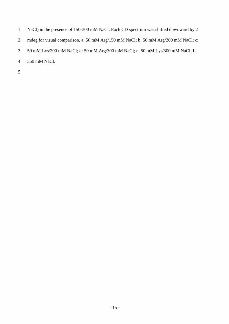
3.1. Measurement of myosin concentration by fluorescence intensity 3
The concentration of myosin in the test solvents was determined by the intrinsic 4
tryptophan fluorescence of myosin as opposed to the absorbance at 280 nm. This approach 5
was used because the formation of filamentous structures causes significant light scattering 6
and makes UV absorbance unreliable for concentration determination. Figure 1 demonstrates 7
the reliability of fluorescence for myosin concentration measurements. Figure 1A shows the 8
fluorescence emission spectra of a serially diluted myosin stock solution of known protein 9
concentration. The spectral shape was independent of dilution with a peak at 334 nm, which 10
indicated no effects of dilution and protein concentration on the tryptophan environments of 11
myosin; note that this fluorescence peak position indicates that the fluorescent tryptophans are 12
at least partially buried inside the tertiary structure of myosin. The fluorescence intensity at 13
334 nm of these myosin samples were plotted against the concentration of myosin. As shown 14
in Fig. 1B, the fluorescence intensity linearly increased with the sample concentration. The 15
straight line corresponds to a linear regression with a correlation coefficient of 0.999. Thus, 16
the myosin concentration can be reliably determined from the fluorescence intensity at 334 17
nm. Furthermore, the fluorescence spectra of myosin were not affected by the additives tested 18
and NaCl concentrations (date not shown). 19
20
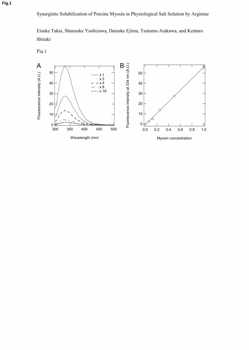
3.2. Effect of arginine on myosin solubility 21
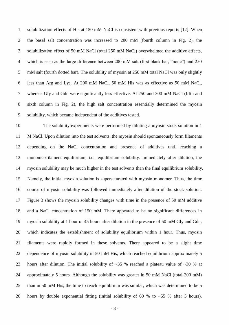
Figure 2 shows the effects of 50 mM Arg, Lys, NaCl, His, Gly and Gdn on the 22
solubility of myosin in the presence of NaCl concentrations (50, 100, 150, 200, 250 and 300 23
mM); note that an additional 50 mM NaCl was present over the basal NaCl concentration (a 24
total of 100 mM NaCl for the first column, which corresponds to the 50 mM basal NaCl 25
- 7 -
concentration). The myosin solubility in the absence of an additional 50 mM NaCl (at the 1
basal salt concentration) is shown as “none” in Fig. 2. As described in the methods section, 2
the myosin solubility was determined in the test solvents by diluting the stock myosin solution 3
in 1 M NaCl, where myosin is largely monomeric, into the test solvents. Thus, myosin 4
filament formation may vary depending on the salt concentration, which suggests that more 5
filaments may be present at lower salt concentrations. The solubility shown in Fig. 2 is the 6
value at 1 hour after dilution and may not be the final equilibrium value, particularly in the 7
presence of 50 mM Arg (for reasons described later). 8
At the 50 mM basal NaCl concentration (first column in Fig. 2), 50 mM Arg and Lys 9
showed insignificant effects on myosin solubility compared with 50 mM NaCl (total 100 mM 10
NaCl), whereas 50 mM His and Gly slightly reduced myosin solubility compared with the 11
three additives. It is interesting that 50 mM Gdn showed reduced myosin solubility, although 12
only slightly. Thus, the effects of these five additives (i.e., Arg, Lys, His, Gly and Gdn) were 13
marginal at this low basal salt concentration. A small but significant effect of 50 mM Arg was 14
observed at a 100 mM NaCl concentration (second column in Fig. 2). Arg and Lys increased 15
the myosin solubility compared to NaCl alone, and Arg was more effective; 50 mM NaCl was 16
slightly effective when compared with its absence (see “none”). At this NaCl concentration 17
(second column in Fig. 2), 50 mM His, Gly and Gdn were essentially ineffective as myosin 18
solubility in these solvents was nearly identical to the solubility in 100 mM NaCl alone 19
(“none”). At a 150 mM NaCl concentration (third column in Fig. 2), a much stronger effect of 20
Arg was observed. At this NaCl concentration, 50 mM Arg increased the myosin solubility by 21
more than twofold over the level achieved by 50 mM NaCl (200 mM total NaCl) and almost 22
threefold over the value at 150 mM NaCl (see first black bar, “none”). Lys was also effective; 23
however, its effect was greatly reduced when compared to Arg. At 150 mM NaCl, 50 mM His, 24
Gly and Gdn were much weaker than NaCl in solubilizing myosin and nearly identical to the 25
value in the absence of 50 mM salt (but in the presence of 150 mM salt). The lack of 26
- 8 -
solubilization effects of His at 150 mM NaCl is consistent with previous reports [12]. When 1
the basal salt concentration was increased to 200 mM (fourth column in Fig. 2), the 2
solubilization effect of 50 mM NaCl (total 250 mM NaCl) overwhelmed the additive effects, 3
which is seen as the large difference between 200 mM salt (first black bar, “none”) and 250 4
mM salt (fourth dotted bar). The solubility of myosin at 250 mM total NaCl was only slightly 5
less than Arg and Lys. At 200 mM NaCl, 50 mM His was as effective as 50 mM NaCl, 6
whereas Gly and Gdn were significantly less effective. At 250 and 300 mM NaCl (fifth and 7
sixth column in Fig. 2), the high salt concentration essentially determined the myosin 8
solubility, which became independent of the additives tested. 9
The solubility experiments were performed by diluting a myosin stock solution in 1 10
M NaCl. Upon dilution into the test solvents, the myosin should spontaneously form filaments 11
depending on the NaCl concentration and presence of additives until reaching a 12
monomer/filament equilibrium, i.e., equilibrium solubility. Immediately after dilution, the 13
myosin solubility may be much higher in the test solvents than the final equilibrium solubility. 14
Namely, the initial myosin solution is supersaturated with myosin monomer. Thus, the time 15
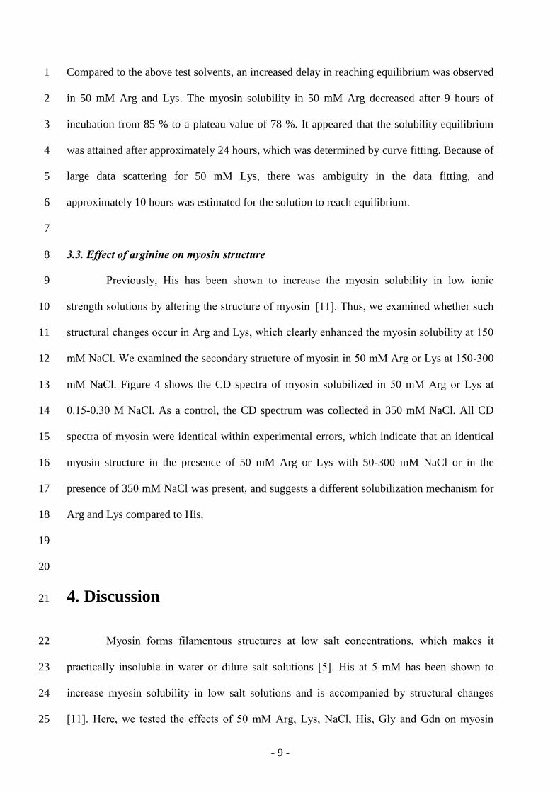
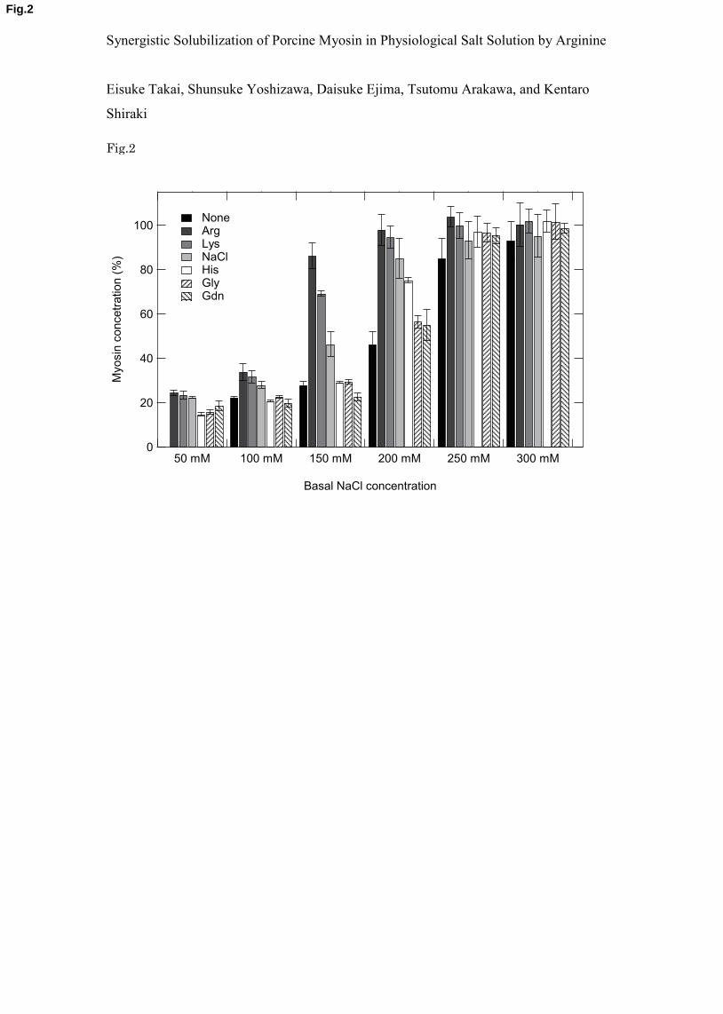
course of myosin solubility was followed immediately after dilution of the stock solution. 16
Figure 3 shows the myosin solubility changes with time in the presence of 50 mM additive 17
and a NaCl concentration of 150 mM. There appeared to be no significant differences in 18
myosin solubility at 1 hour or 45 hours after dilution in the presence of 50 mM Gly and Gdn, 19
which indicates the establishment of solubility equilibrium within 1 hour. Thus, myosin 20
filaments were rapidly formed in these solvents. There appeared to be a slight time 21
dependence of myosin solubility in 50 mM His, which reached equilibrium approximately 5 22
hours after dilution. The initial solubility of ~35 % reached a plateau value of ~30 % at 23
approximately 5 hours. Although the solubility was greater in 50 mM NaCl (total 200 mM) 24
than in 50 mM His, the time to reach equilibrium was similar, which was determined to be 5 25
hours by double exponential fitting (initial solubility of 60 % to ~55 % after 5 hours). 26
- 9 -
Compared to the above test solvents, an increased delay in reaching equilibrium was observed 1
in 50 mM Arg and Lys. The myosin solubility in 50 mM Arg decreased after 9 hours of 2
incubation from 85 % to a plateau value of 78 %. It appeared that the solubility equilibrium 3
was attained after approximately 24 hours, which was determined by curve fitting. Because of 4
large data scattering for 50 mM Lys, there was ambiguity in the data fitting, and 5
approximately 10 hours was estimated for the solution to reach equilibrium. 6
7
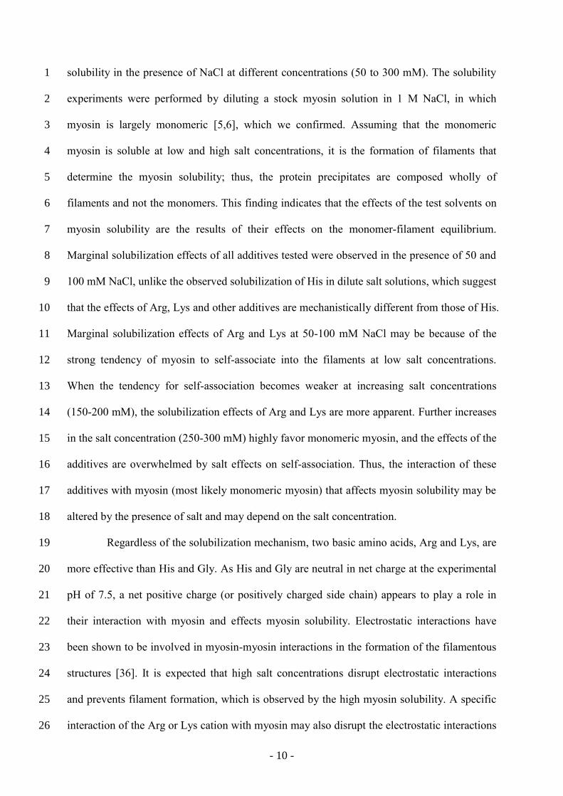
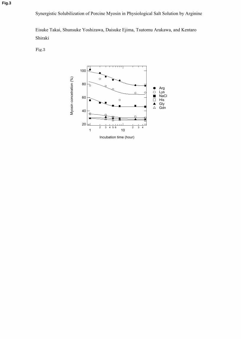
3.3. Effect of arginine on myosin structure 8
Previously, His has been shown to increase the myosin solubility in low ionic 9
strength solutions by altering the structure of myosin [11]. Thus, we examined whether such 10
structural changes occur in Arg and Lys, which clearly enhanced the myosin solubility at 150 11
mM NaCl. We examined the secondary structure of myosin in 50 mM Arg or Lys at 150-300 12
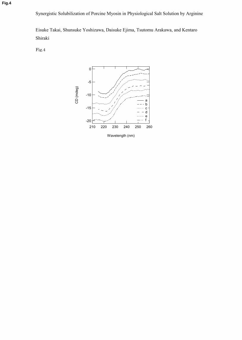
mM NaCl. Figure 4 shows the CD spectra of myosin solubilized in 50 mM Arg or Lys at 13
0.15-0.30 M NaCl. As a control, the CD spectrum was collected in 350 mM NaCl. All CD 14
spectra of myosin were identical within experimental errors, which indicate that an identical 15
myosin structure in the presence of 50 mM Arg or Lys with 50-300 mM NaCl or in the 16
presence of 350 mM NaCl was present, and suggests a different solubilization mechanism for 17
Arg and Lys compared to His. 18
19
20
4. Discussion 21
Myosin forms filamentous structures at low salt concentrations, which makes it 22
practically insoluble in water or dilute salt solutions [5]. His at 5 mM has been shown to 23
increase myosin solubility in low salt solutions and is accompanied by structural changes 24
[11]. Here, we tested the effects of 50 mM Arg, Lys, NaCl, His, Gly and Gdn on myosin 25
- 10 -
solubility in the presence of NaCl at different concentrations (50 to 300 mM). The solubility 1
experiments were performed by diluting a stock myosin solution in 1 M NaCl, in which 2
myosin is largely monomeric [5,6], which we confirmed. Assuming that the monomeric 3
myosin is soluble at low and high salt concentrations, it is the formation of filaments that 4
determine the myosin solubility; thus, the protein precipitates are composed wholly of 5
filaments and not the monomers. This finding indicates that the effects of the test solvents on 6
myosin solubility are the results of their effects on the monomer-filament equilibrium. 7
Marginal solubilization effects of all additives tested were observed in the presence of 50 and 8
100 mM NaCl, unlike the observed solubilization of His in dilute salt solutions, which suggest 9
that the effects of Arg, Lys and other additives are mechanistically different from those of His. 10
Marginal solubilization effects of Arg and Lys at 50-100 mM NaCl may be because of the 11
strong tendency of myosin to self-associate into the filaments at low salt concentrations. 12
When the tendency for self-association becomes weaker at increasing salt concentrations 13
(150-200 mM), the solubilization effects of Arg and Lys are more apparent. Further increases 14
in the salt concentration (250-300 mM) highly favor monomeric myosin, and the effects of the 15
additives are overwhelmed by salt effects on self-association. Thus, the interaction of these 16
additives with myosin (most likely monomeric myosin) that affects myosin solubility may be 17
altered by the presence of salt and may depend on the salt concentration. 18
Regardless of the solubilization mechanism, two basic amino acids, Arg and Lys, are 19
more effective than His and Gly. As His and Gly are neutral in net charge at the experimental 20
pH of 7.5, a net positive charge (or positively charged side chain) appears to play a role in 21
their interaction with myosin and effects myosin solubility. Electrostatic interactions have 22
been shown to be involved in myosin-myosin interactions in the formation of the filamentous 23
structures [36]. It is expected that high salt concentrations disrupt electrostatic interactions 24
and prevents filament formation, which is observed by the high myosin solubility. A specific 25
interaction of the Arg or Lys cation with myosin may also disrupt the electrostatic interactions 26
- 11 -
more effectively than NaCl, which increases the myosin solubility over the solubility achieved 1
by salt alone. Arg was always more effective than Lys, which indicates that the positive 2
charge and structure of the side chain play a role. The critical role of the guanidinium group 3
has been implicated in the effectiveness of Arg in the suppression of protein aggregation 4
[17,18]. The guanidinium group alone was insufficient in myosin solubilization, which was 5
shown by the lack of increased solubilization effects because of Gdn. Under the experimental 6
conditions, His displayed no or marginal solubilization effects, which differed from 7
previously reported results [10–12] . Such His effects were attributed to the structural changes 8
of myosin conferred by His [11], which indicates that His has no effect on myosin structure 9
under the present experimental conditions. Further, the effects of Arg and Lys are mediated 10
by their interaction with native myosin, which was indicated by the lack of structural changes. 11
Interestingly, the equilibrium solubility was attained extremely slowly in the 12
presence of 50 mM Arg with 150 mM NaCl, which took more than 20 hours after dilution of 13
the stock myosin solution in this solvent. Namely, self-association of the myosin monomer to 14
the filaments was slow in this solvent system. The rate of this transition from monomer to 15
filament becomes faster in Lys and further in NaCl. In Gly and Gdn, the transition appeared to 16
occur within one hour. Myosin is in a monomeric structure in 1 M NaCl [5,6]. Upon dilution 17
into 50 mM Gly or Gdn in 150 mM NaCl, the solubility of myosin rapidly reaches 18
equilibrium, which means a low activation energy of the monomer-filament transition. In the 19
presence of 50 mM Arg, the initial solubility is much higher than the equilibrium solubility 20
(~100 % vs. < 80 %). Arg increases the activation energy. Because of this high energy barrier, 21
the supersaturated myosin solution is kinetically stabilized by Arg. The binding of Arg to 22
monomeric myosin may be involved in the stabilization of the monomer as the free energy to 23
dissociate the bound Arg should increase the energy barrier. 24
In conclusion, Arg increased the equilibrium solubility and activation energy of self-25
association of monomeric myosin in a physiological salt solution. This stabilization of the 26
- 12 -
myosin monomer by Arg occurred without altering the structure of myosin. It would be of 1
great interest to test the utility of arginine in processing meat products containing myosin 2
based on its ability to increase the solubility of monomeric myosin. 3
Acknowledgments 4
This work was supported by a Grant-in-Aid for JSPS KAKENHI Grant Numbers 5
23246063, 23550189 and the JSPS Fellows from the Ministry of Education, Culture, Sports, 6
Science, and Technology (MEXT), Japan. 7
8
9
References 10
[1] M. Friedman, J. Agric. Food Chem. 44 (1996) 6. 11
[2] R. Craig, J.L. Woodhead, Curr. Opin. Struct. Biol. 16 (2006) 204. 12
[3] J.Q. Xu, B.A. Harder, P. Uman, R. Craig, J. Cell Biol. 134 (1996) 53. 13
[4] R.L. Sohn, K.L. Vikstrom, M. Strauss, C. Carolyn, A.G. Szent-Gyorgyi, L.A. 14
Leinwand, J. Mol. Biol. 266 (1997) 317. 15
[5] R. Niederman, T.D. Pollard, J. Cell Biol. 67 (1975) 72. 16
[6] J.H. Sinard, W.F. Stafford, T.D. Pollard, J. Cell Biol. 109 (1989) 1537. 17
[7] T.M. Lin, J.W. Park, J. Food Sci. 63 (2008) 215. 18
[8] Y. Tsunashima, T. Akutagawa, Biopolymers 75 (2004) 264. 19
[9] M. Ishioroshi, K. Samejima, T. Yasui, J. Food Sci. 44 (1979) 1280. 20
[10] Y. Ito, R. Tatsumi, J. Wakamatsu, T. Nishimura, A. Hattori, Anim. Sci. J. 74 (2003) 21
417. 22
[11] T. Hayakawa, T. Ito, J. Wakamatsu, T. Nishimura, A. Hattori, Meat Sci. 82 (2009) 151. 23
[12] T. Hayakawa, T. Ito, J. Wakamatsu, T. Nishimura, A. Hattori, Meat Sci. 84 (2010) 742. 24
[13] K.R.C. Reddy, H. Lilie, R. Rudolph, C. Lange, Protein Sci. 14 (2005) 929. 25
[14] T. Arakawa, K. Tsumoto, Biochem. Biophys. Res. Commun. 304 (2003) 148. 26
- 13 -
[15] K. Shiraki, M. Kudou, S. Fujiwara, T. Imanaka, M. Takagi, J. Biochem. 132 (2002) 1
591. 2
[16] E.M. Lyutova, A.S. Kasakov, B.Y. Gurvits, Biotechnol. Prog. 23 (2007) 1411. 3
[17] H. Hamada, R. Takahashi, T. Noguchi, K. Shiraki, Biotechnol. Prog. 24 (2008) 436. 4
[18] T. Matsuoka, H. Hamada, K. Matsumoto, K. Shiraki, Biotechnol. Prog. 25 (2009) 1515. 5
[19] J. Buchner, R. Rudolph, Nat. Biotechnol. 9 (1991) 157. 6
[20] E. De Bernardez Clark, E. Schwarz, R. Rudolph, Methods Enzymol. 309 (1999) 217. 7
[21] C. Lange, R. Rudolph, Curr. Pharm. Biotechnol. 10 (2009) 408. 8
[22] K. Tsumoto, M. Umetsu, I. Kumagai, D. Ejima, J.S. Philo, T. Arakawa, Biotechnol. 9
Prog. 20 (2004) 1301. 10
[23] L. Ito, K. Shiraki, H. Yamaguchi, Acta Crystallogr., Sect. F: Struct. Biol. Cryst. 11
Commun. 66 (2010) 744. 12
[24] D. Ejima, R. Yumioka, K. Tsumoto, T. Arakawa, Anal. Biochem. 345 (2005) 250. 13
[25] D. Ejima, R. Yumioka, T. Arakawa, K. Tsumoto, J. Chromatogr., A 1094 (2005) 49. 14
[26] T. Arakawa, Y. Kita, H. Sato, D. Ejima, Protein Expression Purif. 63 (2009) 158. 15
[27] T. Arakawa, K. Tsumoto, K. Nagase, D. Ejima, Protein Expression Purif. 54 (2007) 16
110. 17
[28] P.E. Mason, C.E. Dempsey, G.W. Neilson, S.R. Kline, J.W. Brady, J. Am. Chem. Soc. 18
131 (2009) 16689. 19
[29] L. Ito, K. Shiraki, T. Matsuura, M. Okumura, K. Hasegawa, S. Baba, H. Yamaguchi, T. 20
Kumasaka, Protein Eng., Des. Sel. 24 (2011) 269. 21
[30] A. Hirano, T. Kameda, T. Arakawa, K. Shiraki, J. Phys. Chem. B 114 (2010) 13455. 22
[31] D. Shukla, B.L. Trout, J. Phys. Chem. B 114 (2010) 13426. 23
[32] T. Arakawa, D. Ejima, K. Tsumoto, N. Obeyama, Y. Tanaka, Y. Kita, S.N. Timasheff, 24
Biophys. Chem. 127 (2007) 1. 25
[33] A. Hirano, T. Arakawa, K. Shiraki, J. Biochem. 144 (2008) 363. 26
[34] B.M. Baynes, D.I.C. Wang, B.L. Trout, Biochemistry 44 (2005) 4919. 27
[35] C.P. Schneider, B.L. Trout, J. Phys. Chem. B 113 (2009) 2050. 28
[36] A.D. Mclachlan, J. Karn, Nature 299 (1982) 226. 29
- 14 -
1
2
Figure Captions 3
Figure 1. (A) The intrinsic emission fluorescence spectra of myosin at different protein 4
concentrations. The stock sample solution was serially diluted (1- to 16-fold) with 20 mM 5
sodium phosphate (pH 7.5). The fluorescence emission spectra were collected at 25 °C with 6
an excitation wavelength of 280 nm. (B) The fluorescence intensity at 334 nm against myosin 7
concentrations. Data are taken from Fig. 1A. The plot was fit by linear regression with a 8
correlation coefficient of 0.999. The measurements were performed three times, and the error 9
bars depict the standard deviation of the mean. 10
11
Figure 2. Myosin solubility in 50 mM additives or “None” indicated in the figure as a 12
function of basal NaCl concentration. A stock myosin solution in 1 M NaCl was diluted into 13
the test solvents and incubated for 1 h. The concentration of myosin in 50 mM additives or 14
“None” in the presence of 50-300 mM NaCl was determined by measuring the intrinsic 15
fluorescence intensity at 334 nm. The myosin solubility in test solvents was normalized to the 16
value in 50 mM Arg and 300 mM NaCl, which was set as 100 %. The measurements were 17
performed three times, and the error bars depict the standard deviation of the mean. 18
19
Figure 3. Time course of concentration changes of myosin. The concentration of myosin in 50 20
mM additives in the presence of 150 mM NaCl was followed with incubation for 1 hour after 21
dilution into the test solvents. The continuous line though the data points is a fit with a double 22
exponential with offset. 23
24
Figure 4. Far-UV CD spectra of myosin in the presence of Arg or Lys as a function of NaCl 25
concentration. The far-UV CD spectra were measured in 50 mM additives (Arg, Lys and 26
- 15 -
NaCl) in the presence of 150-300 mM NaCl. Each CD spectrum was shifted downward by 2 1
mdeg for visual comparison. a: 50 mM Arg/150 mM NaCl; b: 50 mM Arg/200 mM NaCl; c: 2
50 mM Lys/200 mM NaCl; d: 50 mM Arg/300 mM NaCl; e: 50 mM Lys/300 mM NaCl; f: 3
350 mM NaCl. 4
5
50
40
30
20
10
0
Fluo
resc
ence
inte
nsity
(A.U
.)
500450400350300
Wavelength (nm)
x 1 x 2 x 4 x 8 x 16
50
40
30
20
10
0Fl
uore
scen
ce in
tens
ity a
t 334
nm
(A.U
.)1.00.80.60.40.20.0
Myosin concentration
A B
Synergistic Solubilization of Porcine Myosin in Physiological Salt Solution by Arginine
Eisuke Takai, Shunsuke Yoshizawa, Daisuke Ejima, Tsutomu Arakawa, and Kentaro
Shiraki Fig.1
Fig.1
50
40
30
20
10
0
Fluo
resc
ence
inte
nsity
(A.U
.)
500450400350300
Wavelength (nm)
x 1 x 2 x 4 x 8 x 16
50
40
30
20
10
0Fl
uore
scen
ce in
tens
ity a
t 334
nm
(A.U
.)1.00.80.60.40.20.0
Myosin concentration
A B
Synergistic Solubilization of Porcine Myosin in Physiological Salt Solution by Arginine
Eisuke Takai, Shunsuke Yoshizawa, Daisuke Ejima, Tsutomu Arakawa, and Kentaro
Shiraki Fig.2
100
80
60
40
20
0
Myo
sin
conc
etra
tion
(%)
50 mM 100 mM 150 mM 200 mM 250 mM 300 mM
Basal NaCl concentration
None Arg Lys NaCl His Gly Gdn
Fig.2
Synergistic Solubilization of Porcine Myosin in Physiological Salt Solution by Arginine
Eisuke Takai, Shunsuke Yoshizawa, Daisuke Ejima, Tsutomu Arakawa, and Kentaro
Shiraki Fig.3
100
80
60
40
20
Myo
sin
conc
etra
tion
(%)
12 3 4 5 6
102 3 4
Incubation time (hour)
Arg Lys NaCl His Gly Gdn
Fig.3
Synergistic Solubilization of Porcine Myosin in Physiological Salt Solution by Arginine
Eisuke Takai, Shunsuke Yoshizawa, Daisuke Ejima, Tsutomu Arakawa, and Kentaro
Shiraki Fig.4
-20
-15
-10
-5
0
CD
(mde
g)
260250240230220210
Wavelength (nm)
a b c d e f
Fig.4
Supporting Information for Synergistic Solubilization of Porcine Myosin in 1
Physiological Salt Solution by Arginine 2
E. Takai, S. Yoshizawa, D. Ejima, T. Arakawa, K. Shiraki 3
4
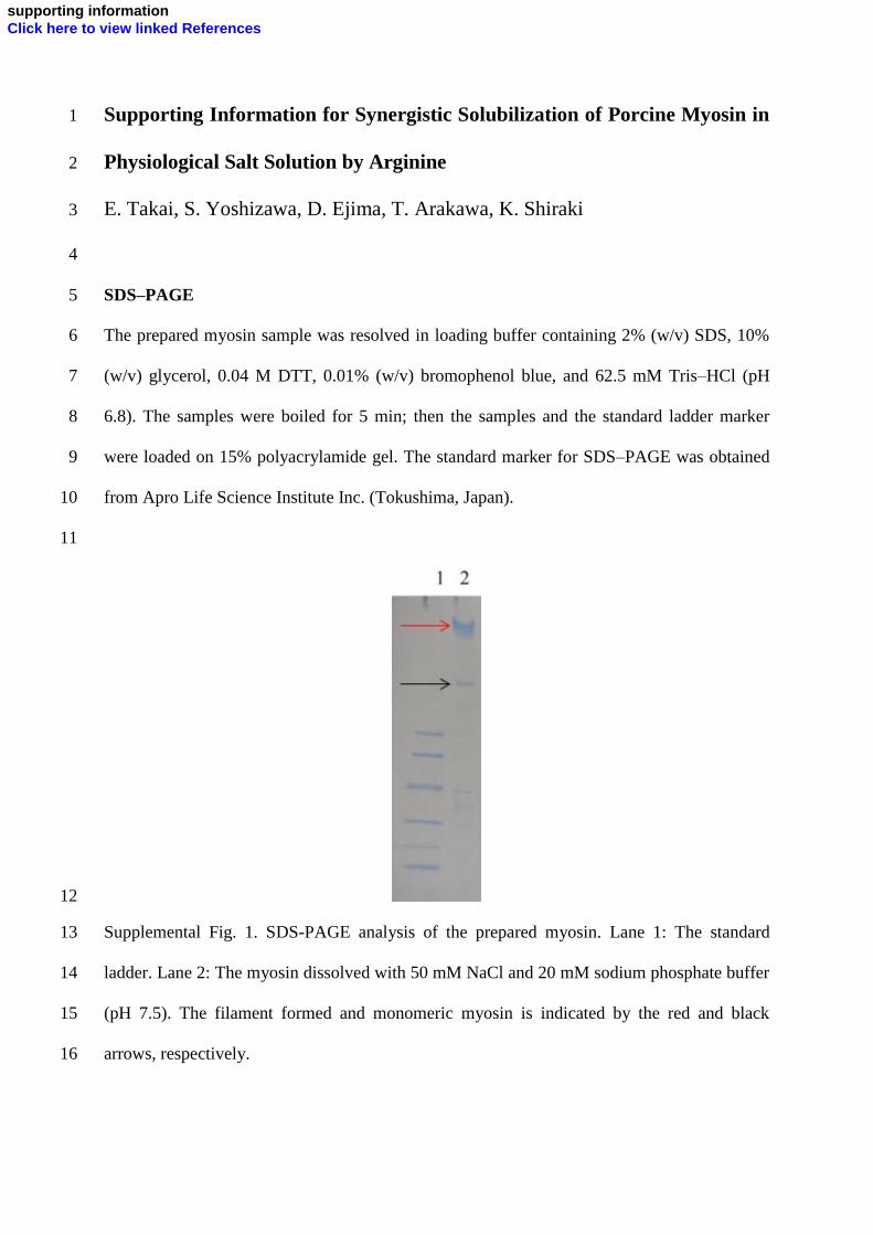
SDS–PAGE 5
The prepared myosin sample was resolved in loading buffer containing 2% (w/v) SDS, 10% 6
(w/v) glycerol, 0.04 M DTT, 0.01% (w/v) bromophenol blue, and 62.5 mM Tris–HCl (pH 7
6.8). The samples were boiled for 5 min; then the samples and the standard ladder marker 8
were loaded on 15% polyacrylamide gel. The standard marker for SDS–PAGE was obtained 9
from Apro Life Science Institute Inc. (Tokushima, Japan). 10
11
12
Supplemental Fig. 1. SDS-PAGE analysis of the prepared myosin. Lane 1: The standard 13
ladder. Lane 2: The myosin dissolved with 50 mM NaCl and 20 mM sodium phosphate buffer 14
(pH 7.5). The filament formed and monomeric myosin is indicated by the red and black 15
arrows, respectively. 16
supporting informationClick here to view linked References