Embed Size (px)
Citation preview

Syntax and the brain: disentangling grammar by selective anomalies
A. Moro1,2, M. Tettamanti3, D. Perani1,3, C. Donati4, S. F. Cappa1, F. Fazio3,5,6
1Università “Vita-Salute” San Raffaele, 2Università degli studi di Bologna, 3Istituto di Neuroscienze e Bioimmagini - C.N.R., Milano 4Università di Urbino,
5Istituto Scientifico San Raffaele HSR, 6Università Statale di Milano-Bicocca
Running title: brain correlates of syntax processes
Address for correspondence
Daniela PeraniInstitute of Neuroscience and Bioimaging C.N.R. and Università “Vita-Salute” San Raffaele, Milano,Via Olgettina 60, 20132 Milano, Italytelephone: 0039-02-26432224-2223fax: [email protected]
1

Abstract
Many paradigms employed so far with functional imaging in language studies do not allow a
clear differentiation of the semantic, morphological and syntactic components, as traditionally
defined within linguistic theory. In fact, many studies simply consider the brain's response to
lists of unrelated words, rather than to syntactic structures, or do not neutralize the
confounding effect of the semantic component. In the present PET experiment, we isolated
the functional correlates of morphological and syntactic processing. The neutralization of the
access to the lexical-semantic component was achieved by requiring the detection of
anomalies in written sentences consisting of pseudo-words. In both syntactic and
morphosyntactic processing, the involvement of a selective deep component of Broca's area
and of a right inferior frontal region was detected. In addition, within this system, the left
caudate nucleus and insula were activated only during syntactic processing, indicating their
role in syntactic computation. These findings provide original in vivo evidence that these
brain structures, whose individual contribution has been highlighted by clinical studies,
constitute a neural network selectively engaged in morphological and syntactic computation.
Key words: syntax, morphosyntax, PET, normal subjects
2
Introduction
Modern linguistics has succeeded in decomposing the complexity of grammars in the
interaction of independent modules. More specifically, for any given sentence in any
language three abstract levels of representation converge to give the associated structure; the
phonological level (where the possible sequences of sounds are checked), the syntactic level
(where words are combined yielding the proper hierarchical structures), the semantic level
(where the meaning of the whole sentence is computed on the basis of the meaning of each
lexical item). Thus, for example, an English native speaker knows that such expressions as
"remnantzry", "dog a barks" and "happiness broke his arm" are not acceptable at the
phonological, syntactic and semantic level, respectively.
Such a modular architecture, which is claimed to reflect the implicit knowledge of
grammar that every human being is endowed with genetically, raises many empirical
questions. A crucial one is whether this threefold abstract partition is actually isomorphic to
some neurophysiological process, and more specifically whether these three levels of
representation are subserved by distinct neural correlates. Of course, although the rules
governing each level are independent, there is no direct way to test each of them in isolation,
since by definition they are activated simultaneously. Several experiments have shown that
semantic information as expressed by the lexicon is independently represented in the brain
(Martin et al., 1995; Martin et al., 1996; Perani et al., 1999b; Vandenberghe et al., 1996);
nevertheless, the fundamental question remains as to whether syntactic operations can be
associated with some dedicated neural networks. Indeed, it must be highlighted that many
neuroimaging experiments on human language have used as stimuli lists of words, rather than
full sentences, which are in fact the actual units of spontaneous speech (Price, 1998).
3
Along with such an abstract model of the knowledge of grammar, the actual process
of interpretation of a sentence of course requires assigning each element of the sentence to the
proper slots in the actual mental representation-grid; this in turn implies the memory load
capacity to keep phrases in an activated state. Syntactic processing during sentence reading
has been addressed by several functional neuroimaging investigations focusing specifically on
syntactic complexity, which showed consistent activations in Broca’s pars opercularis, during
an on-line acceptability-judgment task (Caplan et al., 1998; Stromswold et al., 1996) and
during a post-sentence presentation judgment task (Just et al., 1996). In the latter paper, Just
and colleagues, in addition to left Broca’s and Wernicke’s regions, found activations also in
right hemispheric homologue areas. The reported activations were indeed interpreted as being
due to increasing syntactic complexity and, concerning Broca’s area specifically, to
augmented memory and computational load. Similar findings were reported for sentences
presented in the auditory modality (Caplan et al., 1999): however, in contrast to the above
mentioned studies, the pars triangularis and not the pars opercularis of the inferior frontal
gyrus was activated. Caplan and colleagues (1998), in addition to Broca’s area, found
activations in the anterior cingulate gyrus and in the right medial frontal gyrus (similar
finding are reported in Stromswold et al. (1996). This pattern, in the authors’ opinion,
correlates with phonological encoding and subvocal rehearsal, an hypothesis that is supported
by other imaging studies (Paulesu et al., 1993; Zatorre et al., 1992). Subvocal rehearsal might
be used for assigning the head of the sentence its thematic role, as in relative clauses
(Swinney and Zurif, 1995; Swinney et al., 1996; Zurif et al., 1993).
A recent fMRI experiment (Dapretto and Bookheimer, 1999) used a sentence
comprehension task, in which different relative weights for syntactic and lexico-semantic
processing had been introduced. Subjects were asked to decide whether a certain pair of
sentences had the same meaning. Such pairs were constructed by either changing one single
4
word in the same sequence, called "semantic condition", or changing the full sequence, called
"syntactic condition". The subjects were requested to give “same” or “different” judgments.
For example, in the syntactic condition, sentences such as “The policeman arrested the thief”
or “The thief was arrested by the policeman” were judged as “same” whereas “West of the
bridge is the airport” or “The bridge is west of the airport” as “different”. Again, a selective
activation in Broca’s pars opercularis on the lateral brain surface was found to be associated
with syntactic processing. Clearly, such a task was crucially centered on a major, although
implicit, assumption, namely that changing the syntactic structure of a sentence does not
affect the semantic component. So, the transformation of an active sentence like "the
policeman arrested the thief" into a passive sentence like "the thief was arrested by the
policemen" is considered not to affect the semantic interpretation. Although the
transformation from active to passive construction is surely a syntactic phenomenon, one
cannot be sure that this is not affecting also the semantic component. Indeed, since at least
Jackendoff (1968), it is well-known that passive constructions do not preserve the semantics
content of their active counterparts. Famous examples, often quoted in the linguistic literature
are the pairs like: "many arrows didn't hit the target" and "the target wasn't hit by many
arrows". Clearly, the state of affairs which are compatible with the two sentences differ, since
the target could still be hit by many arrows, if the first sentence is true, whereas this cannot be
the case if the second one is. Indeed, these kind of observations lead Chomsky to formulate
the so called "Extended Standard Theory" -see for example, Chomsky (1975) contra
Chomsky (1965) based on Katz and Postal (1964)-, and have never been dismissed ever since
that time.
The Dapretto and Bookheimer (1999) experiment represents an advancement with
respect to previous works in the field. In the present study, we have adopted an alternative
strategy, which allows to disentangle grammar and isolate syntax from semantics. The
5
innovative strategy we pursued, was suggested on the basis of some crucial problematic
aspects of the previous work in the field. Indeed, all previous neuroimaging experiments,
either using words or sentences, left the access to semantics unaltered. To avoid these
problematic issue, we have designed a paradigm which neutralizes the access to any semantic
component. Such a problem was overcome by using non-words, that is invented words which
are not related to any meaning in the lexicon, like "staze". Functional words and morphemes,
instead, like articles, auxiliaries, prepositions, plural morphemes etc. have been fully
preserved. In such a case, any anomaly in the syntactic structure could not influence any
semantic interpretation which was missing in the input in the first place. All in all, even if a
non-word is in fact assigned a syntactic category on the basis of its morphological structure
and the context where it occurs, it is clearly impossible for it to have a proper semantic status
for at least two reasons: first, it is by definition not assigned an extension in any possible
world; second, which is crucial, it can by no means contribute to the computation of the
semantics of the whole sentence which we take to be its truth functional value (Dowty et al.,
1981).
In our experiment we tested the subjects’ linguistic knowledge at each level by
selectively disrupting one level while maintaining the others intact. More specifically, we
have asked the subjects to detect either phonological, morphosyntactic and syntactic
anomalies in pseudo-word sentences which contained only one type of anomaly for each level
(see methods). This in principle allows one to focus selectively on syntactic processing rather
than on the different amount of syntactic complexity, as done in cited works. A major
problem was also overcome which is implicit in this type of experiment. In fact, when the
syntactic level is disrupted, a potential semantic anomaly is also produced; thus, for example,
if one says "all the eaten have chickens snakes" the anomalous syntactic structure also
disturbs the semantic interpretation which would be impossible to reconstruct.
6
Materials and methods
Subjects
The study was approved by the Ethics Committee of the Scientific Institute H San Raffaele,
and each volunteer gave his written informed consent prior to the admission to the study.
Eleven male volunteer right-handed subjects (mean age 26 years, range 22 to 28 years )
entered the study. All subjects had no history of neurological or psychiatric disorders. Right-
handedness was verified using the Edinburgh Inventory (Oldfield, 1971).
Tasks Design
The study consisted of three experimental and one baseline conditions. Subjects were asked to
covertly read sentences presented visually and, for the three experimental conditions only, to
make acceptability judgments at the corresponding sentence-structure levels. The sentences
all consisted of pseudowords only ('pseudosentences'), so as to neutralize the access to
semantic components: this 'Quasi-Italian', devoid of any open-class word, but maintaining
inflections and function-words, was employed in order to isolate the correlates of
morphosyntactic and syntactic processing. According to the experimental tasks, anomalies
either at the phonotactic, the morphosyntactic or the syntactic level were introduced.
Syntactic anomalies presented sentences with wrong linear order but proper agreements.
Morphosyntactic anomalies presented sentences with proper word order but agreement errors.
Phonotactic anomalies presented sentences containing Italian unlegal consonant strings.
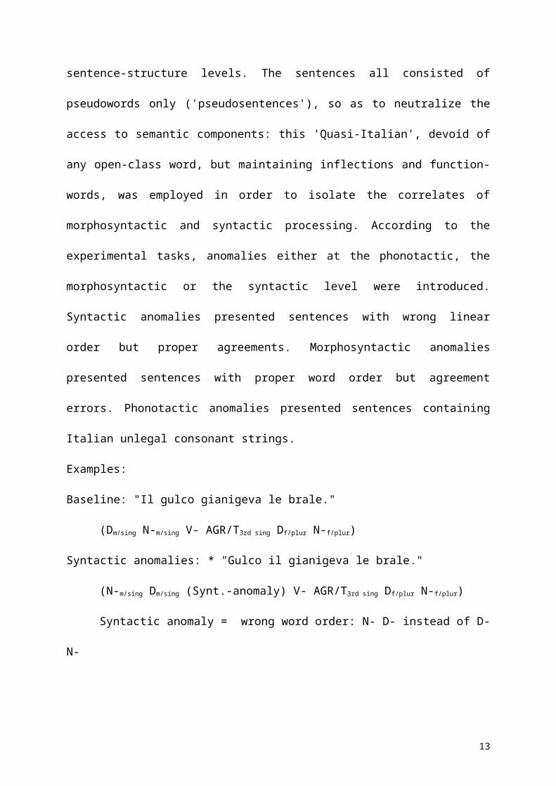
Examples:
Baseline: "Il gulco gianigeva le brale."
(Dm/sing N-m/sing V- AGR/T3rd sing Df/plur N-f/plur)
7
Syntactic anomalies: * "Gulco il gianigeva le brale."
(N-m/sing Dm/sing (Synt.-anomaly) V- AGR/T3rd sing Df/plur N-f/plur)
Syntactic anomaly = wrong word order: N- D- instead of D- N-
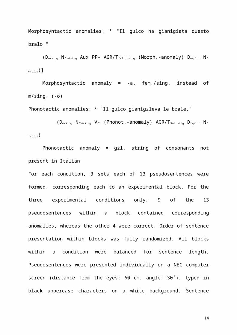
Morphosyntactic anomalies: * "Il gulco ha gianigiata questo bralo."
(Dm/sing N-m/sing Aux PP- AGR/Tf/3rd sing (Morph.-anomaly) Dm/plur N-m/plur)]
Morphosyntactic anomaly = -a, fem./sing. instead of m/sing. (-o)
Phonotactic anomalies: * "Il gulco gianigzleva le brale."
(Dm/sing N-m/sing V- (Phonot.-anomaly) AGR/T3rd sing Df/plur N-f/plur)
Phonotactic anomaly = gzl, string of consonants not present in Italian
For each condition, 3 sets each of 13 pseudosentences were formed, corresponding each to an
experimental block. For the three experimental conditions only, 9 of the 13 pseudosentences
within a block contained corresponding anomalies, whereas the other 4 were correct. Order of
sentence presentation within blocks was fully randomized. All blocks within a condition were
balanced for sentence length. Pseudosentences were presented individually on a NEC
computer screen (distance from the eyes: 60 cm, angle: 30˚), typed in black uppercase
characters on a white background. Sentence presentation time was 4000 ms, with an Inter
Stimulus Interval of 1000 ms. Subjects read the sentences covertly and, either pressed a
response-box button when they had completed sentence reading (for the baseline condition),
or pressed the response-box when they detected an anomaly (for the experimental conditions).
Reaction times and response accuracy were recorded.
A preliminary dyslexia test battery was administered to all subjects, in order to exclude
possible pseudoword processing deficits. All experimental and behavioral subjects included in
the analysis performed as normal.
PET data acquisition
8
Regional cerebral blood flow (rCBF) was assessed with positron emission tomography (PET)
on each of the eleven experimental subjects, while they were instructed to execute one of the
four tasks. Three repetitions of each condition were run for each subject, for a total of 12 PET
scans per subject. Condition-presentation order was balanced across subjects (Latin square
design).
rCBF was measured by recording the distribution of radioactivity following an
intravenous injection of 15O-labeled water (H215O) with a GE-Advance scanner (General
Electric Medical System, Milwaukee,WI) which has a field of view of 15.2 cm. Data were
acquired by scanning in 3D mode. A 5 mCi slow bolus of H215O, 4 cc in 20 sec, plus 4 cc of
saline solution in 20 s, were injected (Silbersweig et al., 1993). After attenuation correction
(measured by a transmission scan using a pair of rotating pin sources filled with 68Ge), the
data were reconstructed as 35 transaxial planes by three-dimensional filtered back projection
with a Hanning filter (cut-off 4 mm filter width) in the transaxial plane, and a Ramp filter
(cut-off 8.5 mm) in the axial direction. The integrated counts collected for 90 s, starting 30 s
after injection time, were used as an index of rCBF.
Image transformations and statistical analysis were performed in MATLAB 4.2 (Math
Works, Natick, MA, USA) using statistical parametric mapping (SPM-96, Wellcome
Department of Cognitive Neurology, London, UK). The original brain images were first
realigned and then transformed into a standard stereotactic space (defined by the International
Consortium for Brain Mapping project (ICBM) (NIH P-20 grant), and closely approximates
the space described in the atlas of Talairach and Tournoux (1988). In order to increase signal
to noise ratio and accommodate normal variability in functional gyral anatomy each image
was smoothed in three dimensions with a Gaussian filter (16 x 16 x 16 mm). A repeated-
measures ANCOVA was used for the comparison of different tasks, in which every subject
was studied under all conditions. Global differences in cerebral blood flow were covaried out
9
for all voxels and comparisons across conditions were made using t statistics with appropriate
linear contrasts (Friston et al., 1995a; Friston et al., 1995b). The set of t values for each voxel
of the image comprise the statistical parametric map (SPM{t}).
The following contrasts were evaluated:
Commonalities: overall main effects masked with each of the individual contrasts:
1. (Ph + M + S) - B; masked with (Ph - B); (M -B); (S - B).
2. (M + S) - Ph; masked with (M - Ph); (S - Ph).
Simple Main effects:
3. M -Ph
4. S - Ph
B = baseline task; Ph = phonotactic task; M = morphosyntactic task; S = syntactic task
10
Results
Behavioral data:
All subjects performed the tasks with high accuracy (range B: 92-100 %; Ph: 92-100 %; M:
77-100 %; S: 69-100 %). A multivariate repeated measure Anova was performed on the
accuracy rates (expressed in % of correct answers; correct answer defined as: answer given
within time < 4000 ms, and correct anomaly detection). Experimental conditions (Means: B =
99,0 %; Ph = 98.8 %; M = 92.5 %; S = 95.5 %) were significantly different: F = 5.175, p =
0.005. Post-hoc Student t-test paired comparisons that reached the p<0.05 significance level
were between M and B conditions (p = 0.0019) and between M and F (p = 0.027). Within-
conditions block presentation order was not significant as a main effect (Means; 1. Block =
96.5 %; 2. Block = 95.6 %; 3. Block = 97.4 %): F = 2.289, p = 0.127. The interaction
between block presentation order and conditions was not significant: F = 0.317; p = 0.925.
The same analysis was also performed on the Reaction Times (RT) of the 11
experimental subjects gave the following results: Experimental conditions (Means: B = 1946
ms; Ph = 1693 ms; M = 1891 ms; S = 1867 ms) were not significantly different: F = 1.977, p
= 0.139). Within-conditions block presentation order was significant as a main effect (Means;
1. Block = 1959 ms; 2. Block = 1806 ms; 3. Block = 1784 ms): F = 4.210, p = 0.030. The
interaction between block presentation order and conditions was not significant: F = 1.230; p
= 0.299.
Functional data :
The 3 experimental conditions share a common neural network as revealed by the main
effect, masked with the individual simple main effects, using the baseline as a reference
condition. The common pattern of significant activations included Broca's area pars
opercularis (Ba 44) and the left inferior parietal lobule (Ba 40); on the right hemisphere, the
lateral premotor area (Ba 6), the cuneus (Ba 18) and the middle occipital gyrus (Ba 19 and
11
18). Bilateral activations included the superior parietal lobule (Ba 7), the precuneus (Ba 7),
the fusiform gyrus (Ba 18/37), the cerebellum and the cerebellar vermis (see table 1, a for
stereotaxic coordinates and fig. 1, A).
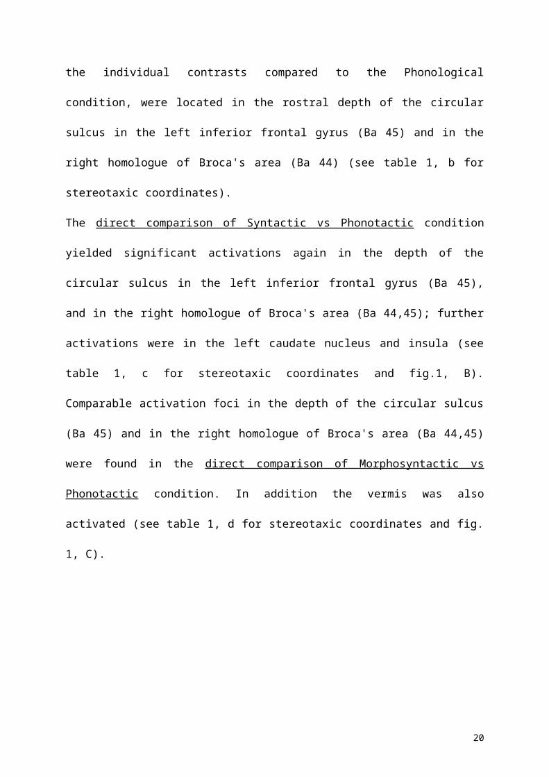
The common activations for Syntactic and Morphosyntactic conditions, as revealed by the
main effect masked with each of the individual contrasts compared to the Phonological
condition, were located in the rostral depth of the circular sulcus in the left inferior frontal
gyrus (Ba 45) and in the right homologue of Broca's area (Ba 44) (see table 1, b for
stereotaxic coordinates).
The direct comparison of Syntactic vs Phonotactic condition yielded significant activations
again in the depth of the circular sulcus in the left inferior frontal gyrus (Ba 45), and in the
right homologue of Broca's area (Ba 44,45); further activations were in the left caudate
nucleus and insula (see table 1, c for stereotaxic coordinates and fig.1, B). Comparable
activation foci in the depth of the circular sulcus (Ba 45) and in the right homologue of
Broca's area (Ba 44,45) were found in the direct comparison of Morphosyntactic vs
Phonotactic condition. In addition the vermis was also activated (see table 1, d for stereotaxic
coordinates and fig. 1, C).
12
Discussion
The detection of errors in pseudosentences is associated with the activation of an
extended network of brain regions, involving the classical language areas as well as several
other associative occipito-temporal and parietal areas (Table 1a). Common to all three
experimental conditions was the activation of high-order visual areas, which may reflects
aspects of visual processing specific for the error detection task in comparison with the
reading condition. In particular, error detection engages more extensive attentional resources
than simple reading, and might thus result in stronger parietal activation (Wojciulik and
Kanwisher, 1999). The main issue underlying the present investigation was to address
sentence processing at the syntactic level, while keeping this component as far as possible
disentangled from lexical semantics: we will discuss the activations specifically related to
morphological and syntactic processing, which included Broca’s area, the caudate nucleus
and the cerebellum.
Broca's area has been traditionally associated with morphosyntactic processing. The
main basis for this association is the fact that the clinical picture of agrammatism,
characterized by morphological errors in production and (inconstantly) by disordered
syntactic comprehension (Caplan et al., 1981), is usually part of the symptom complex of
Broca’s aphasia (see Grodzinsky (2000), for a recent review). The classical syndrome of
Broca's aphasia, however, combines the morphosyntactic disorder with impairments in other
domains, such as articulation and phonological and lexical-semantic processing. It is common
clinical knowledge that the full syndrome of Broca’s aphasia actually follows from extensive
anterior perisylvian damage extending beyond Broca’s area proper. Most patients with this
complex syndrome have been affected by extensive lesions, typically centered on Broca's area
(Ba 44 and 45), but extending towards other brain regions: precentral gyrus, insula, anterior
13
temporal cortex (Déjerine, 1914). There has been a considerable effort in the clinico-
pathological literature to "fractionate" the speech and language components of Broca's
aphasia, and to associate them with specific neural substrates. The most successful aspect of
this endeavor is probably related to the articulatory disorder, variously labeled as apraxia of
speech, cortical dysarthria, aphemia or anarthria. Clinico-pathological studies have indicated
a specific role of the precentral gyrus (Lecours, 1976; Tonkonogy and Goodglass, 1981), and
in particular of its insular part (Dronkers, 1996). When the lesion spares this area, and is
limited to Broca's area proper, the clinical picture is different from typical Broca's aphasia.
According to some early CT studies (Mohr et al., 1978), small lesions in Broca's area are
associate with mild, transient aphasia. Tonkonogy and Goodglass (1981) reported a case with
a clinical picture of anomia. When clinico-radiological correlation studies have attempted to
define the relationship between syntactic disorders and lesion location within what we may
call the Broca’s region, the results have been largely disappointing. Lesions in several areas
within the whole left perisylvian cortex, and in rare cases also in the right homologous region,
have been shown to be associated with defective syntactic processing (Tramo et al., 1988).
An exception is a recent study, which suggested that the effects of Broca’s area involvement
dissociate from those of a more anterior involvement of the left prefrontal cortex: patients
with the latter location of lesions have unimpaired syntactic processing skills, but show
pronounced deficits in narrative serial ordering, i.e. in producing temporally coherent
sequences of actions (Sirigu et al., 1998).
The results of clinico-anatomical correlation studies must now be reconsidered in the
light of the results of functional neuroimaging. The left dorsolateral prefrontal cortex,
including Broca's area proper, has been shown to be activated by a variety of tasks involving
different kinds of linguistic and cognitive processing. In particular, auditory-verbal short term
memory tasks have been shown to be associated with activation of the posterior part of Ba 44,
14
which appears to be involved in phonological recoding and rehearsal processes (Paulesu et
al., 1993). The same region was shown to be activated also in phonological discrimination
tasks (Zatorre et al., 1992). Different areas (Ba 45 and 47) appear to be related to memory
encoding, as well as by lexical-semantic processing (review in Gabrieli et al. (1998). A direct
contrast between these different regions was shown by a fMRI study of word fluency, in
which phonological cueing was associated with activation in the opercular, semantic cueing
with anterior triangular component of Broca’s area (Paulesu et al., 1997). The "semantic" area
appears to be modulated by specific demands of the task, such as the amount of search
required (Thompson-Schill et al., 1997), or the semantic category (Perani et al., 1999a).
The complex contribution of Broca's region to semantic processing, which is
underlined by these studies, represents a problem for the interpretation of the few
investigations of syntactic processing, which failed to unravel the syntactic from the semantic
component (see introduction). Our results, based on a paradigm which aims to disentangle
grammar from the semantic component, suggest that it is a specific portion of Broca’s area,
i.e. Ba 45 within the depth of the lateral sulcus in the inferior frontal gyrus, to be activated by
both the morphosyntactic and the syntactic task. On the other hand, the common activation
for the three experimental conditions in Broca’s area was centered within the pars opercularis
(Ba 44); this activation, observed also by others (Caplan et al., 1998; Dapretto and
Bookheimer, 1999; Just et al., 1996; Stromswold et al., 1996), may not thus be specifically
related to syntactic processing. A similar area was found to be activated by both syntactic and
semantic anomalies in a recent event-related fMR study, in which subject read minimal verb
phrases (of the type "forgot made" or "wrote beers") (Kang et al., 1999).
The activation of the right-sided homologue of Broca’s area is also interesting. Data
from patients who had undergone full or partial callosal section as a treatment for epilepsy
suggest two parallel and complementary functions for Broca’s area and its right hemispheric
15
homologue. The right hemisphere of split-brain patients, though severely limited in its
capacity to use syntactic information in comprehension (Gazzaniga, 1980; Gazzaniga et al.,
1984; Zaidel, 1983) is quite capable of judging whether a spoken sentence is grammatical or
not (Baynes and Gazzaniga, 1988). It thus seems that, while a deep component of Broca's
area is likely to be the preferred locus for syntactic analysis and computation, a right
hemispheric region, homologous to Broca's area is capable of conscious abstractions
pertaining to the level of metalinguistic knowledge, which are clearly required for
acceptability-judgment tasks of the type we have used.
The selective activation we found of the left caudate region for the syntactic anomaly
condition is consistent with the hypothesis that the basal ganglia might be involved in
syntactic processing. Agrammatism can be observed in patients with left subcortical lesions.
Broca’s-like production deficits have been observed, as the result of extensive subcortical
damage affecting connections to and from Broca’s area, leaving the latter region and more
generally the prefrontal cortex intact (Alexander et al., 1987; Mega and Alexander, 1994;
Naeser et al., 1982). Further, neuropsychological studies of patients with Parkinson Disease
(PD) have shown selective deficits in syntactic judgment tasks as well as in the
comprehension of syntactically conveyed discourse meaning (Grossman et al., 1991;
Lieberman et al., 1990). It must be underlined, however, that PD patients have also other
cognitive disorders, pertaining to abstraction, problem solving and working memory
(Cummings and Benson, 1984; Flowers and Robertson, 1985). The problem of the
relationship of working memory with sentence comprehension is complex; it has been
claimed that a "specialization" exists for assigning the syntactic structure of a sentence and
using that structure in determining sentence meaning, separate from the system underlying the
use of sentence meaning (Caplan and Waters, 1999). The visual presentation used in the
present experiment can be expected to reduce the burden on working memory, as the whole
16
pseudo-sentences were always physically present during the task. It must be however
underlined that activations in Broca’s area have been observed in association with the
processing of both written (Caplan et al., 1998) and auditory (Caplan and Waters, 1999)
sentences. A recent case study of a patient with mild parkinsonism due to anoxic damage to
the putamen and the head of the caudate nucleus is indicative of the complex relationship
between syntactic complexity and working memory load (Pickett et al., 1998). The patient
presented "frontal" deficits: she scored below average in sequencing ability and showed
perseverations in rule applications, which required switching from one criterion to the next
one; she also showed an impaired comprehension in sentence meaning conveyed by syntax:
however, her verbal and visual short-term memory were intact. Interestingly, her sentence
comprehension capability increased proportionally with increasing syntactic complexity. The
authors interpret this somewhat striking finding, as an interplay of two cognitive strategies
employed by the patient, namely her tendency to perseverate being overcome by her intact
verbal short-term memory in more complex sentences. These findings might suggest, that
syntactic complexity might in fact relate to an increased verbal memory load. The most
probable location to play this role seems to be Broca’s area (see introduction), particularly in
relation with subvocal rehearsal processes. The left basal ganglia may play an essential role in
establishing an interplay with frontal regions of the cortex, Broca’s area in particular, that
allows sentence word order to be checked, stored and retrieved at the right time, and the
appreciation of hierarchical syntactic structure.
The foci of selective cerebellar activation associated with morphosyntactic anomalies
detection is also consistent with clinical data. There are now a handful of case reports of
production agrammatism after cerebellar damage (Silveri et al., 1994; Zettin et al., 1997),
suggesting an involvement of the cerebellum in the production of morphologically correct
sentences: whether this represents a genuine disorder of language production, or can be
17
interpreted as a consequence of a highly specific impairment in motor planning and execution
requires further investigation.
In conclusion, strong converging evidence appears to be now available, leading to a
better understanding of the anatomo-functional structure of the neural network involved in
sentence processing at the morphosyntactic and syntactic level. The overall pattern resulting
from this experiment suggests that syntactic capacities are not implemented in a single area.
Rather, they constitute an integrated system which involves both left and right neocortical
areas, as well as other portions of the brain, such as the basal ganglia and the cerebellum,
providing independent evidence for the interpretation of clinical data. Furthermore, the lack
of a complete overlap between the neurological correlates of the syntactic and the
morphosyntactic components of the language faculty fits well with the distinction made in
linguistics on theoretical grounds: further experimental work is necessary to clarify this
important issue.
Acknowledgements
We wish to thank Mrs. A. Compierchio for PET data acquisition, Dr. F. Perugini for
radioisotopes production and delivery.
18
References
Alexander, M.P., Naeser, M.A., and Palumbo, C.L. (1987). Correlations of subcortical CT
lesion sites and aphasia profiles, Brain 110 ( Pt 4), 961-91.
Baynes, K., and Gazzaniga, M.S. (1988). Right hemisphere language: insights into normal
language mechanisms?, Res. Publ. Assoc. Res. Nerv. Ment. Dis. 66, 117-26.
Caplan, D., Alpert, N., and Waters, G. (1998). Effects of syntactic structure and propositional
number on patterns of regional cerebral blood flow, J. Cogn. Neurosci. 10, 541-52.
Caplan, D., Alpert, N., and Waters, G. (1999). PET studies of syntactic processing with
auditory sentence presentation, Neuroimage 9, 343-51.
Caplan, D., Matthei, E., and Gigley, H. (1981). Comprehension of gerundive constructions by
Broca's aphasics, Brain Lang. 13, 145-69.
Caplan, D., and Waters, G. (1999). Verbal working memory and sentence comprehension,
Behav. Brain Sci. 22, 77-126.
Chomsky, N. (1965). Aspects in the Theory of Syntax (Cambridge, Massachusetts, The MIT
Press).
Chomsky, N. (1975). Reflections on Languages (New York, Pantheon).
Cummings, J.L., and Benson, D.F. (1984). Subcortical dementia. Review of an emerging
concept, Arch. Neurol. 41, 874-9.
Dapretto, M., and Bookheimer, S.Y. (1999). Form and Content: Dissociating Syntax and
Semantics in Sentence Comprehension, Neuron 24, 427-432.
Déjerine, J. (1914). Semiologie des affections du système nerveux (Paris, Masson).
Dowty, D.R., Wall, R.E., and Peters, S. (1981). Introduction to Montague Semantics
(Dordrecht, Reidel Publishing Company).
19
Dronkers, N.F. (1996). A new brain region for coordinating speech articulation, Nature
384(6605), 159-61.
Flowers, K.A., and Robertson, C. (1985). The effect of Parkinson's disease on the ability to
maintain a mental set, J. Neurol. Neurosurg. Psychiatry 48, 517-29.
Friston, K.J., Ashburner, J., Poline, J.B., Frith, C.D., Heather, J.D., and Frackowiak, R.S.J.
(1995a). Spatial resistration and normalization of images, Human Brain Mapping 2, 165-189.
Friston, K.J., Holmes, A.P., Worsley, K.J., Poline, J.B., Frith, C.D., and Frackowiak, R.S.J.
(1995b). Statistical parametric maps: confidence intervals on p-values, Human Brain
Mapping 2, 189-210.
Gabrieli, J.D., Poldrack, R.A., and Desmond, J.E. (1998). The role of left prefrontal cortex in
language and memory, Proc. Natl. Acad. Sci. U.S.A. 95(3), 906-913.
Gazzaniga, M.S. (1980). The role of language for conscious experience: observations from
split-brain man, Prog. Brain Res. 54, 689-96.
Gazzaniga, M.S., Smylie, C.S., Baynes, K., Hirst, W., and McCleary, C. (1984). Profiles of
right hemisphere language and speech following brain bisection, Brain Lang. 22, 206-20.
Grodzinsky, Y. (2000). The neurology of syntax: Language use without Broca's area, Behav.
Brain Sci. 23.
Grossman, M., Carvell, S., Gollomp, S., Stern, M.B., Vernon, G., and Hurtig, H.I. (1991).
Sentence comprehension and praxis deficits in Parkinson's disease, Neurology 41, 1620-6.
Jackendoff, R.S. (1968). An Interpretive Theory of Negation, Foundation of Language 5,
218-241.
Just, M.A., Carpenter, P.A., Keller, T.A., Eddy, W.F., and Thulborn, K.R. (1996). Brain
activation modulated by sentence comprehension, Science 274, 114-6.
Kang, A.M., Constable, R.T., Gore, J.C., and Avrutin, S. (1999). An event-related fMRI
study of implicit phrase-level syntactic and semantic processing, Neuroimage 10, 555-561.
20
Katz, J., and Postal, P. (1964). An Integrated Theory of Linguistic Descriptions (Cambridge,
Massachusetts, The MIT Press).
Lecours, A.R. (1976). The "Pure Form" of the phonetic disintegration syndrome (pure
anarthria); anatomo-clinical report of a historical case, Brain Lang. 3(1), 88-113.
Lieberman, P., Friedman, J., and Feldman, L.S. (1990). Syntax comprehension deficits in
Parkinson's disease, J. Nerv. Ment. Dis. 178, 360-5.
Martin, A., Haxby, J.V., Lalonde, F.M., Wiggs, C.L., and Ungerleider, L.G. (1995). Discrete
cortical regions associated with knowledge of color and knowledge of action, Science 270,
102-5.
Martin, A., Wiggs, C.L., Ungerleider, L.G., and Haxby, J.V. (1996). Neural correlates of
category-specific knowledge, Nature 379, 649-52.
Mega, M.S., and Alexander, M.P. (1994). Subcortical aphasia: the core profile of
capsulostriatal infarction, Neurology 44, 1824-9.
Mohr, J.P., Pessin, M.S., Finkelstein, S., Funkenstein, H.H., Duncan, G.W., and Davis, K.R.
(1978). Broca aphasia: pathologic and clinical, Neurology 28(4), 311-24.
Naeser, M.A., Alexander, M.P., Helm-Estabrooks, N., Levine, H.L., Laughlin, S.A., and
Geschwind, N. (1982). Aphasia with predominantly subcortical lesion sites: description of
three capsular/putaminal aphasia syndromes, Arch. Neurol. 39, 2-14.
Oldfield, R. C. (1971). The assessment and analysis of handedness: the Edinburgh inventory,
Neuropsychologia 9, 97-113.
Paulesu, E., Frith, C.D., and Frackowiak, R.S. (1993). The neural correlates of the verbal
component of working memory, Nature 362, 342-5.
Paulesu, E., Goldacre, B., Scifo, P., Cappa, S.F., Gilardi, M.C., Castiglioni, I., Perani, D.,
and Fazio, F. (1997). Functional heterogeneity of left inferior frontal cortex as revealed by
fMRI, Neuroreport 8, 2011-7.
21
Perani, D., Cappa, S.F., Schnur, T., Tettamanti, M., Collina, S., Rosa, M.M., and Fazio F.
(1999a). The neural correlates of verb and noun processing: a PET study, Brain 122, 2337-
2344.
Perani, D., Schnur, T., Tettamanti, M., Gorno-Tempini, M., Cappa, S.F., and Fazio, F.
(1999b). Word and picture matching: a PET study of semantic category effects,
Neuropsychologia 37(3), 293-306.
Pickett, E.R., Kuniholm, E., Protopapas, A., Friedman, J., and Lieberman, P. (1998).
Selective speech motor, syntax and cognitive deficits associated with bilateral damage to the
putamen and the head of the caudate nucleus: a case study, Neuropsychologia 36, 173-188.
Price, C.J. (1998). The functional anatomy of word comprehension and production, Trends
Cogn. Sci. 2, 281-288.
Silbersweig, D. A., Stern, E., Frith, C. D., Cahill, C., Schnorr, L., Grootoonk, S., Spinks, T.,
Clark, J., Frackowiak, R. S. J., and Jones, T. (1993). Detection of thirty-second cognitive
activations in single subjects with positron emission tomography: a new low-dose H2(15)O
regional cerebral blood flow three-dimensional imaging technique, J. Cereb. Blood Flow
Metab. 13, 617-29.
Silveri, M.C., Leggio, M.G., and Molinari, M. (1994). The cerebellum contributes to
linguistic production: a case of agrammatic speech following a right cerebellar lesion [see
comments], Neurology 44, 2047-50.
Sirigu, A., Cohen, L., Zalla, T., Pradat-Diehl, P., Van Eeckhout, P., Grafman, J., and Agid Y.
(1998). Distinct frontal regions for processing sentence syntax and story grammar, Cortex 34,
771-8.
Stromswold, K., Caplan, D., Alpert, N., and Rauch, S. (1996). Localization of syntactic
comprehension by positron emission tomography, Brain Lang. 52, 452-73.
Swinney, D., and Zurif, E. (1995). Syntactic processing in aphasia, Brain Lang 50, 225-39.
22
Swinney, D., Zurif, E., Prather, P., and Love, T. (1996). Neurological distribution of
processing resources underlying language comprehension, J. Cogn. Neurosci. 8, 174-184.
Talairach, J., and Tournoux, P. (1988). Co-planar stereotaxic atlas of the human brain
(Stuttgard, Thieme).
Thompson-Schill, S.L., D'Esposito, M., Aguirre, G.K., and Farah, M.J. (1997). Role of left
inferior prefrontal cortex in retrieval of semantic knowledge: a reevaluation, Proc. Natl.
Acad. Sci. U.S.A. 94(26), 14792-7.
Tonkonogy, J., and Goodglass, H. (1981). Language function, foot of the third frontal gyrus,
and rolandic operculum, Arch. Neurol. 38(8), 486-90.
Tramo, M.J., Baynes, K., and Volpe, B.T. (1988). Impaired syntactic comprehension and
production in Broca's aphasia: CT lesion localization and recovery patterns, Neurology 38,
95-8.
Vandenberghe, R., Price, C., Wise, R., Josephs, O., and Frackowiak, R.S. (1996). Functional
anatomy of a common semantic system for words and pictures, Nature 383, 254-6.
Wojciulik, E., and Kanwisher, N. (1999). The generality of parietal involvement in visual
attention, Neuron 23, 747-764.
Zaidel, E. (1983). A response to Gazzaniga. Language in the right hemisphere, convergent
perspectives, Am. Psychol. 38, 542-6.
Zatorre, R.J., Evans, A.C., Meyer, E., and Gjedde, A. (1992). Lateralization of phonetic and
pitch discrimination in speech processing, Science 256, 846-9.
Zettin, M., Cappa, S.F., D'Amico, A., Rago, R., Perino, C., Perani, D., and Fazio, F. (1997).
Agrammatic speech production after a right cerebellar hemorrhage, Neurocase 3, 375-380.
Zurif, E., Swinney, D., Prather, P., Solomon, J., and Bushell, C. (1993). An on-line analysis
of syntactic processing in Broca's and Wernicke's aphasia, Brain Lang. 45, 448-64.
23
Table 1
a. (Ph + M + S) - Baseline masked with simple main effects
x y z Z scores
L inferior frontal gyrus (44) -46 18 24 4.80
L inferior parietal lobule (40) -36 -42 44 3.82
L superior parietal lobule (7) -30 -68 48 5.28
-34 -50 52 4.57
-32 -58 52 4.49
L precuneus (7) -26 -70 40 5.03
L occipital/fusiform gyrus (18/37) -30 -82 -16 3.97
-20 -80 -16 3.30
-22 -84 -8 3.19
L cerebellum -50 -64 -24 3.99
-44 -72 -24 3.77
-20 -42 -48 3.36
R lateral premotor (6) 34 -2 60 4.14
R superior parietal lobule (7) 30 -64 56 4.52
R precuneus (7) 10 -72 56 5.99
20 -76 48 5.37
26 -80 40 5.25
R cuneus (18) 22 -82 4 4.20
14 -78 8 3.87
R occipital/fusiform gyrus (18/37) 22 -84 -8 4.22
16 -80 -20 3.98
14 -82 -8 3.80
24
R middle occipital gyrus (19) 28 -82 20 4.71
30 -94 16 4.36
28 -84 12 4.77
R cerebellum 42 -68 -32 4.02
cerebellar vermis -4 -70 -36 5.39
-8 -52 -32 3.21
25
b. (M + S) - Ph masked with simple main effects
x y z Z scores
L inferior frontal gyrus (circular sulcus) (45) -28 34 8 4.19
R inferior frontal gyrus lateral (44) 56 18 12 3.12
c. S - Ph
L inferior frontal gyrus (circular sulcus) (45) -28 32 4 3.53
L insula -36 -14 16 2.88
-36 -22 24 2.52
L nucleus caudatus -24 -2 20 2.79
R inferior frontal gyrus (44,45) 58 22 8 3.03
60 14 12 2.71
d. M - Ph
L inferior frontal gyrus (circular sulcus) (45) -28 34 8 3.89
R inferior frontal gyrus (44,45) 50 14 12 3.10
58 22 16 2.59
cerebellar vermis 6 -80 -44 3.21
6 -70 -36 2.88
12 -64 -8 3.13
Ph = phonotactic task; M = morphosyntactic task; S = syntactic task
26
Legend for Figure 1
Foci of significant activation for the corresponding contrast are superimposed on a set of axial
slices, derived from a T1 Magnetic Resonance Imaging single-subject image (SPM-96),
which has been normalized to the standard stereotactic space (ICBM), closely approximating
the space described in the atlas of Talairach and Tournoux (1988). Below each axial slice, the
corresponding coordinate level along the z axic is indicated (in mm).
A. (S + M + Ph) - Baseline masked with the individual simple main effects, Z > 3.09 (see
table 1, a).
B. S - Ph, Z > 2.33 (see table 1, c).
C. M - Ph, Z > 2.33 (see table 1, d).
Ph = phonotactic task; M = morphosyntactic task; S = syntactic task
27
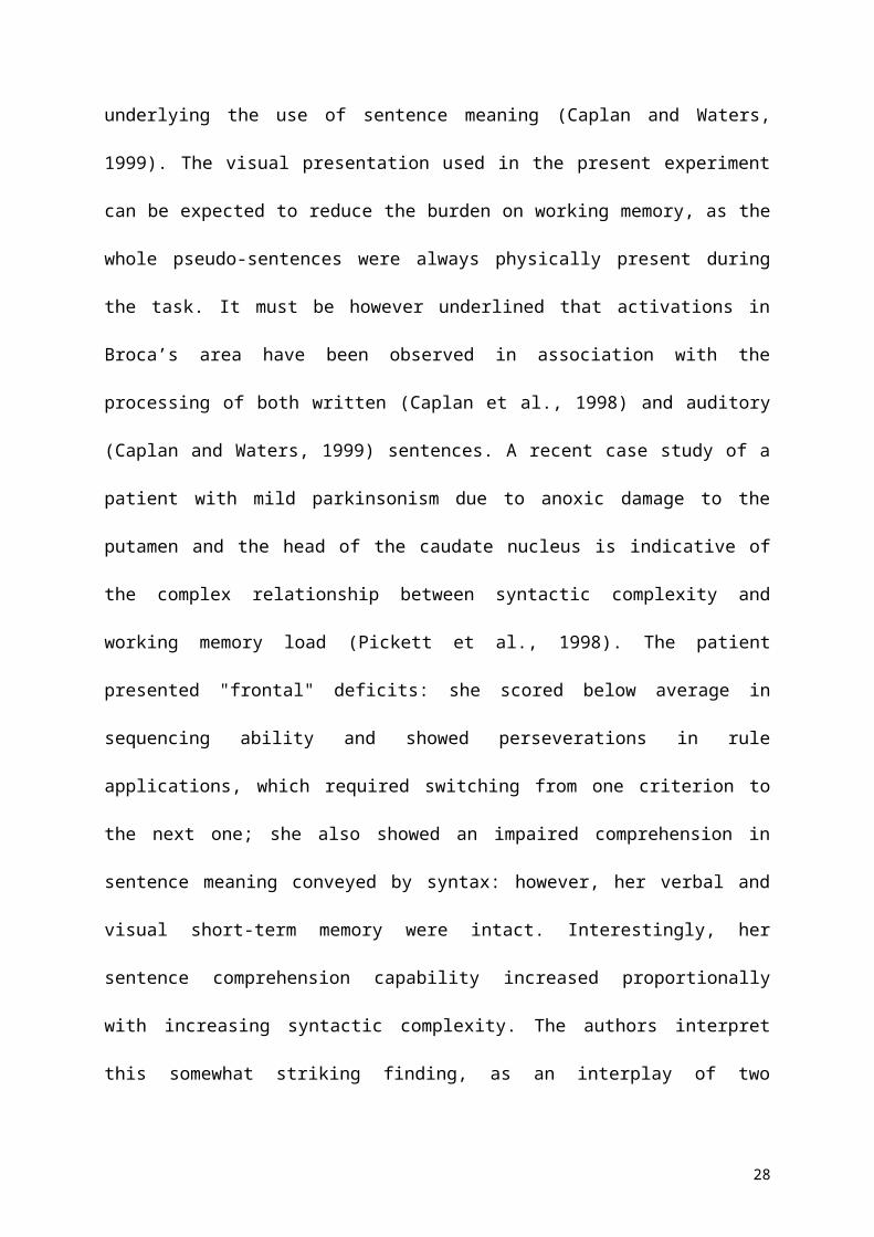
A
- 32- 48 - 16 - 4 + 8
+ 24 + 32 + 40 + 48 + 56
C
+ 240 + 8 + 12 + 16
+ 12- 44 - 36 + 4 + 8
B
28