Embed Size (px)
Citation preview
CERAMICSINTERNATIONAL
Available online at www.sciencedirect.com
http://dx.doi.org/0272-8842/& 20
nCorrespondinE-mail addre
bhamidreza2@li
(2014) 14009–14018
Ceramics International 40 www.elsevier.com/locate/ceramintSynthesis and biodegradation evaluation of nano-Si and nano-Si/TiO2
coatings on biodegradable Mg–Ca alloy in simulated body fluid
H.R. Bakhsheshi-Rada,n, E. Hamzaha, M. Daroonparvara, M. Kasiri-Asgaranib, M. Medrajc
aDepartment of Materials, Manufacturing and Industrial Engineering, Faculty of Mechanical Engineering, Universiti, Teknologi Malaysia,81310 Johor Bahru, Johor, Malaysia
bMaterials Engineering Department, Najafabad Branch, Islamic Azad University, Najafabad, Isfahan, IrancDepartment of Mechanical Engineering, Concordia University, 1455 De Maisonneuve Blvd. West, Montreal, Canada QC H3G 1M8
Received 5 March 2014; received in revised form 8 May 2014; accepted 27 May 2014Available online 4 June 2014
Abstract
In the present study, nano-Si and nano-Si/TiO2 composite coatings have been successfully synthesized on the surface of Mg–1 wt%Ca alloy bythe physical vapor deposition (PVD) method. The surface morphology and compositions of the coated specimens were characterized by X-raydiffraction (XRD), Fourier-transform infrared spectroscopy (FTIR), transmission electron microscopy (TEM), scanning electron microscopy(SEM) and energy dispersive X-ray spectroscopy (EDS). The results show the formation of thin and compact coating layers whichhomogeneously cover the surface of Mg alloy. Some micro-pores and micro-cracks were observed in the Si and Si/TiO2 films. It was also foundthat the Si and TiO2 nanoparticle had a spherical morphology with an average particle size of 30–40 nm and 70–80 nm, respectively.Electrochemical studies revealed that nano-Si/TiO2 coating offers a significant reduction in the corrosion rate (0.57 mm/year) compared to the Sicoated (0.91 mm/year) and the uncoated alloys (6.21 mm/year) in simulated body fluid (SBF). Hydrogen evolution studies showed a lowerdegradation rate of nano-Si/TiO2 (1.57 ml/cm2/day) than that of nano-Si coated alloy (2.22 ml/cm2/day). Immersion test showed that the nano-Si/TiO2 coating presented a greater nucleation site of hydroxyapatite (HA) than the uncoated sample. Thus nano-Si/TiO2 composite coating preparedby PVD on the Mg–Ca alloy is more appropriate for biomedical applications.& 2014 Elsevier Ltd and Techna Group S.r.l. All rights reserved.
Keywords: B. Composites; C. Corrosion; C. Mechanical properties; D. TiO2; E. Biomedical applications
1. Introduction
Magnesium alloys as biodegradable materials have drawnmuch interest due to eliminating the need for a second surgeryto remove the biomedical implants [1,2]. Mg-based biomater-ials have inevitable roles in human metabolism, good biocom-patibility, and superior mechanical properties [3,4]. However,clinical applications of Mg are limited due to its relatively poorcorrosion resistance, rapid degradation rate and hydrogen gasevolution in the human body fluid [5–7]. The deposition ofcoatings by physical vapor deposition (PVD) is an effective
10.1016/j.ceramint.2014.05.12614 Elsevier Ltd and Techna Group S.r.l. All rights reserved.
g author. Tel.: þ0060 147382258.sses: [email protected],ve.utm.my (H.R. Bakhsheshi-Rad).
method to overcome these drawbacks and enhance the corro-sion resistance of these alloys [8]. In addition, PVD is a cleanenvironmental friendly technique, and does not produce wastesor appreciable atmospheric emissions. There are severalstudies related to the surface modifications of magnesiumalloys by TiN, CrN, and AlN PVD coatings and theirprotection ability against corrosion [9,10]. In this study, siliconcoating was selected because it is an essential ion in osteogeniccells and due to its biocompatibility, bioinert nature tobiological tissues and more importantly, silicon deficiencycan cause abnormal bone growth [1,2,11]. Titania (TiO2) isalso a non-toxic and chemically stable material that can beused to improve the biocompatibility of the substrate [12].However, studies on preparation and characterization of nano-Si and nano-Si/TiO2 on magnesium alloy by the physical
H.R. Bakhsheshi-Rad et al. / Ceramics International 40 (2014) 14009–1401814010
vapor deposition technique could not be found in the literature.Therefore, in the present study, PVD is utilized to fabricatenano-Si and nano-Si/TiO2 composite coatings and elaboratetheir surface characteristics and in-vitro corrosion behavior.
2. Experimental details
Pure magnesium (99.98% Mg), and Mg–32%Ca masteralloy were used to prepare magnesium alloy containing 1 wt%Ca. In preparation for further experiments, several Mg–1%Caspecimens, with dimensions of 20 mm� 15 mm� 10 mm,were cut from the ingot. Then, they were mechanically wetground with 320–2000 SiC grit papers until all visiblescratches were removed. A hybrid ion beam deposition system,consisting of a linear ion source and a magnetron sputteringsource, was selected to deposit the coatings on the substrates.The Mg alloys were ultrasonically washed in pure alcohol for5 min before placement inside the vacuum chamber. An ionsource with Ar gas was used to clean the surface of the Mgalloys for 40 min. This pre-treatment was performed, when thebase pressure of the chamber was below 4� 10�3 Torr.Physical vapor deposition was performed at room temperaturewith argon and oxygen as the sputtering gases. It should benoted that Ar gas was used for the Si layer and that bothoxygen and argon gases were employed for TiO2 as a secondlayer. The PVD parameters are as follows: a working pressureof 8� 10�3 Torr, a radio frequency (RF) power of 200 W,a deposition time of 90 min and a bias voltage of �150 V.
AB
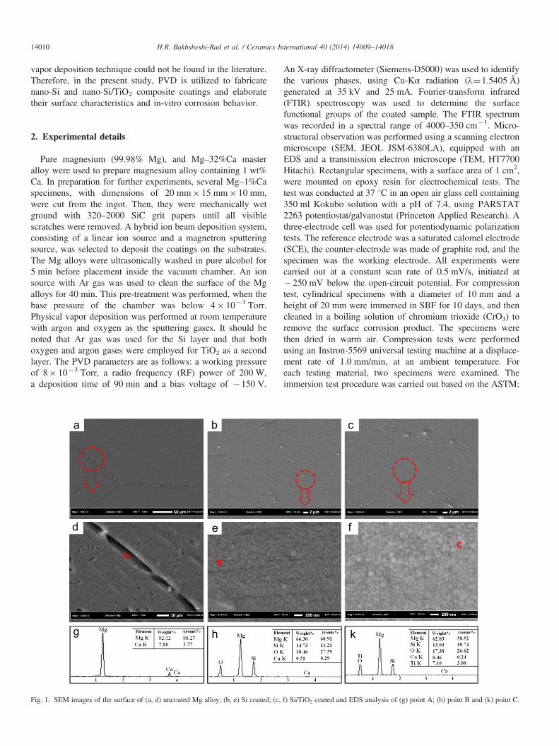
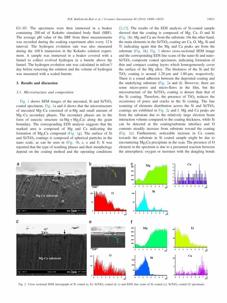
Fig. 1. SEM images of the surface of (a, d) uncoated Mg alloy; (b, e) Si coated; (c,
An X-ray diffractometer (Siemens-D5000) was used to identifythe various phases, using Cu-Kα radiation (λ¼1.5405 Å)generated at 35 kV and 25 mA. Fourier-transform infrared(FTIR) spectroscopy was used to determine the surfacefunctional groups of the coated sample. The FTIR spectrumwas recorded in a spectral range of 4000–350 cm�1. Micro-structural observation was performed using a scanning electronmicroscope (SEM, JEOL JSM-6380LA), equipped with anEDS and a transmission electron microscope (TEM, HT7700Hitachi). Rectangular specimens, with a surface area of 1 cm2,were mounted on epoxy resin for electrochemical tests. Thetest was conducted at 37 1C in an open air glass cell containing350 ml Kokubo solution with a pH of 7.4, using PARSTAT2263 potentiostat/galvanostat (Princeton Applied Research). Athree-electrode cell was used for potentiodynamic polarizationtests. The reference electrode was a saturated calomel electrode(SCE), the counter-electrode was made of graphite rod, and thespecimen was the working electrode. All experiments werecarried out at a constant scan rate of 0.5 mV/s, initiated at�250 mV below the open-circuit potential. For compressiontest, cylindrical specimens with a diameter of 10 mm and aheight of 20 mm were immersed in SBF for 10 days, and thencleaned in a boiling solution of chromium trioxide (CrO3) toremove the surface corrosion product. The specimens werethen dried in warm air. Compression tests were performedusing an Instron-5569 universal testing machine at a displace-ment rate of 1.0 mm/min, at an ambient temperature. Foreach testing material, two specimens were examined. Theimmersion test procedure was carried out based on the ASTM:
C
f) Si/TiO2 coated and EDS analysis of (g) point A; (h) point B and (k) point C.
H.R. Bakhsheshi-Rad et al. / Ceramics International 40 (2014) 14009–14018 14011
G1–03. The specimens were then immersed in a beakercontaining 200 ml of Kokubo simulated body fluid (SBF).The average pH value of the SBF from three measurementswas recorded during the soaking experiment after every 12 hinterval. The hydrogen evolution rate was also measuredduring the 168 h immersion in the Kokubo solution experi-ment. A sample was immersed in a beaker covered with afunnel to collect evolved hydrogen in a burette above thefunnel. The hydrogen evolution rate was calculated in ml/cm2/day before renewing the solution and the volume of hydrogenwas measured with a scaled burette.
3. Results and discussion
3.1. Microstructure and composition
Fig. 1 shows SEM images of the uncoated, Si and Si/TiO2
coated specimens. Fig. 1a and d shows that the microstructureof uncoated Mg–Ca consisted of α-Mg and small amount ofMg–Ca secondary phases. The secondary phases are in theform of eutectic structure (α-MgþMg2Ca) along the grainboundary. The corresponding EDS analysis suggests that themarked area is composed of Mg and Ca indicating theformation of Mg2Ca compound (Fig. 1g). The surface of Siand Si/TiO2 coatings is composed of spherical particles in thenano scale, as can be seen in (Fig. 1b, c, e and f). It wasreported that the type of resulting phases and their morphologydepend on the coating method and the operating conditions
Fig. 2. Cross sectional SEM micrograph of Si coated (a, b); Si/TiO2 coated (d,
[2,13]. The results of the EDS analysis of Si-coated sampleshowed that the coating is composed of Mg, Ca, O and Si(Fig. 1h). Mg and Ca are from the substrate. On the other hand,the main elements in the Si/TiO2 coating are Ca, O, Mg, Si andTi indicating again that the Mg and Ca peaks are from thesubstrate (Fig. 1k). Fig. 2 shows cross-sectional SEM imageand the corresponding EDS line scans of the nano-Si and nano-Si/TiO2 composite coated specimens, indicating formation ofthin and compact coating layers which homogeneously coverthe surface of the Mg alloy. The thickness of the Si and Si/TiO2 coating is around 1.20 mm and 1.80 μm, respectively.There is a sound adhesion between the deposited coating andthe underlying substrate (Fig. 2a and d). However, there aresome micro-pores and micro-flaws in the film, but themicrostructure of the Si/TiO2 coating is denser than that ofthe Si coating. Therefore, the presence of TiO2 reduces theoccurrence of pores and cracks in the Si coating. The linescanning of elements distribution across the Si and Si/TiO2
coatings are exhibited in Fig. 2c and f. Mg and Ca peaks arefrom the substrate due to the relatively large electron beaminteraction volume compared to the coating thickness, while Sican be detected at the coating/substrate interface and Ocontents steadily increase from substrate toward the coating(Fig. 2c). Furthermore, noticeable increase in Ca countstowards the substrate in Si coated sample might be due toencountering Mg2Ca precipitate in the scan. The presence of Oelement in the spectrum is due to a presumed reaction betweenthe atmospheric oxygen or moisture with the dangling bonds
e) and EDX line scans of Si coated (c); Si/TiO2 coated (f) specimens.
(110)
(111)
Si particlesTiO2 particle
(210)
(111)
(200)
50 nm 50 nm
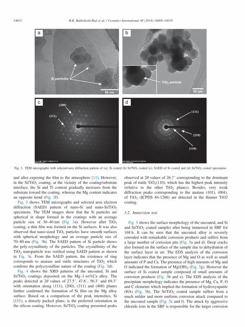
Fig. 3. TEM micrographs with selected-area diffraction pattern of (a); Si coated (b) Si/TiO2 coated (c); SAED of Si coated and (d) Si/TiO2 coated specimens.
H.R. Bakhsheshi-Rad et al. / Ceramics International 40 (2014) 14009–1401814012
and after exposing the film to the atmosphere [11]. However,in the Si/TiO2 coating, at the vicinity of the coating/substrateinterface, the Si and Ti content gradually increases from thesubstrate toward the coating, whereas the Mg content indicatesan opposite trend (Fig. 2f).
Fig. 3 shows TEM micrographs and selected area electrondiffraction (SAED) pattern of nano-Si and nano-Si/TiO2
specimens. The TEM images show that the Si particles arespherical in shape formed in the coatings with an averageparticle size of 30–40 nm (Fig. 3a). However after TiO2
coating, a thin film was formed on the Si surfaces. It was alsoobserved that nano-sized TiO2 particles have smooth surfaceswith spherical morphology and an average particle size of70–80 nm (Fig. 3b). The SAED pattern of Si particle showsthe poly-crystallinity of the particles. The crystallinity of theTiO2 nanoparticle was verified using SAED pattern as shownin Fig. 3c. From the SAED pattern, the existence of ringcorresponds to anatase and rutile structures of TiO2 whichconfirms the polycrystalline nature of the coating (Fig. 3d).
Fig. 4 shows the XRD patterns of the uncoated, Si andSi/TiO2 coatings deposited on the Mg–1 wt%Ca alloy. Thepeaks detected at 2θ values of 27.51, 47.61, 56.31 and 69.31with orientation along (111), (200), (311) and (400) planesfurther confirmed the formation of Si film on the Mg alloysurface. Based on a comparison of the peak intensities, Si(111), a densely packed plane, is the preferred orientation inthe silicon coating. However, Si/TiO2 coating presented peaks
observed at 2θ values of 26.71 corresponding to the dominantpeak of rutile TiO2(110), which has the highest peak intensity(relative to the other TiO2 planes). Besides, very weakdiffraction peaks corresponding to the anatase (101), (004),of TiO2 (JCPDS 84-1286) are detected in the thinner TiO2coating.
3.2. Immersion test
Fig. 5 shows the surface morphology of the uncoated, and Siand Si/TiO2 coated samples after being immersed in SBF for168 h. It can be seen that the uncoated alloy is severelycorroded with remarkable corrosion products and suffers froma large number of corrosion pits (Fig. 5a and d). Deep cracksalso formed on the surface of the sample due to dehydration ofthe surface layer in air. The EDS analysis of the corrosionlayer indicates that the presence of Mg and O as well as smallamounts of P and Ca. The presence of high amounts of Mg andO indicated the formation of Mg(OH)2 (Fig. 5g). However thesurface of Si coated sample composed of small amounts ofcorrosion products (Fig. 5b and e). The EDS analysis of theprecipitate morphology indicates the presence of Mg, Ca, P, Oand C elements which implied the formation of hydroxyapatite(HA) (Fig. 5h). The Si/TiO2 coated sample suffers from amuch milder and more uniform corrosion attack compared tothe uncoated sample (Fig. 5c and f). The attack by aggressivechloride ions in the SBF is responsible for the larger corrosion
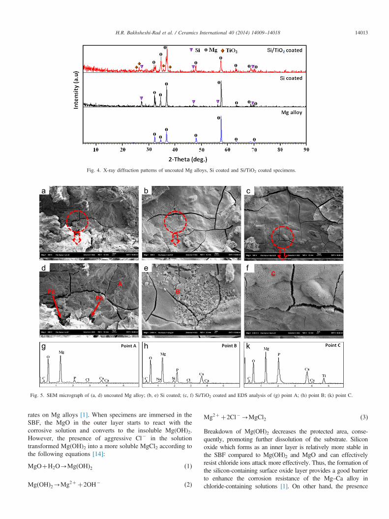
Fig. 4. X-ray diffraction patterns of uncoated Mg alloys, Si coated and Si/TiO2 coated specimens.
PitPit
AB
C
Fig. 5. SEM micrograph of (a, d) uncoated Mg alloy; (b, e) Si coated; (c, f) Si/TiO2 coated and EDS analysis of (g) point A; (h) point B; (k) point C.
H.R. Bakhsheshi-Rad et al. / Ceramics International 40 (2014) 14009–14018 14013
rates on Mg alloys [1]. When specimens are immersed in theSBF, the MgO in the outer layer starts to react with thecorrosive solution and converts to the insoluble Mg(OH)2.However, the presence of aggressive Cl� in the solutiontransformed Mg(OH)2 into a more soluble MgCl2 according tothe following equations [14]:
MgOþH2O-Mg OHð Þ2 ð1Þ
MgðOHÞ2-Mg2þ þ2OH� ð2Þ
Mg2þ þ2Cl�-MgCl2 ð3Þ
Breakdown of Mg(OH)2 decreases the protected area, conse-quently, promoting further dissolution of the substrate. Siliconoxide which forms as an inner layer is relatively more stable inthe SBF compared to Mg(OH)2 and MgO and can effectivelyresist chloride ions attack more effectively. Thus, the formation ofthe silicon-containing surface oxide layer provides a good barrierto enhance the corrosion resistance of the Mg–Ca alloy inchloride-containing solutions [1]. On other hand, the presence
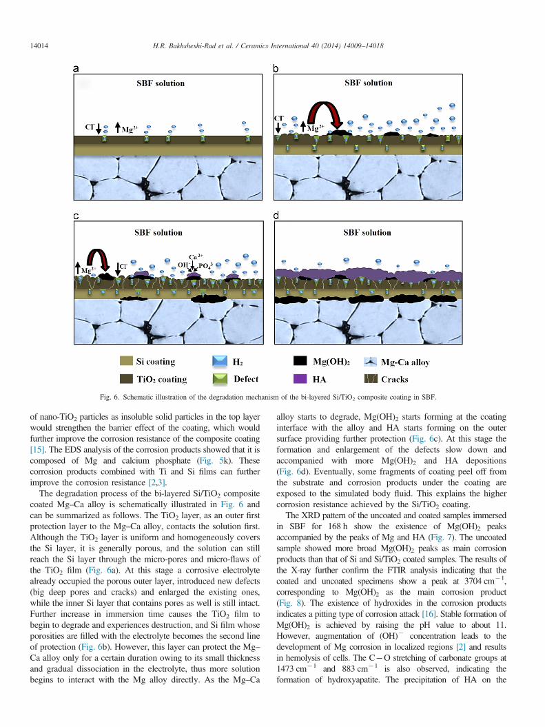
Fig. 6. Schematic illustration of the degradation mechanism of the bi-layered Si/TiO2 composite coating in SBF.
H.R. Bakhsheshi-Rad et al. / Ceramics International 40 (2014) 14009–1401814014
of nano-TiO2 particles as insoluble solid particles in the top layerwould strengthen the barrier effect of the coating, which wouldfurther improve the corrosion resistance of the composite coating[15]. The EDS analysis of the corrosion products showed that it iscomposed of Mg and calcium phosphate (Fig. 5k). Thesecorrosion products combined with Ti and Si films can furtherimprove the corrosion resistance [2,3].
The degradation process of the bi-layered Si/TiO2 compositecoated Mg–Ca alloy is schematically illustrated in Fig. 6 andcan be summarized as follows. The TiO2 layer, as an outer firstprotection layer to the Mg–Ca alloy, contacts the solution first.Although the TiO2 layer is uniform and homogeneously coversthe Si layer, it is generally porous, and the solution can stillreach the Si layer through the micro-pores and micro-flaws ofthe TiO2 film (Fig. 6a). At this stage a corrosive electrolytealready occupied the porous outer layer, introduced new defects(big deep pores and cracks) and enlarged the existing ones,while the inner Si layer that contains pores as well is still intact.Further increase in immersion time causes the TiO2 film tobegin to degrade and experiences destruction, and Si film whoseporosities are filled with the electrolyte becomes the second lineof protection (Fig. 6b). However, this layer can protect the Mg–Ca alloy only for a certain duration owing to its small thicknessand gradual dissociation in the electrolyte, thus more solutionbegins to interact with the Mg alloy directly. As the Mg–Ca
alloy starts to degrade, Mg(OH)2 starts forming at the coatinginterface with the alloy and HA starts forming on the outersurface providing further protection (Fig. 6c). At this stage theformation and enlargement of the defects slow down andaccompanied with more Mg(OH)2 and HA depositions(Fig. 6d). Eventually, some fragments of coating peel off fromthe substrate and corrosion products under the coating areexposed to the simulated body fluid. This explains the highercorrosion resistance achieved by the Si/TiO2 coating.The XRD pattern of the uncoated and coated samples immersed
in SBF for 168 h show the existence of Mg(OH)2 peaksaccompanied by the peaks of Mg and HA (Fig. 7). The uncoatedsample showed more broad Mg(OH)2 peaks as main corrosionproducts than that of Si and Si/TiO2 coated samples. The results ofthe X-ray further confirm the FTIR analysis indicating that thecoated and uncoated specimens show a peak at 3704 cm�1,corresponding to Mg(OH)2 as the main corrosion product(Fig. 8). The existence of hydroxides in the corrosion productsindicates a pitting type of corrosion attack [16]. Stable formation ofMg(OH)2 is achieved by raising the pH value to about 11.However, augmentation of (OH)� concentration leads to thedevelopment of Mg corrosion in localized regions [2] and resultsin hemolysis of cells. The C�O stretching of carbonate groups at1473 cm�1 and 883 cm�1 is also observed, indicating theformation of hydroxyapatite. The precipitation of HA on the
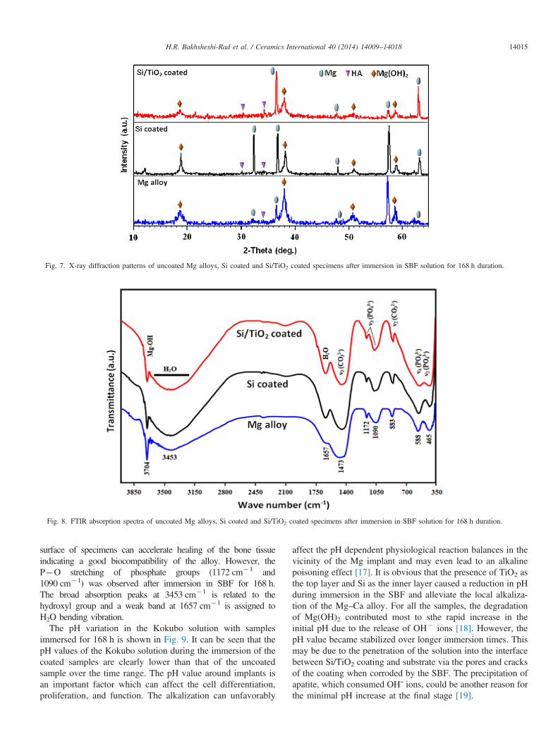
Fig. 7. X-ray diffraction patterns of uncoated Mg alloys, Si coated and Si/TiO2 coated specimens after immersion in SBF solution for 168 h duration.
Fig. 8. FTIR absorption spectra of uncoated Mg alloys, Si coated and Si/TiO2 coated specimens after immersion in SBF solution for 168 h duration.
H.R. Bakhsheshi-Rad et al. / Ceramics International 40 (2014) 14009–14018 14015
surface of specimens can accelerate healing of the bone tissueindicating a good biocompatibility of the alloy. However, theP�O stretching of phosphate groups (1172 cm�1 and1090 cm�1) was observed after immersion in SBF for 168 h.The broad absorption peaks at 3453 cm�1 is related to thehydroxyl group and a weak band at 1657 cm�1 is assigned toH2O bending vibration.
The pH variation in the Kokubo solution with samplesimmersed for 168 h is shown in Fig. 9. It can be seen that thepH values of the Kokubo solution during the immersion of thecoated samples are clearly lower than that of the uncoatedsample over the time range. The pH value around implants isan important factor which can affect the cell differentiation,proliferation, and function. The alkalization can unfavorably
affect the pH dependent physiological reaction balances in thevicinity of the Mg implant and may even lead to an alkalinepoisoning effect [17]. It is obvious that the presence of TiO2 asthe top layer and Si as the inner layer caused a reduction in pHduring immersion in the SBF and alleviate the local alkaliza-tion of the Mg–Ca alloy. For all the samples, the degradationof Mg(OH)2 contributed most to sthe rapid increase in theinitial pH due to the release of OH� ions [18]. However, thepH value became stabilized over longer immersion times. Thismay be due to the penetration of the solution into the interfacebetween Si/TiO2 coating and substrate via the pores and cracksof the coating when corroded by the SBF. The precipitation ofapatite, which consumed OHˉ ions, could be another reason forthe minimal pH increase at the final stage [19].
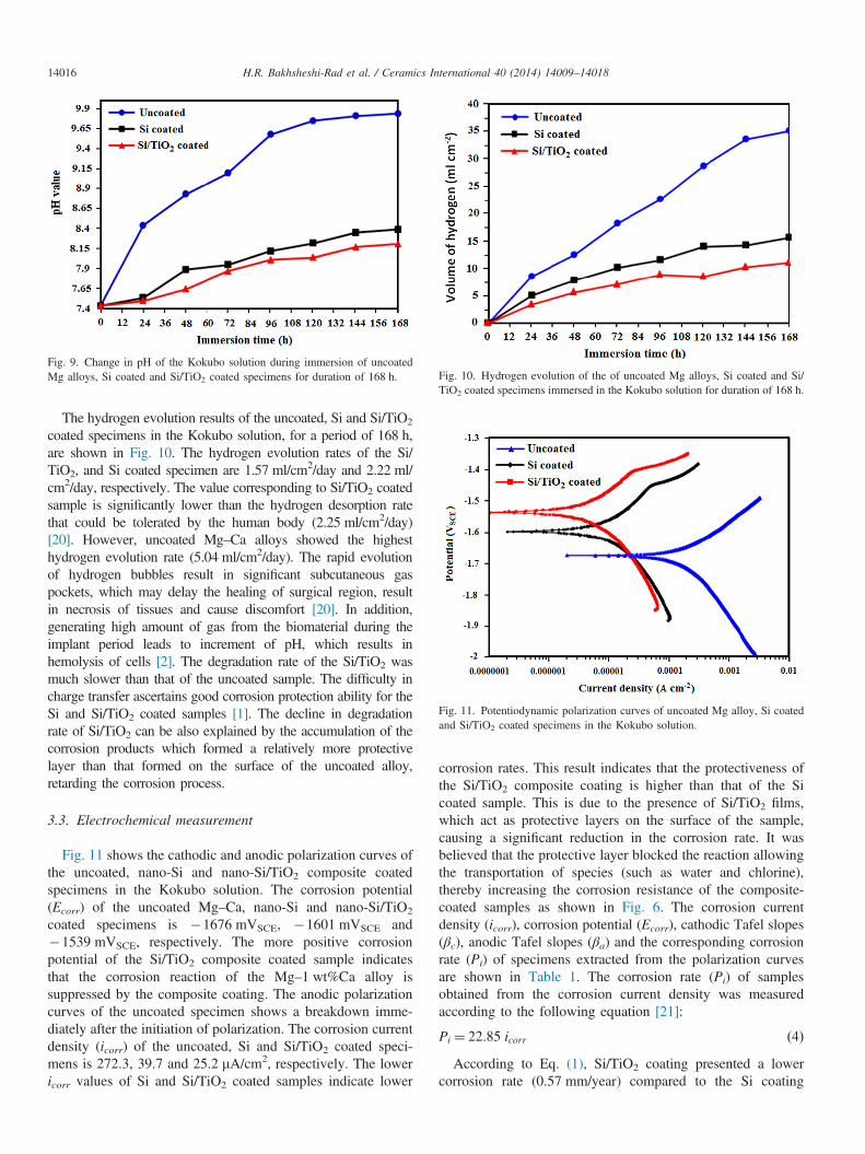
Fig. 9. Change in pH of the Kokubo solution during immersion of uncoatedMg alloys, Si coated and Si/TiO2 coated specimens for duration of 168 h. Fig. 10. Hydrogen evolution of the of uncoated Mg alloys, Si coated and Si/
TiO2 coated specimens immersed in the Kokubo solution for duration of 168 h.
Fig. 11. Potentiodynamic polarization curves of uncoated Mg alloy, Si coatedand Si/TiO2 coated specimens in the Kokubo solution.
H.R. Bakhsheshi-Rad et al. / Ceramics International 40 (2014) 14009–1401814016
The hydrogen evolution results of the uncoated, Si and Si/TiO2
coated specimens in the Kokubo solution, for a period of 168 h,are shown in Fig. 10. The hydrogen evolution rates of the Si/TiO2, and Si coated specimen are 1.57 ml/cm2/day and 2.22 ml/cm2/day, respectively. The value corresponding to Si/TiO2 coatedsample is significantly lower than the hydrogen desorption ratethat could be tolerated by the human body (2.25 ml/cm2/day)[20]. However, uncoated Mg–Ca alloys showed the highesthydrogen evolution rate (5.04 ml/cm2/day). The rapid evolutionof hydrogen bubbles result in significant subcutaneous gaspockets, which may delay the healing of surgical region, resultin necrosis of tissues and cause discomfort [20]. In addition,generating high amount of gas from the biomaterial during theimplant period leads to increment of pH, which results inhemolysis of cells [2]. The degradation rate of the Si/TiO2 wasmuch slower than that of the uncoated sample. The difficulty incharge transfer ascertains good corrosion protection ability for theSi and Si/TiO2 coated samples [1]. The decline in degradationrate of Si/TiO2 can be also explained by the accumulation of thecorrosion products which formed a relatively more protectivelayer than that formed on the surface of the uncoated alloy,retarding the corrosion process.
3.3. Electrochemical measurement
Fig. 11 shows the cathodic and anodic polarization curves ofthe uncoated, nano-Si and nano-Si/TiO2 composite coatedspecimens in the Kokubo solution. The corrosion potential(Ecorr) of the uncoated Mg–Ca, nano-Si and nano-Si/TiO2
coated specimens is �1676 mVSCE, �1601 mVSCE and�1539 mVSCE, respectively. The more positive corrosionpotential of the Si/TiO2 composite coated sample indicatesthat the corrosion reaction of the Mg–1 wt%Ca alloy issuppressed by the composite coating. The anodic polarizationcurves of the uncoated specimen shows a breakdown imme-diately after the initiation of polarization. The corrosion currentdensity (icorr) of the uncoated, Si and Si/TiO2 coated speci-mens is 272.3, 39.7 and 25.2 mA/cm2, respectively. The lowericorr values of Si and Si/TiO2 coated samples indicate lower
corrosion rates. This result indicates that the protectiveness ofthe Si/TiO2 composite coating is higher than that of the Sicoated sample. This is due to the presence of Si/TiO2 films,which act as protective layers on the surface of the sample,causing a significant reduction in the corrosion rate. It wasbelieved that the protective layer blocked the reaction allowingthe transportation of species (such as water and chlorine),thereby increasing the corrosion resistance of the composite-coated samples as shown in Fig. 6. The corrosion currentdensity (icorr), corrosion potential (Ecorr), cathodic Tafel slopes(βc), anodic Tafel slopes (βa) and the corresponding corrosionrate (Pi) of specimens extracted from the polarization curvesare shown in Table 1. The corrosion rate (Pi) of samplesobtained from the corrosion current density was measuredaccording to the following equation [21]:
Pi ¼ 22:85 icorr ð4ÞAccording to Eq. (1), Si/TiO2 coating presented a lower
corrosion rate (0.57 mm/year) compared to the Si coating
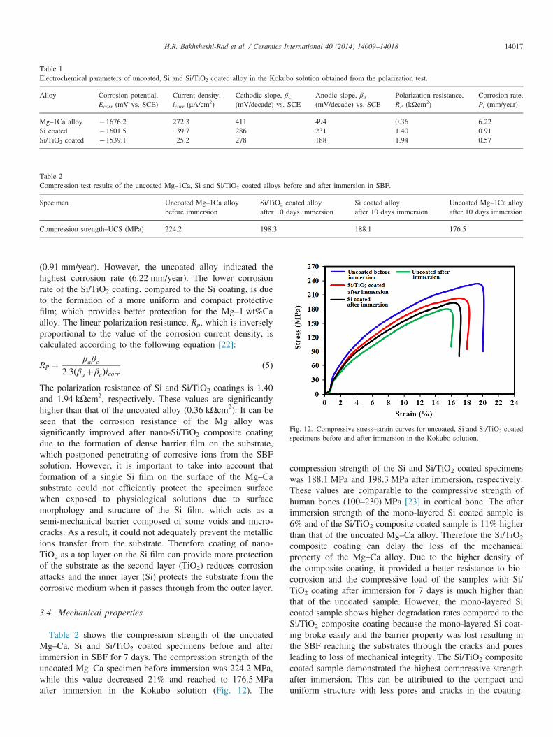
Table 1Electrochemical parameters of uncoated, Si and Si/TiO2 coated alloy in the Kokubo solution obtained from the polarization test.
Alloy Corrosion potential,Ecorr (mV vs. SCE)
Current density,icorr (μA/cm2)
Cathodic slope, βC(mV/decade) vs. SCE
Anodic slope, βa(mV/decade) vs. SCE
Polarization resistance,RP (kΩcm2)
Corrosion rate,Pi (mm/year)
Mg–1Ca alloy �1676.2 272.3 411 494 0.36 6.22Si coated �1601.5 39.7 286 231 1.40 0.91Si/TiO2 coated �1539.1 25.2 278 188 1.94 0.57
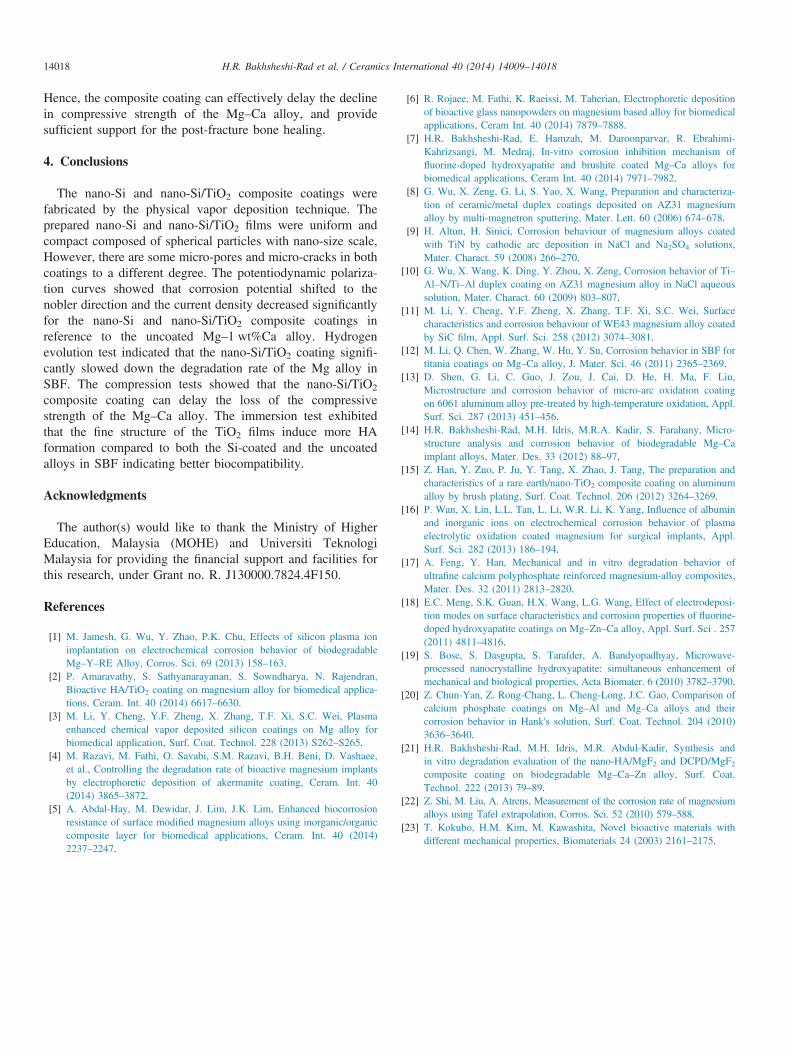
Table 2Compression test results of the uncoated Mg–1Ca, Si and Si/TiO2 coated alloys before and after immersion in SBF.
Specimen Uncoated Mg–1Ca alloybefore immersion
Si/TiO2 coated alloyafter 10 days immersion
Si coated alloyafter 10 days immersion
Uncoated Mg–1Ca alloyafter 10 days immersion
Compression strength–UCS (MPa) 224.2 198.3 188.1 176.5
Fig. 12. Compressive stress–strain curves for uncoated, Si and Si/TiO2 coatedspecimens before and after immersion in the Kokubo solution.
H.R. Bakhsheshi-Rad et al. / Ceramics International 40 (2014) 14009–14018 14017
(0.91 mm/year). However, the uncoated alloy indicated thehighest corrosion rate (6.22 mm/year). The lower corrosionrate of the Si/TiO2 coating, compared to the Si coating, is dueto the formation of a more uniform and compact protectivefilm; which provides better protection for the Mg–1 wt%Caalloy. The linear polarization resistance, Rp, which is inverselyproportional to the value of the corrosion current density, iscalculated according to the following equation [22]:
RP ¼βaβc
2:3ðβaþβcÞicorrð5Þ
The polarization resistance of Si and Si/TiO2 coatings is 1.40and 1.94 kΩcm2, respectively. These values are significantlyhigher than that of the uncoated alloy (0.36 kΩcm2). It can beseen that the corrosion resistance of the Mg alloy wassignificantly improved after nano-Si/TiO2 composite coatingdue to the formation of dense barrier film on the substrate,which postponed penetrating of corrosive ions from the SBFsolution. However, it is important to take into account thatformation of a single Si film on the surface of the Mg–Casubstrate could not efficiently protect the specimen surfacewhen exposed to physiological solutions due to surfacemorphology and structure of the Si film, which acts as asemi-mechanical barrier composed of some voids and micro-cracks. As a result, it could not adequately prevent the metallicions transfer from the substrate. Therefore coating of nano-TiO2 as a top layer on the Si film can provide more protectionof the substrate as the second layer (TiO2) reduces corrosionattacks and the inner layer (Si) protects the substrate from thecorrosive medium when it passes through from the outer layer.
3.4. Mechanical properties
Table 2 shows the compression strength of the uncoatedMg–Ca, Si and Si/TiO2 coated specimens before and afterimmersion in SBF for 7 days. The compression strength of theuncoated Mg–Ca specimen before immersion was 224.2 MPa,while this value decreased 21% and reached to 176.5 MPaafter immersion in the Kokubo solution (Fig. 12). The
compression strength of the Si and Si/TiO2 coated specimenswas 188.1 MPa and 198.3 MPa after immersion, respectively.These values are comparable to the compressive strength ofhuman bones (100–230) MPa [23] in cortical bone. The afterimmersion strength of the mono-layered Si coated sample is6% and of the Si/TiO2 composite coated sample is 11% higherthan that of the uncoated Mg–Ca alloy. Therefore the Si/TiO2
composite coating can delay the loss of the mechanicalproperty of the Mg–Ca alloy. Due to the higher density ofthe composite coating, it provided a better resistance to bio-corrosion and the compressive load of the samples with Si/TiO2 coating after immersion for 7 days is much higher thanthat of the uncoated sample. However, the mono-layered Sicoated sample shows higher degradation rates compared to theSi/TiO2 composite coating because the mono-layered Si coat-ing broke easily and the barrier property was lost resulting inthe SBF reaching the substrates through the cracks and poresleading to loss of mechanical integrity. The Si/TiO2 compositecoated sample demonstrated the highest compressive strengthafter immersion. This can be attributed to the compact anduniform structure with less pores and cracks in the coating.
H.R. Bakhsheshi-Rad et al. / Ceramics International 40 (2014) 14009–1401814018
Hence, the composite coating can effectively delay the declinein compressive strength of the Mg–Ca alloy, and providesufficient support for the post-fracture bone healing.
4. Conclusions
The nano-Si and nano-Si/TiO2 composite coatings werefabricated by the physical vapor deposition technique. Theprepared nano-Si and nano-Si/TiO2 films were uniform andcompact composed of spherical particles with nano-size scale,However, there are some micro-pores and micro-cracks in bothcoatings to a different degree. The potentiodynamic polariza-tion curves showed that corrosion potential shifted to thenobler direction and the current density decreased significantlyfor the nano-Si and nano-Si/TiO2 composite coatings inreference to the uncoated Mg–1 wt%Ca alloy. Hydrogenevolution test indicated that the nano-Si/TiO2 coating signifi-cantly slowed down the degradation rate of the Mg alloy inSBF. The compression tests showed that the nano-Si/TiO2
composite coating can delay the loss of the compressivestrength of the Mg–Ca alloy. The immersion test exhibitedthat the fine structure of the TiO2 films induce more HAformation compared to both the Si-coated and the uncoatedalloys in SBF indicating better biocompatibility.
Acknowledgments
The author(s) would like to thank the Ministry of HigherEducation, Malaysia (MOHE) and Universiti TeknologiMalaysia for providing the financial support and facilities forthis research, under Grant no. R. J130000.7824.4F150.
References
[1] M. Jamesh, G. Wu, Y. Zhao, P.K. Chu, Effects of silicon plasma ionimplantation on electrochemical corrosion behavior of biodegradableMg–Y–RE Alloy, Corros. Sci. 69 (2013) 158–163.
[2] P. Amaravathy, S. Sathyanarayanan, S. Sowndharya, N. Rajendran,Bioactive HA/TiO2 coating on magnesium alloy for biomedical applica-tions, Ceram. Int. 40 (2014) 6617–6630.
[3] M. Li, Y. Cheng, Y.F. Zheng, X. Zhang, T.F. Xi, S.C. Wei, Plasmaenhanced chemical vapor deposited silicon coatings on Mg alloy forbiomedical application, Surf. Coat. Technol. 228 (2013) S262–S265.
[4] M. Razavi, M. Fathi, O. Savabi, S.M. Razavi, B.H. Beni, D. Vashaee,et al., Controlling the degradation rate of bioactive magnesium implantsby electrophoretic deposition of akermanite coating, Ceram. Int. 40(2014) 3865–3872.
[5] A. Abdal-Hay, M. Dewidar, J. Lim, J.K. Lim, Enhanced biocorrosionresistance of surface modified magnesium alloys using inorganic/organiccomposite layer for biomedical applications, Ceram. Int. 40 (2014)2237–2247.
[6] R. Rojaee, M. Fathi, K. Raeissi, M. Taherian, Electrophoretic depositionof bioactive glass nanopowders on magnesium based alloy for biomedicalapplications, Ceram Int. 40 (2014) 7879–7888.
[7] H.R. Bakhsheshi-Rad, E. Hamzah, M. Daroonparvar, R. Ebrahimi-Kahrizsangi, M. Medraj, In-vitro corrosion inhibition mechanism offluorine-doped hydroxyapatite and brushite coated Mg–Ca alloys forbiomedical applications, Ceram Int. 40 (2014) 7971–7982.
[8] G. Wu, X. Zeng, G. Li, S. Yao, X. Wang, Preparation and characteriza-tion of ceramic/metal duplex coatings deposited on AZ31 magnesiumalloy by multi-magnetron sputtering, Mater. Lett. 60 (2006) 674–678.
[9] H. Altun, H. Sinici, Corrosion behaviour of magnesium alloys coatedwith TiN by cathodic arc deposition in NaCl and Na2SO4 solutions,Mater. Charact. 59 (2008) 266–270.
[10] G. Wu, X. Wang, K. Ding, Y. Zhou, X. Zeng, Corrosion behavior of Ti–Al–N/Ti–Al duplex coating on AZ31 magnesium alloy in NaCl aqueoussolution, Mater. Charact. 60 (2009) 803–807.
[11] M. Li, Y. Cheng, Y.F. Zheng, X. Zhang, T.F. Xi, S.C. Wei, Surfacecharacteristics and corrosion behaviour of WE43 magnesium alloy coatedby SiC film, Appl. Surf. Sci. 258 (2012) 3074–3081.
[12] M. Li, Q. Chen, W. Zhang, W. Hu, Y. Su, Corrosion behavior in SBF fortitania coatings on Mg–Ca alloy, J. Mater. Sci. 46 (2011) 2365–2369.
[13] D. Shen, G. Li, C. Guo, J. Zou, J. Cai, D. He, H. Ma, F. Liu,Microstructure and corrosion behavior of micro-arc oxidation coatingon 6061 aluminum alloy pre-treated by high-temperature oxidation, Appl.Surf. Sci. 287 (2013) 451–456.
[14] H.R. Bakhsheshi-Rad, M.H. Idris, M.R.A. Kadir, S. Farahany, Micro-structure analysis and corrosion behavior of biodegradable Mg–Caimplant alloys, Mater. Des. 33 (2012) 88–97.
[15] Z. Han, Y. Zuo, P. Ju, Y. Tang, X. Zhao, J. Tang, The preparation andcharacteristics of a rare earth/nano-TiO2 composite coating on aluminumalloy by brush plating, Surf. Coat. Technol. 206 (2012) 3264–3269.
[16] P. Wan, X. Lin, L.L. Tan, L. Li, W.R. Li, K. Yang, Influence of albuminand inorganic ions on electrochemical corrosion behavior of plasmaelectrolytic oxidation coated magnesium for surgical implants, Appl.Surf. Sci. 282 (2013) 186–194.
[17] A. Feng, Y. Han, Mechanical and in vitro degradation behavior ofultrafine calcium polyphosphate reinforced magnesium-alloy composites,Mater. Des. 32 (2011) 2813–2820.
[18] E.C. Meng, S.K. Guan, H.X. Wang, L.G. Wang, Effect of electrodeposi-tion modes on surface characteristics and corrosion properties of fluorine-doped hydroxyapatite coatings on Mg–Zn–Ca alloy, Appl. Surf. Sci . 257(2011) 4811–4816.
[19] S. Bose, S. Dasgupta, S. Tarafder, A. Bandyopadhyay, Microwave-processed nanocrystalline hydroxyapatite: simultaneous enhancement ofmechanical and biological properties, Acta Biomater. 6 (2010) 3782–3790.
[20] Z. Chun-Yan, Z. Rong-Chang, L. Cheng-Long, J.C. Gao, Comparison ofcalcium phosphate coatings on Mg–Al and Mg–Ca alloys and theircorrosion behavior in Hank's solution, Surf. Coat. Technol. 204 (2010)3636–3640.
[21] H.R. Bakhsheshi-Rad, M.H. Idris, M.R. Abdul-Kadir, Synthesis andin vitro degradation evaluation of the nano-HA/MgF2 and DCPD/MgF2composite coating on biodegradable Mg–Ca–Zn alloy, Surf. Coat.Technol. 222 (2013) 79–89.
[22] Z. Shi, M. Liu, A. Atrens, Measurement of the corrosion rate of magnesiumalloys using Tafel extrapolation, Corros. Sci. 52 (2010) 579–588.
[23] T. Kokubo, H.M. Kim, M. Kawashita, Novel bioactive materials withdifferent mechanical properties, Biomaterials 24 (2003) 2161–2175.