Embed Size (px)
Citation preview

Journal of Physics Conference Series
OPEN ACCESS
Synthesis and characterisation of nanocrystallineiron oxides via ultrasonic spray assisted chemicalvapour depositionTo cite this article L T Chao et al 2006 J Phys Conf Ser 26 073
View the article online for updates and enhancements
You may also likeBPS state counting on singular varietiesElizabeth Gasparim Thomas KoumlppePushan Majumdar et al
-
The Breakthrough Listen Search forIntelligent Life Public Data FormatsReduction and ArchivingMatthew Lebofsky Steve Croft Andrew PV Siemion et al
-
Structural characterisation of doped andundoped nanocrystalline zinc oxidesdeposited by ultrasonic spray assistedchemical vapour depositionMing Wei Dan Zhi and Judith LMacManus-Driscoll
-
Recent citationsAndrea Ehrmann and Tomasz Blachowicz-
Viscosity Reduction of Heavy Oil UsingNanocatalyst in Aquathermolysis ReactionFerry Iskandar et al
-
Synthesis of NixFe3-xO4 Nanoparticles byMicrowave-Assisted Coprecipitation andtheir Application in Viscosity Reduction ofHeavy OilMohamad Insan Nugraha et al
-
This content was downloaded from IP address 21815780200 on 12112021 at 1238
Synthesis and characterisation of nanocrystalline iron oxides via ultrasonic spray assisted chemical vapour deposition
LT Chao M Wei and JL MacManus-Driscoll
Department of Materials Science and Metallurgy University of Cambridge Pembroke Street Cambridge CB2 3QZ UK
E-mail jld35camacuk
Abstract The synthesis and characterisation of nanocrystalline magnetic materials with different morphology have attracted much attention for understanding the fundamental aspects of magnetic-ordering with reduced dimensions and for possible new potential applications in data storage technology We report here a novel synthesis route for nanocrystalline magnetite Fe3O4 by ultrasonic spray assisted chemical vapour deposition in which either iron acetate or iron acetylacetonate are used as precursors Magnetically coupled nano-grains and nano-islands of Fe3O4 with different size and morphology can be produced at different conditions The structure of as-deposited and post-annealed nanoparticles was investigated by field emission gun scanning electron microscopy (FEGSEM) x-ray diffraction (XRD) and transmission electron microscopy (TEM) The surface roughness was measured by atomic force microscopy (AFM)
1 Introduction Magnetite (Fe3O4) has received much interest due to its high Curie temperature (860K) and fully spin polarised half metallic character which can allow for spin-dependent tunnelling magntoresistance (TMR) with quite a high efficiency [12] These characteristics can potentially be utilized in the development of magnetic random access memory (MRAM) devices that operate at room temperature with high performance capabilities [3] From the application point of view growth of epitaxial ferrite films is desirable and epitaxy should improve the magnetic properties of the film Moreover an epitaxial ferrite would allow the deposition of further epitaxial or oriented layers of other materials on the ferrite thus increasing the device-design possibilities There have been several methods to fabricate iron oxide films such as pulsed laser deposition (PLD) [2] oxygen-plasma-assisted molecular beam epitaxy [4] and chemical vapour deposition (CVD) [5] Apart from epitaxial films nanostructured magnetic iron oxides have also attracted increasing attention in recent years It has been expected that with reducing dimensions magnetic materials can bring new potential applications such as high density recording media and bioinspired materials Different processes for the growth and deposition of magnetic iron oxide nanoparticles and 1D materials such as single crystalline Fe3O4 nanowires [6] have been reported
In this report nanostructured iron oxides on various substrates have been studied using a novel ultrasonic spray assisted chemical vapour deposition Both self assembled nanocrystals and epitaxially grown nanoislands were deposited and characterized
Institute of Physics Publishing Journal of Physics Conference Series 26 (2006) 304ndash307doi1010881742-6596261073 EMAGndashNANO 05 Imaging Analysis and Fabrication on the Nanoscale
304copy 2006 IOP Publishing Ltd
2 Experimental The experimental setup is similar to the previous paper of our groups [78] In this study either iron(II) acetate or iron(III) 24-pentanedionate was used as a precursor Before the deposition 50ml-100ml diluted precursor solution (001-005M) was first delivered into a quartz glass container with a 05 cm diameter nozzle and an 8 cm diameter base The base of the quartz vessel was a 0075 mm thick polyethylene sheet which was secured with a clamp The vessel was then placed in a deionised water bath containing two 18 MHz transducers A substrate (Si or MgO) was mounted on the bottom of a hot-plate of which the temperature was kept constant (300-600) during the deposition After the substrate reached the desired temperature the precursor aerosol was generated from the vibration of the transducers and was delivered towards the heated substrate by the carrier gas argon The gas flow rate used was between 3-7 lmin The deposition time for the iron oxide was 10 to 80 minutes The solvent in the precursor solution was evaporated before reaching the heated substrate and the precursor was subjected to decomposition to form iron oxide on the various substrates
X-ray diffraction (XRD) measurements were made using a Phillips XrsquoPert x-ray diffractometer in the Bragg-Brentano geometry with Cu-K 1 radiation All samples were observed by using field emission gun scanning electron microscopy (FEGSEM JEOL 6330F) The determination of surface roughness as well as the size of the individual particulates was carried out using atomic force microscopy (AFM Digital Instrument Nanoscope III) Iron oxide nanoparticles were also characterized using transmission electron microscopy (TEM)
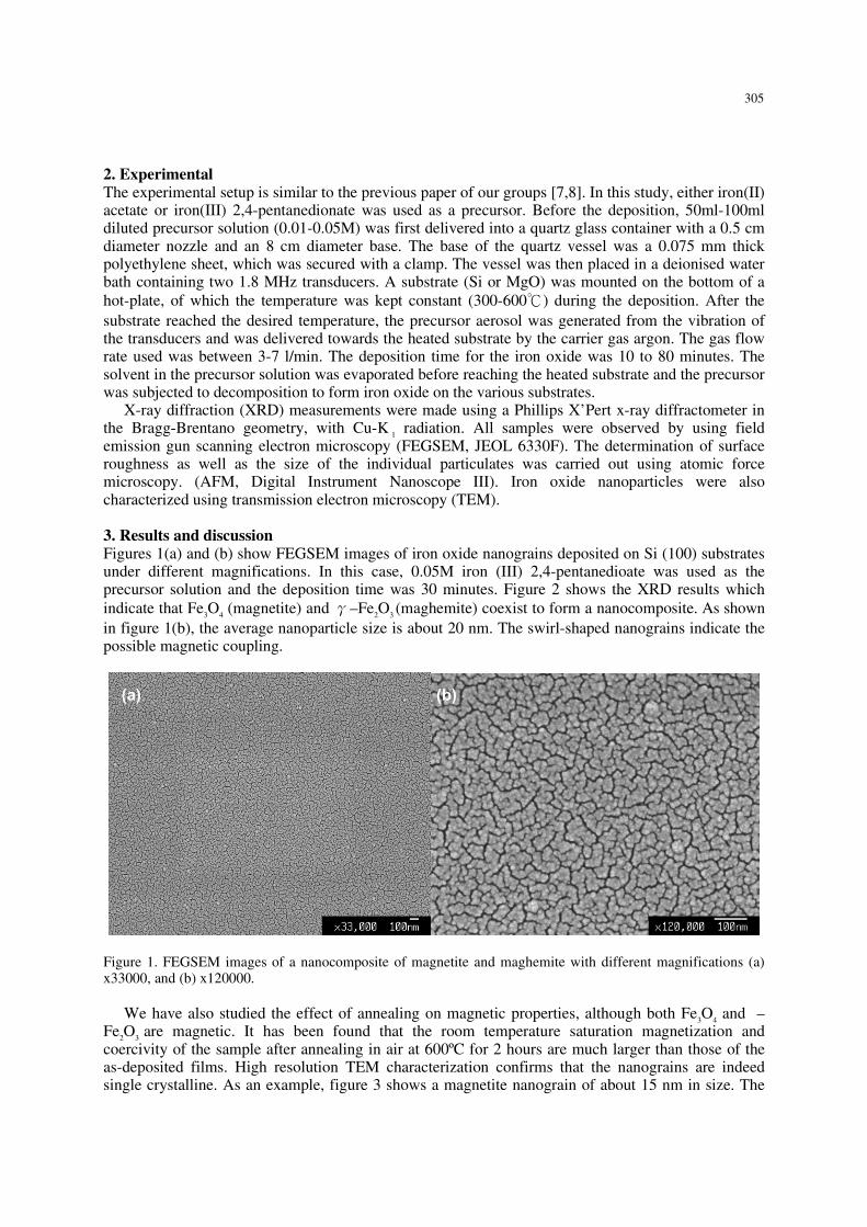
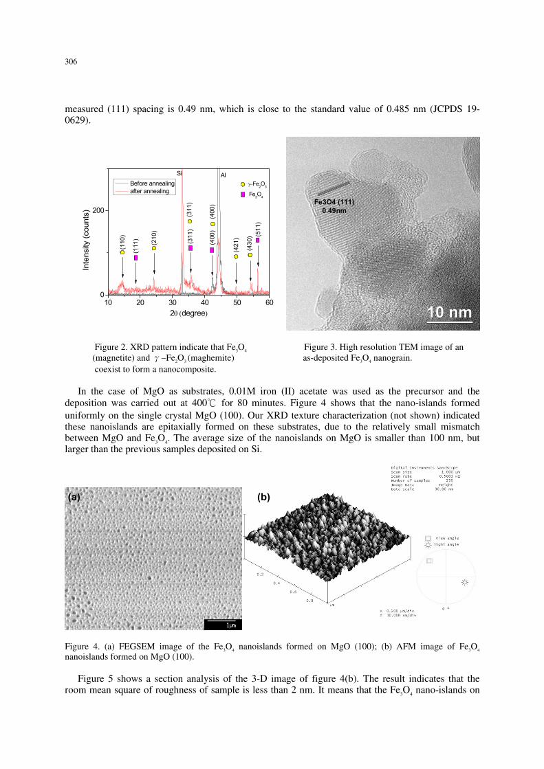
3 Results and discussion Figures 1(a) and (b) show FEGSEM images of iron oxide nanograins deposited on Si (100) substrates under different magnifications In this case 005M iron (III) 24-pentanedioate was used as the precursor solution and the deposition time was 30 minutes Figure 2 shows the XRD results which indicate that Fe3O4 (magnetite) and γndashFe2O3 (maghemite) coexist to form a nanocomposite As shown in figure 1(b) the average nanoparticle size is about 20 nm The swirl-shaped nanograins indicate the possible magnetic coupling
Figure 1 FEGSEM images of a nanocomposite of magnetite and maghemite with different magnifications (a) x33000 and (b) x120000
We have also studied the effect of annealing on magnetic properties although both Fe3O4 and ndash
Fe2O3 are magnetic It has been found that the room temperature saturation magnetization and coercivity of the sample after annealing in air at 600ordmC for 2 hours are much larger than those of the as-deposited films High resolution TEM characterization confirms that the nanograins are indeed single crystalline As an example figure 3 shows a magnetite nanograin of about 15 nm in size The
(a)
(b)
305
measured (111) spacing is 049 nm which is close to the standard value of 0485 nm (JCPDS 19-0629)
10 20 30 40 50 600
200
(110
) (511
)
(311
)
(430
)
(400
)
(421
)
(400
)
(311
)
(210
)
(111
)
Fe3O4
γminusFe2O3
Inte
nsity
(cou
nts)
2θ (degree)
Before annealing after annealing
Si Al
10 nm10 nm
Fe3O4 (111)049nm
Figure 2 XRD pattern indicate that Fe3O4 Figure 3 High resolution TEM image of an (magnetite) and γndashFe2O3 (maghemite) as-deposited Fe3O4 nanograin coexist to form a nanocomposite
In the case of MgO as substrates 001M iron (II) acetate was used as the precursor and the
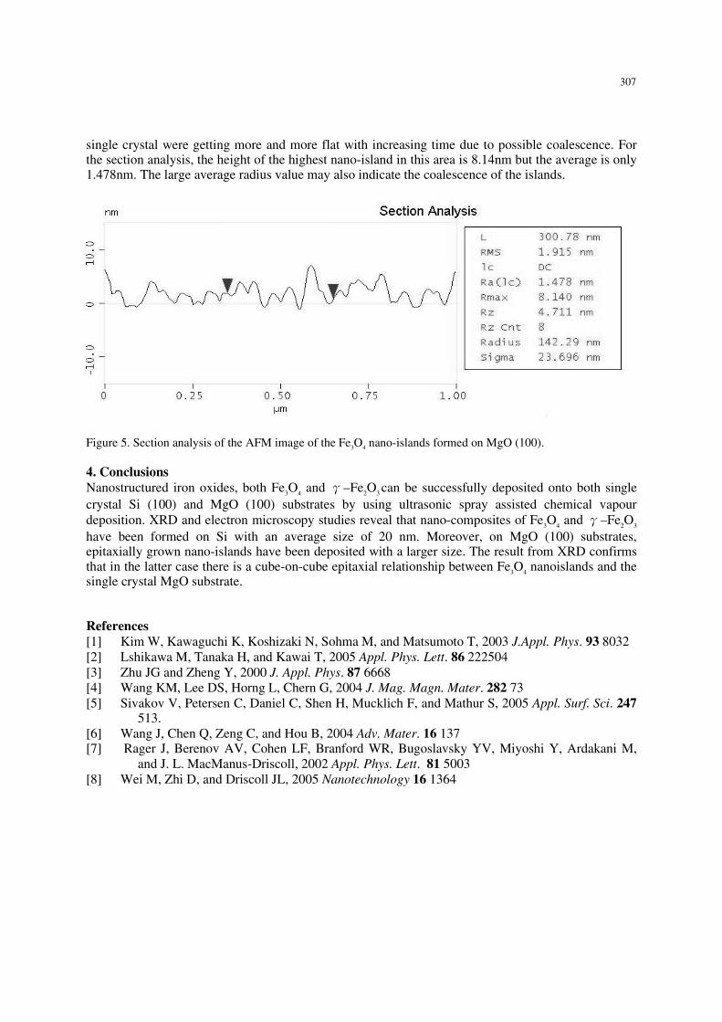
deposition was carried out at 400 for 80 minutes Figure 4 shows that the nano-islands formed uniformly on the single crystal MgO (100) Our XRD texture characterization (not shown) indicated these nanoislands are epitaxially formed on these substrates due to the relatively small mismatch between MgO and Fe3O4 The average size of the nanoislands on MgO is smaller than 100 nm but larger than the previous samples deposited on Si
Figure 4 (a) FEGSEM image of the Fe3O4 nanoislands formed on MgO (100) (b) AFM image of Fe3O4 nanoislands formed on MgO (100)
Figure 5 shows a section analysis of the 3-D image of figure 4(b) The result indicates that the
room mean square of roughness of sample is less than 2 nm It means that the Fe3O4 nano-islands on
(a)
(b)
306
single crystal were getting more and more flat with increasing time due to possible coalescence For the section analysis the height of the highest nano-island in this area is 814nm but the average is only 1478nm The large average radius value may also indicate the coalescence of the islands
Figure 5 Section analysis of the AFM image of the Fe3O4 nano-islands formed on MgO (100)
4 Conclusions Nanostructured iron oxides both Fe3O4 and γndashFe2O3 can be successfully deposited onto both single crystal Si (100) and MgO (100) substrates by using ultrasonic spray assisted chemical vapour deposition XRD and electron microscopy studies reveal that nano-composites of Fe3O4 and γndashFe2O3 have been formed on Si with an average size of 20 nm Moreover on MgO (100) substrates epitaxially grown nano-islands have been deposited with a larger size The result from XRD confirms that in the latter case there is a cube-on-cube epitaxial relationship between Fe3O4 nanoislands and the single crystal MgO substrate
References [1] Kim W Kawaguchi K Koshizaki N Sohma M and Matsumoto T 2003 JAppl Phys 93 8032 [2] Lshikawa M Tanaka H and Kawai T 2005 Appl Phys Lett 86 222504 [3] Zhu JG and Zheng Y 2000 J Appl Phys 87 6668 [4] Wang KM Lee DS Horng L Chern G 2004 J Mag Magn Mater 282 73 [5] Sivakov V Petersen C Daniel C Shen H Mucklich F and Mathur S 2005 Appl Surf Sci 247
513 [6] Wang J Chen Q Zeng C and Hou B 2004 Adv Mater 16 137 [7] Rager J Berenov AV Cohen LF Branford WR Bugoslavsky YV Miyoshi Y Ardakani M
and J L MacManus-Driscoll 2002 Appl Phys Lett 81 5003 [8] Wei M Zhi D and Driscoll JL 2005 Nanotechnology 16 1364
307
Synthesis and characterisation of nanocrystalline iron oxides via ultrasonic spray assisted chemical vapour deposition
LT Chao M Wei and JL MacManus-Driscoll
Department of Materials Science and Metallurgy University of Cambridge Pembroke Street Cambridge CB2 3QZ UK
E-mail jld35camacuk
Abstract The synthesis and characterisation of nanocrystalline magnetic materials with different morphology have attracted much attention for understanding the fundamental aspects of magnetic-ordering with reduced dimensions and for possible new potential applications in data storage technology We report here a novel synthesis route for nanocrystalline magnetite Fe3O4 by ultrasonic spray assisted chemical vapour deposition in which either iron acetate or iron acetylacetonate are used as precursors Magnetically coupled nano-grains and nano-islands of Fe3O4 with different size and morphology can be produced at different conditions The structure of as-deposited and post-annealed nanoparticles was investigated by field emission gun scanning electron microscopy (FEGSEM) x-ray diffraction (XRD) and transmission electron microscopy (TEM) The surface roughness was measured by atomic force microscopy (AFM)
1 Introduction Magnetite (Fe3O4) has received much interest due to its high Curie temperature (860K) and fully spin polarised half metallic character which can allow for spin-dependent tunnelling magntoresistance (TMR) with quite a high efficiency [12] These characteristics can potentially be utilized in the development of magnetic random access memory (MRAM) devices that operate at room temperature with high performance capabilities [3] From the application point of view growth of epitaxial ferrite films is desirable and epitaxy should improve the magnetic properties of the film Moreover an epitaxial ferrite would allow the deposition of further epitaxial or oriented layers of other materials on the ferrite thus increasing the device-design possibilities There have been several methods to fabricate iron oxide films such as pulsed laser deposition (PLD) [2] oxygen-plasma-assisted molecular beam epitaxy [4] and chemical vapour deposition (CVD) [5] Apart from epitaxial films nanostructured magnetic iron oxides have also attracted increasing attention in recent years It has been expected that with reducing dimensions magnetic materials can bring new potential applications such as high density recording media and bioinspired materials Different processes for the growth and deposition of magnetic iron oxide nanoparticles and 1D materials such as single crystalline Fe3O4 nanowires [6] have been reported
In this report nanostructured iron oxides on various substrates have been studied using a novel ultrasonic spray assisted chemical vapour deposition Both self assembled nanocrystals and epitaxially grown nanoislands were deposited and characterized
Institute of Physics Publishing Journal of Physics Conference Series 26 (2006) 304ndash307doi1010881742-6596261073 EMAGndashNANO 05 Imaging Analysis and Fabrication on the Nanoscale
304copy 2006 IOP Publishing Ltd
2 Experimental The experimental setup is similar to the previous paper of our groups [78] In this study either iron(II) acetate or iron(III) 24-pentanedionate was used as a precursor Before the deposition 50ml-100ml diluted precursor solution (001-005M) was first delivered into a quartz glass container with a 05 cm diameter nozzle and an 8 cm diameter base The base of the quartz vessel was a 0075 mm thick polyethylene sheet which was secured with a clamp The vessel was then placed in a deionised water bath containing two 18 MHz transducers A substrate (Si or MgO) was mounted on the bottom of a hot-plate of which the temperature was kept constant (300-600) during the deposition After the substrate reached the desired temperature the precursor aerosol was generated from the vibration of the transducers and was delivered towards the heated substrate by the carrier gas argon The gas flow rate used was between 3-7 lmin The deposition time for the iron oxide was 10 to 80 minutes The solvent in the precursor solution was evaporated before reaching the heated substrate and the precursor was subjected to decomposition to form iron oxide on the various substrates
X-ray diffraction (XRD) measurements were made using a Phillips XrsquoPert x-ray diffractometer in the Bragg-Brentano geometry with Cu-K 1 radiation All samples were observed by using field emission gun scanning electron microscopy (FEGSEM JEOL 6330F) The determination of surface roughness as well as the size of the individual particulates was carried out using atomic force microscopy (AFM Digital Instrument Nanoscope III) Iron oxide nanoparticles were also characterized using transmission electron microscopy (TEM)
3 Results and discussion Figures 1(a) and (b) show FEGSEM images of iron oxide nanograins deposited on Si (100) substrates under different magnifications In this case 005M iron (III) 24-pentanedioate was used as the precursor solution and the deposition time was 30 minutes Figure 2 shows the XRD results which indicate that Fe3O4 (magnetite) and γndashFe2O3 (maghemite) coexist to form a nanocomposite As shown in figure 1(b) the average nanoparticle size is about 20 nm The swirl-shaped nanograins indicate the possible magnetic coupling
Figure 1 FEGSEM images of a nanocomposite of magnetite and maghemite with different magnifications (a) x33000 and (b) x120000
We have also studied the effect of annealing on magnetic properties although both Fe3O4 and ndash
Fe2O3 are magnetic It has been found that the room temperature saturation magnetization and coercivity of the sample after annealing in air at 600ordmC for 2 hours are much larger than those of the as-deposited films High resolution TEM characterization confirms that the nanograins are indeed single crystalline As an example figure 3 shows a magnetite nanograin of about 15 nm in size The
(a)
(b)
305
measured (111) spacing is 049 nm which is close to the standard value of 0485 nm (JCPDS 19-0629)
10 20 30 40 50 600
200
(110
) (511
)
(311
)
(430
)
(400
)
(421
)
(400
)
(311
)
(210
)
(111
)
Fe3O4
γminusFe2O3
Inte
nsity
(cou
nts)
2θ (degree)
Before annealing after annealing
Si Al
10 nm10 nm
Fe3O4 (111)049nm
Figure 2 XRD pattern indicate that Fe3O4 Figure 3 High resolution TEM image of an (magnetite) and γndashFe2O3 (maghemite) as-deposited Fe3O4 nanograin coexist to form a nanocomposite
In the case of MgO as substrates 001M iron (II) acetate was used as the precursor and the
deposition was carried out at 400 for 80 minutes Figure 4 shows that the nano-islands formed uniformly on the single crystal MgO (100) Our XRD texture characterization (not shown) indicated these nanoislands are epitaxially formed on these substrates due to the relatively small mismatch between MgO and Fe3O4 The average size of the nanoislands on MgO is smaller than 100 nm but larger than the previous samples deposited on Si
Figure 4 (a) FEGSEM image of the Fe3O4 nanoislands formed on MgO (100) (b) AFM image of Fe3O4 nanoislands formed on MgO (100)
Figure 5 shows a section analysis of the 3-D image of figure 4(b) The result indicates that the
room mean square of roughness of sample is less than 2 nm It means that the Fe3O4 nano-islands on
(a)
(b)
306
single crystal were getting more and more flat with increasing time due to possible coalescence For the section analysis the height of the highest nano-island in this area is 814nm but the average is only 1478nm The large average radius value may also indicate the coalescence of the islands
Figure 5 Section analysis of the AFM image of the Fe3O4 nano-islands formed on MgO (100)
4 Conclusions Nanostructured iron oxides both Fe3O4 and γndashFe2O3 can be successfully deposited onto both single crystal Si (100) and MgO (100) substrates by using ultrasonic spray assisted chemical vapour deposition XRD and electron microscopy studies reveal that nano-composites of Fe3O4 and γndashFe2O3 have been formed on Si with an average size of 20 nm Moreover on MgO (100) substrates epitaxially grown nano-islands have been deposited with a larger size The result from XRD confirms that in the latter case there is a cube-on-cube epitaxial relationship between Fe3O4 nanoislands and the single crystal MgO substrate
References [1] Kim W Kawaguchi K Koshizaki N Sohma M and Matsumoto T 2003 JAppl Phys 93 8032 [2] Lshikawa M Tanaka H and Kawai T 2005 Appl Phys Lett 86 222504 [3] Zhu JG and Zheng Y 2000 J Appl Phys 87 6668 [4] Wang KM Lee DS Horng L Chern G 2004 J Mag Magn Mater 282 73 [5] Sivakov V Petersen C Daniel C Shen H Mucklich F and Mathur S 2005 Appl Surf Sci 247
513 [6] Wang J Chen Q Zeng C and Hou B 2004 Adv Mater 16 137 [7] Rager J Berenov AV Cohen LF Branford WR Bugoslavsky YV Miyoshi Y Ardakani M
and J L MacManus-Driscoll 2002 Appl Phys Lett 81 5003 [8] Wei M Zhi D and Driscoll JL 2005 Nanotechnology 16 1364
307


![Magnetic and Structural Properties of … and Structural Properties of Nanocrystalline Iron Oxides A. Kihala,b*, B. Bouzabataa, ... (SPM) regime at high temperature [6], but with blocking](https://img.pdfslide.net/doc/110x75/5ac1f9567f8b9ad73f8d8671/magnetic-and-structural-properties-of-and-structural-properties-of-nanocrystalline.jpg)