Embed Size (px)
Citation preview

Synthesis and characterization of hyaluronic acid hydrogelscrosslinked using a solvent-free process for potential biomedicalapplications.Larrañeta, E., Henry, M., Irwin, N. J., Trotter, J., Perminova, A. A., & Donnelly, R. F. (2017). Synthesis andcharacterization of hyaluronic acid hydrogels crosslinked using a solvent-free process for potential biomedicalapplications. Carbohydrate Polymers. DOI: 10.1016/j.carbpol.2017.12.015
Published in:Carbohydrate Polymers
Document Version:Peer reviewed version
Queen's University Belfast - Research Portal:Link to publication record in Queen's University Belfast Research Portal
Publisher rightsCopyright 2017 the authors.This is an open access article published under a Creative Commons Attribution License (https://creativecommons.org/licenses/by/4.0/),which permits unrestricted use, distribution and reproduction in any medium, provided the author and source are cited.
General rightsCopyright for the publications made accessible via the Queen's University Belfast Research Portal is retained by the author(s) and / or othercopyright owners and it is a condition of accessing these publications that users recognise and abide by the legal requirements associatedwith these rights.
Take down policyThe Research Portal is Queen's institutional repository that provides access to Queen's research output. Every effort has been made toensure that content in the Research Portal does not infringe any person's rights, or applicable UK laws. If you discover content in theResearch Portal that you believe breaches copyright or violates any law, please contact [email protected].
Download date:04. Jan. 2018
brought to you by COREView metadata, citation and similar papers at core.ac.uk
provided by Queen's University Research Portal

Accepted Manuscript
Title: Synthesis and characterization of hyaluronic acidhydrogels crosslinked using a solvent-free process forpotential biomedical applications.
Authors: Eneko Larraneta, Megan Henry, Nicola J. Irwin,Johann Trotter, Anastasia A. Perminova, Ryan F. Donnelly
PII: S0144-8617(17)31412-1DOI: https://doi.org/10.1016/j.carbpol.2017.12.015Reference: CARP 13077
To appear in:
Received date: 9-10-2017Revised date: 22-11-2017Accepted date: 6-12-2017
Please cite this article as: Larraneta, Eneko., Henry, Megan., Irwin, Nicola J.,Trotter, Johann., Perminova, Anastasia A., & Donnelly, Ryan F., Synthesisand characterization of hyaluronic acid hydrogels crosslinked using a solvent-free process for potential biomedical applications.Carbohydrate Polymershttps://doi.org/10.1016/j.carbpol.2017.12.015
This is a PDF file of an unedited manuscript that has been accepted for publication.As a service to our customers we are providing this early version of the manuscript.The manuscript will undergo copyediting, typesetting, and review of the resulting proofbefore it is published in its final form. Please note that during the production processerrors may be discovered which could affect the content, and all legal disclaimers thatapply to the journal pertain.
1
Synthesis and characterization of hyaluronic acid hydrogels crosslinked using a solvent‐free process
for potential biomedical applications.
Eneko Larrañeta*, Megan Henry, Nicola J. Irwin, Johann Trotter, Anastasia A. Perminova, Ryan F.
Donnelly.
School of Pharmacy, Queens University Belfast, Medical Biology Centre, 97 Lisburn Road, Belfast BT9
7BL, Northern Ireland, UK.
* Corresponding author
Dr. Eneko Larrañeta
Lecturer in Pharmaceutical Sciences
School of Pharmacy,
Queens University Belfast,
Medical Biology Centre,
97 Lisburn Road,
Belfast
BT9 7BL, UK
Tel: +44 (0) 28 9097 2360
Fax: +44 (0) 28 9024 7794
Email: [email protected]
ACCEPTED MANUSCRIP
T

2
Graphical Abstract
Highlights
Hyaluronic acid hydrogels have been prepared by using a single step crosslinking reaction in
solid state with a synthetic polymer containing multiple acid groups through an esterification
reaction.
The use of microwave radiation reduces significantly the crosslinking time.
The synthesized materials allowed sustained release of a model molecule (methylene blue)
for a period of up to 2 days.
The material can be used to prepare micro‐engineered devices such as microneedles
through a micromoulding process.
The resulting hydrogels showed anti‐infective and bacteriostatic properties.
ACCEPTED MANUSCRIP
T

3
Abstract
Hyaluronic acid (HA) is a natural linear polysaccharide that has been used extensively in the biomedical
field as it is a biocompatible, biodegradable, nontoxic and non‐immunogenic polymer with high water
affinity. Besides, the presence of multiple acid and hydroxyl groups in the HA molecule makes it an
ideal candidate for chemical modification. The present paper describes the synthesis and
characterization of HA‐based hydrogels. For this purpose, aqueous mixtures containing 5% (w/w) of
HA and different concentrations of Gantrez S97 (GAN) (1, 3 and 5% w/w) were used to prepare HA‐
based hydrogels. The mixtures were dried and the hydrogels were obtained after heating the solid
material at 80ºC for 24h. GAN is the acid form of an methylvinylether and maleic anhydride copolymer
and contains multiple acid groups that can form ester bonds when reacting with the multiple hydroxyl
groups present in HA chains. The method described here present potential to be applied for the
preparation of HA‐based biomaterials with a defined form as the crosslinking reaction between HA
and the crosslinker takes place in solid phase. Besides, the method can be considered an
environmental‐friendly process as no organic solvents or potentially toxic substances were used. The
esterification reaction was confirmed by infrared spectroscopy and dynamic scanning calorimetry
measurements. The loading and release capabilities of the hydrogels were evaluating by using
methylene blue (MB) as a model molecule. The hydrogels showed a high affinity for MB showing
loadings up to 0.35 mg MB per mg of hydrogel. Moreover, the hydrogels were capable of sustaining
the MB release over two days. The use of microwave radiation was evaluated to reduce the
crosslinking time from 24 h to 1 h, but this procedure needs to be optimized in future studies. As the
crosslinking procedure takes place in solid state, the HA/GAN hydrogels were used to prepare micro‐
engineered device, microneedle arrays. Finally, the antimicrobial properties of the hydrogels were
evaluated. The results showed that the hydrogels presented anti‐infective properties.
Keywords: Hyaluronic acid; Hydrogels; Microneedles
ACCEPTED MANUSCRIP
T

4
1. Introduction
Hyaluronic acid (HA) is a natural linear polysaccharide formed by repeating units of D‐glucoronic acid
and N‐acetyl‐D‐glucosamine disaccharide that was first isolated in 1934 from the vitreous humour of
bovine eyes (Mero and Campisi, 2014). This biomacromolecule is one of the major constituents of the
skin (Mero and Campisi, 2014) and can be found in extracellular tissues of various parts of the body
(Collins and Birkinshaw, 2013). Hyaluronic acid plays a role in several biological processes, including
cell growth, migration and differentiation (Hemshekhar, et al., 2016).
HA presents a suite of desirable properties for application in the biomedical field; specifically HA is a
biocompatible, biodegradable, nontoxic and non‐immunogenic polymer with high water affinity
(Highley, et al., 2016, Mero and Campisi, 2014, Tripodo, et al., 2015). Besides, the presence of multiple
acid and hydroxyl groups in the HA molecule makes it an ideal candidate for chemical modification
(Schanté, et al., 2011, Tripodo, et al., 2015). Accordingly, this material is showing increased importance
in biomaterials science, with applications ranging from tissue culture scaffolds to cosmetic materials
(Collins and Birkinshaw, 2013). In the biomedical field, HA has to‐date been used mainly to prepare
scaffolds for tissue engineering (Chen, et al., 2017, Collins and Birkinshaw, 2013, Cui, et al., 2015,
Hemshekhar, et al., 2016) and drug delivery systems (Fiorica, et al., 2017, Jiao, et al., 2016, Tripodo,
et al., 2015).
Recently, HA has been used in the preparation of hydrogels (Tripodo, et al., 2015). Hydrogels are a
three‐dimensional network of polymer chains, crosslinked by covalent or non‐covalent interactions,
capable of absorbing large amounts of water (Caló and Khutoryanskiy, 2015, Hoare and Kohane, 2008,
Peppas, et al., 2000). Chemical crosslinking provides enhanced stability to HA‐based materials (Segura,
et al., 2005, Tripodo, et al., 2015). HA hydrogels are commonly prepared via chemical modification in
solution using organic solvents and/or toxic reagents (Xu, et al., 2012).
Alternative methods for the preparation of hydrogels, which importantly avoid the use of organic
solvents and reagents that can present toxicity problems for biological applications, have been
described during recent years. For example, Caló et al. developed hydrogels based on poly(methyl
vinyl ether‐alt‐maleic anhydride) and poly(vinyl alcohol), which were facilely crosslinked in the
absence of organic solvents (Calo, et al., 2016b). In this work, an autoclave process was employed to
yield crosslinked and sterile hydrogels (Calo, et al., 2016b). Alternatively, Donnelly et al. developed
microneedle arrays based on hydrogel materials which were crosslinked in their solid state by a
thermal process (Donnelly, et al., 2012b). In addition, microwave radiation has shown potential for
the preparation of hydrogels in aqueous solutions (Cook, et al., 2012) or in solid state (Larrañeta, et
al., 2015a). Importantly, these aforementioned hydrogel synthetic procedures are environmental
ACCEPTED MANUSCRIP
T
5
friendly and can be easily scaled up for industrial applications. Besides, the ability to crosslink in solid
phase enables hydrogels to be prepared with defined shapes and, consequently, these methods may
be applied for the development of hydrogel‐based biomedical microdevices.
The combination of novel, environmentally‐friendly and readily scalable hydrogel preparation
methods with a natural and widely available material such as HA presents exciting potential for
application in the biomedical field. In the present work, we describe the preparation of HA‐based
hydrogels crosslinked with poly(methyl vinyl ether‐alt‐maleic acid) by thermal‐ and microwave‐based
processes as promising candidate wound care, drug delivery and medical materials. Synthesised
hydrogels were characterized and evaluated as drug delivery systems using methylene blue as a model
drug. In addition, the materials were successfully used to produce microneedle (MN) arrays for
potential transdermal delivery and, finally, the antimicrobial properties of the resulting hydrogels
were evaluated in vitro.
2. Material and methods
2.1 Materials
Gantrez® S‐97 (GAN) (acid form of methylvinylether and maleic anhydride copolymer) (Mw = 1.2 × 10⁶
Da), was provided by Ashland (Tadworth, Surrey, UK). Hyabest®(S) LF‐P (sodium hyaluronate 99.9%
purity, MW 250‐400 kDa range) was obtained from Kewpie Corporation Fine Chemical Division (Tokyo,
Japan). Methylene blue (MB) was purchased from Sigma–Aldrich (Steinheim, Germany). Poly(vinyl
chloride) (PVC) sheets (unplasticised) with a thickness of 0.2 mm were obtained from Goodfellow Ltd
(Cambridge, UK). Phosphate‐buffered saline (PBS), tryptone soya broth (TSB), quarter‐strength
Ringer’s solution (QSRS) and Mueller‐Hinton broth (MHB) were obtained from Oxoid Ltd (Hampshire,
UK). Proteus mirabilis ATCC 35508 and Staphylococcus aureus ATCC 6538 (LGC Standards, Middlesex,
UK) were maintained on cryopreservative beads (Protect Bacterial Preservation System, Technical
Service Consultants Ltd., UK) in 10% glycerol at ‐80C and cultivated in MHB at 37C when required
for the microbiological assessments.
2.2 Preparation of hyaluronic acid hydrogels
Aqueous solutions containing different ratios of HA and GAN were prepared (Table 1) and 30 g of
these solutions were casted in 10x10 moulds. Solutions were allowed to dry over at least 48 hours.
The resulting films were cut in pieces of 1x1 cm and subsequently they were placed inside an oven at
ACCEPTED MANUSCRIP
T
6
80°C during 24 hours. The hydrogels prepared using a microwave assisted process were prepared
following the same process. Instead of placing the films in a convection oven for the crosslinking
process, they were placed in the middle of the oven cavity in a Panasonic NN‐CF778S microwave oven
(Panasonic UK Ltd, Bracknell, UK). The films were crosslinked during 1 hour with the oven at the
highest output power (1000 W).
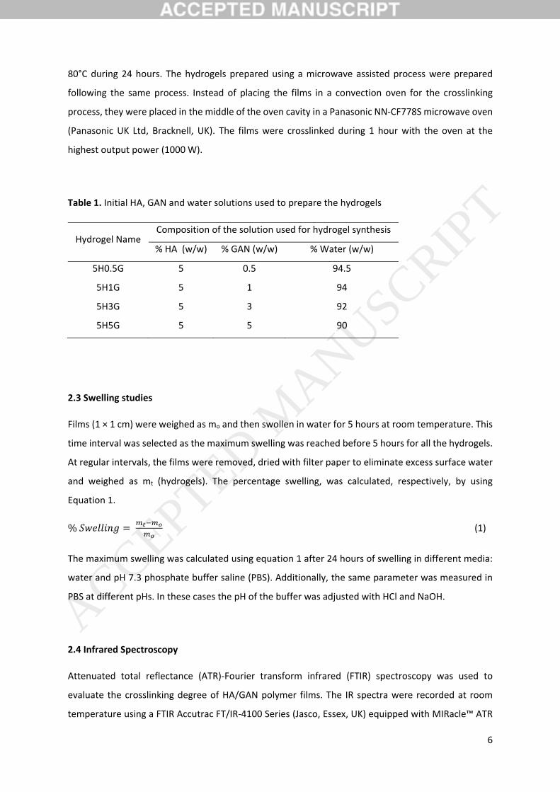
Table 1. Initial HA, GAN and water solutions used to prepare the hydrogels
Hydrogel Name Composition of the solution used for hydrogel synthesis
% HA (w/w) % GAN (w/w) % Water (w/w)
5H0.5G 5 0.5 94.5
5H1G 5 1 94
5H3G 5 3 92
5H5G 5 5 90
2.3 Swelling studies
Films (1 × 1 cm) were weighed as mo and then swollen in water for 5 hours at room temperature. This
time interval was selected as the maximum swelling was reached before 5 hours for all the hydrogels.
At regular intervals, the films were removed, dried with filter paper to eliminate excess surface water
and weighed as mt (hydrogels). The percentage swelling, was calculated, respectively, by using
Equation 1.
% (1)
The maximum swelling was calculated using equation 1 after 24 hours of swelling in different media:
water and pH 7.3 phosphate buffer saline (PBS). Additionally, the same parameter was measured in
PBS at different pHs. In these cases the pH of the buffer was adjusted with HCl and NaOH.
2.4 Infrared Spectroscopy
Attenuated total reflectance (ATR)‐Fourier transform infrared (FTIR) spectroscopy was used to
evaluate the crosslinking degree of HA/GAN polymer films. The IR spectra were recorded at room
temperature using a FTIR Accutrac FT/IR‐4100 Series (Jasco, Essex, UK) equipped with MIRacle™ ATR
ACCEPTED MANUSCRIP
T

7
accesorie between 4000–600 cm−1 with a resolution of 4.0 cm−1. The obtained spectra were the result
of averaging 64 scans.
2.5 Differential Scanning Calorimetry
All the materials were analysed using a differential scanning calorimeter (DSC Q100) (TA Instruments,
New Castle, USA). Due to the presence of broad peaks of water specially in HA containing samples a
drying cycle was introduce prior to the analysis. This cycle runs between 0 and 150°C at a heating
speed of 10°C/min. Samples were subsequently analysed from 0 to 200°C at a heating speed of
10°C/min.
2.6 Microscopy
The morphology of the hydrogels was evaluated by using electronic microscopy. A Hitachi TM3030
environmental scanning electron microscope (SEM) (Tokyo, Japan) was used. Prior to the analysis,
hydrogels were previously swollen for at least 24 hours and subsequently freeze dried. In order to
evaluate the transparency of the synthesized hydrogel films a Leica EZ4 D digital microscope (Leica,
Wetzlar, Germany) was used.
2.7 Methylene Blue loading and release
HA/GAN hydrogels were loaded with MB by immersing the 1 cm2 dry film in 5 mL of a 2mg/mL solution
of the dye. The film was left inside the solution for a defined period of time. In this study the selected
loading times were 1 hour and 24 hours. The loading was evaluated by measuring the absorbance of
the initial solution before and after placing the hydrogel film in the solution at 664 nm in an UV‐visible
plate reader (PowerWave XS Microplate Spectrophotometer, Bio‐Tek, Winooski, USA).
After the loading process films were removed, dried with filter paper to eliminate the superficial
excess of MB solution and placed in 20 mL of PBS. The tubes containing the samples were placed in a
shaking incubator (40 rpm and 37°C). Samples were collected at different times. The concentration of
MB in the solution was evaluated using a UV–vis plate reader at a wavelength of 664 nm.
2.8 Microneedle preparation and testing
ACCEPTED MANUSCRIP
T

8
Aqueous blends containing HA (5% w/w) and GAN S‐97 (3% w/w) were used to fabricate MNs. This
formulation was poured into laser‐engineered silicone micromould templates (19x19 , centrifuged for
15 min at 3,500 rpm and allowed to dry under ambient conditions for 48 h (Larrañeta, et al., 2015a).
Finally, they were placed inside a convection oven at 80 °C for 24 hours. All the arrays contained
19 × 19 conical needles. The dimensions were: 600 μm needle height, 300 μm width at the base and
50 μm interspacing. Formed MN arrays were visualized using a Keyence VHX‐700F digital microscope.
Parafilm® M (PF) film was used as a skin simulant for MN insertion studies as described previously
(Larrañeta, et al., 2014). A sheet of Parafilm® was folded to get an 8‐layer film (≈ 1 mm thickness) and
placed on a sheet of expanded poly(ethylene) for support. MN arrays were inserted using a TA‐XT2
Texture Analyser (Stable Micro Systems, Surrey, UK), with the probe lowered onto the artificial
membrane at a speed of 0.5 mm s−1 with an exerted force per array held for 30 s. Different forces were
tested. Once the target force was reached, the probe was moved upwards at a speed of 0.5 mm s−1.
The MN arrays were removed from the polymeric sheet after insertion, the PF sheet unfolded and the
number of holes in each layer was evaluated using a Leica EZ4 D digital microscope (Leica, Wetzlar,
Germany). In order to ease the detection of the created holes in the PF layers, the sample was placed
between two polarizer filters. The thickness of each PF layer was determined previously (126 ± 7 μm)
(Larrañeta, et al., 2014) and was used to calculate the percentage of MN inserted as a function of the
depth.
The morphology of the MN arrays was studied using a Keyence VHX‐700F Digital Microscope (Keyence,
Osaka, Japan). Finally, optical coherence tomography (OCT) images of the MN arrays inserted in
excised neonatal porcine skin were obtained using an OCT VivoSight™ Topical Multi‐Beam OCT
Handheld Probe (Michelson Diagnostics Ltd, Kent, UK). In this study, the MN arrays were inserted
using the TA‐XT2 Texture Analyser under the same conditions described above. The applied force was
32 N.
2.9 In vitro microbiological analysis
Bacterial suspensions of P. mirabilis and S. aureus were adjusted to a density of 1 x 10⁶ cfumL‐1 in PBS
supplemented with 0.5% TSB. Replicate samples (10 x 10 mm) of 5H1G and 5H3G hydrogels, and PVC
(as control) were placed in individual wells of a sterile 24‐well flat bottom tissue culture plate (Corning
Inc., Corning, NY) containing 1 mL of the respective bacterial suspensions (1 x 10⁶ cfumL‐1). The plates
were incubated at 37C under continuous shaking at 100 rpm. After designated time intervals of 4 h
and 24 h, samples were removed from the bacterial suspension using sterile forceps and non‐adherent
bacteria removed by rinsing three times with QSRS (Wang, et al., 2012). Samples were transferred
ACCEPTED MANUSCRIP
T

9
into fresh QSRS (5 mL) and adherent bacteria subsequently removed by sonicating for 10 min in an
ultrasonic bath and vortexing for 30 sec. The sonication technique has previously been demonstrated
not to affect bacterial viability or morphology (Jones, et al., 1997). Viable counting of the resulting
QSRS was performed by the Miles and Misra serial dilution technique (Miles, et al., 1938), with plating
onto low‐swarm (LSW) agar (P. mirabilis) or Mueller‐Hinton agar (S. aureus) to determine the number
of adherent bacteria on each sample surface. Percentage reductions in the number of adherent
bacteria to each sample relative to the PVC control were calculated. In addition, densities of the
planktonic bacterial suspensions in wells containing samples and in wells with no added samples were
quantitated at each time interval by colony counting as before.
2.10. Statistical Analysis
All data were expressed as mean ± standard deviation. Data were compared using a paired, two‐tailed
Student's t‐test when comparing two means and One‐Way Analysis of Variance (ANOVA), with Tukey's
HSD post‐hoc test for more than two means. In all cases, p < 0.05 was the minimum value considered
acceptable for rejection of the null hypothesis.
3. Results and Discussion
GAN polymers have been used in the past to prepare hydrogels (Calo, et al., 2016b, Donnelly, et al.,
2012c, Moreno, et al., 2014). All these works are based on the crosslinking of GAN with other synthetic
polymers. In the present work the possibility of using this type of polymers to obtain hydrogels based
on biomacromolecules for potential pharmaceutical/medical applications is explored. For this
purpose, HA was selected as it is biocompatible and extensively used for biomedical applications
(Collins and Birkinshaw, 2013). Based on previously published papers describing the preparation of
GAN‐based hydrogels (Larrañeta, et al., 2015b) the proposed reaction mechanism is the esterification
between the acid groups of GAN and the multiple alcohol groups of HA (Figure 1A).
The preparation of the hydrogels described in this work is a simple solvent‐free procedure. After
mixing both macromolecules, films were prepared by casting the solutions inside moulds and allowing
the water to evaporate. Subsequently, films were placed in a convection over at 80°C for crosslinking.
The process is simple, does not require of organic solvents and as it is an esterification the main by‐
product is water. Therefore, it presents potential advantages over alternative synthetic procedures to
obtain HA hydrogels that require the use of organic solvents or potentially toxic reagents. The lack of
toxic reagents/byproducts during the synthesis is crucial for biomedical applications. Furthermore, the
ACCEPTED MANUSCRIP
T

10
crosslinking process was carried out in solid state allowing to prepare HA‐based hydrogels with a
defined form/pattern. Consequently, the material could be potentially used in biomedical engineering
applications.
Figure 1. Chemical structures of Gantrez® S97 and sodium hyaluronate (top). Proposed crosslinking
mechanism between Gantrez® S97 and sodium hyaluronate (A). Swelling kinetics of different HA/GAN
hydrogels in water (B). Maximum swelling of different HA/GAN hydrogels in water and PBS (C).
3.1 Hydrogel Characterization
The main evidence of the crosslinking process is that after taking the films from the oven and placing
them in water they start to swell. The non‐crosslinked films dissolve quickly when placed in water. The
swelling kinetics in water of the HA/GAN hydrogels can be seen in Figure 1B. 5H0.5G hydrogels showed
the highest swelling capacity. However, after 15 minutes the hydrogels yielded lower swelling values.
This is due to a weight loss of the sample. These hydrogels present small GAN/HA ratio and,
consequently, the amount of GAN present in the hydrogel is not enough to achieve a complete
ACCEPTED MANUSCRIP
T
11
crosslinking of the HA molecules. Therefore, the non‐crosslinked polymeric chains dissolved in water,
producing the weight loss seen in the swelling plot. Consequently, 5H0.5G hydrogels were discarded
from this study.
The 5H5G, 5H3G and 5H1G swelling curves (Figure 1B) presented a quick maximum swelling after 15
minutes that was maintained for the rest of the study. The swelling capacity can be related directly
with the amount of GAN in the sample. Higher crosslinking densities and, consequently, lower swelling
capacities, are expected in samples with higher amounts of GAN, as there are more acid groups to
react with the alcohol groups in HA. 5H3G and 5H5G hydrogels showed similar profiles despite the
difference in GAN concentrations. Besides. it can be seen that 5H1G presented a small swelling
reductions after 30 min. This can be explained in the same way as described before. However, in this
case the weight loss was almost imperceptible. The maximum swelling capacity of the hydrogels in
water was measured after 24h (Figure 1C). It can be seen that the HA‐based hydrogels maintain their
water retention capacity during this longer period of time suggesting a covalent crosslinking between
HA and GAN molecules.
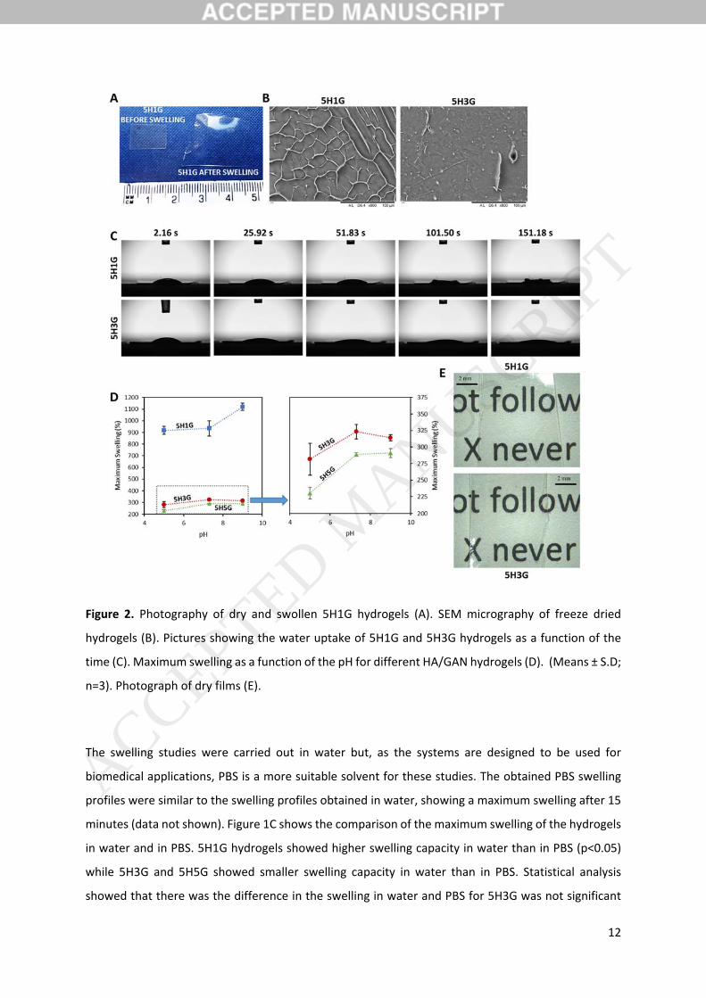
Images of 5H1G before and after swelling can be seen in Figure 2A to illustrate the swelling of the
material. Besides, Figure 2B shows SEM images of freeze dried 5H1G and 5H3G after maximum
swelling. The images showed than 5H1G present a more porous structure due to its higher water
uptake, confirming what was seen in the swelling studies.
It is important to note that all hydrogels showed a relatively fast water uptake as the maximum
swelling values were obtained after only 15 minutes. As an example of this, Figure 2C shows the
behaviour of a droplet of water on the top of 5H1G and 5H3G hydrogels as a function of the time. It
can be seen that in less than 2 minutes the water is absorbed by 5H1G while it takes longer time for
5H3G. HA has a high water affinity and the presence of this molecule in the hydrogels could explain
this behaviour.
ACCEPTED MANUSCRIP
T
12
Figure 2. Photography of dry and swollen 5H1G hydrogels (A). SEM micrography of freeze dried
hydrogels (B). Pictures showing the water uptake of 5H1G and 5H3G hydrogels as a function of the
time (C). Maximum swelling as a function of the pH for different HA/GAN hydrogels (D). (Means ± S.D;
n=3). Photograph of dry films (E).
The swelling studies were carried out in water but, as the systems are designed to be used for
biomedical applications, PBS is a more suitable solvent for these studies. The obtained PBS swelling
profiles were similar to the swelling profiles obtained in water, showing a maximum swelling after 15
minutes (data not shown). Figure 1C shows the comparison of the maximum swelling of the hydrogels
in water and in PBS. 5H1G hydrogels showed higher swelling capacity in water than in PBS (p<0.05)
while 5H3G and 5H5G showed smaller swelling capacity in water than in PBS. Statistical analysis
showed that there was the difference in the swelling in water and PBS for 5H3G was not significant
ACCEPTED MANUSCRIP
T
13
(p>0.05). On the other hand, 5H5G showed higher fluid uptake in PBS than in water (p>0.05) In the
literature hyaluronic acid gels showed that the water uptake is influenced by the ionic strength and
pH of the medium (Mráček, et al., 2008, Shah and Barnett, 1992). The behaviour is different for 5H3G
and 5H5G due to the highest amount of GAN in the hydrogel. GAN is a poly‐acid and only a few acid
groups will react to form the hydrogel (Larrañeta, et al., 2015b) leaving a large amount of chemical
groups that can be ionized as a function of the pH of the medium. Deionized water has a slightly acid
pH while PBS is a buffer with a defined pH of 7.3. The difference in the pH can easily explain the
different swelling behaviour.
The results described in the previous paragraph suggest that the HA/GAN hydrogels present a certain
degree of pH responsiveness. This property can be important, as one of the potential application of
these materials is wound dressings and the pH of wounds can change over time (Shukla, et al., 2007).
Therefore, the swelling of the hydrogels was evaluated in a range of values representative of wound
pHs (5‐9) (Schneider, et al., 2007, Shukla, et al., 2007). The ability to respond to pH changes can be
really interesting for biomaterials. Therefore, the influence of the pH in the swelling was studied.
Figure 2D shows the maximum swelling of 5H1G, 5H3G and 5H5G as a function of the pH. It can be
seen that the behaviour of 5H1G is different than the swelling obtained at different pH values for the
other hydrogels. 5H1G presented the highest swelling capacities at pH 9 (p<0.05). Besides there was
no significant difference between the swelling capacity of 5H1G at pH 7.3 and pH 5 (p>0.05). Hydrogels
containing higher amounts of GAN in their composition presented comparable swelling in alkaline and
neutral pHs (p>0.05) and lower water absorption at acidic pHs (p<0.05). Again, 5H5G hydrogels
presented slightly lower swelling than 5H3G due to the higher amount of GAN and consequently a
higher crosslinking degree. The difference in the swelling behaviour of 5H1G can be easily explained
by the hydrogel composition. In this case the material is mainly formed by HA. It has been showed
previously than HA hydrogels presented higher swelling capabilities at alkaline pH values (Shah and
Barnett, 1992). 5H3G and 5H5G showed lower fluid uptake at lower pHs due to the higher
concentration of GAN in the hydrogels. The increase in pH of solution causes dissociation of COOH
groups of GAN, preventing intermolecular –COOH⋯HO– bonds (Calo, et al., 2016a) and consequently
yielding higher swellings. On the other hand, 5H1G hydrogels presented more unreacted –OH groups.
These groups can be ionized at alkaline pH values (pKa = 10) (Schanté, et al., 2011) and at pH 9 some
of them will be ionized. The presence of extra negative charges in the structure will lead to an extra
expansion of the network, due to the repulsion of the negatively charged chains.
The obtained results suggest than the influence of the pH in the swelling can be useful for wound care.
The pH of wounds changes over the healing process (Schneider, et al., 2007, Shukla, et al., 2007). The
pH of the wound changes from acidic (betwenn 5 and 6) after the injury to alkaline (7‐8) during the
ACCEPTED MANUSCRIP
T

14
last stages of the healing process in acute wounds. For chronic wounds the pH changes from acidic to
alkaline, and during the chronic phase, the pH is maintained at alkaline pHs (7‐8). However, it has been
shown that, in certain cases, the pH of the wounds can be higher than 9 (Shukla, et al., 2007).
Consequently, 5H1G could be a good candidate as a wound dressing. It presents high swelling capacity
at lower pHs allowing the absorption of the wound exudate at the initial states of the healing process
while having extra capabilities of absorbing more fluid in the later stages of the healing process
(alkaline pHs) (Schneider, et al., 2007). In addition to its pH responsiveness and its fluid uptake
capacity, its transparency makes it a good candidate for wound dressing materials. Figure 2E includes
photographs of 5H1G and 5H3G showing its transparency. Transparency is key for a wound dressing
as it allows to follow wound evolution over time without removing the dressing.
In order to ascertain the crosslinking mechanism, FTIR was used. Figure 3A shows the FTIR spectra of
HA, GAN and the non‐crosslinked (NC) films. All the peaks that were present in the spectra of the pure
compounds were present in the spectra of the non‐crosslinked films. By having a closer look at the
region between 1800 and 1500 cm‐1, it can be seen that some interactions took place between HA and
GAN. The carbonyl peak at around 1700 cm‐1 from the acid groups in GAN is displaced at higher
wavenumber when combined with HA in all the mixtures. Moreover, the same behaviour can be
observed for the carbonyl peaks of the amide and the acid group in HA (1500 and ca. 1600 cm‐1). The
displacement to higher wavenumbers could be explained by the mixture of both type of
macromolecular chains. In pure HA, there are non‐covalent interactions, mainly hydrogen bonds,
between the polysaccharide chains (Jia, et al., 2015). When combining GAN and HA, the HA‐HA
interactions are not as feasible anymore. As both macromolecules are mixed GAN chains will be
located in‐between HA chains preventing the HA‐HA interactions (Jia, et al., 2015). The same
explanation can be applied to GAN. This phenomenon can be observed by having a close look at the
hydroxyl vibration region (3800‐3000 cm‐1) in this case, the peak of the mixtures is shifted to higher
frequencies when increasing the GAN concentration in the sample. These results suggest that both
polymers are mixed during the formulation process.
ACCEPTED MANUSCRIP
T

15
Figure 3. FTIR spectra of HA, GAN and HA/GAN mixtures before crosslinking (A). FTIR spectra of
HA/GAN hydrogels before and after crosslinking (B). Magnified areas of the original spectra can be
seen on the right hand in each case. DSC thermograms of HA, GAN and HA/GAN mixtures before
crosslinking (C). DSC thermograms of HA/GAN hydrogels before and after crosslinking between: 60
and 250°C (D) and 100 and 180°C (E). The cross indicates the Tg value in the curves. Tg values as a
function of the GAN % in the material (F). For all thermograms: Exo Up.
Figure 3B shows the FTIR spectra of the crosslinked hydrogels compared with the spectra of the non‐
crosslinked films. There is no appreciable difference between non‐crosslinked films and hydrogels.
However, by having a closer look at the carbonyl region, it is noticeable that the GAN acid peak is
slightly broader now. The infrared carbonyl peaks for the carboxylic acids and esters are overlapping
and the broadening of the peak suggests the presence of a new ester peak overlapping with the
previous acid peak (Sclavons, et al., 2000). Additionally, a new peak appears at around 1780 cm‐1, due
to the formation of anhydride groups between two acid groups in the GAN molecule (Larrañeta, et al.,
2015b, Sclavons, et al., 2000). This effect will be more noticeable after microwave‐assisted crosslinking
of the hydrogels described in later sections of this article.
ACCEPTED MANUSCRIP
T

16
DSC measurements were performed to confirm the results obtained from the swelling and the FTIR
measurements. From the swelling studies, it was shown that the two polymers have reacted to form
a hydrogel. Furthermore, FTIR measurements showed that an esterification reaction could have
happened between the polymers, as there are slight changes in the carbonyl region of the spectra.
The first step was to analyse the pure substances and the non‐crosslinked films. Due to the presence
of water strongly bound to HA, a dehydration cycle was performed.
Figure 3C shows the DSC curves of pure HA, pure GAN and the non‐crosslinked HA/GAN films. The DSC
curves of the pure HA acid showed an exothermic peak at around 240ºC that can be attributed to the
degradation of the polysaccharide (Collins and Birkinshaw, 2007). On the other hand, the GAN DSC
curve showed a broad peak at around 160ºC that can be attributed to the formation of anhydrides
between two acid groups (Chung, et al., 1990). The DSC curves of HA/GAN mixtures presented
differences with the curves of the pure compounds. It is noticeable that endothermic peaks can be
found between 180 and 220ºC. We hypothesize that these peaks can be attributed to the formation
of anhydrides, as described before and to the formation of ester bonds between GAN COOH groups
and HA OH groups. It is noticeable that these peaks appear at lower temperatures when the amount
of GAN in the sample is higher, getting closer to the peak observed for pure GAN. Besides it is
important to notice that the degradation peak of HA cannot be found in the mixtures. This confirms
that there is a good mixture between these two macromolecules.
After the crosslinking process the main difference that can be observed in the DSC curves is that the
esterification/anhydride formation peak can be found at higher temperatures (Figure 3D). As some of
the COOH and OH groups in the mixture had already reacted, the energy needed to carry on with the
reaction is higher. Consequently, these peaks can be found at higher temperatures. Due to the lower
crosslinking degree of 5H1G, this behaviour cannot be observed for this type of hydrogel. In addition
to the presence of these endothermic peaks, all the samples presented a glass transition temperature
(Tg) between 135 and 160ºC (Figure 3E). For the non crosslkined samples, the Tg shifted to higher
values when the proportion of GAN in the samples increased (Figure 3E and 3F). This shows that the
inclusion of GAN yields a more compact structure. On the other hand, after the crosslinking process
an increase of the Tg of the samples can be seen. This suggest that the esterification reaction is
generating a more compact structure. However, the difference is higher for 5H3G than for 5H5G
suggesting that the system is reaching a limit in the esterification reaction. Accordingly, the addition
of more GAN will not yield a significantly higher degree of crosslinking. This is consistent with the
results obtained for the swelling of 5H3G and 5H5G that were similar (Figure 1). On the other hand,
5H1G showed a different behaviour that 5H3G and 5H5G. as in this case the glass transition of the
ACCEPTED MANUSCRIP
T
17
uncrosslinked films was higher than the one of the hydrogels. This suggest that when lower amounts
of GAN the system present a less ordered structure.
After evaluating the swelling and crosslinking of the hydrogels 5H1G and 5H3G hydrogels were used
for further testing. 5H5G showed similar swelling parameters to 5H3G and, due to the high viscosity
of the aqueous mixtures obtaining films with consistent thickness was difficult. Consequently, this
type of hydrogels was not used in further studies.
The obtained results show that HA‐based hydrogels can be obtained through an esterification process
in a solvent‐free crosslinking process. Some of the procedures to synthesize HA hydrogels described
in the literature are not easy to achieve due to the complexity of the chemistry and, additionally, to
the toxicity of preparation (Burdick and Prestwich, 2011). The proposed process can be easily scaled
up, as it involves only a thermal treatment of a solid product and the raw materials have been
demonstrated to be safe and biocompatible. Additionally, the degradation products of HA backbone
have been shown to increase wound healing (Mast, et al., 1995). Therefore, the presence of high
quantities of HA in the dressing could potentially improve wound healing. So the designed hydrogels
have potential in this field as they can be used to absorb wound exudate and protect the lesion and
finally accelerate the healing process.
3.2. Methylene blue loading and release studies
MB was selected as a model molecule to evaluate the drug loading and release capabilities of the
hydrogels. This molecule has been widely used as a model compound for drug release studies. Besides,
this molecule has been used previously as an antibacterial agent for wound dressings (Edwards, 2016).
It is a cationic molecule and therefore it is expected to present a high loading in HA/GAN hydrogels
that are heavily negatively charged.
To load MB inside the hydrogels, dry hydrogel films were placed inside a MB solution. Due to its quick
swelling, two different loading times were studied, 1 h and 24 h. The 1h loading process was evaluated
as the materials could present potential applications in wound care. Consequently, films can be loaded
with a drug molecule (such as an antibiotic to prevent wound infections) immediately before applying
the wound dressing to the patient.
Figure 4A shows all the loading obtained for the different hydrogels. The loading achieved for both
types of hydrogels after 1 hours is lower than after 24 hours. Besides, 5H1G shows a slightly higher
loading capacity in all cases. However, due to the higher variability of the results obtained for 5H1G
ACCEPTED MANUSCRIP
T

18
the statistical analysis shows that there are no significant differences between the MB loading
capabilities of 5H1G and 5H3G in all cases (p>0.05).
Figure 4. MB loading for 5H1G and 5H3G for two different loading times (1h and 24h) (A). Cumulative
MB release from 5H1G and 5H3G hydrogels prepared using two different loading times (1h and 24h)
(B). (Means ± S.D; n=3)
Figure 4B shows the release profiles of MB from 5H1G and 5H3G hydrogels loaded under two different
conditions. 5H1G hydrogels showed a higher release capacity than 5H3G. As expected the hydrogels
loaded over 24 hours released larger amounts of MB over a period of two days. The limited release of
MB from hydrogels containing higher amounts of GAN can be explained by its lower loadings/swelling
and its higher presence of negatively charged groups. HA contains a negatively charged acid group
(Figure 1A) and GAN is a poly‐acid containing two acid groups per monomer (Figure 1A) that at pH 7.3
are ionized (pKa1 = 3.47 and pKa2 = 6.47) (Zong, et al., 2013) and so is positively charged under such
conditions. Consequently, MB can be strongly retained inside the hydrogel due to electrostatic
interactions. As 5H3G contains more GAN in its structure than 5H1G, it is understandable that these
hydrogels showed lower release of a positively charged molecule. Besides, the MB release curves from
5H3G loaded during 1 hour and during 24 hours are similar. This fact suggests that MB is strongly
retained in the hydrogel matrix limiting its release.
ACCEPTED MANUSCRIP
T
19
It is important to note that the amount of MB released after 51 hours of experiment is lower than 50%
of the total amount of MB loaded in all cases (5H1G 1h: 40.5%; 5H1G 24h: 31.3%; 5H3G 1h: 17.7%;
5H3G 24h: 9.9%). This suggest a strong binding of the drug to the negatively charged hydrogel
backbone. This behaviour has been described in the literature before for the release of positively
charged drugs from polymeric matrices containing negative charges (Gustafson, et al., 2015,
Korogiannaki, et al., 2015). Consequently, due to its higher fluid uptake and better release properties,
5H1G seems to be the most promising hydrogel for wound dressing applications. These hydrogels
could potentially be loaded and applied to a patient in 1 hour.
MB has not only been used as antimicrobial agent. Several papers can be found in the literature
describing its use in photodynamic therapy in wound healing (Heckenkamp, et al., 2000, Sperandio, et
al., 2010). The light activation of photosensitizers, such as MB, produces reactive oxygen species and
free radicals that could potentially inhibit experimental intimal hyperplasia (Heckenkamp, et al., 2000).
Due to the transparency of the hydrogels described in the present work (Figure 2E), visible light can
be applied through the dressing to reach the wound after the delivery of MB to enhance wound
healing. However, this point needs to be evaluated carefully as self‐shielding phenomena can happen
due to the absorption of the incident light by the MB molecules present in the dressing rather than
the molecules delivered to the wound.
3.3. Microwave‐assisted crosslinking of the hydrogels
The results presented in the previous section showed a potential method to synthesized HA‐based
hydrogels in the solid state. The method is simple and does not require any organic solvent or
potentially toxic reaction initiators. However, the crosslinking time is long and it requires
temperatures of 80ºC over 24 hours. This is acceptable at laboratory scale but it is a limiting factor if
the material should be prepared at an industrial scale. In order to obtain HA/GAN hydrogels using a
shorter process we propose the use of microwave (MW) radiation as a way to crosslink these
macromolecules.
5H1G and 5H3G hydrogels were crosslinked in a MW oven during 1 hour by selecting a power output
of 1000 W. The water uptake of the obtained hydrogels was evaluated (Figure 5A). It is obvious that
the MW process yields hydrogels with higher crosslinking degrees as the maximum swelling is lower
than the one obtained for the oven crosslinked hydrogels (p<0.05). The same behaviour can be
observed for 5H3G hydrogels (p<0.05).
ACCEPTED MANUSCRIP
T
20
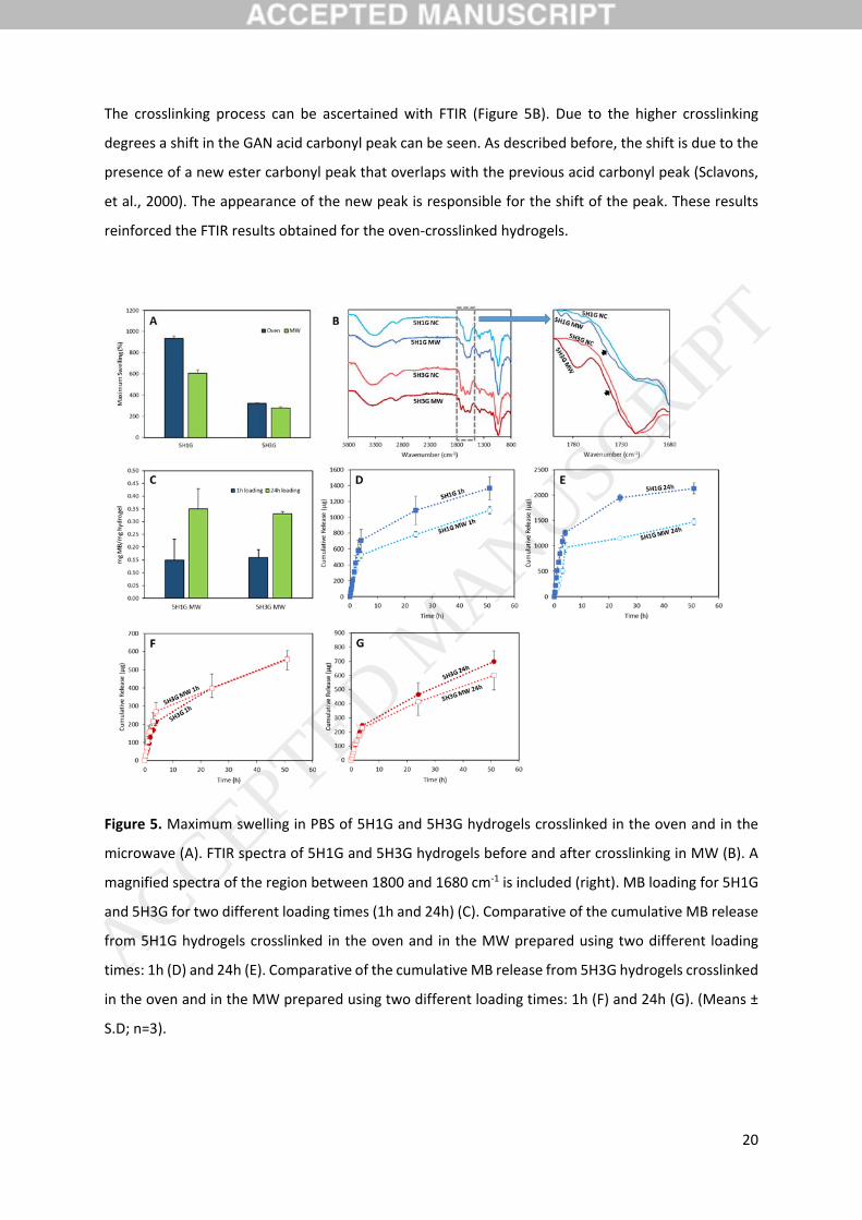
The crosslinking process can be ascertained with FTIR (Figure 5B). Due to the higher crosslinking
degrees a shift in the GAN acid carbonyl peak can be seen. As described before, the shift is due to the
presence of a new ester carbonyl peak that overlaps with the previous acid carbonyl peak (Sclavons,
et al., 2000). The appearance of the new peak is responsible for the shift of the peak. These results
reinforced the FTIR results obtained for the oven‐crosslinked hydrogels.
Figure 5. Maximum swelling in PBS of 5H1G and 5H3G hydrogels crosslinked in the oven and in the
microwave (A). FTIR spectra of 5H1G and 5H3G hydrogels before and after crosslinking in MW (B). A
magnified spectra of the region between 1800 and 1680 cm‐1 is included (right). MB loading for 5H1G
and 5H3G for two different loading times (1h and 24h) (C). Comparative of the cumulative MB release
from 5H1G hydrogels crosslinked in the oven and in the MW prepared using two different loading
times: 1h (D) and 24h (E). Comparative of the cumulative MB release from 5H3G hydrogels crosslinked
in the oven and in the MW prepared using two different loading times: 1h (F) and 24h (G). (Means ±
S.D; n=3).
ACCEPTED MANUSCRIP
T
21
MW‐crosslinked hydrogels were compared with oven‐crosslinked hydrogels by evaluating their MB
release capabilities. Identical loading conditions were used for these sets of experiments. Figure 5C
shows the MB blue loadings for MW‐crosslinked hydrogels. 5H1G hydrogels showed similar loadings
regardless of the crosslinking method (Figure 4A and 5C) for both loading times (p>0.05). However,
5H3G crosslinked in the MW oven showed a higher MB cargo after the 24 loading process (Figure 5C)
than the one obtained for the oven process materials (Figure 4A) (p<0.05). It is interesting that again
there are no significant difference in the loading capacity of 5H1G and 5H3G in all cases (p>0.05).
Figures 8B, 8C, 8D and 8E show the release profiles of MB from all the hydrogels crosslinked using the
MW‐assisted process. The release profiles of MB from the oven‐crosslinked hydrogels have been
included in order to compare both types of systems. Figures 5D and 5E show that the amount of MB
blue release from MW‐treated 5H1G hydrogels is inferior to the one release from the oven‐treated
5H1G hydrogels. This behaviour can be observed for hydrogels loaded during 1 hour and during 24
hours. It is noticeable that the release curves are similar during the first hours. However, after a few
hours the oven‐treated hydrogels showed a superior release capacity. This can be correlated with the
difference in their swelling capacity. 5H1G MW‐treated hydrogels showed a lower swelling capacity
and consequently this can influence the release process. On the other hand, 5H3G MW‐treated
hydrogels showed similar MB release profiles to their oven‐treated counterparts (Figure 5F and 5G).
This is an interesting point as the MW‐treated hydrogels presented slightly higher loading capacity
than the oven‐treated ones. This reinforces the point that the release of MB is not limited by the
loading and that an electrostatic interaction could be responsible of the limited release of MB from
these hydrogels. However, despite the higher release from 5H1G hydrogels, all MW treated hydrogels
showed a relatively limited MB release taking into account its high loading. (5H1G 1h: 48.1%; 5H1G
24h: 18.3%; 5H3G 1h: 12.2%; 5H3G 24h: 5.5%).
The results suggest that MW can be used to crosslink HA/GAN hydrogels in shorter periods of time.
Besides, MW processing is cheaper as it is more energetically efficient and shorter. This is crucial when
considering the scaled up production of the material. MW crosslinking has been used as a proof of
concept, as the equipment was a conventional MW oven that does not offer total control over the
process and the resulting batches were small. For a more complete control over the crosslinking
process a laboratory MW oven should be used and the process should be optimized. As the
conventional oven procedure allowed the preparation of larger batches, the rest of the experiments
were carried out using the conventional oven process.
ACCEPTED MANUSCRIP
T
22
The present paper describes the use of HA but the described methods can be easily applied to other
polysaccharides as GAN is a versatile polymer and can react with a wide variety of polysaccharides, as
they contain multiple hydroxyl groups.
3.4. Preparation of microneedle arrays using HA‐based hydrogels
Micro‐engineered devices are becoming increasingly important in medicine (Donnelly, et al., 2012a).
MNs are a type of micro‐engineered devices that have been extensively used for transdermal and
intradermal delivery of drugs and vaccines (Donnelly, et al., 2012a, Larrañeta, et al., 2016). They are
minimally‐invasive devices used to by‐pass the outermost layer of the skin (Donnelly, et al., 2012a).
Hyaluronic acid has been used on several occasions to prepare MN arrays that dissolve after insertion
in the skin releasing an active molecule included in the array (González‐Vázquez, et al., 2017,
Larrañeta, et al., 2016). The amount of drug that can be delivered with dissolving MN arrays is normally
limited by the dose loaded in the needle tips (Larrañeta, et al., 2016). Consequently, hydrogel‐forming
MN arrays were designed to overcome this limitation (Donnelly, et al., 2012b). This type of MN arrays
swells after insertion in the skin and they contain the drug in a separate layer. Consequently, the drug
can diffuse through the hydrogel matrix into the skin (Donnelly, et al., 2012b). In order to prepare
hydrogel‐forming MN arrays, the mixture of the polymers should be dissolved in an appropriate
solvent, casted into a mould, dried and finally crosslinked (Donnelly, et al., 2012b). As the MN patch
should be cross‐linked in the solid state HA‐based hydrogels are an ideal candidate for this purpose.
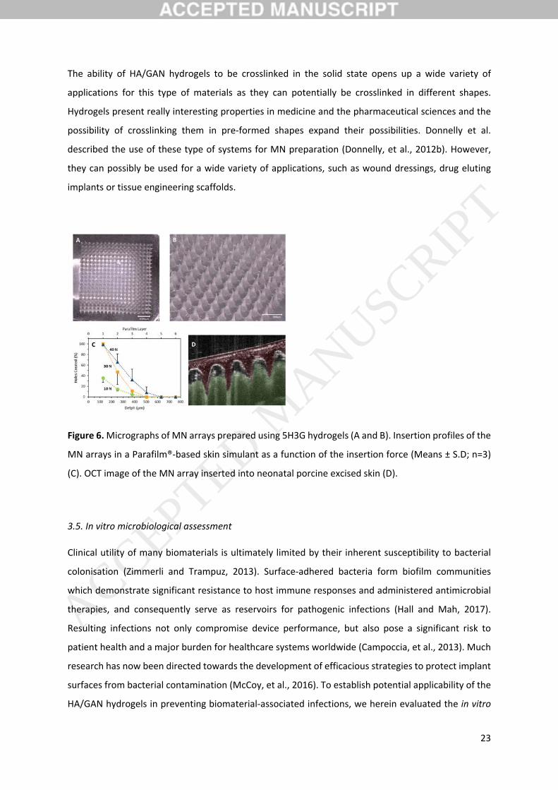
5H3G was selected as a good candidate for MN preparation as these hydrogels did not present higher
swelling capabilities than 5H1G (Figure 1) and can be used for a more sustained drug release (Singh,
et al., 2012). Figure 6A and 6B shows micrographs of 5H3A MN arrays. Figure 6C shows the insertion
profile in a skin simulant for three different application forces and Figure 6D shows an OCT image of
MN insertion into excised neonatal porcine skin. These results shows that the insertion depth is
strongly dependant on the application force. The insertion was not successful when the application
force was 10N. This is due to the shape of the baseplate. The baseplate of the arrays was not
completely flat after the drying process. Consequently, when lower forces were applied only one side
of the array was inserted. However, when using 30 N or higher forces the insertion profile is equivalent
to the one obtained in the past for hydrogel‐forming MN arrays prepared using the same geometries
and different materials (Larrañeta, et al., 2014). As described previously, the average force that
patients apply for MN insertion is around 30N (Larrañeta, et al., 2014, Lutton, et al., 2015, Ripolin, et
al., 2017). Finally, the OCT image confirm that the arrays can be easily inserted inside excised skin.
ACCEPTED MANUSCRIP
T
23
The ability of HA/GAN hydrogels to be crosslinked in the solid state opens up a wide variety of
applications for this type of materials as they can potentially be crosslinked in different shapes.
Hydrogels present really interesting properties in medicine and the pharmaceutical sciences and the
possibility of crosslinking them in pre‐formed shapes expand their possibilities. Donnelly et al.
described the use of these type of systems for MN preparation (Donnelly, et al., 2012b). However,
they can possibly be used for a wide variety of applications, such as wound dressings, drug eluting
implants or tissue engineering scaffolds.
Figure 6. Micrographs of MN arrays prepared using 5H3G hydrogels (A and B). Insertion profiles of the
MN arrays in a Parafilm®‐based skin simulant as a function of the insertion force (Means ± S.D; n=3)
(C). OCT image of the MN array inserted into neonatal porcine excised skin (D).
3.5. In vitro microbiological assessment
Clinical utility of many biomaterials is ultimately limited by their inherent susceptibility to bacterial
colonisation (Zimmerli and Trampuz, 2013). Surface‐adhered bacteria form biofilm communities
which demonstrate significant resistance to host immune responses and administered antimicrobial
therapies, and consequently serve as reservoirs for pathogenic infections (Hall and Mah, 2017).
Resulting infections not only compromise device performance, but also pose a significant risk to
patient health and a major burden for healthcare systems worldwide (Campoccia, et al., 2013). Much
research has now been directed towards the development of efficacious strategies to protect implant
surfaces from bacterial contamination (McCoy, et al., 2016). To establish potential applicability of the
HA/GAN hydrogels in preventing biomaterial‐associated infections, we herein evaluated the in vitro
ACCEPTED MANUSCRIP
T
24
resistance of the 5H1G and 5H3G hydrogels to adherence of the Gram‐positive pathogen,
Staphylococcus aureus, a leading cause of medical device‐associated infections (Tong, et al., 2015) and
Proteus mirabilis, a Gram‐negative pathogen responsible for the majority of catheter‐associated
urinary tract infections (Norsworthy and Pearson, 2017), relative to a widely employed material in
healthcare and medical devices, PVC (McKeen, 2014).
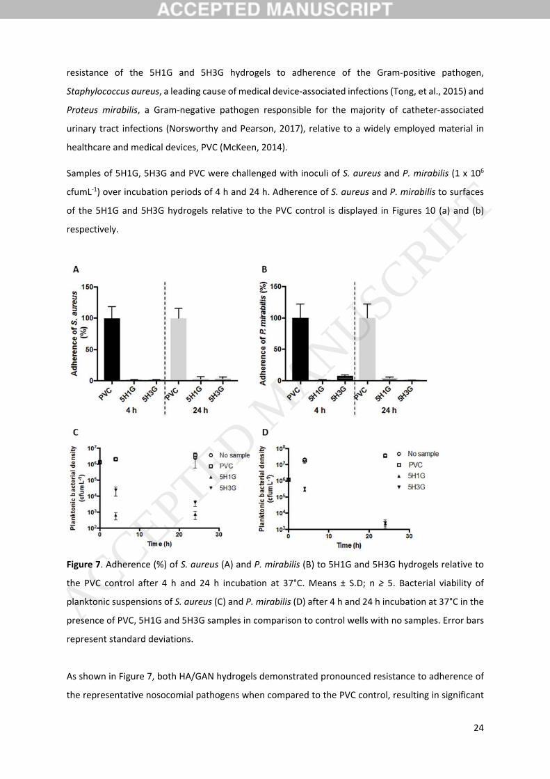
Samples of 5H1G, 5H3G and PVC were challenged with inoculi of S. aureus and P. mirabilis (1 x 10⁶
cfumL‐1) over incubation periods of 4 h and 24 h. Adherence of S. aureus and P. mirabilis to surfaces
of the 5H1G and 5H3G hydrogels relative to the PVC control is displayed in Figures 10 (a) and (b)
respectively.
Figure 7. Adherence (%) of S. aureus (A) and P. mirabilis (B) to 5H1G and 5H3G hydrogels relative to
the PVC control after 4 h and 24 h incubation at 37°C. Means ± S.D; n ≥ 5. Bacterial viability of
planktonic suspensions of S. aureus (C) and P. mirabilis (D) after 4 h and 24 h incubation at 37°C in the
presence of PVC, 5H1G and 5H3G samples in comparison to control wells with no samples. Error bars
represent standard deviations.
As shown in Figure 7, both HA/GAN hydrogels demonstrated pronounced resistance to adherence of
the representative nosocomial pathogens when compared to the PVC control, resulting in significant
ACCEPTED MANUSCRIP
T
25
reductions of up to 98.4% and 98.2% in adherence of S. aureus and P. mirabilis after 4 h incubation
and respective reductions of up to 96.6% and 99.0% after 24 h. Further statistical analysis
demonstrated similar resistance of the two HA/GAN hydrogels to bacterial adherence, with greater
than one‐logarithmic reductions achieved by both hydrogels after challenge periods of 4 h and 24 h.
While this is the first report of the antibacterial properties of GAN‐crosslinked HA, this polysaccharide
has previously demonstrated efficacy in reducing adherence of bacteria to cellular and polymeric
substrates through proposed mechanisms involving interference with bacterial ligand‐surface
receptor site interactions and shielding of underlying substrates by hydration layers formed through
polysaccharide‐water interactions (Cassinelli, et al., 2000, Morra and Cassineli, 1999, Romanò, et al.,
2017). With regards to the antimicrobial activity of the GAN crosslinking agent, biodegradable
microneedles prepared from GAN AN 169 BF have previously demonstrated efficacy in inhibiting in
vitro growth of a range of Gram‐positive and –negative pathogens, including S. aureus and
Enterococcus faecalis, on inoculated agar plates (Boehm, et al., 2012).
In addition to their anti‐adherent properties, the GAN/HA hydrogels synthesised herein demonstrated
significant antimicrobial activity against planktonic bacterial suspensions, as shown in Figure 7.
Logarithmic reductions of up to 3.57 and 1.82 in populations of S. aureus and P. mirabilis respectively
were achieved after 4 h incubation with the HA/GAN hydrogels, whereas respective bacterial densities
increased to ~2 x 10⁶ cfumL‐1 and ~2 x 10⁷ cfumL‐1 over this same time period in the presence of the
PVC controls.
A bacteriostatic effect of HA against planktonic bacterial pathogens, including Pseudomonas
aeruginosa, S. aureus and Escherichia coli has previously been reported, with the resultant activity
highly variable between individual strains and dependent on HA concentration and molecular weight
(Ardizzoni, et al., 2011, Pirnazar, et al., 1999). The pronounced activity of the HA/GAN hydrogels
against planktonic pathogens observed herein may play an important role in protecting surfaces from
contamination in consideration of the “race to the surface” between host and bacterial cells following
device implantation (Gristina, 1987). Furthermore, the drug release capabilities of the HA/GAN
hydrogels offer exciting potential for enhancement of the intrinsic antibacterial properties of the
synthesised materials.
4. Conclusions
The present paper describes the synthesis and characterization of HA‐based hydrogels using GAN as
a chemical crosslinker. The synthetic process takes place in solid phase inside an oven, does not
ACCEPTED MANUSCRIP
T

26
require the use of any type of organic solvent and the only by‐product generated after the
esterification reaction is water. Consequently, it can be considered a green process. Besides, it can be
accelerated by using microwave radiation. However, this procedure was described as a proof of
concept and should be optimized.
These hydrogels present potential to be used for biomedical materials. They are capable of releasing
drugs over a period of several days. Moreover, and based on initial microbiological assessment, the
HA/GAN hydrogels developed herein represent highly promising candidates for non‐fouling materials
to ultimately mitigate the impact of device‐associated infections. These properties make HA/GAN
hydrogels ideal to be used as drug delivery devices or biomaterials, such as microneedle arrays for
transdermal drug delivery, medicated wound dressings or anti‐infective coating for catheters.
5. Acknowledgements
This work was supported in part by the Wellcome Trust (WT094085MA).
6. ReferencesReferences
Ardizzoni A., Neglia R., Baschieri M. C., Cermelli C., Caratozzolo M., Righi E., Palmieri B., & Blasi E. (2011). Influence of hyaluronic acid on bacterial and fungal species, including clinically relevant opportunistic pathogens. Journal of Materials Science: Materials in Medicine 22, 2329‐2338.
Boehm R. D., Miller P. R., Singh R., Shah A., Stafslien S., Daniels J., & Narayan R. J. (2012). Indirect rapid prototyping of antibacterial acid anhydride copolymer microneedles. Biofabrication 4, 011002.
Burdick J. A. & Prestwich G. D. (2011). Hyaluronic Acid Hydrogels for Biomedical Applications. Advanced Materials 23, H41‐H56.
Calo E., Barros J., Ballamy L., & Khutoryanskiy V. V. (2016a). Poly(vinyl alcohol)‐Gantrez® AN cryogels for wound care applications. RSC Advances 6, 105487‐105494.
Calo E., Barros J. M. S. d., Fernandez‐Gutierrez M., San Roman J., Ballamy L., & Khutoryanskiy V. V. (2016b). Antimicrobial hydrogels based on autoclaved poly(vinyl alcohol) and poly(methyl vinyl ether‐alt‐maleic anhydride) mixtures for wound care applications. RSC Advances 6, 55211‐55219.
Caló E. & Khutoryanskiy V. V. (2015). Biomedical applications of hydrogels: A review of patents and commercial products. European Polymer Journal 65, 252‐267.
Campoccia D., Montanaro L., & Arciola C. R. (2013). A review of the clinical implications of anti‐infective biomaterials and infection‐resistant surfaces. Biomaterials 34, 8018‐8029.
ACCEPTED MANUSCRIP
T
27
Cassinelli C., Morra M., Pavesio A., & Renier D. (2000). Evaluation of interfacial properties of hyaluronan coated poly(methylmethacrylate) intraocular lenses. Journal of Biomaterials Science, Polymer Edition 11, 961‐977.
Chen F., Ni Y., Liu B., Zhou T., Yu C., Su Y., Zhu X., Yu X., & Zhou Y. (2017). Self‐crosslinking and injectable hyaluronic acid/RGD‐functionalized pectin hydrogel for cartilage tissue engineering. Carbohydrate Polymers 166, 31‐44.
Chung K. H., Wu C. S., & Malawer E. G. (1990). Glass transition temperatures of poly(methyl vinyl ether‐co‐maleic anhydride) (PMVEMA) and poly(methyl vinyl ether‐co‐maleic acid) (PMVEMAC) and the kinetics of dehydration of PMVEMAC by thermal analysis. Journal of Applied Polymer Science 41, 793‐803.
Collins M. N. & Birkinshaw C. (2013). Hyaluronic acid based scaffolds for tissue engineering—A review. Carbohydrate Polymers 92, 1262‐1279.
Collins M. N. & Birkinshaw C. (2007). Comparison of the effectiveness of four different crosslinking agents with hyaluronic acid hydrogel films for tissue‐culture applications. Journal of Applied Polymer Science 104, 3183‐3191.
Cook J. P., Goodall G. W., Khutoryanskaya O. V., & Khutoryanskiy V. V. (2012). Microwave‐assisted hydrogel synthesis: a new method for crosslinking polymers in aqueous solutions. Macromolecular Rapid Communications 33, 332‐336.
Cui N., Qian J., Liu T., Zhao N., & Wang H. (2015). Hyaluronic acid hydrogel scaffolds with a triple degradation behavior for bone tissue engineering. Carbohydrate Polymers 126, 192‐198.
Donnelly, R. F., Singh, T. R. R., Morrow, D. I. J., & Woolfson, A. D. (2012a). Microneedle‐mediated Transdermal and Intradermal Drug Delivery. : Wiley.
Donnelly R. F., Singh T. R. R., Garland M. J., Migalska K., Majithiya R., McCrudden C. M., Kole P. L., Mahmood T. M. T., McCarthy H. O., & Woolfson A. D. (2012b). Hydrogel‐Forming Microneedle Arrays for Enhanced Transdermal Drug Delivery. Advanced Functional Materials 22, 4879‐4890.
Donnelly R. F., Singh T. R. R., Garland M. J., Migalska K., Majithiya R., McCrudden C. M., Kole P. L., Mahmood T. M. T., McCarthy H. O., & Woolfson A. D. (2012c). Hydrogel‐Forming Microneedle Arrays for Enhanced Transdermal Drug Delivery. Advanced Functional Materials 22, 4879‐4890.
Edwards K. (2016). New Twist on an Old Favorite: Gentian Violet and Methylene Blue Antibacterial Foams. Advances in Wound Care 5, 11‐18.
Fiorica C., Palumbo F. S., Pitarresi G., Bongiovì F., & Giammona G. (2017). Hyaluronic acid and beta cyclodextrins films for the release of corneal epithelial cells and dexamethasone. Carbohydrate Polymers 166, 281‐290.
González‐Vázquez P., Larrañeta E., McCrudden M. T. C., Jarrahian C., Rein‐Weston A., Quintanar‐Solares M., Zehrung D., McCarthy H., Courtenay A. J., & Donnelly R. F. (2017). Transdermal delivery of gentamicin using dissolving microneedle arrays for potential treatment of neonatal sepsis. Journal of Controlled Release .
ACCEPTED MANUSCRIP
T

28
Gristina A. G. (1987). Biomaterial‐centered infection: microbial adhesion versus tissue integration. Science; Science 237, 1588‐1595.
Gustafson C. T., Boakye‐Agyeman F., Brinkman C. L., Reid J. M., Patel R., Bajzer Z., Dadsetan M., & Yaszemski M. J. (2015). Controlled Delivery of Vancomycin via Charged Hydrogels. PLoS ONE 11, e0146401.
Hall C. W. & Mah T. F. (2017). Molecular mechanisms of biofilm‐based antibiotic resistance and tolerance in pathogenic bacteria. FEMS Microbiology Reviews 41, 276‐301.
Heckenkamp J., Adili F., Kishimoto J., Koch M., & LaMuraglia G. M. (2000). Local photodynamic action of methylene blue favorably modulates the postinterventional vascular wound healing response. Journal of Vascular Surgery 31, 1168‐1177.
Hemshekhar M., Thushara R. M., Chandranayaka S., Sherman L. S., Kemparaju K., & Girish K. S. (2016). Emerging roles of hyaluronic acid bioscaffolds in tissue engineering and regenerative medicine. International Journal of Biological Macromolecules; International Journal of Biological Macromolecules 86, 917‐928.
Highley C. B., Prestwich G. D., & Burdick J. A. (2016). Recent advances in hyaluronic acid hydrogels for biomedical applications. Current Opinion in Biotechnology 40, 35‐40.
Hoare T. & Kohane D. (2008). Hydrogels in drug delivery: Progress and challenges. Polymer 49, 1993‐2007.
Jia Y., Huo M., Huang H., Fu W., Wang Y., Zhang J., & Jia S. (2015). Preparation and characterization of bacterial cellulose/hyaluronic acid composites. Proceedings of the Institution of Mechanical Engineers, Part N: Journal of Nanoengineering and Nanosystems 229, 41‐48.
Jiao Y., Pang X., & Zhai G. (2016). Advances in Hyaluronic Acid‐Based Drug Delivery Systems. Current Drug Targets; Current Drug Targets 17, 720‐730.
Jones D. S., McGovern J. G., Woolfson A. D., & Gorman S. P. (1997). Role of physiological conditions in the oropharynx on the adherence of respiratory bacterial isolates to endotracheal tube poly(vinyl chloride). Biomaterials 18, 503‐510.
Korogiannaki M., Guidi G., Jones L., & Sheardown H. (2015). Timolol maleate release from hyaluronic acid‐containing model silicone hydrogel contact lens materials. J Biomater Appl 30, 361‐376.
Larrañeta E., Lutton R. E. M., Brady A. J., Vicente‐Pérez E. M., Woolfson A. D., Thakur R. R. S., & Donnelly R. F. (2015a). Microwave‐Assisted Preparation of Hydrogel‐Forming Microneedle Arrays for Transdermal Drug Delivery Applications. Macromolecular Materials and Engineering 300, 586‐595.
Larrañeta E., Lutton R. E. M., Brady A. J., Vicente‐Pérez E. M., Woolfson A. D., Thakur R. R. S., & Donnelly R. F. (2015b). Microwave‐Assisted Preparation of Hydrogel‐Forming Microneedle Arrays for Transdermal Drug Delivery Applications. Macromolecular Materials and Engineering 300, 586‐595.
Larrañeta E., Moore J., Vicente‐Pérez E. M., González‐Vázquez P., Lutton R., Woolfson A. D., & Donnelly R. F. (2014). A proposed model membrane and test method for microneedle insertion studies. International Journal of Pharmaceutics 472, 65‐73.
ACCEPTED MANUSCRIP
T
29
Larrañeta E., Lutton R. E. M., Woolfson A. D., & Donnelly R. F. (2016). Microneedle arrays as transdermal and intradermal drug delivery systems: Materials science, manufacture and commercial development. Materials Science and Engineering: R: Reports 104, 1‐32.
Lutton R. E. M., Larrañeta E., Kearney M. C., Boyd P., Woolfson A. D., & Donnelly R. F. (2015). A novel scalable manufacturing process for the production of hydrogel‐forming microneedle arrays. International Journal of Pharmaceutics 494, 417‐429.
Mast B. A., Frantz F. W., Diegelmann R. F., Krummel T. M., & Cohen I. K. (1995). Hyaluronic acid degradation products induce neovascularization and fibroplasia in fetal rabbit wounds. Wound Repair and Regeneration 3, 66‐72.
McCoy C. P., Irwin N. J., Brady C., Jones D. S., Carson L., Andrews G. P., & Gorman S. P. (2016). An Infection‐Responsive Approach To Reduce Bacterial Adhesion in Urinary Biomaterials. Molecular Pharmaceutics 13, 2817‐2822.
McKeen, L. W. (2014). 3 ‐ Plastics Used in Medical Devices. In K. Modjarrad & S. Ebnesajjad (Eds.). Handbook of Polymer Applications in Medicine and Medical Devices (pp. 21‐53). Oxford: William Andrew Publishing.
Mero A. & Campisi M. (2014). Hyaluronic Acid Bioconjugates for the Delivery of Bioactive Molecules. Polymers 6, 346‐369.
Miles A. A., Misra S. S., & Irwin J. O. (1938). The estimation of the bactericidal power of the blood. The Journal of Hygiene 38, 732‐749.
Moreno E., Schwartz J., Larrañeta E., Nguewa P. A., Sanmartín C., Agüeros M., Irache J. M., & Espuelas S. (2014). Thermosensitive hydrogels of poly(methyl vinyl ether‐co‐maleic anhydride) ‐ Pluronic(®) F127 copolymers for controlled protein release. International Journal of Pharmaceutics 459, 1‐9.
Morra M. & Cassineli C. (1999). Non‐fouling properties of polysaccharide‐coated surfaces. Journal of Biomaterials Science, Polymer Edition 10, 1107‐1124.
Mráček A., Varhaníková J., Lehocký M., Gřundělová L., Pokopcová A., & Velebný V. (2008). The Influence of Hofmeister Series Ions on Hyaluronan Swelling and Viscosity. Molecules 13, 1025‐1034.
Norsworthy A. N. & Pearson M. M. (2017). From Catheter to Kidney Stone: The Uropathogenic Lifestyle of Proteus mirabilis. Trends in Microbiology 25, 304‐315.
Peppas N. A., Bures P., Leobandung W., & Ichikawa H. (2000). Hydrogels in pharmaceutical formulations. European Journal of Pharmaceutics and Biopharmaceutics 50, 27‐46.
Pirnazar P., Wolinsky L., Nachnani S., Haake S., Pilloni A., & Bernard G. W. (1999). Bacteriostatic Effects of Hyaluronic Acid. Journal of Periodontology 70, 370‐374.
Ripolin A., Quinn J., Larrañeta E., Vicente‐Perez E. M., Barry J., & Donnelly R. F. (2017). Successful application of large microneedle patches by human volunteers. International Journal of Pharmaceutics 521, 92‐101.
ACCEPTED MANUSCRIP
T
30
Romanò C. L., De Vecchi E., Bortolin M., Morelli I., & Drago L. (2017). Hyaluronic Acid and Its Composites as a Local Antimicrobial/Antiadhesive Barrier. Journal of Bone and Joint Infection 2, 63‐72.
Schanté C. E., Zuber G., Herlin C., & Vandamme T. F. (2011). Chemical modifications of hyaluronic acid for the synthesis of derivatives for a broad range of biomedical applications. Carbohydrate Polymers 85, 469‐489.
Schneider L. A., Korber A., Grabbe S., & Dissemond J. (2007). Influence of pH on wound‐healing: a new perspective for wound‐therapy? Archives of Dermatological Research 298, 413‐420.
Sclavons M., Franquinet P., Carlier V., Verfaillie G., Fallais I., Legras R., Laurent M., & Thyrion F. C. (2000). Quantification of the maleic anhydride grafted onto polypropylene by chemical and viscosimetric titrations, and FTIR spectroscopy. Polymer 41, 1989‐1999.
Segura T., Anderson B. C., Chung P. H., Webber R. E., Shull K. R., & Shea L. D. (2005). Crosslinked hyaluronic acid hydrogels: a strategy to functionalize and pattern. Biomaterials 26, 359‐371.
Shah C. B. & Barnett S. M. (1992). Swelling behavior of hyaluronic acid gels. Journal of Applied Polymer Science 45, 293‐298.
Shukla V. K., Shukla D., Tiwary S. K., Agrawal S., & Rastogi A. (2007). Evaluation of pH measurement as a method of wound assessment. Journal of Wound Care 16, 291‐294.
Singh T. R. R., Garland M. J., Migalska K., Caffarel‐Salvador E., Shaikh R., McCarthy H. O., Woolfson A. D., & Donnelly R. F. (2012). Influence of a pore‐forming agent on swelling, network parameters, and permeability of poly(ethylene glycol)‐crosslinked poly(methyl vinyl ether‐co‐maleic acid) hydrogels: Application in transdermal delivery systems. Journal of Applied Polymer Science; Journal of Applied Polymer Science 125, 2680‐2694.
Sperandio F. F., Simões A., Corrêa Aranha A. C., Corrêa L., & Machado de Sousa O. (2010). Photodynamic Therapy Mediated by Methylene Blue Dye in Wound Healing. Photomedicine and Laser Surgery 28, 581‐587.
Tong S. Y. C., Davis J. S., Eichenberger E., Holland T. L., & Fowler V. G. (2015). Staphylococcus aureus Infections: Epidemiology, Pathophysiology, Clinical Manifestations, and Management. Clinical Microbiology Reviews 28, 603‐661.
Tripodo G., Trapani A., Torre M. L., Giammona G., Trapani G., & Mandracchia D. (2015). Hyaluronic acid and its derivatives in drug delivery and imaging: Recent advances and challenges. European Journal of Pharmaceutics and Biopharmaceutics 97, 400‐416.
Wang R., Neoh K. G., Shi Z., Kang E. T., Tambyah P. A., & Chiong E. (2012). Inhibition of escherichia coli and proteus mirabilis adhesion and biofilm formation on medical grade silicone surface. Biotechnology and Bioengineering 109, 336‐345.
Xu X., Jha A. K., Harrington D. A., Farach‐Carson M., & Jia X. (2012). Hyaluronic Acid‐Based Hydrogels: from a Natural Polysaccharide to Complex Networks. Soft Matter 8, 3280‐3294.
ACCEPTED MANUSCRIP
T

31
Zimmerli, W. & Trampuz, A. (2013). Biomaterial‐Associated Infection: A Perspective from the Clinic. In T. F. Moriarty, S. A. J. Zaat, & H. J. Busscher (Eds.). Biomaterials Associated Infection: Immunological Aspects and Antimicrobial Strategies (pp. 3‐24). New York, NY: Springer New York.
Zong, Y., Wei, Y., & Morgan, S. E. (2013). Adsorption/Desorption Processes of pH‐Responsive Copolymers on Model Dental Surfaces via QCM and AFM Analysis. In Anonymous Polymers for Personal Care and Cosmetics (Vol. 1148, pp. 301‐318). : American Chemical Society.
ACCEPTED MANUSCRIP
T