Embed Size (px)
Citation preview

Eular On-line Course on Rheumatic Diseases – module n°17 Ricard Cervera, Gerard Espinosa, David D’Cruz
1
©2007-2009 EULAR
SYSTEMIC LUPUS ERYTHEMATOSUS: PATHOGENESIS, CLINICAL MANIFESTATIONS AND DIAGNOSIS
INTRODUCTION Systemic lupus erythematosus (SLE) is a multisystem autoimmune disorder with a broad
spectrum of clinical presentations (1). There is a peak age of onset among young women between the
late teens and early 40’s and a female to male ratio of 9:1. Ethnic groups such as those with African
or Asian ancestry are more at risk of developing the disorder and it may be more severe compared to
Caucasian patients. SLE is a chronic illness that may be life-threatening when major organs are
affected but more commonly results in chronic debilitating ill health. No single cause for SLE has been
identified though factors such as sunlight and drugs may precipitate the condition and there is a
complex genetic basis.
This module will describe in detail the epidemiology, pathogenesis, clinical features and
diagnosis of SLE.
Learning Outcomes: At the end of this module participants should be able to:
1. Outline the epidemiology of SLE
2. Describe, explain and critically evaluate the evidence for the pathogenesis of SLE in terms of
genetics and environmental and hormonal factors.
3. Describe the clinical manifestations of SLE in the musculoskeletal, dermatological, renal,
respiratory, cardiovascular, central nervous, gastrointestinal and haematological systems.
4. Describe and evaluate the evidence for the existence of patterns of SLE expression in
specific subsets of patients depending on age, gender, ethnicity and social class.
5. Classify and assess patients according to their severity of systems and use appropriate
diagnostic criteria to influence both the morbidity and mortality of patients with SLE.
Eular On-line Course on Rheumatic Diseases – module n°17 Ricard Cervera, Gerard Espinosa, David D’Cruz
2
©2007-2009 EULAR
EPIDEMIOLOGY There are many epidemiological studies on SLE from around the world and there is extensive
data from the European Union (EU) and the United States of America (USA) (2-14).
Incidence The incidence of SLE in the general population varies according to the characteristics of the
population studied, i.e. age, gender, race, ethnic/national origin or period of time studied, but also
depending on changes in classification criteria. In the EU, the annual incidence ranges between 3.3
cases per 100,000 persons per year in Iceland (7) and 4.8 cases per 100,000 persons per year in
Sweden (5) (Table 1). In the USA, the annual incidence of SLE ranges from 1.8 to 7.6 cases per
100,000 persons per year (Table 2). The incidence of SLE may be increasing - for example the
incidence of SLE in Rochester, Minnesota increased by a factor of 2.5 from the years 1950-1954 (4)
to the years 1975-1979 (10).
Table 1: Incidence of SLE in several studies in Europe.
Location Date Incidence*
Sweden (5) 1982 4.8
Nottingham(6) 1989 3.7
Iceland (7) 1990 3.3
Birmingham (9) 1991 3.8
* Incidence rates per 100,000 persons per year; including males and females.
Table 2: Incidence of SLE in several studies in the USA.
Location Date Incidence*
San Francisco (2) 1973 7.6
Baltimore (3) 1977 4.6
Rochester (4) 1979 2.2
Rochester (10) 1992 5.8
* Incidence rates per 100.000 persons per year; including males and females.

Eular On-line Course on Rheumatic Diseases – module n°17 Ricard Cervera, Gerard Espinosa, David D’Cruz
3
©2007-2009 EULAR
Prevalence Several prevalence studies in the general population also show marked variation. This
variability may result from methodological differences in case ascertainment and socio-economic
factors. However, true geographic differences cannot be excluded and may result from differences in
genetic or environmental factors.
In 1982, Hochberg et al. (12) in England and Wales reported a prevalence of 12.5 cases per
100,000 women of all ages, and was higher at 17.7 in women of 15 to 64 years of age. More recent
studies by Hopkinson et al. (6) indicate a prevalence of 25 cases per 100,000 persons in Nottingham
and those of Johnson et al. (9) a prevalence of 28 cases per 100,000 persons. The highest
prevalence in Europe has been described in Sweden with 39 cases per 100,000 persons (Table 3).
The overall prevalence in the USA ranges between 14.6 and 50 cases per 100,000 persons (including
white and black people) (Table 4).
Table 3: Prevalence of SLE in several studies in Europe.
Location Date Prevalence*
Finland (11) 1978 28.0
England-Wales (12) 1982 12.5**
Sweden (5) 1982 39.0
England (Leicester) (8) 1989 26.1
Iceland (7) 1990 36.0
England (Nottingham) (6) 1990 24.6
England (Birmingham) (9) 1991 27.7
Ireland (14) 1993 25.4
* Prevalence rates per 100.000 persons; both sexes combined.
** Females only.
Table 4: Prevalence of SLE in several studies in the USA.
Location Date Prevalence*
San Francisco (2) 1973 50.8
Rochester (3) 1980 40.0
Hawaii (13) 1989 41.8
* Prevalence rates per 100.000 persons; including males and females.

Eular On-line Course on Rheumatic Diseases – module n°17 Ricard Cervera, Gerard Espinosa, David D’Cruz
4
©2007-2009 EULAR
PATHOGENESIS
The pathogenesis of lupus remains unclear although the concept of apoptosis goes some way
to explaining how the immune system may recognise predominantly intracellular antigens.
Autoantigens are released by necrotic as well as apoptotic cells. Defects in the clearance of apoptotic
cells have been described in SLE which may lead to aberrant uptake by macrophages which then
present the previously intracellular antigens to T and B cells thus driving the autoimmune process
(15). Recent work has expanded these concepts and dissected out possible defects in clearance of
apoptotic bodies including complement deficiencies, defects in macrophage handling and
presentation of these antigens to the immune system.
The most striking recent studies have demonstrated the development of autoantibodies years
before the onset of clinical features of SLE and the antiphospholipid syndrome (APS) (16,17). These
investigators utilised the United States Department of Defence serum repository containing some 30
million samples from service personnel taken at regular intervals. They identified 130 individuals with
SLE and showed that autoantibodies to DNA developed in 72 patients on average 2.7 years prior to
diagnosis and up to 9.3 years earlier. They also described the prevalence of other autoantibodies
such as anti-nuclear, anti-Ro, anti-La, anti-Sm, anti-RNP (16) and anti-phospholipid antibodies
(aPL)(17) prior to the development of clinical SLE. Antinuclear antibodies occurred earlier than anti-
DNA antibodies and a significant number of these patients had a rise in the anti-DNA titres just prior
to diagnosis. Interestingly, anti-Sm and anti-RNP antibodies appeared shortly before diagnosis
suggesting a crescendo of autoimmunity resulting in clinical illness. This data also suggests that
autoantibodies alone do not necessarily result in clinical disease and that other factors possibly
genetic and environmental may be important. It may be possible in the future to predict the onset of
clinical features of lupus by clinical assessment and monitoring the development of various lupus
autoantibodies.
The potential pathogenicity of anti-DNA antibodies remains controversial. Several animal
models and experiments suggest that these auto-antibodies are capable of producing renal lesions in
severe combined immunodeficiency (SCID) mice. However, the evidence is less convincing in
humans. This is especially true with the insights gained from therapy with B cell depleting agents such
as rituximab where there is often a rapid clinical response with only moderate reductions in anti-DNA
antibody levels (18).
Cytokine patterns may also be important in the pathogenesis of lupus. Recent studies have
highlighted the over-expression of the type I interferon pathway in patients – the so-called ‘interferon
signature’. A large study has shown an association of a common interferon regulatory factor 5 (IRF5)
haplotype driving elevated expression of multiple unique isoforms of IRF5 as an important genetic risk
factor for SLE (19).

Eular On-line Course on Rheumatic Diseases – module n°17 Ricard Cervera, Gerard Espinosa, David D’Cruz
5
©2007-2009 EULAR
Abnormal signal transduction may also be important in the pathogenesis of SLE. For example
decreased expression of TCR zeta chain, PKC theta, decreased PKC dependent protein
phosphorylation, impaired translocation of NF-kB p65 and decreased production of IL-2 as well as
functional variants in the signal transducer and activator of transcription 4 (STAT4) genes have all
been described in T cells from SLE patients (19, 20).
Genetics Genetic susceptibility to lupus is inherited as a complex trait and studies have suggested that
several genes may be important. In particular an interval on the long arm of chromosome 1, 1q23–24,
is linked with SLE in multiple populations. Clinically it is widely accepted that active SLE is
characterised by elevated erythrocyte sedimentation rates but normal C-reactive protein (CRP) levels.
Both CRP and complement as well as serum amyloid P protein are important in clearing apoptotic cell
debris and the genes for CRP have been mapped to chromosome 1, 1q23–24, the so-called pentraxin
locus. Russell and colleagues examined the inheritance of polymorphisms at the pentraxin locus in a
family-based association study of SLE and found strong linkage disequilibrium within each of the CRP
and serum amyloid P genes (21). They demonstrated that an allele of CRP 4 was associated with
SLE. Furthermore there were two haplotypes that were significantly associated with reduced basal
CRP expression: CRP 2 and CRP 4 and an allele of CRP 4 was associated with ANA production.
Thus the authors proposed a genetic explanation of the link between low CRP levels, antinuclear
autoantibody production and the contribution of these to the development of human SLE.
Another large study of individuals and multi-case families with SLE suggested that a single
nucleotide polymorphism (SNP) within the programmed cell death 1 gene (PDCD1) is associated with
the development of lupus in both European and Mexican populations. The authors showed that the
associated allele of this SNP alters a binding site for a transcription factor located in an intronic
enhancer, suggesting a mechanism through which it can contribute to the development of SLE (22).
Environmental factors
Sunlight is the most obvious environmental factor that may exacerbate SLE. Other factors
have been considered and crystalline silica was the focus of studies from the south eastern United
States where occupational exposure was hypothesised as a risk for developing lupus. A case control
study found that more patients (19%) than controls (8%) had a history of medium- or high-level silica
exposure from farming or trades suggesting that this may be associated with the development of SLE
in a proportion of individuals though occupational exposure may be often difficult to quantify
accurately (23). A further study found associations with self-reported occupational exposure to
mercury, mixing pesticides for agricultural work and among dental workers although the actual
numbers exposed was relatively small. Unlike scleroderma though there was no association with
solvent use (24).

Eular On-line Course on Rheumatic Diseases – module n°17 Ricard Cervera, Gerard Espinosa, David D’Cruz
6
©2007-2009 EULAR
Epstein Barr virus (EBV) has also been identified as a possible factor in the development of
lupus. EBV may reside in and interact with B cells. Gross et al (25) found a high frequency of EBV
infected B cells in lupus patients compared to controls and these infected cells are predominantly
memory B cells. There was no relationship with immunosuppressive therapy and furthermore patients
with active lupus flares had more infected cells than patients with quiescent disease. Although other
studies have suggested a causative role for EBV in SLE, these authors are more cautious and despite
their findings of increased frequencies of infected cells, increased viral loads, and viral gene
expression, they have not interpreted this as directly implicating EBV in the development of SLE and
argue that it is also possible that the immune dysregulation of SLE may result in aberrant EBV
expression. In contrast, studies in a mouse model found that direct introduction of the whole EBV
nuclear antigen 1 protein can elicit IgG antibodies to Sm and to double-stranded DNA (dsDNA) thus
supporting a putative role for EBV in the development of lupus. The paradox remains that although
90% of the adult population are infected by EBV, the prevalence of SLE remains low emphasising the
multi-factorial nature of the pathogenesis of SLE.
Hormonal factors SLE is a disease of women of child-bearing age and there have been many anecdotal reports
of exogenous oestrogens exacerbating lupus or increasing the risk of developing this disorder. Oral
contraceptive use in the Nurses Health Study was associated with a slightly increased risk of
developing SLE with a relative risk versus never users of 1.9 (26). Hormone replacement therapy
(HRT) has been associated with an increased risk of developing SLE though another study failed to
show any increased risk. Several small studies have suggested that HRT is unlikely to increase the
risk of flares in SLE though these studies in general have been small retrospective case series. The
definitive prospective study is the SELENA trial which randomised women with SLE to receive HRT or
placebo (27). Although there was no increase in major flares in the HRT group, there were
significantly more mild to moderate flares in the HRT group compared to the placebo group. A number
of women, including one in the placebo group, developed thrombotic events. Recent data has swung
against the long term use of HRT although there may still be a role for using it for limited periods in
women with SLE who are aPL negative who have severe menopausal symptoms. This study will
inform patients and clinicians of the potential risks and confirms the view that HRT is contraindicated
with women with aPL.
The use of the combined oestrogen containing oral contraceptive pill has been discouraged in
lupus patients following anecdotal reports of serious disease flares. Two randomised controlled trials
investigated the oral contraceptive pill in women with lupus. Petri et al randomised 183 women with
inactive or stable low grade lupus activity to receive either a combined low dose oestrogen containing
oral contraceptive pill or a placebo for one year (28). All participants practised other effective birth-
control methods.

Eular On-line Course on Rheumatic Diseases – module n°17 Ricard Cervera, Gerard Espinosa, David D’Cruz
7
©2007-2009 EULAR
There were no differences in the rates of severe or mild to moderate disease flares in either treatment
group and the authors suggest that this type of contraception may be considered in women with lupus
who need effective birth control especially when receiving cytotoxics, for amelioration of menstrual
disease flares and protection against steroid related bone loss.
Furthermore, Sánchez-Guererro et al. (29) randomised 162 women with SLE to combined oral
contraceptive pills, progestin only pills or a copper intra-uterine device. At the end of one year there
were no differences in disease activity scores or flare rates. This study included asymptomatic aPL
positive patients and 4 patients (2 in each of the hormone groups) suffered venous thrombotic events.
There was a higher infection rate in those women assigned to the intra-uterine device.
Taken together these studies provide some reassurance for lupus patients with mild stable
disease who are aPL negative who wish to consider the use of the oral contraceptive pill. Both studies
however highlight the thrombotic risk inherent in lupus patients, even if aPL are absent.
CLINICAL MANIFESTATIONS
Musculoskeletal involvement Joints
Arthralgia occurs in about 90% of all patients with SLE. Characteristically, it is polyarticular,
symmetrical, episodic and flitting in nature. The patients’ symptoms often exceed the objective clinical
findings and usually there is no clinically overt arthritis. Synovial effusions are uncommon and of small
volume when they do occur. However, approximately 10% of SLE patients do have a deforming
Jaccoud’s arthritis. In contrast to patients with rheumatoid arthritis, the deformities are not usually
associated with synovial hypertrophy or bony erosions. In fact, tenosynovitis is more common than
erosive synovitis and is the cause of the “swan-neck” deformities and ulnar deviation seen in the
Jaccoud’s arthritis of lupus. Examination of the synovial fluid usually reveals a white cell count of less
than 3000/mm3, predominantly mononuclear cells. The fluid is often positive for rheumatoid factor and
anti-nuclear antibody. Approximately, 1-2% of SLE patients also meet the American College of
Rheumatology (ACR) criteria for definite or classical rheumatoid arthritis and have an erosive
arthropathy .
Muscles Clinically obvious muscle involvement has been reported in 30-50% of SLE patients. However,
myalgia, muscle weakness and tenderness, may be due to a variety of other complications. Thus both
corticosteroid and rarely chloroquine therapy may cause a myopathy. In addition, myalgia may be
induced by an adjacent arthralgia, although only 5% of lupus patients have met the ACR criteria for
both SLE and polymyositis.

Eular On-line Course on Rheumatic Diseases – module n°17 Ricard Cervera, Gerard Espinosa, David D’Cruz
8
©2007-2009 EULAR
Dermatological involvement Cutaneous lesions may occur in up to 85% of SLE patients. The butterfly rash is
erythematous, often blotchy, and found mainly over the malar bones and across the bridge of the
nose (Fig. 1). Although it is the best known skin lesion, it is merely one of numerous ways in which
lupus manifests cutaneously. Lesions such as maculopapular and discoid lesions, splinter
haemorrhages, dilated capillaries at the nail base, bullous lesions, angioneurotic oedema, livedo
reticularis (Fig. 2) and buccal, genital and nasal ulceration have also been described. Vasculitic skin
lesions are usually found at the nailfolds and finger tips (Fig 3) or on the extensor surface of the
forearm. When they occur around the malleoli, they may lead to tender, deep, leg ulcers which can
take months to heal.
Many SLE rashes are exacerbated by ultraviolet light and indeed generalized lupus flares may
follow exposure to direct sunlight with inadequate protection (Fig 4). A particularly photosensitive rash
is subacute cutaneous lupus erythematosus (SCLE) which is often associated with anti-Ro antibodies
(Fig. 5). Babies born to mothers with anti-Ro and/or anti-La antibodies are at risk of neonatal lupus
syndrome (Fig 6).
The deposition of immunoglobulins at the dermal-epidermal junction in skin biopsies from
patients with lupus was first reported over 40 years ago. These immunoglobulins are usually of the
IgG or IgM isotype. Approximately, 90% of biopsies from lupus skin lesions have such
immunoglobulin deposits which usually appear as a band along the dermal-epidermal junction, giving
rise to the name the “lupus band test”. In patients with SLE, deposition of immunoglobulin and
complement may be found in clinically normal skin and is thus a useful adjunct to diagnosis since no
such deposition is found in patients with discoid lupus or control subjects.
Figure 1 - Malar rash

Eular On-line Course on Rheumatic Diseases – module n°17 Ricard Cervera, Gerard Espinosa, David D’Cruz
9
©2007-2009 EULAR
Figure 2 - Livedo reticularis
Figure 3 – Periungual erythema and nailfold vasculitis

Eular On-line Course on Rheumatic Diseases – module n°17 Ricard Cervera, Gerard Espinosa, David D’Cruz
10
©2007-2009 EULAR
Figure 4 - Acute diffuse cutaneous lupus
Figure 5 - Subacute cutaneous lupus
erythematosus Figure 6 - Neonatal lupus

Eular On-line Course on Rheumatic Diseases – module n°17 Ricard Cervera, Gerard Espinosa, David D’Cruz
11
©2007-2009 EULAR
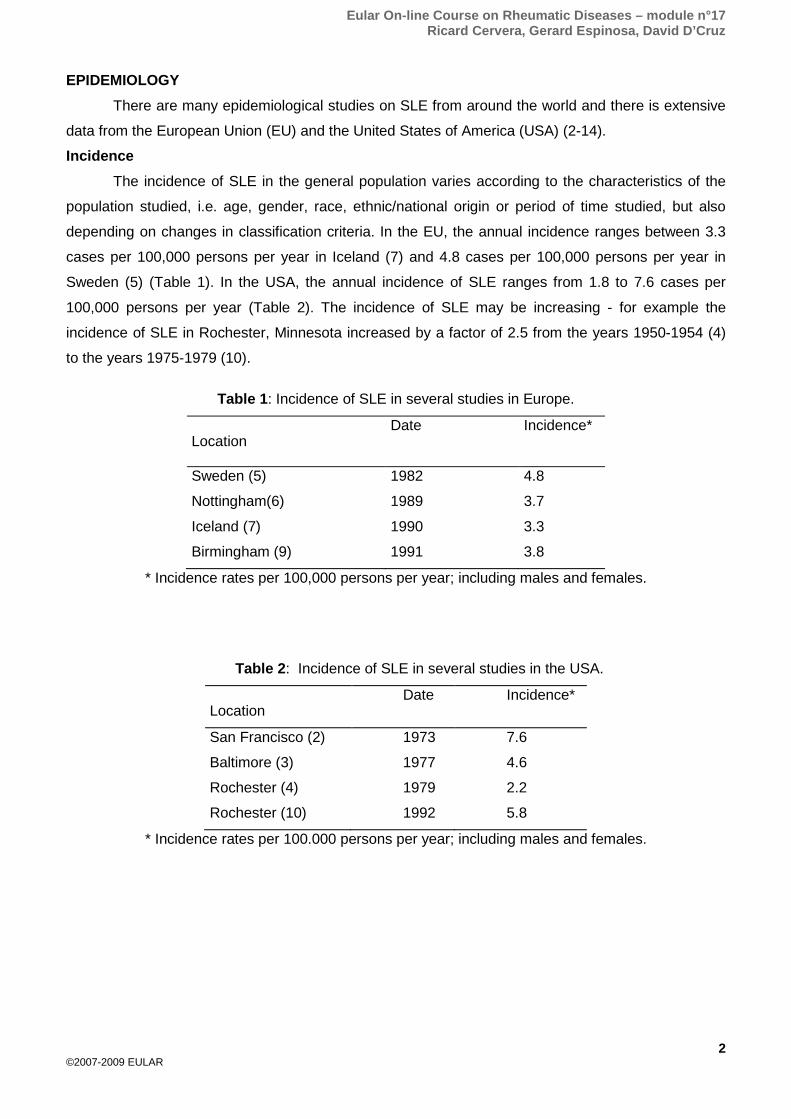
Lupus nephritis More than 70% of patients with SLE have renal involvement at some stage of their disease.
The World Health Organisation (WHO) classification for lupus nephritis has been updated to allow
more accurate descriptions of renal histopathology specimens by the International Society of
Nephrology (ISN) and the Renal Pathology Society (RPS) (30) (Fig. 7 - 9) (Table 5). These
descriptions allow better communication between pathologists translating static images from histology
slides into meaningful descriptions of the huge variety of biopsy appearances for clinicians. Of the
different pathological classes, diffuse proliferative glomerulonephritis (Class IV) has the worst
prognosis, resulting in 11-48% of patients with end stage renal disease at 5 years.
Figure 7 - Kidney Biopsy: Class III (Focal Proliferative) Lupus Nephropathy
Eular On-line Course on Rheumatic Diseases – module n°17 Ricard Cervera, Gerard Espinosa, David D’Cruz
12
©2007-2009 EULAR
Figure 8 - Kidney Biopsy: Class IV (Diffuse Proliferative) Lupus Nephropathy
Figure 9 - Kidney Biopsy: Class V (Membranous) Lupus Nephropathy

Eular On-line Course on Rheumatic Diseases – module n°17 Ricard Cervera, Gerard Espinosa, David D’Cruz
13
©2007-2009 EULAR
Table 5: International Society of Nephrology/Renal Pathology Society 2003 classification of lupus nephritis
Class I Minimal mesangial lupus nephritis Normal glomeruli by light microscopy, but mesangial immune deposits by immunofluorescence Class II Mesangial proliferative lupus nephritis Purely mesangial hyper-cellularity of any degree or mesangial matrix expansion by light microscopy, with mesangial immune deposits May be a few isolated sub-epithelial or sub-endothelial deposits visible by immunofluorescence or electron microscopy, but not by light microscopy Class III Focal lupus nephritisa Active or inactive focal, segmental or global endo- or extra-capillary glomerulonephritis involving <50% of all glomeruli, typically with focal sub-endothelial immune deposits, with or without mesangial alterations Class III (A) Active lesions: focal proliferative lupus nephritis Class III (A/C) Active and chronic lesions: focal proliferative and sclerosing lupus nephritis Class III (C) Chronic inactive lesions with glomerular scars: focal sclerosing lupus nephritis Class IV Diffuse lupus nephritisb Active or inactive diffuse, segmental or global endo- or extra-capillary glomerulonephritis involving 50% of all glomeruli, typically with diffuse sub-endothelial immune deposits, with or without mesangial alterations. This class is divided into diffuse segmental(IV-S) lupus nephritis when 50% of the involved glomeruli have segmental lesions, and diffuse global (IV-G) lupus nephritis when 50% of the involved glomeruli have global lesions. Segmental is defined as a glomerular lesion that involves less than half of the glomerular tuft. This class includes cases with diffuse wire loop deposits but with little or no glomerular proliferation Class IV-S (A) Active lesions: diffuse segmental proliferative lupus nephritis Class IV-G (A) Active lesions: diffuse global proliferative lupus nephritis Class IV-S (A/C) Active and chronic lesions: diffuse segmental proliferative and sclerosing lupus nephritis Active and chronic lesions: diffuse global proliferative and sclerosing lupus nephritis Class IV-S (C) Chronic inactive lesions with scars: diffuse segmental sclerosing lupus nephritis Class IV-G (C) Chronic inactive lesions with scars: diffuse global sclerosing lupus nephritis Class V Membranous lupus nephritis Global or segmental sub-epithelial immune deposits or their morphologic sequelae by light microscopy and by immunofluorescence or electron microscopy, with or without mesangial alterations Class V lupus nephritis may occur in combination with class III or IV in which case both will be diagnosed Class V lupus nephritis show advanced sclerosis Class VI Advanced sclerosis lupus nephritis 90% of glomeruli globally sclerosed without residual activity
a Indicate the proportion of glomeruli with active and with sclerotic lesions. b Indicate the proportion of glomeruli with fibrinoid necrosis and/or cellular crescents. Indicate and grade (mild, moderate, severe) tubular atrophy, interstitial inflammation and fibrosis, severity of arteriosclerosis or other vascular lesions.

Eular On-line Course on Rheumatic Diseases – module n°17 Ricard Cervera, Gerard Espinosa, David D’Cruz
14
©2007-2009 EULAR
Lungs The immunosuppressive therapy required by many SLE patients predisposes them to
concurrent infection. The lungs are a frequent target for this “secondary” infection and bacteria
(including tubercule bacilli), viruses and fungi may all cause pneumonia in lupus patients.
Parenchymal alterations, attributable to SLE itself, have been described in 18% of patients.
These patients had interstitial fibrosis, pulmonary vasculitis and interstitial pneumonitis. However,
many non-specific pulmonary lesions previously attributed to SLE, such as alveolar haemorrhage
(Fig. 9), alveolar wall necrosis, oedema and hyaline membranes, are probably secondary to factors
such as intercurrent infection, congestive heart failure, renal failure and oxygen toxicity.
In the relatively few cases studied, immune complex deposition has been closely correlated
with histological evidence of inflammatory lesions in the pleural (and pericardial) membrane.
Abnormal pulmonary function tests, notably diminished total lung capacity and flow rates, in
clinically mild patients with dyspnoea, poor diaphragmatic movement, basal crepitations and
occasionally cyanosis and clubbing, are found in up to 50% of SLE patients. A similar proportion of
SLE patients may have an acute lupus pneumonitis with a mononuclear cell infiltrate detectable in the
alveolar septae. These patients frequently complain of dyspnoea, pleuritic chest pain and coughs.
Haemoptysis is less common and true pulmonary haemorrhage from necrotizing alveolar capillaritis is
rare.
Pleural effusions may be found in about half of these patients (and in other SLE patients
especially during generalized disease flares). The effusions are normally small to moderate in size
and are usually exudates (i.e. protein content >3 g/100 ml). They are rarely haemorrhagic and usually
have a glucose concentration double that found in rheumatoid effusions (normally, 20 mg/100 ml or
less).
Figure 10 - Alveolar Haemorrhage

Eular On-line Course on Rheumatic Diseases – module n°17 Ricard Cervera, Gerard Espinosa, David D’Cruz
15
©2007-2009 EULAR
Heart Pericardium
Abnormalities of the electrocardiogram, notably of the T wave, are the most frequent
manifestation. A pericardial rub may be more common than a significant pericardial effusion.
Histological abnormalities vary from occasional foci of fibrinoid degeneration and inflammatory cell
infiltrates to far more extensive lesions. Adhesive chronic pericarditis and very large effusions causing
tamponade are very rare.
Myocardium Whilst true myocardial involvement is less frequent than pericardial disease, prolongation of
the PR interval (approximately 10%), fibrinoid degeneration, myocardial infarction and coronary
stenosis due to arteritis are occasionally seen. New imaging techniques such as cardiac MRI suggest
that myocardial involvement may be more common than previously thought.
There is increasing evidence that premature accelerated atherosclerosis considerably
increases the risk of cardiovascular events in patients with SLE and this is described in a separate
module of this course.
Valves Systolic murmurs are frequently heard in around 30% of SLE patients. However, they probably
reflect the hyperdynamic circulation consequent upon the anaemia often found in these individuals. In
contrast, diastolic murmurs are uncommon.
Libman-Sacks endocarditis has long been described as a feature of SLE. Although found in up
to 50% of autopsied cases, it rarely causes clinically significant lesions. Histologically, the lesions are
small (1-4 mm) vegetations (verrucae) comprising proliferating and degenerating valve tissue with
fibrin and platelet thrombi. They are most frequently found adjacent to the edges of the mitral and
tricuspid valves. aPL may contribute to the development of Libman-Sacks endocarditis and studies
suggest that there is a selective deposition of aPL and complement within the walls of the small
junctional vessels in the active portions of the verrucous endocardial lesions.
Central nervous system lupus The ACR classification criteria for central nervous system (CNS) lupus has changed
considerably from seizures and psychosis. The ACR nomenclature now includes 19 different
syndromes that are classifiable (31) (Table 6). An emerging concept is the distinction between CNS
manifestations due to lupus and those due to the APS. A wide variety of neuropsychiatric
manifestations attributable to APS have been described including strokes, seizures, movement
disorders, transverse myelopathy, demyelination syndromes, transient ischaemic attacks, cognitive
dysfunction, visual loss and headaches including migraine.

Eular On-line Course on Rheumatic Diseases – module n°17 Ricard Cervera, Gerard Espinosa, David D’Cruz
16
©2007-2009 EULAR
Table 6: Neuropsychiatric syndromes observed in SLE. Central nervous system: Aseptic meningitis Cerebrovascular disease Demyelinating syndrome Headache (including migraine and benign intracranial hypertension) Movement disorder (chorea) Myelopathy Seizure disorders Acute confusional state Anxiety disorder Cognitive dysfunction Mood disorder Psychosis Peripheral nervous system: Acute inflammatory demyelinating polyradiculoneuropathy (Guillain-Barré syndrome) Autonomic disorder Mononeuropathy, single/multiplex Myasthenia gravis Neuropathy, cranial Plexopathy Polyneuropathy
Gastrointestinal problems
A wide variety of non-specific gastrointestinal clinical features has been described amongst
SLE patients. Abdominal pain (especially common in children) occurs in about 20% of cases. Its
precise cause is rarely determined though ileal and colonic perforations and regional enteritis have
been described. Pathologically, necrotizing vasculitis is usually found when perforation occurs.
Ascites, dysphagia and pancreatitis are occasionally seen. Hepatomegaly and/or liver function test
abnormalities may be found in up to 30% of patients. However, the laboratory abnormalities may be
related to SLE therapy.
Haematological abnormalities Red blood cells
A normochromic, normocytic anaemia is frequently found in SLE patients, with concomitant
low levels of both the serum iron and iron binding capacity. This abnormality appears to be related, as
in other diseases, to chronic inflammation and shunting of elemental iron from erythroblasts to
macrophages.
Iron-deficiency anaemia may be induced by non-steroidal anti-inflammatory drugs, which can
cause gastrointestinal haemorrhage. Excessive blood loss from menorrhagia, sometimes related to
severe thrombocytopenia, may have the same effect.

Eular On-line Course on Rheumatic Diseases – module n°17 Ricard Cervera, Gerard Espinosa, David D’Cruz
17
©2007-2009 EULAR
Haemolytic anaemia as detected by the Coombs’ test is another rare feature of SLE.
Autoimmune thrombocytopenia occasionally manifests simultaneously with haemolytic anaemia: this
condition is known as Evan’s syndrome.
Platelets Two forms of thrombocytopenia (platelet count < 100 x 109/l) are found in SLE. Firstly, it may
be encountered in a chronic form, generally associated with mild disease. Secondly, it may occur in
an acute form, similar to idiopathic autoimmune thrombocytopenic purpura. This latter association is
with disease carrying a greater morbidity and mortality.
Platelet destruction appears to be mediated by anti-platelet antibodies and aPL are also
associated with thrombocytopenia as well as with thrombosis.
White blood cells Persistent leucopenia (< 4.0 x 10⁹/l) is one of the ACR criteria for the classification of SLE. It
probably results from a combination of destruction of white cells by autoantibodies, decreased marrow
production, increased or marginal splenic pooling, and complement activation. It should also be noted
that the immunosuppressive drugs used in the treatment of SLE may cause a marked leucopenia.
Serological abnormalities The serum from SLE patients may bind to an extensive array of molecules including nucleic
acids (antinuclear antibodies) and phospholipid binding proteins (lupus anticoagulant, anticardiolipin
antibodies, β2 glycoprotein 1 antibodies). Antibodies may also be detected against diverse cells
including leukocytes, erythrocytes, platelets and neurones. In addition to these autoantibodies,
numerous other abnormalities are evident, including the LE cell phenomenon,
hypocomplementaemia, elevated levels of acute phase proteins, gamma globulins and circulating
immune complexes.
Non-specific features Fever, lymphadenopathy, hair loss and Raynaud’s phenomenon are all commonly found in
SLE patients. Fever in lupus patients may be striking and often requires extensive investigation to
exclude concurrent infection, although a normal CRP in this context usually suggests a low likelihood
of sepsis.
Lymphadenopathy may also be dramatic in SLE, to such an extent that lymph node biopsy
may have to be performed to exclude malignancy. Some patients seem more prone to this feature
than others and in this group the degree of lymphadenopathy may reflect general disease activity.
Splenomegaly occurs in about 10% of patients.
Eular On-line Course on Rheumatic Diseases – module n°17 Ricard Cervera, Gerard Espinosa, David D’Cruz
18
©2007-2009 EULAR
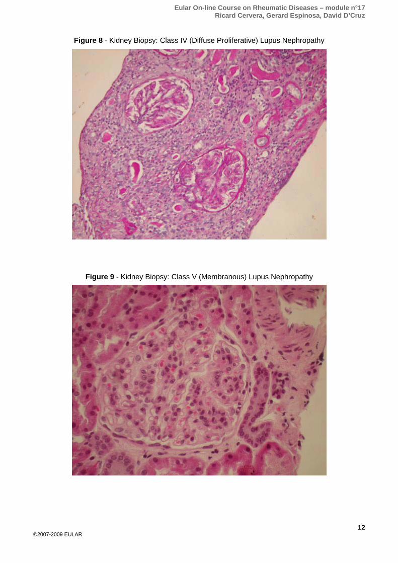
PATTERNS OF DISEASE EXPRESSION IN SPECIFIC SUBSETS An important question is whether the age at onset of the disease, the gender, or the
autoantibody pattern, can modify the disease expression and define specific SLE subsets. The “Euro-
Lupus” studies have provided major insights into these areas of inquiry. This cohort is composed of
1,000 patients with SLE that have been followed prospectively since 1991. These patients have been
gathered by a European consortium - the “Euro-Lupus Project Group” - that includes more than 40
investigators from seven European countries who have substantial experience in the management of
SLE patients. This consortium was originated as part of the network promoted by the “European
Working Party on SLE”. The general characteristics of this cohort at the beginning of the study were
published in 1993 (Tables 7 and 8) (32).
Table 7: Clinical manifestations in a series of 1,000 European SLE patients.
SLE manifestation Prevalence (%)
Arthritis 84
Malar rash 58
Fever 52
Photosensitivity 45
Nephropathy 39
Serositis 36
Raynaud’s phenomenon 34
Neurologic involvement 27
Oral ulcers 24
Thrombocytopenia 22
Sicca syndrome 16
Livedo reticularis 14
Thrombosis 14
Lymphadenopathy 12
Discoid lesions 10
Myositis 9
Haemolytic anemia 8
Lung involvement 7
Subacute cutaneous lesions 6
Chorea 2
Eular On-line Course on Rheumatic Diseases – module n°17 Ricard Cervera, Gerard Espinosa, David D’Cruz
19
©2007-2009 EULAR
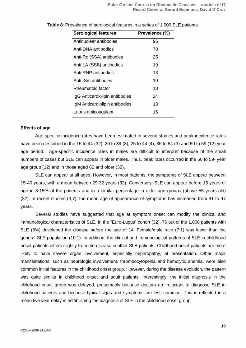
Table 8: Prevalence of serological features in a series of 1,000 SLE patients.
Serological features Prevalence (%)
Antinuclear antibodies 96
Anti-DNA antibodies 78
Anti-Ro (SSA) antibodies 25
Anti-LA (SSB) antibodies 19
Anti-RNP antibodies 13
Anti -Sm antibodies 10
Rheumatoid factor 18
IgG Anticardiolipin antibodies 24
IgM Anticardiolipin antibodies 13
Lupus anticoagulant 15
Effects of age Age-specific incidence rates have been estimated in several studies and peak incidence rates
have been described in the 15 to 44 (32), 20 to 39 (8), 25 to 44 (4), 35 to 54 (3) and 50 to 59 (12) year
age period. Age-specific incidence rates in males are difficult to interpret because of the small
numbers of cases but SLE can appear in older males. Thus, peak rates occurred in the 50 to 59- year
age group (12) and in those aged 65 and older (32).
SLE can appear at all ages. However, in most patients, the symptoms of SLE appear between
15-40 years, with a mean between 29-32 years (32). Conversely, SLE can appear before 15 years of
age in 8-15% of the patients and in a similar percentage in older age groups (above 55 years-old)
(32). In recent studies (3,7), the mean age of appearance of symptoms has increased from 41 to 47
years.
Several studies have suggested that age at symptom onset can modify the clinical and
immunological characteristics of SLE. In the “Euro-Lupus” cohort (32), 76 out of the 1,000 patients with
SLE (8%) developed the disease before the age of 14. Female/male ratio (7:1) was lower than the
general SLE population (10:1). In addition, the clinical and immunological patterns of SLE in childhood
onset patients differs slightly from the disease in other SLE patients. Childhood onset patients are more
likely to have severe organ involvement, especially nephropathy, at presentation. Other major
manifestations, such as neurologic involvement, thrombocytopenia and hemolytic anemia, were also
common initial features in the childhood onset group. However, during the disease evolution, the pattern
was quite similar in childhood onset and adult patients. Interestingly, the initial diagnosis in the
childhood onset group was delayed, presumably because doctors are reluctant to diagnose SLE in
childhood patients and because typical signs and symptoms are less common. This is reflected in a
mean five year delay in establishing the diagnosis of SLE in the childhood onset group.

Eular On-line Course on Rheumatic Diseases – module n°17 Ricard Cervera, Gerard Espinosa, David D’Cruz
20
©2007-2009 EULAR
On the other hand, although SLE has traditionally been considered a disease of young women,
several reports have described SLE in older populations. In the “Euro-Lupus” cohort (32), 90 patients
(9%) developed the disease after the age of 50. Although some authors have found no differences in
the female/male ratio related with aging, the observations of this cohort suggest that female
predominance is not so pronounced in the older onset group (5:1). The clinical expression of SLE in
older patients differs in several aspects from the disease in young adults. The most common
manifestations in the older-onset patients are themselves interesting, and the clinical picture best
resembles patients with drug-induced SLE, primary Sjögren's syndrome, or polymyalgia rheumatica.
Thus, in the “Euro-Lupus” cohort, typical SLE manifestations, such as malar rash, photosensitivity,
arthritis or nephropathy, were less common than in the younger patients. In contrast, sicca syndrome
was common.
Although the explanation for this apparent age-related variability in the expression of the disease
is still unclear, demographic factors and differences in genetic predisposition or responsiveness of an
aging immune system may be implicated. It has been speculated that older and younger patients may
have different genetic determinants of disease and respond to different triggering mechanisms.
Alternatively, the less florid expression of SLE both clinically and immunologically in older patients may
reflect senescence of the immune system.
Effects of gender Clinical studies have consistently demonstrated a female predominance. Thus, in the largest
American series of 1,103 patients (33), 88% were females and in the largest European series (32)
with 1,000 patients, 91% were females. In general, this percentage ranges between 78 and 96% in
the majority of studies, with a female/male ratio of approximately 10:1. This excess of females is
especially noteworthy in the 15 to 64 year age group, where ratios of age and sex specific incidence
rates show a six to tenfold female excess. No such excess was noted in the 14 and younger and in
the 65 and older age groups. These age-related differences in the female/male ratios may well be
related to hormonal changes.
In the “Euro-Lupus Cohort” (32), 92 out of the 1,000 (9%) patients with SLE were men. Overall
experience with male SLE patients is not extensive and the precise frequency of clinical and serological
features differs from study to study. The clinical expression and immunological features of SLE in men
and women both at disease onset and during the follow-up period noted several interesting clinical
differences. Firstly, a higher prevalence of serositis was found in the male patients at presentation. In
contrast, arthritis tended to occur less commonly in these patients, although the difference was not
statistically significant. This atypical presentation is relevant because it can lead to a delay in diagnosis.
Secondly, during disease evolution, a lower prevalence of arthritis was found in the males. The
prevalence of nephropathy, neurological involvement, thrombocytopenia, vasculitis and serositis was
similar in both groups. In addition, no significant immunological differences were found between men
and women
Eular On-line Course on Rheumatic Diseases – module n°17 Ricard Cervera, Gerard Espinosa, David D’Cruz
21
©2007-2009 EULAR
Effects of ethnic and social factors The incidence and prevalence of SLE has consistently been found to be higher in patients with
African ancestry. For example, a study in Birmingham, England, found a higher age-adjusted
incidence and prevalence in Afro-Caribbeans than in whites (9). Incidence rates (age-adjusted) were
25.8 and 4.3 per 100,000 persons per year in Afro-Caribbeans and whites, respectively, and
prevalence rates were 112 and 21 per 100,000 persons. In this study, the age distribution of incident
cases differed significantly, with a younger median age in Afro-Caribbean females of 34.5 years,
compared with 41 years in white females. There was also an excess prevalence of SLE among
Asians from the Indian sub-continent compared with whites. In Birmingham (9), the age-adjusted
incidence and prevalence rates of SLE in Asians were 20.7 and 46.7 per 100,000 persons compared
with 4.3 and 20.7 per 100,000 persons in whites, respectively.
SLE is more common in women with African ancestry but is thought to be uncommon in West
Africa suggesting that environmental factors, possibly infections, may contribute to the development
of lupus in women whose ancestors migrated from West Africa. However, when this was examined in
women who had recently migrated from West Africa, the prevalence of lupus was similar to that in
Afro-Caribbean women but much lower in European women (34). This data suggests that SLE is not
uncommon in West Africa and that there is a genetic basis for the higher risk of lupus in these
women.
Effects of familial or hereditary factors There is a genetic basis for lupus highlighted by the significant concordance rates in identical
twin studies and increased risk of having affected siblings/parents. However, the frequency of SLE in
relatives is relatively low and ranges between 3 and 18% and there are no major differences in the
clinical expression of the disease between patients with an affected relative (familial SLE) and
patients with sporadic SLE (35).
Effects of the autoantibody pattern
SLE with high titer anti-dsDNA antibodies High titers of anti-dsDNA are associated with disease activity in SLE. In the “Euro-Lupus” cohort
(32), anti-dsDNA antibodies were associated with a higher prevalence of nephropathy, hemolytic
anemia and fever. In contrast, patients with high titer anti-dsDNA antibodies have a lower prevalence of
thrombosis and sicca syndrome.
SLE with anti-ENA antibodies Anti-Ro (SSA) antibodies, often accompanied by anti-La (SSB), are found in 20 and 30% of
SLE patients. The former have been found to be associated with a higher prevalence of subacute
cutaneous lesions and sicca syndrome, but with a lower prevalence of thrombocytopenia. Anti-La (SSB)
may be associated with malar rash, subacute cutaneous lesions, photosensitivity, arthritis, serositis, and
thrombosis. The prevalence of anti-U1-snRNP was 13%.

Eular On-line Course on Rheumatic Diseases – module n°17 Ricard Cervera, Gerard Espinosa, David D’Cruz
22
©2007-2009 EULAR
Patients with these antibodies had a higher incidence of Raynaud's phenomenon, myositis and
lymphadenopathy. Anti-Sm antibodies occurred in 10% of patients and was more prevalent in those
with oral ulcers and myositis, but less in those with sicca syndrome (32).
SLE with rheumatoid factor The presence of rheumatoid factor has been found in 18% of the patients. Interestingly, these
patients have a higher prevalence of sicca syndrome, but a lower prevalence of nephropathy (32).
SLE with aPL aPL are strongly associated with thrombosis, spontaneous fetal losses and thrombocytopenia.
In the “Euro-Lupus” cohort, IgM anticardiolipin antibodies were also associated with haemolytic anaemia
and although this has rarely been reported it has been suggested that aPL may react with the cell wall
of either erythrocytes or platelets, causing their destruction either by complement or by receptor
mediated entrapment by the reticulo-endothelial system.
CLINICAL DIAGNOSIS, CLASSIFICATION CRITERIA AND ASSESSMENT OF DISEASE ACTIVITY AND DAMAGE
The clinical diagnosis of SLE hinges on careful and very thorough assessment of the
presenting clinical features, examination of all the organ systems and selected investigations.
Symptoms often occur intermittently and cumulatively over many months and years. Oral ulcers,
arthralgia, hair fall, Raynaud’s phenomenon, photosensitive rashes, pleuritic chest pains, headaches,
fatigue, fevers and lymphadenopathy are just a few of the many non-specific presenting features of
this disease. Clinical examination of all organ systems including routine urinalysis and blood pressure
measurement is mandatory. Simple investigations may yield useful information. For example, a
grossly elevated erythrocyte sedimentation rate (ESR) with a normal C-reactive protein (CRP) is a
strong pointer to lupus and related connective tissue diseases. Blood count abnormalities such as
anaemia, neutropenia, lymphopenia and thrombocytopenia are also common. Serologically, anti-
nuclear antibodies are highly sensitive but not specific and anti-dsDNA antibodies are specific but not
sensitive and it is important to recognise that a negative result for anti-dsDNA antibodies does not
exclude a diagnosis of lupus.
There are no diagnostic criteria for lupus and the ACR classification criteria (1) are often
misused in this context and can result in missed diagnosis/under-treatment. For example a patient
may present with arthritis, Raynaud’s phenomenon, malaise, fevers, lymphadenopathy, oral ulcers
and a positive ANA. This patient clearly may have SLE but does not fulfil the 4 criteria needed for
classification by the ACR criteria but investigation and treatment should not be delayed until these
criteria are fulfilled. The ACR criteria were specifically designed to be highly specific for research
studies to enable consistency between studies and have been updated to include antiphospholipid
antibodies in the criteria (Table 9).
The objective assessment of lupus has depended on a number of disease activity scoring
systems which usually give a single numeric value.

Eular On-line Course on Rheumatic Diseases – module n°17 Ricard Cervera, Gerard Espinosa, David D’Cruz
23
©2007-2009 EULAR
The British Isles Lupus Assessment Group (BILAG) is emerging as a useful tool in clinical trials as it
describes disease activity based on the physician’s intention to treat and also gives a clear picture of
affected organs and systems. It has recently undergone revision and is being validated (36). Other
disease activity scoring systems have also been updated including the SLEDAI 2K and an adjusted
mean SLEDAI-AMS that describes disease activity over time.
Table 9: The 1997 modified classification criteria for SLE
Classification criteria Malar rash Fixed erythema, flat or raised, over the malar eminences,
tending to spare the nasolabial folds Discoid rash Erythematous raised patches with adherent keratotic
scaling and follicular plugging; atrophic scarring may occur in older lesions
Photosensitivity Skin rash as a result of unusual reaction to sunlight, by patient history or physician observation
Oral ulcer Oral or nasopharyngeal ulceration, usually painless, observed by a physician
Arthritis Non-erosive arthritis involving 2 or more peripheral joints, characterized by tenderness, swelling or effusion
Serositis a) Pleuritis b) Pericarditis
Pleuritis: Convincing history of pleuritic pain or rub heard by a physician or evidence of pleural effusion Pericarditis: documented by ECG or rub or evidence of pericardial effusion
Renal disorder a) Persistent proteinuria b) Cellular casts
Proteinuria: greater than 0.5 grams per day or greater than +++ if quantification not performed Casts: may be red cell, haemoglobin, granular, tubular or mixed
Neurologic disorder See ACR definitions of 19 separate syndromes (Ref 91) Hematologic disorder
a) Hemolytic anemia b) Leukopenia c) Lymphopenia d) Thrombocytopenia
With reticulocytosis Less than 4000/mm3 total on 2 or more occasions Less than 1500/mm3 total on 2 or more occasions Less than 100,000/mm3 in the absence of offending drugs
Immunologic disorder a) Anti-DNA b) Anti-Sm c) Positive finding of antiphospholipid antibodies
Antibody to native DNA in abnormal titre Presence of antibody to Sm nuclear antigen
1. Abnormal serum level of IgG or IgM anticardiolipin antibodies 2. A positive test result for lupus anticoagulant using a standard method 3. A false positive serologic test for syphilis, known to be positive for at least 6 months and confirmed by Treponema pallidum immobilization or fluorescent treponema antibody absorption test
Antinuclear antibody Abnormal titer of antinuclear antibody by
immunofluorescence or an equivalent assay at any point in time, and in the absence of drugs known to be associated with ‘drug induced lupus’ syndrome

Eular On-line Course on Rheumatic Diseases – module n°17 Ricard Cervera, Gerard Espinosa, David D’Cruz
24
©2007-2009 EULAR
This classification is based on 11 criteria. For the purposes of identifying patients in clinical studies, a person must have SLE if any four or more of the 11 criteria are present, serially or simultaneously, during any interval of observation (Refs 91,131).
Damage describes irreversible events resulting from lupus disease activity and its treatment. The
Systemic Lupus International Collaborating Clinics/American College of Rheumatology (SLICC/ACR)
Damage Index is validated and widely used to describe damage (37). The link between damage and
an increased risk of morbidity and mortality is now clear. Clearly therefore it behoves clinicians to try,
as far as possible, to achieve disease remission although studies highlight the inadequacies of current
therapies in achieving this aim and prolonged disease remission is quite a rare achievement.
Another important outcome measure is the risk of cancer associated with lupus. This has been
a controversial area but a recent very large study of 9,547 patients from 23 centres confirmed an
increased risk of lymphoma, especially non-Hodgkin's lymphoma, among patients with SLE (38). An
update of this study did not show a strong association between treatment with immunosuppressive
agents and overall risk of cancer although older studies have documented the well known risk of
bladder cancer with long term cyclophosphamide use (39).
Table 10: Clinical manifestations related to SLE in the “Euro-Lupus Cohort” during the 10-year prospective study (1990-2000).
SLE manifestations 1990-2000 1990-1995 1995-2000
(n=1,000) (n=1,000) (n=840)* p**
No. (%) No. (%) No. (%)
Malar rash 311 (31.1) 264 (26.4) 144 (17.1) <0.001
Discoid lesions 78 (7.8) 54 (5.4) 50 (5.9)
Subacute cutaneous lesions 67 (6.7) 46 (4.6) 21 (2.5) 0.023
Photosensitivity 229 (22.9) 187 (18.7) 112 (13.3) 0.002
Oral ulcers 125 (12.5) 89 (8.9) 61 (7.3)
Arthritis 481 (48.1) 413 (41.3) 240 (28.6) <0.001
Serositis 160 (16) 129 (12.9) 52 (6.2) <0.001
Nephropathy 279 (27.9) 222 (22.2) 57 (6.8) <0.001
Neurologic involvement 194 (19.4) 136 (13.6) 97 (11.5)
Thrombocytopenia 134 (13.4) 95 (9.5) 76 (9.0)
Hemolytic anemia 48 (4.8) 33 (3.3) 24 (2.9)
Fever 166 (16.6) 139 (13.9) 62 (7.4) <0.001
Raynaud’s phenomenon 163 (16.3) 132 (13.2) 74 (8.9) 0.003
Livedo reticularis 70 (7.0) 55 (5.5) 30 (3.6)
Thrombosis 92 (9.2) 72 (7.2) 41 (4.9) 0.049
Myositis 43 (4.3) 40 (4) 11 (1.3) <0.001
*Number of patients that continued in the study in 1995. **All p values are a comparison between the frequencies in the 1990-1995 and in the 1995-2000 periods.

Eular On-line Course on Rheumatic Diseases – module n°17 Ricard Cervera, Gerard Espinosa, David D’Cruz
25
©2007-2009 EULAR
MORBIDITY AND MORTALITY STUDIES The natural history of SLE is characterized by episodes of relapses or flares, interchanging with
remissions, and the outcome is highly variable ranging from permanent remission to death. However,
both morbidity and mortality have improved over the years due to a number of reasons, including the
more conservative use of corticosteroids and of modified immunosuppressive regimens. Additionally,
there is much more information on factors such as organ involvement and accelerated atherosclerosis
that may predict morbidity and mortality. The “Euro-Lupus Cohort” has been instrumental in clarifying
some of these factors (40). (See also Atherosclerosis section in this module).
The frequencies of the main lupus manifestations during the initial 10 years of the prospective
“Euro-Lupus Cohort” (Table 8) are slightly lower than those reported in several large series from
America (41,42) and Asia (43) in the last decade (Table 11). In this European cohort, active
nephropathy was diagnosed in 27.9 % of the patients (40), and ranges between 40.2% in an American
series (41) and 74% in an Asian series (43). These lower frequencies of SLE clinical manifestations
could be due to genetic or environmental differences between Europeans and Americans or Asians but
could also reflect the effect of medical care during the study. Furthermore, there was a lower frequency
of most SLE manifestations during the last 5 years of this prospective study (1995-2000) (40),
compared with the cumulative clinical manifestations during the initial 5 years of the study (1990-1995).
For instance, the frequency of active lupus nephropathy during the last 5 years was 6.8% compared to a
cumulative prevalence of 22.2% during the initial 5 years of the study. These lower frequencies in the
last 5 years probably reflect the effect of therapy and of medical care during the study, but may also
reflect natural remissions which may occur with advancing age and the menopause.
Table 11: Comparison of the main clinical manifestations related to SLE in several large series reported during the last decade.
Authors Petri et al. (40) Wang et al.
(41)
Alarcón et al.
(39)
“Euro-Lupus
Cohort”
No. of patients 574 539 555 1,000
Geographical area America Asia America Europe
Malar rash 331(57.7) 410 (76.1) 322 (58) 311 (31.1)
Discoid lesions 162 (28.2) 30 (5.6) 107 (19.3) 78 (7.8)
Photosensitivity 335 (58.4) 222 (41.2) 334 (60.2) 229 (22.9)
Oral ulcers 219 (38.2) 185 (34.3) 293 (52.8) 125 (12.5)
Arthritis NR 272 (50.5) 489 (88.1) 481 (48.1)
Nephropathy 319 (55.6) 399 (74) 223 (40.2) 279 (27.9)
Neurologic involvement NR 123 (22.8) 67 (12.1) 194 (19.4)
Thrombocytopenia NR 161 (29.9) NR 134 (13.4)
Haemolytic anaemia NR 102 (18.9) NR 48 (4.8)
NR: Not reported

Eular On-line Course on Rheumatic Diseases – module n°17 Ricard Cervera, Gerard Espinosa, David D’Cruz
26
©2007-2009 EULAR
Over the past 50 years, survival has improved dramatically in patients with SLE. Whereas earlier
studies in the 50´s, reported a survival rate of less than 50% at 5 years, more recent studies show that
over 93 % of patients with SLE survive for 5 years and 85 % survive for 10 years. In the “Euro-Lupus
Cohort”, 10 years from entry into the study survival was 92 % (40). These improved survival rates may
be related to the advances in general medical therapies such as antihypertensive agents, antibiotics,
availability of renal dialysis and transplantation and the wider availability of intensive therapy units.
Improvements in the understanding of the pathogenesis of the disease, earlier diagnosis and inclusion
of milder cases in recent studies are also relevant. In particular, advances in the careful use of cytotoxic
drugs, immunosuppressive drugs and high-dose prednisolone. Furthermore, the slightly higher survival
in this European cohort when compared with the American series may be also due to predominance of
Caucasian patients in the present cohort (97.1%); it is known that race influences outcome in SLE and
Blacks and Hispanic Americans of mestizo or native Indian origin have a poorer outcome.
The improved survival of patients with SLE has been associated with an alteration in the
patterns of mortality. The “Euro-Lupus Cohort” showed a similar percentage of active SLE (26.5%),
thromboses (26.5%) and infections (25%) as the main causes of death in the 10 year observational
period. However, it is important to stress that when the causes of death during the initial 5 years were
compared with those during the ensuing 5 years, active SLE and infections (28.9%, each) appeared to
be the most common causes during the initial 5 years, while thromboses (26.1%) became the most
common cause of death during the last 5 years (40) (Table 12).

Eular On-line Course on Rheumatic Diseases – module n°17 Ricard Cervera, Gerard Espinosa, David D’Cruz
27
©2007-2009 EULAR
Table 12: Causes of death in the “Euro-Lupus Cohort” during the 10-year prospective study
(1990-2000).
Causes of death 1990-2000 1990-1995 1995-2000 (total = 68) (total = 45) (total = 23) No. (%) No. (%) No. (%) Active SLE 18 (26.5) 13 (28.9) 5 (21.7) Multi-system 5 (7.4) 4 (8.9) 1 (4.3) Renal 6 (8.8) 4 (8.9) 2 (8.7) Cardio-pulmonary 3 (4.4) 3 (6.7) 0 (0) Hematologic 1 (1.5) 1 (2.2) 0 (0) Neurologic 3 (4.4) 1 (2.2) 2 (8.7) Infections 17 (25) 13 (28.9)* 4 (17.4)*** Bacterial sepsis 15 (22.1) 11 (24.4) 4 (17.4) Pulmonary 6 (8.8) 4 (8.9) 2 (8.7) Abdominal 5 (7.4) 4 (8.9) 1 (4.3) Urinary 4 (5.9) 3 (6.7) 1 (4.3) Fungal 1 (1.5) 1 (2.2) 0 Viral 1 (1.5) 1 (2.2) 0 Thromboses 18 (26.5) 12 (26.7) 6 (26.1) Cerebral 8 (11.8) 5 (11.1) 3 (13) Pulmonary 4 (5.9) 3 (6.7) 1 (4.3) Coronary 5 (7.4) 3 (6.7) 2 (8.7) Other 1 (1.5) 1 (2.2) 0 (0) Malignancies 4 (5.9) 3 (6.7) 1 (4.3) Breast 1 (1.5) 1 (2.2) 0 (0) Lung 2 (2.9) 1 (2.2) 0 (0) Lymphoma 1 (1.5) 1 (2.2) 0 (0) Gastric bleeding 2 (2.9) 2 (4.4)** 0 (0) Obstetric 1 (1.5) 1 (2.2) 0 (0) Suicide 1 (1.5) 1 (2.2) 0 (0) Surgical 1 (1.5) 1 (2.2) 0 (0) Accident 1 (1.5) 0 (0) 1 (4.3) Unknown 14 (20.6) 7 (15.6) 7 (30.4)
*In 6 patients, the cause of death was attributed to infection plus other factors (active SLE in 5 and
thrombosis in 1).
**In 2 patients, the cause of death was attributed to gastric bleeding plus other factors (active SLE in
1 and infection in 1).
*** In 1 patient, the cause of death was attributed to infections plus active SLE.
Eular On-line Course on Rheumatic Diseases – module n°17 Ricard Cervera, Gerard Espinosa, David D’Cruz
28
©2007-2009 EULAR
SUMMARY POINTS
• SLE is a multisystem autoimmune disorder with a broad spectrum of clinical presentations.
• There is a peak age of onset among women between the late teens and early 40’s and a
female to male ratio of 9:1.
• Ethnicity, age at onset, gender and clinical and immunological features at onset can all
influence the prevalence and clinical disease evolution.
• The pathogenesis of SLE is complex and includes genetic, environmental, ethnic and
immunological factors.
• The diagnosis of SLE depends on thorough clinical assessment and careful investigation.
There are no diagnostic criteria.
• Criteria for classification of SLE as well as for describing central nervous system disorders and
the pathologic description of lupus nephritis have been validated.
• Several systems have been validated for describing disease activity and the SLICC/ACR
criteria are used to describe damage.
• The antiphospholipid syndrome may co-exist with SLE and contribute to morbidity and
mortality. Classification criteria for APS have been updated.
• There have been significant improvements in long term survival but patients with SLE still
have higher risks of premature mortality compared to the general population.
• Factors contributing to mortality include major organ involvement, especially nephropathy,
thrombosis, accelerated atherosclerosis and an increased risk of cancer.
RECOMMENDED TEXTS 1. Dubois lupus erythematosus. Eds Wallace D, Hahn BHH. 6th Edition Lipincott Williams and Wilkins
2001.
2. Systemic lupus erythematosus. Ed Lahita RG 3rd Edition Academic Press 1999.
3. Hughes syndrome. Ed Khamashta MA. 2nd Edition Springer 2006.
SLE: Internet links http://www.lupus.org The Lupus Foundation of America
http://www.lupusuk.com The official website of Lupus UK
http://www.mayoclinic.com/health/lupus/DS00115
http://www.lupus.org.uk St Thomas' Lupus Trust
http://www.lupusresearchinstitute.org
Eular On-line Course on Rheumatic Diseases – module n°17 Ricard Cervera, Gerard Espinosa, David D’Cruz
29
©2007-2009 EULAR
REFERENCES 1. Tan EM, Cohen AS, Fries J, et al. The 1982 revised criteria for classification of SLE. Arthritis Rheum 1982;
25: 1271-1272.
2. Fessel WJ. Systemic lupus erythematosus in the community: incidence, prevalence, outcome and first
symptoms; the high prevalence in black women. Arch Intern Med 1974; 134: 1027-1035.
3. Hochberg MC, Perlmutter SL, Medsger TA et al. Prevalence of self-reported physician-diagnosed systemic
lupus erythematosus in the USA. Lupus 1995; 4: 454-456.
4. Michet CJ, McKenna CH, Elveback LR, Kaslow RA, Kurland LT. Epidemiology of systemic lupus
erythematosus and other connective tissues disease in Rochester, Minnesota, 1950 though 1979. Mayo
Clin Proc 1985; 60: 105-113.
5. Nived O, Sturfelt G, Wolheim F. Systemic lupus erythematosus in an adult population in southern Sweden:
incidence/prevalence and validity of ARA revised criteria. Br J Rheumatol 1985; 24: 147-154.
6. Hopkinson ND, Doherty M, Powell RJ. Clinical features and race-specific incidence/prevalence rates of
systemic lupus erythematosus in geographically complete cohort of patients. Ann Rheum Dis 1994; 53: 675-
680.
7. Gudmundsson S, Steisson K. Systemic lupus erythematosus in Iceland 1975 though 1984. A nationwide
epidemiological study in an unselected population. J Rheumatol 1990; 17: 1162-1167.
8. McCarty DJ, Manzi S, Medsger TA, Ramsy-Goldman R, La Porte PE, Kwoh CK. Incidence of systemic
lupus erythematosus. Race and gender differences. Arthritis Rheum 1995; 38: 1260-1270.
9. Johnson AE, Gordon C, Palmer RG, Bacon PA. The prevalence and incidence ofd systemic lupus
erythematosus in Birmingham, England. Arthritis Rheum 1995; 38: 551-558.
10. Uramoto KM, Michet CJ, Thumboo J, et al. Trends in the incidence and mortality od systemic lupus
erythematosus (SLE) 1950-1992. Arthritis Rheum 1997; 40 (suppl 9): S161.
11. Helve T. Prevalence and mortality rates of systemic lupus erythematosus and causes of death in SLE
patients in Finland. Scand J Rheumatol 1985: 14: 43-46.
12. Hochberg M. Prevalence of systemic lupus erythematosus in England and Wales, 1981-2. Ann Rheum Dis
1987; 46: 664-666.
13. Maskarinec G, Katz AR. Prevalence of systemic lupus erythematosus in Hawaii: It is there a difference
between ethnic groups? Hawaii Med J 1995; 54: 406.
14. Gourley IS, Patterson CC, Bell AL. The prevalence of systemic lupus erythematosus in Nothern Ireland.
Lupus 1997; 6: 399-403.
15. Munoz LE, Gaipl US, Franz S, Sheriff A, Voll RE, Kalden JR, Herrmann M. SLE--a disease of clearance
deficiency? Rheumatology (Oxford). 2005;44:1101-7.
16. Arbuckle MR, McClain MT, Rubertone MV, Scofield RH, Dennis GJ, James JA, Harley JB. Development of
autoantibodies before the clinical onset of systemic lupus erythematosus. N Engl J Med. 2003;349:1526-33.
17. McClain MT, Arbuckle MR, Heinlen LD, Dennis GJ, Roebuck J, Rubertone MV, Harley JB, James JA. The
prevalence, onset, and clinical significance of antiphospholipid antibodies prior to diagnosis of systemic
lupus erythematosus. Arthritis Rheum. 2004;50:1226-32.
18. Use of rituximab in patients with systemic lupus erythematosus: An update. García-Carrasco M, Jiménez-Hernández M, Escárcega RO, Mendoza-Pinto C, Galarza-Maldonado C, Sandoval-Cruz M, Zamudio-Huerta L, López-Colombo A, Cervera R. Autoimmun Rev. 2008 Nov 23. [Epub ahead of print]

Eular On-line Course on Rheumatic Diseases – module n°17 Ricard Cervera, Gerard Espinosa, David D’Cruz
30
©2007-2009 EULAR
19. STAT4 Associates with SLE through two independent effects that correlate with gene expression and act
additively with IRF5 to increase risk.Abelson AK, Delgado-Vega AM, Kozyrev SV, Sánchez E, Velázquez-
Cruz R, Eriksson N, Wojcik J, Linga Reddy P, Lima G, D'Alfonso S, Migliaresi S, Baca V, Orozco L, Witte T,
Ortego-Centeno N, Abderrahim H, Pons-Estel BA, Gutiérrez C, Suárez A, González-Escribano MF, Martin
J, Alarcón-Riquelme ME. Ann Rheum Dis. 2008 Nov 19. [Epub ahead of print]
20. Fujii Y, Fujii K, Tanaka Y. Attempt to correct abnormal signal transduction in T lymphocytes from systemic
lupus erythematosus patients. Autoimmun Rev. 2006;5:143-4.
21. Russell AI, Cunninghame Graham DS, Shepherd C, Roberton CA, Whittaker J, Meeks J, Powell RJ,
Isenberg DA, Walport MJ, Vyse TJ. Polymorphism at the C-reactive protein locus influences gene
expression and predisposes to systemic lupus erythematosus. Hum Mol Genet. 2004;13:137-47.
22. Prokunina L, Castillejo-Lopez C, Oberg F, Gunnarsson I, Berg L, Magnusson V, Brookes AJ, Tentler D,
Kristjansdottir H, Grondal G, Bolstad AI, Svenungsson E, Lundberg I, Sturfelt G, Jonssen A, Truedsson L,
Lima G, Alcocer-Varela J, Jonsson R, Gyllensten UB, Harley JB, Alarcon-Segovia D, Steinsson K, Alarcon-
Riquelme ME. A regulatory polymorphism in PDCD1 is associated with susceptibility to systemic lupus
erythematosus in humans. Nat Genet. 2002;32:666-9.
23. Parks CG, Cooper GS, Nylander-French LA, Sanderson WT, Dement JM, Cohen PL, Dooley MA, Treadwell
EL, St Clair EW, Gilkeson GS, Hoppin JA, Savitz DA. Occupational exposure to crystalline silica and risk of
systemic lupus erythematosus: a population-based, case-control study in the southeastern United States.
Arthritis Rheum. 2002;46:1840-50.
24. Cooper GS, Parks CG, Treadwell EL, St Clair EW, Gilkeson GS, Dooley MA. Occupational risk factors for
the development of systemic lupus erythematosus. J Rheumatol. 2004 ;31:1928-33.
25. Gross AJ, Hochberg D, Rand WM, Thorley-Lawson DA. EBV and Systemic Lupus Erythematosus: A New
Perspective. J Immunol. 2005;174:6599-607.
26. Sanchez-Guerrero J, Karlson EW, Liang MH, Hunter DJ, Speizer FE, Colditz GA. Past use of oral
contraceptives and the risk of developing systemic lupus erythematosus. Arthritis Rheum. 1997;40:804-8.
27. Buyon JP, Petri MA, Kim MY, Kalunian KC, Grossman J, Hahn BH, Merrill JT, Sammaritano L, Lockshin M,
Alarcon GS, Manzi S, Belmont HM, Askanase AD, Sigler L, Dooley MA, Von Feldt J, McCune WJ, Friedman
A, Wachs J, Cronin M, Hearth-Holmes M, Tan M, Licciardi F. The effect of combined estrogen and
progesterone hormone replacement therapy on disease activity in systemic lupus erythematosus: a
randomized trial. Ann Intern Med. 2005;142:953-62.
28. Petri M, Kim MY, Kalunian KC, Grossman J, Hahn BH, Sammaritano LR, Lockshin M, Merrill JT, Belmont
HM, Askanase AD, McCune WJ, Hearth-Holmes M, Dooley MA, Von Feldt J, Friedman A, Tan M, Davis J,
Cronin M, Diamond B, Mackay M, Sigler L, Fillius M, Rupel A, Licciardi F, Buyon JP; OC-SELENA Trial.
Combined oral contraceptives in women with systemic lupus erythematosus. N Engl J Med. 2005;353:2550-
8.
29. Sanchez-Guerrero J, Uribe AG, Jimenez-Santana L, Mestanza-Peralta M, Lara-Reyes P, Seuc AH, Cravioto
MD. A trial of contraceptive methods in women with systemic lupus erythematosus. N Engl J Med.
2005;353:2539-49.
30. Weening JJ, D'Agati VD, Schwartz MM, Seshan SV, Alpers CE, Appel GB, Balow JE, Bruijn JA, Cook T,
Ferrario F, Fogo AB, Ginzler EM, Hebert L, Hill G, Hill P, Jennette JC, Kong NC, Lesavre P, Lockshin M,

Eular On-line Course on Rheumatic Diseases – module n°17 Ricard Cervera, Gerard Espinosa, David D’Cruz
31
©2007-2009 EULAR
Looi LM, Makino H, Moura LA, Nagata M. The classification of glomerulonephritis in systemic lupus
erythematosus revisited. J Am Soc Nephrol. 2004;15:241-50.
31. The American College of Rheumatology nomenclature for neuropsychiatric lupus syndromes. Arthritis
Rheum 1999; 42: 599-608.
32. Cervera R, Khamashta MA, Font J, et al. Systemic lupus erythematosus: Clinical and imunological patterns
of disease in a cohort of 1000 patients. Medicine (Baltimore) 1993; 72: 113-124.
33. Ginzler EM, Diamond HS, Weiner M, et al. A multicenter study of outcome in systemic lupus erythematosus.
Arthritis Rheum 1982; 25: 601-617.
34. Molokhia M, Hoggart C, Patrick AL, Shriver M, Parra E, Ye J, Silman AJ, McKeigue PM. Relation of risk of
systemic lupus erythematosus to west African admixture in a Caribbean population. Hum Genet.
2003;112:310-8.
35. Michel M, Johanet C, Meyer C, et al. Familial lupus erythematosus: Clinical and immunological features of
125 multiplex families. Medicien (Baltimore) 2001; 80: 153-158.
36. Isenberg DA, Rahman A, Allen E, Farewell V, Akil M, Bruce IN, D'Cruz D, Griffiths B, Khamashta M,
Maddison P, McHugh N, Snaith M, Teh LS, Yee CS, Zoma A, Gordon C. BILAG 2004. Development and
initial validation of an updated version of the British Isles Lupus Assessment Group's disease activity index
for patients with systemic lupus erythematosus. Rheumatology (Oxford). 2005;44:902-6.
37. Gladman DD, Urowitz MB, Goldsmith CH, Fortin P, Ginzler E, Gordon C, Hanly JG, Isenberg DA, Kalunian
K, Nived O, Petri M, Sanchez-Guerrero J, Snaith M, Sturfelt G. The reliability of the Systemic Lupus
International Collaborating Clinics/American College of Rheumatology Damage Index in patients with
systemic lupus erythematosus. Arthritis Rheum. 1997;40:809-13.
38. Bernatsky S, Boivin JF, Joseph L, Rajan R, Zoma A, Manzi S, Ginzler E, Urowitz M, Gladman D, Fortin
PR, Petri M, Edworthy S, Barr S, Gordon C, Bae SC, Sibley J, Isenberg D, Rahman A, Aranow C, Dooley
MA, Steinsson K, Nived O, Sturfelt G, Alarcon G, Senecal JL, Zummer M, Hanly J, Ensworth S, Pope J, El-
Gabalawy H, McCarthy T, St Pierre Y, Ramsey-Goldman R, Clarke A. An international cohort study of
cancer in systemic lupus erythematosus. Arthritis Rheum. 2005;52:1481-90.
39. Bernatsky S, Joseph L, Boivin JF, Gordon C, Urowitz M, Gladman D, Fortin PR, Ginzler E, Bae SC, Barr S,
Edworthy S, Isenberg D, Rahman A, Petri M, Alarcón GS, Aranow C, Dooley MA, Rajan R, Sénécal JL,
Zummer M, Manzi S, Ramsey-Goldman R, Clarke AE. The relationship between cancer and medication
exposures in systemic lupus erythaematosus: a case-cohort study. Ann Rheum Dis. 2008;67:74-9.
40. Cervera R, Khamashta MA, Font J, Sebastiani GD, Gil A, Lavilla P, et al. Morbidity and mortality in systemic
lupus erythematosus. A multicenter prospective study of 1000 patients. Medicina (Baltimore) 1999; 78: 167-
175.
41. Alarcón GS, McGwin G Jr, Petri M, Reveille JD, Ramsey-Goldman R, Kimberly RP. Baseline characteristics of
a multiethnic lupus cohort: PROFILE. Lupus 2002; 11: 95-101.
42. Petri M. The effect of race on the presentation and course of SLE in the United States. Arthritis Rheum 1997;
40: S162 (abstract).
43. Wang F, Wang CL, Tan CT, Manivasagar M. Systemic lupus erythematosus in Malaysia: a study of 539
patients and comparison of prevalence and disease expression in different racial and gender groups. Lupus
1997; 6: 248-253.