Embed Size (px)
Citation preview

Studies of Immune Functions of Patients with
Systemic Lupus Erythematosus
T-CELL SUBSETSANDANTIBODIES TO T-CELL SUBSETS
TSUYOSHISAKANE, ALFREDD. STEINBERG, J. PATTONREEVES, and IRA GREEN,Laboratory of Immunology, National Institute of Allergy and InfectiousDiseases and Arthritis and Rheumatism Branch, National Institute of Arthritis,Metabolism, and Digestive Diseases, National Institutes of Health, Bethesda,Maryland 20205
A B S T R A C T Antibodies to T cells present in theplasma of patients with active systemic lupus ery-thematosus (SLE) plus complement are able toeliminate concanavalin A-induced suppressor functionfor the proliferative responses of T cells to allogeneiclymphocytes (MLR) and of B cells to pokeweedmitogen (PWM). Such antibodies were found to beeffective in eliminating suppressor function only whenT cells were treated before activation; there was noeffect when treatment was performed after activation.These studies indicate that the antibodies preferen-tially interact with a T cell necessary for the generationof suppressor cells, rather than with mature, activatedsuppressor cells. Studies of individual SLE patientsindicate that the same defects observed in SLE T cellswere induced in normal T cells by plasma from thatpatient. Such observations suggest that many T-celldefects associated with active SLE may not be intrinsicT-cell abnormalities, but, rather, secondary effects ofanti-T-cell antibodies.
Studies of the T-cell subpopulations responsible forsuppression of the MLRand PWMresponses indicatethat only T, cells (T cells bearing receptors for theFc portion of immunoglobulin [Ig]G) acted as pre-cursors of suppressor cells for the MLR, whereas bothT, and Tn.n-, cells (T cells not bearing receptors forthe Fc portion of IgG) could be activated to suppressthe PWMresponse. Consistent with this observation,SLE anti-T-cell antibodies that preferentially killed T,cells preferentially eliminated suppressor cells forthe MLR.
This is paper No. 4 in a series. Papers No. 1, 2, and 3 arereferences 24, 28, and 14, respectively.
Address reprint requests to Dr. Alfred D. Steinberg.Received for publication 9 April 1979 and in revised form
18 June 1979.
INTRODUCTION
Systemic lupus erythematosus (SLE)l is a multisystemdisease characterized by the production of large quan-tities of antibodies reactive with self-antigens (1, 2).The immune system of patients with active SLE ischaracterized by generalized B-cell hyperactivity (3-7)and impaired T-cell function (8-14). The latter mayresult, at least in part, from a reduction in numbers ofT lymphocytes (15-17). Patients with active SLEproduce antilymphocyte antibodies, some of whichreact preferentially with T cells (18-22). Such anti-bodies may be responsible for the observed loss of Tlymphocytes, especially those with a high density ofT-cell antigens on their surface membranes (23).
Wehave previously demonstrated (24) that patientswith active SLE have a defect in suppressor T-cellfunction and that the defect is related to impairedgeneration of suppressor cells rather than in the responseto suppressor signals (which was found to be normal).Others have reported similar observations of impairedsuppressor cell activity in patients with SLE (25-27).Moreover, patients with active SLE frequently haveantibodies to T cells that are capable of inhibiting thegeneration of suppressor cells in populations of normalT lymphocytes (28-30). These observations suggestthat defects in suppressor function in patients with SLEcould be related to the presence of such antibodies.
In view of the subdivision of human T cells on thebasis of receptors for the Fc portion of immunoglobulin
' Abbreviations used in this paper: Con A, concanavalin A;MLR, mixed lymphocyte reaction; PWM,pokeweed mitogen;SLE, systemic lupus erythematosus; SRBC, sheep erythro-cytes; T, cells, T cells bearing receptors for the Fc portionof IgG; Tnon-e cells, T cells not bearing receptors for the Feportion of IgG; T, cells, T cells bearing receptors for the Feportion of IgM.
The Journal of Clinical Investigation Volume 64 November 1979 *1260-12691260

(1g)G or IgM (31-34), we have analyzed which of thesesubpopulations of T cells are normally responsible forsuppression of proliferation of responder T cells toallogeneic cells and of responder B cells to pokeweedmitogen. In addition we have performed experimentsto determine with which subpopulations the SLE anti-T-cell antibodies react to inhibit the generation of thesesuppressor functions.
METHODS
Isolation of T cells, non-T cells, and monocytes. Mono-nuclear cells from the peripheral blood of healthy humandonors were isolated by Ficoll-Hypaque (Pharmacia FineChemicals, Div. of Pharmacia, Inc., Piscataway, N. J.) gradientcentrifugation. T cells, non-T cells, and monocytes wereseparated from the mononuclear cells as previously describedin detail (35). Briefly, spontaneous rosette formation betweenhumiiian lymphocytes and sheep erythrocytes (SRBC) wasperformed with neuraminidase-treated SRBC. The rosettingcells were separated on another Ficoll-Hypaque gradientfrom the nonrosetting cells. Both rosetting and nonrosettingfractions were further purified by a repeated rosette formationwith SRBCand subsequent density gradient centrifugation.The doubly purified rosetting cell population was recoveredafter lysis of SRBCby a Tris-buffered ammonium chloridesolution. This population consisted of >95% T cells as de-termined by rerosetting, and will be referred to as T cells.The doubly purified nonrosetting population was depleted ofmonocytes by removal of cells adhering to Petri dishes. Mono-cytes were obtained by collecting the cells adhering firmlyto the dishes. The nonadherent, nonrosetting cell populationconsisted of B cells (42.4+3.4%), L cells (54.5+±3.4%) deter-mined by rerosetting, and was contaminated with <1% ofT cells (the criteria used for these assignments of cell typeare cited by Sakane and Green [35]). We will refer to thispopulation as non-T cells. More than 95% of "monocyte"preparations were monocytes as judged by morphology afterGiemsa staining.
Fractionation of T cells. Purified T cells were subjectedto further fractionation into T cells bearing receptors for theFc portion of IgG (T,) cells and T cells not bearing receptorsfor the Fc portion of IgG (Tn0n -) by employing the preferenitialabilitv of T, cells to form rosettes with IgG-coated ox erythro-cytes (36). These rosetting cells were separated from non-rosetting cells by centrifugation over Ficoll-Hypaque. The T,cell fraction contained 80-85% Fc(IgG)-rosetting cells,whereas in the Tnon-, cell fraction, the proportion of cellsforming Fc(IgG)-rosettes was <1%.
Patients and source of plasma. Patients satisfying thediagnostic criteria of the American Rheumatism Associa-tion for SLE were admitted to the Arthritis Branch of theNational Institute of Arthritis, Metabolism, and DigestiveDiseases at the Clinical Center, National Institutes of Health.Plasmas were obtained from patients with active disease beforetreatment. Patients with inactive disease had previously beentreated with corticosteroids and occasionally with azathioprine;these patients had not received such treatment for many wveeksor months and were untreated at the time of study. Clinicalactivity was assessed at the time of blood drawing by twkophysicians on the basis of signs and symptoms (active rash,serositis, arthritis, active central nervous systemn disease,active renal disease). Patients lacking these symptoms ordetectable signs of activity were categorized as inactive. Theactive patients in this study had at least three of the abovecriteria of activity. In addition, they all hadl high titers of
antibodies to native DNA. The antibodies to purified humanT cells were meastured by indirect immunofluoreseence usingflow microfluorometry as previously described (23). All theSLE plasma used had been fresh-frozen and had never beenpreviously thawed. Normal fresh-frozen plasma were obtainedfrom healthy adults (between 16 and 48 yr old). All plasmawere centrifuged at 105,000 g for 2 h at 4°C to remove ag-gregated materials before use. (Individual patients are desig-nated by a capital letter.)
Adsorption of plasma with normal T cells or non-T cells.T cells or non-T cells used for adsorption were prepared fromnormal individuals as described above. Plasma from SLE pa-tients was incubated with 2.5-3.0 x 10⁸ packed T cells ornon-T cells/ml plasma at 4°C overnight. Thereafter the cellswere removed by centrifugation at 105,000 g for 2 h at 4°C andthe supernatant plasma collected.
Preparation of IgG and IgMfractions of plasma by SephadexG-200 column chromatography. Plasma was precipitatedwith 50% ammonium sulfate, dialyzed against buffer (0.2 Mborate buffer, pH 8.0), and applied to a 1.5-m long SephadexG-200 (Pharmacia Fine Chemicals) column. Individual fractionswere collected and the optical density at 280 nm of each frac-tion measured in a spectrophotometer. Marker proteins wererun to confirm the approximate size of molecules obtainedfrom the resulting peaks. A good separation of IgM and IgGpeaks was observed. The purity of each fraction was con-firmed by radial immunodiffusion of 20-fold concentratedsamples using immunoplates impregnated with highly purifiedantisera specific for human IgG or IgM. The IgM and IgGpeaks were separately pooled and concentrated. Individualfractions were dialyzed overnight against phosphate-bufferedsaline, pH 7.2, before use in in vitro studies.
Experimental design of suppression sttudies. SuppressorT cells were generated by coneanavalin (Con A, PharmaciaFine Chemicals) A activation in a first cultuire. These cellswere added to responder cells in a second assay culture systemthat were stimulated with either mitogens or allogeneic cells.The suppressor cells from the first culture and the respondercells in the second culture were from the same individual(24, 28, 37). In detail, 3 x 10⁶ normal T cells (unfractionatedT cells, T, cells, aind Tnon-e cells) were incubated in 3 ml cutlturemedium, RPMI 1640 (Grand Island Biological Co., GrandIsland, N. Y.), supplemented with 100 U penicillin/ml, 100 ,ugstreptomycin/ml, 2 mML-glutamine, 25 mMHepes buffer,and 10% fetal bovine serumi (Microbiological Associates,XValkersville, Md.) wvith Con A, 30 ,ug (Con A-activated T cells),or without Con A (nonactivated control T cells) at 37°C in ahtumidified 5%C02/95% air environment. To both nonactivatedcontrol and Con A-activated cultuires, 0.2 x 10⁶ mitomnycin-treated (Sigmla Chemical Co., St. Louis, Mo.) monocytes wereadded (38). 60 h later, the cells were harvested, washed fouirtimes, treated with mitomycin, and then tested for their sUp-pressor activity in the second assay culture system. For thisassay, responder cells were obtained 3 d later from a newbleeding of the same individual who originally provided thesuppressor cells. Stimulatory mitogens or allogeneic stimulat-ing cells wvere added to 1 x 10⁵ responder cells in microtiterplates; in addition, 1 x 10⁵ mitomycin-treated Con A-activatedor nonactivated control T cells from the first culture wereadded. To fullx develop the suppressor activity by the ConA-activated cells, mitomvcin-treated monocytes (5,000 percuiltture) were also added to the second cutlture systemii (26).Where T lymphocytes were used as responder cells, theywere stimulated by either phvtohemagglutinin (The WellcomeResearch Laboratories, Beckenham, England), 0.1 fitg/ml, or1 x 10; mitomycin-treated allogeneic stimulating cells. Whennon-T lymphocytes were used as responder cells, pokeweedmitogen (PWNM; Grand Island Biological Co.), 2 ,tg/ml, was the
Heterogetneity of Systemic Lu pus Erythematosus Anti-T-Cell Antibodies 1261

stimulant in the culture. As the non-T-cell response to PWMis clearly T-cell dependent, freshly prepared autologous Tcells that had been treated with mitomycin were also added(5 x 10⁴ per culture). T-cell cultures stimulated by phyto-hemagglutinin were harvested on day 4; B-cell culturesstimulated with PWMwere harvested on day 6. In both cases,DNAsynthesis in the second culture period was assayed byaddition of 1 j,Ci of [methyl-3H]thymidine (5 Ci/mmol; Amer-sham Corp., Arlington Heights, Ill.) to the culture for the final20 h of the second culture period.
The degree of suppression was calculated with the followingformula:
%suppression =
previously found that active SLE patients frequently,but not always, have defects in suppressor cell genera-tion (24). Furthermore, we have found that plasma frommany active SLE patients can inhibit the generation ofsuppressor cells in T-cell populations obtained fromnormal individuals (28). We therefore asked whetherthe pattern of suppressor activity observed in lympho-cytes from particular SLE patients could be impartedto normal T cells by the plasma from these same pa-tients. Wefound that patients with defects in suppres-
Mean cpm of stimulated cultures containing Con A-activated T cells( 1 -
- Mean cpm of unstimulated cultures containing Con A-activated T cells x 100.Mean cpm of stimulated cultures containing nonactivated T cells- Mean cpm of unstimulated cultures containing nonactivated T cells
The basic suppressor system was then perturbed by anumber of different procedures. In some experiments unfrac-tionated T cells were treated with SLE plasma plus comple-ment either before or after the first culture; in some casesthey were treated with SLE plasma plus complement bothbefore and after the first culture. In other experiments,adsorbed plasma or IgG or IgM fractions were substituted.
Treatment of normal T cells with SLE plasma plus comple-ment. 1 x 10⁷ normal T cells before or after activation byCon A in 1 ml RPMI 1640 were mixed with 1 ml SLE plasma(either unadsorbed or adsorbed with normal T cells or non-Tcells); these mixtures were kept at 4°C for 1 h. These T cellswere then washed, resuspended in 0.5 ml RPMI 1640, andincubated with 0.5 ml fresh normal human serum as a comple-ment source at room temperature for another 3 h. Thereafterthese cells were washed three times, resuspended in culturemedium, and viable cell yield was determined by trypan blueexclusion. The percentage decrease in the number of viablecells resulting from such treatment was also calculated (seeformula below), based on the original viable cell numberpresent before such treatment.
Whennormal T cells were treated with the IgM or the IgGfraction of SLE plasma (prepared by Sephadex G-200 columnchromatography), the same above procedure was employedexcept that 1 mg of the fraction was added to 1 x 10⁷ normalT cells in a final volume of 1 ml, instead of addition of theplasma.
Evaluation of specificities of SLE anti-T-cell antibodiesagainst normal human T-cell subsets. To determine whetheror not SLE anti-T-cell antibodies could preferentially killhuman T-cell subsets, normal T cells fractionated into Ty andTnon_y cells were examined for their susceptibility to killingby the anti-T-cell antibodies. Thus, 2 x 10⁵ normal T cells in0.1 ml RPMI 1640 were mixed with 0.1 ml plasma from eitherSLE patients or normal individuals at 4°C for 5 h. These Tcells were then incubated with 0.05 ml fresh normal humanserum as a complement source at room temperature over-night. Thereafter viable cell yield was determined by trypanblue exclusion and the percent of cell killing was calculatedwith the following formula:
sor cell generation had anti-T-cell antibodies in theirplasma which caused the same defects in a normalT-cell population. A representative group of patientsis shown in Table I. Patient A with active SLE, andwith defects in generation of suppressor cells for boththe allogeneic lymphocyte (MLR) and PWMresponses,had anti-T-cell antibodies that induced the same defectsin normal T cells. However, when the same patient Abecame inactive, normal suppressor function of herlymphocytes, as well as a lack of inhibition of sup-pressor cell generation in normal T cells by her plasmawas observed. Patient K also had inactive SLE andnormal suppressor function. She did not have antibodiesthat inhibited suppressor cell generation of normal Tcells. Of particular interest was patient U. Her T lympho-cytes had a defect in the generation of suppressorfunction for the MLRresponse, but not for the suppres-sor function of the PWMresponse. Exposure of normalT lymphocytes to her plasma induced in these lympho-cytes the same pattern of suppressor function defectsas was observed in the lymphocytes of patient U.
Antibodies interact with cells necessary for thegeneration of suppressor cells rather than with maturesuppressor cells. The first series of experiments in-volved attempts to inactivate suppressor T cells fromnormal donors with SLE anti-T-cell antibodies. Treat-ment of normal T cells with anti-T-cell antibodies pluscomplement was performed either before or after Con Aactivation, or at both times. A representative experi-ment is shown in Table II. Plasma containing anti-T-cell antibodies plus complement added to normal Tcells before Con A activation markedly inhibited thegeneration of functional suppressor cells. In contrast,
( Number of viable T cells after treatment with plasma plus complement 1%cliiNumber of viable T cells after treatment with complement alone /
RESULTSAntibodies from SLE patients confer on normal T
cells the defectsfound in these SLE patients. Wehave
treatment of T cells at the end of the Con A activationstep had no effect on suppressor cell function. Whenthe treatment was performed both at the beginning and
1262 T. Sakane, A. D. Steinberg, J. P. Reeves, and I. Green
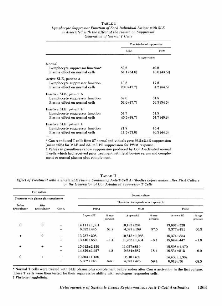
TABLE ILymphocyte Suppressor Function of Each Individual Patient wvith SLE
is Associated with the Effect of the Plasma on SuppressorGeneration of Normal T Cells
Con A-induced suppression
MLR PWM
%suppression
NormalLymphocyte suppressor function* 52.2 40.2Plasma effect on normal cells 51.1 (54.6) 43.0 (43.5)1
Active SLE, patient ALymphocyte suppressor function 13.8 17.8Plasma effect on normal cells 20.0 (47.7) 4.2 (54.5)
Inactive SLE, patient ALymphocyte suppressor function 62.6 81.5Plasma effect on normal cells 52.6 (47.7) 53.5 (54.5)
Inactive SLE, patient KLymphocyte suppressor function 54.7 51.5Plasma effect on normal cells 45.5 (48.7) 51.7 (46.8)
Inactive SLE, patient ULymphocyte suppressor function 21.9 45.4Plasma effect on normal cells 11.5 (53.6) 40.5 (44.3)
* Con A-induced T cells from 27 normal individuals gave 56.2+2.4% suppression(mean+SE) for MLRand 53.1+3.1% suppression for PWMresponse.4 Values in parentheses show suppression produced by Con A-activated normalT cells which had received prior treatment with fetal bovine serum and comple-ment or normal plasma plus complement.
TABLE IIEffect of Treatment with a Single SLE Plasma Containing Anti-T-Cell Antibodies before andlor after First Culture
on the Generation of Con A-induced Suppressor T Cells
First cultureSecond culture
Treatment with plasma plus complementThymidine incorporation in response to
Before Afterfirst culture* first culture* Con A PHAt MLR PWM
A cpm±SE %sup- Acprn -SE %sup- Acpm±SE %sup-pressioni pression pression
0 0 - 14,111+ 1,331 10,182+204 13,607+528+ 6,822±445 51.7 4,327* 169 57.5 5,377+491 60.5
+ 0 - 13,257+208 10,613+*1,056 15,374+854+ 13,440+650 -1.4 11,265 1,434 -6.1 15,649*447 -1.8
+ + - 15,612*2,159 11,057±819 15,596±1,479+ 14,856 1,937 4.8 9,684*687 18.4 16,534+512 -6.0
0 + - 19,303+ 1,136 9,910+459 14,488+ 1,362+ 5,862±748 69.6 4,023+426 59.4 6,018+56 68.5
* Normal T cells were treated with SLE plasma plus complement before and/or after Con A activation in the first culture.These T cells were then tested for their suppressive ability with autologous responder cells.t Phytohemagglutinin.
Heterogeneity of Systemic Lupus Erythematosus Anti-T-Cell Antibodies 1263

at the end, the result was not much different from thatobtained when treatment occurred only at the begin-ning. As a result, we omitted the double treatmentdata from the remainder of the studies.
A summary of studies of plasma from 14 patientswith SLE is shown in Table III. Plasma from normaldonors had no effect upon suppressor cell function.Plasma from patients with inactive SLE or active SLEwithout anti-T-cell antibodies did not markedly inhibitthe generation of suppressor function. In contrast,plasma containing anti-T-cell antibodies from five activepatients markedly inhibited the generation of suppres-sor function. This effect occurred when the antibodiesand complement were added before Con A activation;however, again there was no significant effect if theT cells were exposed to these plasma at the end of theCon A activation step (Table III). It was also noted thatthere was a 37.2+±6.1% decrease in viable cell numberwhen these five plasma containing anti-T-cell anti-bodies were added before Con A activation, but onlya 7.5+7.6% decrease in vidble cell number when addedat the end of the culture. These observations suggestedthat SLE anti-T-cell antibodies could kill a cell neces-sary for the generation of suppressor cells, but couldnot easily kill mature suppressor cells.
In the couise of the present studies, a curious phe-nomenon (as already noted in Table I with plasma frompatient U) was observed with plasma obtained frompatients U and L. Plasma of patient U, when inactive,was able to inhibit the generation of suppresor func-tion of normal T cells for the MLR(Table IV). In con-trast, her plasma did not inhibit the generation of sup-pressor function for PWM-induced proliferation.Furthermore, plasma from another patient, L, withmildly active disease, exhibited the same dichotomouseffect (Table IV). Her plasma also inhibited the genera-tion of suppressor function for the MLR, but not forthe response to PWM. These observations led us toprospectively evaluate the T-cell subpopulations medi-ating the suppression of the two functions.
Role of T, and Tnon-y cells in the generation of sup-pressor function for the MLRand PWMproliferativeresponses. Unfractionated T cells and T cells frac-tionated into Ty and Tnon-Y populations obtained fromnormal individuals were tested for their ability to beactivated by Con A to mediate suppression of the T-cellproliferative response to MLRor the non-T-cell pro-liferative response to PWM.Results of such experimentsare shown in Table V. We found that unfractionatedT cells activated by Con A led to suppression of both
TABLE IIIEffect of Treatment with Plasma from Patients with Active or Inactive SLE before or after First Culture
on Generation of Con A-induced Suppressor T Cells: Summary
First ctultture
Treatment with plasma Second ciltturePlasma plus complement
Con A-indtuced stuppressionNumber Before After
Sotirce tested first culture* first culture* MLR PWM
%supppression-SE
Nonet 0 0 57.4+4.5 53.0±2.6
Normal 5§ + 0 51.6±2.0 52.4±4.70 + 58.5±4.1 50.0±4.4
Inactive SLE 6§ + 0 41.6±5.0 49.3±3.00 + 51.1±2.5 54.7+2.6
Active SLE without anti- 3§ + 0 56.1±9.3 52.6+1.3T-cell antibodies 0 + 50.4±3.9 62.8+4.2
Active SLE with anti- 5§ + 0 8.8±3.4"1 8.5±+13.4"T-cell antibodies 0 + 60.0±8.0 50.7±3.8
* Same as Table II.This control group represents the results from 12 experiments performed simultaneously to those
with plasma in which the cells of the first culture were not treated with any plasma.§ The number of individual plasma donors tested is shown.
Significantly different from a group where T cells were treated with normal plasma plus complement(P < 0.02) and significantly different from a control where T cells were not treated with plasma pluscomplement (P < 0.001).
1264 T. Sakane, A. D. Steinberg, J. P. Reeves, and I. Green

TABLE IVDifferential Effect of SLE Plasma on the Killing of
Precursors of Suppressor T Cells for Either theMLRor the PWMResponse
First cultiureSecond eciltulre
Treatmenit with plasmaplus compleinent Con A-iniduced
suppressor activityBefore After
Plasma souirce first culture* first culture* M1LRI PWMI
%suppression
Active SLE, 0 0 48.7 46.8patient L + 0 -1.4 49.2
0 + 52.9 59.7
Inactive SLE, 0 0 53.6 44.3patient U + 0 11.5 40.5
0 + 47.0 49.5
* Same as Table II.t Same as Table II.§ Same as Table LI.
reactions. Similarly, T, cells led to suippression of bothreactions, whereas the Tnon-y cells activated by Con Afailed to suppress the MLR; however, they were capableof suppressing the PWM-induced proliferation.
In the above experiments, a single dose of suppres-sor cells (10 x 104) from the first culture was used totest for suppressor activity. In the next experiment,varying numbers of Con A-activated T, or Tnon-y cellswere employed; a dose-response curve was constructedby plotting the number of Con A-activated cells addedto the assay culture vs. the percent suppression (Fig. 1).Only T, cells could suppress the MLR; four times the
TABLE VDifferential Suppressor Activity of Ty and T Cells: Using
T Cells from Six Different Normal Individuals*
First culture Second ctiltuire
T-cell fraction uised MLRt PWN'MI
Mean %ofsuppression-SE
Unfractionated T cells 46.5±4.2 74.0+4.2T, cells 56.1±3.2 77.7±4.5Tnon_y cells 9.9+3.3 49.5±4.9
* Normal T cells, unfractionated or fractionated, were pre-cultured with or without Con A in the first culture. These Tcells were assayed for their suppressor activity in the secondculture.t Unfractionated T cells were used as responder cells in thesecond assay culture.§ Non-T cells were used as responder cells in the secondassay culture.
usual number (105) of Tnon-y cells were ineffective. Withregard to the suppression of PWMresponse, both TYand Tnon-y cells could suppress; however, T, cells weremore effective than Tnony cells. Of particular interest,at low cell numbers, Con A-activated cells tended tobe stimulatory rather than suppressive of the PWMresponse (Fig. 1).
Effect of selected SLE anti-T-cell antibodies on sub-populations of T cells. In view of the observationsdescribed above that (a) some SLE anti-T-cell antibodiesinterfere with the generationi of suppressor cells for theMLRbut not for PWMresponses, and (b) that the MLRis suppressed only by T, cells, whereas the PWMresponse is suppressed by both T, and Tnon e cells,we further studied the effect of plasma from patients
80 MLR70 Ty
60-50 -
40 -
30 -
20 K10 Tnon-y
0-10
z -20w ) 30
C: -40L 0.5 1 2 5 10 40D
807 PWMZ Tyw 70Ku
LU 60tz0-50
/ ~~~Tnon-Y40
3020
0
-10
-20-
-30
-400.5 1 2 5 10 40
NUMBEROFSUPPRESSORCELLS ADDED
X10-4
FIGURE 1 Dose-response analysis of suppressive ability offractionated T cells. Responder cells, 1 x 10⁵, were stimulatedwith either MLRor PWMin cultures containing increasingnumbers of Con A-activated T, or Tn.n-, cells.
Heterogeneity of Systemic Lupus Erythematosus Anti-T-Cell Antibodies 1265

z0
U)
w0-D
z.u
uLJ0L
60 Plasma from Active SLE, L
60
1:2 1:10 1:50 1:250 Untreated
with Plasma
Plasma from Inactive SLE, U
60
1:2 1:10 1:50 1:250 Untreatedwith Plasma
50
40
30-I
20KI
1:2 1:10 1:50 1:250 Untreated
with Plasma
PLASMACONCENTRATION
FIGURE 2 Dose-response analysis of anti-T-cell antibodiesobtained fronm SLE patients L and U. Normal T cells weretreated with various concentrations of SLE plasma and thenexposed to normal serum as a complement source. After Con Aactivation of these cells, their suppressive ability was testedin the second culture. =, percent suppression of MLR; *,percent suppression of PWMresponse.
L and U, which could block the generation of suppres-sor cells for the MLR, but not for the PWMresponse.The first study was a dose-response analysis of eachanti-T-cell antibody and the effect upon suppressor cellgeneration (Fig. 2). Plasma from mildly active patientL, even when diluted to 1:250, still had a profoundinhibiting effect on the generation of suppressor cellsfor the MLR. Plasma from inactive patient U, lowerpanel of Fig. 2, only inhibited MLR suppressor cellgeneration at a dilution of 1:2.
When the specificities of those two plasma againstnormal human T-cell subsets were further examined,active plasma from patient L, in which anti-T-cell anti-bodies had already been demonstrated by indirect im-munofluorescence, could kill 28% of whole unfrac-tionated T cells by trypan blue exclusion. This plasmahad preferential ability to kill T, cells of normal donors(65% killing for T, cells and 17% for T000n- cells). Inac-tive plasma from patient U could kill only 7% of un-fractionated normal T cells by trypan blue exclusion.However, when fractionated normal T cells were used,74% of T, cells and none of the Tnon-, cells could bekilled by this plasma. Thus, there was a correlationbetween the subpopulation killed and the suppressorfunction abrogated. That is, these two plasma, whichwere preferentially cytotoxic for T, cells, preferentiallyinterfered with the generation of suppressor functionfor the MLR, and T,-mediated function. In contrast,
most active SLE anti-T-cell antibodies killed both T,and Tnony cells and interfered with generation of sup-pressor cells for both the MLRand the PWM.To bespecific, the average percent of killing by four separateSLE plasma containing anti-T-cell antibodies (exclud-ing patient L) was as follows: 48.3±5.5% of unfrac-tionated T cells, 53.4+9.1% of Ty cells, and 37.1±6.4%of Tnon-y cells.
The possible specificity of the factor present in theplasma of patients L and U responsible for the elimnina-tion of suppressor cells for the MLRbut not for thePWMresponse was further studied by adsorption ofthese plasma with either normal T cells or non-T cells(Fig. 3). Adsorption with T cells almost completelyremoved the activity of the SLE plasma. In contrast,adsorption with non-T cells did not remove the activity.
When plasma from these two patients were frac-tionated by Sephadex G200 chromatography, the IgM,but not the IgG, fraction contained the active antibodythat prevented the generation of suppressor cells foronly the MLR, but not for the PWMresponse (Table VI).
DISCUSSION
Wehave previously reported that anti-T-cell antibodiesobtained from patients with active SLE are capable ofinhibiting the generation of suppressor T cells forproliferation in response to stimulation with both MLRand PWM(28). In the present study we found that thisinhibition occurs when the antibody treatment is per-formed before activation, whereas it is ineffective afteractivation. Thus, the SLE anti-T-cell antibodies inter-fere with a cell necessary for the generation of suppres-sor T cells, probably by killing suppressor cell pre-cursors, although it is possible that another T cell, suchas an initiator cell, is both necessary for suppressorcell activation and killed by the anti-T-cell antibodies.When patients with SLE were studied with regard to(a) their own T-cell function and (b) effects of theirplasma on normal T-cell function, the same defect(s)observed with the patient's T cells were induced bytreating normal T cells with their plasma. Thus thepresence of antibodies, rather than intrinsic T-cellabnormalities, may be responsible for the T-cell defectsobserved in patients with active SLE.
Although most anti-T-cell antibodies from patientswith active SLE eliminated suppressor function forboth the MLRand PWMresponses, one patient withmildly active SLE and one with inactive disease werefound to have anti-T-cell antibodies that preferentiallyeliminated suppressor cell precursors for the MLR, butdid not interfere with the generation of suppressor Tcells for the PWMresponse. Wefound that such anti-bodies were of the IgM class, were adsorbed with Tcells (but not non-T cells), and that they preferentiallykilled T, cells as compared with Tnon-Y cells. The ob-
1266 T. Sakane, A. D. Steinberg, J. P. Reeves, and I. Green

SUPPRESSORACTIVITY IN ASSAYCULTURE
PLASMASOURCE
Active SLE, NoneL Unabsorbed
Absorbed with Normal T Cells
Absorbed with Normal Non-T Cells
Inactive SlU
MLR PWM
LE, NoneUnabsorbed
Absorbed with Normal Cells
@ \I I\\Absorbed with Normal Non-T Cells
0 20 40 60 80 0 20 40 60 80
PERCENTSUPPRESSION
FIGURE 3 Effect of SLE plasma adsorbed with normal T cells or non-T cells on the generationof suppressor T cells. Plasma from two SLE patients, L and U, which could inhibit the suppressorcell generation for the MLRbut not for the PWMresponse, was first adsorbed with either normalT cells or non-T cells. The adsorbed plasma to be studied was mixed with normal T cells.Thereafter these mixtures were exposed to complement. Such treated T cells were activatedwith Con A, and then these Con A-activated cells were tested for their suppressor activity.
servation that the antibodies were of the IgM classstrongly suggested that they were not interfering withT-cell functions by binding to Fc receptors on the Tycells; however, we have not formally demonstrated thatthey bound to the Ty cells through the antibody-com-bining site. The finding that selected anti-T-cell anti-bodies had specificity for both T, cells and precursorsof suppressor T cells for the MLRbut not for the PWM
TABLE VIEffect of IgG and 1gM Fractions of SLE Plasma Obtained
by Sephadex G-200 Column Chromatography on theGeneration of Suppressor T Cells*
Suppressor activityFractions in second cultureadded in
Source of plasma fractions first culture MLRt PWM§
%suppression
Active SLE, patient L"' None 65.5 69.6IgG 52.5 59.9IgM 6.2 62.0
Inactive SLE, patient Ull None 95.1 35.2IgG 91.5 40.1IgM 26.5 36.8
* Normal T cells were treated with either IgG or IgM fractionof SLE plasma plus complement and activated with Con A.These Con A-activated T cells were then tested for theirsuppressor activity in the second culture.t T cells were used as responder cells.§ Non-T cells were used as responder cells.11 These experiments were performed on different days andemployed different donors for normal cells.
responses led us to examine the nature of the T-cellsubsets responsible for suppression of the MLRandthe PWM.
Several studies have suggested that subpopulationsof human T cells can be separated on the basis of re-ceptors for the Fc portion of different immunoglobulinclasses (isotypes) (27, 31-34, 36). Moretta and col-leagues initially felt that T, cells were specific for thefunction of suppression, whereas T cells bearing re-ceptors for the Fc portion of IgM (T, cells) were specificfor the function of help (31, 34). Those studies examinedthe effect of the different T-cell subpopulation on B-cellfunctions. Subsequently, it has been found in an assayof Con A-activated suppressor cells that T, cells mayunder certain circumstances function as suppressorcells (32). In the present study, we found that T, cellswere more effective than Tnon- cells at suppressingboth the PWMand MLRreactions. Tnon_y cells signifi-cantly suppressed the PWMresponse; however, theywere unable to suppress the MLR. These studies helpedto explain the effects of the two selected SLE anti-bodies described above. Those anti-T-cell antibodiescapable of killing only T. cells interfered only withthe generation of suppressor cells for the MLR. Incontrast, those antibodies capable of killing both Tyand Tnon_y cells interfered with the generation of sup-pressor cells for both responses. Because T, cells aloneare involved in the generation of suppressor cell func-tion for the MLR, the antibodies that preferentiallykilled those cells would be expected to preferentiallyeliminate the function mediated by those cells. Whetheror not Ty cells alone are responsible for suppressionof all T-cell functions remains to be determined; it is
Heterogeneity of Systemic Lupus Erythematosus Anti-T-Cell Antibodies
FIRST CULTURE
1267
,.\N
J. i-I I I t

clear, however, that the non-T-cell proliferative responseto PWMis suippressible by both Ty and Tnon_Y cells.This helps to explain previous reports which claimedthat both Ty cells and T, cells could act as suppressorcells (31, 32, 34).
Furthermore, it was noted that small numbers of Tyand Tnon_y cells led to stimulation of the PWMresponserather than suppression. Such an observation underlinesthe importance of dose-response curves before decidingthat a particular subset of T cells subserves a particularfunction. Wepresume that there are both helper andsuppressor subsets of T. cells to explain these observa-tions. This idea is also supported by the finding thatT cells treated with SLE anti-T-cell antibodies pluscomplement and activated with Con A often producedan increase in the PWMresponse rather than a decrease.However, further dose-response studies using SLEanti-T-cell antibodies plus Fc (IgG) receptor separationwill be necessary to discriminate between helper andsuppressor Ty cells.
Previous studies of SLE anti-T-cell antibodies suggestthat some may be of the IgG class (18, 20, 22, 30).The present study measured the effects of complement-mediated killing of T cells. It has previously been shownin mice that IgM antibodies to T cells are 500 timesmore efficient at complement-mediated lysis than areIgG antibodies; therefore, the activity of the IgM anti-bodies might be the only one observed despite thepresence of large amounts of anti-T-cell antibodies ofthe IgG class (39).
Furthermore, at least one mechanism by which IgGantibodies eliminate T cells is antibody-dependentcellular cytotoxicity (ADCC) (22). Because in thepresent study only highly purified T-cell populationswere treated with SLE antibodies, effector cells forantibody-dependent cellular cytotoxicity might nothave been present in sufficient numbers to mediateT-cell killing. Thus, the present studies do not deny thepresence of IgG anti-T-cell antibodies in SLE. How-ever, since they were designed to study complement-mediated T-cell killing, they may have preferentiallyselected for IgM antibody activity.
Recent studies have demonstrated that sera frompatients with juvenile rheumatoid arthritis containantibodies capable of recognizing suppressor cells(40, 41). In addition the patients with such antibodieslack suppressor cells and have increased numbers ofimmunoglobulin-secreting cells (41). Thus, in bothjuvenile rheumatoid arthritis and SLE the presence ofthe antibodies and the loss of suppressor cells correlatedwith disease activity. Circumstantially, these resultssuggest that in some patients disease may be initiatedby some factor or factors which lead to formation ofanti-T-cell antibodies and subsequent loss of suppressorcells. Once formed, anti-T-cell antibodies would becapable of eliminating cells that would serve to reduce
B-cell activity. Thus, a self-perpetuating cycle wouldoccuir with continued high level production of auto-antibodies and continued loss of the very cells thatserve to suppress such excessive antibody production.
ACKNOWLEDGMENTS
The auithors are grateful to Dr. Paul V. Holland, Dr. RichardDavey, and Ms. Jane E. Kendall, Blood Bank Department,Clinical Center, National Institutes of Health, for their helpand cooperation in supplying the blood from normal humansin these studies.
This work was supported in part by the Cancer ResearchInstitute Inc., New York, and the Naite Foundation, Tokyo,Japan.
REFERENCES
1. Koffler, D., P. H. Schur, and H. G. Kunkel. 1967. Immuno-logical studies concerning the nephritis of systemic lupuserythematosus. J. Exp. Med. 126: 607-623.
2. Harbeck, R. J., E. J. Bardana, P. F. Kohler, and R. I. Carr.1973. DNA:anti-DNA complexes: their detection in sys-temic lupus erythematosus sera. J. Clin. Invest. 52:785-795.
3. Vaughan, J. H., and T. Chihara. 1973. Lymphocyte func-tion in rheumatic disorders. Arch. Intern. Med. 135:1324-1328.
4. Bell, D. A., C. Clark, S. E. Blomgren, and J. H. Vaughan.1973. Anti-DNA antibody production by lymphoid cellsof NZB/Wmice and human systemic lupus erythematosus(SLE). Clin. Immunol. Immunopathol. 1: 293-303.
5. Delbarre, F., A. L. Go, and A. Kahan. 1975. Hyperbaso-philic immunoblasts in the circulating blood in chronicinflammatory rheumatic and collagen diseases. Ann.Rheum. Dis. 34: 422-430.
6. Budman, D. R., E. B. Merchant, A. D. Steinberg, B. Doft,M. E. Gershwin, E. Lizzio, and J. P. Reeves. 1977. In-creased spontaneous activity of antibody-forming cellsin the peripheral blood of patients with active SLE.Arthritis Rheum. 20: 829-833.
7. Morimoto, C., T. Abe, M. Hara, and M. Homma. 1977.In vitro TNP-specific antibody formation by peripherallymphocytes from patients with systemic lupus erythem-atosus. Scand. J. Immunol. 6: 575-579.
8. Horwitz, D. A. 1972. Impaired delayed hypersensitivityin systemic lupus erythematosus. Arthritis Rheum. 15:353-359.
9. Hahn, B. H., M. K. Bagby, and C. K. Osterland. 1973.Abnormalities of delayed hypersensitivity in systemiclupus erythematosus. Am. J. Med. 55: 25-3 1.
10. Suciu-Foca, N., J. A. Buda, T. Thiem, and K. Reemtsma.1974. Impaired responsiveness of lymphocytes in patientswith systemic lupus erythematosus. Clin. Exp. Immunol.18: 295-301.
11. Rosenthal, C. J., and E. C. Franklin. 1975. Depressionof cellular-mediated immunity in systemic lupus erythem-atosus. Relation to disease activity. Arthritis Rheum.18: 207-217.
12. Sakane, T. 1976. Studies on cell-mediated immuno re-sponses by human lymphocytes in vitro. III. Lympho-cyte response to phytohemmaglutinin and concanavalinA in patients with autoimmune diseases. Acta Haematol.Jpn. 39: 29-42.
13. Sakane, T., A. D. Steinberg, and I, Green. 1978. Failureof autologous mixed lymphocyte reactions between T and
1268 T. Sakane, A. D. Steinberg, J. P. Reeves, and I. Green

non-T cells in patients with systemic lupus erythem-atosus. Proc. Natl. Acad. Sci. U. S. A. 75: 3464-3468.
14. Sakane, T., A. D. Steinberg, F. C. Arnett, J. L. Reinertsen,and I. Green. 1979. Studies of immune functions of pa-tients with systemic lupus erythematosus. III. Characteri-zation of lymphocyte subpopulations responsible fordefective autologous mixed lymphocyte reactions. ArthritisRheum. 22: 770-776.
15. Messner, P. P., F. D. Lindstrom, and R. C. Williams, Jr.1973. Peripheral blood lymphocyte cell surface markersduring the course of systemic lupus erythematosus. J.Clin. Invest. 52: 3046-3056.
16. Scheinberg, M. A., and E. S. Cathcart. 1974. B cell and Tcell lymphopenia in systemic lupus erythematosus. Cell.Immunol. 12: 309-314.
17. Glinski, W., M. E. Gershwin, D. R. Budman, and A. D.Steinberg. 1976. Study of lymphocyte subpopulations innormal humans and patients with systemic lupuserythematosus by fractionation of peripheral bloodlymphocytes on a discontinuous Ficoll gradient. Clin.Exp. Immunol. 26: 228-238.
18. Stastney, P., and M. Ziff. 1971. Antibodies against cell mem-brane constituents in systemic lupus erythematosus andrelated diseases. I. Cytotoxic effect of serum from pa-tients with systemic lupus erythematosus (SLE) forallogeneic and for autologous lymphocytes. Clin. Exp.Immunol. 8: 543-550.
19. Lies, R. B., R. P. Messner, and R. C. Williams, Jr. 1973.Relative T-cell specificity of lymphocytotoxins from pa-tients with systemic lupus erythematosus. ArthritisRheumn. 16: 369-375.
20. Wernet, P., and H. G. Kunkel. 1973. Antibodies to aspecific surface antigen of T cells in human sera inhibitingmixed leukocyte culture reactions. J. Exp. Med. 138:1021- 1026.
21. Winfield, J. B., R. J. Winchester, P., Wernet, S. M. Fu,and H. G. Kunkel. 1975. Nature of cold-reactive anti-bodies to lymphocyte surface determinants in systemiclupus erythematosus. Arthritis Rheum. 18: 1-5.
22. Glinski, W., M. E. Gershwin, and A. D. Steinberg. 1976.Fractionation of cells on a discontinuous Ficoll gradient:study of subpopulations of human T cells using anti-T-cellantibodies from patients with systemic lupus erythem-atosus.J. Clin. Invest. 57: 604-614.
23. Steinberg, A. D., L. W. Klassen, D. R. Budman, and G. W.Williams. 1979. Immunofluorescence studies of anti-Tcell antibodies and T cells in systemic lupus erythem-atosus. Selective loss of brightly staining T cells in activedisease. Arthritis Rheuin. 22: 114-122.
24. Sakane, T., A. D. Steinberg, and I. Green. 1978. Studiesof immune functions of patients with systemic lupuserythematosus. I. Dysfunction of suppressor T-cell activityrelated to impaired generation of, rather than responseto, suppressor cells. Arthritis Rheum. 21: 657-664.
25. Horowitz, S. W., Borcherding, A. V., Moorthy, R. Chesney,H. Schulte-Wissermann, R. Hong, and A. Goldstein. 1977.Induction of suppressor T cells in systemic lupuserythematosus by thymosin and cultured thymic epi-thelium. Scietnce (Wash. D. C.) 197: 999-1001.
26. Sagawa, A., and N. I. Abdou. 1978. Suppressor-celldysfunction in system-ic luipuis erythematosus. Cell in-volved and irn vitro correction. J. Clin. Invest. 62: 789-796.
27. Fauci, A. S., A. D. Steinberg, B. F. Haynes, and G. Whalen.1978. Immunoregulatory aberrations in systemic lupuserythematosus. J. Immunol. 121: 1473-1479.
28. Sakane, T., A. D. Steinberg, J. P. Reeves, and I, Green.1979. Studies of immune functions of patients withsystemic lupus erythematosus: complement-dependentimmunoglobulin M anti-thymus-derived cell antibodiespreferentially inactivate suppressor cells. J. Clin. Invest.63: 954-965.
29. Twomey, J. J., A. H. Laughter, and A. D. Steinberg.1978. A serum inhibitor of immune regulation in pa-tients with systemic lupus erythematosus. J. Clin.Invest. 62: 713-715.
30. Sagawa, A., and N. I. Abdou. 1979. Suppressor cellantibody in systemic lupus erythematosus: possiblemechanism for suppressor-cell dysfunction. J. Clin.Invest. 63: 536-539.
31. Moretta, L., S. R. Webb, C. E. Grossi, P. M. Lydyard, andM. D. Cooper. 1977. Functional analysis of two humanT-cell subpopulations: help and suppression of B-cellresponses by T cells bearing receptors for IgM or IgG.
J. Exp. Med. 146: 184-200.32. Hayward, A. R., L. Layward, P. M. Lydyard, L. Moretta,
M. Dagg, and A. R. Lawton. 1978. Fe-receptor hetero-geneity of human suppressor T cells. J. Immunol. 121:1-5.
33. Haynes, B. F., and A. S. Fauci. 1978. Activation of humanB lymphocytes. X. Heterogeneity of coneanavalin A-generated suppressor cells of the pokeweed mitogen-induced plaque-forming cell response of human peripheralblood lymphocytes. J. I mmunol. 121: 559-565.
34. Moretta, L., M. C. Mingari, A. Moretta, and M. D. Cooper.1979. Human T lymphocyte subpopulations: studies ofthe mechanism by which T cells bearing Fc receptorsfor IgG suppress T-dependent B cell differentiationinduced by pokeweed mitogen.]. Immunol. 122: 984-990.
35. Sakane, T., and I. Green. 1978. Protein A from Staph ylo-coccus aureus-a mitogen for human T lymphocytes andB lymphocytes but no L lymphocytes. J. Immunol. 120:302-311.
36. Moretta, L., M. Ferrarini, M. C. Mingari, A. Moretta, andS. R. Webb. 1976. Subpopulations of human T cellsidentified by receptors for immunoglobulin and mitogenresponsiveness.J. Iii mmunol. 117: 2171-2174.
37. Sakane, T., and I. Green. 1977. Humansuppressor T cellsinduced by concanavalinl A: suppressor T cells belong todistinetive T cell subelasses.j. Immmunol. 119: 1169-1178.
38. Raff, H. V., K. C. Cochrumil, and J. D. Stobo. 1978. Macro-phage-T cell interaction in the Con A induction of humansuppressive T cells. J. Im munol. 121: 2311-2315.
39. Gershwin, M. E., T. M. Chused, and A. D. Steinberg.1974. Cytotoxic activity of anti-O antibody found in theYM fraction. Tranisplantationt (Baltimore). 18: 377-380.
40. Strelkauskas, A. J., V. Schaluif, B. S. Wilson, L. Chess, andS. F. Schlossman. 1978. Isolation and chalracterization ofnaturally occurring subeclasses of human peripheral bloodT cells with regulatory functions. J. Immunnol. 120:1278- 1282.
41. Strelkaiskats, A. J., R. T. Callery, J. McDowell, Y. Borel,andcl S. F. Schlossmanll. 1978. Direct evidence for loss ofhumanlt suippressor cells d(iriong active atutoimmiiiiune disease.Proc. Natl. Acad. Sci. U. S. A. 75: 5150-5154.
Heterogeneity of Systemic Lu pus Erytthem atosu.s An ti-T-Cell Antiboclies 1269