Embed Size (px)
Citation preview

Systemic mastocytosis with associatedmyeloproliferative disease and precursor Blymphoblastic leukaemia with t(13;13)(q12;q22)involving FLT3
A Tzankov,1 K Sotlar,2 D Muhlematter,3 A Theocharides,4 P Went,1 M Jotterand,3
H-P Horny,5 S Dirnhofer1
1 Institute of Pathology,University of Basel, Switzerland;2 Institute of Pathology,University of Munchen,Germany; 3 Institute of MedicalGenetics, University ofLausanne, Switzerland;4 Department of Hematology,University of Basel, Switzerland;5 Institute of Pathology,Ansbach, Germany
Correspondence to:Dr A Tzankov, University HospitalBasel, Institute of Pathology,Schoenbeinstr. 40, CH-4031Basel, Switzerland; [email protected]
Accepted 22 May 2008
ABSTRACTSystemic mastocytoses represent neoplastic prolifera-tions of mast cells. In about 20% of cases systemicmastocytoses are accompanied by clonal haematopoieticnon-mast cell-lineage disorders, most commonly myeloidneoplasms. A case of systemic mastocytosis carrying thecharacteristic mutation at codon 816 (D816V) in the KITgene of mast cells, with two concurrent accompanyingclonal haematopoietic non-mast cell-lineage disorders,chronic myeloproliferative disease, unclassifiable andprecursor B lymphoblastic leukaemia is documented. Bothaccompanying clonal haematopoietic non-mast cell-line-age disorders carried the wild-type KIT gene, but had anovel t(13;13)(q12;q22) involving the FLT3 locus at13q12. The chronic myeloproliferative disease, unclassifi-able and the precursor B lymphoblastic leukaemia werecured by syngenous stem cell transplantation, but thesystemic mastocytosis persisted for more than 10 years.The additional impact of molecular techniques on thecorrect diagnosis in haematological malignancies ishighlighted, and evidence is provided that, apart frominternal tandem duplications and mutations, FLT3 can beactivated by translocations.
Associated clonal haematopoietic non-mast cell-lineage disorders (AHNMDs) can be observed inabout 20% of patients with systemic mastocytosis(SM).1 2 SM and AHNMDs can be either clonallyrelated or not.3 4 Concurrent SM and precursorlymphoblastic leukaemia (ALL) have not beenreported. Here, we describe a case of SM withtwo AHNMDs, namely chronic myeloproliferativedisease, unclassifiable (CMPD, U) and B-ALL, bothAHNMDs carrying a novel t(13;13)(q12;q22)involving the FLT3 locus.
MATERIALS AND METHODSHistologyBone marrow biopsy samples prior to 2000 werefixed and decalcified in Susa’s medium, and after2000 in 4% buffered formaldehyde solution andEDTA; they were then embedded in paraffin.Sections (3 mm) were stained with H&E, Giemsaand Gomori stains.
ImmunohistochemistryImmunohistochemistry for CD3, CD10, CD20,CD25, CD34, CD79a, CD117 and tryptase wasperformed using an automated immunostainer(Nexes, Ventana, USA), and for terminal deoxy-
nucleotidyl transferase (TdT) and phosphorylatedSTAT5 manually.
Detection of the D816V mutation in the KIT geneand the V617F mutation in the JAK2 geneDNA from the bone marrow biopsy specimensfrom 1996 and 2006 was evaluated for the KITmutation D816V by peptide nucleic acid-mediatedPCR-clamping and melting point analysis of theproducts as described previously.5 DNA frommicrodissected mast cells from 2006 and micro-dissected lymphoblasts from 1996 was amplifiedby nested PCR and analogously analysed for KITmutations.4 PCR for the JAK2 mutation V617F wasperformed on bone marrow aspirates from 1995and 2006 as described previously.6
Cytogenetic analysis and t(13;13)(q12;q22)breakpoint mappingCell culture and chromosome preparations werestandard. Chromosomes were stained in G-bands.To map the chromosomal breakpoints int(13;13)(q12;q22), the following bacterial artificialchromosome probes were identified in the www.genome.UCSC.edu database and obtained fromBACPAC Resources (Children’s Hospital, Oakland,California,USA): RP11-94A1, RP11-274P12 (brid-ging ZNF198 locus), RP11-80J14, RP11-367C11(bridging FLT3 locus), RP11-87C7 (proximalFLT3), RP11-85P8 (proximal/inside FLT3), RP11-35M5 (distal/inside FLT3) and RP11-89P22 (distalFLT3). Direct labelling of probes with FITC or Cy3and metaphase fluorescence in situ hybridisation(FISH) was standard. Interphase FISH with Cy3-labelled RP11-87C7 and FITC-labelled RP11-35M5was performed on bone marrow samples from theinitial diagnosis of SM-AHNMD (1996) andpersistent SM (2006) to determine the FLT3 statusin mast cells in consecutive FISH and tryptase-stained sections.
RESULTS
Clinicopathological case historyIn November 1995, a 46-year-old man wasadmitted to our hospital with antiphlogistictreatment-resistant bone pain, pruritus, raisedwhite blood cell count (16.16109/l) with increasedneutrophilic granulocytes (12.246109/l), eosinophi-lia of 0.486109/l and monocytosis of 1.376109/l,and splenomegaly of 17.5 cm, as assessed by
Case report
958 J Clin Pathol 2008;61:958–961. doi:10.1136/jcp.2008.058073
group.bmj.com on July 8, 2013 - Published by jcp.bmj.comDownloaded from
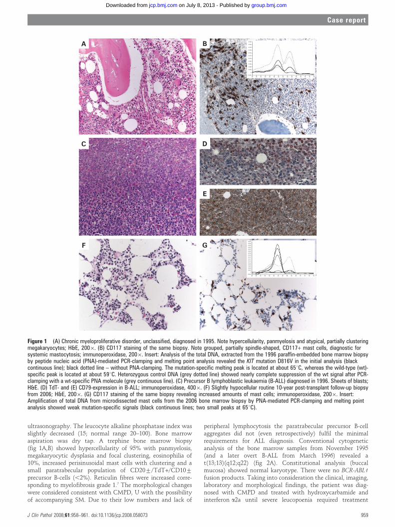
ultrasonography. The leucocyte alkaline phosphatase index wasslightly decreased (15; normal range 20–100). Bone marrowaspiration was dry tap. A trephine bone marrow biopsy(fig 1A,B) showed hypercellularity of 95% with panmyelosis,megakaryocytic dysplasia and focal clustering, eosinophilia of10%, increased perisinusoidal mast cells with clustering and asmall paratrabecular population of CD20¡/TdT+/CD10¡
precursor B-cells (,2%). Reticulin fibres were increased corre-sponding to myelofibrosis grade 1.7 The morphological changeswere considered consistent with CMPD, U with the possibilityof accompanying SM. Due to their low numbers and lack of
peripheral lymphocytosis the paratrabecular precursor B-cellaggregates did not (even retrospectively) fulfil the minimalrequirements for ALL diagnosis. Conventional cytogeneticanalysis of the bone marrow samples from November 1995(and a later overt B-ALL from March 1996) revealed at(13;13)(q12;q22) (fig 2A). Constitutional analysis (buccalmucosa) showed normal karyotype. There were no BCR-ABL1fusion products. Taking into consideration the clinical, imaging,laboratory and morphological findings, the patient was diag-nosed with CMPD and treated with hydroxycarbamide andinterferon a2a until severe leucopoenia required treatment
Figure 1 (A) Chronic myeloproliferative disorder, unclassified, diagnosed in 1995. Note hypercellularity, panmyelosis and atypical, partially clusteringmegakaryocytes; H&E, 2006. (B) CD117 staining of the same biopsy. Note grouped, partially spindle-shaped, CD117+ mast cells, diagnostic forsystemic mastocytosis; immunoperoxidase, 2006. Insert: Analysis of the total DNA, extracted from the 1996 paraffin-embedded bone marrow biopsyby peptide nucleic acid (PNA)-mediated PCR-clamping and melting point analysis revealed the KIT mutation D816V in the initial analysis (blackcontinuous line); black dotted line – without PNA-clamping. The mutation-specific melting peak is located at about 65uC, whereas the wild-type (wt)-specific peak is located at about 59uC. Heterozygous control DNA (grey dotted line) showed nearly complete suppression of the wt signal after PCR-clamping with a wt-specific PNA molecule (grey continuous line). (C) Precursor B lymphoblastic leukaemia (B-ALL) diagnosed in 1996. Sheets of blasts;H&E. (D) TdT- and (E) CD79-expression in B-ALL; immunoperoxidase, 4006. (F) Slightly hypocellular routine 10-year post-transplant follow-up biopsyfrom 2006; H&E, 2006. (G) CD117 staining of the same biopsy revealing increased amounts of mast cells; immunoperoxidase, 2006. Insert:Amplification of total DNA from microdissected mast cells from the 2006 bone marrow biopsy by PNA-mediated PCR-clamping and melting pointanalysis showed weak mutation-specific signals (black continuous lines; two small peaks at 65uC).
Case report
J Clin Pathol 2008;61:958–961. doi:10.1136/jcp.2008.058073 959
group.bmj.com on July 8, 2013 - Published by jcp.bmj.comDownloaded from

discontinuation. Since he had a monozygous twin, syngenousbone marrow transplantation was considered.
At the admission for graft procedures atypical lymphoid cellswere detected in the peripheral blood smear. Bone marrowaspiration revealed hypercellularity as well as 50% precursor B-cells with an aberrant flow cytometry-phenotype (CD19+/sIgM+/CD34+/TdT+/CD10¡/CD20¡/CD22+/CD43+). Trephine bonemarrow biopsy revealed packed marrow with diffuse sheaths oflymphoblasts, focally displacing the haematopoiesis, as well aspersistent mast cell hyperplasia and focal myeloproliferativefeatures (fig 1C–E). The lymphoblasts were immunohistochemi-cally CD20¡/CD79a+/CD34+/TdT+/CD10¡. B-ALL with reac-tive mast cell hyperplasia was diagnosed. The patient was treated
with chemotherapy consisting of dexamethasone, vincristine,ifosphamide, carboplatin and etoposide followed by conditioningwith etoposide and cyclophosphamide prior to total bodyirradiation and syngenous peripheral stem cell transplantation.Complete haematological remission to the last control examina-tion in April 2008 was achieved.
In the follow-up period from 1996 to 2001, the patientcomplained of hyperpigmentation, relapsing pruritus, rashes,intermittent diarrhoea, and poor circulation in his fingertips.The routine 3-, 6- and 12-month post-transplant biopsiesshowed complete remission of B-ALL and CMPD, U andpersistent minor mast cell hyperplasia. A routine 10-yearfollow-up trephine bone marrow biopsy in August 2006 wasnormocellular, with persistent complete morphological remis-sion of B-ALL and CMPD, U. An increased amount ofperivascular, focally-grouped mast cells was detected (fig 1F,G).These mast cells were tryptase+/CD117+/CD25+, indicating aneoplastic phenotype.
Genetic analysisDual-colour metaphase FISH carried out in 2007 on archivedbone marrow cells from 1996 pointed to the involvement of theFLT3 gene in the t(13;13)(q12;q22) (fig 2B; localised in theproximal gene part). Only the bone marrow probe from 2006was evaluable for interphase FISH. Breaks involving FLT3 couldnot be detected in 50 identifiable mast cells or 200 additionalhaemopoietic cells.
To study the functional consequences of the FLT3 rearrange-ment, the phosphorylation status of its downstream target,STAT5,8 was immunohistochemically analysed and comparedto other acute leukaemia cases without FLT3 abnormalities(data not shown). Nuclear phosphorylated STAT5 was detect-able in 20% of B-ALL cells of the present case (fig 2B, insert), butnot in blasts of acute leukaemias without FLT3 abnormalities.
DNA analysis showed the KIT mutation D816V in the initialbone marrow biopsy from 1996 only with peptide nucleic acid-mediated PCR-clamping (fig 1A, insert) and in microdissectedmast cells from the bone marrow biopsy in 2006 (fig 1G, insert).In contrast, the mutation was not detected in microdissectedlymphoblasts from the bone marrow biopsy in 1996. JAK2 waswild-type.
Final integrative interpretation considering molecular analysesTaking into consideration the presence of three minor criteria(.25% spindle-shaped mast cells, co-expression of CD117 andCD25, and KIT mutation D816V, but normal serum tryptase,assessed only in 2006)1 and all other molecular findings, theintegrative diagnosis of persistent indolent SM with completeremission of the coexistent AHNMDs (CMPD, U and B-ALL)was finally established in 2006.
DISCUSSIONSMs in SM-AHNMDs are most commonly accompanied bymyeloid malignancies and rarely by lymphoproliferative dis-orders, but thus far association with ALL has not beendocumented.2 Importantly, clonal relationship between SMand AHNMD is currently not required to classify a coincidentalhaematological malignancy as AHNMD; if SM diagnosis isestablished, any accompanying haematological malignancyshould be classified as AHNMD.1 The observed co-occurrenceof SM and ALL in our patient may be related to a possiblelymphoblastic transformation (blast phase) of the initialCMPD, U. This assumption is supported by the clinical
Figure 2 Cytogenetic results. (A) G-banded karyotype:46,XY,t(13;13)(q12;q22). (B) Dual-colour metaphase fluorescence in situhybridisation with RP11-85P8- (Cy3, red) and RP11-35M5 probes (FITC,green) showing a small red signal on the short der(13) and two fusionsignals on the long der(13)(DAPI counterstaining). Insert: nuclearphosphorylated STAT5 expression in B-ALL cells from 1996 (anti-phospho-STAT5 antibody from Cell Signaling (Boston, Massachusetts,USA); dilution: 1:50, antigen retrieval: microwave, 100uC/15 min, citratebuffer pH 6).
Case report
960 J Clin Pathol 2008;61:958–961. doi:10.1136/jcp.2008.058073
group.bmj.com on July 8, 2013 - Published by jcp.bmj.comDownloaded from

chronology as well as by the detection of the t(13;13)(q12;q22)in both aspirates from 1995 (CMPD, U) and 1996 (B-ALL).Importantly, mast cells of SM did not carry t(13;13)(q12;q22),whereas B-ALL and CMPD, U did not carry the KIT mutationD816V, which is highly suggestive of differential clonal originsof SM and AHNMDs.
Regarding the novel t(13;13)(q12;q22), our observationsimply that FLT3 can be activated not only by internal tandemduplications and mutations8 but also by translocations as in thecase of another tyrosin kinase-encoding oncogene JAK2.9
Although investigated on a single case, this assumption isfurther supported by the higher amount of phosphorylatedSTAT5 (flt3 downstream target)8 in B-ALL cells in the index(fig 2B, insert) compared to reference cases (data not shown). Inline with this hypothesis, a CMPD case with hypereosinophilia
and t(12;13)(p13;q12) leading to FLT3/ETV6 fusion has beenreported,10 thereby clearly demonstrating the oncogenic poten-tial of dimerisable chimeric flt3.
Competing interests: None.
REFERENCES1. Valent P, Horny HP, Li CY, et al. Mastocytosis. In: Jaffe ES, Harris NL, Stein H,
Vardiman JW, eds. Pathology and genetics of tumours of haematopoietic andlymphoid tissues. Lyon: IARC Press, 2001:293–302.
2. Horny HP, Sotlar K, Sperr WR, et al. Systemic mastocytosis with associated clonalhaematological non-mast cell lineage diseases: a histopathological challenge. J ClinPathol 2004;57:604–8.
3. Sperr WR, Walchshofer S, Horny HP, et al. Systemic mastocytosis associated withacute myeloid leukaemia: report of two cases and detection of the c-kit mutationAsp-816 to Val. Br J Haematol 1998;103:740–9.
4. Sotlar K, Fridrich C, Mall A, et al. Detection of c-kit point mutation Asp-816–. Val inmicrodissected pooled single mast cells and leukemic cells in a patient with systemicmastocytosis and concomitant chronic myelomonocytic leukemia. Leuk Res2002;26:979–84.
5. Sotlar K, Escribano L, Landt O, et al. One-step detection of c-kit point mutationsusing peptide nucleic acid-mediated polymerase chain reaction clamping andhybridization probes. Am J Pathol 2003;162:737–46.
6. Kralovics R, Teo SS, Li S, et al. Acquisition of the V617F mutation of JAK2 is a lategenetic event in a subset of patients with myeloproliferative disorders. Blood2006;108:1377–80.
7. Thiele J, Kvasnicka HM, Facchetti F, et al. European consensus on grading bonemarrow fibrosis and assessment of cellularity. Haematologica 2005;90:1128–32.
8. Choudhary C, Muller-Tidow C, Berdel WE, et al. Signal transduction of oncogenicFlt3. Int J Hematol 2005;82:93–9.
9. Tzankov A, Heiss S. Myeloproliferative diseases with the recurrent t(8;9)(p23;p24) –reply. Hum Pathol 2006;37:500–2.
10. Vu HA, Xinh PT, Masuda M, et al. FLT3 is fused to ETV6 in a myeloproliferativedisorder with hypereosinophilia and a t(12;13)(p13;q12) translocation. Leukemia2006;20:1414–21.
Take-home messages
c Systemic mastocytosis can be accompanied by precursor celllymphoblastic leukaemia.
c In addition to internal tandem duplications and mutations, FLT3can be activated by translocations.
c Systemic mastocytosis might be resistant to stemtransplantation and persist after eradication of associatedclonal haematopoietic non-mast cell-lineage disorders.
Case report
J Clin Pathol 2008;61:958–961. doi:10.1136/jcp.2008.058073 961
group.bmj.com on July 8, 2013 - Published by jcp.bmj.comDownloaded from

doi: 10.1136/jcp.2008.058073 2008 61: 958-961J Clin Pathol
A Tzankov, K Sotlar, D Muhlematter, et al. t(13;13)(q12;q22) involving FLT3lymphoblastic leukaemia withmyeloproliferative disease and precursor B Systemic mastocytosis with associated
http://jcp.bmj.com/content/61/8/958.full.htmlUpdated information and services can be found at:
These include:
References
http://jcp.bmj.com/content/61/8/958.full.html#related-urlsArticle cited in:
http://jcp.bmj.com/content/61/8/958.full.html#ref-list-1This article cites 9 articles, 3 of which can be accessed free at:
serviceEmail alerting
the box at the top right corner of the online article.Receive free email alerts when new articles cite this article. Sign up in
CollectionsTopic
(1397 articles)Immunology (including allergy) � (195 articles)Dermatology �
Articles on similar topics can be found in the following collections
Notes
http://group.bmj.com/group/rights-licensing/permissionsTo request permissions go to:
http://journals.bmj.com/cgi/reprintformTo order reprints go to:
http://group.bmj.com/subscribe/To subscribe to BMJ go to:
group.bmj.com on July 8, 2013 - Published by jcp.bmj.comDownloaded from