Embed Size (px)
Citation preview
S t i MNethealthbook.com
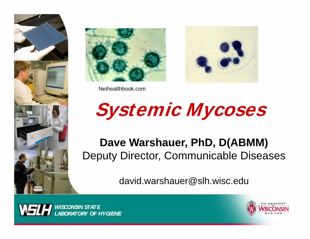
Systemic MycosesDave Warshauer, PhD, D(ABMM)
Deputy Director, Communicable Diseases
WISCONSIN STATE LABORATORY OF HYGIENEWISCONSIN STATE LABORATORY OF HYGIENE
Systemic MycosesSystemic Mycoses
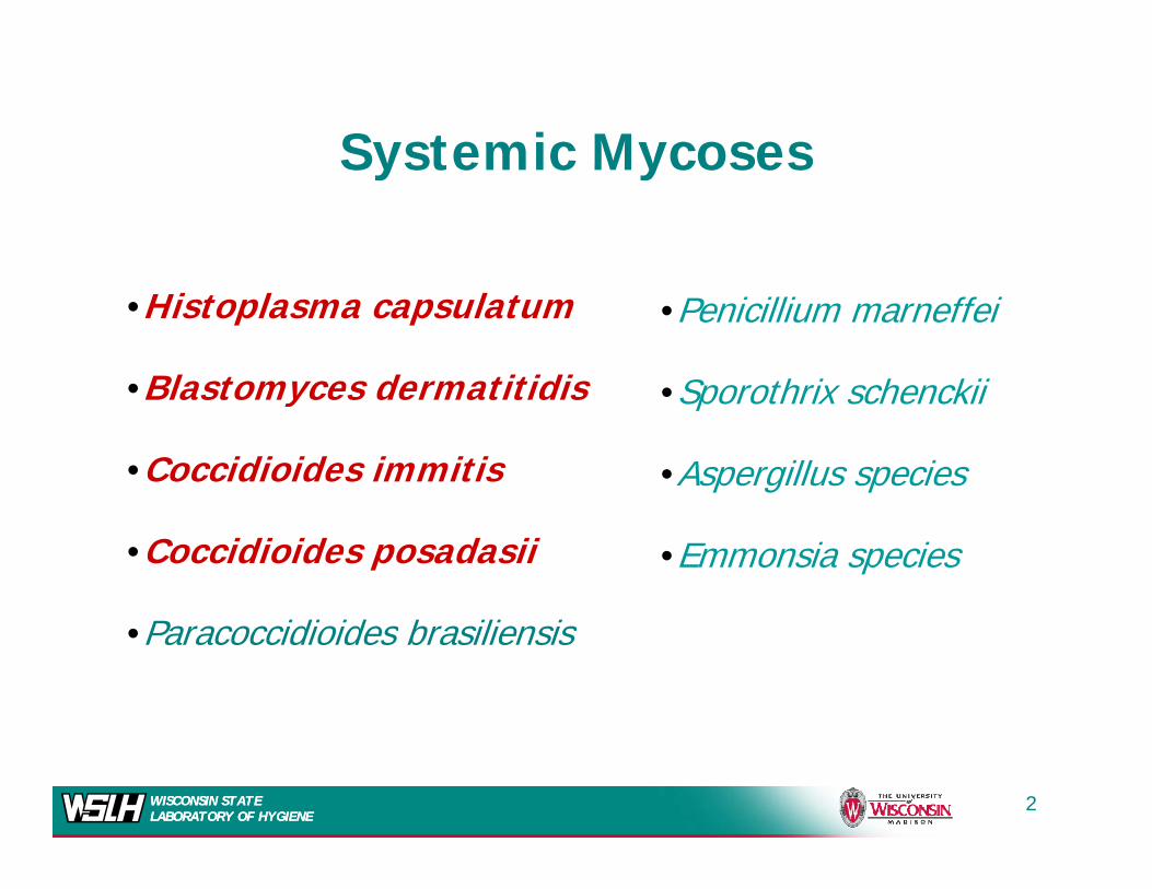
•Histoplasma capsulatum
•Blastomyces dermatitidis
•Penicillium marneffei
•Sporothrix schenckii•Blastomyces dermatitidis
•Coccidioides immitis
•Sporothrix schenckii
•Aspergillus species
•Coccidioides posadasii •Emmonsia species
•Paracoccidioides brasiliensis
WISCONSIN STATE LABORATORY OF HYGIENEWISCONSIN STATE LABORATORY OF HYGIENE
2
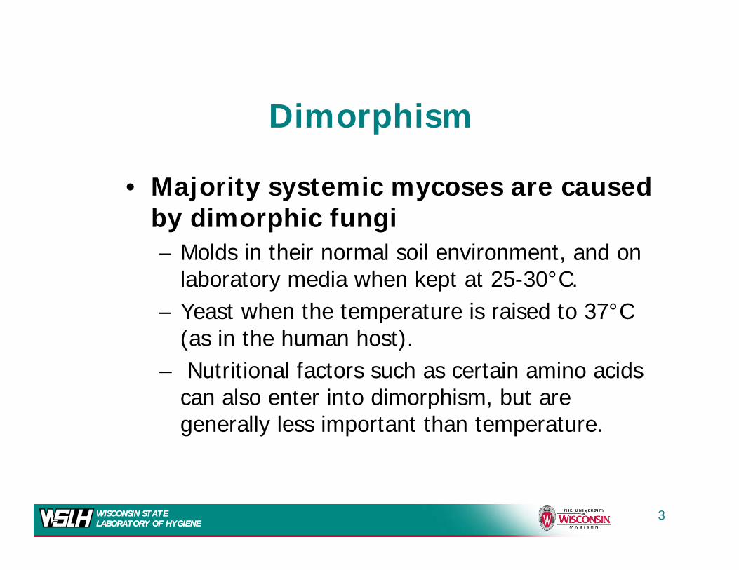
Di hiDimorphism
M j it t i d• Majority systemic mycoses are caused by dimorphic fungi
Molds in their normal soil environment and on– Molds in their normal soil environment, and on laboratory media when kept at 25-30°C.
– Yeast when the temperature is raised to 37°C p(as in the human host).
– Nutritional factors such as certain amino acids can also enter into dimorphism but arecan also enter into dimorphism, but are generally less important than temperature.
WISCONSIN STATE LABORATORY OF HYGIENEWISCONSIN STATE LABORATORY OF HYGIENE
3
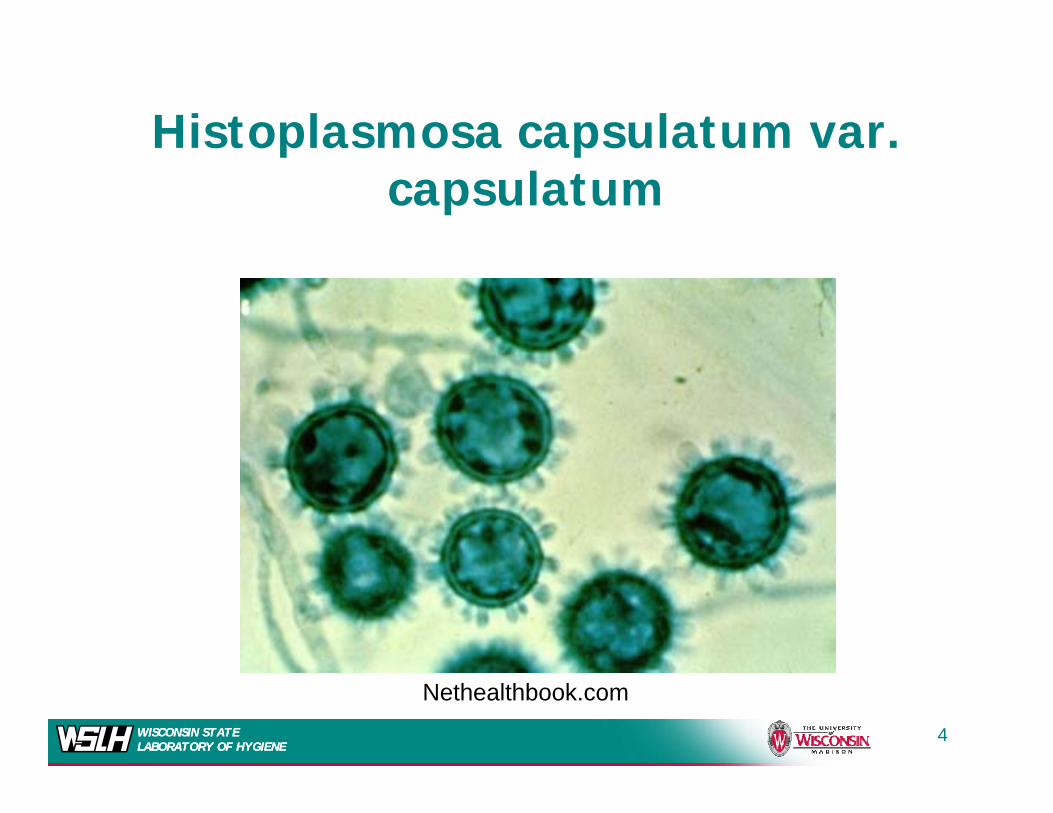
Histoplasmosa capsulatum var. p pcapsulatum
WISCONSIN STATE LABORATORY OF HYGIENEWISCONSIN STATE LABORATORY OF HYGIENE
4
Nethealthbook.com
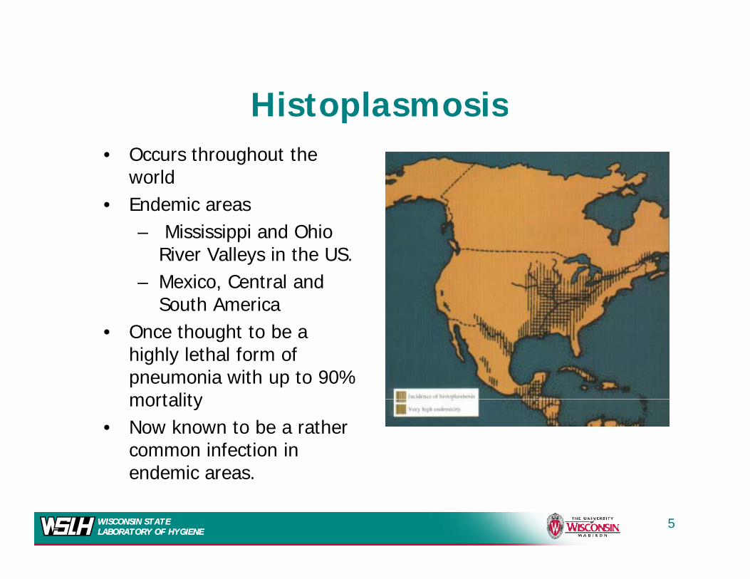
HistoplasmosisHistoplasmosis• Occurs throughout the
worldworld• Endemic areas
– Mississippi and Ohio River Valleys in the USRiver Valleys in the US.
– Mexico, Central and South America
O th ht t b• Once thought to be a highly lethal form of pneumonia with up to 90% mortalitymortality
• Now known to be a rather common infection in endemic areas
WISCONSIN STATE LABORATORY OF HYGIENEWISCONSIN STATE LABORATORY OF HYGIENE
endemic areas.
5
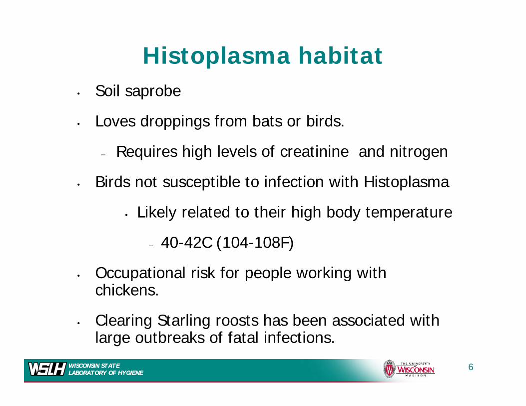
Histoplasma habitat• Soil saprobe
• Loves droppings from bats or birds.Loves droppings from bats or birds.
– Requires high levels of creatinine and nitrogen
Bi d t tibl t i f ti ith Hi t l• Birds not susceptible to infection with Histoplasma
• Likely related to their high body temperature
– 40-42C (104-108F)
• Occupational risk for people working with p p p gchickens.
• Clearing Starling roosts has been associated with l b k f f l f
WISCONSIN STATE LABORATORY OF HYGIENEWISCONSIN STATE LABORATORY OF HYGIENE
large outbreaks of fatal infections.
6
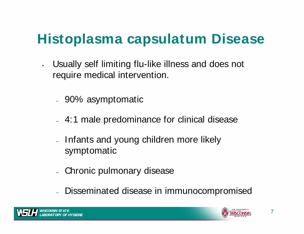
Histoplasma capsulatum Disease
• Usually self limiting flu-like illness and does not i di l i t ti
Histoplasma capsulatum Disease
require medical intervention.
– 90% asymptomatic90% asymptomatic
– 4:1 male predominance for clinical disease
– Infants and young children more likely symptomatic
– Chronic pulmonary disease
Disseminated disease in immunocompromised
WISCONSIN STATE LABORATORY OF HYGIENEWISCONSIN STATE LABORATORY OF HYGIENE
– Disseminated disease in immunocompromised
7

Pathobiology
I h i f ti ll t 2 5 i
Pathobiology
• In human infection---small yeast 2-5 µm in diameter.
d i tl i h– predominantly in macrophages. • Nonactivated macrophages do not
effectively kill H capsulatum and caneffectively kill H. capsulatum and can actually spread the disease.– Can multiply intracellularly kill the– Can multiply intracellularly, kill the
phagocyte, and infect additional cells
WISCONSIN STATE LABORATORY OF HYGIENEWISCONSIN STATE LABORATORY OF HYGIENE
8
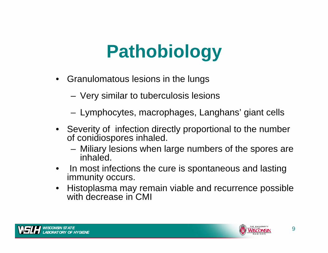
P th bi lPathobiology• Granulomatous lesions in the lungs• Granulomatous lesions in the lungs
– Very similar to tuberculosis lesions
L mphoc tes macrophages Langhans’ giant cells– Lymphocytes, macrophages, Langhans’ giant cells
• Severity of infection directly proportional to the number of conidiospores inhaled. p– Miliary lesions when large numbers of the spores are
inhaled.• In most infections the cure is spontaneous and lasting
i itimmunity occurs. • Histoplasma may remain viable and recurrence possible
with decrease in CMI
WISCONSIN STATE LABORATORY OF HYGIENEWISCONSIN STATE LABORATORY OF HYGIENE
9
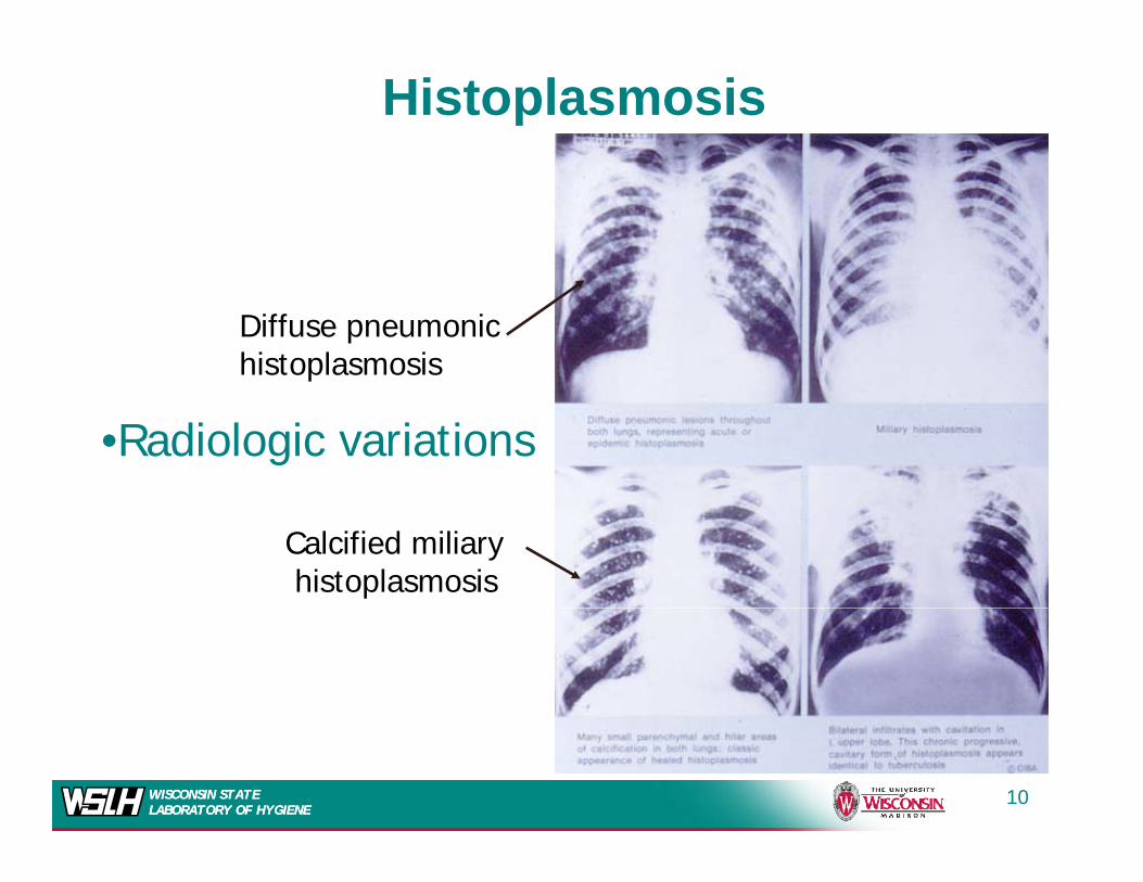
Histoplasmosis
Diffuse pneumonichistoplasmosis
•Radiologic variations
Calcified miliaryhistoplasmosis
WISCONSIN STATE LABORATORY OF HYGIENEWISCONSIN STATE LABORATORY OF HYGIENE
10
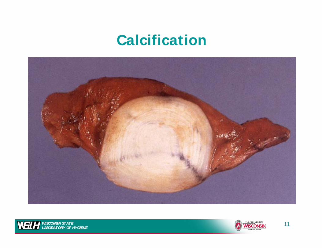
Calcification
WISCONSIN STATE LABORATORY OF HYGIENEWISCONSIN STATE LABORATORY OF HYGIENE
11

Immunity and TreatmentImmunity and Treatment
• Immunity dependent on CMI. – Antibody is of little importance
• Healing of lesions leads to calcified granulomassimilar to that seen in tuberculosissimilar to that seen in tuberculosis.
– Old calcified nodules on chest x-ray not uncommonuncommon
• Treatment reserved for life-threatening infections
Amphotericin B– Amphotericin B
– Itraconazole
WISCONSIN STATE LABORATORY OF HYGIENEWISCONSIN STATE LABORATORY OF HYGIENE
12
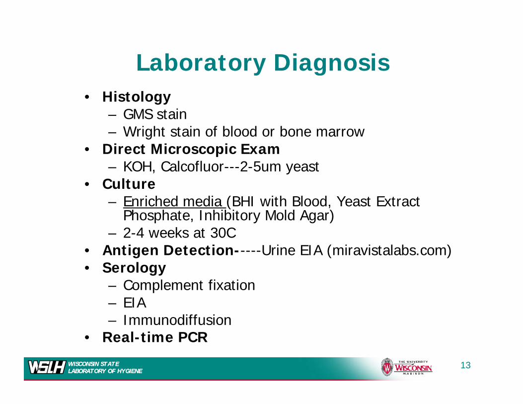
Laboratory Diagnosis• Histology
– GMS stain h f bl d b
y g
– Wright stain of blood or bone marrow• Direct Microscopic Exam
– KOH, Calcofluor---2-5um yeast• Culture
– Enriched media (BHI with Blood, Yeast Extract Phosphate, Inhibitory Mold Agar)2 4 k t 30C– 2-4 weeks at 30C
• Antigen Detection-----Urine EIA (miravistalabs.com)• Serology
– Complement fixation– EIA– Immunodiffusion
WISCONSIN STATE LABORATORY OF HYGIENEWISCONSIN STATE LABORATORY OF HYGIENE
• Real-time PCR
13
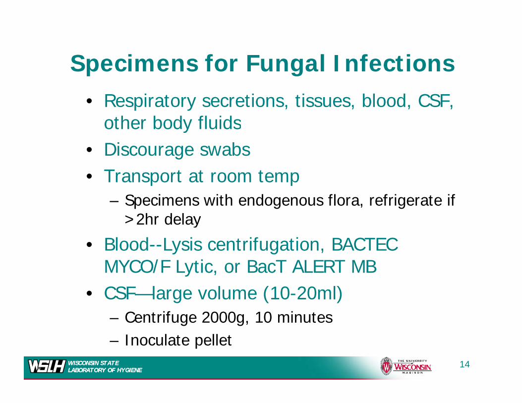
Specimens for Fungal Infectionsp g• Respiratory secretions, tissues, blood, CSF,
other body fluidsother body fluids• Discourage swabs• Transport at room temp• Transport at room temp
– Specimens with endogenous flora, refrigerate if >2hr delayy
• Blood--Lysis centrifugation, BACTEC MYCO/F Lytic, or BacT ALERT MB
• CSF—large volume (10-20ml)– Centrifuge 2000g, 10 minutes
WISCONSIN STATE LABORATORY OF HYGIENEWISCONSIN STATE LABORATORY OF HYGIENE
– Inoculate pellet14
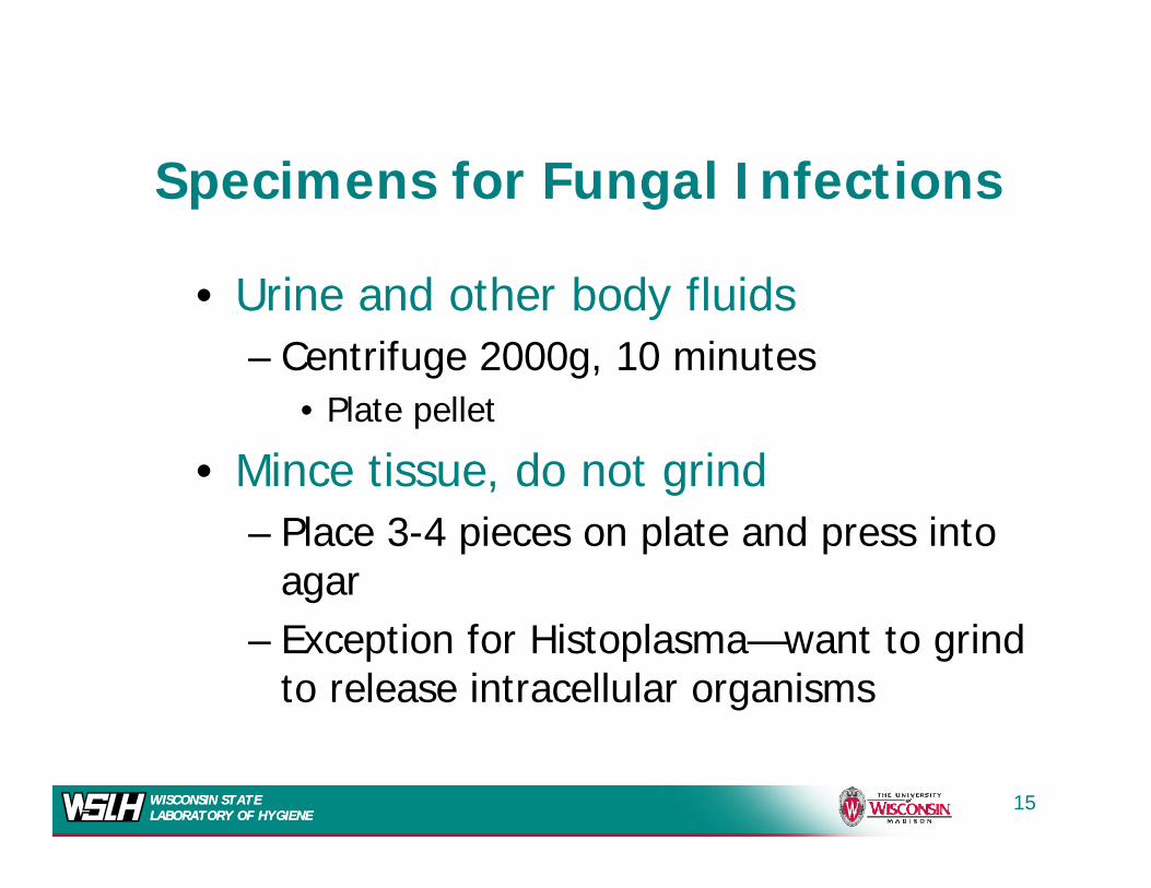
S i f F l I f tiSpecimens for Fungal Infections
d h b d fl d• Urine and other body fluids– Centrifuge 2000g, 10 minutes
• Plate pellet
• Mince tissue, do not grind– Place 3-4 pieces on plate and press into
agar– Exception for Histoplasma—want to grind
to release intracellular organisms
WISCONSIN STATE LABORATORY OF HYGIENEWISCONSIN STATE LABORATORY OF HYGIENE
15
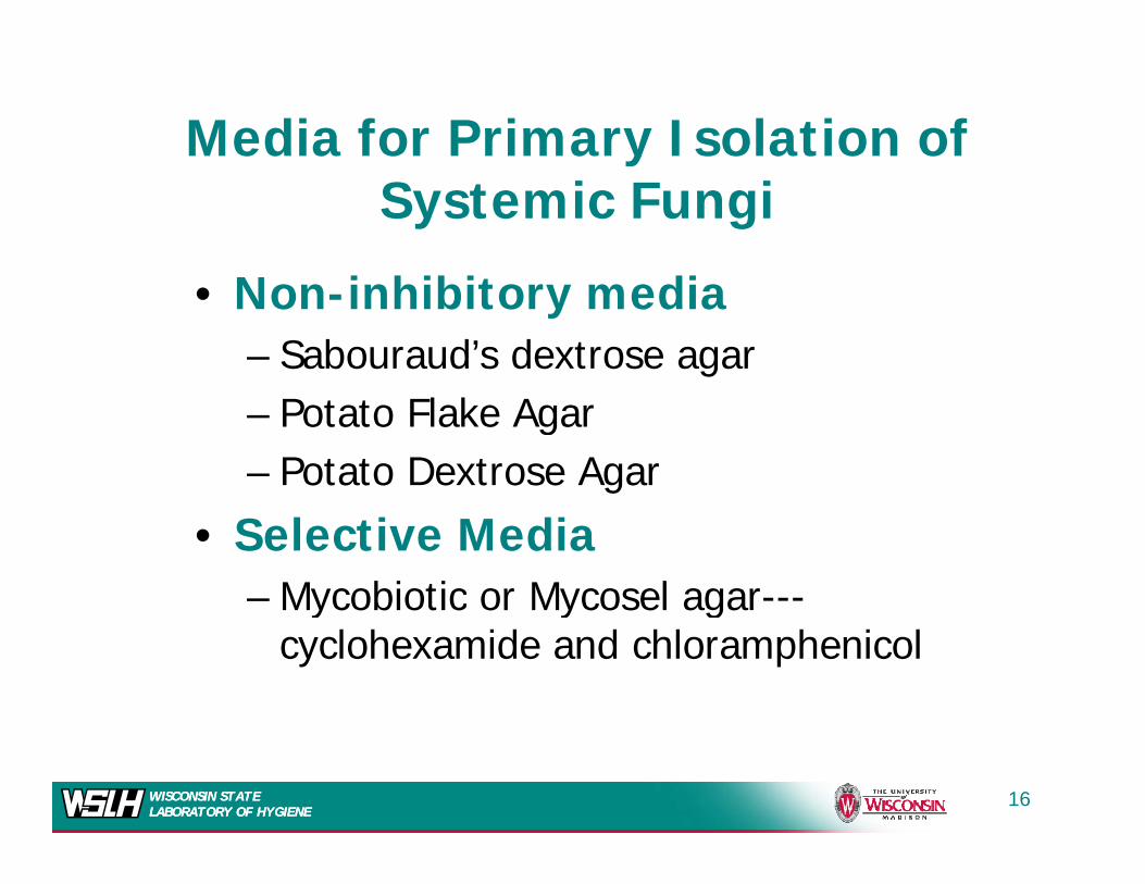
Media for Primary Isolation of ySystemic Fungi
i hibi di• Non-inhibitory media– Sabouraud’s dextrose agar– Potato Flake Agar– Potato Dextrose Agar
• Selective Media– Mycobiotic or Mycosel agar---Mycobiotic or Mycosel agar
cyclohexamide and chloramphenicol
WISCONSIN STATE LABORATORY OF HYGIENEWISCONSIN STATE LABORATORY OF HYGIENE
16

Media for Primary Isolation of ySystemic Fungi (2)
• Enriched media w/ or w/o antibiotics– Inhibitory mold agar---chloramphenicol and cyclohexamide– BHI with sheep blood w/wo antibioticsp /– Yeast extract phosphate agar with ammonia
• Incubate plates or tubes at 30oC or 25oCIncubate plates or tubes at 30 C or 25 C – Hold 4 weeks
• For Blood Cultures– Lysis Centrifugation
– BACTEC MYCO/F or BacT ALERT MB
WISCONSIN STATE LABORATORY OF HYGIENEWISCONSIN STATE LABORATORY OF HYGIENE
17
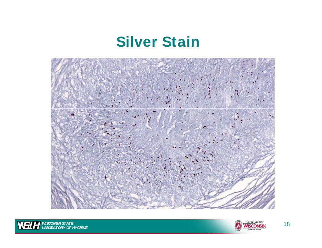
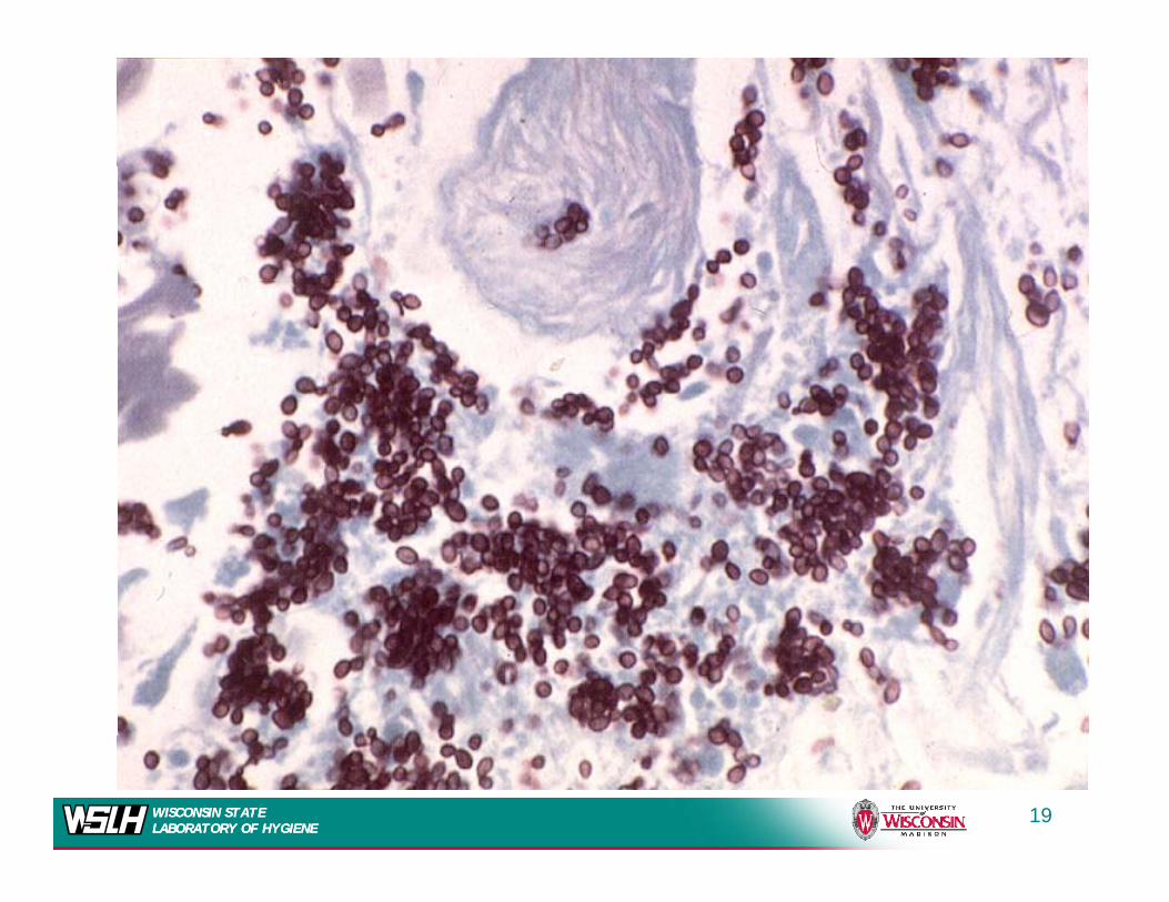
Silver StainSilver Stain
WISCONSIN STATE LABORATORY OF HYGIENEWISCONSIN STATE LABORATORY OF HYGIENE
18
WISCONSIN STATE LABORATORY OF HYGIENEWISCONSIN STATE LABORATORY OF HYGIENE
19
WISCONSIN STATE LABORATORY OF HYGIENEWISCONSIN STATE LABORATORY OF HYGIENE
20
WISCONSIN STATE LABORATORY OF HYGIENEWISCONSIN STATE LABORATORY OF HYGIENE
21

Culture
WISCONSIN STATE LABORATORY OF HYGIENEWISCONSIN STATE LABORATORY OF HYGIENE
Histo at 3 weeks, 30C22
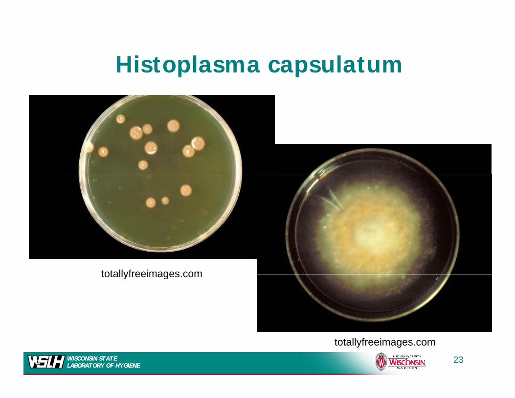
Histoplasma capsulatump p
totallyfreeimages comtotallyfreeimages.com
WISCONSIN STATE LABORATORY OF HYGIENEWISCONSIN STATE LABORATORY OF HYGIENE
23
totallyfreeimages.com
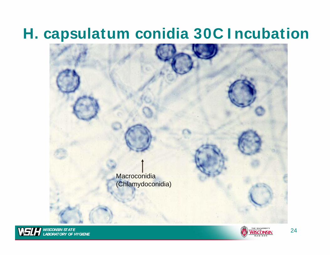
H. capsulatum conidia 30C Incubation
MacroconidiaMacroconidia (Chlamydoconidia)
WISCONSIN STATE LABORATORY OF HYGIENEWISCONSIN STATE LABORATORY OF HYGIENE
24
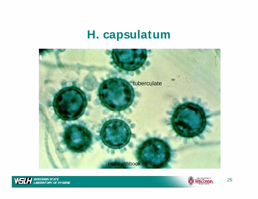
H. capsulatumH. capsulatum
tuberculate
th lthb k
WISCONSIN STATE LABORATORY OF HYGIENEWISCONSIN STATE LABORATORY OF HYGIENE
nethealthbook.com
25
H. capsulatum—Yeast phase
WISCONSIN STATE LABORATORY OF HYGIENEWISCONSIN STATE LABORATORY OF HYGIENE
26

Diff ti ti f th F iDifferentiation from other Fungi
M t diff ti t f S d i d• Must differentiate from Sepedonium and Chrysosporium species that produce tuberculate macroconidiatuberculate macroconidia– More rapid growing
Not dimorphic– Not dimorphic– Usually will not grow in the presence of
cycloheximidecycloheximide– Distinguish using DNA probe
WISCONSIN STATE LABORATORY OF HYGIENEWISCONSIN STATE LABORATORY OF HYGIENE
27
Nucleic Acid Probe IdentificationNucleic Acid Probe Identification
• GenProbe® Assay• GenProbe® Assay– Rapid
Chemiluminescent assay using labeled– Chemiluminescent assay using labeled probes specific for each agent
– Labeled DNA probe hybridizes with rRNA– Labeled DNA probe hybridizes with rRNAof the fungus
– Available for H capsulatum BlastomycesAvailable for H. capsulatum, Blastomycesdermatitidis, and C. immitis
WISCONSIN STATE LABORATORY OF HYGIENEWISCONSIN STATE LABORATORY OF HYGIENE
28

BLASTO!
WISCONSIN STATE LABORATORY OF HYGIENEWISCONSIN STATE LABORATORY OF HYGIENE
29
Blastomycosis
• Blastomyces dermatitidis
Blastomycosis
• Blastomyces dermatitidis• Agent of North American
Bl iBlastomycosis,• Geographical distribution is similar
to H. capsulatum• More common in Wisconsin than H.
capsulatum.
WISCONSIN STATE LABORATORY OF HYGIENEWISCONSIN STATE LABORATORY OF HYGIENE
30

Kurt Reed et. al PLOSone 3(4): e2034, 2008
WISCONSIN STATE LABORATORY OF HYGIENEWISCONSIN STATE LABORATORY OF HYGIENE
31
Blastomycosis
• The epidemiology is poorly understood
y
– Lack of a good skin test reagent
– Ecologic niche not well established
• Difficult to recover from the soil in endemic areas.
• Eagle River, Wisconsin outbreak 1985
– First time Blastomyces isolated from the environment at the site of an outbreak
– Isolated from soil containing decayed t ti tt d f d d d
WISCONSIN STATE LABORATORY OF HYGIENEWISCONSIN STATE LABORATORY OF HYGIENE
vegetative matter and from decomposed wood.
32
Clinical Manifestations
• Two clinical presentations– A primary cutaneous infection which usually remainsA primary cutaneous infection which usually remains
localized to one area of the body
• May indicate systemic disease – Primary pulmonary infection with possible secondary
dissemination.
• 30-45 day incubation30 45 day incubation
• Mimics flu progressing to cough, weight loss, chest pain, low grade fever75% i h i l d l di• 75% with isolated pulmonary disease
• Infection may involve any organ• Secondary cutaneous infection
WISCONSIN STATE LABORATORY OF HYGIENEWISCONSIN STATE LABORATORY OF HYGIENE
– Asymptomatic in >50% of those infected
33
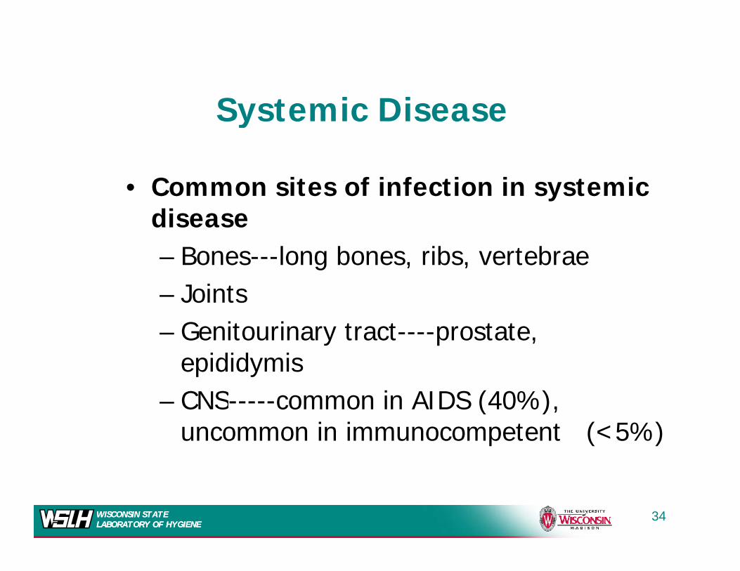
Systemic DiseaseSystemic Disease
C it f i f ti i t i• Common sites of infection in systemic disease
B l b ib t b– Bones---long bones, ribs, vertebrae– Joints
G i i– Genitourinary tract----prostate, epididymisCNS i AIDS (40%)– CNS-----common in AIDS (40%), uncommon in immunocompetent (<5%)
WISCONSIN STATE LABORATORY OF HYGIENEWISCONSIN STATE LABORATORY OF HYGIENE
34

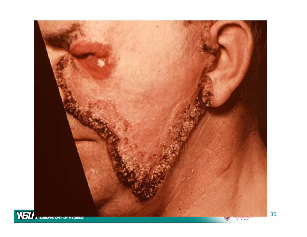
Cutaneous
A h i ti
Form• A chronic suppurative
granulomatous lesion. • The presence of epithelial
microabscesses and characteristic yeasts in the tissues is considered diagnostic.
• It is important to obtain urine and sputum samples from aand sputum samples from a patient with cutaneousblastomycosis since systemic spread may occur
WISCONSIN STATE LABORATORY OF HYGIENEWISCONSIN STATE LABORATORY OF HYGIENE
spread may occur.
35
WISCONSIN STATE LABORATORY OF HYGIENEWISCONSIN STATE LABORATORY OF HYGIENE
36
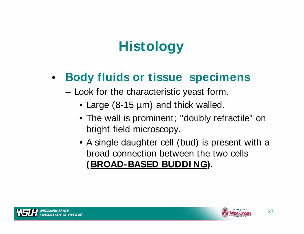
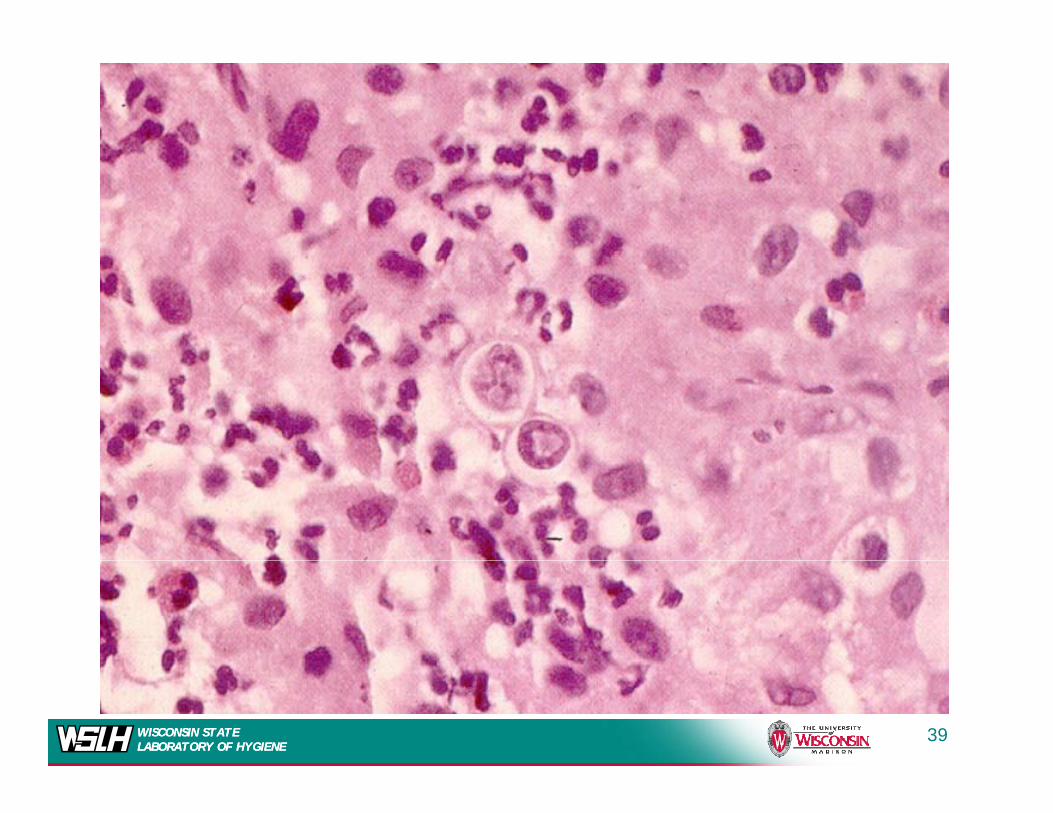
Histology
B d fl id i i
Histology
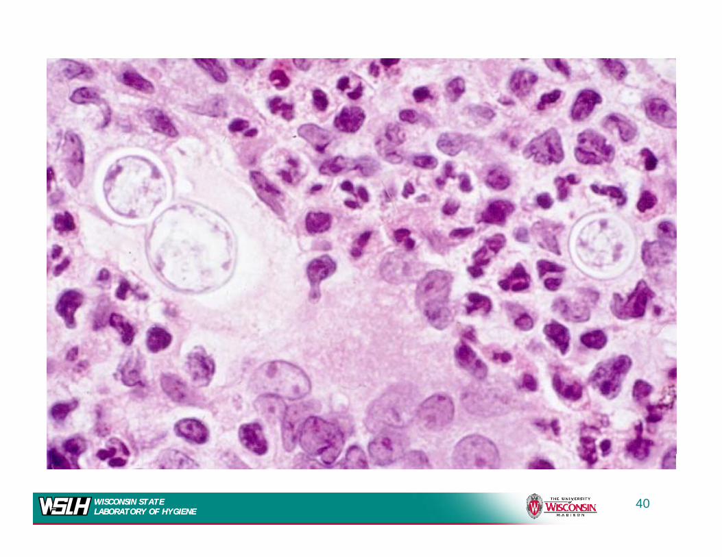
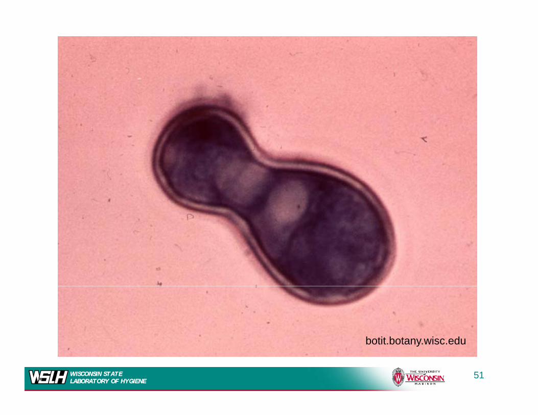
• Body fluids or tissue specimens – Look for the characteristic yeast form.
L (8 15 ) d thi k ll d• Large (8-15 µm) and thick walled. • The wall is prominent; "doubly refractile" on
bright field microscopy.bright field microscopy. • A single daughter cell (bud) is present with a
broad connection between the two cells (BROAD BASED BUDDING)(BROAD-BASED BUDDING).
WISCONSIN STATE LABORATORY OF HYGIENEWISCONSIN STATE LABORATORY OF HYGIENE
37

WISCONSIN STATE LABORATORY OF HYGIENEWISCONSIN STATE LABORATORY OF HYGIENE
38
WISCONSIN STATE LABORATORY OF HYGIENEWISCONSIN STATE LABORATORY OF HYGIENE
39
WISCONSIN STATE LABORATORY OF HYGIENEWISCONSIN STATE LABORATORY OF HYGIENE
40
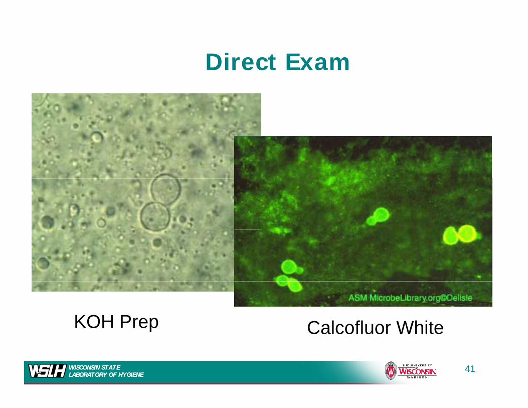
Direct Exam
KO• KO
KOH Prep Calcofluor White
WISCONSIN STATE LABORATORY OF HYGIENEWISCONSIN STATE LABORATORY OF HYGIENE
Calcofluor White
41
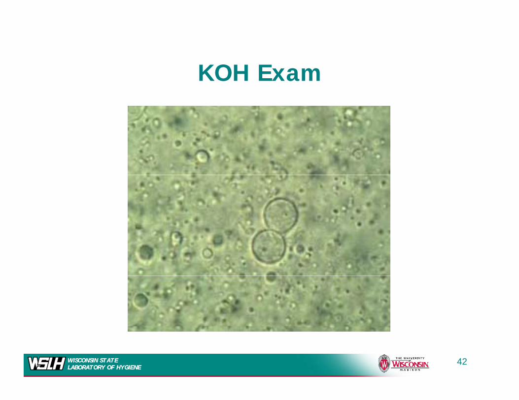
KOH ExamKOH Exam
WISCONSIN STATE LABORATORY OF HYGIENEWISCONSIN STATE LABORATORY OF HYGIENE
42
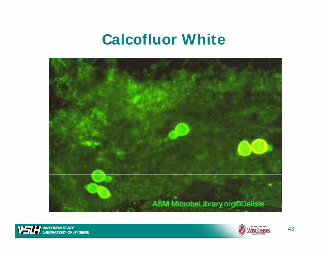
Calcofluor White
WISCONSIN STATE LABORATORY OF HYGIENEWISCONSIN STATE LABORATORY OF HYGIENE
43
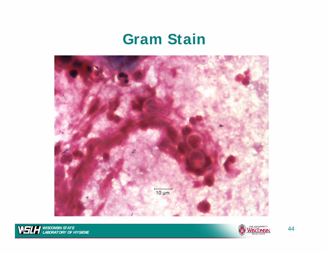
Gram Stain
WISCONSIN STATE LABORATORY OF HYGIENEWISCONSIN STATE LABORATORY OF HYGIENE
44
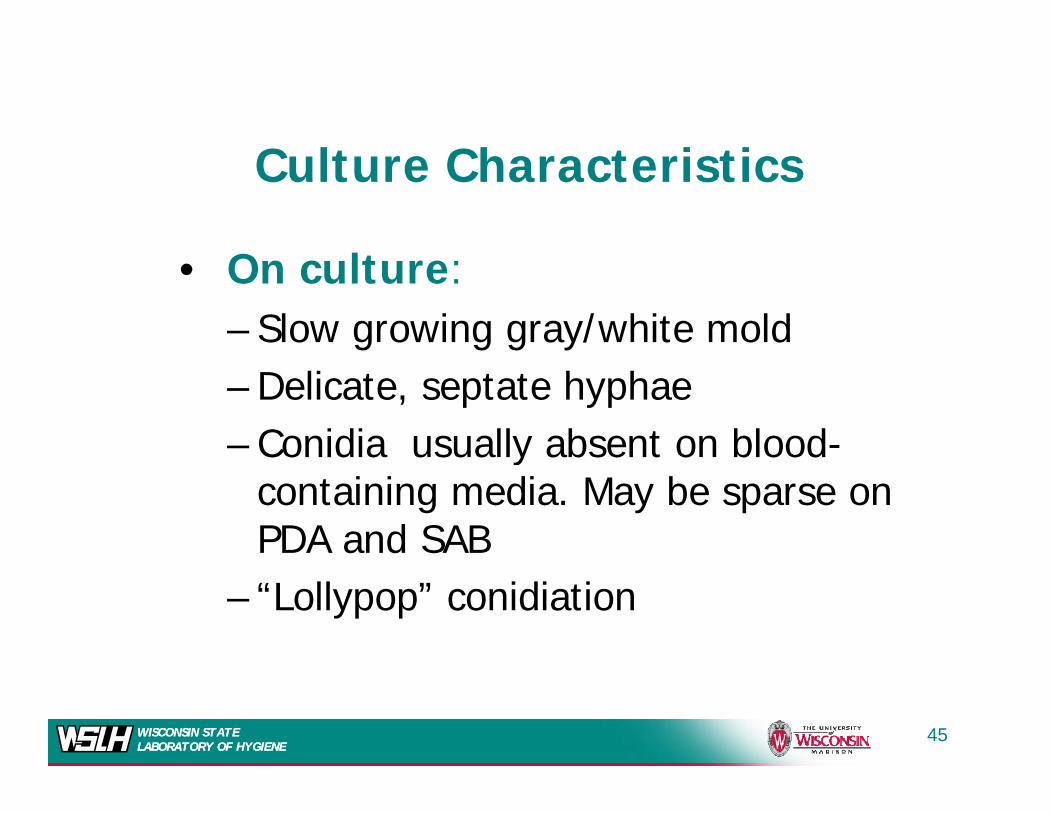
C lt Ch t i tiCulture Characteristics
O l• On culture:– Slow growing gray/white mold– Delicate, septate hyphae– Conidia usually absent on blood-y
containing media. May be sparse on PDA and SAB
– “Lollypop” conidiation
WISCONSIN STATE LABORATORY OF HYGIENEWISCONSIN STATE LABORATORY OF HYGIENE
45
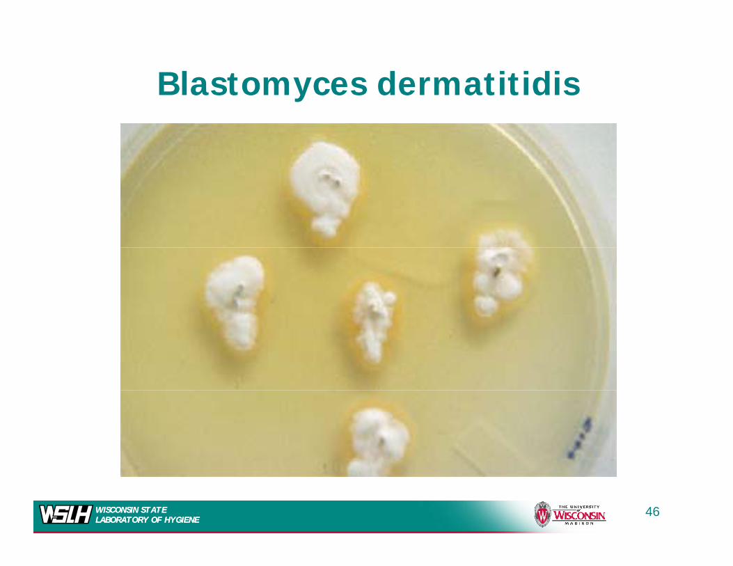
Blastomyces dermatitidis
WISCONSIN STATE LABORATORY OF HYGIENEWISCONSIN STATE LABORATORY OF HYGIENE
46
WISCONSIN STATE LABORATORY OF HYGIENEWISCONSIN STATE LABORATORY OF HYGIENE
47
Blastomyces Mold Phase
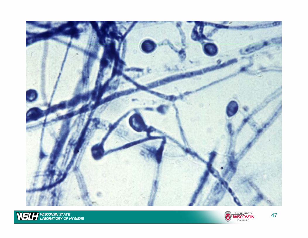
Thi f f idi i l f d i h f i
Blastomyces Mold Phase
• This form of conidia is also found in such fungi as Chrysosporium sp., Pseudallescheria boydii (Scedosporium), and various Trichophyton sp. ( p ) p y p
• Differentiation from these other species can be made by the following characteristics:
Sl th– Slower growth– Growth in the presence of cycloheximide
Dimorphism– Dimorphism– Nucleic acid probes
WISCONSIN STATE LABORATORY OF HYGIENEWISCONSIN STATE LABORATORY OF HYGIENE
48
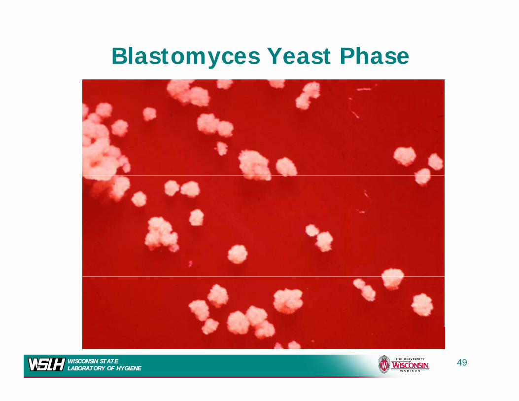
Blastomyces Yeast Phase
WISCONSIN STATE LABORATORY OF HYGIENEWISCONSIN STATE LABORATORY OF HYGIENE
49
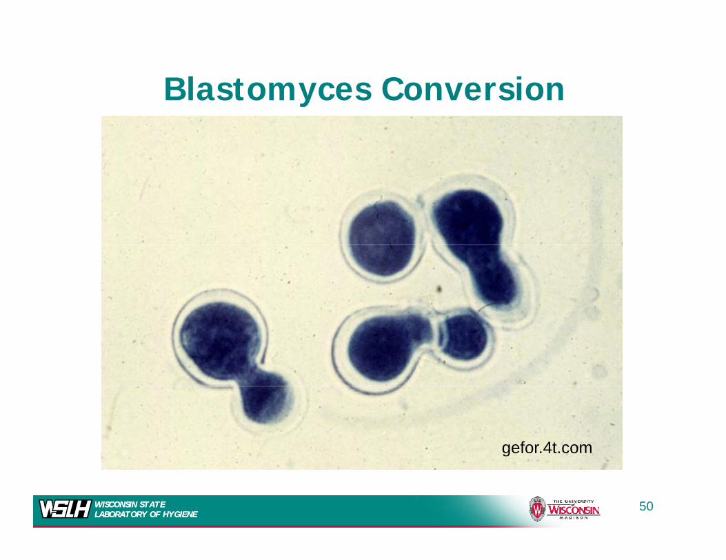
Blastomyces Conversiony
gefor.4t.com
WISCONSIN STATE LABORATORY OF HYGIENEWISCONSIN STATE LABORATORY OF HYGIENE
50
b tit b t i d
WISCONSIN STATE LABORATORY OF HYGIENEWISCONSIN STATE LABORATORY OF HYGIENE
botit.botany.wisc.edu
51
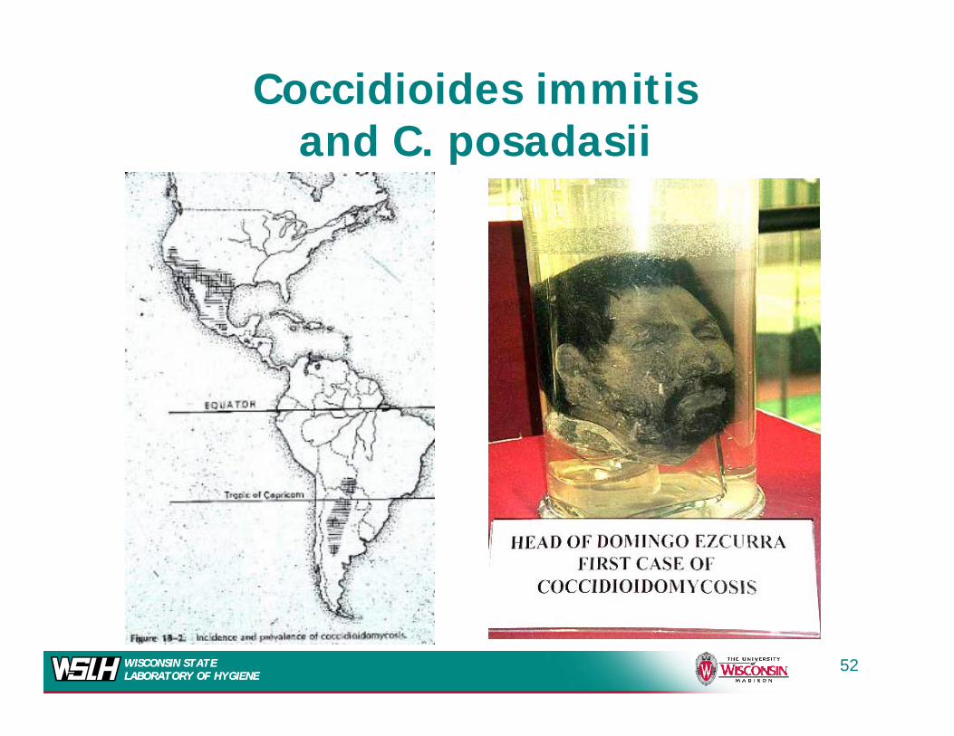
Coccidioides immitisand C posadasiiand C. posadasii
WISCONSIN STATE LABORATORY OF HYGIENEWISCONSIN STATE LABORATORY OF HYGIENE
52
WISCONSIN STATE LABORATORY OF HYGIENEWISCONSIN STATE LABORATORY OF HYGIENE
53
Coccidiodes immitis
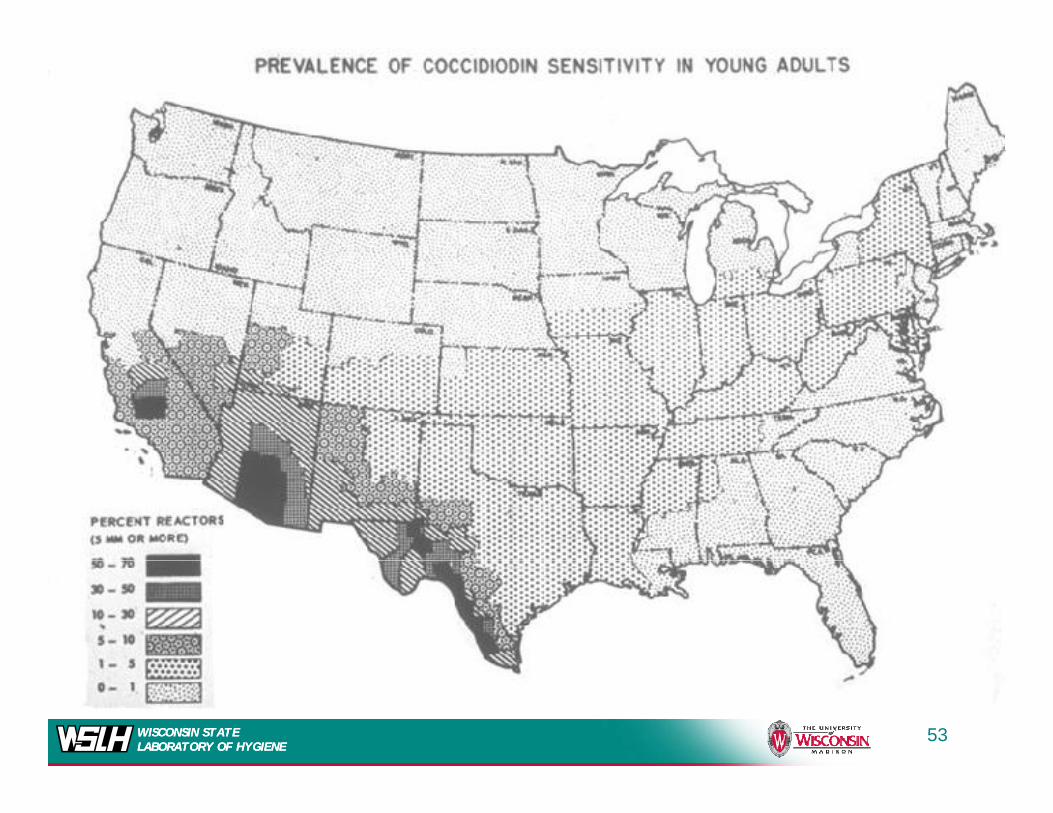
• Coccidiomycosis is sometimes known as “San Joaquin Valley fever” Up to 95% of the residents of theValley fever . Up to 95% of the residents of the endemic area are skin test positive (coccidioidin test positive)
• Lower Sonoran Life Zone– Arid climate, hot summers, few winter freezes, low altitude,
alkaline soil, sparse flora– Drought followed by heavy rains---Increased infections– 100,000 infected annually in U.S.
• Variety of animals infected• Variety of animals infected– Positive cultures around rodent burrows
• Archaeology students discover new “infected” sites
WISCONSIN STATE LABORATORY OF HYGIENEWISCONSIN STATE LABORATORY OF HYGIENE
Archaeology students discover new infected sites
54
Clinical ManifestationsClinical Manifestations
The primary disease is pulmonary• The primary disease is pulmonary, secondary to inhalation of small numbers of arthrosporesnumbers of arthrospores
• Usually resolves spontaneously as an influenza-like infection. – 60% asymptomatic – 40% influenza-like illness, LRI or
systemic illnesssystemic illness• Cough, sputum, chest pain, malaise, fever,
chills, night sweats, arthralgias, anorexia
WISCONSIN STATE LABORATORY OF HYGIENEWISCONSIN STATE LABORATORY OF HYGIENE
55
Cli i l M if t tiClinical Manifestations
f h• In a minority of cases a more chronic pulmonary infection occurs– Granulomatous lesions of the lung– Can lead to cavitation
• In rare cases (0.5%) dissemination occurs which can lead to rapidly fatal p yresults.
• Reactivation infection occurs
WISCONSIN STATE LABORATORY OF HYGIENEWISCONSIN STATE LABORATORY OF HYGIENE
Reactivation infection occurs
56
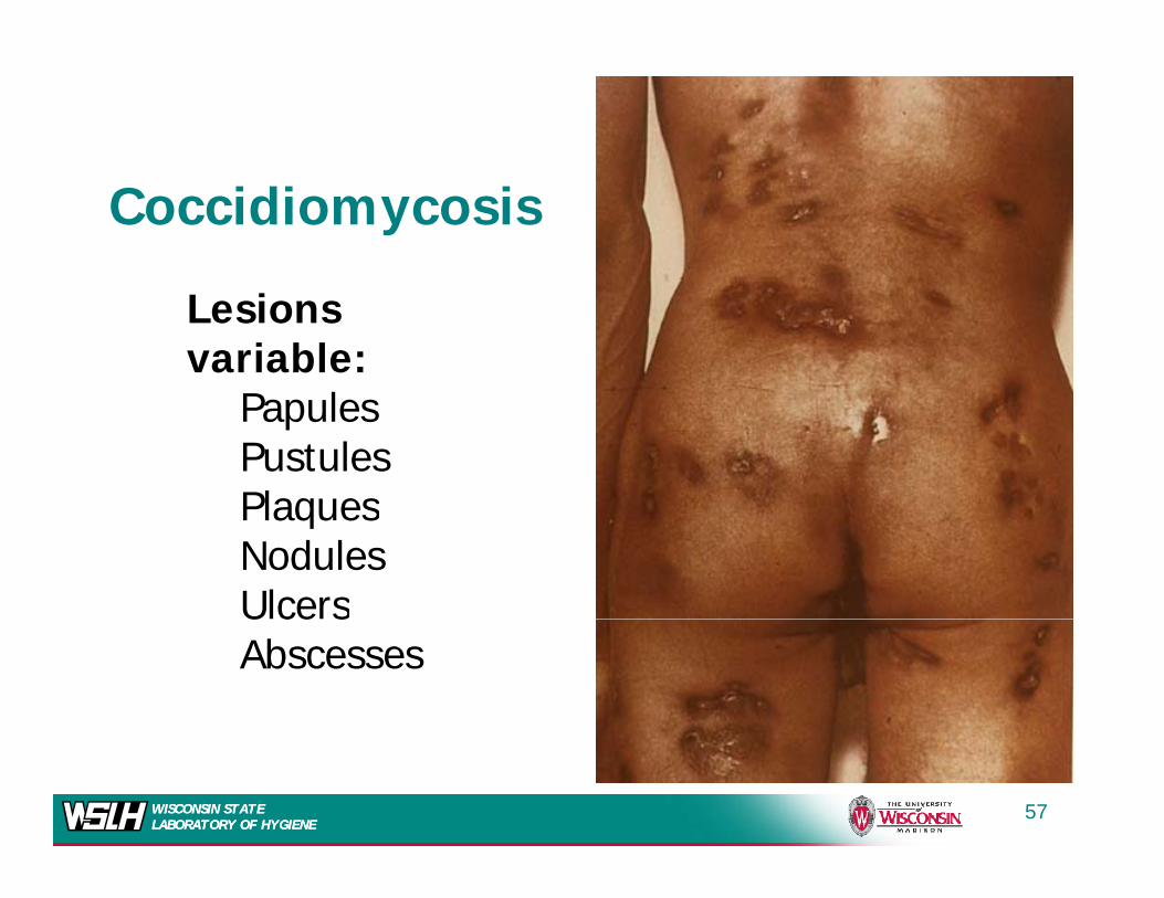
Coccidiomycosis
Lesions variable:
P lPapulesPustulesPlaquesPlaquesNodulesUlcersU ce sAbscesses
WISCONSIN STATE LABORATORY OF HYGIENEWISCONSIN STATE LABORATORY OF HYGIENE
57
HistologyHistology
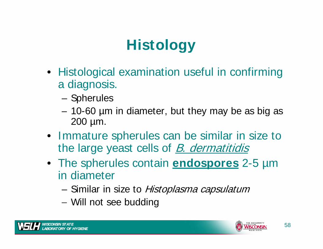
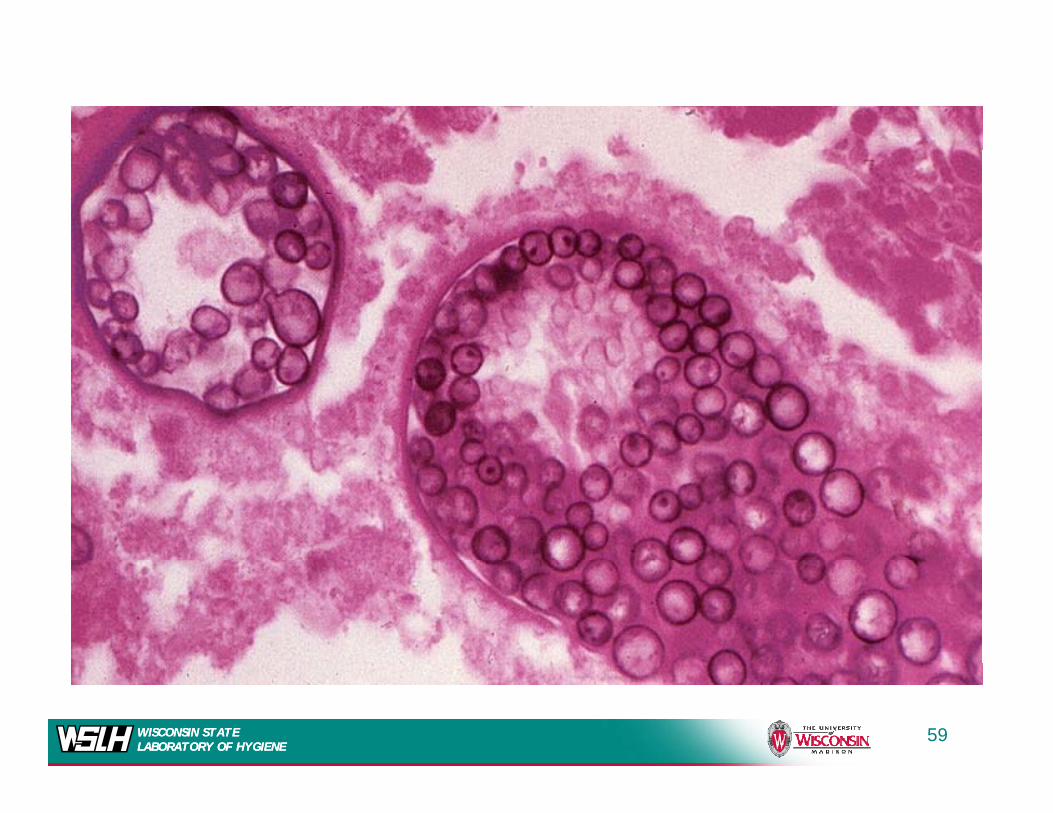
• Histological examination useful in confirming g ga diagnosis.– Spherules
10 60 µm in diameter but they may be as big as– 10-60 µm in diameter, but they may be as big as 200 µm.
• Immature spherules can be similar in size to pthe large yeast cells of B. dermatitidis
• The spherules contain endospores 2-5 µm i di tin diameter– Similar in size to Histoplasma capsulatum– Will not see budding
WISCONSIN STATE LABORATORY OF HYGIENEWISCONSIN STATE LABORATORY OF HYGIENE
Will not see budding
58
WISCONSIN STATE LABORATORY OF HYGIENEWISCONSIN STATE LABORATORY OF HYGIENE
59
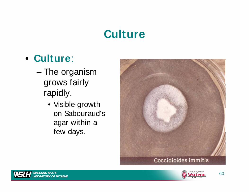
Culture
• Culture:
Culture
• Culture:– The organism
grows fairlygrows fairly rapidly.• Visible growth g
on Sabouraud'sagar within a few daysfew days.
WISCONSIN STATE LABORATORY OF HYGIENEWISCONSIN STATE LABORATORY OF HYGIENE
60
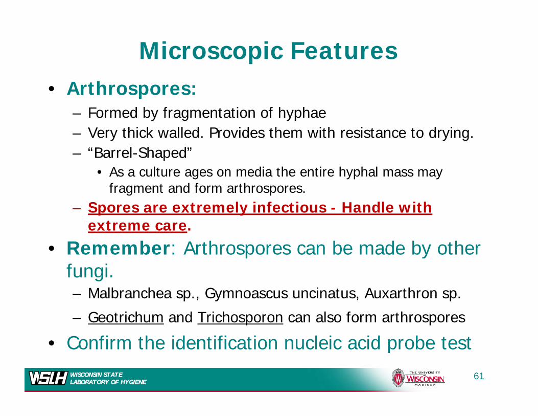
Microscopic Features• Arthrospores:
– Formed by fragmentation of hyphaeVe thi k alled P o ides them ith esistan e to d ing– Very thick walled. Provides them with resistance to drying.
– “Barrel-Shaped” • As a culture ages on media the entire hyphal mass may
fragment and form arthrospores. – Spores are extremely infectious - Handle with
extreme care.
• Remember: Arthrospores can be made by other fungi.– Malbranchea sp., Gymnoascus uncinatus, Auxarthron sp.
– Geotrichum and Trichosporon can also form arthrospores
Confirm the identification nucleic acid probe testWISCONSIN STATE LABORATORY OF HYGIENEWISCONSIN STATE LABORATORY OF HYGIENE
• Confirm the identification nucleic acid probe test
61
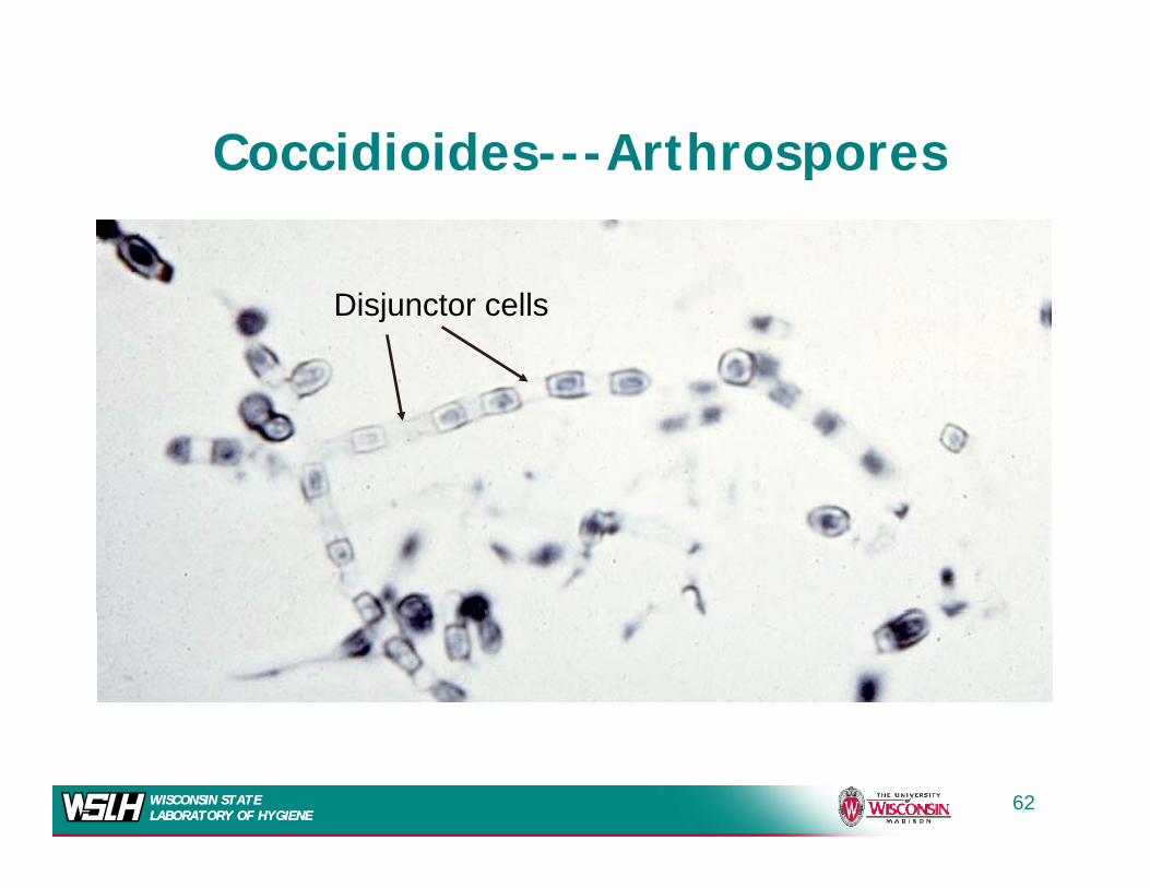
Coccidioides---ArthrosporesCoccidioides Arthrospores
Disjunctor cells
WISCONSIN STATE LABORATORY OF HYGIENEWISCONSIN STATE LABORATORY OF HYGIENE
62
Select Agent RegulationsSelect Agent Regulations
• Report to CDC within 7 days of ID• Report to CDC within 7 days of ID– Responsibility of lab confirming ID
S l A APHIS/CDC F 4– Select Agent APHIS/CDC Form 4
• Secure against loss, theft, or release• Destroy all subcultures and specimens• Good NewsGood News
– Proposed to remove Coccidioides from SA list
WISCONSIN STATE LABORATORY OF HYGIENEWISCONSIN STATE LABORATORY OF HYGIENE
list
63
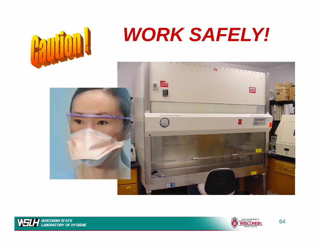
WORK SAFELY!
WISCONSIN STATE LABORATORY OF HYGIENEWISCONSIN STATE LABORATORY OF HYGIENE
64
WISCONSIN STATE LABORATORY OF HYGIENEWISCONSIN STATE LABORATORY OF HYGIENE
65