Embed Size (px)
Citation preview
University of Rhode IslandDigitalCommons@URIInstitute for Immunology and Informatics FacultyPublications Institute for Immunology and Informatics (iCubed)
2013
T-cell dependent immunogenicity of proteintherapeutics: Preclinical assessment and mitigationVibha Jawa
Leslie P. Cousens
See next page for additional authors
Creative Commons License
This work is licensed under a Creative Commons Attribution-Noncommercial-No Derivative Works 3.0License.
Follow this and additional works at: https://digitalcommons.uri.edu/immunology_facpubs
Terms of UseAll rights reserved under copyright.
This Article is brought to you for free and open access by the Institute for Immunology and Informatics (iCubed) at DigitalCommons@URI. It hasbeen accepted for inclusion in Institute for Immunology and Informatics Faculty Publications by an authorized administrator ofDigitalCommons@URI. For more information, please contact [email protected].
Citation/Publisher AttributionJawa, V., Cousens, L.P., Awwad, M., Wakshull, E., Kropshofer, H., & De Groot, A.S. (2013). T-cell dependent immunogenicity ofprotein therapeutics: Preclinical assessment and mitigation. Clinical Immunology, 149(3.B), 534-555.Available at: http://dx.doi.org/10.1016/j.clim.2013.09.006

AuthorsVibha Jawa, Leslie P. Cousens, Michel Awwad, Eric Wakshull, Harald Kropshofer, and Anne S. De Groot
This article is available at DigitalCommons@URI: https://digitalcommons.uri.edu/immunology_facpubs/3
REVIEW
T-cell dependent immunogenicity of proteintherapeutics: Preclinical assessmentand mitigation☆
Vibha Jawaa, Leslie P. Cousensb,1, Michel Awwadc,2, Eric Wakshull d,Harald Kropshofer e, Anne S. De Grootb,f,⁎
a Amgen, USAb EpiVax, USAc Pfizer, USAd Genentech, USAe Roche, Switzerlandf University of Rhode Island, USA
Received 8 July 2013; accepted with revision 14 September 2013Available online 25 September 2013
KEYWORDSQuality-by-Design;T cell;Immunogenicity;Cell-mediated immunity
Abstract Protein therapeutics hold a prominent and rapidly expanding place among medicinalproducts. Purified blood products, recombinant cytokines, growth factors, enzyme replacementfactors, monoclonal antibodies, fusion proteins, and chimeric fusion proteins are all examples oftherapeutic proteins that have been developed in the past few decades and approved for use inthe treatment of human disease. Despite early belief that the fully human nature of theseproteins would represent a significant advantage, adverse effects associated with immuneresponses to some biologic therapies have become a topic of some concern. As a result, drugdevelopers are devising strategies to assess immune responses to protein therapeutics duringboth the preclinical and the clinical phases of development. While there are many factors thatcontribute to protein immunogenicity, T cell- (thymus-) dependent (Td) responses appear toplay a critical role in the development of antibody responses to biologic therapeutics. A rangeof methodologies to predict and measure Td immune responses to protein drugs has beendeveloped. This review will focus on the Td contribution to immunogenicity, summarizing
Abbreviations: Td, T-cell dependent, thymus dependent; T, thymus; ADA, anti-drug antibodies; Ti, T-cell independent; APC,antigen-presenting cells; HLA, human leukocyte antigen; MHC, major histocompatibility complex; TCR, T cell receptor; Treg, regulatory Tcells; FVIII, factor VIII; nTregs, natural regulatory T cells; aTreg, adaptive regulatory T cells; iTreg, induced regulatory T cells; IEDB, ImmuneEpitope Database Analysis Resource; IC50, 50% inhibitory concentration; ELISpot, enzyme-linked immunosorbent spot-forming; ELISA,enzyme-linked immunosorbent assay; CFSE, carboxyfluorescein succinimidyl ester; PBMC, peripheral blood mononuclear cells; ALN, artificiallymph node; ORG, unmodified original epitopes; FPX, recombinant Fc fusion protein; SFC, spot-forming cells.☆ This is an open-access article distributed under the terms of the Creative Commons Attribution-NonCommercial-No Derivative Works License,which permits non-commercial use, distribution, and reproduction in any medium, provided the original author and source are credited.⁎ Corresponding author at: 146 Clifford Street, Providence, RI USA. Fax: +1 401 272 7562.E-mail address: [email protected] (A.S. De Groot).
1 Denotes equal first-authorship.2 Present affiliation: Merrimack Pharmaceuticals, USA.
1521-6616/$ - see front matter © 2013 The Authors. Published by Elsevier Inc. All rights reserved.http://dx.doi.org/10.1016/j.clim.2013.09.006
ava i l ab l e a t www.sc i enced i r ec t . com
C l i n i ca l Immuno logy
www.e l sev i e r . com / l oca te / yc l im
Clinical Immunology (2013) 149, 534–555
current approaches for the prediction and measurement of T cell-dependent immune responsesto protein biologics, discussing the advantages and limitations of these technologies, andsuggesting a practical approach for assessing and mitigating Td immunogenicity.© 2013 The Authors. Published by Elsevier Inc. All rights reserved.
Contents
1. Introduction . . . . . . . . . . . . . . . . . . . . . . . . . . . . . . . . . . . . . . . . . . . . . . . . . . . . . . . . . 5351.1. The immunogenicity of protein therapeutics . . . . . . . . . . . . . . . . . . . . . . . . . . . . . . . . . . . . 535
2. The central role of T cells in immunogenicity . . . . . . . . . . . . . . . . . . . . . . . . . . . . . . . . . . . . . . 5372.1. The T cell contribution to antibody responses . . . . . . . . . . . . . . . . . . . . . . . . . . . . . . . . . . . 5372.2. T cell epitope stability and immunogenicity . . . . . . . . . . . . . . . . . . . . . . . . . . . . . . . . . . . . 5372.3. Td immunity to therapeutic proteins . . . . . . . . . . . . . . . . . . . . . . . . . . . . . . . . . . . . . . . . 5382.4. The role of central and peripheral tolerance to biologics . . . . . . . . . . . . . . . . . . . . . . . . . . . . . 538
3. Methods for predicting Td immune responses . . . . . . . . . . . . . . . . . . . . . . . . . . . . . . . . . . . . . . 5383.1. In silico T cell epitope-screening methods . . . . . . . . . . . . . . . . . . . . . . . . . . . . . . . . . . . . . 5383.2. Strengths and limitations of in silico analysis. . . . . . . . . . . . . . . . . . . . . . . . . . . . . . . . . . . . 539
3.2.1. Antigen processing . . . . . . . . . . . . . . . . . . . . . . . . . . . . . . . . . . . . . . . . . . . . . . 5393.2.2. MHC affinity . . . . . . . . . . . . . . . . . . . . . . . . . . . . . . . . . . . . . . . . . . . . . . . . . 5393.2.3. T cell phenotype . . . . . . . . . . . . . . . . . . . . . . . . . . . . . . . . . . . . . . . . . . . . . . . 5403.2.4. Individual versus population-level predictions . . . . . . . . . . . . . . . . . . . . . . . . . . . . . . 5403.2.5. Post-translational factors . . . . . . . . . . . . . . . . . . . . . . . . . . . . . . . . . . . . . . . . . . 540
3.3. HLA binding assays . . . . . . . . . . . . . . . . . . . . . . . . . . . . . . . . . . . . . . . . . . . . . . . . . . 5403.3.1. Competition binding assays . . . . . . . . . . . . . . . . . . . . . . . . . . . . . . . . . . . . . . . . . 5403.3.2. Direct binding assays. . . . . . . . . . . . . . . . . . . . . . . . . . . . . . . . . . . . . . . . . . . . . 5413.3.3. Real-time kinetic measurements . . . . . . . . . . . . . . . . . . . . . . . . . . . . . . . . . . . . . . 541
3.4. Strengths and limitations of HLA binding assays . . . . . . . . . . . . . . . . . . . . . . . . . . . . . . . . . . 5413.5. In vitro T cell assay methods for Td immunogenicity analysis. . . . . . . . . . . . . . . . . . . . . . . . . . . 541
3.5.1. Measurement of T cell cytokine responses. . . . . . . . . . . . . . . . . . . . . . . . . . . . . . . . . 5413.5.2. T cell proliferation. . . . . . . . . . . . . . . . . . . . . . . . . . . . . . . . . . . . . . . . . . . . . . 5423.5.3. Tetramers. . . . . . . . . . . . . . . . . . . . . . . . . . . . . . . . . . . . . . . . . . . . . . . . . . . 5423.5.4. Naïve T cell in vitro assays . . . . . . . . . . . . . . . . . . . . . . . . . . . . . . . . . . . . . . . . . 5423.5.5. T cell assays using whole antigens . . . . . . . . . . . . . . . . . . . . . . . . . . . . . . . . . . . . . 5423.5.6. T cell re-stimulation assays using “exposed” donors . . . . . . . . . . . . . . . . . . . . . . . . . . . 5423.5.7. Reconstitution of T cell immune responses in vitro . . . . . . . . . . . . . . . . . . . . . . . . . . . . 542
3.6. Strengths and limitations of in vitro T cell assays for Td immunogenicity analysis . . . . . . . . . . . . . . . 5433.7. Mouse models of in vivo Td immunogenicity of human therapeutic proteins . . . . . . . . . . . . . . . . . . 543
3.7.1. HLA transgenic mice . . . . . . . . . . . . . . . . . . . . . . . . . . . . . . . . . . . . . . . . . . . . . 5433.7.2. Humanized mouse models . . . . . . . . . . . . . . . . . . . . . . . . . . . . . . . . . . . . . . . . . . 544
3.8. Strengths and limitations of mouse models of in vivo Td immunogenicity . . . . . . . . . . . . . . . . . . . . 5444. Applied Td immunogenicity prediction . . . . . . . . . . . . . . . . . . . . . . . . . . . . . . . . . . . . . . . . . . 545
4.1. In silico prediction supported by subsequent clinical data . . . . . . . . . . . . . . . . . . . . . . . . . . . . 5454.2. Clinical link between MHC class II haplotype and IFN-β immunogenicity . . . . . . . . . . . . . . . . . . . . 545
5. Mitigation of T cell-dependent immunogenicity . . . . . . . . . . . . . . . . . . . . . . . . . . . . . . . . . . . . . 5465.1. Deimmunization . . . . . . . . . . . . . . . . . . . . . . . . . . . . . . . . . . . . . . . . . . . . . . . . . . . . 5475.2. Tolerization . . . . . . . . . . . . . . . . . . . . . . . . . . . . . . . . . . . . . . . . . . . . . . . . . . . . . . 547
6. Considerations: assessing immunogenicity of therapeutic proteins . . . . . . . . . . . . . . . . . . . . . . . . . . . 5477. Conclusion . . . . . . . . . . . . . . . . . . . . . . . . . . . . . . . . . . . . . . . . . . . . . . . . . . . . . . . . . . 548Conflict of interest statement . . . . . . . . . . . . . . . . . . . . . . . . . . . . . . . . . . . . . . . . . . . . . . . . . . 549References. . . . . . . . . . . . . . . . . . . . . . . . . . . . . . . . . . . . . . . . . . . . . . . . . . . . . . . . . . . . . 549
1. Introduction
1.1. The immunogenicity of protein therapeutics
Since the approval of the first recombinant biological thera-peutic, insulin, in October 1982, more than 165 biotherapeutic
agents have entered the marketplace and have generated anestimated $99 billion in sales worldwide [1–4]. Therapeuticbiologics offer the advantages of increased specificity andreduced toxicity compared to small molecules. However, whenadministered to patients, these protein-based drugs have thepotential to elicit immune responses that may directly impactdrug safety, efficacy, and potency. For example, anti-drug
535Td immunogenicity of biologics
antibodies (ADA) that develop in response to a therapeuticprotein may alter the drug's pharmacokinetic profile andabrogate its pharmacodynamic effect (neutralizing activity)[5–8].
Immune responses to proteins are characterized by thegeneration of antibodies (humoral immune response) thatcould be T cell dependent or independent. T-independentantibody responses may be generated when B cells recognizea repeated pattern (motif) in the therapeutic protein andrespond by transiently producing low-affinity, predominate-ly IgM antibodies [9]. Antibodies that are generated inconjunction with T cell help are referred to as T cell-dependent or thymus-dependent (Td) antibodies. Thisprocess, described in the next section, involves a complexinterplay among antigen presenting cells, T cells, secretedcytokines, and B cells, emphasizing the importance ofgenetic factors such as HLA haplotype expression and Tcell/B cell repertoire in immune responses to administeredproteins. Thus, measurement of ADA IgG responses usuallyindicates that T cells are involved in the immune response tothe protein. Moreover, a number of clinical studies nowsuggest that high levels of T cell-driven IgG ADA have thepotential to cross-react with the endogenous counterpart,an adverse effect that can have serious consequences[10–12].
T cell responses contribute to the generation of ADA.Proteins with therapeutic potential are produced in cell linesthat are derived from a variety of sources, includingmammals (both human and non-human), insects, bacteria,plants, yeast, and viruses. Small differences in the proteinsequence and/or post-translational modifications (e.g.,glycosylation, oxidation, deamidation, acylation, and alkyl-ation) can contribute to the immunogenic potential of thetherapeutic protein. Furthermore, the manufacturing pro-cess may involve multiple steps (e.g., regulation of geneexpression, purification, concentration, formulation, andlong-term stabilization) to produce recombinant proteins ofsufficient quantity and quality to meet clinical releasecriteria. At each of these steps, there is potential for theintroduction of biochemical or biophysical modifications intothe molecules that may influence the immunogenicityprofile of the biologic product. While these aspects ofprotein production may not impact function of the thera-peutic protein, modifications inherent in the manufacturingprocess can have a major impact on host immune responses.For example, consider the case of Erbitux, a protein withnon-human type glycosylations that caused anaphylaxis inselected patients who had pre-existing antibodies targetingthese carbohydrate structures [13,14]. Host cell proteinsderived from recombinant protein-producing cell lines mayco-purify with a therapeutic product to become part of thefinal formulation [15,16]. These impurities, even in smallquantities, have the potential to stimulate an unwantedimmune response. In at least one recent case, they inducedanti-host cell protein antibodies and contributed to thesuspension of a clinical trial [15,16].
So as to provide biologics developers with a structuredapproach to measuring immunogenicity risk, the EuropeanMedicines Agency (EMA) has published a “Guideline onImmunogenicity Assessment of Biotechnology-Derived Ther-apeutic Proteins” [17], in which factors influencing theimmunogenicity of therapeutic proteins were classified
into patient-, disease-, or product-related categories. Thepatient- and disease-related categories describe factorsthat may pre-dispose a particular individual to an immuneresponse. In contrast, product-related factors, i.e. factorsintrinsic to the final drug product itself that contributeto immunogenicity, may include modifications in theglycosylation profile [18–20], biophysical and biochemicalattributes [21–24], or factors introduced during formulation[14,20,25,26]. Table 1 summarizes some of these patient-,disease-, and product-related factors that have the poten-tial to influence immune responses to a biologic therapeutic.Many of the factors described in Table 1 that mightpredispose a therapeutic protein to be immunogenic havealso been identified as “critical quality attributes” in theFDA-sponsored Quality-by-Design initiative [27] focused onmanufacturing “process development”. For protein-baseddrugs, the primary amino acid sequence itself can be astrong determinant of immunogenic potential. Unfortunate-ly, primary sequence is difficult, if not impossible, to modifyin the manufacturing process.
Although many protein drugs are associated with ADA, asmentioned above, only a few have been associated withsevere adverse events such as profound anemia or thrombo-cytopenia when the antibodies cross-reacted with the drug'sendogenous counterparts [10,11,28]. These types of seriousoutcomes resulting from cross-reactive ADA have inspiredthe development of a multitude of in vitro methods formeasuring the presence of ADA, which have been carefullyreviewed in several white papers and regulatory guidancedocuments [17,22,29–33]. In addition, methods for identi-fying drivers of Td immune responses have also expandedand have been described in a number of publications[34–42].
The goal of this paper is to review recent developments inthe understanding and identification of Td immune response
Table 1 Factors that can influence immune responses to atherapeutic protein.
FACTORS IMPLICATIONS
Intrinsic immunogenic dominant epitopes
Sequence differences between therapeuticprotein and endogenous protein
– Aggregation– Oxidation– Deamidation and degradation– Conformational changes
Route of delivery–
–
–
– Differences in antigen presentationfollowing intravenous and subcutaneousdelivery
– Presence of leachates from syringe (i.e.tungsten or silicone) can facilitate immuneresponses
– Exposure to high dose proteins informulations can lead to an immuneresponse
Patient–related factors
–
––
•
•
• Age
–
–
Host Cell Proteins (HCP)The homology of HCP with human proteinsraises concern over the inherent risk ofanti–self immune responses.
Cross–reactive neutralizing antibodiesto endogenous proteins
Breaking of tolerance due to
Route/frequency of administration
Syringe environment and associatedreconstitution
Non–physiologic concentrations
Immune statusImmunosuppression due tosupportive therapiesAutoimmune–diseased stateGenetic features like HLAhaplotypes
Intensity of treatment at the first dose
Recombinant protein therapeutics producedin mammalian cell lines may containprocess–related contaminants.
Higher risk of immunogenicity based onreactivity to HLA (DR, DP, DQ) alleles
Differences in glycosylation patterns. Structuralalterations induced by storage condition,production/purification and formulation
Immune status can enhance or reducethe likelihood of an immune responsePresence of HLA alleles that have highpotential to bind to immune dominantepitopes
536 V. Jawa et al.
drivers, and to organize these into a framework to approachthe prediction and measurement of Td immune responses asone component of a risk assessment strategy. Case studieswill be presented to illustrate Td immunogenicity and thepractical application of these methods. An evidence-basedroadmap is proposed for identifying Td responses in proteintherapeutics and developing criteria for assessing immuno-genicity by (i) sequence-driven assessment using in silicoalgorithms, (ii) in vitro assays, and (iii) in vivo models.Lastly, we touch on the emergence of methods for mitigatingTd immunogenicity, such as de-immunization and toleranceinduction.
2. The central role of T cells in immunogenicity
2.1. The T cell contribution to antibody responses
Immune responses to therapeutic proteins can be broadlydivided into two general categories. The first involvesactivation of the classic adaptive immune system by whatare considered to be “foreign” proteins, like the responseelicited against pathogens, vaccines, or allotypic antigens.Replacement proteins would be viewed as foreign to theimmune system of an individual who is lacking, in whole or inpart, the endogenous counterpart. The second involves abreach of B and/or T cell tolerance, like the responseelicited to autologous self-proteins in certain autoimmunediseases. The mechanisms for the breach of immunetolerance are not as well defined as the mechanisms forimmune induction to microbial infections, but may includeepitope mimicry, cross-reactivity of T cells, presence oftrace levels of innate immune activators such as toll-likereceptor agonists [25,43,44], and/or multivalency as mightbe presented by aggregated proteins [45].
For the classic immune pathway, production of anti-therapeutic protein responses is the culmination of a seriesof events that eventually leads to B cell activation andantibody secretion. This antibody production can be thymusindependent [T-cell independent, (Ti)] or Td in origin[46,47]. B cells are activated in a Ti manner when particularstructural patterns, such as polymeric repeats or carbohy-drate molecules, directly induce activation of B cells. Tiactivation of B cells can be distinguished from Td activation,as the antibodies resulting from Ti activation are limited inboth isotype and affinity and if memory B cells are generatedthey are not long-lived [48,49]. In contrast, Td activation ofB cells is characterized by class switching (IgM to IgG) anddevelopment of memory B cells that produce higher-affinity,more robust, and longer-lived antibody responses. Thedevelopment of IgG-class antibodies following administra-tion of a biological protein generally indicates that thetherapeutic is driving a Td immune response.
Td responses, by definition, are contingent upon T cellrecognition of therapeutic protein-derived epitopes throughthe basic processes of protein antigen processing andpresentation. The therapeutic protein is taken up byantigen-presenting cells (APC) and proteolytically processedinto small peptides. Certain of these peptides will bind tohuman leukocyte antigen (HLA)/major histocompatibilitycomplex (MHC) class II molecules, where interactions of
sufficiently high stability with MHC will facilitate presenta-tion of the peptide/MHC complex on the APC cell surface3
[50]. Because human populations express a number ofdifferent HLA class II alleles, the interaction betweenantigenic epitope and HLA may exhibit a full spectrum ofbinding stabilities. HLA polymorphism and its impact on thebinding of specific peptides (HLA restriction) are primarymechanisms by which patient genetics contributes toimmune responses to particular protein therapeutics. Exam-ples of HLA-associated immune responses to therapeuticproteins are provided below.
CD4+ T cell recognition of a specific antigen-MHC class IIcomplex, combined with co-stimulatory signals delivered bythe APC, culminates in robust T cell activation that in turnfacilitates a mature antibody response. In the absence ofactivated T helper cells, naïve B cells do not fully mature,and activated antigen-specific B cells are rendered anergicor undergo apoptosis. Therefore, T cell recognition ofpeptide epitopes derived from a protein therapeutic is thekey determinant of Td antibody formation.
2.2. T cell epitope stability and immunogenicity
Immunogenicity prediction is based on well-defined interac-tions between the amino acids comprising a proteinsequence and an individual's HLA molecules. Setting asidethe influence of T cell receptor (TCR) affinity, the greaterthe stability of a given peptide within the binding groove of aparticular HLA class II molecule, the greater the likelihoodthat this peptide will elicit a T cell response [50]. Thus, theidentification of peptide sequences in a biologic drug thatbind to HLA is highly relevant for achieving the goal ofscreening for immunogenic potential.
The presence of HLA-binding sequences in a proteintherapeutic is part of the larger biological process of antigenpresentation, T cell receptor (TCR) recognition, and T cellactivation, and thus is not an absolute guarantee that T cellresponses will occur. As elegantly described by others,peptide processing and abundance, intracellular editing andMHC loading by the DM protein, peptide–MHC stability, TCRspecificity and abundance, phenotype of the responding Tcell, and presence or absence of secondary signals allcontribute to initiating and shaping the quality and quantityof a T cell response [18,20,25,26,50]. While the detection ofa T cell response to a putative epitope is an importantpredictor of immunogenic potential, it has been much moredifficult to reliably model the intricate yet critical steps thatoccur between the HLA binding event and the activation of Tcell response. Furthermore, the important contribution ofregulatory T cells (Treg), which may proliferate and produceinhibitory signals in response to particular T cell epitopes,has gained appreciation in recent years. Methods fordetecting and discriminating between regulatory and helperT cell responses (Treg and T helper, respectively) arediscussed in greater detail below. Quite a few clinical trialsof protein therapeutics have now linked the presence of Thelper responses to immunogenicity, confirming the direct
3 The terms HLA (human) and MHC (encompasses both human andmouse) are used interchangeably in this manuscript.
537Td immunogenicity of biologics
relationship between T cell epitopes and the immunogenic-ity of biologics [6,39,51–54].
2.3. Td immunity to therapeutic proteins
A classic T cell and Td antibody response to a therapeuticprotein antigen can occur after one or two administrationsof a particular protein therapeutic, persist for prolongedperiods of time, and ultimately inhibit the efficacy of thetherapeutic product. A classic Td immune response to aprotein therapeutic is illustrated by the induction of ADA“inhibitors” in response to blood replacement factor VIII(FVIII) in hemophilia patients. Although the incidence andintensity of the immune response to FVIII can vary dependingon the extent to which endogenous FVIII is expressed in theindividual patient, immune responses to FVIII are driven by Tcell epitopes [55]. The differential between effector andregulatory T cell immune responses to FVIII epitopes, on theindividual level, may be a factor that determines whetherthat individual develops anti-FVIII ADA [56].
2.4. The role of central and peripheral tolerance tobiologics
The absence of an immune response to autologous proteins isattributed to central (thymus-derived) immune tolerance toproteins present in the respective individual's proteome.According to the theory of central tolerance, T cells thatrespond to epitopes derived from autologous proteinsexpressed in the thymus undergo deletion or are renderedanergic during thymic development [57]. Nonetheless,tolerance to autologous proteins is now known to beincomplete, since autoreactive T cells to self-antigens havebeen uncovered in peripheral circulation in the context ofautoimmunity and are also present in the circulation ofhealthy individuals. Natural regulatory T cells (nTregs) are Tcells generated in the thymus that circulate in theperiphery. Upon antigen-specific activation through theirTCR, CD4+CD25+FoxP3+ nTregs are able to suppress bystand-er effector T cell responses to unrelated antigens bycontact-dependent and -independent mechanisms [58,59].nTregs may hold immune responses to some autologousproteins (such as erythropoietin) in check.
Adaptive Tregs, developed in reaction to foreign antigensto which central tolerance may not exist, are an additionalsource of control over immune responses [60]. Sustainedtolerance (to exogenous proteins) may require the existenceof these ‘adaptive’ or ‘induced’ Treg cells (aTreg, alsoknown as iTreg, [58]) with the same antigen specificity asthe self-reactive effector T cells [58,61–63]. Administrationof antigen in the absence of an innate-immune stimulator(danger signal) can lead to tolerance; this approach has beenused for the induction of tolerance to allergens. A strong linkconnecting HLA, presentation of T cell epitopes (bothregulatory and effector) in the context of HLA, and themaintenance of peripheral tolerance has been described[64]. The implications of immune control by nTregs and theinduction of tolerance via aTregs will be discussed in greaterdetail below.
3. Methods for predicting Td immune responses
Detailed knowledge of the regions of localized immunoge-nicity within the protein sequence may facilitate immuno-genicity mitigation efforts. Here we begin with a review ofimmunogenicity prediction methods, followed by a briefdescription of strategies that may mitigate Td immunoge-nicity including tolerization, deimmunization, and thecoadministration of the drug with immunosuppressivetherapies such as methotrexate, prednisone, cyclophospha-mide, or Rituxan [65–68].
Predictive immunogenicity screening often involves morethan one approach, as each method has strengths andweaknesses. A first step in the process may be to screen aprotein for the presence of T cell epitopes by sequenceanalysis in silico. This step can be followed by in vitro studiesusing a variety of methodologies, including HLA bindingassays, naïve blood assays, or humanized mouse models (seebelow for descriptions of these assays).
3.1. In silico T cell epitope-screening methods
The core residues of the T cell epitope sequence that mainlydefine the affinity and stability of binding to HLA pockets arelimited in length to 9–10 amino acids; thus prediction of T cellepitopes based on the amino acid sequence of a peptide iscomputationally feasible when sufficient information on a set ofpeptides that are known to bind to a particular MHC is available.Databases such as the Immune Epitope Database AnalysisResource (IEDB; www.tools.immunoepitope.org) [69] providethe raw material for developing T cell epitope prediction tools.
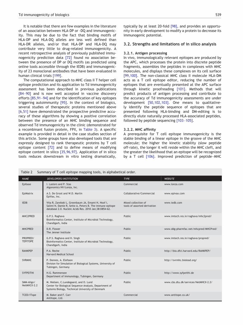
Presentation of epitopes by both MHC I and MHC IIcontributes to the initiation of an immune response. MHC IIis more relevant to anti-drug antibody responses, as MHCClass II-restricted T helper cells are responsible for drivinghumoral immunity. HLA DR, DQ, and DP are the three lociof peptide-carrying HLA class II molecules. In vitro andin vivo observations indicate that HLA DR binding peptidesare generally 12 amino acids in length, as the flankingsequences serve to stabilize the peptide (by hydrogenbonds) in the HLA binding groove [70]. Systematic assess-ments of MHC class II peptide binding domains of proteinshave been developed using machine learning methods,software algorithms, and data transformations [71–73].For a brief overview of T cell epitope mapping tools, seeTable 2 [69,74–80]. A common denominator among thesetools is the ability to quickly screen large datasets,including whole genomes or proteomes, for putative T cellepitopes. Several common HLA-DR types share HLA bindingpocket repertoires [81], meaning their ability to bindpeptides is more promiscuous than for class I alleles.Moreover, analysis focused on as few as eight representa-tives of the 875 known HLA-DR alleles can “cover” thegenetic backgrounds of most humans worldwide [82].Additionally, MHC class II-binding T cell epitopes have beenobserved to occur in clusters of up to 25 amino acids in length[72,83]. Thus identifying MHC class II-binding T cell epitopeclusters can be a strong indication for T cell responses becausethey represent regions of the protein in which sequences thathave high affinity across multiple HLA alleles and multipleframes are located [79].
538 V. Jawa et al.
It is notable that there are few examples in the literatureof an association between HLA-DP or -DQ and immunogenic-ity. This may be due to the fact that binding motifs ofHLA-DP and HLA-DQ alleles are less well defined thanHLA-DR alleles, and/or that HLA-DP and HLA-DQ maycontribute very little to drug-related immunogenicity. Arecent retrospective analysis of previously published immu-nogenicity prediction data [72] found no association be-tween the presence of DP or DQ motifs (as predicted usingonline tools accessible through the IEDB) and immunogenic-ity of 23 monoclonal antibodies that have been evaluated inhuman clinical trials [199].
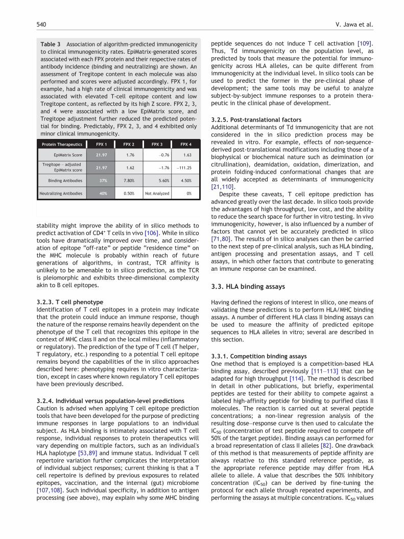
The computational approach to MHC class II T helper cellepitope prediction and its application to Td immunogenicityassessment has been described in previous publications[84–90] and is now well accepted in vaccine discoveryefforts [85,91–94] and for the identification of key epitopestriggering autoimmunity [95]. In the context of biologics,several studies of therapeutic proteins mentioned above[6,51] have demonstrated the prospective predictive accu-racy of these algorithms by showing a positive correlationbetween the presence of an MHC binding sequence andobserved Td immunogenicity in the clinic (demonstrated fora recombinant fusion protein, FPX, in Table 3). A specificexample is provided in detail in the case studies section ofthis article. Some groups have also developed tools that areexpressly designed to rank therapeutic proteins by T cellepitope content [72] and to define means of modifyingepitope content in silico [35,96,97]. Application of in silicotools reduces downstream in vitro testing dramatically,
typically by at least 20-fold [98], and provides an opportu-nity in early development to modify a protein to decrease itsimmunogenic potential.
3.2. Strengths and limitations of in silico analysis
3.2.1. Antigen processingIn vivo, immunologically relevant epitopes are produced bythe APC, which processes the protein into discrete peptidefragments, assembles the peptides in complexes with MHCmolecules, and displays these complexes on its cell surface[99,100]. The non-classical MHC class II molecule HLA-DMacts as a T cell epitope editor, reducing the number ofepitopes that are eventually presented at the APC surfacethrough kinetic proofreading [101]. Methods that willpredict products of antigen processing and contribute tothe accuracy of Td immunogenicity assessments are underdevelopment [50,102,103]. One means to qualitative-ly identify the peptide sequence of epitopes that arepresented following HLA-binding and DM-editing is todirectly elute naturally processed HLA-associated peptides,followed by peptide sequencing [103–105].
3.2.2. MHC affinityA prerequisite for T cell epitope immunogenicity is thestable binding of a linear epitope in the groove of the MHCmolecule; the higher the kinetic stability (slow peptideoff-rate), the longer it will reside within the MHC cleft, andthe greater the likelihood that an epitope will be recognizedby a T cell [106]. Improved prediction of peptide–MHC
Table 2 Summary of T cell epitope mapping tools, in alphabetical order.
NAME DEVELOPERS/INSTITUTION TYPE WEBSITE
Epibase I. Lasters and P. StasAlgonomics NV/Lonza, Inc.
Commercial www.lonza.com
EpiMatrix A.S. De Groot and W.D. MartinEpiVax, Inc.
Collaborative/Commercial www.epivax.com
IEDB Vita R, Zarebski L, Greenbaum JA, Emami H, Hoof I,Salimi N, Damle R, Sette A, Peters B. The immune epitopedatabase 2.0. Nucleic Acids Res. 2010 Jan;38:D854-62.
Mixed collection oftools of assorted derivation
www.iedb.com
MHC2PRED G.P.S. RaghavaBioinformatics Center, Institute of Microbial Technology,Chandigarh, India
Public www.imtech.res.in/raghava/mhc2pred/
MHCPRED D.R. FlowerThe Jenner Institute
Public www.ddg-pharmfac.net/mhcpred/MHCPred/
PROPRED/TEPITOPE
G.P.S. Raghava and H. SinghBioinformatics Center, Institute of Microbial Technology,Chandigarh, India
Public www.imtech.res.in/raghava/propred/
RANKPEP P.A. RecheHarvard Medical School
Public http://bio.dfci.harvard.edu/RANKPEP/
SVRMHC P. Donnes, A. ElofssonDivision for Simulation of Biological Systems, University ofTubingen, Germany
Public http://svrmhc.biolead.org/
SYFPEITHI H.G. RammenseeDepartment of Immunology, Tubingen, Germany
Public http://www.syfpeithi.de
SMM-Align/NetMHCII-2.2
M. Nielsen, C.Lundegaard, and O. LundCenter for Biological Sequence Analysis, Department ofSystems Biology, Technical University of Denmark
Public www.cbs.dtu.dk/services/NetMHCII-2.2/
TCED/iTope M. Baker and F. CarrAntitope, Ltd.
Commercial www.antitope.co.uk/
539Td immunogenicity of biologics
stability might improve the ability of in silico methods topredict activation of CD4+ T cells in vivo [106]. While in silicotools have dramatically improved over time, and consider-ation of epitope “off-rate” or peptide “residence time” onthe MHC molecule is probably within reach of futuregenerations of algorithms, in contrast, TCR affinity isunlikely to be amenable to in silico prediction, as the TCRis pleiomorphic and exhibits three-dimensional complexityakin to B cell epitopes.
3.2.3. T cell phenotypeIdentification of T cell epitopes in a protein may indicatethat the protein could induce an immune response, thoughthe nature of the response remains heavily dependent on thephenotype of the T cell that recognizes this epitope in thecontext of MHC class II and on the local milieu (inflammatoryor regulatory). The prediction of the type of T cell (T helper,T regulatory, etc.) responding to a potential T cell epitoperemains beyond the capabilities of the in silico approachesdescribed here: phenotyping requires in vitro characteriza-tion, except in cases where known regulatory T cell epitopeshave been previously described.
3.2.4. Individual versus population-level predictionsCaution is advised when applying T cell epitope predictiontools that have been developed for the purpose of predictingimmune responses in large populations to an individualsubject. As HLA binding is intimately associated with T cellresponse, individual responses to protein therapeutics willvary depending on multiple factors, such as an individual'sHLA haplotype [53,89] and immune status. Individual T cellrepertoire variation further complicates the interpretationof individual subject responses; current thinking is that a Tcell repertoire is defined by previous exposures to relatedepitopes, vaccination, and the internal (gut) microbiome[107,108]. Such individual specificity, in addition to antigenprocessing (see above), may explain why some MHC binding
peptide sequences do not induce T cell activation [109].Thus, Td immunogenicity on the population level, aspredicted by tools that measure the potential for immuno-genicity across HLA alleles, can be quite different fromimmunogenicity at the individual level. In silico tools can beused to predict the former in the pre-clinical phase ofdevelopment; the same tools may be useful to analyzesubject-by-subject immune responses to a protein thera-peutic in the clinical phase of development.
3.2.5. Post-translational factorsAdditional determinants of Td immunogenicity that are notconsidered in the in silico prediction process may berevealed in vitro. For example, effects of non-sequence-derived post-translational modifications including those of abiophysical or biochemical nature such as deimination (orcitrullination), deamidation, oxidation, dimerization, andprotein folding-induced conformational changes that areall widely accepted as determinants of immunogenicity[21,110].
Despite these caveats, T cell epitope prediction hasadvanced greatly over the last decade. In silico tools providethe advantages of high throughput, low cost, and the abilityto reduce the search space for further in vitro testing. In vivoimmunogenicity, however, is also influenced by a number offactors that cannot yet be accurately predicted in silico[71,80]. The results of in silico analyses can then be carriedto the next step of pre-clinical analysis, such as HLA binding,antigen processing and presentation assays, and T cellassays, in which other factors that contribute to generatingan immune response can be examined.
3.3. HLA binding assays
Having defined the regions of interest in silico, one means ofvalidating these predictions is to perform HLA/MHC bindingassays. A number of different HLA class II binding assays canbe used to measure the affinity of predicted epitopesequences to HLA alleles in vitro; several are described inthis section.
3.3.1. Competition binding assaysOne method that is employed is a competition-based HLAbinding assay, described previously [111–113] that can beadapted for high throughput [114]. The method is describedin detail in other publications, but briefly, experimentalpeptides are tested for their ability to compete against alabeled high-affinity peptide for binding to purified class IImolecules. The reaction is carried out at several peptideconcentrations; a non-linear regression analysis of theresulting dose–response curve is then used to calculate theIC50 (concentration of test peptide required to compete off50% of the target peptide). Binding assays can performed fora broad representation of class II alleles [82]. One drawbackof this method is that measurements of peptide affinity arealways relative to this standard reference peptide, asthe appropriate reference peptide may differ from HLAallele to allele. A value that describes the 50% inhibitoryconcentration (IC50) can be derived by fine-tuning theprotocol for each allele through repeated experiments, andperforming the assays at multiple concentrations. IC50 values
Table 3 Association of algorithm-predicted immunogenicityto clinical immunogenicity rates. EpiMatrix-generated scoresassociated with each FPX protein and their respective rates ofantibody incidence (binding and neutralizing) are shown. Anassessment of Tregitope content in each molecule was alsoperformed and scores were adjusted accordingly. FPX 1, forexample, had a high rate of clinical immunogenicity and wasassociated with elevated T-cell epitope content and lowTregitope content, as reflected by its high Z score. FPX 2, 3,and 4 were associated with a low EpiMatrix score, andTregitope adjustment further reduced the predicted poten-tial for binding. Predictably, FPX 2, 3, and 4 exhibited onlyminor clinical immunogenicity.
Protein Therapeutics FPX 1 FPX 2 FPX 3 FPX 4
EpiMatrix Score 21.97 1.76 —0.76 1.63
Tregitope — adjusted EpiMatrix score 21.97 1.62 —1.76 —111.25
Binding Antibodies 37% 7.80% 5.60% 4.50%
Neutralizing Antibodies 40% 0.50% Not Analyzed 0%
540 V. Jawa et al.
can be converted to method-independent Ki values via theCheng–Prusoff equation for comparison of peptide affinitiesacross platforms [115].
3.3.2. Direct binding assaysIt is also possible to measure peptide affinity directly, ratherthan observing its ability to displace a known ligand. In suchexperiments, a variety of methods may be used to separatepeptide bound to HLA molecules from free peptide insolution, thus allowing for assessment of the peptide'saffinity. Spin-column filtration and gel electrophoresis areways to accomplish this distinction by molecular weight,since peptide–HLA complexes will weigh more than unboundpeptides. These methods, however, are tedious and cansuffer from complex dissociation during the separationprocess. Other novel methods have been proposed, such asproximity-based detection utilizing a Luminescent OxygenChanneling Immunoassay [116]. All of these techniques allowmore direct binding affinity determination than competitionstudies, but have methodological limitations and requirecostly equipment.
3.3.3. Real-time kinetic measurementsApart from determining the quantity of displaced referencepeptide (competition assay) or bound vs. free test peptide(direct assay), the rate at which peptides interact withHLA molecules is a dimension of epitope strength that canbe measured in vitro. Protocols based on fluorescencepolarization are suitable for kinetic studies due to theabsence of interference from ELISA wash steps, allowingmultiple readings to be taken throughout the bindingreaction [117]. Additionally, surface plasmon resonancemethods have also been employed to measure peptidebinding to MHC II [118].
3.4. Strengths and limitations of HLA binding assays
In vitro HLA binding assays are relatively straightforward andeasier to perform than live-cell experiments. However, eachof the assay formats can be affected by peptide purity andlength as described here. Peptides that are synthesized forHLA binding assays must include carboxy- and amino-terminal flanks that stabilize the peptide in the MHC groove[119]. Selection of peptides based on artificially definedoverlapping sequences (e.g., 15mers overlapping by tenamino acids) can lead to the truncation of MHC bindingmotifs and elimination of critical flanking regions, whichlimits the accuracy of the binding assay. Furthermore,artificially synthesized peptides may contain impurities,truncations, and errors in the sequence that can lower oralter the binding of the pure peptides; thus high-qualitypeptides (greater than 90% purity) are required to obtainmore accurate results in binding assays. Long peptides canfold or peptides can aggregate in solution, leading tounderperformance in binding assays. Solvents used in theexperiment may interact negatively with certain aminoacids, causing oxidation or the formation of unwanteddisulfide bonds. Quality-controlled peptide manufacturingand storage, along with proper reagent selection, canminimize the impact of these problems [113,114].
While epitopes can be predicted (in silico) and validatedin binding assays (in vitro), the final impact of the T cellepitope is through its activation of a T cell. Activation isrelated to TCR specificity, avidity and T cell phenotype(none of which are measured in HLA binding assays), whichmakes straightforward interpretation of immunogenicitybased upon HLA binding assays impossible. Thus, HLA bindingassays may improve the accuracy of immunogenicity predic-tions when applied in a step-wise process after in silicoepitope prediction and before the employment of abiological assay such as enzyme-linked immunosorbentspot-forming (ELISpot) assays or HLA transgenic mousestudies, but additional assays that assess T cell responsesmay need to be performed [120].
3.5. In vitro T cell assay methods for Tdimmunogenicity analysis
For many years, in vitro assays based on HLA haplotype havebeen used in transplantation research to assess the risk ofengrafted T cells responding to host tissue (graft-versus-hostdisease). Adaptation of these assays to protein therapeuticsmay improve the pre-clinical assessment of the potential forTd immunogenicity. The presence of T cells that activelysuppress immune responses to autologous proteins is asignificant confounding factor in their development andevaluation. In addition, these assays are dependent on theselection of a culture milieu that accurately accounts for thein vivo conditions of human immune stimulation [40,54,121].In this respect, bulk cultures of PBMC, either with or withoutco-stimulatory signals (anti-CD28 antibody, IL-2, IL-7, etc.),have been utilized to assess immunogenicity of therapeuticproteins.
A number of biological outcomes for T cell activation canbe measured in these in vitro assays, including cytokinesecretion (IFN-γ, IL-2, IL-4, etc.), regulation of cell surfacemarkers of activation, signal transduction events, andproliferation [122,123]. Supported by such evidence, insilico-identified peptides that stimulate multifunctional Tcell responses in vitro can be considered bona fide T cellepitopes with the potential to contribute to an ADAresponse.
3.5.1. Measurement of T cell cytokine responsesELISAs and ELISpots are two related methods of measuringcytokines secreted by T cells (i.e., IFN-γ, IL-2, IL-4, andIL-10). The enzyme-linked immunosorbent assay (ELISA) canmeasure cytokine levels in culture supernatants generatedunder conditions of T cell stimulation. Cytokine levelsmeasured in an ELISA can provide information about themagnitude of the response (how much cytokine is secretedinto the supernatant) as well as the type and quality of theresponse (which cytokines are or are not secreted into thesupernatant). Multi-analyte, high throughput cytokine test-ing can also be performed in cell-derived supernatants inbead-based assays [124–127]. Enzyme-linked immunosor-bent spot-forming (ELISpot) assays provide informationregarding the number of cytokine-producing cells (down to1 cell per million) within a cell population stimulated exvivo; these assays are considered to be more sensitive and
541Td immunogenicity of biologics
quantitative whereas ELISA and bead-based assays areslightly less sensitive and more qualitative.
Intracellular cytokine staining measured by flow cytom-etry is another method for detecting cytokines and linkingtheir expression to the phenotype of individual cells.These assays can be used to accurately measure T cellpolyfunctionality relative to the phenotypic classificationsof CD4+ T cells based on cell surface markers.
3.5.2. T cell proliferationT cell proliferation in response to stimulation by a peptide–MHC complex can be measured by (1) the incorporationof the radioactive nucleotide tritiated thymidine into theDNA of dividing but not resting cells or (2) the dilution ofa fluorescent dye, carboxyfluorescein succinimidyl ester(CFSE), that decreases in fluorescence intensity by halfwith each round of cell division and can be measured byflow cytometry. Thymidine incorporation assays are graduallybeing replaced by CFSE staining, which presents significantadvantages in terms of ease-of-use. In addition to CFSE labeling,cells can be co-stained for expression of other cell surfacemarkers, transcription factors, and/or intracellular cytokinesthat distinguish between regulatory T cells and effectorT-helper cell phenotypes, including Th1, Th2, Th17, Th22,etc. as reviewed in detail elsewhere [128–130].
3.5.3. TetramersFluorescently labeled tetrameric complexes of MHC class IImolecules loaded with the peptide of interest (i.e., “tetra-mers”) can also be used to enumerate T cells recognizing aparticular epitope. However, relative toMHC I tetramers, MHCII tetramer staining has proven problematic and so their utilityhas been restricted. The technical limitations experiencedspecifically with MHC II tetramers may be due to weaker TCR–peptide–MHC II interactions or the fact that CD4 does notparticipate in the stability of tetramer binding as does CD8[131]; either one or both of these features may contribute toincreased variability and poor quality of the experimentaloutput. Efforts continue to improve MHC II tetramer–TCRinteractions towards the goal of increasing the utility of MHC IItetramers so that they may be more widely used for theidentification of specific CD4 T cell responses, including thoseagainst protein therapeutics.
3.5.4. Naïve T cell in vitro assaysNaïve peripheral blood mononuclear cells (PBMC) have beenused to describe Td immunogenicity of therapeutic proteins[34,40,132,133]. Compared to a recall response, the precursorfrequency of antigen-specific cells in a naïve population isquite low; it was postulated that the higher the precursorfrequency reactive with a certain peptide or protein, thehigher the potential of the respective peptide or protein toinduce an immune response [134]. Multiple rounds of antigenstimulation, sometimes over several weeks, are required toexpand sufficient numbers of antigen-specific T cells forreliablemeasurement. By conditioning naïve blood samples exvivo through prolonged and/or repeated exposure to experi-mental antigen, immune responses can be expanded to thepoint where they can be measured. What is not known is howexpansion affects the phenotype of the T cell response; eitherregulatory or effector responders may eventually dominate inin vitro cultures.
3.5.5. T cell assays using whole antigensT cell assays usingwhole antigens also can be used tomeasure Tcell responses to protein therapeutics in vitro. The recognitionof these antigens requires the presence of an APC that iscapable of processing and presenting peptides derived from theantigen. Advantages of whole PBMC assays include the abilityto set up several assays and/or assay conditions with a limitedblood sample volume and the ease of assay performance,features which lend themselves to high-throughput assaydevelopment [40]. Human monocyte-derived dendritic cellscan be manipulated in vitro to model antigen processing byprofessional APC in vivo (H. Kropshofer, unpublished data).However, as applied to evaluating immunogenicity of biologics,optimization of variables such as the ratio of DCs to autologousT cells are important factors for ensuring that the in vitroresults are relevant to the clinical scenario.
3.5.6. T cell re-stimulation assays using “exposed” donorsT cells re-stimulation assays are generally used to identifyand measure a recall or memory response in PBMC derivedfrom subjects who have been exposed at a distant time pointto a protein or a given biologic product. Whereas ‘exposedblood’ assays cannot be performed for novel therapeutics,this type of assay can be used to evaluate the impact ofpre-existing immune responses to a new version of anexisting biologic in use such as a re-engineered FVIII. Epitopemapping of the recall response can be performed usingspecific peptides from the whole protein; however, thisapproach may be over-predictive. Therefore, studies usingwhole therapeutic protein should ideally be performed inparallel with studies using sets of predicted peptides.Antigen-specific T cell responses can be assessed afterre-stimulation ex vivo by ELISA, ELISpot, proliferation, andflow cytometric analysis of activation markers and intracel-lular cytokine expression.
3.5.7. Reconstitution of T cell immune responses in vitroNew methods for “reconstituting T cell immune responses”in artificial media may permit improved in vitro assess-ments of the interactions between the professional APC andthe T cells. To this end, several artificial lymph node (ALN)systems have been developed [135–137]. These methodsattempt to replicate, in three dimensional structures and inAPC:PBMC ratios, the natural immune environment [138]. Inat least one approach, human blood-derived dendritic cellsare cultured in transwells partitioned by human vascularendothelial cells. Addition of autologous CD4+ T cells to theco-culture allows activated APC expressing chemokinereceptors to respond to inflammatory chemokines andmigrate through the transwells, mimicking the migrationof APC from the periphery to the lymph nodes. Theantigen-specific CD4+ T cell response is monitored byCD154, IFN-γ, IL-2, IL-5, IL-17, and IL-21 expression. Agood correlation between previously established immuneresponses in vivo and ALN immunogenicity has beenobserved, at least for protein-based vaccines [139]. Eventhough the human ALN model is primarily being used for invitro evaluation of vaccine efficacy [140], the application ofthis technology for the prediction of therapeutic proteinimmunogenicity is feasible.
Alternatively, some groups have used small flow-throughsystems that induce PBMC to self-assemble into lymph
542 V. Jawa et al.
node-like structures [141]. Pre-activation of the T cellsimproves the readouts; however, the introduction ofspecialized (non-self) APC may generate false positives forsome biologic proteins. In other cases, these ALN systemshave given results that correlate with predicted immuneresponses [135,142].
3.6. Strengths and limitations of in vitro T cellassays for Td immunogenicity analysis
The benefit of in vitro T cell assays utilizing easily accessiblehuman peripheral blood is that the assays may provide apreview of the immunogenicity of a therapeutic proteinwithout the risks typically associated with first-in-humanuse. Careful consideration of the composition of the cells inculture, such as monocyte-derived macrophages, immature/mature dendritic cells, and T and B cells, may improve theability of a protein to initiate and propagate an immuneresponse. Furthermore, these assays incorporate antigenprocessing and presentation pathways for whole proteins aswell as for discrete peptides.
A limitation of ELISpot and ELISA assays is that PBMC containseveral cell types capable of secreting particular cytokines,e.g., IFN-γ (NK cells, NKT cells, CD4+ or CD8+ T cells). ThusPBMC-based assays tomeasure cytokine production by a specificlymphocyte subsetmay need to be fine-tuned such as by the useof subset-depleted or subset-enriched PBMC preparations.Another potential issue related to T cell assays is that theconcentration of whole protein or peptide in the cell culturemay need to be titrated relative to the number of T cells. Theoptimal concentration of protein required for proper evaluationof immune responses in vitro may be non-physiologic due to alimited antigen-presenting population and/or co-stimulation. Tcell assays also require support with homeostatic cytokines(IL-2, IL-7, IL-15, etc.) to reduce bystander effects [40]. Inaddition, optimization of T cell concentrations may berequired [143].
While “irrelevant” cells secreting cytokines in vitro maycontribute to overestimation of the frequency of activatedT cells, their presence also contributes to the overall levelof activation of cells in the assay. Cells such as NK cells,CD8+ T cells, and basophils may play a supportive role inthese cultures [144], thus their removal can modify theoutcome of a truly representative immune response.Finding the right balance between minimization of irrele-vant immune responses and support for the in vitro immuneresponse is one of the major obstacles to widespreadadaptation of in vitro T cell assays for pre-clinical screeningof protein therapeutics.
Viable and functional T cells are also required for theassay; standardized and optimized procedures for handlingand storing whole blood are needed to ensure the accuracyof subsequent assays. Furthermore, blood from naïve anddrug-exposed individuals can differ in the content ofantigen-specific T effector and T regulatory cells. Based onthe nature of response (in vivo primed vs. stimulated andrecalled), stimulation methods and the amount of antigenrequired for challenge can also differ. Naïve cells willrequire multiple in vitro stimulations to amplify detection,while antigen-specific recall responses can be elicited evenwith a single challenge.
Similarly, stimulation can be performed with peptides orwhole proteins alone, peptides in the context of tetramers,or APC pulsed with whole proteins. New advances intetramer/multimer technology can enhance detection ofepitope-specific T cells and thus should allow more sensitiveand standardized approaches to evaluate individual re-sponses to T cell epitopes identified in protein therapeutics[145].
Finally, the number of individual blood (PBMC) donorsthat would normally be required to address the HLA diversityof a patient population is quite large (more than forty), andthe volume of blood required ranges from 15 mL to morethan 50 mL. Maintenance of a large supply of blood samplesfrom pre-qualified donors that is sufficient to reduceassay-to-assay variation can be done, but is cost prohibitivefor most preclinical laboratories.
Much remains to be done to improve the accuracy of in vitroT cell assays in predicting clinical immunogenicity. Futureconsiderations for improving in vitro T cell assays include:better linkage between the spectrum of immune responses to atherapeutic protein and predictive power in clinical trialsusing statistically derived criteria, such as fold-increase orstimulation-index; improved means to distinguish respondersfrom non-responders; evaluation of T cell responses fromdiseased states associated with inflammation or immunesuppression; selection of the optimal set of markers for theidentification of activated T cells; and improved ability todifferentiate Treg and CD4+ Teff responses (one schema hasbeen offered by the HIPC consortium [146]). Additionalimprovements require establishing clear parameters that definememory versus naïve T cell populations, influence due tobystander cells, and standard methods for PBMC harvest,preparation, and storage. Concurrent with efforts to standard-ize, in vitro immunogenicity screening assays are beingincorporated into the preclinical pipeline by a number of drugdevelopers.
3.7. Mouse models of in vivo Td immunogenicity ofhuman therapeutic proteins
Important advances in understanding MHC restriction, map-ping of epitope recognition for murine epitopes presented bymurine MHC, and T cell function have been achieved with invivo mouse studies. However, when we turn to in vivo mousestudies for prediction and validation of Td immunogenicity forclinically relevant proteins, there are two major limitationsthat must be taken into consideration. The first is that humanand murine proteins are not identical, thus administration ofprotein therapeutics to mice may result in responses tocomponents of the protein that are foreign to mice; andsecond, murine MHC will present mouse, not human, T cellepitopes. Since murine models provide a means of evaluatingimmunogenicity and an important bridge to the clinic, anumber of enhanced models have been developed.
3.7.1. HLA transgenic miceThe HLA transgenic lines are generated by incorporation ofspecific human HLA genes into murine MHC class II-deficientmice, producing a mouse strain that expresses human class IIHLA in the absence of mouse class II MHC [147,148]. Thus,these mice process and present epitopes in the context of
543Td immunogenicity of biologics
human HLA, and their T cells recognize epitopes presented byhuman HLA. They aremost useful when directly comparing twoproteins that are very similar (such as FVIII and versions of FVIIIthat have fewer epitopes or new glycosylations) [97]. A directcorrelation has been found between epitopes that elicit T cellresponses in infected humans and those that induce T cellresponses in immunized HLA transgenic mice [149–152].HLA transgenic mice are now routinely used to test andoptimize (human) epitope-driven vaccines in preclinicalstudies [153–155]. For example, Hanke et al. mapped HIVepitopes in transgenicmice, and thenmoved their DNA vaccinethrough abbreviated primate studies after proving that the onenon-human primate epitope engineered into the vaccinestimulated T cells [156].
The formation of anti-FVIII antibodies, also known asinhibitors, is a major obstacle to FVIII gene replacementtherapy in hemophilia A patients. After intravenous adminis-tration of FVIII, the immune response mounted is dependenton CD4+ T helper cells, as has been demonstrated by numerousstudies in mice and humans [157–162]. More currently,interference with T–B cell interactions in hemophilic micewas shown to reduce inhibitor formation [159,160,162].
An example of the use of HLA transgenic mice for evaluatingthe application of immunogenicity prediction tools towardsthe goal of deimmunizing a therapeutic protein was recentlypublished in Clinical Immunology [97]. In silico tools wereutilized to predict immunogenic peptides within the C2 domainof FVIII. Changes to amino acids in positions predicted to beimportant for binding to the HLA DR3 MHC class II pocket weremodified with the intent of disrupting peptide–MHC binding.The same predictive tools were reapplied to assess the bindingpotential of the modified peptides. This process was reiterateduntil the predicted binding to HLA DR3 was reduced.The de-novo immunogenicity of these modified peptides wastested in hemophilic E16 mice (H-2b; [163]) and in HLA-DR3transgenic mice [164]. The initial immunogenicity studyresults, in which mice were immunized with the unmodifiedoriginal (ORG) epitopes predicted by in silico analysis of the C2domain, were consistent with predicted responses for eitherH-2b- or HLA-DR3-expressing mice. These two mouse strainswere crossed to produce E16xDR3 mice, in which an immuneresponse of intermediate magnitude was observed. Specifi-cally, the immunogenicity of epitopes derived from FVIII in theE16xDR3 mice was consistent with the absence of toleranceinduction to this sequence (the E16 mice do not expressfull-length FVIII) and the presence of MHC (I-Ab) bindingmotifsin the sequence [97]. In proliferation assays, modified epitopepeptides were less antigenic than ORG peptides. In general,lower antigenicity was observed for those peptides that hadtwo rather than just one amino acid substitution. Similarly, interms of de novo immunogenicity, the more mutations, thelower the observed proliferative response in general.
In addition to providing an example of how in silicotools can be applied early in the development process tomitigate immunogenicity risk, this study also highlightscertain limitations of available mouse models for riskassessment. While certain epitopes are predicted to bindpromiscuously to both human HLA DR3 and I-Ab MHC, othersdemonstrate greater restriction by either the human or themouse MHC. Thus the lack of observed immunogenicity maybe attributed to the absence of a relevant MHC expressed inthe mouse model.
With regard to theHLA DR transgenicmodels, those peptidespredicted to be HLA ligands were only immunogenic when thesequence contained mismatches between the human FVIIIsequence used in the immunization and the correspondingsequence in the native mouse FVIII protein [97]. To beimmunogenic, the mismatches had to occur within 9-mersequences that also contained HLA DR3 or DR4 binding motifs.When presented to the mouse T cell, these 9-mers wouldappear “foreign” and thus stimulate T cell proliferation.Peptides that were predicted to bind HLA DR3 and/or DR4 butdid not stimulate immune responses were found to contain nomouse/human sequence mismatches.
In summary, identification of T cell epitopes anddevelopment of de-immunized versions by targeted se-quence modification can lower HLA binding and proliferationresponses, but the process has the potential to impactprotein function
3.7.2. Humanized mouse models“Humanized” mice engrafted with a functional humanimmune system are now being used to study humanhematopoiesis, immunity, regeneration, stem cell function,cancer, and human-specific infectious agents. Immunocom-promised SCID/NOD/γ chain−/− or RAG2−/−/γ chain−/−mice, utilized as recipients to facilitate acceptance ofhuman tissue, are engrafted with functional human hema-topoietic stem cells (CD34+), liver, and thymus [165]. Theresult is a cohort of mice in which human myeloid andlymphoid lineages are reconstituted from a single humandonor, and the interactions of these cells in a complexbiological environment can be studied. XenoMouse® (de-scribed below), in addition to the humanized mice suchas NOD/Shi-scid/IL-2Rγnull (NOG), NOD scid IL2 receptorgamma chain knockout mice (NSG), bone marrow, liver,thymus transplanted mouse (BLT), and bone marrowtransplanted mouse (BMT) have all been used as animalmodels to evaluate human immune responses [165–168].
3.8. Strengths and limitations of mouse models of invivo Td immunogenicity
These evolving mouse models could provide functional andtestable elements of the innate and adaptive human immunesystem without putting patients at risk [169]. The speciesspecificity of a number of cells and molecules critical for afully functional immune system remains a limitation in thesemodels. For example, in HLA transgenic mice, the T cellrepertoire will be shaped by epitopes derived from mouseproteins presented by a single human HLA allele to mouseTCR. This confounds the application of HLA transgenic micefor determination of immunogenicity of a human proteintherapeutic. And while humanized (SCID/Hu) mouse modelsare improving, certain aspects of a fully functional immuneresponse relevant to immunogenicity prediction and mitiga-tion are lacking, such as the ability to elicit the completespectrum of B cell antibody responses or the ability toproteolytically process antigens in a way that recapitulateswhat has been observed for human endosomal/lysosomalproteases.
The XenoMouse®model has been of particular interest, as itis transgenic for nearly the complete human immunoglobulin
544 V. Jawa et al.
locus, thus is tolerant for human IgG2/kλ antibodies, but isdeficient for mouse IgH and Igk chains. The human-like humoralimmune response in XenoMouse® is restricted by mouse MHCand T cell help but is not as robust as in wild type mice,potentially due to inefficient signal transduction and isotypeswitching mediated by accessory factors that are necessary forB cell maturation. Hence the utility of such a model to studyimmune responses to human proteins remains somewhatlimited.
Clearly, more work is required to develop these mousemodels to accurately reflect human immune responses toprotein therapeutics before they can become accurate, useful,and routine components of a Td immunogenicity screeningprogram.
4. Applied Td immunogenicity prediction
4.1. In silico prediction supported by subsequentclinical data
Koren et al. [54] demonstrated a correlation between the insilico evaluation of T helper epitope content of a proteintherapeutic and its observed immunogenicity when adminis-tered to human subjects in a clinical trial. The therapeuticprotein of interest was a recombinant Fc fusion protein (FPX)consisting of human germ line Fc γ fragment with two identical,biologically active, 24-amino-acid peptides attached to theamino terminal end of the Fc fragment. In the in silico analysis,the carboxy terminal region of the peptide scored high forbinding to five of eight common HLAmolecules, suggesting thatthis peptide had the potential to be presented by five differentHLA molecules to T cells. Moreover, the C-terminus peptideswere associated with a cluster of overlapping 9-mers that couldbind across several HLA DR alleles.
The antibody response to FPX was consistent with the highimmunogenic potential predicted in silico. A single subcutane-ous or intravenous administration of the protein resulted inhigh-affinity binding antibodies in 40% and 33% of totalindividuals, respectively (Table 3). T cell-mediated recallresponses to the therapeutic protein were also assessedin vitro for donors exhibiting a strong humoral responsein vivo. In vitro PBMC activation by the FPX peptide, and theamino-terminal and carboxy-terminal fragments thereof, wasmeasured as a function of the number of IFN-γ and IL-4spot-forming cells (SFC) in a standard ELISpot assay. Theantibody data suggested a strong T cell-driven response, whichwas corroborated by the in vitro cytokine responses observed inPBMC culture. Thus, the in silico prediction of immunogenicT helper cell epitope(s) within the carboxy-terminal region ofthe FPX peptide correlated with the in vitro T cell assays andthe in vivo antibody responses.
HLA typing confirmed the predicted binding promiscuityof the carboxy-terminal epitope(s), as antibody-positivesubjects possessed all of the eight most common HLA alleles.The magnitude of the immune response also appeared tocorrelate with the HLA haplotype and with the bestcarboxy-terminal peptide binding scores. One subject whopossessed the DRB1*0701 allele had the highest antibodyconcentration as well as the highest number of IFN-γ andIL-4 SFCs, as was predicted based on the in silico EpiMatrixresults. Another subject who had DRB1*0701 allele also
showed relatively high antibody concentration with lower,but measurable, SFCs in vitro. Immune responses (both invivo antibody and in vitro T cell) to different regions ofthe protein correlated with in silico predictions. Thecarboxy-terminal region of the FPX peptide showed thehighest MHC binding score in the context of the DRB1*0701allele; T cell and antibody responses to this fragment wereobserved in vitro and in vivo, respectively, for individualspossessing that allele. In contrast, the DRB1*0301 allele hadvery low MHC binding scores, and patients who possessedDRB1*0301 but not any of the other higher binding allelesdemonstrated low responses in ELISpot and no evidence ofan antibody response to the protein therapeutic. Theimmunogenicity of the FPX fragments and the associationbetween clinical results and the HLA class II alleles weresupported in the naïve blood T cell assays in furtherstudies performed by Jawa et al. [40,54] the reactivity ofnaïve (pre-exposure) PBMC to FPX1 was associated withtherapeutic-induced antibody responses observed in theclinic as well as with expression of specific HLA class IIalleles that were predicted in silico to present FPX1-derivedepitopes.
This case study illustrates several important princi-ples regarding the immunogenicity assessment of proteintherapeutics:
(i) Clinical correlation: Clinical incidence of high immuno-genicity to FPX1 from exposed donors was retrospec-tively associated with the in silico immunogenicitypredictions.
(ii) Promiscuous epitopes: This study demonstrated thatimmunodominance was associated with clusters ofepitopes within the sequence of the FPX1 peptide.The clustering of epitopes was associated with greaterimmunogenicity as measured by a high incidence ofbinding and neutralizing antibodies to FPX fusionprotein. Moreover, due to the clustering, the peptidewas more promiscuous and was able to bind acrossseveral HLA DR alleles. This was validated when ADApositive subjects were observed to express high-bindingalleles.
(iii) Antibody–HLA correlation: The responders with highantibody titers expressed HLA-DR alleles that had beenpredicted by the in silico algorithm to be the bestepitope binders.
(iv) In silico–T cell assay correlation: The C-terminal regionof the FPX1 peptide elicited T cell responses in PBMC fromFPX1-exposed, antibody-positive donors, supporting acorrelation between in silico prediction and observedclinical immunogenicity.
(v) Correlation to in vitro naïve response: The predictedimmunodominant regions of the peptide were able toelicit response from naïve PBMC with the HLA DRpredicted in silico.
4.2. Clinical link between MHC class II haplotype andIFN-β immunogenicity [53]
A similar association between the HLA DRB1*0701 allele anda strong antibody response to recombinant beta-interferonwas observed by Barbosa et al. The IFN-β epitopes were
545Td immunogenicity of biologics
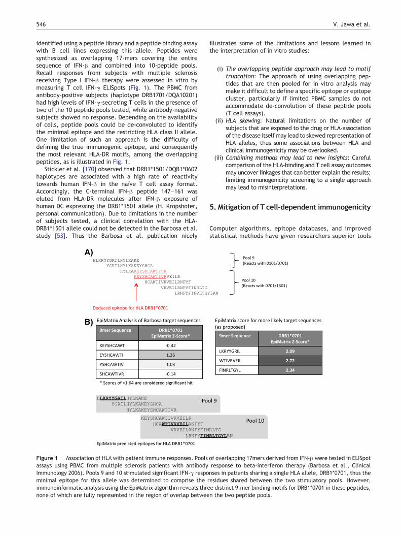
identified using a peptide library and a peptide binding assaywith B cell lines expressing this allele. Peptides weresynthesized as overlapping 17-mers covering the entiresequence of IFN-β and combined into 10-peptide pools.Recall responses from subjects with multiple sclerosisreceiving Type I IFN-β therapy were assessed in vitro bymeasuring T cell IFN-γ ELISpots (Fig. 1). The PBMC fromantibody-positive subjects (haplotype DRB1701/DQA10201)had high levels of IFN-γ-secreting T cells in the presence oftwo of the 10 peptide pools tested, while antibody-negativesubjects showed no response. Depending on the availabilityof cells, peptide pools could be de-convoluted to identifythe minimal epitope and the restricting HLA class II allele.One limitation of such an approach is the difficulty ofdefining the true immunogenic epitope, and consequentlythe most relevant HLA-DR motifs, among the overlappingpeptides, as is illustrated in Fig. 1.
Stickler et al. [170] observed that DRB1*1501/DQB1*0602haplotypes are associated with a high rate of reactivitytowards human IFN-β in the naïve T cell assay format.Accordingly, the C-terminal IFN-β peptide 147–161 waseluted from HLA-DR molecules after IFN-β exposure ofhuman DC expressing the DRB1*1501 allele (H. Kropshofer,personal communication). Due to limitations in the numberof subjects tested, a clinical correlation with the HLA-DRB1*1501 allele could not be detected in the Barbosa et al.study [53]. Thus the Barbosa et al. publication nicely
illustrates some of the limitations and lessons learned inthe interpretation of in vitro studies:
(i) The overlapping peptide approach may lead to motiftruncation: The approach of using overlapping pep-tides that are then pooled for in vitro analysis maymake it difficult to define a specific epitope or epitopecluster, particularly if limited PBMC samples do notaccommodate de-convolution of these peptide pools(T cell assays).
(ii) HLA skewing: Natural limitations on the number ofsubjects that are exposed to the drug or HLA-associationof the disease itselfmay lead to skewed representation ofHLA alleles, thus some associations between HLA andclinical immunogenicity may be overlooked.
(iii) Combining methods may lead to new insights: Carefulcomparison of the HLA-binding and T cell assay outcomesmay uncover linkages that can better explain the results;limiting immunogenicity screening to a single approachmay lead to misinterpretations.
5. Mitigation of T cell-dependent immunogenicity
Computer algorithms, epitope databases, and improvedstatistical methods have given researchers superior tools
A)
B)
Figure 1 Association of HLA with patient immune responses. Pools of overlapping 17mers derived from IFN-β were tested in ELISpotassays using PBMC from multiple sclerosis patients with antibody response to beta-interferon therapy (Barbosa et al., ClinicalImmunology 2006). Pools 9 and 10 stimulated significant IFN-γ responses in patients sharing a single HLA allele, DRB1*0701, thus theminimal epitope for this allele was determined to comprise the residues shared between the two stimulatory pools. However,immunoinformatic analysis using the EpiMatrix algorithm reveals three distinct 9-mer binding motifs for DRB1*0701 in these peptides,none of which are fully represented in the region of overlap between the two peptide pools.
546 V. Jawa et al.
for assessing the potential of T cell immunogenicity oftherapeutic proteins [42]. As a result, a number of differentapproaches to mitigate Td immunogenicity of therapeuticproteins are now under consideration. These approachesinclude: direct modification of the therapeutic protein bypegylation and/or glycosylation to mask “immunogenic epi-topes”, thereby reducing recognition by the immune system;modification of HLA class II anchor residues of immunodominantepitopes to disrupt presentation; and application of strategiesthat tolerize the immune system to the therapeutic protein.The primary focus of this review is on methods for predictingand measuring Td immunogenicity; hence, a thorough discus-sion of methods for mitigating immunogenicity is beyond itsscope. However, it is important here to link our evolving abilityto identify contributors to a therapeutic protein's immunoge-nicity with our ability to modify that feature to mitigateunwanted immune responses. Currently, tools to predict T cellepitopes can be applied to remove T cell epitopes. Indeed, amethod for deimmunization (protein/sequence re-engineering)has been introduced, which involves the elimination ofpredicted T cell epitopes or a reduction in the total numberof T cell epitopes. The approach has been described in detailin a number of publications [41,171–175], and readers arereferred to those articles for details on the methodology.Efforts to render effector T cells non-responsive through theactions of immunosuppressive drugs or induction of Treg cellsare evolving. Finally, the ability to identify drug-induced ordrug-responsive T cells may in the future bring opportunitiesto specifically deplete them.
5.1. Deimmunization
The first published attempt to deimmunize a proteininvolved the introduction of alanine substitutions at theMHC anchoring residues Y73, K74, R77, E80, and D82 ofstaphylokinase, alone or in combination. These modifica-tions to the native protein were subsequently shown toreduce or eliminate a T cell response and clinical immuno-genicity [176]; this was followed by a number of additionaldeimmunization studies [29,41,171–175,177,178]. Ongoingefforts include deimmunization of (1) botulinum neurotoxintype A, (2) lysostaphin, and (3) Factor VIII [97], as well as(4) removing T regulatory epitopes from amonoclonal antibody(anti-DEC205) for vaccine applications. Epitope modificationhas also been applied to several other therapeutic proteinsin studies performed by researchers at Biovation [179],Epimmune [52], Genencor [180], and elsewhere, using arange of methodologies.
Clearly, modification of the sequence of a protein can havean impact on the secondary or tertiary structure. Thus theutility of this approach depends on the location and extent ofamino acid changes and the impact of those changes on thepharmacological activity of the modified protein. On a case-by-case basis, an immunoinformatics-driven and cell-based ap-proach to modifying protein sequences may eventually lead toless immunogenic proteins while preserving their therapeuticpotential. Several de-immunized therapeutic proteins are inclinical use [176]; however, in the majority of instances, theimmunogenic potential of the parent molecule was determinedby the presence of T cell epitopes and not through clinicaltesting. This is the key limitation of all predictive methods, and
of course, clinical testing is the only way to “validate” any ofthemethods discussed in this review. Retrospective studiesmaybe the only approach to testing the concept, but without ahead-to-head comparison of the original and deimmunizedproteins (FVIII, for example) in a clinical setting, all else will becorrelative.
5.2. Tolerization
Active interference with T cell responses to proteintherapeutics by inducing tolerance to the drug is anapproach that has garnered significant interest in the lastfew years. Tolerance can be induced with non-depletinganti-CD4 antibodies [181,182]. IVIG has also been used toinduce tolerance in solid organ transplant [183], to reduceFVIII inhibitors [184–189], and to inhibit ADA in Pompepatients undergoing Myozyme treatment [190]. Tolerance-inducing protocols combining Rituxan, methotrexate andIVIG that have been effectively applied to mitigate existinganti-therapeutic responses in the context of life-savingenzyme replacement therapy may provide an immediatesolution for these patients [68,190,191]. However, there arenew opportunities opening up along the developmentalpipeline to build next-generation protein therapeutics towhich patients are more tolerant. Towards this goal, DeGroot et al. have identified a set of natural, humanregulatory T cell epitopes (“Tregitopes”) present in the Fcand Fab domains of IgG that have also been shown to inducetolerance to co-administered proteins; these Tregitopes maybe responsible for tolerance to idiotypic epitopes. Whenincubated with PBMC in vitro, Tregitopes specificallyactivate CD4+ T cells, increase CD25/Foxp3 expression, andincrease expression of regulatory cytokines and chemokines[41]. Administration of Tregitopes with protein antigens invivo inhibits T cell proliferation and effector cytokineexpression, and induces antigen-specific adaptive toleranceinduction. Methods for co-administering Tregitopes withprotein therapeutics are currently under development[192–195].
6. Considerations: assessing immunogenicity oftherapeutic proteins
Given the potential impact of immunogenicity on the safety,efficacy, and utility of a final therapeutic protein drugproduct, there is increasing interest in developing ap-proaches that will help to establish an accurate immunoge-nicity profile for a protein therapeutic. Immunogenicitypredictions provide the greatest opportunity, and have thegreatest benefit, early during selection of a lead molecule.Introduction of changes to the structure of a lead moleculecould be prohibitively expensive with respect to time andresources necessary to repeat prior development and safetystudies. We suggest consideration of a multi-step approachsuch as the following (Fig. 2) to evaluate the Td immunoge-nicity potential of a therapeutic protein:
Step 1 HLA binding prediction in silico and confirmatoryassessment in vitro. A high-throughput in silicoepitope-mapping algorithm applicable across com-mon HLA class II haplotypes could identify areas of
547Td immunogenicity of biologics
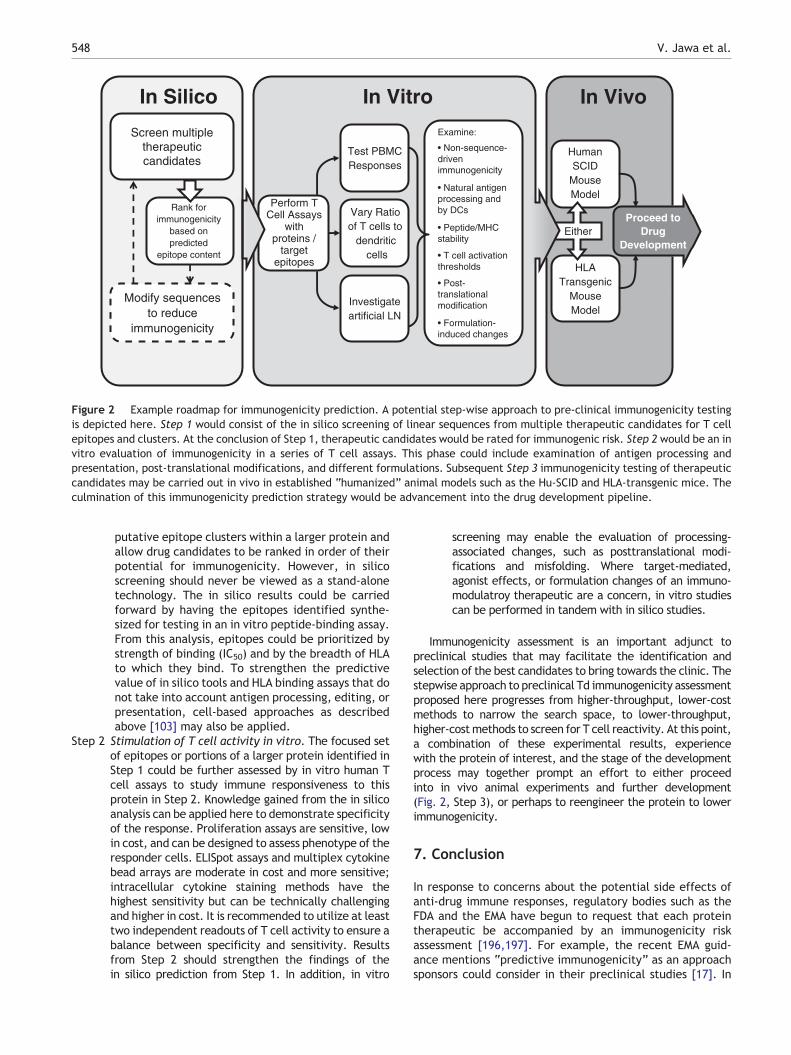
putative epitope clusters within a larger protein andallow drug candidates to be ranked in order of theirpotential for immunogenicity. However, in silicoscreening should never be viewed as a stand-alonetechnology. The in silico results could be carriedforward by having the epitopes identified synthe-sized for testing in an in vitro peptide-binding assay.From this analysis, epitopes could be prioritized bystrength of binding (IC50) and by the breadth of HLAto which they bind. To strengthen the predictivevalue of in silico tools and HLA binding assays that donot take into account antigen processing, editing, orpresentation, cell-based approaches as describedabove [103] may also be applied.
Step 2 Stimulation of T cell activity in vitro. The focused setof epitopes or portions of a larger protein identified inStep 1 could be further assessed by in vitro human Tcell assays to study immune responsiveness to thisprotein in Step 2. Knowledge gained from the in silicoanalysis can be applied here to demonstrate specificityof the response. Proliferation assays are sensitive, lowin cost, and can be designed to assess phenotype of theresponder cells. ELISpot assays and multiplex cytokinebead arrays are moderate in cost and more sensitive;intracellular cytokine staining methods have thehighest sensitivity but can be technically challengingand higher in cost. It is recommended to utilize at leasttwo independent readouts of T cell activity to ensure abalance between specificity and sensitivity. Resultsfrom Step 2 should strengthen the findings of thein silico prediction from Step 1. In addition, in vitro
screening may enable the evaluation of processing-associated changes, such as posttranslational modi-fications and misfolding. Where target-mediated,agonist effects, or formulation changes of an immuno-modulatroy therapeutic are a concern, in vitro studiescan be performed in tandem with in silico studies.
Immunogenicity assessment is an important adjunct topreclinical studies that may facilitate the identification andselection of the best candidates to bring towards the clinic. Thestepwise approach to preclinical Td immunogenicity assessmentproposed here progresses from higher-throughput, lower-costmethods to narrow the search space, to lower-throughput,higher-costmethods to screen for T cell reactivity. At this point,a combination of these experimental results, experiencewith the protein of interest, and the stage of the developmentprocess may together prompt an effort to either proceedinto in vivo animal experiments and further development(Fig. 2, Step 3), or perhaps to reengineer the protein to lowerimmunogenicity.
7. Conclusion
In response to concerns about the potential side effects ofanti-drug immune responses, regulatory bodies such as theFDA and the EMA have begun to request that each proteintherapeutic be accompanied by an immunogenicity riskassessment [196,197]. For example, the recent EMA guid-ance mentions “predictive immunogenicity” as an approachsponsors could consider in their preclinical studies [17]. In
In VivoIn VitroIn Silico
Modify sequencesto reduce
immunogenicity
Screen multipletherapeuticcandidates
HumanSCID
MouseModel
HLATransgenic
MouseModel
Either
Examine:
• Non-sequence-drivenimmunogenicity
• Natural antigenprocessing andby DCs
• Peptide/MHCstability
• T cell activationthresholds
• Post-translationalmodification
• Formulation-induced changes
Perform TCell Assays
withproteins /
targetepitopes
Rank forimmunogenicity
based onpredicted
epitope content
Proceed toDrug
Development
Investigateartificial LN
Vary Ratioof T cells to
dendriticcells
Test PBMCResponses
Figure 2 Example roadmap for immunogenicity prediction. A potential step-wise approach to pre-clinical immunogenicity testingis depicted here. Step 1 would consist of the in silico screening of linear sequences from multiple therapeutic candidates for T cellepitopes and clusters. At the conclusion of Step 1, therapeutic candidates would be rated for immunogenic risk. Step 2 would be an invitro evaluation of immunogenicity in a series of T cell assays. This phase could include examination of antigen processing andpresentation, post-translational modifications, and different formulations. Subsequent Step 3 immunogenicity testing of therapeuticcandidates may be carried out in vivo in established “humanized” animal models such as the Hu-SCID and HLA-transgenic mice. Theculmination of this immunogenicity prediction strategy would be advancement into the drug development pipeline.
548 V. Jawa et al.
this context, the historical focus has been on measurementof antibody responses as the read-out for immunogenicity,supported by the obvious consequences of ADA responses onprotein therapeutic pharmacokinetics and efficacy. Tdimmunogenicity assessment has been considered by manydrug developers to be an “upstream” activity, associatedwith the lead candidate optimization/selection process.Given the contribution of T cell responses to the develop-ment of a detrimental ADA response and the emerging suiteof tools for predicting Td immunogenicity, this focus isshifting in some instances. For example, mitigating immu-nogenicity by modification of T cell responses, with carefulattention to potential Treg epitopes, is now mentioned inthe recently released FDA draft guidance entitled “Immu-nogenicity Assessment for Therapeutic Protein Products”[120].
The drug discovery and early development phases presentprime opportunities to extract the greatest value fromimmunogenicity prediction tools. The cost of modifying or, inextreme cases, abandoning drugs that are near or have reachedclinical trial stage can be quite significant [6]. Thus applying Tdimmunogenicity screening earlier in the drug-developmentpathway may be an important adjunct to preclinical studies insome circumstances.
At present, drug developers are applying a range ofstrategies and assays to assess and modulate humoral and/orcell-mediated immune responses to protein therapeutic drugsat both the preclinical and clinical phases of development.Guidelines for standardizing immunogenicity testing of proteintherapeutics across the industry are emerging from theseactivities and parallel assay standardization and validationefforts in the vaccine discovery field. Approaches for reducingbiologic protein immunogenicity are also proceeding apace; anumber of drugs that have been de-immunized or tolerized arenow being introduced into the clinic. Additional methods, suchas tolerization using cytotoxic and immunosuppressive drugs,are currently in use in high-risk enzyme replacement therapypatients [66,68,191,198].
A wide range of Td immunogenicity screening methodsexamining different aspects of the process by which aprotein therapeutic may trigger an immune response hasbeen presented here. No single method has emerged as adefinitive tool for determining whether or not a proteintherapeutic will elicit a detrimental immune response inpatients, and given the complexity of the immune system,a singular solution may not be realistic. Rather, the fieldis evolving to apply strategic combinations of multiple methodsto most closely predict and mitigate immunogenicity risk. Tdimmunogenicity screening is a rapidly advancing science thatcould eventually impact drug development, reducing risks topatients and costs to industry. As more preclinical immunoge-nicity testing is performed and clinical correlations becomeavailable, the accuracy of preclinical immunogenicity screeningmethods and their utility to industry are bound to improve.
Conflict of interest statement
Anne S. De Groot is a senior officer/majority shareholder andLeslie Cousens is an employee at EpiVax, Inc., a privately-owned immunotherapeutics company located in ProvidenceRI. These authors acknowledge that there is a potential
conflict of interest related to their relationship with EpiVaxand attest that the work contained in this report is free of anybias that might be associatedwith the commercial goals of thecompany.
References
[1] D.S. Dimitrov, Therapeutic proteins, Methods Mol. Biol. 899(2012) 1–26.
[2] W.R. Strohl, D.M. Knight, Discovery and development ofbiopharmaceuticals: current issues, Curr. Opin. Biotechnol.20 (2009) 668–672.
[3] G. Walsh, Biopharmaceutical benchmarks 2010, Nat. Biotechnol.28 (2010) 917–924.
[4] M.D. Barbosa, Immunogenicity of biotherapeutics in thecontext of developing biosimilars and biobetters, Drug Discov.Today 16 (2011) 345–353.
[5] K.U. Eckardt, N. Casadevall, Pure red-cell aplasia due toanti-erythropoietin antibodies, Nephrol. Dial. Transplant. 18(2003) 865–869.
[6] S.M. Tatarewicz, X. Wei, S. Gupta, D. Masterman, S.J.Swanson, M.S. Moxness, Development of a maturing T-cell-mediated immune response in patients with idiopathicParkinson's disease receiving r-metHuGDNF via continuousintraputaminal infusion, J. Clin. Immunol. 27 (2007) 620–627.
[7] V. Jawa, M. Hokom, Z. Hu, N. El-Abaadi, Y. Zhuang, D. Berger,S. Gupta, S.J. Swanson, N. Chirmule, Assessment of immuno-genicity of romiplostim in clinical studies with ITP subjects,Ann. Hematol. 89 (2010) S75–S85.
[8] G. Shankar, C. Pendley, K.E. Stein, A risk-based bioanalyticalstrategy for the assessment of antibody immune responsesagainst biological drugs, Nat. Biotechnol. 25 (2007) 555–561.
[9] M. Sauerborn, E. van de Vosse, D. Delawi, J.T. van Dissel, V.Brinks, H. Schellekens, Natural antibodies against bonemorphogenic proteins and interferons in healthy donors andin patients with infections linked to type-1 cytokine re-sponses, J. Interferon Cytokine Res. 31 (2011) 661–669.
[10] N. Casadevall, J. Nataf, B. Viron, A. Kolta, J.J. Kiladjian, P.Martin-Dupont, P. Michaud, T. Papo, V. Ugo, I. Teyssandier, B.Varet, P. Mayeux, Pure red-cell aplasia and antierythropoietinantibodies in patients treated with recombinant erythropoietin,N. Engl. J. Med. 346 (2002) 469–475.
[11] J. Li, C. Yang, Y. Xia, A. Bertino, J. Glaspy, M. Roberts, D.J.Kuter, Thrombocytopenia caused by the development ofantibodies to thrombopoietin, Blood 98 (2001) 3241–3248.
[12] F. Baert, M. Noman, S. Vermeire, G. Van Assche, D.H.G., A.Carbonez, P. Rutgeerts, Influence of immunogenicity on thelong-term efficacy of infliximab in Crohn's disease, N. Engl. J.Med. 348 (2003) 601–608.
[13] C.H. Chung, B. Mirakhur, E. Chan, Q.T. Le, J. Berlin, M.Morse, B.A. Murphy, S.M. Satinover, J. Hosen, D. Mauro, R.J.Slebos, Q. Zhou, D. Gold, T. Hatley, D.J. Hicklin, T.A.Platts-Mills, Cetuximab-induced anaphylaxis and IgE specificfor galactose-alpha-1,3-galactose, N. Engl. J. Med. 358 (2008)1109–1117.
[14] S.K. Singh, Impact of product-related factors on immunoge-nicity of biotherapeutics, J. Pharm. Sci. 100 (2011) 354–387.
[15] A.H. Gutierrez, L. Moise, A.S. De Groot, Of [hamsters] andmen: a new perspective on host cell proteins, Hum. Vaccin.Immunother. 8 (2012).
[16] K. Lewis, Inspiration Biopharmaceuticals Announces ClinicalHold of Clinical Trials Evaluating IB1001 for the Treatmentand Prevention of Bleeding in Hemophilia B, InspirationBiopharmaceuticals, 2012.
[17] In: E.M. Agency (Ed.), Guideline on immunogenicity assess-ment of biotechnology-derived therapeutic proteins EMEA/CHMP/BMWP/14327/2007, 2007.
549Td immunogenicity of biologics
[18] B. Monzavi-Karbassi, S. Shamloo, M. Kieber-Emmons, F.Jousheghany, P. Luo, K.Y. Lin, G. Cunto-Amesty, D.B. Weiner,T. Kieber-Emmons, Priming characteristics of peptidemimotopesof carbohydrate antigens, Vaccine 21 (2003) 753–760.
[19] A. Beck, E. Wagner-Rousset, M.C. Bussat, M. Lokteff, C.Klinguer-Hamour, J.F. Haeuw, L. Goetsch, T. Wurch, A. VanDorsselaer, N. Corvaia, Trends in glycosylation, glycoanalysisand glycoengineering of therapeutic antibodies and Fc-fusionproteins, Curr. Pharm. Biotechnol. 9 (2008) 482–501.
[20] R. Mueller, A. Karle, A. Vogt, H. Kropshofer, A. Ross, K. Maeder,H.C. Mahler, Evaluation of the immuno-stimulatory potential ofstopper extractables and leachables by using dendritic cells asreadout, J. Pharm. Sci. 98 (2009) 3548–3561.
[21] A.S. Rosenberg, Effects of protein aggregates: an immuno-logic perspective, AAPS J. 8 (2006) E501–E507.
[22] I.C. Buttel, P. Chamberlain, Y. Chowers, F. Ehmann, A.Greinacher, R. Jefferis, D. Kramer, H. Kropshofer, P. Lloyd,A. Lubiniecki, R. Krause, A. Mire-Sluis, T. Platts-Mills, J.A.Ragheb, B.M. Reipert, H. Schellekens, R. Seitz, P. Stas, M.Subramanyam, R. Thorpe, J.H. Trouvin, M. Weise, J.Windisch, C.K. Schneider, Taking immunogenicity assessmentof therapeutic proteins to the next level, Biologicals 39 (2011)100–109.
[23] M.K. Joubert, M. Hokom, C. Eakin, L. Zhou, M. Deshpande,M.P. Baker, T.J. Goletz, B.A. Kerwin, N. Chirmule, L.O. Narhi,V. Jawa, Highly aggregated antibody therapeutics canenhance the in vitro innate and late-stage T-cell immuneresponses, J. Biol. Chem. 287 (2012) 25266–25279.
[24] M.K. Joubert, Q. Luo, Y. Nashed-Samuel, J. Wypych, L.O.Narhi, Classification and characterization of therapeuticantibody aggregates, J. Biol. Chem. 286 (2011) 25118–25133.
[25] D. Verthelyi, V. Wang, Trace levels of innate immuneresponse modulating impurities (IIRMIs) synergize to breaktolerance to therapeutic proteins, PLoS One 5 (2010) e15252.
[26] M.M. van Beers, W. Jiskoot, H. Schellekens, On the role ofaggregates in the immunogenicity of recombinant humaninterferon beta in patients with multiple sclerosis, J.Interferon Cytokine Res. 30 (2010) 767–775.
[27] A.S. Rathore, H.Winkle, Quality by design for biopharmaceuticals,Nat. Biotechnol. 27 (2009) 26–34.
[28] K. Praditpornsilpa, P. Kupatawintu, W. Mongkonsritagoon, O.Supasyndh, S. Jootar, T. Intarakumthornchai, C. Pongskul, W.Prasithsirikul, B. Achavanuntakul, P. Ruangkarnchanasetr, S.Laohavinij, S. Eiam-Ong, The association of anti-r-HuEpo-associated pure red cell aplasia with HLA-DRB1*09-DQB1*0309,Nephrol. Dial. Transplant. 24 (2009) 1545–1549.
[29] A.R. Mire-Sluis, Y.C. Barrett, V. Devanarayan, E. Koren, H.Liu, M. Maia, T. Parish, G. Scott, G. Shankar, E. Shores, S.J.Swanson, G. Taniguchi, D. Wierda, L.A. Zuckerman, Recom-mendations for the design and optimization of immunoassaysused in the detection of host antibodies against biotechnologyproducts, J. Immunol. Methods 289 (2004) 1–16.
[30] S. Gupta, S.R. Indelicato, V. Jethwa, T. Kawabata, M. Kelley,A.R. Mire-Sluis, S.M. Richards, B. Rup, E. Shores, S.J.Swanson, E. Wakshull, Recommendations for the design,optimization, and qualification of cell-based assays used forthe detection of neutralizing antibody responses elicited tobiological therapeutics, J. Immunol. Methods 321 (2007) 1–18.
[31] E. Koren, H.W. Smith, E. Shores, G. Shankar, D. Finco-Kent, B.Rup, Y.C. Barrett, V. Devanarayan, B. Gorovits, S. Gupta, T.Parish, V. Quarmby, M. Moxness, S.J. Swanson, G. Taniguchi,L.A. Zuckerman, C.C. Stebbins, A. Mire-Sluis, Recommenda-tions on risk-based strategies for detection and characterizationof antibodies against biotechnology products, J. Immunol.Methods 333 (2008) 1–9.
[32] G. Shankar, V. Devanarayan, L. Amaravadi, Y.C. Barrett, R.Bowsher, D. Finco-Kent, M. Fiscella, B. Gorovits, S. Kirschner,M. Moxness, T. Parish, V. Quarmby, H. Smith, W. Smith, L.A.
Zuckerman, E. Koren, Recommendations for the validation ofimmunoassays used for detection of host antibodies againstbiotechnology products, J. Pharm. Biomed. Anal. 48 (2008)1267–1281.
[33] In: U.F.C. CBER (Ed.), Assay development for immunogenicitytesting of therapeutic proteins, 2009.
[34] M. Stickler, N. Rochanayon, O.J. Razo, J. Mucha, W. Gebel, N.Faravashi, R. Chin, S. Holmes, F.A. Harding, An in vitro humancell-based assay to rank the relative immunogenicity ofproteins, Toxicol. Sci. 77 (2004) 280–289.
[35] A.S. De Groot, D.W. Scott, Immunogenicity of proteintherapeutics, Trends Immunol. 28 (2007) 482–490.
[36] A.S. De Groot, J. McMurry, L. Moise, Prediction of immuno-genicity: in silico paradigms, ex vivo and in vivo correlates,Curr. Opin. Pharmacol. 8 (2008) 620–626.
[37] L.C. Perry, T.D. Jones, M.P. Baker, New approaches toprediction of immune responses to therapeutic proteinsduring preclinical development, Drugs R D 9 (2008) 385–396.
[38] P. Stas, I. Lasters, Strategies for preclinical immunoge-nicity assessment of protein therapeutics, IDrugs 12 (2009)169–173.
[39] F.A. Harding, M.M. Stickler, J. Razo, R.B. DuBridge, Theimmunogenicity of humanized and fully human antibodies:residual immunogenicity resides in the CDR regions, MAbs 2(2010) 256–265.
[40] D. Wullner, L. Zhou, E. Bramhall, A. Kuck, T.J. Goletz, S.Swanson, N. Chirmule, V. Jawa, Considerations for optimiza-tion and validation of an in vitro PBMC derived T cell assay forimmunogenicity prediction of biotherapeutics, Clin. Immunol.137 (2010) 5–14.
[41] A.S. De Groot, L. Moise, J.A. McMurry, E. Wambre, L. VanOvertvelt, P. Moingeon, D.W. Scott, W. Martin, Activationof natural regulatory T cells by IgG Fc-derived peptide“Tregitopes”, Blood 112 (2008) 3303–3311.
[42] C.J. Bryson, T.D. Jones, M.P. Baker, Prediction of immuno-genicity of therapeutic proteins: validity of computationaltools, BioDrugs 24 (2010) 1–8.
[43] M.C. Honeyman, N.L. Stone, B.A. Falk, G. Nepom, L.C.Harrison, Evidence for molecular mimicry between human Tcell epitopes in rotavirus and pancreatic islet autoantigens,J. Immunol. 184 (2010) 2204–2210.
[44] M.F. Cusick, J.E. Libbey, R.S. Fujinami, Molecular mimicry asa mechanism of autoimmune disease, Clin. Rev. AllergyImmunol. 42 (1) (2012) 102–111.
[45] R.F. Siliciano, A.D. Keegan, R.Z. Dintzis, H.M. Dintzis, H.S.Shin, The interaction of nominal antigen with T cell antigenreceptors. I. Specific binding of multivalent nominal antigento cytolytic T cell clones, J. Immunol. 135 (1985) 906–914.
[46] R.H. Zubler, Naive and memory B cells in T-cell-dependent andT-independent responses, Springer Semin. Immunopathol. 23(2001) 405–419.
[47] C.C. Goodnow, C.G. Vinuesa, K.L. Randall, F. Mackay, R. Brink,Control systems and decision making for antibody production,Nat. Immunol. 11 (2010) 681–688.
[48] M. Onda, Reducing the immunogenicity of protein therapeu-tics, Curr. Drug Targets 10 (2009) 131–139.
[49] T. Defrance, M. Taillardet, L. Genestier, T cell-independent Bcell memory, Curr. Opin. Immunol. 23 (2011) 330–336.
[50] J.M. Weaver, C.A. Lazarski, K.A. Richards, F.A. Chaves, S.A.Jenks, P.R. Menges, A.J. Sant, Immunodominance of CD4 Tcells to foreign antigens is peptide intrinsic and independentof molecular context: implications for vaccine design, J.Immunol. 181 (2008) 3039–3048.
[51] M. Moxness, S. Tatarewicz, D. Weeraratne, N. Murakami, D.Wullner, D. Mytych, V. Jawa, E. Koren, S.J. Swanson,Immunogenicity testing by electrochemiluminescent detec-tion for antibodies directed against therapeutic humanmonoclonal antibodies, Clin. Chem. 51 (2005) 1983–1985.
550 V. Jawa et al.
[52] S. Tangri, B.R. Mothe, J. Eisenbraun, J. Sidney, S. Southwood,K. Briggs, J. Zinckgraf, P. Bilsel, M. Newman, R. Chesnut,C. Licalsi, A. Sette, Rationally engineered therapeuticproteins with reduced immunogenicity, J. Immunol. 174 (2005)3187–3196.
[53] M.D. Barbosa, J. Vielmetter, S. Chu, D.D. Smith, J. Jacinto,Clinical link between MHC class II haplotype and interferon-beta (IFN-beta) immunogenicity, Clin. Immunol. 118 (2006)42–50.
[54] E. Koren, A.S. De Groot, V. Jawa, K.D. Beck, T. Boone, D.Rivera, L. Li, D. Mytych, M. Koscec, D. Weeraratne, S.Swanson, W. Martin, Clinical validation of the “in silico”prediction of immunogenicity of a human recombinanttherapeutic protein, Clin. Immunol. 124 (2007) 26–32.
[55] K.P. Pratt, A.R. Thompson, B-cell and T-cell epitopesin anti-factor VIII immune responses, Clin. Rev. AllergyImmunol. 37 (2009) 80–95.
[56] C. Kamate, P.J. Lenting, H.M. van den Berg, T. Mutis,Depletion of CD4+/CD25high regulatory T cells may enhanceor uncover factor VIII-specific T-cell responses in healthyindividuals, J. Thromb. Haemost. 5 (2007) 611–613.
[57] S. Yarkoni, A. Kaminitz, Y. Sagiv, I. Yaniv, N. Askenasy,Involvement of IL-2 in homeostasis of regulatory T cells: theIL-2 cycle, Bioessays 30 (2008) 875–888.
[58] J.A. Bluestone, A.K. Abbas, Natural versus adaptive regula-tory T cells, Nat. Rev. Immunol. 3 (2003) 253–257.
[59] N.M. Tsuji, A. Kosaka, Oral tolerance: intestinal homeostasisand antigen-specific regulatory T cells, Trends Immunol. 29(2008) 532–540.
[60] M.C. Eyerman, X. Zhang, L.J. Wysocki, T cell recognitionand tolerance of antibody diversity, J. Immunol. 157 (1996)1037–1046.
[61] D.T. Umetsu, R.H. DeKruyff, The regulation of allergy andasthma, Immunol. Rev. 212 (2006) 238–255.
[62] M.R. Karlsson, J. Rugtveit, P. Brandtzaeg, Allergen-responsiveCD4+CD25+ regulatory T cells in children who have outgrowncow's milk allergy, J. Exp. Med. 199 (2004) 1679–1688.
[63] A.R. Kendal, Y. Chen, F.S. Regateiro, J. Ma, E. Adams, S.P.Cobbold, S. Hori, H. Waldmann, Sustained suppression byFoxp3+ regulatory T cells is vital for infectious transplanta-tion tolerance, J. Exp. Med. 208 (2011) 2043–2053.
[64] M. Congia, S. Patel, A.P. Cope, S. De Virgiliis, G. Sonderstrup,T cell epitopes of insulin defined in HLA-DR4 transgenic miceare derived from preproinsulin and proinsulin, Proc. Natl.Acad. Sci. U. S. A. 95 (1998) 3833–3838.
[65] R.J. Farrell, M. Alsahli, Y.T. Jeen, K.R. Falchuk,M.A. Peppercorn,P. Michetti, Intravenous hydrocortisone premedication reducesantibodies to infliximab in Crohn's disease: a randomizedcontrolled trial, Gastroenterology 124 (2003) 917–924.
[66] M. Rohrbach, A. Klein, A. Kohli-Wiesner, D. Veraguth, I.Scheer, C. Balmer, R. Lauener, M.R. Baumgartner, CRIM-negative infantile Pompe disease: 42-month treatmentoutcome, J. Inherit. Metab. Dis. 33 (2010) 751–757.
[67] E. Moran, F. Carbone, V. Augusti, F. Patrone, A. Ballestrero,A. Nencioni, Proteasome inhibitors as immunosuppressants:biological rationale and clinical experience, Semin. Hematol.49 (2012) 270–276.
[68] A. Joseph, K. Neff, J. Richard, L. Gao, D. Bangari, M. Joly, K.Culm-Merdek, R. Garman, J. Williams, S. Richards, M. Ruzek,Transient low-dose methotrexate induces tolerance to murineanti-thymocyte globulin and together they promote long-termallograft survival, J. Immunol. 189 (2012) 732–743.
[69] Y. Kim, A. Sette, B. Peters, Applications for T-cell epitopequeries and tools in the Immune Epitope Database andAnalysis Resource, J. Immunol. Methods 374 (1–2) (2011)62–69.
[70] A.J. Godkin, K.J. Smith, A. Willis, M.V. Tejada-Simon, J.Zhang, T. Elliott, A.V. Hill, Naturally processed HLA class II
peptides reveal highly conserved immunogenic flankingregion sequence preferences that reflect antigen processingrather than peptide–MHC interactions, J. Immunol. 166(2001) 6720–6727.
[71] I.A. Doytchinova, V.A. Walshe, N.A. Jones, S.E. Gloster, P.Borrow, D.R. Flower, Coupling in silico and in vitro analysis ofpeptide–MHC binding: a bioinformatic approach enablingprediction of superbinding peptides and anchorless epitopes,J. Immunol. 172 (2004) 7495–7502.
[72] A.S. De Groot, W. Martin, Reducing risk, improving outcomes:bioengineering less immunogenic protein therapeutics, Clin.Immunol. 131 (2009) 189–201.
[73] Y. El-Manzalawy, D. Dobbs, V. Honavar, Predicting MHC-IIbinding affinity using multiple instance regression, IEEE/ACMTrans. Comput. Biol. Bioinform. 8 (4) (2011) 1067–1079.
[74] A. Sette, S. Buus, E. Appella, J.A. Smith, R. Chesnut, C. Miles,S.M. Colon, H.M. Grey, Prediction of major histocompatibilitycomplex binding regions of protein antigens by sequencepattern analysis, Proc. Natl. Acad. Sci. U. S. A. 86 (1989)3296–3300.
[75] A.S. De Groot, J. Rayner, W. Martin, Modelling the immuno-genicity of therapeutic proteins using T cell epitope mapping,Dev. Biol. (Basel) 112 (2003) 71–80.
[76] A.S. De Groot, J.A. Berzofsky, From genome to vaccine—newimmunoinformatics tools for vaccine design, Methods 34(2004) 425–428.
[77] G.L. Zhang, N. Petrovsky, C.K. Kwoh, J.T. August, V. Brusic,PRED(TAP): a system for prediction of peptide binding tothe human transporter associated with antigen processing,Immunome Res. 2 (2006) 3.
[78] J.C. Tong, Z.H. Zhang, J.T. August, V. Brusic, T.W. Tan, S.Ranganathan, In silico characterization of immunogenicepitopes presented by HLA-Cw*0401, Immunome Res. 3(2007) 7.
[79] G.L. Zhang, A.M. Khan, K.N. Srinivasan, A. Heiny, K. Lee, C.K.Kwoh, J.T. August, V. Brusic, Hotspot Hunter: a computa-tional system for large-scale screening and selection ofcandidate immunological hotspots in pathogen proteomes,BMC Bioinforma. 9 (Suppl. 1) (2008) S19.
[80] D.R. Flower, K. Phadwal, I.K. Macdonald, P.V. Coveney, M.N.Davies, S. Wan, T-cell epitope prediction and immunecomplex simulation using molecular dynamics: state of theart and persisting challenges, Immunome Res. 6 (Suppl. 2)(2010) S4.
[81] T. Sturniolo, E. Bono, J. Ding, L. Raddrizzani, O. Tuereci, U.Sahin, M. Braxenthaler, F. Gallazzi, M.P. Protti, F. Sinigaglia,J. Hammer, Generation of tissue-specific and promiscuousHLA ligand databases using DNA microarrays and virtual HLAclass II matrices, Nat. Biotechnol. 17 (1999) 555–561.
[82] S. Southwood, J. Sidney, A. Kondo, M.F. del Guercio, E. Appella,S. Hoffman, R.T. Kubo, R.W. Chesnut, H.M. Grey, A. Sette,Several common HLA-DR types share largely overlapping peptidebinding repertoires, J. Immunol. 160 (1998) 3363–3373.
[83] D. Valmori, F. Levy, E. Godefroy, L. Scotto, N.E. Souleimanian, J.Karbach, V. Tosello, C.S. Hesdorffer, L.J. Old, E. Jager, M.Ayyoub, Epitope clustering in regions undergoing efficientproteasomal processing defines immunodominant CTL regions ofa tumor antigen, Clin. Immunol. 122 (2007) 163–172.
[84] K.B. Bond, B. Sriwanthana, T.W. Hodge, A.S. De Groot,T.D. Mastro, N.L. Young, N. Promadej, J.D. Altman, K.Limpakarnjanarat, J.M. McNicholl, An HLA-directed mo-lecular and bioinformatics approach identifies new HLA-A11HIV-1 subtype E cytotoxic T lymphocyte epitopes inHIV-1-infectedThais, AIDS Res. Hum. Retroviruses 17 (2001) 703–717.
[85] A.S. De Groot, A. Bosma, N. Chinai, J. Frost, B.M. Jesdale,M.A. Gonzalez, W. Martin, C. Saint-Aubin, From genome tovaccine: in silico predictions, ex vivo verification, Vaccine 19(2001) 4385–4395.
551Td immunogenicity of biologics

[86] Y. Dong, S. Demaria, X. Sun, F.R. Santori, B.M. Jesdale, A.S. DeGroot, W.N. Rom, Y. Bushkin, HLA-A2-restricted CD8+-cytotoxic-T-cell responses to novel epitopes inMycobacterium tuberculosissuperoxide dismutase, alanine dehydrogenase, and glutaminesynthetase, Infect. Immun. 72 (2004) 2412–2415.
[87] J. McMurry, H. Sbai, M.L. Gennaro, E.J. Carter, W. Martin, A.S.De Groot, AnalyzingMycobacterium tuberculosis proteomes forcandidate vaccine epitopes, Tuberculosis (Edinb.) 85 (2005)95–105.
[88] O.A. Koita, D. Dabitao, I. Mahamadou, M. Tall, S. Dao, A.Tounkara, H. Guiteye, C. Noumsi, O. Thiero, M. Kone, D.Rivera, J.A. McMurry, W. Martin, A.S. De Groot, Confirmationof immunogenic consensus sequence HIV-1 T-cell epitopes inBamako, Mali and Providence, Rhode Island, Hum. Vaccin. 2(2006) 119–128.
[89] T. Cohen, L. Moise, M. Ardito, W. Martin, A.S. De Groot, Amethod for individualizing the prediction of immunogenicityof protein vaccines and biologic therapeutics: individualizedT cell epitope measure (iTEM), J. Biomed. Biotechnol. 2010(2010).
[90] L. Moise, R.M. Buller, J. Schriewer, J. Lee, S.E. Frey, D.B.Weiner, W. Martin, A.S. De Groot, VennVax, a DNA-prime,peptide-boost multi-T-cell epitope poxvirus vaccine, inducesprotective immunity against vaccinia infection by T cellresponse alone, Vaccine 29 (3) (2011) 501–511.
[91] J.D. Ahlers, I.M. Belyakov, E.K. Thomas, J.A. Berzofsky,High-affinity T helper epitope induces complementary helperand APC polarization, increased CTL, and protection againstviral infection, J. Clin. Invest. 108 (2001) 1677–1685.
[92] A.S. De Groot, W. Martin, From immunome to vaccine:epitope mapping and vaccine design tools, Novartis Found.Symp. 254 (2003) 57–72(discussion 72-56, 98-101, 250-102).
[93] A.S. De Groot, D.S. Rivera, J.A. McMurry, S. Buus, W. Martin,Identification of immunogenic HLA-B7 “Achilles' heel” epi-topes within highly conserved regions of HIV, Vaccine 26(2008) 3059–3071.
[94] A.S. De Groot, M. Ardito, E.M. McClaine, L. Moise, W.D.Martin, Immunoinformatic comparison of T-cell epitopescontained in novel swine-origin influenza A (H1N1) viruswith epitopes in 2008–2009 conventional influenza vaccine,Vaccine 27 (2009) 5740–5747.
[95] H. Inaba, W. Martin, A.S. De Groot, S. Qin, L.J. De Groot,Thyrotropin receptor epitopes and their relation to histocom-patibility leukocyte antigen-DR molecules in Graves' disease,J. Clin. Endocrinol. Metab. 91 (2006) 2286–2294.
[96] C.A. Weber, P.J. Mehta, M. Ardito, L. Moise, B. Martin, A.S.De Groot, T cell epitope: friend or foe? Immunogenicityof biologics in context, Adv. Drug Deliv. Rev. 61 (2009)965–976.
[97] L. Moise, C. Song, W.D. Martin, R. Tassone, A.S. De Groot, D.W.Scott, Effect of HLA DR epitope de-immunization of Factor VIIIin vitro and in vivo, Clin. Immunol. 142 (2012) 320–331.
[98] M. Nielsen, O. Lund, NN-align. An artificial neural network-based alignment algorithm for MHC class II peptide bindingprediction, BMC Bioinforma. 10 (2009) 296.
[99] H.G. Rammensee, Antigen presentation—recent develop-ments, Int. Arch. Allergy Immunol. 110 (1996) 299–307.
[100] D.R. Fooksman, G.K. Gronvall, Q. Tang, M. Edidin, Clusteringclass I MHC modulates sensitivity of T cell recognition, J.Immunol. 176 (2006) 6673–6680.
[101] A.J. Sant, F.A. Chaves, S.A. Jenks, K.A. Richards, P. Menges,J.M. Weaver, C.A. Lazarski, The relationship betweenimmunodominance, DM editing, and the kinetic stability ofMHC class II:peptide complexes, Immunol. Rev. 207 (2005)261–278.
[102] C. Kuttler, A.K. Nussbaum, T.P. Dick, H.G. Rammensee, H.Schild, K.P. Hadeler, An algorithm for the prediction ofproteasomal cleavages, J. Mol. Biol. 298 (2000) 417–429.
[103] H. Kropshofer, T. Singer, Overview of cell-based tools forpre-clinical assessment of immunogenicity of biotherapeutics, J.Immunotoxicol. 3 (2006) 131–136.
[104] T.A. Rohn, A. Reitz, A. Paschen, X.D. Nguyen, D. Schadendorf,A.B. Vogt, H. Kropshofer, A novel strategy for the discoveryof MHC class II-restricted tumor antigens: identification of amelanotransferrin helper T-cell epitope, Cancer Res. 65 (2005)10068–10078.
[105] R.M. Chicz, R.G. Urban, Analysis of MHC-presented peptides:applications in autoimmunity and vaccine development,Immunol. Today 15 (1994) 155–160.
[106] C.A. Lazarski, F.A. Chaves, S.A. Jenks, S.Wu, K.A. Richards, J.M.Weaver, A.J. Sant, The kinetic stability of MHC class II:peptidecomplexes is a key parameter that dictates immunodominance,Immunity 23 (2005) 29–40.
[107] K. Berer, G. Krishnamoorthy, Commensal gut flora and brainautoimmunity: a love or hate affair? Acta Neuropathol. 123(2012) 639–651.
[108] R.M. Welsh, J.W. Che, M.A. Brehm, L.K. Selin, Heterolo-gous immunity between viruses, Immunol. Rev. 235 (2010)244–266.
[109] P. Wang, J. Sidney, C. Dow, B. Mothe, A. Sette, B. Peters, Asystematic assessment of MHC class II peptide bindingpredictions and evaluation of a consensus approach, PLoSComput. Biol. 4 (2008) e1000048.
[110] L.A. Trouw, T.W. Huizinga, R.E. Toes, Autoimmunity inrheumatoid arthritis: different antigens—common principles,Ann. Rheum. Dis. 72 (Suppl. 2) (2013) ii132–ii136.
[111] J.H. Kessler, W.E. Benckhuijsen, T. Mutis, C.J. Melief, S.H.van der Burg, J.W. Drijfhout, Competition-based cellularpeptide binding assay for HLA class I, Curr. Protoc. Immunol.(2004)(Chapter 18, Unit 18 12).
[112] J.H. Kessler, B. Mommaas, T. Mutis, I. Huijbers, D. Vissers, W.E.Benckhuijsen, G.M. Schreuder, R. Offringa, E. Goulmy, C.J.Melief, S.H. van der Burg, J.W. Drijfhout, Competition-basedcellular peptide binding assays for 13 prevalent HLA class I allelesusing fluorescein-labeled synthetic peptides, Hum. Immunol. 64(2003) 245–255.
[113] A.C. Steere, W. Klitz, E.E. Drouin, B.A. Falk, W.W. Kwok, G.T.Nepom, L.A. Baxter-Lowe, Antibiotic-refractory Lyme arthritisis associated with HLA-DR molecules that bind a Borreliaburgdorferi peptide, J. Exp. Med. 203 (2006) 961–971.
[114] J.A. McMurry, S.H. Gregory, L. Moise, D. Rivera, S. Buus, A.S.De Groot, Diversity of Francisella tularensis Schu4 antigensrecognized by T lymphocytes after natural infections inhumans: identification of candidate epitopes for inclusion ina rationally designed tularemia vaccine, Vaccine 25 (2007)3179–3191.
[115] Y. Cheng, W.H. Prusoff, Relationship between the inhibitionconstant (K1) and the concentration of inhibitor which causes50 per cent inhibition (I50) of an enzymatic reaction,Biochem. Pharmacol. 22 (1973) 3099–3108.
[116] S. Justesen, M. Harndahl, K. Lamberth, L.L. Nielsen, S. Buus,Functional recombinant MHC class II molecules and high-throughput peptide-binding assays, Immunome Res. 5 (2009) 2.
[117] R. Buchli, R.S. VanGundy, H.D. Hickman-Miller, C.F. Giberson,W.Bardet, W.H. Hildebrand, Development and validation of afluorescence polarization-based competitive peptide-bindingassay for HLA-A*0201—a new tool for epitope discovery,Biochemistry 44 (2005) 12491–12507.
[118] L.C. Wu, D.S. Tuot, D.S. Lyons, K.C. Garcia, M.M. Davis,Two-step binding mechanism for T-cell receptor recognitionof peptide MHC, Nature 418 (2002) 552–556.
[119] S.T. Chang, D. Ghosh, D.E. Kirschner, J.J. Linderman, Peptidelength-based prediction of peptide–MHC class II binding,Bioinformatics 22 (2006) 2761–2767.
[120] Guidance for industry: immunogenicity assessment for therapeu-tic protein products, DHHS FDA Docket No. FDA-2013-D-0092,
552 V. Jawa et al.
Document No. 2013-03019, 2013, (https://federalregister.gov/a/2013-03019).
[121] M.P. Baker, T.D. Jones, Identification and removal ofimmunogenicity in therapeutic proteins, Curr. Opin. DrugDiscov. Devel. 10 (2007) 219–227.
[122] L. Findlay, D. Eastwood, C. Ball, C.J. Robinson, C. Bird, M.Wadhwa, S.J. Thorpe, R. Thorpe, R. Stebbings, S. Poole,Comparison of novel methods for predicting the risk ofpro-inflammatory clinical infusion reactions during monoclonalantibody therapy, J. Immunol. Methods 371 (2011) 134–142.
[123] G. Suntharalingam, M.R. Perry, S. Ward, S.J. Brett, A.Castello-Cortes, M.D. Brunner, N. Panoskaltsis, Cytokinestorm in a phase 1 trial of the anti-CD28 monoclonal antibodyTGN1412, N. Engl. J. Med. 355 (2006) 1018–1028.
[124] C. Czerkinsky, G. Andersson, H.P. Ekre, L.A. Nilsson, L. Klareskog,O. Ouchterlony, Reverse ELISpot assay for clonal analysis ofcytokine production. I. Enumeration of gamma-interferon-secreting cells, J. Immunol. Methods 110 (1988) 29–36.
[125] C.C. Czerkinsky, L.A. Nilsson, H. Nygren, O. Ouchterlony, A.Tarkowski, A solid-phase enzyme-linked immunospot (ELISpot)assay for enumeration of specific antibody-secreting cells, J.Immunol. Methods 65 (1983) 109–121.
[126] R.T. Carson, D.A. Vignali, Simultaneous quantitation of 15cytokines using a multiplexed flow cytometric assay, J. Immunol.Methods 227 (1999) 41–52.
[127] B. Makitalo, M. Andersson, I. Arestrom, K. Karlen, F. Villinger,A. Ansari, S. Paulie, R. Thorstensson, N. Ahlborg, ELISpotand ELISA analysis of spontaneous, mitogen-induced andantigen-specific cytokine production in cynomolgus andrhesus macaques, J. Immunol. Methods 270 (2002) 85–97.
[128] R. Basu, R.D. Hatton, C.T. Weaver, The Th17 family:flexibility follows function, Immunol. Rev. 252 (2013) 89–103.
[129] B. Foster, C. Prussin, F. Liu, J.K. Whitmire, J.L. Whitton,Detection of intracellular cytokines by flow cytometry,Curr. Protoc. Immunol. John Wiley and Sons, 2007, (Chapter6; Unit 6.24).
[130] P. Pala, T. Hussell, P.J. Openshaw, Flow cytometric mea-surement of intracellular cytokines, J. Immunol. Methods 243(2000) 107–124.
[131] L. Wooldridge, A. Lissina, D.K. Cole, H.A. van den Berg, D.A.Price, A.K. Sewell, Tricks with tetramers: how to get themost from multimeric peptide–MHC, Immunology 126 (2009)147–164.
[132] A. Jaber, M. Baker, Assessment of the immunogenicity ofdifferent interferon beta-1a formulations using ex vivo T-cellassays, J. Pharm. Biomed. Anal. 43 (2007) 1256–1261.
[133] M.M. Stickler, D.A. Estell, F.A. Harding, CD4+ T-cell epitopedetermination using unexposed human donor peripheral bloodmononuclear cells, J. Immunother. 23 (2000) 654–660.
[134] S. Delluc, G. Ravot, B. Maillere, Quantification of thepreexisting CD4 T-cell repertoire specific for human erythro-poietin reveals its immunogenicity potential, Blood 116(2010) 4542–4545.
[135] R.G. Higbee, A.M. Byers, V. Dhir, D. Drake, H.G. Fahlenkamp,J. Gangur, A. Kachurin, O. Kachurina, D. Leistritz, Y. Ma, R.Mehta, E. Mishkin, J. Moser, L. Mosquera, M. Nguyen, R. Parkhill,S. Pawar, L. Poisson, G. Sanchez-Schmitz, B. Schanen, I. Singh, H.Song, T. Tapia, W. Warren, V. Wittman, An immunologic modelfor rapid vaccine assessment — a clinical trial in a test tube,Altern. Lab. Anim. 37 (Suppl. 1) (2009) 19–27.
[136] C. Giese, A. Lubitz, C.D. Demmler, J. Reuschel, K. Bergner, U.Marx, Immunological substance testing on human lymphaticmicro-organoids in vitro, J. Biotechnol. 148 (2010) 38–45.
[137] Y. Ma, L. Poisson, G. Sanchez-Schmitz, S. Pawar, C. Qu, G.J.Randolph, W.L. Warren, E.M. Mishkin, R.G. Higbee, Assessingthe immunopotency of Toll-like receptor agonists in an invitro tissue-engineered immunological model, Immunology130 (2010) 374–387.
[138] C. Qu, V.A. Nguyen, M. Merad, G.J. Randolph, MHC classI/peptide transfer between dendritic cells overcomes poorcross-presentation by monocyte-derived APCs that engulfdying cells, J. Immunol. 182 (2009) 3650–3659.
[139] B.C. Schanen, A.S. De Groot, L. Moise, M. Ardito, E. McClaine,W. Martin, V. Wittman, W.L. Warren, D.R. Drake III, Couplingsensitive in vitro and in silico techniques to assess cross-reactive CD4(+) T cells against the swine-origin H1N1influenza virus, Vaccine 29 (2011) 3299–3309.
[140] V. Dhir, M. Fort, A. Mahmood, R. Higbee, W. Warren, P.Narayanan, V. Wittman, A predictive biomimetic model ofcytokine release induced by TGN1412 and other therapeuticmonoclonal antibodies, J. Immunotoxicol. 9 (1) (2012) 34–42.
[141] C. Giese, C.D. Demmler, R. Ammer, S. Hartmann, A. Lubitz,L. Miller, R. Muller, U. Marx, A human lymph node in vitro—challenges and progress, Artif. Organs 30 (2006) 803–808.
[142] J.M. Moser, E.R. Sassano, C. Leistritz del, J.M. Eatrides, S.Phogat, W. Koff, D.R. Drake III, Optimization of a dendriticcell-based assay for the in vitro priming of naive human CD4+T cells, J. Immunol. Methods 353 (2010) 8–19.
[143] R.A. Subbramanian, S. Basha, R.C. Brady, S. Hazenfeld, M.T.Shata, D.I. Bernstein, Age-related changes in magnitude anddiversity of cross-reactive CD4+ T-cell responses to the novelpandemic H1N1 influenza hemagglutinin, Hum. Immunol. 71(2010) 957–963.
[144] H. Karasuyama, K. Mukai, Y. Tsujimura, K. Obata, Newlydiscovered roles for basophils: a neglected minority gains newrespect, Nat. Rev. Immunol. 9 (2009) 9–13.
[145] M.M. Davis, J.D. Altman, E.W. Newell, Interrogating therepertoire: broadening the scope of peptide–MHC multimeranalysis, Nat. Rev. Immunol. 11 (2011) 551–558.
[146] H.T. Maecker, J.P. McCoy, R. Nussenblatt, Standardizingimmunophenotyping for the Human Immunology Project, Nat.Rev. Immunol. 12 (2012) 191–200.
[147] S. Pan, T. Trejo, J. Hansen, M. Smart, C.S. David, HLA-DR4(DRB1*0401) transgenic mice expressing an altered CD4-bindingsite: specificity and magnitude of DR4-restricted T cell response,J. Immunol. 161 (1998) 2925–2929.
[148] Y.C. Kong, L.C. Lomo, R.W. Motte, A.A. Giraldo, J. Baisch, G.Strauss, G.J. Hammerling, C.S. David, HLA-DRB1 polymorphismdetermines susceptibility to autoimmune thyroiditis in trans-genic mice: definitive association with HLA-DRB1*0301 (DR3)gene, J. Exp. Med. 184 (1996) 1167–1172.
[149] M. Shirai, T. Arichi, M. Nishioka, T. Nomura, K. Ikeda, K.Kawanishi, V.H. Engelhard, S.M. Feinstone, J.A. Berzofsky,CTL responses of HLA-A2.1-transgenic mice specific forhepatitis C viral peptides predict epitopes for CTL of humanscarrying HLA-A2.1, J. Immunol. 154 (1995) 2733–2742.
[150] A.X. Le, E.J. Bernhard, M.J. Holterman, S. Strub, P. Parham,E. Lacy, V.H. Engelhard, Cytotoxic T cell responses inHLA-A2.1 transgenic mice. Recognition of HLA alloantigensand utilization of HLA-A2.1 as a restriction element, J.Immunol. 142 (1989) 1366–1371.
[151] A. Didierlaurent, J.C. Ramirez, M. Gherardi, S.C. Zimmerli, M.Graf, H.A. Orbea, G. Pantaleo, R. Wagner, M. Esteban, J.P.Kraehenbuhl, J.C. Sirard, Attenuated poxviruses expressing asynthetic HIV protein stimulate HLA-A2-restricted cytotoxicT-cell responses, Vaccine 22 (2004) 3395–3403.
[152] S. Man, M.H. Newberg, V.L. Crotzer, C.J. Luckey, N.S.Williams, Y. Chen, E.L. Huczko, J.P. Ridge, V.H. Engelhard,Definition of a human T cell epitope from influenza Anon-structural protein 1 using HLA-A2.1 transgenic mice, Int.Immunol. 7 (1995) 597–605.
[153] J. Charo, M. Sundback, A. Geluk, T. Ottenhoff, R. Kiessling,DNA immunization of HLA transgenic mice with a plasmidexpressing mycobacterial heat shock protein 65 results in HLAclass I- and II-restricted T cell responses that can be augmentedby cytokines, Hum. Gene Ther. 12 (2001) 1797–1804.
553Td immunogenicity of biologics
[154] G.Y. Ishioka, J. Fikes, G. Hermanson, B. Livingston, C. Crimi,M. Qin, M.F. del Guercio, C. Oseroff, C. Dahlberg, J.Alexander, R.W. Chesnut, A. Sette, Utilization of MHC class Itransgenic mice for development of minigene DNA vaccinesencoding multiple HLA-restricted CTL epitopes, J. Immunol.162 (1999) 3915–3925.
[155] B.D. Livingston, M. Newman, C. Crimi, D. McKinney, R.Chesnut, A. Sette, Optimization of epitope processingenhances immunogenicity of multiepitope DNA vaccines,Vaccine 19 (2001) 4652–4660.
[156] T. Hanke, A.J. McMichael, M. Mwau, E.G. Wee, I. Ceberej, S.Patel, J. Sutton, M. Tomlinson, R.V. Samuel, Development ofa DNA-MVA/HIVA vaccine for Kenya, Vaccine 20 (2002)1995–1998.
[157] M.V. Ragni, F.A. Bontempo, J.H. Lewis, Disappearance ofinhibitor to factor VIII in HIV-infected hemophiliacs withprogression to AIDS or severe ARC, Transfusion 29 (1989)447–449.
[158] E.A. Clark, J.A. Ledbetter, How B and T cells talk to eachother, Nature 367 (1994) 425–428.
[159] B.M. Reipert, M. Sasgary, R.U. Ahmad, W. Auer, P.L. Turecek,H.P. Schwarz, Blockade of CD40/CD40 ligand interactionsprevents induction of factor VIII inhibitors in hemophilic micebut does not induce lasting immune tolerance, Thromb.Haemost. 86 (2001) 1345–1352.
[160] M.J. Holder, H. Wang, A.E. Milner, M. Casamayor, R.Armitage, M.K. Spriggs, W.C. Fanslow, I.C. MacLennan, C.D.Gregory, J. Gordon, et al., Suppression of apoptosis in normaland neoplastic human B lymphocytes by CD40 ligand isindependent of Bc1-2 induction, Eur. J. Immunol. 23 (1993)2368–2371.
[161] G.G. Klaus, M.S. Choi, E.W. Lam, C. Johnson-Leger, J. Cliff,CD40: a pivotal receptor in the determination of life/deathdecisions in B lymphocytes, Int. Rev. Immunol. 15 (1997)5–31.
[162] J. Qian, L.C. Burkly, E.P. Smith, J.L. Ferrant, L.W. Hoyer,D.W. Scott, C.C. Haudenschild, Role of CD154 in thesecondary immune response: the reduction of pre-existingsplenic germinal centers and anti-factor VIII inhibitor titer,Eur. J. Immunol. 30 (2000) 2548–2554.
[163] L. Bi, A.M. Lawler, S.E. Antonarakis, K.A. High, J.D. Gearhart,H.H. Kazazian Jr., Targeted disruption of the mouse factorVIII gene produces a model of haemophilia A, Nat. Genet. 10(1995) 119–121.
[164] A. Mangalam, M. Rodriguez, C. David, A new humanized HLAtransgenic mouse model of multiple sclerosis expressing classII on mouse CD4 T cells, Ann. N. Y. Acad. Sci. 1103 (2007)112–117.
[165] M.A. Brehm, A. Cuthbert, C. Yang, D.M. Miller, P. DiIorio, J.Laning, L. Burzenski, B. Gott, O. Foreman, A. Kavirayani, M.Herlihy, A.A. Rossini, L.D. Shultz, D.L. Greiner, Parametersfor establishing humanized mouse models to study humanimmunity: analysis of human hematopoietic stem cell en-graftment in three immunodeficient strains of mice bearingthe IL2rgamma(null) mutation, Clin. Immunol. 135 (2010)84–98.
[166] F. Ishikawa, Y. Saito, S. Yoshida, M. Harada, L.D. Shultz, Thedifferentiative and regenerative properties of human hema-topoietic stem/progenitor cells in NOD-SCID/IL2rgamma(null) mice, Curr. Top. Microbiol. Immunol. 324 (2008)87–94.
[167] A.K. Wege, M.W. Melkus, P.W. Denton, J.D. Estes, J.V.Garcia, Functional and phenotypic characterization of thehumanized BLT mouse model, Curr. Top. Microbiol. Immunol.324 (2008) 149–165.
[168] P. Reddy, R. Negrin, G.R. Hill, Mouse models of bone marrowtransplantation, Biol. Blood Marrow Transplant. 14 (2008)129–135.
[169] T. Willinger, A. Rongvaux, T. Strowig, M.G. Manz, R.A. Flavell,Improving human hemato-lymphoid-system mice by cytokineknock-in gene replacement, Trends Immunol. 32 (2011) 321–327.
[170] M. Stickler, A.M. Valdes, W. Gebel, O.J. Razo, N. Faravashi,R. Chin, N. Rochanayon, F.A. Harding, The HLA-DR2 haplo-type is associated with an increased proliferative responseto the immunodominant CD4(+) T-cell epitope in humaninterferon-beta, Genes Immun. 5 (2004) 1–7.
[171] A.S. Parker, W. Zheng, K.E. Griswold, C. Bailey-Kellogg,Optimization algorithms for functional deimmunization oftherapeutic proteins, BMC Bioinforma. 11 (2010) 180.
[172] J.R. Cantor, T.H. Yoo, A. Dixit, B.L. Iverson, T.G. Forsthuber,G. Georgiou, Therapeutic enzyme deimmunization by combi-natorial T-cell epitope removal using neutral drift, Proc. Natl.Acad. Sci. U. S. A. 108 (2011) 1272–1277.
[173] R.G. Holgate, M.P. Baker, Circumventing immunogenicity inthe development of therapeutic antibodies, IDrugs 12 (2009)233–237.
[174] T.D. Jones, L.J. Crompton, F.J. Carr, M.P. Baker, Deimmunizationof monoclonal antibodies, Methods Mol. Biol. 525 (2009)405–423(xiv).
[175] A.S. De Groot, M. Goldberg, L. Moise, W. Martin, Evolutionarydeimmunization: an ancillary mechanism for self-tolerance?Cell. Immunol. 244 (2006) 148–153.
[176] P.A. Warmerdam, S. Plaisance, K. Vanderlick, P. Vandervoort, K.Brepoels, D. Collen, M. De Maeyer, Elimination of a human T-cellregion in staphylokinase by T-cell screening and computermodeling, Thromb. Haemost. 87 (2002) 666–673.
[177] A.S. De Groot, P.M. Knopp, W. Martin, De-immunization oftherapeutic proteins by T-cell epitope modification, Dev.Biol. (Basel) 122 (2005) 171–194.
[178] D.W. Scott, A.S. De Groot, Can we prevent immunogenicity ofhuman protein drugs? Ann. Rheum. Dis. 69 (Suppl. 1) (2010)i72–i76.
[179] K. Hellendoorn, T. Jones, J. Watkins, M. Baker, A. Hamilton,F. Carr, Limiting the risk of immunogenicity by identificationand removal of T-cell epitopes (DeImmunisationTM), CancerCell Int. 4 (2004) S20.
[180] V.P. Yeung, J. Chang, J. Miller, C. Barnett, M. Stickler, F.A.Harding, Elimination of an immunodominant CD4+ T cellepitope in human IFN-beta does not result in an in vivoresponse directed at the subdominant epitope, J. Immunol.172 (2004) 6658–6665.
[181] S.P. Cobbold, S.X. Qin, H. Waldmann, Reprogramming theimmune system for tolerance with monoclonal antibodies,Semin. Immunol. 2 (1990) 377–387.
[182] D. Winsor-Hines, C. Merrill, M. O'Mahony, P.E. Rao, S.P.Cobbold, H. Waldmann, D.J. Ringler, P.D. Ponath, Inductionof immunological tolerance/hyporesponsiveness in baboonswith a nondepleting CD4 antibody, J. Immunol. 173 (2004)4715–4723.
[183] S.C. Jordan, M. Toyoda, A.A. Vo, Regulation of immunity andinflammation by intravenous immunoglobulin: relevance tosolid organ transplantation, Expert. Rev. Clin. Immunol. 7(2011) 341–348.
[184] G. Zandman-Goddard, A. Krauthammer, Y. Levy, P. Langevitz, Y.Shoenfeld, Long-term therapy with intravenous immunoglobulinis beneficial in patients with autoimmune diseases, Clin. Rev.Allergy Immunol. 42 (2) (2012) 247–255.
[185] A.C. Dykes, I.D. Walker, G.D. Lowe, R.C. Tait, Combinedprednisolone and intravenous immunoglobulin treatment foracquired factor VIII inhibitors: a 2-year review, Haemophilia 7(2001) 160–163.
[186] D. Provan, Characteristics of immune thrombocytopenicpurpura: a guide for clinical practice, Eur. J. Haematol.Suppl. (2009) 8–12.
[187] D.B. Cines, H. Liebman, R. Stasi, Pathobiology of secondaryimmune thrombocytopenia, Semin. Hematol. 46 (2009) S2–S14.
554 V. Jawa et al.
[188] P. Lozeron, D. Adams, Advances in the treatment of chronicinflammatory demyelinating neuropathies in 2010, J. Neurol.258 (2011) 1737–1741.
[189] M. Mahdi-Rogers, Y.A. Rajabally, Overview of the pathogen-esis and treatment of chronic inflammatory demyelinatingpolyneuropathy with intravenous immunoglobulins, Biologics4 (2010) 45–49.
[190] N.J. Mendelsohn, Y.H. Messinger, A.S. Rosenberg, P.S.Kishnani, Elimination of antibodies to recombinant enzymein Pompe's disease, N. Engl. J. Med. 360 (2009) 194–195.
[191] R.D. Garman, K. Munroe, S.M. Richards, Methotrexatereduces antibody responses to recombinant human alpha-galactosidase A therapy in a mouse model of Fabry disease,Clin. Exp. Immunol. 137 (2004) 496–502.
[192] Y. Su, R. Rossi, A.S. De Groot, D.W. Scott, Regulatory T cellepitopes (Tregitopes) in IgG induce tolerance in vivo and lackimmunogenicity per se, J. Leukoc. Biol. 94 (2) (2013) 377–383.
[193] L.P. Cousens, Y. Su, E. McClaine, X. Li, F. Terry, R. Smith, J.Lee, W. Martin, D.W. Scott, A.S. De Groot, Applicationof IgG-derived natural Treg epitopes (IgG tregitopes) toantigen-specific tolerance induction in a murine model oftype 1 diabetes, J. Diabetes Res. 2013 (2013) 621693.
[194] F. Mingozzi, K.A. High, Immune responses to AAV vectors:overcoming barriers to successful gene therapy, Blood 122 (1)(2013) 23–26.
[195] S. van der Marel, A. Majowicz, K. Kwikkers, R. vanLogtenstein, A.A. te Velde, A.S. De Groot, S.L. Meijer, S.J.van Deventer, H. Petry, D.W. Hommes, V. Ferreira, Adeno-associated virus mediated delivery of Tregitope 167 amelio-rates experimental colitis, World J. Gastroenterol. 18 (2012)4288–4299.
[196] D.T. Mytych, S. La, T. Barger, J. Ferbas, S.J. Swanson,The development and validation of a sensitive, dual-flowcell, SPR-based biosensor immunoassay for the detection,semi-quantitation, and characterization of antibodies todarbepoetin alfa and epoetin alfa in human serum, J.Pharm. Biomed. Anal. 49 (2009) 415–426.
[197] M. Wadhwa, R. Thorpe, Unwanted immunogenicity: les-sons learned and future challenges, Bioanalysis 2 (2010)1073–1084.
[198] J.M. Cruzado, O. Bestard, J.M. Grinyo, New immunosuppres-sive protocols with the advent of novel biological drugs,Transplantation 88 (2009) S20–S23.
[199] A.S. De Groot, R. Tassone, L.P. Cousens, F. Terry, R. Martin,M. Ardito, W. Martin, In silico prediction of HLA-DP and ?DQepitope content is poorly correlated with clinical immunoge-nicity of therapeutic proteins, manuscript in preparation(2013), Abstract presented at the American Association ofPharmaceutical Scientists (AAPS) National BiotechnologyConference (NBC), San Diego, CA, USA. May 20-22, 2013.
555Td immunogenicity of biologics


![Antigenicity and immunogenicity of collagenminants of collagen can be classified into one of three cate-gories: 1. Helical [Fig. 1(a)]—recognition by antibodies dependent on 3D](https://img.pdfslide.net/doc/110x75/601cf96b708ca92366160580/antigenicity-and-immunogenicity-of-collagen-minants-of-collagen-can-be-classiied.jpg)