Embed Size (px)
Citation preview

T2Candida® Panel Instructions for UseFor use exclusively on the T2Dx® Instrument.
For In Vitro Diagnostic Use.
T2Candida® Cartridge
Customer Catalog #: 95-95000
Reference #: 90-0179412
15 - 30 ° C
T2Candida® Reagent Pack
Customer Catalog #: 95-95000
Reference #: 80-0168912
2 - 8 ° C
T2 Biosystems, Inc.
101 Hartwell Avenue, Lexington, MA 02421T2 Biosystems®, T2MR®, T2Candida® and T2Dx® are registered trademarks of T2 Biosystems, Inc.©2016 T2 Biosystems. All rights reserved.DES-00177 V9 05/16


T2 BIOSYSTEMS, INC. | i
Table of Contents
Proprietary Name ........................................................................................1
Intended Use ...............................................................................................2
Summary & Explanation of the T2Candida Panel .......................................3
Principles of the Procedure ..........................................................................5
T2Candida Automated Processing on the T2Dx Instrument ..................................................................................5
Warnings & Precautions ..............................................................................6
T2Candida Panel Components ...................................................................8
T2Candida Panel Storage & Handling ......................................................10
Specimen Collection, Handling & Storage ................................................11
Materials Provided ...............................................................................11
Equipment & Materials Required but not Provided ............................12
Materials Available but not Provided ...................................................13
T2Candida Panel Instructions ...................................................................14
Work Area Preparation ........................................................................14
Procedure ............................................................................................15
Unloading the T2Dx Instrument ..............................................................22
Quality Control ........................................................................................24
T2Dx Instrument ................................................................................24
Internal Control ..................................................................................24
External Controls ................................................................................24
Data Retrieval & Interpreting Results ........................................................26
Performance Characteristics.......................................................................27
Limit of Detection (LoD) ....................................................................27
Analytical Reactivity (Inclusivity) ........................................................27

ii | T2Candida Instructions for Use
Analytical Specificity (Exclusivity) .......................................................27
Reproducibility ....................................................................................28
Interfering Substances ..........................................................................28
Competitive Inhibition ........................................................................29
Clinical Performance ...........................................................................29
Limitations of the Procedure .....................................................................33
Bibliography ..............................................................................................35
Customer Support & Contact Information ...............................................37
Customer Support ...............................................................................37
Manufacturer.......................................................................................37
T2 Biosystems, Inc. .............................................................................37
Understanding the Symbols .......................................................................38
Notice to Purchaser ...................................................................................39

T2 BIOSYSTEMS, INC. | 1
Proprietary NameT2Candida® Panel.

2 | T2Candida Instructions for Use
Intended UseT2Candida® Panel and T2Dx® Instrument is a qualitative T2 Magnetic Resonance (T2MR®) assay for the direct detection of Candida species in K2EDTA human whole blood specimens from patients with symptoms of, or medical conditions predisposing the patient to, invasive fungal infections. The T2Candida Panel identifies five species of Candida and categorizes them into the following three species groups:
1. Candida albicans and/or Candida tropicalis
2. Candida parapsilosis
3. Candida glabrata and/or Candida krusei
The T2Candida Panel does not distinguish between C. albicans and C. tropicalis. The T2Candida Panel does not distinguish between C. glabrata and C. krusei.
The T2Candida Panel is indicated for the presumptive diagnosis of candidemia. The test is performed independent of blood culture. Concomitant blood cultures are necessary to recover organisms for susceptibility testing or further identification.
In clinical studies, the T2Candida Panel demonstrated superiority to blood culture for the detection of candidemia and invasive candidiasis. In head to head comparative studies for the detection of Candida, the T2Candida Panel results indicated a sensitivity of 96.4% compared to a sensitivity of 60% for blood culture.1
Multiple peer reviewed publications reported on the improved ability of the T2Candida Panel to detect invasive candidiasis vs. blood culture.1,2 See Clinical Performance section for additional information.
The T2Candida positive and negative External Controls are intended to be used as quality control samples with the T2Candida Panel when run on the T2Dx Instrument. These controls are not intended for use with other assays or systems.

T2 BIOSYSTEMS, INC. | 3
Summary & Explanation of the T2Candida PanelCandidemia is a fungal infection that occurs when Candida species enter the bloodstream. Candida is the fourth leading cause of hospital acquired bloodstream infections and responsible for approximately 10% of all bloodstream infection cases, currently estimated at over 135,000 annual candidemia cases in the United States.3,4 Invasive infections by Candida species have increased worldwide in recent decades, and patients with a critical illness or malignant disease demonstrate particular susceptibility.5 Persons at high risk for candidemia include individuals at the extremes of age (<1 - >70 yrs), very-low-birth-weight babies, patients who have undergone gastrointestinal surgery, patients hospitalized in the ICU, patients with a central venous catheter, and those whose immune systems are compromised (e.g. chemotherapy, corticosteroids).6
The symptoms of candidemia are not specific, and patients will most frequently present with an unexplained fever. If targeted treatment is delayed for a patient, mortality increases dramatically with time, exceeding 40% when the infection is treated after two to three days from the time of an initial blood culture draw.7,8 In these studies, mortality decreases to as low as 11% if appropriate therapy is administered within twelve hours of the presentation of symptoms.7,8 Species identification is needed for a clinician to determine the most appropriate targeted therapy for the patient.9
Candidemia is usually diagnosed by a series of analyses that begin with blood culture. After a blood culture is positive, the blood culture specimen will typically be analyzed through a gram staining procedure. In the case of Candida, the gram stain would reveal a yeast organism, prompting further characterization of the blood culture specimen to identify the specific species of the pathogen. These procedures require multiple days and can be as long as six days.10 Further, cellular growth of Candida cells in blood culture may be inhibited by the presence of antifungal therapy in a patient’s bloodstream, while certain species, notably C. glabrata, have extended growth periods that may not reach a high enough concentration to be detectable by blood culture within standard clinical protocols.11
Candidemia may result when a person’s own Candida organisms, present within their digestive tract flora, enter the bloodstream. It can also occur when medical equipment (i.e. indwelling central venous catheters) or devices become contaminated with Candida. In all cases, the infection may become widely disseminated throughout the body.3
The T2Candida Panel is run on the T2Dx Instrument, a fully automated, bench top instrument that starts with a blood sample and provides a qualitative result. The T2Dx Instrument utilizes T2MR technology, our innovative and proprietary technology platform. The T2Dx Instrument performs whole blood DNA amplification and utilizes T2MR for direct detection of species-specific amplicons at a limit of detection as low as 1

4 | T2Candida Instructions for Use
CFU/mL in 3-5 hours. The T2Candida Panel is performed directly on human whole blood specimens on the T2Dx Instrument to qualitatively detect five species of Candida by category: Candida albicans and/or Candida tropicalis, Candida parapsilosis, Candida glabrata and/or Candida krusei. The T2Candida Panel incorporates an Internal Control for monitoring performance in each reaction.

T2 BIOSYSTEMS, INC. | 5
Principles of the Procedure
T2Candida Automated Processing on the T2Dx InstrumentThe T2Candida Panel is performed on the T2Dx Instrument, a bench top, fully automated sample-to-result system, which performs all steps in the T2Candida Panel after specimen loading. A K2EDTA whole blood sample is first directly loaded onto the T2Candida Sample Inlet, which is then placed on the T2Candida Base that is loaded with the T2Candida Reagent Tray. The T2Dx Instrument lyses the red blood cells, concentrates the pathogen cells and cellular debris, lyses the Candida cells, amplifies the Candida DNA, and finally detects the amplified product directly in the whole-blood matrix by amplicon-induced agglomeration of superparamagnetic particles and T2MR measurement.2 At the end of each T2Candida Panel, the T2Dx Instrument uses a bleach solution to neutralize all liquids on the T2Candida Cartridge to mitigate the risk of laboratory contamination. An Internal Control is introduced into each specimen to monitor the integrity of the T2Candida Panel results.

6 | T2Candida Instructions for Use
Warnings & Precautions ♦ Caution: Federal law restricts this device to sale by or on the order of a
licensed practitioner.
♦ T2Candida Panel is intended for in vitro diagnostic use. T2Candida Panel is intended for use only with the T2Dx Instrument.
♦ T2Candida Panel is intended only for use with human whole blood collected with K2EDTA as the anticoagulant.
♦ A minimum blood specimen volume of 3 milliliters (mL) is required to assure optimal T2Candida Panel performance.
♦ Specimens should not be drawn from a central line or port through which anti-fungal therapy has been administered.
♦ Aseptic blood collection technique, similar to blood culture sample acquisition, is recommended to avoid specimen contamination.
♦ Avoidance of contamination is critical for the effectiveness of the T2Candida Panel.
♦ Establish a clean environment in which to perform the test as described in the Work Area Preparation section.
♦ Change gloves as specified in the T2Candida Panel Instructions section when preparing T2Candida Panel components or processing specimens.
♦ Biohazard: The T2Candida Panel contains all waste, including human whole blood samples, generated by the T2Candida Panel. Specimens should be handled as if infectious, using Universal Precautions and safe laboratory procedures.13,14
♦ Handle all specimens and controls as if they are capable of transmitting infectious agents. Wear protective clothing and disposable gloves when handling reagents and specimens.
♦ Wear protective clothing and disposable gloves when handling T2Candida Panel components. Wash hands thoroughly prior to and upon completion of the T2Candida Panel.
♦ Do not smoke, eat, or drink in areas where specimens or reagents are being handled.
♦ Do not use T2Candida Panel reagents after their expiration date.
♦ Handle all materials containing specimens and controls according to good laboratory practices in order to prevent cross-contamination of specimens or controls.
♦ Avoid contact of T2Candida Panel reagents with skin eyes or mucous membranes. Wash with water if contact occurs.
♦ Dispose of used and unused reagents and waste in accordance with

T2 BIOSYSTEMS, INC. | 7
country, federal, state and local regulations.
♦ This product contains sodium azide as a preservative. Reagents containing azide compounds should not be disposed of in a plumbing system to avoid the potential accumulation of deposits in metal piping in which explosive conditions could develop.
♦ Safety Data Sheets are available on request from your T2 Biosystems representative.

8 | T2Candida Instructions for Use
T2Candida Panel ComponentsThe T2Candida Panel is comprised of the T2Candida Cartridge (which consists of the T2Candida Sample Inlet and T2Candida Base) and the T2Candida Reagent Tray (Table 1). The T2Candida Panel is packaged in two boxes: one box contains twelve (12) T2Candida Reagent Trays, and a second box contains twelve (12) T2Candida Bases and twelve (12) T2Candida Sample Inlets. Each box is labeled with the component(s) name, part number, lot number and expiration date.

T2 BIOSYSTEMS, INC. | 9
Table 1. T2Candida Panel Components
Component Name Picture Description
T2C
andi
da C
artr
idge
T2Candida Sample Inlet
The T2Candida component used to load the patient sample for a T2Candida Panel. This, along
with the T2Candida Base, make up the T2Candida Cartridge.
T2Candida Base
The T2Candida component that contains disposables and
the lysis reagent to perform the T2Candida Panel. This, along with the T2Candida Sample
Inlet, make up the T2Candida Cartridge.
T2Candida Reagent Tray
The T2Candida component that contains the refrigerated reagents used to perform the T2Candida
Panel.
T2Candida Panel Fully Assembled T2Candida Panel

10 | T2Candida Instructions for Use
T2Candida Panel Storage & Handling ♦ Store T2Candida Cartridge at 15-30°C. Note the expiration date present
on the label and consult T2 Biosystems customer support if you have questions.
♦ Store T2Candida Reagent Trays at 2-8°C. Note the expiration date present on the label and consult T2 Biosystems customer support if you have questions.

T2 BIOSYSTEMS, INC. | 11
Specimen Collection, Handling & Storage
Handle all specimens and controls as if they are capable of transmitting infectious agents.
Assure that the volume of whole blood collected is at least 3 mL.
Draw blood specimens for the T2Candida Panel using the same technique and the same anatomical location that would be used to obtain a blood culture specimen.
1. After blood collection, invert the tube 8-10 times to thoroughly mix the blood with the anticoagulant in the blood collection tube.
2. Transportation of whole blood must comply with country, federal, state, and local regulations for the transport of etiologic agents.13
3. Store whole blood in the K2EDTA collection tube at 15-25°C for no longer than 12 hours before running the T2Candida Panel.
4. Specimens should be tested as soon as possible after collection.
5. Specimens held for longer than one day at 2-8°C may result in a decrease of viable organisms in the specimen.
6. If stored at 2-8°C, ensure that the whole blood collection tube has equilibrated to room temperature (15-25°C) before running the T2Candida Panel.
Materials ProvidedT2Candida CartridgeT2Candida Base Twelve (12) single-useT2Candida Bases per box. Each base contains Calcium Hypochlorite and a lysis reagent comprised of a detergent mix and 0.09% sodium azide in an aqueous buffer solution.
T2Candida Sample Inlet Twelve (12) single-use T2Candida Sample Inlets per box.
T2Candida Reagent Pack T2Candida Reagent Pack
12 | T2Candida Instructions for Use
Twelve (12) single-use T2Candida Reagent Trays per pack. Each single use T2Candida Reagent Tray is preloaded with all necessary solutions for the T2Candida Panel, including:
♦ Internal Control Aqueous buffered solution containing Internal Control DNA, carrier DNA and 0.09% sodium azide as a preservative.
♦ Reagent A Aqueous buffered solution containing dNTPs, Candida primers, and 0.09% sodium azide as a preservative.
♦ Enzyme Solution Polymerase.
♦ Candida albicans / Candida tropicalis particles Probe-coupled superparamagnetic particles that hybridize to either C. albicans or C. tropicalis amplicons in an aqueous buffered solution containing 0.09% sodium azide as a preservative.
♦ Candida parapsilosis particles Probe-coupled superparamagnetic particles that hybridize to C. parapsilosis amplicons in an aqueous buffered solution containing 0.09% sodium azide as a preservative.
♦ Candida krusei / Candida glabrata particles Probe-coupled superparamagnetic particles that hybridize to either C. krusei or C. glabrata amplicons in an aqueous buffered solution containing 0.09% sodium azide as a preservative.
♦ Candida Internal Control particles Probe-coupled superparamagnetic particles that hybridize to an Internal Control sequence amplicon in an aqueous buffered solution containing 0.09% sodium azide as a preservative.
Composition of each component is proprietary to T2 Biosystems. All components are contained within the T2Candida Reagent Tray and never handled individually. T2Candida Safety Data Sheets (SDS) are available on request from your T2 Biosystems sales representative or through T2 Biosystems customer support at: [email protected].
Equipment & Materials Required but not Provided ♦ T2Dx Instrument, barcode scanner and T2Dx Instrument Operator
Manual
♦ 4 mL plastic blood collection tube (lavender top, Vacutainer® plastic K2EDTA Venous Blood Collection Tubes 13mm, Becton-Dickinson catalog #367862)
♦ Powderless disposable gloves
♦ Eye protection
♦ Lab coat
T2 BIOSYSTEMS, INC. | 13
♦ Pre-diluted, stabilized bleach solution consisting of ≥ 0.525% sodium hypochlorite (10% bleach) (BleachRite, IRADECON, or equivalent)
♦ 70% Isopropyl alcohol
♦ Lint-free wipes
♦ Absorbent pads
♦ Biohazard waste bags
Materials Available but not Provided ♦ APG External Control, positive
♦ TPK External Control, positive
♦ External Control, negative

14 | T2Candida Instructions for Use
T2Candida Panel InstructionsRefer to T2Dx Instrument Operator Manual (DES-00166) for detailed instructions.
NOTE: Should gloves become soiled during any of the steps of this procedure, remove and replace with a clean pair of gloves following standard lab procedures. Do not spray any liquid directly on the instrument.
Work Area PreparationThe T2Candida Panel has a limit of detection (LoD) as low as 1 CFU/mL, and therefore has high sensitivity for the five target species of Candida. Appropriate precautions should be taken to avoid contamination of specimens or cartridges with cellular or DNA material. Gloves should be changed as indicated.
All work surfaces and common touch points should be disinfected with a pre-diluted, stabilized bleach solution consisting of ≥ 0.525% sodium hypochlorite (10% bleach) (BleachRite, IRADECON, or equivalent), 70% isopropyl alcohol and lint-free wipes prior to specimen loading. These surfaces include the T2Dx Instrument barcode scanner, touchscreen, drawer fronts and the bench top area where the T2Dx Instrument is located.
The following is required for effective disinfection:
1. Wearing fresh gloves, generously spray bleach solution onto a new lint-free wipe and wipe the preparation area bench top in a unidirectional motion, then discard the wipe.
2. Generously spray bleach solution onto a new lint-free wipe and wipe the drawer panel and front cover in a unidirectional motion, then discard the wipe.
3. Generously spray bleach solution onto a new lint-free wipe and wipe the bench top around the instrument in a unidirectional motion, then discard the wipe.
4. Lightly spray bleach solution onto a new lint-free wipe and wipe the instrument touchscreen in a unidirectional motion, then discard the wipe.
5. Lightly spray bleach solution onto a new lint-free wipe and wipe the barcode scanner in a unidirectional motion, then discard the wipe.
6. Allow the bleach solution to sit for at least 3 minutes.

T2 BIOSYSTEMS, INC. | 15
7. Wearing fresh gloves, repeat steps 1-6 using 70% isopropyl alcohol to wipe all surfaces instead of a bleach solution.
NOTE: Do not spray any liquids directly unto the instrument. Do not allow cleaning solution to drip into the touchscreen bezel.
Figure 1. Areas to Clean on the T2Dx Instrument and Unidirectional Wiping
ProcedureSTEP 1. Setting Up a T2Candida PanelA. Confirm that the T2Dx Instrument is operational and that a drawer is
available for each sample to be loaded.
B. Wearing fresh gloves, place a disposable absorbent pad and a bench top biohazard waste container on the work area bench top. Obtain the specimens and/or External Controls from storage and ensure that there is sufficient sample volume (≥3 mL) in the blood collection tubes and that the specimen or External Control barcode is legible and undamaged. If there is any blood or fluid present on the exterior of the tubes, clean the tubes using standard lab practices.
C. Allow the sample and/or External Controls to equilibrate to room temperature (~20 minutes for refrigerated samples and a minimum of two hours for External Controls).

16 | T2Candida Instructions for Use
D. Wearing fresh gloves, obtain the required number of T2Candida Reagent Trays and T2Candida Cartridges (T2Candida Bases and T2Candida Sample Inlets) from proper storage. Open the packaging without touching the inner contents. The outer packaging is considered potentially contaminated, therefore, do not remove from the package at this time.
STEP 2. Preparing an Individual Sample
A. Wearing fresh gloves, take out one T2Candida Sample Inlet, one T2Candida Base and one T2Candida Reagent Tray and place them on the clean absorbent pad being careful not to touch the outside of the packaging or the foil on top of the T2Candida Reagent Tray. Check the labels and barcodes for integrity.
B. Holding the Reagent Tray on the sides and being careful not to touch the foil on top, mix by agitating in a horizontal motion for 3-5 seconds. Visually inspect the contents to ensure all solutions are homogeneous (Figure 2, left). If, by visual inspection, settled particles are present at the bottom of the well(s), repeat up to three times until homogeneous. If the Reagent Tray is not homogeneous after agitation, discard and replace with a new Reagent Tray. Once homogeneous, gently tap on the bench top to displace any trapped air bubbles, and visually confirm that air bubbles are removed (Figure 2, right).
Figure 2. Proper Re-Suspension of Reagents and Removal of Air Bubble(s)
C. Insert the T2Candida Reagent Tray onto the T2Candida Base using the orientation notch (Figure 3) of the T2Candida Reagent Tray to assist in properly aligning the two components. While taking care to avoid the foil seal over the wells, push down on the edge of the T2Candida Reagent Tray at the site of the barcode until an audible snap-in sound is heard, indicating that the T2Candida Reagent Tray is fixed onto the T2Candida Base (Figure 4).

T2 BIOSYSTEMS, INC. | 17
Figure 3. Orientation Notch on the Reagent Tray
Figure 4. Properly Seating the Reagent Tray onto T2Candida Base
STEP 3. Loading Patient Samples and/or External Controls
WARNING: In the event of sample spillage, use standard biohazardous spill cleanup procedures.
A. Assure that the specimen is at room temperature. Re-suspend the patient sample in a capped blood collection tube by inverting the blood collection tube a minimum of 8-10 times. A blood specimen rocker may also be used to ensure sample homogeneity. Do not use the blood sample if the mixture does not visually appear to be homogenous after re-suspension.
B. Uncap the blood collection tube following standard laboratory procedures. Exercise care to not spill or aerosolize the sample. Dispose of cap as biohazard waste according to laboratory procedures.
C. Invert the T2Candida Sample Inlet and use it to “re-cap” the blood collection tube, using a push and twist motion to secure the blood collection tube (Figure 5). It is critical to ensure that a good seal has formed between the blood collection tube and the Sample Inlet seal (the soft portion of the inlet). Ensure that the blood collection tube is seated firmly in the Sample Inlet before proceeding to the next step.

18 | T2Candida Instructions for Use
Figure 5. Blood Collection Tube Loading onto the T2Candida Sample Inlet
D. Invert the Sample Inlet and blood collection tube and assure that the sample level in the blood collection tube drops as the blood transfers from the collection tube to the T2Candida Sample Inlet reservoir (Figure 6). If the sample level does not drop after one minute, gently tap the sample inlet module against the bench top several times to allow sample to flow into the sample inlet. If the T2Candida Sample Inlet reservoir fails to fill or this issue occurs again, contact T2 Biosystems customer support.
Figure 6. Loaded T2Candida Sample Inlet Unit
E. Once it is verified that sample is flowing into the T2Candida Sample Inlet reservoir, place the T2Candida Base on a flat surface and snap the T2Candida Sample Inlet onto it (Figure 7). Taking care to avoid contact with the foil seal, push the T2Candida Sample Inlet down until an audible snap-in sound is heard, indicating that the T2Candida Sample Inlet is properly attached to the T2Candida Base. This is the fully assembled T2Candida Panel.
NOTE: Do not place the T2Candida Panel in front of the instrument to avoid accidental drops due to automated drawers.
Patient sample level will drop as the sample is transferred into the T2Candida Sample Inlet reservoir.
Visually verify that sample is flowing and not leaking around the seal.

T2 BIOSYSTEMS, INC. | 19
Figure 7. Connecting the T2Candida Sample Inlet Unit
onto T2Candida Base
STEP 4. Loading the T2Candida Panel onto the T2Dx Instrument
A. Wearing fresh gloves, press “Load” on the touchscreen of the T2Dx Instrument.
B. When prompted by the instrument, use the handheld barcode scanner on the T2Dx Instrument to scan the patient sample barcode, the T2Candida Reagent Tray barcode and the T2Candida Base barcode. When dictated by laboratory protocol, you may enter a unique sample identifier using the touchscreen keyboard instead of scanning the blood collection tube barcode.
C. The T2Dx Instrument will open an available drawer and prompt the user to load the T2Candida Panel onto the T2Dx Instrument drawer. Ensure that the T2Candida Panel is level when seated in the drawer and fully in contact with the metal rails and the location pins (Figure 8).

20 | T2Candida Instructions for Use
Figure 8. T2Candida Panel should be Visibly Level with Drawer
D. Once the T2Candida Panel is properly positioned in the T2Dx Instrument drawer, press “Next” on the touchscreen.
E. The instrument will prompt the user to “Tear Off Label.” Hold the T2Candida Base with one hand for stability and gently remove the label by pulling on the tab taking care to avoid contact with the foil seal of the Reagent Tray.
F. Ensure that all components are present in the T2Candida Base before proceeding (Figure 9). If any components are missing, contact T2 Biosystems Customer Support. DO NOT load additional T2Candida Panels until resolved.
Figure 9. Ensure that all T2Candida Base Components are Present
G. Press “Confirm” on the T2Dx Instrument touchscreen. The locking mechanism will engage to hold the T2Candida Panel in place (Figure 10) and the drawer will close.

T2 BIOSYSTEMS, INC. | 21
Figure 10. Locking Mechanism
NOTE: Watch the locking mechanism to ensure that it engages the T2Candida Cartridge as the drawer closes. If the metal clips do not move over the cartridge tabs, stop the loading procedure and contact T2 Biosystems immediately.
H. The T2Dx Instrument automatically selects the next available drawer for use (1 through 7). Drawer 7 (orange STAT drawer) may be used at any time to process priority samples.
NOTE: Repeat steps 2-4 for each additional sample with a fresh pair of gloves if there are available drawers in the T2Dx Instrument. The T2Dx Instrument touchscreen “Drawer Status” display will indicate whether drawers are available.
NOTE: If failure occurs, contact T2 Biosystems customer support.

22 | T2Candida Instructions for Use
Unloading the T2Dx Instrument Once the T2Candida Panel is finished, the “Run Complete” indicator will appear on the T2Dx Instrument touchscreen. All the used and unused disposables along with reagents, sample and liquid waste are contained in the T2Candida Panel.
CAUTION: The used T2Candida Panel contains blood and may contain DNA amplification products. Handle with care.
A. Wearing a fresh pair of gloves, press “Unload” on the T2Dx Instrument touchscreen. Follow the T2Dx Instrument prompts to open the drawer.
B. Using a 9” x 12” biohazard wastebag as a glove, remove the used T2Candida Panel from the drawer keeping it and its contents upright to avoid spilling. Use your other hand to invert the biohazard waste bag over the T2Candida Panel as an additional precaution to limit the risk of inadvertent cross-contamination (Figure11).
NOTE: Be careful when removing the used T2Candida Panel to avoid spilling of reagents, samples and disposables. Do not tip or invert the used T2Candida Panel. Ensure that the T2Candida Panel remains upright while unloading.
C. Immediately close and secure the biohazard waste bag with a twist tie, and then discard in a biohazardous waste container in accordance with local waste disposal regulations. Dispose of gloves immediately into biohazard waste container.
Figure 11. Proper Technique for Unloading the T2Candida Panel
D. Wearing fresh gloves, press “Next” twice more in the T2Dx Instrument touchscreen and the drawer will close.

T2 BIOSYSTEMS, INC. | 23
E. Repeat for all other drawers that display a “Run Complete” indication, wearing fresh gloves for each T2Candida Panel.

24 | T2Candida Instructions for Use
Quality Control
T2Dx InstrumentThe T2Dx Instrument incorporates multiple in-line sensor checks to monitor and assure proper T2Candida Panel loading, system operation and sub-system functionality. Flags and comments may be generated by the T2Dx Instrument during the performance of the T2Candida Panel. The user must check the results of all printouts for flags and comments to verify the test result is valid. Refer to the T2Dx Instrument Operator Manual (DES-00166) for further instructions.
Internal ControlThe T2Candida Panel includes an Internal Control, which is introduced into each specimen during sample processing on the T2Dx Instrument and then processed with the clinical specimen. The Internal Control is designed to ensure that specimens do not contain inhibitors that would interfere with the detection of the Candida species targeted by the T2Candida Panel. If the Internal Control is Invalid, and the specimen result is reported as negative, the specimen result cannot be determined and “Invalid” will be displayed as the Internal Control result. In this case, repeat the panel from a different sample from that patient.
External ControlsT2 Biosystems provides positive and negative whole blood T2Candida External Controls separately, to be used for periodic quality control checks with the T2Candida Panel and the T2Dx Instrument. Users should refer to the T2Candida External Controls Instructions for Use (DES-00292) for additional information regarding the components and instructions.
Users should follow all laboratory procedures, local, state, and/or federal requirements and accrediting organizations guidelines for the testing of all external positive and negative control materials regardless of source.
Based on studies conducted at seven clinical laboratory sites and the performance characteristics of the T2Candida Panel, it is recommended that external quality control testing be run as follows and is recommended at least once per month:
♦ A single positive (APG or TPK) External Control Tube and a single negative Control Tube from the respective T2Candida External Controls are run at least once every 30 days in order to verify the continuing performance of the T2Dx Instrument and the T2Candida Panel reagents. Users should alternate the multiplex blend between APG and TPK positive External Control with each quality control check.
♦ A positive APG, a positive TPK and a negative control tube should be run when either of the following events occurs:
• A new reagent lot is received into the laboratory

T2 BIOSYSTEMS, INC. | 25
• Significant maintenance (including software upgrades) to the T2Dx Instrument
Each panel result generated by the T2Candida Panel reagents is an independent analysis. As such, there are no recommendations regarding the necessity to validate patient specimen results solely on the basis of the results obtained for external controls. The laboratory should follow its own established standard operating procedure for appropriate action(s).
NOTE: In the event of two successive failed External Control tests, or in the event that system contamination is suspected, immediately contact customer support at T2 Biosystems.

26 | T2Candida Instructions for Use
Data Retrieval & Interpreting ResultsT2Candida Panel results can be automatically transmitted from the T2Dx Instrument to a connected Laboratory Information System (LIS). In addition, T2Candida Panel results are accessible via printed form or via the T2Dx Instrument touchscreen display.
Flags and comments may be generated by the T2Dx Instrument during the performance of the T2Candida Panel. The operator must check the results printout for flags and comments to verify the T2Candida Panel result is valid.
To print the T2Candida Panel results, on the T2Dx Instrument touchscreen, press “Results”, choose the result to print, and open the file (which prints to a laboratory designated printer).
Each valid T2Candida Panel will yield three reportable results (positive or negative) for:
1. C. albicans / C. tropicalis (A/T)
2. C. parapsilosis (P) and
3. C. glabrata / C. krusei (K/G)
In addition, an Internal Control (IC) result will be reported:
1. Valid or Invalid
Table 2. T2Candida Panel Results
T2Dx Instrument Result Interpretation
A/T: Positive and IC: Valid* Candida albicans and/or Candida tropicalis detected.
A/T: Negative and IC: Valid Candida albicans and/or Candida tropicalis not detected. The Internal Control is valid.
P: Positive and IC: Valid* Candida parapsilosis detected.
P: Negative and IC: Valid Candida parapsilosis not detected. The Internal Control is valid.
K/G: Positive and IC: Valid* Candida glabrata and/or Candida krusei detected.
K/G: Negative and IC: Valid Candida glabrata and/or Candida krusei not detected. The Internal Control is valid.
IC: Invalid and all species detection results: Negative
The Internal Control is invalid and the specimen result cannot be determined.
* Any A/T, P and/or K/G positive results indicate that amplification was successful. In these cases the Internal Control will always be valid.

T2 BIOSYSTEMS, INC. | 27
Performance Characteristics
Limit of Detection (LoD)The T2Candida Panel has a LoD of 1-3 CFU/mL for all five target species (Table 3) as determined by measuring the % positive detection at a given CFU/mL level for spiked specimens (N ≥20) prepared with quantitative CFU/mL plating on an aliquot of the final spike solution.
Table 3. LoD of T2Candida Panel
Species Strain 1 Strain 2 Final LoD
Positive / #Total (%
Sensitivity)
CFU/mL #Positive/ #Total (%
Sensitivity)
CFU/mL CFU/mL
C. albicans 19/20 (95%) 1 21/21 (100%) 2 2
C. tropicalis 20/20 (100%) 1 21/21 (100%) 1 1
C. parapsilosis 20/20 (100%) 2 20/20 (100%) 3 3
C. glabrata 20/20 (100%) 2 20/20 (100%) 2 2
C. krusei 19/20 (95%) 1 19/10 (95%) 1 1
Analytical Reactivity (Inclusivity)To confirm detection of multiple clinically relevant strains of the Candida species that make up the T2Candida Panel, 15 strains of each, C. albicans, C tropicalis, C. parapsilosis, C. glabrata and C. krusei were tested on the T2Dx Instrument. The strain identities were confirmed by sequence analysis of the ITS2 region of the ribosomal operon. All 75 strains were tested at or near LoD levels and all were successfully detected by the T2Candida Panel.
Analytical Specificity (Exclusivity)To establish the analytical specificity of the T2Candida Panel, 80 organisms commonly found to be pathogenically or environmentally relevant were tested on the T2Dx Instrument. Twenty five species of bacteria, 25 species of fungi, 9 viruses and 21 species of yeast were tested at concentrations of 106 CFU/mL and at 100 CFU/mL and below if any cross-reactivity or interference was detected. Results indicated that assay interference was detected for some organisms tested at concentrations of 106 CFU/mL and nine organisms gave false positive results. Five organisms, A. Iowffii, C. rugosa, C. sojae, E. coli and E. faecium had signals above the T2Candida Panel cutoffs when tested at 106, but not when tested at ≤100 CFU/mL. Of all 80 species tested, only S. cerevisiae, C. bracarensis, C. orthopsilosis and C. metapsilosis produced signals above the cutoffs for T2Candida Panel when tested at concentrations ≤100 CFU/mL; all others were undetectable at ≤100 CFU/mL. C. orthopsilosis and C. metapsilosis were previously classified as C. parapsilosis Group II and Group III based on restriction fragment length polymorphism analysis15 and both were detected by the C. parapsilosis channel. Both S. cerevisiae and C. bracarensis are genetically related to C. glabrata and both were detected by the C. krusei / C. glabrata channel.

28 | T2Candida Instructions for Use
ReproducibilityClinical results agreement was analyzed across three sites over six days using seven individual samples and two T2Candida Reagent Pack lots. All samples were tested in triplicate, twice daily for a total of six days with each lot of Reagent Packs for a total of 756 results. In three samples a second Candida species was detected in addition to the known spiked species. Reproducibility results across sites, days, runs, reagent lots, and sample type were acceptable with a range of 95.4 - 100% agreement with expected results for each Candida species tested (Table 4).
Table 4. Summary of Reproducibility Results
Organism Concentration Test Site No. Detected
No. Not Detected
% Agreement with Expected
C. parapsilosis
1-2 X LOD
Site 1 36 0
108/108 (100%)Site 2 36 0
Site 3 36 0
All Sites 108 0
3-4 X LOD
Site 1 36 0
108/108 (100%)Site 2 36 0
Site 3 36 0
All Sites 108 0
C. glabrata
1-2 X LOD
Site 1 35 0
105/108 (97.2%)Site 2 34 0
Site 3 36 0
All Sites 105 0
3-4 X LOD
Site 1 36 0
106/108 (98.1%)Site 2 35 0
Site 3 35 0
All Sites 106 0
C. albicans
1-2 X LOD
Site 1 35 0
103/108 (95.4%)Site 2 35 0
Site 3 33 0
All Sites 103 0
3-4 X LOD
Site 1 35 0
107/108 (99.1%)Site 2 36 0
Site 3 36 0
All Sites 107 0
Negative N/A
Site 1 0 36
108/108 (100%)Site 2 0 36
Site 3 0 36
All Sites 0 108
Interfering SubstancesTo determine and characterize the effects of potential endogenous and exogenous interfering substances on the performance of the T2Candida Panel, 7 endogenous and 24 exogenous substances were spiked into Candida positive and Candida negative samples and tested on the T2Dx Instrument.

T2 BIOSYSTEMS, INC. | 29
The following substances were tested at or above clinically relevant concentrations to mimic the noted underlying conditions. These substances were found not to interfere: human serum albumin and IgG to mimic hyperproteinemia; white cell buffy coat to mimic leukocytosis; creatinine and urea to mimic renal failure; ALT, AST, and bilirubin to mimic icterus; and hemoglobin to mimic hemolysis. Additionally human genomic DNA was tested and found not to interfere. The following exogenous substances were tested at or above clinically relevant concentrations and were found not to interfere: Fluconazole, Pipercillin, Heparin, Vancomycin, Micafungin, Pipercillin/Tazobactam, Ciprofloxacin, Amphotericin B Trihydrate, Metronidazole, Imipenem, Lisinopril, Gentamicin Sulfate, Caspofungin, Ambisome, Azithromycin, Cytarabine, Linezolid, and Clindamycin. The following substances were found to interfere at the specified concentrations: Calcium Hypochlorite (20 mg/mL), K2EDTA (≥3.0 mg/mL), Feraheme (≥76.5 mg/mL), Magnevist (≥1.7 mg/mL), Ablavar (≥0.39 mg/mL), and Intralipid to mimic Lipemia (≥1.6 mg/dL). In all cases, the presence of the interfering substance was detected by the internal control.
Competitive InhibitionSensitivity of the T2Candida Panel was tested in whole blood samples spiked with two organisms to mimic co-infection in a patient sample. The conditions tested included: co-infection of two Candida species both at, or near, the LoD, co-infection of two Candida species where one species is at high titer and the other is at or near LoD and co-infection of Candida at or near LoD with bacterial organisms at high titer. Bacterial species tested included: S. aureus, S. pneumoniae, S. epidermidis, P. aeruginosa and S. saprophyticus. Of the 31 samples tested at or near LoD, 95.2% of the Candida replicates were detected. When co-infection with another Candida species at high titer was mimicked, 96.8% of the replicates were detected, while 94.5% of the replicates were detected when co-infection was mimicked with bacterial species at high titer. In all 146 combinations tested, all Candida species were successfully detected.
Clinical Performance2
A prospective study of 1,501 subjects referred for a blood culture per routine standard of care was conducted to establish the clinical performance of the T2Candida Panel. In addition, due to the low prevalence of Candida infections in patients suspected of sepsis, a total of 300 additional subjects from the same target population as the prospective arm of the study were enrolled. These samples were used to prepare the 250 contrived positive and 50 contrived negative samples. For the contrived arm of the study, 250 different sequence-verified clinical isolates were spiked at specific concentrations (Reference Table 5 of single Candida species into whole blood specimens). For all contrived patient samples, negative blood cultures were obtained.
All samples were analyzed at independent clinical sites in a blinded fashion. In the analysis population of 1,801 prospective and contrived subjects,

30 | T2Candida Instructions for Use
5,403 T2Candida Panel results were generated (3 results per sample). A total of 2,179 prospective and contrived patient samples were tested with the T2Candida Panel. Of the 2,179 samples tested, 96% successfully completed processing on the T2Dx Instrument and 93% produced valid T2Candida Panel results.
The positive percent agreement (PPA), or sensitivity, and negative percent agreement (NPA), or specificity, were calculated for the prospective and contrived arms of the study for each T2Candida Panel detection channel.
Table 5. T2Candida Panel Performance Characteristics by Channel1
Detection Channel
Sample Type
Sensitivity or PPA 95% CI Specificity or
NPA 95% CI
C. albicans / Contrived 94/100 (94.0%) 87.5-97.2% 200/200 (100.0%) 98.1-100.0%
C. tropicalis Prospective 2/42 (50.0%) 15.0-85.0% 1479/1497 (98.8%) 98.1-99.2%
C. parapsilosisContrived 47/50 (94.0%) 83.8-97.9% 249/250 (99.6%) 97.8-99.9%
Prospective 2/2 (100.0%) 34.2-100.0% 1487/1499 (99.2%) 98.6-99.5%
C. glabrata / Contrived 88/100 (88.0%) 80.2-93.0% 200/200 (100.0%) 98.1-100.0%
C. krusei Prospective 1/1 (100.0%) 20.6-100.0% 1499/1500 (99.9%) 99.6-100.0%
1 Sensitivityandspecificityrefertoprospectiveperformance.PPAandNPArefertocontrivedperformance.2 Oneofthesesubjectswasnegativeinthefirstbloodsampletestedandpositiveineachoftwoconcurrentlycollectedsamples.
In clinical studies, the T2Candida Panel demonstrated superiority to blood culture for the detection of candidemia and invasive candidiasis. In head to head comparative studies for the detection of Candida, the T2Candida Panel results indicated a sensitivity of 96.4% compared to a sensitivity of 60% for blood culture.1
Multiple peer reviewed publications reported on the improved ability of the T2Candida Panel to detect invasive candidiasis vs. blood culture.1,2
Table 6. Summary of the T2Candida Panel Detection of Invasive Candidiasis and Candidemia2
Disease Detected T2Candida Blood Culture Total Candida Infections
Candidemia 31 33 33
Invasive Candidiasis 12 0 12
Probable or suspected invasive candidiasis 10 0 10
Total Cases 53 33 55
Sensitivity 96.4 (53/55) 60% (33/55)
T2 BIOSYSTEMS, INC. | 31
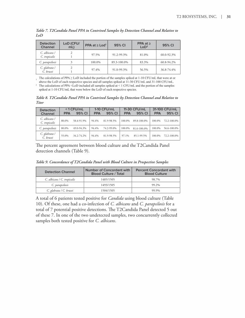
Table 7. T2Candida Panel PPA in Contrived Samples by Detection Channel and Relative to LoD
Detection Channel
LoD (CFU/mL) PPA at ≥ Lod1 95% CI PPA at ≥
LoD2 95% CI
C. albicans / C. tropicalis
2 1 97.5% 91.2-99.3% 81.0% 60.0-92.3%
C. parapsilosis 3 100.0% 89.3-100.0% 83.3% 60.8-94.2%
C. glabrata / C. krusei
2 1 97.4% 91.0-99.3% 56.5% 36.8-74.4%
1 ThecalculationsofPPA≥LoDincludedtheportionofthesamplesspikedat1-10CFU/mLthatwereatorabovetheLoDofeachrespectivespeciesandallsamplesspikedat11-30CFU/mLand31-100CFU/mL.
2 ThecalculationsofPPA<LoDincludedallsamplesspikedat<1CFU/mLandtheportionofthesamplesspikedat1-10CFU/mLthatwerebelowtheLoDofeachrespectivespecies.
Table 8. T2Candida Panel PPA in Contrived Samples by Detection Channel and Relative to Titer
Detection Channel
< 1 CFU/mL 1-10 CFU/mL 11-30 CFU/mL 31-100 CFU/mLPPA 95% CI PPA 95% CI PPA 95% CI PPA 95% CI
C. albicans / C. tropicalis
80.0% 58.4-91.9% 94.4% 81.9-98.5% 100.0% 89.8-100.0% 100.0% 72.2-100.0%
C. parapsilosis 80.0% 49.0-94.3% 94.4% 74.2-99.0% 100.0% 81.6-100.0% 100.0% 56.6-100.0%
C. glabrata / C. krusei
55.0% 34.2-74.2% 94.4% 81.9-98.5% 97.1% 85.1-99.5% 100.0% 72.2-100.0%
The percent agreement between blood culture and the T2Candida Panel detection channels (Table 9).
Table 9. Concordance of T2Candida Panel with Blood Culture in Prospective Samples
Detection Channel Number of Concordant with Blood Culture / Total
Percent Concordant with Blood Culture
C. albicans / C. tropicalis 1485/1505 98.7%
C. parapsilosis 1493/1505 99.2%
C. glabrata / C. krusei 1504/1505 99.9%
A total of 6 patients tested positive for Candida using blood culture (Table 10). Of these, one had a co-infection of C. albicans and C. parapsilosis for a total of 7 potential positive detections. The T2Candida Panel detected 5 out of these 7. In one of the two undetected samples, two concurrently collected samples both tested positive for C. albicans.

32 | T2Candida Instructions for Use
Table 10. Candida Detection in Prospective Blood Culture Positive Samples1
Sample A/T P K/G Blood Culture Result
1 Positive Positive Negative C. albicans and C. parapsilosis
2 Negative Positive Negative C. parapsilosis
3 Negative Negative Positive C. glabrata
4 Positive Negative Negative C. albicans
52 Negative Negative Negative C. albicans
6 Negative Negative Negative C. albicans
1AnadditionalC. albicanspositivebloodcultureresultwasconfirmedbyT2Candidatestbutwasexcludedduetoaprotocoldeviation.
2T2CandidaresultsineachoftwoconcurrentlycollectedsampleswerepositiveforC. albicans.
The T2Candida Panel results indicated 31 positive detections in 29 patient samples for which Candida was not detected via blood culture. One sample was obtained from a patient who had proven intra-abdominal candidiasis with no positive blood cultures. In addition, six of the 29 patients were receiving anti-fungal therapy and four patients were colonized with Candida species. Repeat testing of 26 of the 29 false positive specimens using concurrently collected samples gave negative results for all specimens with the T2Candida Panel assay.
The T2Candida Panel reduced the average time to speciation for Candida positive patients from 129.88 hours to 4.4 hours and the average time for negative diagnosis from > 120 hours to 4.2 hours (Table 11).
Table 11. Time to Result
Negative Result Positive Result
Blood Culture
Mean Time to Result1 (hours) > 120.0 129.882+ 26.32
Number of concordant samples 14663 44
T2Candida
Mean Time to Result5 (hours) 4.2 + 0.9 4.4 + 1.0
Number of concordant samples 14663 44
1 Timetonegativebloodcultureresultwasdefinedas>120.0hoursperInstitutionalprotocols.2 Thetimetopositiveresultisbasedonthetimetospeciationcalculatedfromthestartofincubation.Averagetimetopositivebloodculturepriortospeciationwas43.6hours.
3 Numberofconcordantprospectivenegativeresults.4 Numberofconcordantprospectivepositiveresults.5 ThetimetoT2CandidaPanelresultisdefinedasthetimefromtheinstrument-recordedstarttimetotheinstrument-recordedcompletiontime.

T2 BIOSYSTEMS, INC. | 33
Limitations of the Procedure ♦ As with any procedure, good laboratory technique is important for the
proper performance of the T2Candida Panel. Results are dependent on proper specimen collection, handling and storage. Failure to observe proper procedures in any one of these steps can lead to incorrect results.
♦ The T2Candida Panel has been validated for use only with whole blood collected with K2EDTA as the anticoagulant.
♦ T2Candida Panel performance was established in adult subjects. T2Candida Panel performance in neonates, infants, and pediatric patients has not been established.
♦ Due to the low prevalence of positive specimens collected during the prospective clinical study, performance characteristics for all Candida species detected by the T2Candida Panel were established primarily with contrived specimens.
♦ Candida organisms may persist in vivo independent of organism viability. Detection of Candida species does not imply that the corresponding organisms are infectious nor are the causative agents for clinical symptoms.
♦ Positive and negative predictive values are highly dependent on prevalence. False positive test results are more likely in low prevalence populations.
♦ The T2Candida Panel should only be processed by technicians who have been trained on use of the T2Candida Panel and the T2Dx Instrument.
♦ There is a risk of false positive values resulting from cross-contamination by target organisms, their nucleic acids or amplified products.
♦ The T2Candida Panel is prescription use only.
♦ The following conditions can interfere with an accurate T2Candida Panel result:
• Clotted blood specimen.
• Specimen not at room temperature.
• Insufficient specimen volume. A minimum of 3 mL whole blood is required.
• Contaminated specimen.
• False negatives may occur. The T2Candida Internal Control has been incorporated into the workflow to permit identification of patient specimens, which contain substances that interfere with the T2Candida Panel result.
• The effect of interfering substances has only been evaluated for those listed in the labeling. Interference by substances other than those described in the Interference section could lead to erroneous results.
♦ A negative T2Candida Panel result does not exclude the possibility of

34 | T2Candida Instructions for Use
bloodstream infection. Negative T2Candida Panel results may occur from sequence variants in the region targeted by the assay, the presence of inhibitors, technical error, sample mix-up, or an infection caused by an organism not detected by the panel. Panel results may also be affected by levels of organism in the sample that are below the limit of detection of the test. Negative results should not be used as the sole basis for diagnosis, treatment or other management decisions.
♦ The T2Candida Panel may only be performed on the T2Dx Instrument.
♦ A trained health care professional should interpret results together with the patient’s medical history, clinical signs and symptoms, and the results of other diagnostic tests.

T2 BIOSYSTEMS, INC. | 35
Bibliography1. Pfaller, M. A., Wolk, D. M., & Lowery, T. J. (2016). T2MR and
T2Candida: novel technology for the rapid diagnosis of candidemia and invasive candidiasis. Future microbiology, 11(1), 103-117
2. Mylonakis, E., Clancy, C. J., Ostrosky-Zeichner, L., Garey, K. W., Alangaden, G. J., Vazquez, J. A., ... & Zervou, F. N. (2015). T2 magnetic resonance assay for the rapid diagnosis of candidemia in whole blood: a clinical trial. Clinical infectious diseases, ciu959
3. Pfaller, M. A., &Diekema, D. J. (2007). Epidemiology of invasive candidiasis: a persistent public health problem. Clinical microbiology reviews, 20(1), 133-163.
4. Elixhauser, A., Friedman, B., & Stranges, E. (2011). Septicemia in US hospitals, 2009.
5. Wisplinghoff, H., Bischoff, T., Tallent, S. M., Seifert, H., Wenzel, R. P., & Edmond, M. B. (2004). Nosocomial bloodstream infections in US hospitals: analysis of 24,179 cases from a prospective nationwide surveillance study. Clinical infectious diseases, 39(3), 309-317.
6. Evans, S. E. (2010). Coping with Candida infections. Proceedings of the American Thoracic Society, 7(3), 197-203.
7. Garey, K. W., Rege, M., Pai, M. P., Mingo, D. E., Suda, K. J., Turpin, R. S., & Bearden, D. T. (2006). Time to initiation of fluconazole therapy impacts mortality in patients with candidemia: a multi-institutional study. Clinical infectious diseases, 43(1), 25-31.
8. Morrell, M., Fraser, V. J., &Kollef, M. H. (2005). Delaying the empiric treatment of Candida bloodstream infection until positive blood culture results are obtained: a potential risk factor for hospital mortality. Antimicrobial agents and chemotherapy, 49(9), 3640-3645.
9. Pappas, P. G., Kauffman, C. A., Andes, D., Benjamin, D. K., Calandra, T. F., Edwards, J. E. &Sobe, J. D. (2009). Clinical practice guidelines for the management candidiasis: 2009 update by the Infectious Diseases Society of America. Clinical infectious diseases, 48(5), 503-535.
10. Fernandez, J., Erstad, B. L., Petty, W., & Nix, D. E. (2009). Time to positive culture and identification for Candida blood stream infections. Diagnostic microbiology and infectious disease, 64(4), 402-407.
11. Beyda, N. D., Alam, M. J., &Garey, K. W. (2013). Comparison of the T2Dx instrument with T2Candida assay and automated blood culture in the detection of Candida species using seeded blood samples. Diagnostic microbiology and infectious disease, 77(4), 324-326.
12. Neely, L. A., Audeh, M., Phung, N. A., Min, M., Suchocki, A., Plourde, D., & Lowery, T. J. (2013). T2 magnetic resonance enables

36 | T2Candida Instructions for Use
nanoparticle-mediated rapid detection of candidemia in whole blood. Science translational medicine, 5(182), 182ra54-182ra54.
13. Richmond, J. Y., & McKinney, R. W. Biosafety in Microbiological and Biomedical Laboratories. 1999. US Department of Health and Human Services, Centers for Disease Control and Prevention and National Institutes of Health, Washington DC.
14. Protection of Laboratory Workers from Occupationally Acquired Infections. Approved Guideline-Third Edition. CLSI Document. Clinical and Laboratory Standards Institute (CLSI). M29-A3 Wayne, PA: CLSI, 2005.
15. Tavanti, A., Davidson, A. D., Gow, N. A., Maiden, M. C., & Odds, F. C. (2005). Candida orthopsilosis and Candida metapsilosis spp. nov.to replace Candida parapsilosis groups II and III. Journal of Clinical Microbiology, 43(1), 284-292.

T2 BIOSYSTEMS, INC. | 37
Customer Support & Contact Information
Customer SupportT2 Biosystems, Inc.101 Hartwell AvenueLexington, MA 02421Phone: 781-457-1200 877-504-T2T2 (8282)Fax: 781-357-3080Email: [email protected]
T2 Biosystems, Inc.101 Hartwell AvenueLexington, MA 02421Phone: 781-457-1200Fax: 781-357-3080Email: [email protected]
EC REPMedical Consulting PlusPO Box 31126202 NC MaastrichtNetherlandsPhone: +31-65-3926378Email: [email protected]

38 | T2Candida Instructions for Use
Understanding the SymbolsThe following symbols may appear on the packaging and labeling:
Symbol Meaning
IVD In Vitro Diagnostic Medical Device
Use By <YYYY-MM-DD>
REF Reference Number or Catalog Number
Manufacturer
2 Do Not Reuse
LOT Batch Code or Lot Number
Temperature Limitation
Sufficient For <N tests>
Consult Instructions For Use
Caution
Complies with European Conformity Requirements
EC REP Authorized Representative in the European Community

T2 BIOSYSTEMS, INC. | 39
Notice to PurchaserSee www.t2biosystems.com to download the latest version of the T2Candida Panel Instructions for Use.
This product and its use are covered under one or more of the patents found at www.t2biosystems.com.
Vacutainer® is a registered trademark of Becton Dickinson and Co.
Bleach-Rite® is a registered trademark of Current Technologies, Inc.
Clorox® is a registered trademark of The Clorox Company, Inc.


![Prolonged Outbreak of Candida krusei Candidemia in ... · per 1000 admissions) [1]. Among Candida species causing candidemia, non-albicans Candida (NAC) species are the leading agents](https://img.pdfslide.net/doc/110x75/5ec53fd6a9dc5f3c0426d811/prolonged-outbreak-of-candida-krusei-candidemia-in-per-1000-admissions-1.jpg)


![Ihre persönliche Teemischung für: Sorte Menge [g] · Candida Parapsilosis Fibroma pendulum Mastopathia cystica Prolactin inquiry Toxoplasmosis ... Carc. mammae Glioma Milzbrand](https://img.pdfslide.net/doc/110x75/5c84074c09d3f290718c6de3/ihre-persoenliche-teemischung-fuer-sorte-menge-g-candida-parapsilosis-fibroma.jpg)


![PARIPEX - INDIAN JOURNAL OF RESEARCH | Volume-8 | …...The less commonly identified species are Candida tropcalis, Candida glabrata, Candida parapsilosis, and Candida krusei [5].Identification](https://img.pdfslide.net/doc/110x75/60d53496ab798671291c20a1/paripex-indian-journal-of-research-volume-8-the-less-commonly-identified.jpg)