Embed Size (px)
Citation preview
12/29/2015 2
• Define and identify benign breast conditions• Identify the symptoms of benign breast conditions• Identify the correlation of benign breast
conditions and the risk of breast cancer• Demonstrate the diagnosis of benign breast
conditions
Objectives
Benign Breast Conditions

12/29/2015 3
Benign Breast Conditions
Definition of Benign Breast Disease
Benign breast conditions (also known as benign breast diseases) are non-cancerous disorders or changes in the breast. There are a variety of benign breast conditions.
12/29/2015 4
Benign Breast Conditions
Benign Breast Diseases• Cysts• Fibroadenomas• Hyperplasia (Typical and Atypical)• Intraductal Papillomas• Sclerosing Adenosis• Radial Scars• Galactocele• Lipoma• LCIS• Phyllodes Tumor
12/29/2015 5
Benign Breast Conditions
Symptoms
• Benign breast conditions are not life-threatening, but can cause bothersome symptoms such as a lump or nipple discharge.
• Certain benign conditions are linked with a higher risk of developing breast cancer in the future.
12/29/2015 6
Benign Breast Conditions
Cysts• Small fluid-filled sacs found in the breast• Most commonly found in premenopausal women • Do not increase the risk of developing breast cancer• May be felt by patient if large enough, otherwise seen
using mammography and breast ultrasound• Cause of cysts are unknown• No necessary follow-up unless causing discomfort to
the patient• Cyst aspiration is a treatment option
12/29/2015 7
Benign Breast Conditions
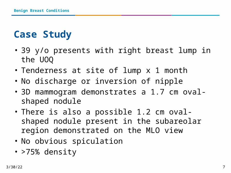
Case Study• 39 y/o presents with right breast lump in the UOQ• Tenderness at site of lump x 1 month• No discharge or inversion of nipple• 3D mammogram demonstrates a 1.7 cm oval-shaped nodule• There is also a possible 1.2 cm oval-shaped nodule present in
the subareolar region demonstrated on the MLO view• No obvious spiculation• >75% density
12/29/2015 8
Benign Breast Conditions
C-View Images

12/29/2015 9
Benign Breast Conditions
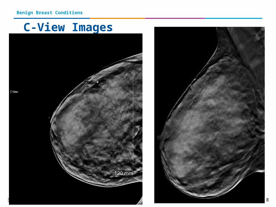
Ultrasound
Ultrasound demonstrates a simple cyst in the 10-11:00 position, 6 cmfn. It measures 2.7 x 1.5 x 2.5 cm. Several smaller cysts are seen within its immediate vicinity

12/29/2015 10
Benign Breast Conditions
Fibroadenomas• Fibroadenomas are benign tumors • They are most common in younger women (between the
ages of 15 and 35) • Most fibroadenomas do not increase the risk of breast
cancer• If a fibroadenoma causes discomfort, it may be excised• Blood flow may be present• Often followed using ultrasound every six months (up to 1
to 2 years) to track any change in size

12/29/2015 11
Benign Breast Conditions
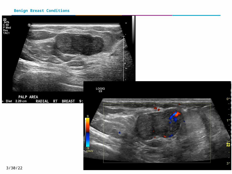
Case Study
• 23 y.o. female• Right palpable mobile lump• Measures 2.3 x 1.2 x 1.6 cm• Lobulated homogeneous solid mass• Blood flow present• Followed over a sixth month period with no
significant change in size
12/29/2015 12
Benign Breast Conditions

12/29/2015 13
Benign Breast Conditions
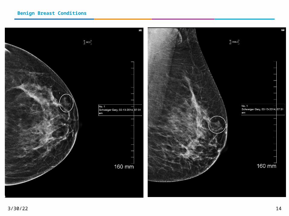
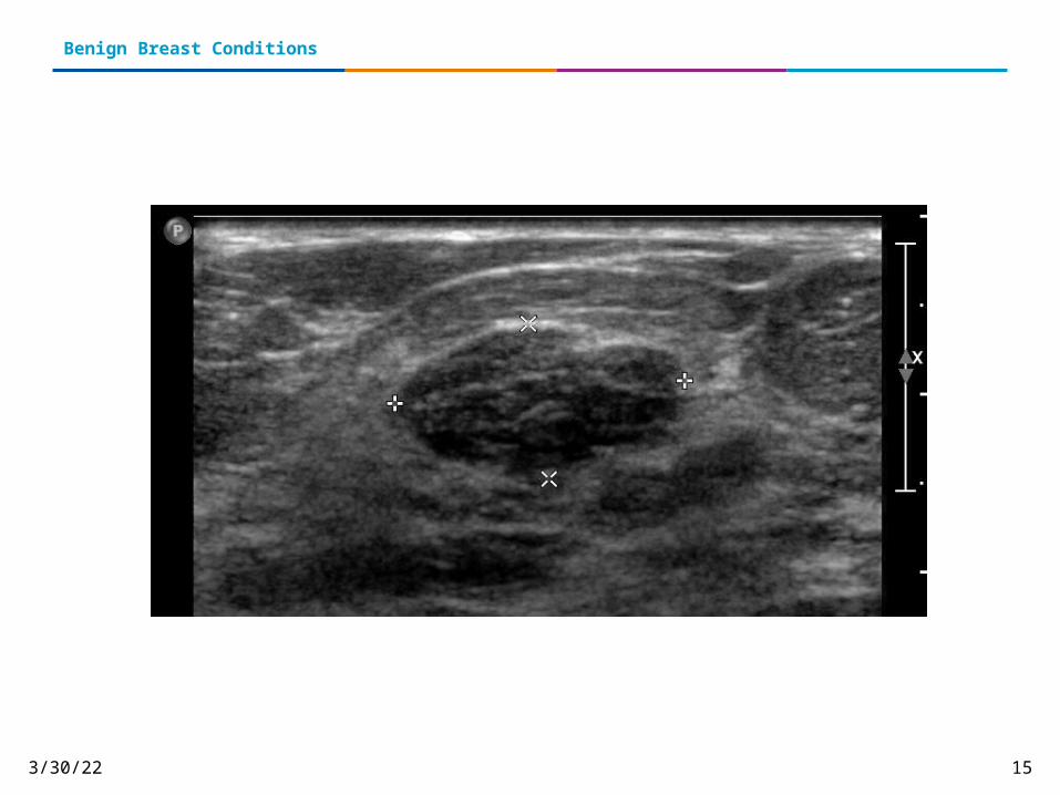
Case Study
• 40 y.o. female• 0.7 x 1 x 1.3 cm hypoechoic mass• Right breast; 7:00 position• Moderate suspicion for malignancy• U/S of right breast demonstrates a well-circumscribed
macrolobulated hypoechoic lesion• No shadowing present• Given the solid appearance, biopsy recommended
12/29/2015 14
Benign Breast Conditions
12/29/2015 15
Benign Breast Conditions
12/29/2015 16
Benign Breast Conditions
Biopsy Results
• Patient underwent ultrasound-guided core biopsy• Pathology findings confirm a benign fibroadenoma• Six month follow-up ultrasound recommended to
look for any post-procedure changes
12/29/2015 17
Benign Breast Conditions
Hyperplasia
• High rate of mitosis or overgrowth (proliferation) of cells
• Most often occurs on the inside of the lobules (milk ducts) in the breast
• Two types of hyperplasia• Typical (Usual)• Atypical
12/29/2015 18
Benign Breast Conditions
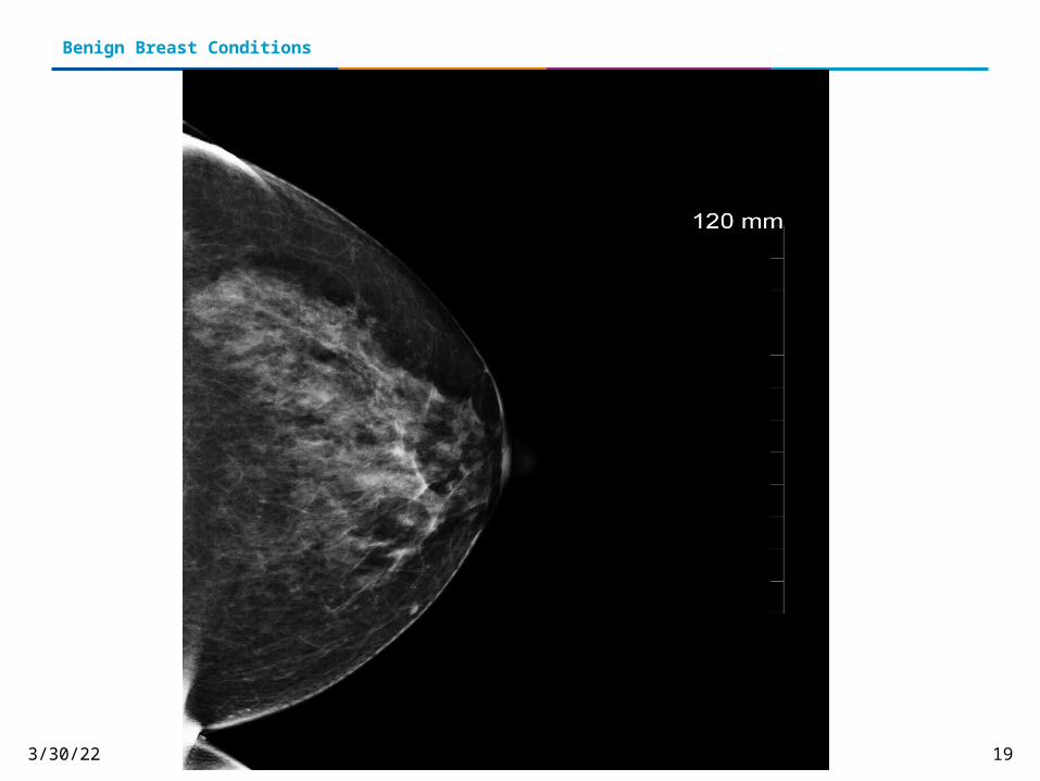
• 50 y.o. female • Presents with 5 day history of spontaneous
bloody discharge; left breast• No breast lump or skin changes• Mild diffused breast pain• Physical exam demonstrated blood coming from a
single duct at the 3:00 position
Case Study
12/29/2015 19
Benign Breast Conditions
12/29/2015 20
Benign Breast Conditions

12/29/2015 21
Benign Breast Conditions
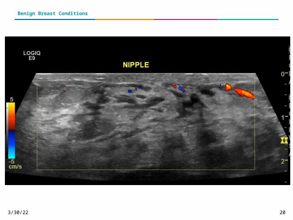
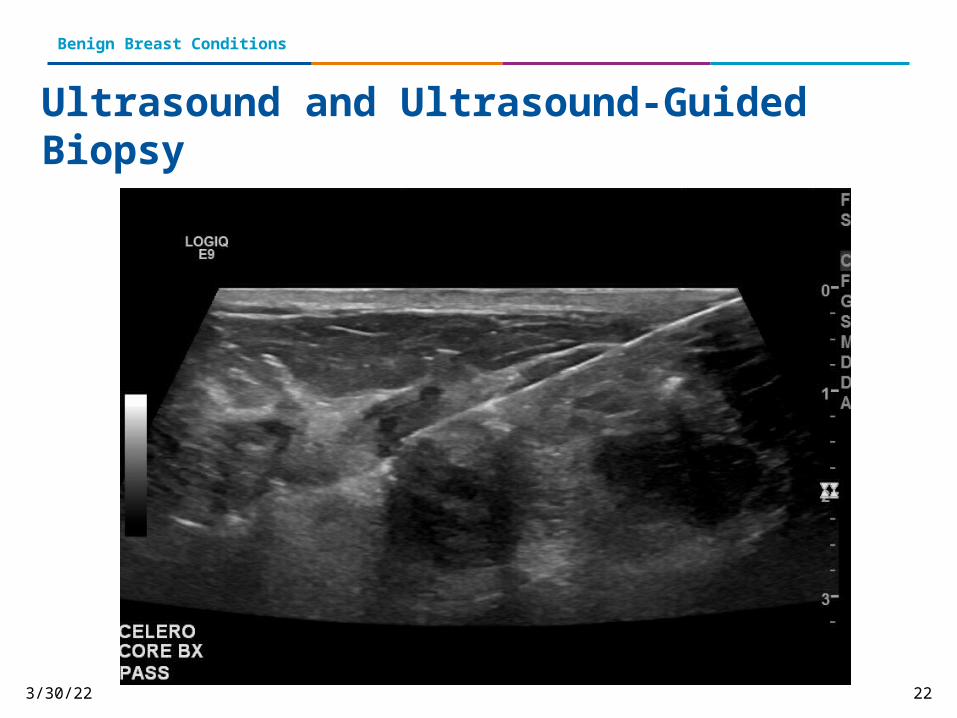
Ultrasound-Guided Biopsy
• Subareolar ultrasound demonstrates slightly prominent ducts with no debris
• Fairly well circumscribed hypoechoic lesion is seen measuring 9x4 mm
• No shadowing, but of solid nature
12/29/2015 22
Benign Breast Conditions
Ultrasound and Ultrasound-Guided Biopsy
12/29/2015 23
Benign Breast Conditions
Pathology
• Ultrasound-guided biopsy was performed and sent to pathology
• Indication: Hypoechoic lesion• Pathologic diagnosis: Atypical ductal
hyperplasia• Recommended follow-up: Excision of area

12/29/2015 24
Benign Breast Conditions
ADH• Classification between TDH and ADH is dependent on
histological features• High reproducibility is important classification• Typical cells appear heterogeneous and atypical are
homogeneous in cell type• ADH is more related to DCIS than typical• ADH is rare and seen in approx. 4% of symptomatic benign
biopsies• ADH is more commonly linked to sreen-detected benign calcs
(31%) and is more commonly an incidental finding• Patients are at a 4-5x higher risk of developing BrCA with a h/o
ADH• Risk is even higher if patient has a first degree relative with BrCA

12/29/2015 25
Benign Breast Conditions
ADH
Three components to the diagnosis of ADH:1. Pattern2. Cytology3. Disease Extent

12/29/2015 26
Benign Breast Conditions
Intraductal Papillomas• Small growths that occur in the lobules• Can cause nipple discharge• Sometimes presents as a lump• Most commonly seen in women 35-55 years old• May be removed by a breast surgeon• No follow-up necessary• Does not increase a woman’s risk of developing
breast cancer
12/29/2015 27
Benign Breast Conditions
Case Study• 51 y.o. female• Perimenopasual • Presents with bloody discharge from the right
nipple• Physical examination revealed no evidence of
lumps, asymmetry or dimpling of the skin • Small amount of bloody fluid appeared when
pressure was exerted on the nipple

12/29/2015 28
Benign Breast Conditions
Ultrasound demonstrates a solid mass measuring 2.45 mm within a dilated duct
Ultrasound
12/29/2015 29
Benign Breast Conditions
Recommendation• After reviewing the negative mammogram from six
months prior, immediate surgical consultation was recommended
• Mass was excised • Histological examination revealed it was benign
(intraductal papilloma, central)• Following excision, all of the patient's symptoms
disappeared• No follow-up required
12/29/2015 30
Benign Breast Conditions
Sclerosing Adenosis• A group of small breast lumps caused by enlarged lobules• A lump may be felt and may be painful• Can be found mammographically• May be mistaken for breast cancer due to its shape and feel• A biopsy is often necessary to rule out breast cancer• Sclerosing adenosis does not need medical treatment• This can be found alongside an existing or developing cancer
and/or atypical hyperplasia
12/29/2015 31
Benign Breast Conditions
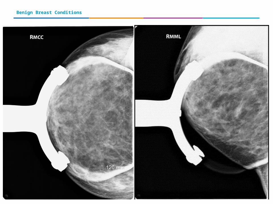
Case Study
• 40 y.o. female• Presents with right breast lump at 6:00• Imaging also shows amorphous calcifications• U/S guided biopsy recommended
12/29/2015 32
Benign Breast Conditions

12/29/2015 33
Benign Breast Conditions
12/29/2015 34
Benign Breast Conditions
Biopsy Results
• Right stereo core biopsy preformed at 6:00• Results show proliferative fibrocystic disease with cellular
change and focal sclerosing adenosis• Calcifications look to be benign; repeat magnification
views in six months• Microscopic descriptions in addition to above
demonstrates typical ductal hyperplasia and intraductal cells and cysts
• 6 month f/u recommended

12/29/2015 35
Benign Breast Conditions
Case Study
• 50 y.o. female• Cluster of pleomorphic calcifications seen on the
CC and MLO views• Left UOQ of breast• Due to irregular borders, biopsy recommended• Stereo-core biopsy

12/29/2015 36
Benign Breast Conditions
12/29/2015 37
Benign Breast Conditions
Biopsy Results
• Pathology demonstrates non-proliferative fibrocystic disease
• Rare benign focus suggesting early sclerosing adenosis
• Calcifications present in benign ducts• Findings are concordant with imaging features
12/29/2015 38
Benign Breast Conditions
Radial Scars (Also known as complex sclerosing lesions)
• Made up of connective tissue fibers from which milk ducts and lobules grow from
• Present similar to breast cancer on a mammogram, but they are not cancer
• Because of their appearance on imaging, they are often biopsied and excised
• After they are removed, no further treatment is necessary
12/29/2015 39
Benign Breast Conditions
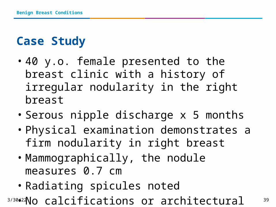
Case Study• 40 y.o. female presented to the breast clinic with a
history of irregular nodularity in the right breast • Serous nipple discharge x 5 months• Physical examination demonstrates a firm nodularity
in right breast• Mammographically, the nodule measures 0.7 cm• Radiating spicules noted• No calcifications or architectural distortion was
appreciated
12/29/2015 40
Benign Breast Conditions
Biopsy and Diagnosis
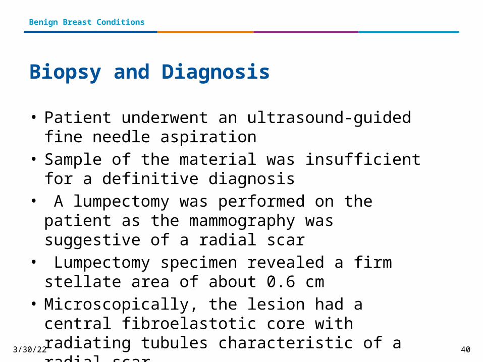
• Patient underwent an ultrasound-guided fine needle aspiration
• Sample of the material was insufficient for a definitive diagnosis
• A lumpectomy was performed on the patient as the mammography was suggestive of a radial scar
• Lumpectomy specimen revealed a firm stellate area of about 0.6 cm
• Microscopically, the lesion had a central fibroelastotic core with radiating tubules characteristic of a radial scar

12/29/2015 41
Benign Breast Conditions
12/29/2015 42
Benign Breast Conditions
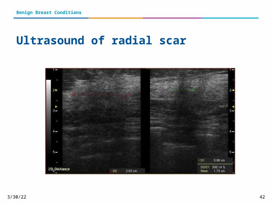
Ultrasound of radial scar
12/29/2015 43
Benign Breast Conditions
Galactocele
• Most common benign breast lesion• Typically occurs in young lactating women• Is also referred to as a lactocele• Presents with a painless breast lump occurring over weeks to
months • Can present as a single or multiple nodule(s) • Unilateral or bilateral• Diagnosis can be done with an aspiration • Most likely located near the sub-areolar region

12/29/2015 44
Benign Breast Conditions
Case Study• Presents with lump x 2 weeks• Patient currently breast feeding• No pain• No skin changes• Physical exam of lump demonstrates a firm and mobile lesion• Ultrasound demonstrates a well-circumscribed hyperechoic
lesion• Mild shadowing• Mild doppler flow• Ultrasound-guided biopsy recommended

12/29/2015 45
Benign Breast Conditions
12/29/2015 46
Benign Breast Conditions
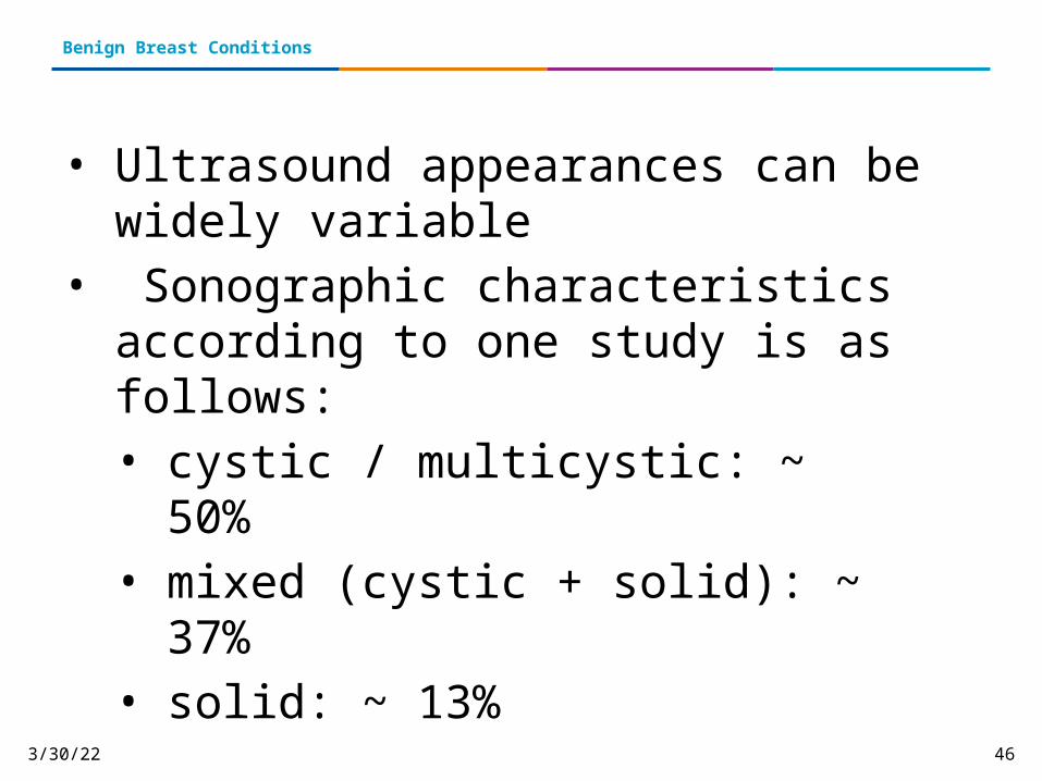
• Ultrasound appearances can be widely variable
• Sonographic characteristics according to one study is as follows:• cystic / multicystic: ~ 50% • mixed (cystic + solid): ~ 37%• solid: ~ 13%
12/29/2015 47
Benign Breast Conditions
Biopsy Results
Benign breast tissue with lactational change and features consistent with galactocle
12/29/2015 48
Benign Breast Conditions
Lipoma• Slow growing, fatty lump • Are mobile and “squishy” to the touch • Are not usually tender• Most commonly found in middle age • Some people have more than one at a time• Lipomas are most often harmless and go untreated• If the lipoma is bothersome, painful or growing, patient
may have it removed
12/29/2015 49
Benign Breast Conditions
Case Study
• 40 y.o. female• Density seen on baseline mammogram• Right UOQ• Persists with spot compression views• Ultrasound recommended
12/29/2015 50
Benign Breast Conditions
12/29/2015 51
Benign Breast Conditions

12/29/2015 52
Benign Breast Conditions
Ultrasound and Ultrasound-guided biopsy
• Ultrasound demonstrates a circumscribed almost completely isoechoic lesion
• Lesion measures 2.2 x 1.3 cm • Biopsy recommended• Ultrasound- guided biopsy performed• Biopsy results demonstrate a fatty benign lesion; lipoma• Six month follow-up recommended• Patient did not undergo surgery or treatment
12/29/2015 53
Benign Breast Conditions
Lobular Carcinoma (LCIS)
• An area or areas of abnormal cellular growth • Increases a person’s risk of developing invasive breast cancer later
on in life• Lobular means that the abnormal cells start growing in the lobules,
the milk-producing glands at the end of breast ducts• Carcinoma refers to any cancer that begins in the skin or other
tissues that cover internal organs — such as breast tissue• In situ or “in its original place” means that the abnormal growth
remains inside the lobule and does not spread to surrounding tissues
• Diagnosis of LCIS tends to have more than one lobule affected

12/29/2015 54
Benign Breast Conditions
LCIS
Despite the fact that its name includes the term “carcinoma,” LCIS is not a true breast cancer.
LCIS is an indication that a person is at higher-than-average risk for getting breast cancer at some point in the future.
12/29/2015 55
Benign Breast Conditions
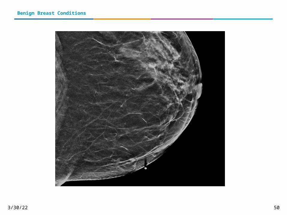
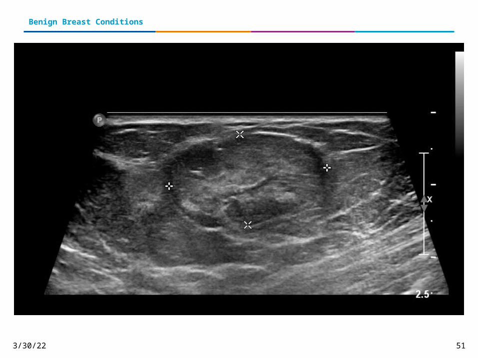
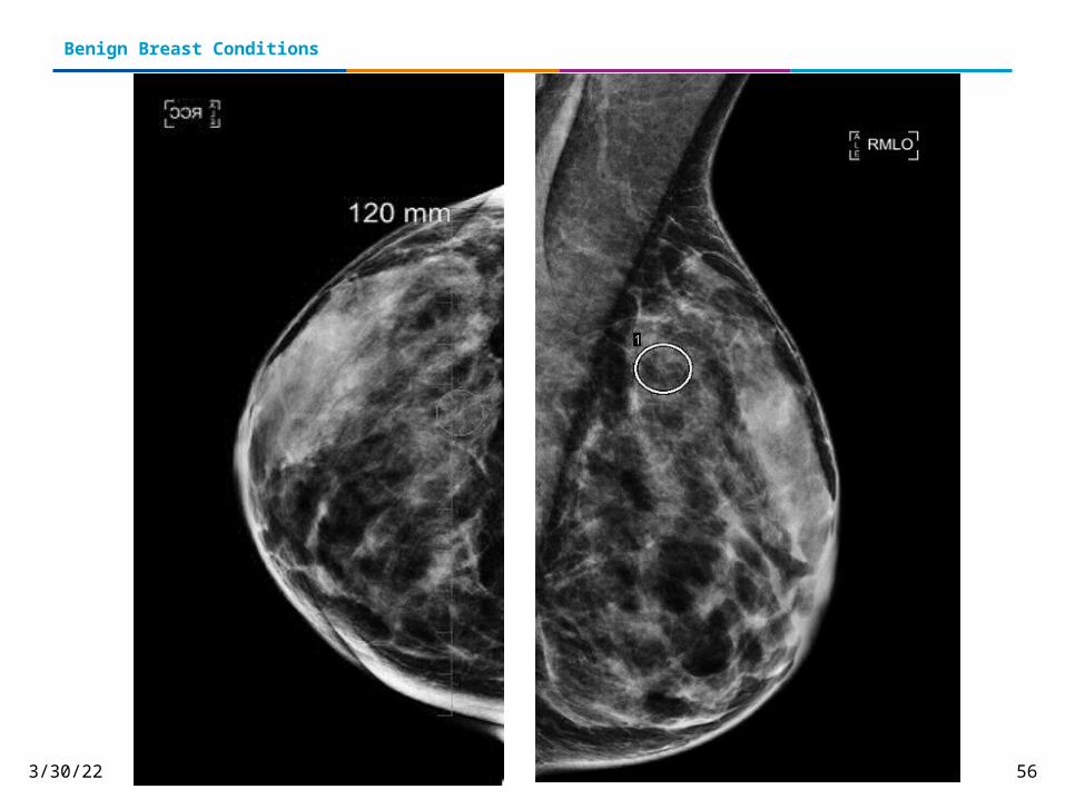
Case Study
• 48 y/o • Mammographic findings demonstrate a
small group of calcifications in the upper outer posterior right breast
• Magnification views recommended
12/29/2015 56
Benign Breast Conditions
12/29/2015 57
Benign Breast Conditions
12/29/2015 58
Benign Breast Conditions
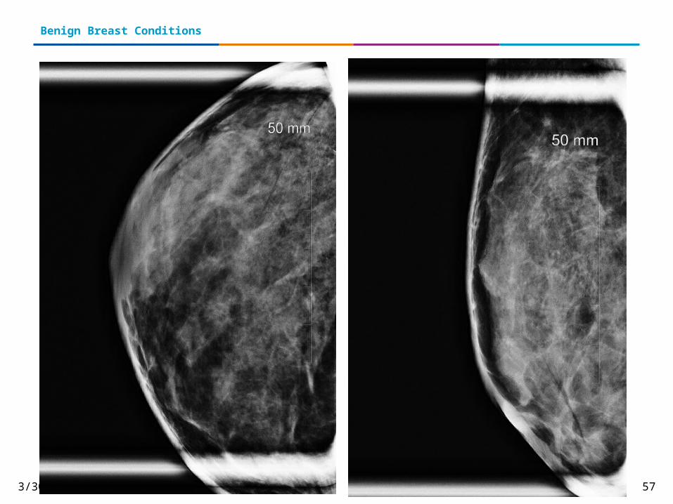
Mammographic Findings
• Calcifications appear to be new compared to prior studies• Indeterminate• BIRADS 4: Suspicious findings• Stereotactic scout attempted• Breast tissue measures too thin for stereotactic biopsy• Patent was referred for a surgical specimen• Needle localization recommended and undertaken• Specimen reveals LCIS and area is excised
12/29/2015 59
Benign Breast Conditions
Phyllodes Tumor• Rare breast tumors that, like fibroadenomas, contain 2 types of breast
tissue: stromal (connective) tissue and glandular (lobule and duct) tissue
• Most commonly found in women in their 30s and 40s, but they may be found in women of any age
Link to cancer risk• Not usually cancerous, but in rare cases they may be related• One third of these tumors are classified as malignant based on how
they look under the microscope, less than 5% of phyllodes tumors overall are clearly true cancers
• Can reappear; close follow-up with frequent breast exams and imaging is usually recommended after treatment
12/29/2015 60
Benign Breast Conditions
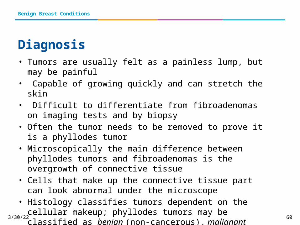
Diagnosis• Tumors are usually felt as a painless lump, but may be painful• Capable of growing quickly and can stretch the skin• Difficult to differentiate from fibroadenomas on imaging tests and by
biopsy• Often the tumor needs to be removed to prove it is a phyllodes tumor• Microscopically the main difference between phyllodes tumors and
fibroadenomas is the overgrowth of connective tissue• Cells that make up the connective tissue part can look abnormal under
the microscope• Histology classifies tumors dependent on the cellular makeup;
phyllodes tumors may be classified as benign (non-cancerous), malignant (cancerous), or borderline (looking more abnormal than benign tumors, but not quite malignant)
12/29/2015 61
Benign Breast Conditions
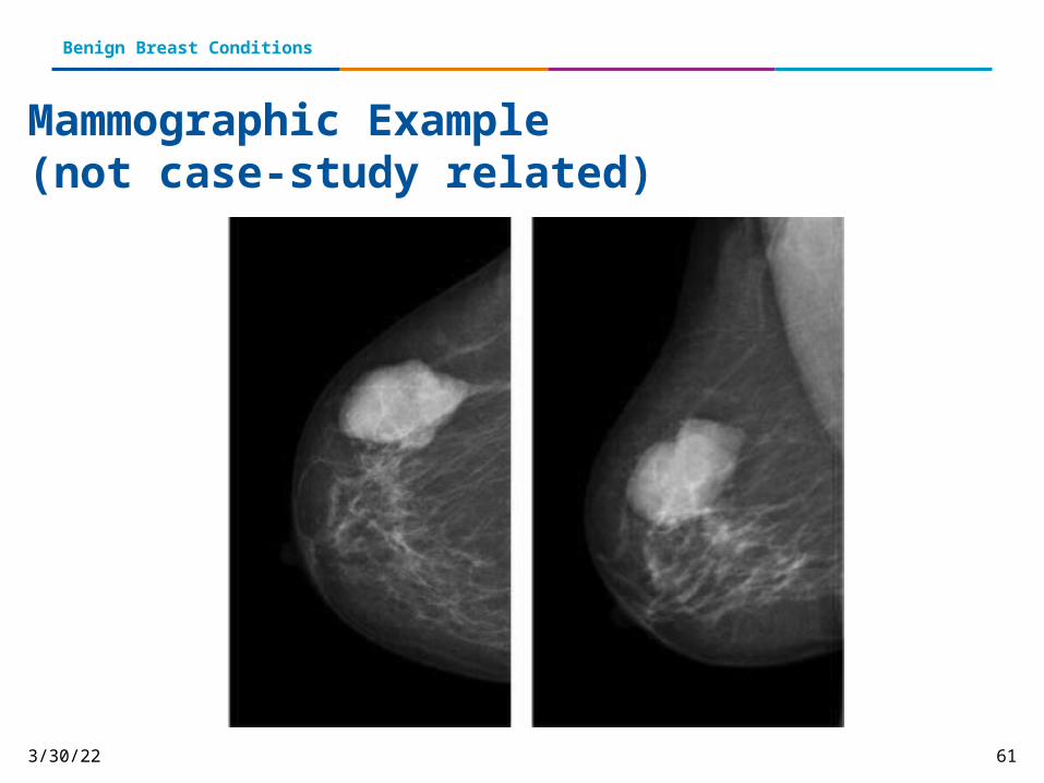
Mammographic Example (not case-study related)

12/29/2015 62
Benign Breast conditions
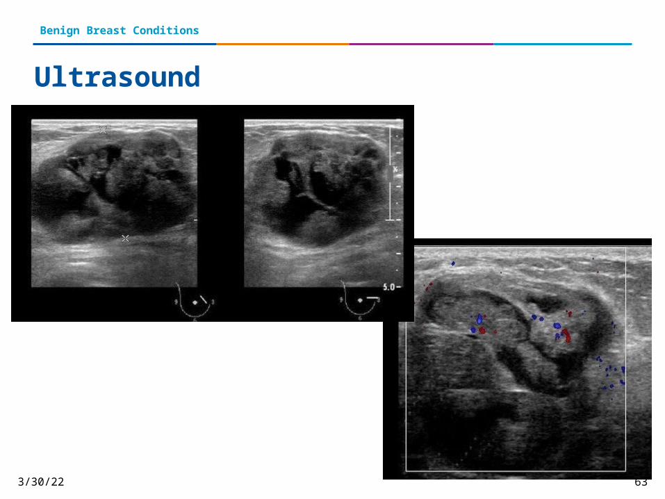
Case Study• 48 y/o female• Presents with large lump in the medial aspect of the left
breast• Ultrasound images of the breast demonstrate large
heterogenous mass of 5.6 x 3.4 cm• Contains multiple lobulations and cystic spaces • The appearance of the tumor is “leaf-like” in its internal
architecture• There is also posterior acoustic enhancement
12/29/2015 63
Benign Breast Conditions
Ultrasound

12/29/2015 64
Benign Breast Conditions
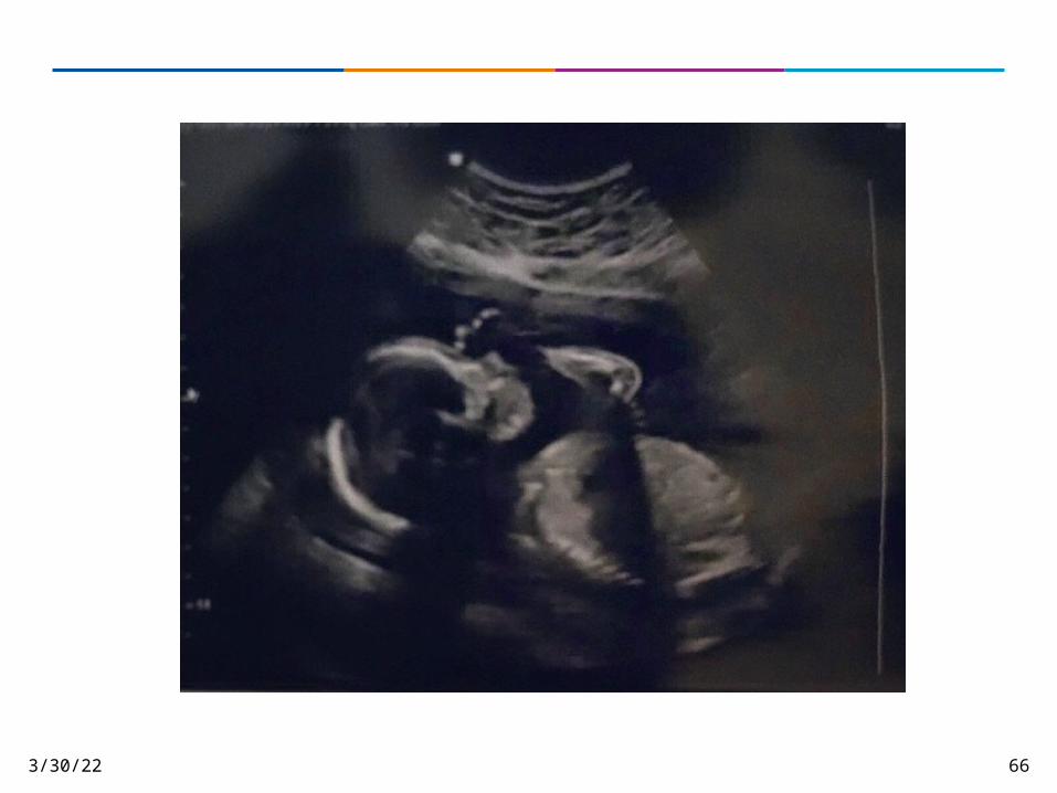
Ultrasound Images
• The images show minimal internal vascularity on color Doppler ultrasound
• Internal lobulation with typical leaf like pattern on sonography
• Age of the patient, large size of tumor and typical ultrasound features are highly suggestive of this being a Phyllodes tumor of the left breast

12/29/2015 65
Benign Breast Conditions
Treatment• Phyllodes tumors (even benign ones) can re-grow in the same place if
they are removed without taking enough of the normal tissue around them
• Treated by removing the tumor and at least a 1 cm (a little less than ½ inch) area of normal breast tissue around the tumor
• Malignant phyllodes tumors are treated by removing them along with a wider margin of normal tissue, or by mastectomy if necessary
• Malignant phyllodes tumors are different from the more common types of BrCA
• They do not respond to hormone therapy and are less likely than most breast cancers to respond to RT or chemo
• Phyllodes tumors that have spread to distant areas are often treated more like sarcomas (soft-tissue cancers) than breast cancers
12/29/2015 66
12/29/2015 67
Benign Breast Conditions
ReferencesAmerican Cancer Society (2014). Non-cancerous breast conditions. Retrieved December 19, 2014 from http://www.cancer.org/healthy/findcancerearly/womenshealth/non-cancerousbreastconditions/non-cancerous-breast-conditions-intro
Bortolotto, C., & Canepari, E., & Tarallo, V. (2012). Intraductal papilloma of the breast: A case report. Retrieved March 3, 2015 from http://www.ncbi.nlm.nih.gov/pmc/articles/PMC3558092/
Breastcancer.org (2015). LCIS-Lobular carcinoma in situ. Retrieved February 3, 2015 from http://www.breastcancer.org/symptoms/types/lcis
Department of Radiation Oncology, Tata Memorial Hospital (2009). Secretory carcinoma arising in radial scars of the breast: A case report and review of literature. Retrieved March 2, 2015 from http://www.ijpmonline.org/article.asp?issn=0377-4929;year=2009;volume=52;issue=1;spage=83;epage=85;aulast=Menon
Mayo Clinic (2014). Atypical Hyperplasia of the breast. Retrieved December 19, 2014 from http://www.mayoclinic.org/diseases-conditions/atypical-hyperplasia/basics/definition/CON-20032601
Pinder, S. E., & Ellis, I. O. (2003). The diagnosis and management of pre-invasive breast disease DCIS and ADH- current definitions and classification. University of Nottingham. Retrieved April 1, 2015.

12/29/2015 68
Benign Breast Conditions
ReferencesRadiopaedia.org (2015). Galactocele. Retrieved January 29, 2015 from http://radiopaedia.org/articles/galactocele
Susan G. Komen (2014). Benign breast conditions. Retrieved December, 19, 2014 from http://ww5.komen.org/BreastCancer/BenignConditions.html


![Benign Breast Disease[1]](https://img.pdfslide.net/doc/110x75/5571f7b649795991698bd982/benign-breast-disease1.jpg)

