Embed Size (px)
Citation preview
Eur. J. Immunol. 2013. 43: 595–605 Antigen processingDOI: 10.1002/eji.201242799 595
Targeting antigen to bone marrow stromal cell-2expressed by conventional and plasmacytoid dendriticcells elicits efficient antigen presentation
Jessica M. Moffat1,2, Elodie Segura3,4, Gabriela Khoury5,6,Irene Caminschi1,7, Paul U. Cameron6,8,9, Sharon R. Lewin6,8,9,Jose A. Villadangos1,2,10 and Justine D. Mintern1,10
1 Walter and Eliza Hall Institute, Parkville, Victoria, Australia2 Department of Microbiology and Immunology, The University of Melbourne, Parkville,
Victoria, Australia3 Institut National de la Sante et de la Recherche Medicale Unite 932, Paris, France4 Center de Recherche, Institut Curie, Paris, France5 Department of Medicine, Monash University, Victoria, Australia6 Centre for Virology, Burnet Institute, Prahran, Victoria, Australia7 Center for Immunology, Burnet Institute, Prahran, Victoria, Australia8 Department of Infectious Diseases, Monash University, Victoria, Australia9 Infectious Diseases Unit, Alfred Hospital, Prahran, Victoria, Australia10 Department of Biochemistry and Molecular Biology, The University of Melbourne, Parkville,
Victoria, Australia
Bone marrow stromal cell-2 (BST-2) has major roles in viral tethering and modulation ofinterferon production. Here we investigate BST-2 as a receptor for the delivery of anti-gen to dendritic cells (DCs). We show that BST-2 is expressed by a panel of mouse andhuman DC subsets, particularly under inflammatory conditions. The outcome of deliver-ing antigen to BST-2 expressed by steady state and activated plasmacytoid DC (pDC) orconventional CD8+ and CD8− DCs was determined. T-cell responses were measured forboth MHC class I (MHCI) and MHC class II (MHCII) antigen presentation pathways in vitro.Delivering antigen via BST-2 was compared with that via receptors DEC205 or Siglec-H.We show that despite a higher antigen load and faster receptor internalisation, whenantigen is delivered to steady state or activated pDC via BST-2, BST-2-targeted activatedconventional DCs present antigen more efficiently. Relative to DEC205, BST-2 was infe-rior in its capacity to deliver antigen to the MHCI cross-presentation pathway. In contrast,BST-2 was superior to Siglec-H at initiating either MHCI or MHCII antigen presentation.In summary, BST-2 is a useful receptor to target with antigen, given its broad expressionpattern and ability to access both MHCI and MHCII presentation pathways with relativeefficiency.
Keywords: Antigen-presenting cells � Antigen presentation/processing � Dendritic cells �
Vaccination
� Additional supporting information may be found in the online version of this article at thepublisher’s web-site
Correspondence: Dr. Justine D. Minterne-mail: [email protected]
C© 2013 WILEY-VCH Verlag GmbH & Co. KGaA, Weinheim www.eji-journal.eu
596 Jessica M. Moffat et al. Eur. J. Immunol. 2013. 43: 595–605
Introduction
Bone marrow stromal cell antigen-2 (BST-2; also CD317, PDCA-1,tetherin and HM1.24) is a widely utilised plasmacytoid dendriticcell (pDC) marker due to its high expression by pDCs in steadystate mouse model systems [1]. BST-2 is an interesting moleculewith unusual topology in that it contains a large extracellulardomain, a cytoplasmic N-terminus and a C-terminus that is GPI-anchored in the plasma membrane [2]. Major functions for BST-2include modulation of pDC interferon (IFN)-α production [1, 3]and the tethering of newly formed enveloped viral particles tothe infected cell surface [4]. BST-2 virus tethering capacity wasfirst described for HIV [4] and is now considered to occur formany, if not all, enveloped viruses [5]. Here we speculate thatBST-2 tethering facilitates pDC antigen presentation. A major rolefor pDCs in efficient antigen presentation, particularly relativeto conventional DCs (cDCs) is controversial [6], however, it isgenerally accepted that pDCs, particularly following activation,have the capacity to present antigen via MHC class I (MHCI) andMHC class II (MHCII) [7]. BST-2 may act as a receptor that confersanti-viral antigen presentation properties to pDCs, given the highexpression of BST-2 by this cell type. In support of this hypothesisis the observation that BST-2 can be utilised as a target moleculefor antigen delivery in vivo [8–10] and can modulate CD8+ T-cellpriming to viral antigen [11].
Incorporating antigen into a monoclonal antibody to targetspecific DC surface markers is an effective strategy for elicitingprotective (and tolerising) immune responses [12]. A number ofDC surface molecules have been targeted to date, including BST-2.Administration of BST-2-targeted ovalbumin (OVA) in vivo elicitsantigen presentation [8–10] and importantly can initiate immu-nity that is capable of protecting mice from vaccinia virus-OVAand reducing tumour growth following implantation of B16-OVAmelanoma [9]. In these studies, BST-2 targeting in vivo was under-taken utilising an adjuvant regime that ensured BST-2 presen-tation was restricted to pDCs, with the role of cDCs excluded[8–10]. Here we will evaluate the outcomes of targeting BST-2 bya broader panel of DC subsets in order to evaluate its efficiencyas an antigen receptor by DC, including both CD8+ and CD8−
DCs that have the capacity to express it under specific conditions.Translating antibody-mediated targeting of DC molecules into set-tings of vaccination against human disease is the obvious next step.Vaccines based on DC targeting are currently being evaluated byCelldex Therapeutics in PhaseI/II clinical trials for the treatmentof solid cancers. DEC205 is the focus of several clinical studies.How would this strategy fare for human BST-2? BST-2 is alreadya target for antibody-induced cytotoxicity as an immunother-apy to eliminate tumours [13, 14]. Utilising anti-BST-2to target antigen for vaccine-mediated immunotherapy relies onBST-2 expression by human DC subsets. Previous studies suggestBST-2 is expressed at low levels by human peripheral blood pDCs,but can be up-regulated by activation [3,15]. Here we will analyseBST-2 expression by a panel of human DC subsets.
While there is a growing list of molecules that can act as recip-ients for targeted antigen, the relative efficiency of unique DC
receptors that are expressed by different DC subsets is mostlyunknown. Factors that will impact the outcome of DC targetinginclude the specific DC subset that is targeted, its activation sta-tus, the amount of antigen delivered (antigen load), the rate ofreceptor internalisation and the trafficking route accessed by thetargeted molecule. In one study, a direct comparison of targetingBST-2 to Siglec-H showed that while targeting BST-2 elicited arobust antibody response, targeting Siglec-H did not [10]. Thisis a pertinent example of the divergent outcomes that can resultfrom targeting distinct DC markers. The underlying mechanismsthat determine the outcome of antigen delivery are of interest ifthis strategy is to be effectively utilised therapeutically. Here wehave investigated BST-2 in the context of antigen delivery andexamined the efficiency of BST-2 targeting, relative to DEC205and Siglec-H by delivering antigen to defined DC subsets, includ-ing both pDCs and cDCs. Evaluating BST-2 targeting outcomes bya panel of different DC subsets has not been previously examined.The ability of BST-2 to facilitate MHCI and MHCII antigen pre-sentation by steady state and activated CD8+ DCs, CD8− DCs andpDCs and the underlying factors that contribute to the presenta-tion of delivered antigen were determined.
Results
Expression profile of BST-2 transcription and surfaceprotein by mouse immune cells
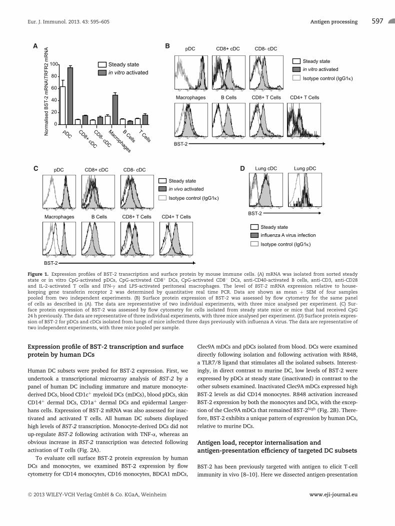
BST-2 expression for a panel of immune cells, including pDCs(CD11cint Ly6c+), CD8+ DCs (CD11c+ CD8+), CD8− DCs (CD11c+
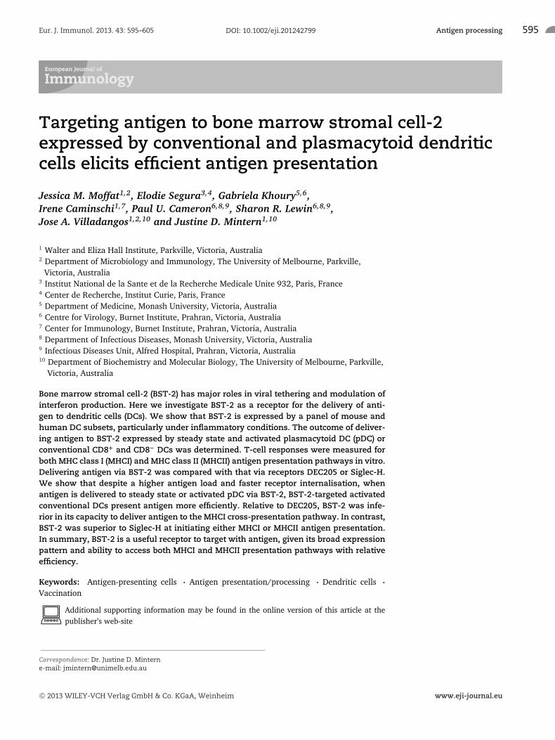
CD8−), macrophages (CD11b+ F480+), B (B220+ CD19+) andT lymphocytes (CD3+), was analysed by quantitative real-timePCR to assess BST-2 transcription. To assess BST-2 transcription,cells were analysed immediately following isolation (steady state)or following treatment with specific stimuli to elicit activation(in vitro activated). Analysis of the steady state samples showedmouse spleen pDCs expressed the highest level of BST-2 mRNA.Following in vitro activation, all of the immune cell types, withthe exception of B cells, increased BST-2 transcription (Fig. 1A).
BST-2 cell surface protein expression was monitored by flowcytometry for a similar panel of immune cells. In this case, cellactivation was achieved either by stimulating with cell specificstimuli as performed for the transcriptional analysis (Fig. 1B) orfollowing isolation of cells from mice that had received nonmethy-lated cytosine-guanosine oligonucleotides (CpG) intravenously asan inflammatory stimulus in vivo (Fig. 1C). At steady state, pDCsexpress high levels of surface BST-2, while other immune cell typesexpress it low levels. Following in vitro activation with cell spe-cific stimuli, cDCs, macrophages, B cells and T cells all up-regulateBST-2 surface expression. Interestingly, activation via the T-cellreceptor increased BST-2 expression by T cells (Fig. 1B). BST-2surface expression was also elevated on all immune cell subsetsisolated 24 h following in vivo CpG administration (Fig. 1C) andon DC subsets isolated from the lung parenchyma of mice infectedwith influenza A virus three days previously (Fig. 1D).
C© 2013 WILEY-VCH Verlag GmbH & Co. KGaA, Weinheim www.eji-journal.eu
Eur. J. Immunol. 2013. 43: 595–605 Antigen processing 597
Figure 1. Expression profiles of BST-2 transcription and surface protein by mouse immune cells. (A) mRNA was isolated from sorted steadystate or in vitro CpG-activated pDCs, CpG-activated CD8+ DCs, CpG-activated CD8− DCs, anti-CD40-activated B cells, anti-CD3, anti-CD28and IL-2-activated T cells and IFN-γ and LPS-activated peritoneal macrophages. The level of BST-2 mRNA expression relative to house-keeping gene transferin receptor 2 was determined by quantitative real time PCR. Data are shown as mean + SEM of four samplespooled from two independent experiments. (B) Surface protein expression of BST-2 was assessed by flow cytometry for the same panelof cells as described in (A). The data are representative of two individual experiments, with three mice analysed per experiment. (C) Sur-face protein expression of BST-2 was assessed by flow cytometry for cells isolated from steady state mice or mice that had received CpG24 h previously. The data are representative of three individual experiments, with three mice analysed per experiment. (D) Surface protein expres-sion of BST-2 for pDCs and cDCs isolated from lungs of mice infected three days previously with influenza A virus. The data are representative oftwo independent experiments, with three mice pooled per sample.
Expression profile of BST-2 transcription and surfaceprotein by human DCs
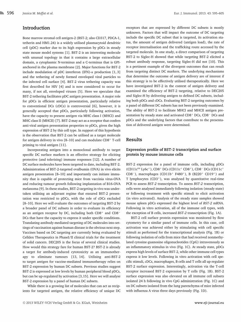
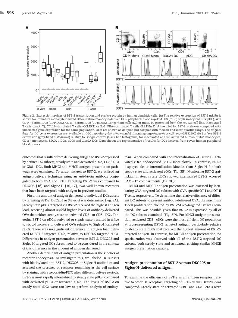
Human DC subsets were probed for BST-2 expression. First, weundertook a transcriptional microarray analysis of BST-2 by apanel of human DC including immature and mature monocyte-derived DCs, blood CD1c+ myeloid DCs (mDCs), blood pDCs, skinCD14+ dermal DCs, CD1a+ dermal DCs and epidermal Langer-hans cells. Expression of BST-2 mRNA was also assessed for inac-tivated and activated T cells. All human DC subsets displayedhigh levels of BST-2 transcription. Monocyte-derived DCs did notup-regulate BST-2 following activation with TNF-α, whereas anobvious increase in BST-2 transcription was detected followingactivation of T cells (Fig. 2A).
To evaluate cell surface BST-2 protein expression by humanDCs and monocytes, we examined BST-2 expression by flowcytometry for CD14 monocytes, CD16 monocytes, BDCA1 mDCs,
Clec9A mDCs and pDCs isolated from blood. DCs were examineddirectly following isolation and following activation with R848,a TLR7/8 ligand that stimulates all the isolated subsets. Interest-ingly, in direct contrast to murine DC, low levels of BST-2 wereexpressed by pDCs at steady state (inactivated) in contrast to theother subsets examined. Inactivated Clec9A mDCs expressed highBST-2 levels as did CD14 monocytes. R848 activation increasedBST-2 expression by both the monocytes and DCs, with the excep-tion of the Clec9A mDCs that remained BST-2high (Fig. 2B). There-fore, BST-2 exhibits a unique pattern of expression by human DCs,relative to murine DCs.
Antigen load, receptor internalisation andantigen-presentation efficiency of targeted DC subsets
BST-2 has been previously targeted with antigen to elicit T-cellimmunity in vivo [8–10]. Here we dissected antigen-presentation
C© 2013 WILEY-VCH Verlag GmbH & Co. KGaA, Weinheim www.eji-journal.eu
598 Jessica M. Moffat et al. Eur. J. Immunol. 2013. 43: 595–605
Figure 2. Expression profiles of BST-2 transcription and surface protein by human dendritic cells. (A) The relative expression of BST-2 mRNA isshown for immature monocyte-derived DC or mature monocyte-derived DCs, peripheral blood myeloid DCs (mDC) or plasmacytoid DCs (pDC), skinCD14+ dermal DCs (CD14DDC), CD1a+ dermal DCs (CD1aDDC), Langerhans cells (LC) or mutz. LC generated from the MUTZ3 cell line, inactivatedT cells (inact. T), CCL19-stimulated T cells (CCL19.T) or IL-2, PHA-stimulated T cells (IL2.PHA.T). A box plot for BST-2 is shown compared withunselected gene expression for the same population. Data are shown as dot plot and box plot with median and inter-quartile range. The originaldata for DC gene expression are available at GEO repository (http://www.ncbi.nlm.nih.gov/geo/query/acc.cgi? acc=GSE32400) (B) Surface BST-2expression (grey-filled histograms) relative to isotype control (black line histograms) for inactivated or R848-activated human CD14+ monocytes,CD16+ monocytes, BDCA-1 DCs, pDCs and Clec9A DCs. Data shown are representative of results for DCs isolated from seven human peripheralblood donors.
outcomes that resulted from delivering antigen to BST-2 expressedby defined DC subsets; steady state and activated pDCs, CD8+ DCsor CD8− DCs. Both MHCI and MHCII antigen-presentation path-ways were examined. To target antigen to BST-2, we utilised anantigen-delivery technique using an anti-biotin antibody conju-gated to both OVA and FITC. Targeting BST-2 was compared toDEC205 [16] and Siglec-H [10, 17], two well-known receptorsthat have been targeted with antigen in previous studies.
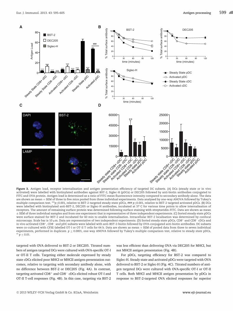
First, the amount of antigen delivered to individual DC subsetsby targeting BST-2, DEC205 or Siglec-H was determined (Fig. 3A).Steady state pDCs targeted via BST-2 received the highest antigenload, receiving almost sixfold higher levels of antibody-deliveredOVA than either steady state or activated CD8+ or CD8− DCs. Tar-geting BST-2 on pDCs, activated or steady state, resulted in a fiveto sixfold increase in delivered OVA relative to Siglec-H-targetedpDCs. There was no significant difference in antigen load deliv-ered to BST-2-targeted cDCs, relative to DEC205-targeted cDCs.Differences in antigen presentation between BST-2, DEC205 andSiglec-H targeted DC subsets need to be considered in the contextof this difference in the amount of antigen delivered.
Another determinant of antigen presentation is the kinetics ofreceptor endocytosis. To investigate this, we labeled DC subsetswith biotinylated anti-BST-2, DEC205 or Siglec-H antibodies andassessed the presence of receptor remaining at the cell surfaceby staining with streptavidin-FITC after different culture periods.BST-2 is most rapidly internalised by steady state pDCs, comparedwith activated pDCs or activated cDCs. The levels of BST-2 onsteady state cDCs were too low to perform analysis of endocy-
tosis. When compared with the internalisation of DEC205, acti-vated cDCs endocytosed BST-2 more slowly. In contrast, BST-2displayed faster internalisation kinetics than Siglec-H for bothsteady state and activated pDCs (Fig. 3B). Monitoring BST-2 traf-ficking in steady state pDCs showed internalised BST-2 accessedLAMP-1+ compartments (Fig. 3C).
MHCI and MHCII antigen presentation was assessed by incu-bating OVA-targeted DC subsets with OVA-specific OT-I and OT-IIT cells, respectively. To determine the relative efficiency of differ-ent DC subsets to present antibody-delivered OVA, the maximumT-cell proliferation elicited by BST-2-OVA-targeted DC was com-pared. This was possible given that BST-2 is expressed by all ofthe DC subsets examined (Fig. 3D). For MHCI antigen presenta-tion, activated CD8+ cDCs were the most efficient DC populationat cross-presenting BST-2 targeted antigen, particularly relativeto steady state pDCs that received the highest amount of BST-2-targeted antigen. In contrast, for MHCII antigen presentation, nospecialization was observed with all of the BST-2-targeted DCsubsets, both steady state and activated, eliciting similar MHCIIantigen presentation capacity.
Antigen presentation of BST-2 versus DEC205 orSiglec-H-delivered antigen
To examine the efficiency of BST-2 as an antigen receptor, rela-tive to other DC receptors, targeting of BST-2 versus DEC205 wascompared. Steady state or activated CD8+ and CD8− cDCs were
C© 2013 WILEY-VCH Verlag GmbH & Co. KGaA, Weinheim www.eji-journal.eu
Eur. J. Immunol. 2013. 43: 595–605 Antigen processing 599
Figure 3. Antigen load, receptor internalisation and antigen presentation efficiency of targeted DC subsets. (A) DCs (steady state or in vivoactivated) were labelled with biotinylated antibodies against BST-2, Siglec-H (pDCs) or DEC205 followed by anti-biotin antibodies conjugated toFITC and OVA protein. Antigen load is determined as a ratio of FITC mean fluorescence intensity compared to secondary antibody alone. The dataare shown as mean + SEM of three to five mice pooled from three individual experiments. Data analysed by one-way ANOVA followed by Tukey’smultiple comparison test. ***p≤0.001, relative to BST-2-targeted steady state pDCs, ### p≤0.001, relative to BST-2 targeted activated pDCs. (B) DCswere labelled with biotinylated anti-BST-2, DEC205 or Siglec-H antibodies, incubated at 37◦C for various time points to allow internalisation ofreceptors. The amount of remaining surface protein was determined following surface staining with streptavidin-FITC. Data are shown as mean± SEM of three individual samples and from one experiment that is representative of three independent experiments. (C) Sorted steady state pDCswere surface stained for BST-2 and incubated for 60 min to enable internalisation. Intracellular BST-2 localisation was determined by confocalmicroscopy. Scale bar is 10 μm. Data are representative of two independent experiments. (D) Sorted steady state pDCs, CD8+ and CD8− cDCs andin vivo activated CD8+, CD8− and pDC subsets were labeled with anti-BST-2-biotin followed by OVA-conjugated anti-biotin antibodies. DC subsetswere co-cultured with CFSE-labelled OT-I or OT-II T cells for 64 h. Data are shown as mean + SEM of pooled data from three to seven individualexperiments, performed in duplicate. p ≤ 0.0001, one-way ANOVA followed by Tukey’s multiple comparison test, relative to steady state pDCs,** p ≤ 0.01.
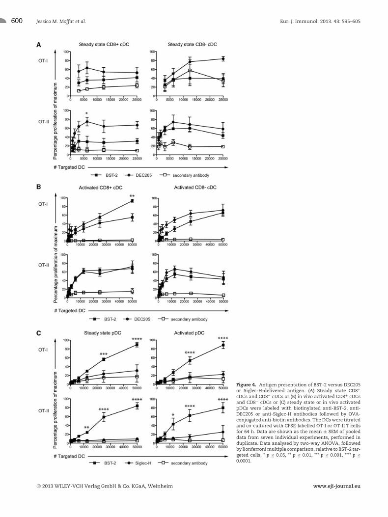
targeted with OVA delivered to BST-2 or DEC205. Titrated num-bers of antigen targeted DCs were cultured with OVA-specific OT-Ior OT-II T cells. Targeting either molecule expressed by steadystate cDCs elicited poor MHCI or MHCII antigen-presentation out-comes, relative to targeting with secondary antibody alone, withno difference between BST-2 or DEC205 (Fig. 4A). In contrast,targeting activated CD8+ and CD8− cDCs elicited robust OT-I andOT-II T-cell responses (Fig. 4B). In this case, targeting via BST-2
was less efficient than delivering OVA via DEC205 for MHCI, butnot MHCII antigen presentation (Fig. 4B).
For pDCs, targeting efficiency for BST-2 was compared toSiglec-H. Steady state and activated pDCs were targeted with OVAdelivered to BST-2 or Siglec-H (Fig. 4C). Titrated numbers of anti-gen targeted DCs were cultured with OVA-specific OT-I or OT-IIT cells. Both MHCI and MHCII antigen presentation by pDCs inresponse to BST-2-targeted OVA elicited responses far superior
C© 2013 WILEY-VCH Verlag GmbH & Co. KGaA, Weinheim www.eji-journal.eu
600 Jessica M. Moffat et al. Eur. J. Immunol. 2013. 43: 595–605
Figure 4. Antigen presentation of BST-2 versus DEC205or Siglec-H-delivered antigen. (A) Steady state CD8+
cDCs and CD8− cDCs or (B) in vivo activated CD8+ cDCsand CD8− cDCs or (C) steady state or in vivo activatedpDCs were labeled with biotinylated anti-BST-2, anti-DEC205 or anti-Siglec-H antibodies followed by OVA-conjugated anti-biotin antibodies. The DCs were titratedand co-cultured with CFSE-labelled OT-I or OT-II T cellsfor 64 h. Data are shown as the mean ± SEM of pooleddata from seven individual experiments, performed induplicate. Data analysed by two-way ANOVA, followedby Bonferroni multiple comparison, relative to BST-2 tar-geted cells, * p ≤ 0.05, ** p ≤ 0.01, *** p ≤ 0.001, **** p ≤0.0001.
C© 2013 WILEY-VCH Verlag GmbH & Co. KGaA, Weinheim www.eji-journal.eu
Eur. J. Immunol. 2013. 43: 595–605 Antigen processing 601
to that elicited by Siglec-H-targeted OVA. Indeed, for MHCI, tar-geting Siglec-H failed to elicit OVA-specific responses above sec-ondary control antibody alone, while for MHCII only very weakSiglec-H-mediated OVA presentation was observed (Fig. 4C).
In addition to targeting DCs in vitro, outcomes of target-ing BST-2 in vivo were also assessed. To do this, anti-rat IgGantibody responses were measured 14 days after immunisationwith 2 μg of rat anti-BST-2 antibody (120G8) or a rat isotypecontrol antibody (GL113). Mice received anti-BST-2 antibodyalone, anti-BST-2 antibody together with 20 nmol CpG or anti-BST-2 antibody 24 h after CpG treatment. In all cases, anti-BST-2administered in vivo did not elicit anti-rat IgG responses (Sup-porting Information Fig. 1). This indicates that targeting BST-2is inferior to targeting Clec9A [18] or Clec12A [19] that elicitanti-rat IgG responses by this protocol.
Discussion
Here we have examined targeting antigen via BST-2, a knownreceptor for the delivery of antigen in vivo [8–10]. We detectexpression of BST-2 by several mouse and human DC subsetsmaking it a useful receptor to target with antigen. We evalu-ated the outcomes of targeting BST-2-expressed by a panel ofdifferent DC subsets. Contrary to our proposal that BST-2 facili-tates pDC antigen presentation, BST-2 targeted antigen was moreefficiently presented by cDC. This occurs despite the high antigenload and efficient endocytosis following antigen delivery to BST-2expressing pDC. While BST-2 is less efficient at eliciting MHCIcross-presentation relative to DEC205, BST-2 is a more efficientantigen delivery receptor than Siglec-H.
pDCs express high levels of BST-2 mRNA and cell-surface pro-tein, while other immune cell subsets increase BST-2 transcrip-tion and surface expression following activation. The BST-2 sur-face protein expression pattern observed here is similar to thatreported previously [1], while BST-2 transcription had not pre-viously been examined for a panel of immune cell subsets. Inaccordance with previous reports [1,20], BST-2 exhibits a promis-cuous expression pattern following activation, with expressionup-regulated at the surface of all immune subsets examined. Weobserved expression at high levels by both major DC populationsat sites of live viral infection. BST-2 is an IFN responsive gene[1, 20, 21] with its promoter containing an IFN regulatory fac-tor binding site [22], together with response elements for severalinflammatory cytokines [23] that are likely to mediate the increasein BST-2 expression observed following in vitro and in vivo acti-vation. Notably, surface BST-2 levels differ between mouse andhuman pDCs. As we have shown here and supported by otherstudies, human blood pDCs express low or no BST-2, but up-regulate its expression after activation [3, 15]. Interestingly, wedetect high BST-2 expression by two major human myeloid DCsubsets; BDCA1 and Clec9A DCs. This has not previously beenexamined and serves to highlight the importance of targeting anti-gen to BST-2 expressed by DC subsets other than pDCs. This isimportant if this strategy is to be translated into clinical settings.
Given the broad expression pattern of BST-2 by various DC sub-sets following activation, we assessed whether purified CD8+ andCD8− cDC subsets present BST-2-targeted antigen. Both MHCI andMHCII antigen presentation by BST-2-targeted activated CD8+
and CD8− cDCs was observed. Targeting BST-2 expressed by apanel of DC subsets enabled us to undertake a direct compari-son of the antigen-presentation efficiency of different DCs. Thishas not been undertaken previously, as previous in vivo target-ing with BST-2 was performed under conditions where BST-2was not expressed by cDCs [8–10]. Despite large differences inantigen load, for cross-presentation via MHCI, CD8+ DCs weresuperior to the other DC subsets analysed. This is in accordancewith the known role for CD8+ DCs as the major cross-presentingDC subset [24, 25]. Delivering antigen via DEC205 elicited moreefficient cross-presentation than BST-2, which indicates a moreefficient ability of this receptor to access the cross-presentationcompartment. Activated CD8− DCs and pDCs were also capable ofcross-presenting antibody-targeted antigen, although at a lowerefficiency than CD8+ DCs. This has been previously describedfor DEC205 [16] and BST-2 [9]-targeted antigen. Notably, acti-vation of CD8+ and CD8− DCs did not impair their ability topresent targeted antigen, unlike soluble protein or cell-associatedantigen that cannot be presented by activated DCs [26]. Thisagrees with previous reports where antibody-mediated deliveryof OVA to DEC205 elicited MHCII antigen presentation by acti-vated DCs [27, 28]. Therefore, while activated DCs shut downmacropinocytosis and phagocytosis, they retain the capacity toundertake receptor-mediated endocytosis. This is a useful attributeto exploit when targeting antigen to DCs in immunisation settings.Finally, our data suggest that pDCs, even following activation,are inferior antigen-presenting cells when compared to cDCs forreceptor-targeted antigen. Despite receiving approximately sixfoldhigher amounts of antigen than their cDC counterparts, steadystate and activated pDCs elicited inferior MHCI cross-presentationand did not promote enhanced MHCII responses compared withthat elicited by cDC subsets receiving significantly less antigen.This concurs with previous reports, where pDCs are less efficientpresenters than cDCs, of soluble OVA protein [7, 29]. Given thereduced antigen-presentation efficiency with which pDCs presentBST-2-targeted OVA relative to BST-2 targeted to cDCs, we wouldexpect cDCs to contribute to BST-2-mediated immunity in vivo.Therefore, to ensure robust immunity is elicited by DC targetingstrategies, utilising receptors that are preferentially expressed bycDCs, rather than pDCs, would be the more advantageous strategy.
Targeting antigen to BST-2 was compared to two well-knownDC surface markers, DEC205 and Siglec-H. DEC205 has pioneeredstudies of antigen targeting to DC receptors. It is expressed bycDCs and elicits robust T-cell immunity when targeted with anti-gen [16]. Studies targeting antigen to DEC205 have pioneeredthe development of T-cell based vaccines for HIV [30]. Siglec-H, unlike DEC205, is not expressed by cDCs but is restrictedto pDCs. Targeting antigen to Siglec-H is capable of initiatingCD4+ T-cell responses, although the cells that develop are ulti-mately hypo-responsive [10], and can elicit CD8+ T-cell immuneresponses in vivo [17]. Relative to DEC205, BST-2 was the inferior
C© 2013 WILEY-VCH Verlag GmbH & Co. KGaA, Weinheim www.eji-journal.eu
602 Jessica M. Moffat et al. Eur. J. Immunol. 2013. 43: 595–605
receptor to target on activated cDCs in order to initiate MHCIantigen presentation. In contrast, for MHCII antigen presentationBST-2 and DEC205 were mostly comparable. BST-2 was a far supe-rior target receptor, relative to Siglec-H, when delivering antigento steady state or activated pDCs for MHCI or MHCII antigenpresentation.
In summary, several mechanisms contribute to successful out-comes for targeting specific DC molecules including the specific DCsubset targeted, the kinetics of receptor internalisation and thedelivered antigen load. Here, we have defined these for BST-2,an interesting molecule that is already the focus of antigen tar-geting strategies. In-depth understanding of the parameters ofantigen targeting is required if this strategy is to be successfullyapplied to clinical settings.
Materials and methods
Mice
C57BL/6, OT-I [31] and OT-II [32] mice were bred under spe-cific pathogen-free conditions at the Walter and Eliza Hall Insti-tute. Gender- and age-matched (6–12 weeks) animals were used.For activation of immune cells by CpG, mice were injected intra-venously with fully phosphorothioated 20 nmol CpG 1668, type B(Geneworks) 24 h prior to organ harvest. Mice were infected with1 × 104 PFU of HKx31 influenza A virus. All experiments were con-ducted in accordance with guidelines provided by National Healthand Medical Research Council of Australia. Experimental proce-dures were approved by the Animal Ethics Committee, MelbourneHealth Research Directorate.
Isolation of mouse immune cells
DCs were isolated from the spleen or lung. Organs were digestedwith 140 μg/mL DNAse (Roche Applied Science) and 1 mg/mLcollagenase type 3 (Worthington Biochemicals) and intercellu-lar clusters disrupted by 0.01M EDTA treatment. Low-densitycells were isolated by density centrifugation (1.077 g/cm3 Nyco-denz). DC was enriched by magnetic depletion of unwanted cellsusing a cocktail of rat anti-mouse antibodies (KT3–1.1, anti-CD3;T24/31.7, anti-Thy1; TER119, anti-erythrocytes; RA36B2, anti-CD45R or 1D3 anti-CD19; RB6–8C5, anti-Ly6C/G or 1A8, Ly6G)and magnetic beads conjugated to anti-rat antibodies (QiagenBiomags). DC isolation yielded cell preparations with approxi-mately 70–85% CD11c+ purity.
Lymphocytes were isolated from spleen and LN. OT-I and OT-IIT cells were isolated from LNs and enriched by magnetic deple-tion of unwanted cells using a cocktail of rat anti-mouse antibod-ies T24/31.7, anti-CD90; RB6-8C5, anti-Gr-1; anti-B220 (RA3–6B2), anti-erythrocyte (Ter-119)) and magnetic beads conjugated
to anti-rat antibodies (Qiagen Biomags). Macrophages were iso-lated from the peritoneal cavity of mice using 10 mL PBS washes.
Cells were identified for flow cytometry using the followingmarkers (antibody clones); cDC: CD11c (N418), CD8 (YTS169),pDC; Ly6c (5075–3.6) and/or CD45RA (14.8), B cells; CD19(1D3) or B220 (RA36B2), T cells: CD3 (KT3–1.1), CD4 (GK1.5),CD8 (YTS169), macrophages; CD11b (M1/70), F4/80 (F4/80).BST-2 was identified using the rat anti-mouse antibody 120.G8compared to isotype control IgG1κ. All antibodies were generatedin house, except the isotype control (Biolegend). Samples wereanalysed using an FACS Calibur or LSR2 (BD Biosciences) andsorted using a Moflo (Dake Cytomation) or FACSAria (BD Bio-sciences). Refer to Supporting Information Fig. 2 for flow cyto-metry gating strategy.
For activation in vitro, all cells were cultured in RPMI1640 medium containing 10% FCS, 100 U/mL penicillin and100 μg/mL streptomycin, 10−4 M 2-mercaptoethanol at 37◦C,10% CO2. DC was cultured for 24 h in media supplemented with10 ng/mL GM-CSF and 0.5 nmol/mL CpG (1668, type B). Peri-toneal macrophages were cultured with 100 ng/mL of IFN-γ for8 h, followed by 16-h incubation in media containing 1 μg/mLLPS. B cells were cultured for 24 h in media containing 50 μg/mLof anti-CD40 antibody (clone FGK45.5). T cells were cultured for48 h in the presence of 5 μg/ml anti-CD28 antibody (clone37.51), 10 U/mL IL-2 and plate-bound anti-CD3 antibody (clone145.2CII).
Quantitative realtime PCR
RNA was isolated from sorted cell populations (Qiagen RNeasyPlus kit). A total of 100 ng of RNA was used to generatecDNA (Qiagen QuantiTec RT PCR kit). SYBR green master mix(Roche) was used in quantitative PCR reactions on the Light-cycler 480 (Roche). BST-2 mRNA expression was normalised tohousekeeping gene transferin receptor 2. BST-2 mRNA was iden-tified by forward 5′ATGGCGCCCTCTTTCTATCAC3′ and reverse5′GTCTCTACAGGCCACGCTGTTC3′ primers. Gene transferinreceptor 2 mRNA was identified by forward 5′ TATCGGCTGGGAC-CTGGGCC 3′ and reverse 5′GCTCCTGGGCCCCATGCATC3′
primers.
Isolation of human immune cells
Peripheral blood mononuclear cells (PBMCs) were isolated fromseronegative healthy donors provided by the Australian Red CrossBank. Monocyte-derived DCs were differentiated from CD14+
monocytes by culture in IL-4 and GM-CSF to generate immaturemonocyte-derived DCs as previously described [33, 34]. Maturemonocyte-derived DCs were generated following culture in thepresence of TNF-α. Total blood DCs and monocytes were enrichedfrom PBMCs using magnetic bead depletion (MiltenyiBiotec) aspreviously described [35]. DCs were further sorted into myeloid
C© 2013 WILEY-VCH Verlag GmbH & Co. KGaA, Weinheim www.eji-journal.eu
Eur. J. Immunol. 2013. 43: 595–605 Antigen processing 603
(HLA-DR+ CD11c+) and plamacytoid (HLA-DR+ CD123+) subsetsusing flow cytometry. Isolation of skin DC subsets (CD14 dermalDCs, CD1a dermal DCs and Langerhans cells) was performed aspreviously described [36]. Human CD34+ acute myeloid leukemiaMUTZ3 cells were provided by S. Santegoets from VU UniversityMedical Center, Netherlands [37]. Resting CD4+ T cells were puri-fied as previously described [38]. Purified T cells were culturedalone, with CCL19 or PHA and IL-2.
For human monocyte and DC analysis, buffy coats from healthydonors were obtained from Etablissement Francais du Sang.PBMCs were prepared by centrifugation on a Ficoll gradient (Lym-phoprep, Greiner Bio-One). PBMCs were either analysed afterisolation or after overnight culture in RPMI-Glutamax medium(Gibco) containing 10% FCS and 1 μg/mL of R848 (Invitro-gen). Cells were stained with anti-CD16 (BD Biosciences), anti-BDCA4 (Miltenyi Biotec), anti-BDCA-1 (eBioscience), anti-CD4(BD Biosciences), anti-CD14 (Miltenyi Biotec), anti-Clec9A (Mil-tenyi Biotec), anti-BST2 (Biolegend) or isotype-matched controlantibody. Cell viability was assessed with DAPI. Cells were anal-ysed on an FACSVerse (BD Biosciences) instrument. Data wereanalysed with FlowJo (Tree Star).
Microarray hybridisation and data analysis
Total RNA was extracted from purified cell populations from indi-vidual donors and processed for hybridisation to 1 of 55 cDNAgene arrays (Human ResGen 8k, Australian Genome ResearchFacility, Melbourne, Australia) using a common monocyte-derivedDC reference, or 24 bead arrays (sentrix human 6 v2 expres-sion chips), Illumina, San Diego, CA). The RNA extraction,labelling, hybridisation, data processing and analysis proceduresare described previously for the cDNA gene array and Illuminaarrays, respectively [33, 39]. Clustered data were further pro-cessed in PARTEK Genomics Suite (Partek Inc., St. Lois, MO,USA) to exclude genes not showing detectable expression inmore than 80% of arrays and to remove batch effects. Microarraydata is available through the Gene Expression Omnibus database(http://www.ncbi.nlm.nih.gov/geo/).
Microscopy
Steady state pDCs were stained with biotin-BST-2 andstreptavidin-Alexa 488 (Invitrogen, Molecular Probes) at 4◦C andthen incubated at 37◦C for 60 min. Cells were attached to coverslips with anti-MHCI antibody (Y3) for 10 min at room tempera-ture. Cover slips were washed in PBS, samples were fixed in 4%PFA (Sigma Aldrich) and permabilised with 0.3% Triton X 100(Sigma Aldrich) prior to staining for LAMP-1 (Abcam). Nucleiwere stained with 5 μg/mL DAPI (Invitrogen). Cover slips weremounted with DAKO mounting media. Images were acquired onan LSM700 confocal microscope (Zeiss) and analysed with ImageJsoftware.
Antigen-presentation assay
Sorted pDC and CD8+ or CD8− cDC were labelled with anti-BST-2 biotin (clone 120.G8), anti-DEC205-biotin (cloneNLDC145) or anti-Siglec-H-biotin (clone 551.3D3) on ice for30 min, washed twice and labelled with anti-biotin OVA (Mil-tenyi Biotec) for 15 min at 4◦C. Cells were then washed twiceand re-suspended in culture media containing CpG (1688, type B)and 10 ng/mL GM-CSF, titrated and incubated with CFSE-labelledOT-I or OT-II transgenic T cells for 64 h. Dividing cells were deter-mined as T cells that had diluted the CFSE dye by flow cytometry.Refer to Supporting Information Fig. 3 for flow cytometry gatingstrategy.
Internalisation assay
Cells were stained either with biotinylated anti-BST-2 (clone120.G8), anti-DEC205 (clone NLDC145) or anti-Siglec-H (clone551.3D3) antibodies on ice for 30 min, washed twice and incu-bated for various time periods at 37◦C allowing the BST-2 antibodyto be internalised. Cells were then stained for surface markers andstreptavidin-FITC and analysed on the LSR II (Becton Dickinson).
Acknowledgements: JDM is an NHMRC (Australia) CareerDevelopment Fellow. GK is the recipient of a NHMRC (Australia)Postgraduate Scholarship. This research was funded by a Creativeand Novel Ideas in HIV Research Award from the InternationalAIDS Society and National Institute of Health, USA. We acknowl-edge the helpful advice of Dr. Sandy Clarke at The Statistical Con-sulting Center, The Department of Mathematics and Statistics, TheUniversity of Melbourne.
Conflict of interest: The authors declare no financial or commer-cial conflict of interest.
References
1 Blasius, A. L., Giurisato, E., Cella, M., Schreiber, R. D., Shaw, A. S. and
Colonna, M., Bone marrow stromal cell antigen 2 is a specific marker
of type I IFN-producing cells in the naive mouse, but a promiscuous
cell surface antigen following IFN stimulation. J. Immunol. 2006. 177:
3260–3265.
2 Swiecki, M., Scheaffer, S. M., Allaire, M., Fremont, D. H., Colonna, M.
and Brett, T. J., Structural and biophysical analysis of BST-2/tetherin
ectodomains reveals an evolutionary conserved design to inhibit virus
release. J. Biol. Chem. 2011. 286: 2987–2997.
3 Cao, W., Bover, L., Cho, M., Wen, X., Hanabuchi, S., Bao, M., Rosen, D. B.
et al., Regulation of TLR7/9 responses in plasmacytoid dendritic cells by
BST2 and ILT7 receptor interaction. J. Exp. Med. 2009. 206: 1603–1614.
C© 2013 WILEY-VCH Verlag GmbH & Co. KGaA, Weinheim www.eji-journal.eu

604 Jessica M. Moffat et al. Eur. J. Immunol. 2013. 43: 595–605
4 Neil, S. J., Zang, T. and Bieniasz, P. D., Tetherin inhibits retrovirus release
and is antagonized by HIV-1 Vpu. Nature 2008. 451: 425–430.
5 Evans, D. T., Serra-Moreno, R., Singh, R. K. and Guatelli, J. C., BST-
2/tetherin: a new component of the innate immune response to
enveloped viruses. Trends Microbiol. 2010. 18: 388–396.
6 Villadangos, J. A. and Young, L., Antigen-presentation properties of plas-
macytoid dendritic cells. Immunity 2008. 29: 352–361.
7 Kool, M., Geurtsvankessel, C., Muskens, F., Madeira, F. B., van Nimwe-
gen, M., Kuipers, H., Thielemans, K. et al., Facilitated antigen uptake and
timed exposure to TLR ligands dictate the antigen-presenting potential
of plasmacytoid DCs. J. Leukoc. Biol. 2011. 90: 1177–1190.
8 Sapoznikov, A., Fischer, J. A., Zaft, T., Krauthgamer, R., Dzionek, A.
and Jung, S., Organ-dependent in vivo priming of naive CD4+, but not
CD8+, T cells by plasmacytoid dendritic cells. J. Exp. Med. 2007. 204:
1923–1933.
9 Loschko, J., Schlitzer, A., Dudziak, D., Drexler, I., Sandholzer, N.,
Bourquin, C., Reindl, W. et al., Antigen delivery to plasmacytoid dendritic
cells via BST2 induces protective T-cell-mediated immunity. J. Immunol.
2011. 186: 6718–6725.
10 Loschko, J., Heink, S., Hackl, D., Dudziak, D., Reindl, W., Korn, T.
and Krug, A. B., Antigen targeting to plasmacytoid dendritic cells via
Siglec-H inhibits Th cell-dependent autoimmunity. J. Immunol. 2011. 187:
6346–6356.
11 Swiecki, M., Wang, Y., Gilfillan, S., Lenschow, D. J. and Colonna, M.,
Cutting edge: paradoxical roles of BST2/tetherin in promoting type I IFN
response and viral infection. J. Immunol. 2012. 188: 2488–2492.
12 Caminschi, I., Lahoud, M. H. and Shortman, K., Enhancing immune
responses by targeting antigen to DC. Eur. J. Immunol. 2009. 39: 931–938.
13 Chiriva-Internati, M., Liu, Y., Weidanz, J. A., Grizzi, F., You, H., Zhou,
W., Bumm, K. et al., Testing recombinant adeno-associated virus-gene
loading of dendritic cells for generating potent cytotoxic T lymphocytes
against a prototype self-antigen, multiple myeloma HM1.24. Blood 2003.
102: 3100–3107.
14 Jalili, A., Ozaki, S., Hara, T., Shibata, H., Hashimoto, T., Abe, M., Nishioka,
Y. et al., Induction of HM1.24 peptide-specific cytotoxic T lymphocytes
by using peripheral-blood stem-cell harvests in patients with multiple
myeloma. Blood 2005. 106: 3538–3545.
15 Erikson, E., Adam, T., Schmidt, S., Lehmann-Koch, J., Over, B., Goffinet,
C., Harter, C. et al., In vivo expression profile of the antiviral restric-
tion factor and tumor-targeting antigen CD317/BST-2/HM1.24/tetherin
in humans. Proc. Natl. Acad. Sci. USA 2011. 108: 13688–13693.
16 Dudziak, D., Kamphorst, A. O., Heidkamp, G. F., Buchholz, V. R.,
Trumpfheller, C., Yamazaki, S., Cheong, C. et al., Differential antigen
processing by dendritic cell subsets in vivo. Science 2007. 315: 107–111.
17 Zhang, J., Raper, A., Sugita, N., Hingorani, R., Salio, M., Palmowski,
M. J., Cerundolo, V. et al., Characterization of Siglec-H as a novel endo-
cytic receptor expressed on murine plasmacytoid dendritic cell precur-
sors. Blood 2006. 107: 3600–3608.
18 Caminschi, I., Proietto, A. I., Ahmet, F., Kitsoulis, S., Shin Teh, J., Lo,
J. C., Rizzitelli, A. et al., The dendritic cell subtype-restricted C-type lectin
Clec9A is a target for vaccine enhancement. Blood 2008. 112: 3264–3273.
19 Lahoud, M. H., Proietto, A. I., Ahmet, F., Kitsoulis, S., Eidsmo, L., Wu, L.,
Sathe, P. et al., The C-type lectin Clec12A present on mouse and human
dendritic cells can serve as a target for antigen delivery and enhancement
of antibody responses. J. Immunol. 2009. 182: 7587–7594.
20 Liberatore, R. A. and Bieniasz, P. D., Tetherin is a key effector of the
antiretroviral activity of type I interferon in vitro and in vivo. Proc. Natl.
Acad. Sci. USA 2011. 108: 18097–18101.
21 Kawai, S., Azuma, Y., Fujii, E., Furugaki, K., Ozaki, S., Matsumoto, T.,
Kosaka, M. et al., Interferon-alpha enhances CD317 expression and the
antitumor activity of anti-CD317 monoclonal antibody in renal cell car-
cinoma xenograft models. Cancer Sci. 2008. 99: 2461–2466.
22 Bego, M. G., Mercier, J. and Cohen, E. A., Virus-activated interferon
regulatory factor 7 upregulates expression of the interferon-regulated
BST2 gene independently of interferon signaling. J. Virol. 2012. 86:
3513–3527.
23 Ohtomo, T., Sugamata, Y., Ozaki, Y., Ono, K., Yoshimura, Y., Kawai, S.,
Koishihara, Y. et al., Molecular cloning and characterization of a surface
antigen preferentially overexpressed on multiple myeloma cells. Biochem.
Biophys. Res. Commun. 1999. 258: 583–591.
24 den Haan, J. M., Lehar, S. M. and Bevan, M. J., CD8(+) but not CD8(-)
dendritic cells cross-prime cytotoxic T cells in vivo. J. Exp. Med. 2000. 192:
1685–1696.
25 Schnorrer, P., Behrens, G. M., Wilson, N. S., Pooley, J. L., Smith, C. M.,
El-Sukkari, D., Davey, G. et al., The dominant role of CD8+ dendritic cells
in cross-presentation is not dictated by antigen capture. Proc. Natl. Acad.
Sci. USA 2006. 103: 10729–10734.
26 Wilson, N. S., Behrens, G. M., Lundie, R. J., Smith, C. M., Waithman, J.,
Young, L., Forehan, S. P. et al., Systemic activation of dendritic cells by
Toll-like receptor ligands or malaria infection impairs cross-presentation
and antiviral immunity. Nat. Immunol. 2006. 7: 165–172.
27 Platt, C. D., Ma, J. K., Chalouni, C., Ebersold, M., Bou-Reslan, H., Carano,
R. A., Mellman, I. et al., Mature dendritic cells use endocytic receptors
to capture and present antigens. Proc. Natl. Acad. Sci. USA 2010. 107:
4287–4292.
28 Kamphorst, A. O., Guermonprez, P., Dudziak, D. and Nussenzweig, M. C.,
Route of antigen uptake differentially impacts presentation by dendritic
cells and activated monocytes. J. Immunol. 2010. 185: 3426–3435.
29 Young, L. J., Wilson, N. S., Schnorrer, P., Proietto, A., ten Broeke, T.,
Matsuki, Y., Mount, A. M. et al., Differential MHC class II synthesis
and ubiquitination confers distinct antigen-presenting properties on
conventional and plasmacytoid dendritic cells. Nat. Immunol. 2008. 9:
1244–1252.
30 Cheong, C., Choi, J. H., Vitale, L., He, L. Z., Trumpfheller, C., Bozzacco,
L., Do, Y. et al., Improved cellular and humoral immune responses in
vivo following targeting of HIV Gag to dendritic cells within human anti-
human DEC205 monoclonal antibody. Blood 2010. 116: 3828–3838.
31 Clarke, S. M. R., Barnden, M., Kurts, C., Carbone, F. R., Miller, J. F. A. P.
and Heath, W. R., Characterisation of the OVA-specific TCR transgenic
line OT-I: MHC elements for positive and negative selection. Immunol. Cell
Biol. 2000.78: 110–117.
32 Barnden, M. J., Allison, J., Heath, W. R. and Carbone, F. R., Defective TCR
expression in transgenic mice constructed using cDNA- based alpha- and
beta-chain genes under the control of heterologous regulatory elements.
Immunol. Cell Biol. 1998. 76: 34–40.
33 Harman, A. N., Wilkinson, J., Bye, C. R., Bosnjak, L., Stern, J. L., Nicholle,
M., Lai, J. et al., HIV induces maturation of monocyte-derived dendritic
cells and Langerhans cells. J. Immunol. 2006. 177: 7103–7113.
34 Turville, S. G., Santos, J. J., Frank, I., Cameron, P. U., Wilkinson, J.,
Miranda-Saksena, M., Dable, J. et al., Immunodeficiency virus uptake,
turnover, and 2-phase transfer in human dendritic cells. Blood 2004. 103:
2170–2179.
35 Cameron, P. U., Handley, A. J., Baylis, D. C., Solomon, A. E., Bernard, N.,
Purcell, D. F. and Lewin, S. R., Preferential infection of dendritic cells dur-
ing human immunodeficiency virus type 1 infection of blood leukocytes.
J. Virol. 2007. 81: 2297–2306.
C© 2013 WILEY-VCH Verlag GmbH & Co. KGaA, Weinheim www.eji-journal.eu
Eur. J. Immunol. 2013. 43: 595–605 Antigen processing 605
36 McLellan, A. D., Heiser, A., Sorg, R. V., Fearnley, D. B. and Hart, D.
N., Dermal dendritic cells associated with T lymphocytes in normal
human skin display an activated phenotype. J. Invest. Dermatol. 1998. 111:
841–849.
37 de Jong, M. A., de Witte, L., Santegoets, S. J., Fluitsma, D., Taylor,
M. E., de Gruijl, T. D. and Geijtenbeek, T. B., Mutz-3-derived Langerhans
cells are a model to study HIV-1 transmission and potential inhibitors.
J. Leukoc. Biol. 2010. 87: 637–643.
38 Saleh, S., Solomon, A., Wightman, F., Xhilaga, M., Cameron, P. U. and
Lewin, S. R., CCR7 ligands CCL19 and CCL21 increase permissiveness of
resting memory CD4+ T cells to HIV-1 infection: a novel model of HIV-1
latency. Blood 2007. 110: 4161–4164.
39 Harman, A. N., Kraus, M., Bye, C. R., Byth, K., Turville, S. G., Tang, O.,
Mercier, S. K. et al., HIV-1-infected dendritic cells show 2 phases of gene
expression changes, with lysosomal enzyme activity decreased during
the second phase. Blood 2009. 114: 85–94.
Abbreviations: BST-2: bone marrow stromal cell-2 · cDC: conventional
dendritic cell · pDC: plasmacytoid dendritic cell
Full correspondence: Dr. Justine D. Mintern, The University ofMelbourne, Bio21, 30 Flemington Rd, Parkville Victoria 3010, AustraliaFax: +61-3-9348-1421e-mail: [email protected]
Additional correspondence: Prof. Jose A. Villadangos, The University ofMelbourne, Bio21, 30 Flemington Rd, Parkville Victoria 3010, AustraliaFax: +61-3-9348-1421e-mail: [email protected]
Received: 5/7/2012Revised: 19/11/2012Accepted: 4/1/2013Accepted article online: 9/1/2013
C© 2013 WILEY-VCH Verlag GmbH & Co. KGaA, Weinheim www.eji-journal.eu