Embed Size (px)
Citation preview
of April 4, 2019.This information is current as
T Cell Activation In Vivo+CD8 and+Opsonized Antigen Leading to CD4
Plasmacytoid Dendritic Cells Take Up
Negrin and Edgar G. EnglemanPia Björck, Andreas Beilhack, Edward I. Herman, Robert S.
http://www.jimmunol.org/content/181/6/3811doi: 10.4049/jimmunol.181.6.3811
2008; 181:3811-3817; ;J Immunol
Referenceshttp://www.jimmunol.org/content/181/6/3811.full#ref-list-1
, 20 of which you can access for free at: cites 30 articlesThis article
average*
4 weeks from acceptance to publicationFast Publication! •
Every submission reviewed by practicing scientistsNo Triage! •
from submission to initial decisionRapid Reviews! 30 days* •
Submit online. ?The JIWhy
Subscriptionhttp://jimmunol.org/subscription
is online at: The Journal of ImmunologyInformation about subscribing to
Permissionshttp://www.aai.org/About/Publications/JI/copyright.htmlSubmit copyright permission requests at:
Email Alertshttp://jimmunol.org/alertsReceive free email-alerts when new articles cite this article. Sign up at:
Print ISSN: 0022-1767 Online ISSN: 1550-6606. Immunologists All rights reserved.Copyright © 2008 by The American Association of1451 Rockville Pike, Suite 650, Rockville, MD 20852The American Association of Immunologists, Inc.,
is published twice each month byThe Journal of Immunology
by guest on April 4, 2019
http://ww
w.jim
munol.org/
Dow
nloaded from
by guest on April 4, 2019
http://ww
w.jim
munol.org/
Dow
nloaded from
Plasmacytoid Dendritic Cells Take Up Opsonized AntigenLeading to CD4� and CD8� T Cell Activation In Vivo1
Pia Bjorck,2* Andreas Beilhack,3† Edward I. Herman,† Robert S. Negrin,†
and Edgar G. Engleman*
Plasmacytoid dendritic cells (pDC) are the body’s main source of IFN-�, but, unlike classical myeloid DC (myDC), they lackphagocytic activity and are generally perceived as playing only a minor role in Ag processing and presentation. We show thatmurine pDC, as well as myDC, express Fc� receptors (CD16/CD32) and can use these receptors to acquire Ag from immunecomplexes (IC), resulting in the induction of robust Ag-specific CD4� and CD8� T cell responses. IC-loaded pDC stimulate CD4�
T cells to proliferate and secrete a mixture of IL-4 and IFN-�, and they induce CD8� T cells to secrete IL-10 as well as IFN-�.In contrast, IC-loaded myDC induce both CD4� and CD8� T cells to secrete mainly IFN-�. These results indicate that pDC canshape an immune response by acquiring and processing opsonized Ag, leading to a predominantly Th2 response. The Journal ofImmunology, 2008, 181: 3811–3817.
P lasmacytoid dendritic cells (pDC)4 are the body’s first lineof defense against invading pathogens, and can efficientlyfight viruses through their high and immediate production
of IFN-� (1). In their resting state, pDC do not resemble classicalmyeloid dendritic cells (myDC), with long dendritic protrusions,but rather have a rounded morphology more like lymphocytes (2,3). Unlike myDC, pDC do not phagocytose or take up exogenousprotein Ags efficiently, mainly because they lack DEC-205 andmannose receptors (4). In addition, they express low levels ofMHC Ags and accessory molecules (1–3). In turn, this leads toweak activation of naive Ag-specific T cells. However, endoge-nous Ags in the form of self-Ags or viral products may be effi-ciently presented by pDC, suggesting a role for these cells in bothviral defense and maintenance of tolerance in the steady state (5,6). Moreover, pDC can activate Ag-experienced and memory Tcells (7), further indicating a role for these cells in shaping theadaptive immune response.
In addition to acquiring Ags via phagocytosis and pinocytosis,monocytes, macrophages, and myDC express and take up Ags inimmune complexes (IC) via their Fc�R (8), resulting in Ag pro-cessing and presentation to T cells. Such opsonized Ags were de-scribed more than a century ago (9), and opsonization is known toplay a central role in the induction of innate immune responses tovarious pathogens. However, despite the well-established role of
pDC in innate immunity, their ability to acquire and process op-sonized Ags is unknown. This led us to ask whether pDC, whichexpress Fc�R (CD16/CD32, Fc�RIII/Fc�RII), can take up exog-enous Ags via this receptor, and, if so, whether such Ags would beprocessed in a manner that promotes Ag-specific T cell activation.
Materials and MethodsMice
Female or male C57BL/6, OT-I, and OT-II OVA-TCR transgenic (OVA-TCR Tg) mice were obtained from The Jackson Laboratory and used at8–12 wk of age. OT-I and OT-II mice crossed onto RAG�/� backgroundwere purchased from Taconic Farms and bred in-house. All experimentswere performed under institutional guidelines according to an approvedprotocol.
Reagents
All fluorochrome-labeled Abs were purchased from BD Pharmingen.IFN-� was detected using a kit from R&D Systems, according to the man-ufacturer’s instructions. IL-6 and TNF-� were detected using Abs pur-chased from eBioscience. Mouse anti-chicken OVA (IgG1) was obtainedas ascites from Sigma-Aldrich. The Ab was dialyzed and filtered before thetiter was confirmed in direct OVA ELISA. Sterile aliquots were stored at–20°C until used. DQ-OVA, a self-quenched dye emitting green fluores-cence upon degradation, and CFSE were purchased from Invitrogen. LPS,PMA, ionomycin, and brefeldin A were all purchased from Sigma-Aldrich.OVA323–339 (ISQAVHAAHAEINEAGR) and OVA257–264 (SIINFEKL)were synthesized at the University of Pittsburgh and purified using HPLC.OVA/Anti-OVA-IC were prepared by incubating DQ-OVA (100 �g/ml)with mouse-anti-OVA (titer 1:20) for 1 h at 37°C. The complexes wereused at a final concentration of 10 �g/ml DQ-OVA and 1/200 dilution ofthe Ab.
Uptake of IC in vivo
To determine uptake of IC by dendritic cells (DC) in vivo, mice wereinjected s.c. in the footpad with 50 �l of DQ-OVA-containing IC. Five to24 h later, mice were sacrificed, and draining and nondraining lymph nodeswere harvested. Single-cell suspensions were prepared in the presence ofcollagenase IV (Worthington Biochemical), washed, and stained with di-rectly conjugated Abs. Cells were analyzed by four-color flow cytometryusing anti-CD11c allophycocyanin, anti-CD19 PE, anti-CD3 PE, and ICmeasured in the FITC channel. B220 PE-Cy5 and CD11b PE-Cy5 wereused to detect pDC and myDC, respectively.
Cell preparation and flow cytometry
Bone marrow-derived DC were prepared, as described by Gilliet et al. (10).Briefly, bone marrow cells were obtained by flushing the femur and tibia
*Department of Pathology/Stanford Blood Center and †Department of Medicine, Di-vision of Blood and Marrow Transplantation, Stanford University, Palo Alto, CA94304
Received for publication January 7, 2008. Accepted for publication July 11, 2008.
The costs of publication of this article were defrayed in part by the payment of pagecharges. This article must therefore be hereby marked advertisement in accordancewith 18 U.S.C. Section 1734 solely to indicate this fact.1 This work was supported by Grants AR051748 (to E.G.E.) and CA80006 (toR.S.N.).2 Address correspondence and reprint requests to Dr. Pia Bjorck, Stanford University,Department of Pathology/Stanford Blood Center, 3373 Hillview Avenue, Palo Alto,CA 94304. E-mail address: [email protected] Current address: Department of Medicine II, Wurzburg University, Wurzburg,Germany.4 Abbreviations used in this paper: pDC, plasmacytoid dendritic cell; DC, dendriticcell; IC, immune complex; myDC, myeloid DC; Tg, transgenic.
Copyright © 2008 by The American Association of Immunologists, Inc. 0022-1767/08/$2.00
The Journal of Immunology
www.jimmunol.org
by guest on April 4, 2019
http://ww
w.jim
munol.org/
Dow
nloaded from
using a 23G needle. After red cell lysis, total leukocytes were cultured inHEPES-buffered RPMI 1640 supplemented with 5% FCS, antibiotics, 2mM L-glutamine, 5 � 10�5 M 2-ME, nonessential amino acids, Na-pyru-vate, and 100 ng/ml human rFlt3 ligand. Cultures were performed at 1 �106 cells/ml in six-well plates (Costar). After 10 days, DC were harvested,and after washing, directly conjugated anti-B220 allophycocyanin and anti-CD11b allophycocyanin-Cy7 Abs were added and cells were further incu-bated on ice for 15 min. FcR-blocking Abs were deliberately excludedfrom the cell preparation protocol, because they would have interfered withour subsequent studies. After final washing, cells were sorted on aFACSVantage (BD Biosciences). Purity exceeded 96% for both pDC andmDC. Postsort analyses showed all cells expressed the mouse DC markerCD11c. OVA-specific CD4� T cells were isolated from the spleens of OT-II(C57BL/6) mice. Spleens were dispersed into single-cell suspensions and, afterlysis of red cells, anti-CD4 Ab-conjugated magnetic beads (Miltenyi Biotec)were added and cells were further incubated for 20 min at 4°C. Purity was�90%. T cells were labeled with CFSE (10 �M) for 10 min at 37°C, washed,and resuspended in HEPES-buffered RPMI 1640 supplemented with 5% FCS,antibiotics, and 2 mM L-glutamine.
A total of 5 � 104 DC and 2 � 105 T cells was cultured in duplicatewells in 96-well round-bottom plates at a final volume of 200 �l. Cultureswere supplemented with either OVA-IC or DQ-OVA. Control culturescontaining OVA peptide were set up in parallel.
In vivo T cell activation
Sorted DC subsets were incubated together with OVA-IC overnight at37°C. After washing in PBS, 1–2 � 106 DC pulsed with IC were injecteds.c. in the footpad. OVA Tg, splenic T cells were isolated using eitheranti-CD4- or anti-CD8-conjugated magnetic beads (Miltenyi Biotec) andlabeled with CFSE (1 �M). A total of 8–10 � 106 T cells was administeredby i.v. injection in the tail. Three to 4 days later, mice were sacrificed, anddraining popliteal and superficial inguinal nodes were harvested and CD4�
or CD8� T cells were reisolated using MACS beads, stained with directlyfluorochrome-labeled Abs, and analyzed by flow cytometry.
CTL assay
For determination of CTL activity, EG7-OVA were labeled with a highconcentration of CFSE (1 �M/ml), and control EL-4 tumor cells werelabeled with a lower CFSE concentration (0.1 �M/ml) (11), mixed in equalparts, and used as target cells. Graded numbers of effector OT-I T cells,either unstimulated or cultured for 6 days with pDC-IC or myDC-IC, wereadded to the labeled target cells and incubated overnight at 37°C. Percent-age of specific lysis of target cells was determined using FACS accordingto the following formula: ratio � (percentage of CFSElow/CFSEhigh); per-centage of specific lysis � (1 � (ratio unprimed/ratio primed)) � 100.
ELISA for OVA-IC
Mouse anti-chicken OVA (Sigma-Aldrich) was dialyzed against PBS, ster-ile filtered, and stored at –20°C. ELISA plates (Costar) were coated withDQ-OVA (10 �g/ml) in PBS at 100 �l/well and incubated overnight at4°C. The plates were blocked for 1 h at 37°C using 3% BSA in PBS-Tween20, and washed, and serial dilutions of mouse-anti-OVA mAb were addedto duplicate wells. After incubation overnight at 4°C, the plates werewashed and biotinylated F(ab�)2 goat anti-mouse IgG (Jackson Immuno-Research Laboratories) was added. After 1-h incubation at room temper-ature, the plate was washed and extravidin-peroxidase (Sigma-Aldrich)was added. After an additional 1-h incubation and washing, the plates weredeveloped using tetramethylbenzidine substrate (Kirkegaard & Perry Lab-oratories). Ab titer was determined as highest OD450 (typically 1:10,000).
Statistical analysis
For comparisons between groups, Student’s t test was performed.
ResultspDC express Fc�R and capture IC in vivo
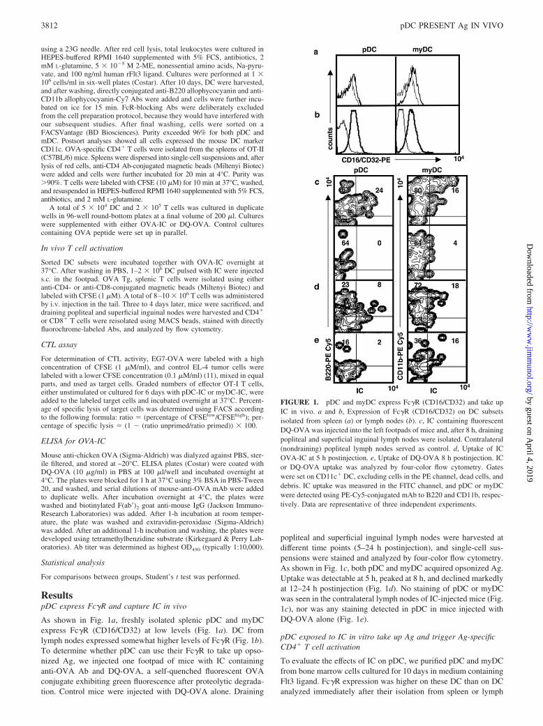
As shown in Fig. 1a, freshly isolated splenic pDC and myDCexpress Fc�R (CD16/CD32) at low levels (Fig. 1a). DC fromlymph nodes expressed somewhat higher levels of Fc�R (Fig. 1b).To determine whether pDC can use their Fc�R to take up opso-nized Ag, we injected one footpad of mice with IC containinganti-OVA Ab and DQ-OVA, a self-quenched fluorescent OVAconjugate exhibiting green fluorescence after proteolytic degrada-tion. Control mice were injected with DQ-OVA alone. Draining
popliteal and superficial inguinal lymph nodes were harvested atdifferent time points (5–24 h postinjection), and single-cell sus-pensions were stained and analyzed by four-color flow cytometry.As shown in Fig. 1c, both pDC and myDC acquired opsonized Ag.Uptake was detectable at 5 h, peaked at 8 h, and declined markedlyat 12–24 h postinjection (Fig. 1d). No staining of pDC or myDCwas seen in the contralateral lymph nodes of IC-injected mice (Fig.1c), nor was any staining detected in pDC in mice injected withDQ-OVA alone (Fig. 1e).
pDC exposed to IC in vitro take up Ag and trigger Ag-specificCD4� T cell activation
To evaluate the effects of IC on pDC, we purified pDC and myDCfrom bone marrow cells cultured for 10 days in medium containingFlt3 ligand. Fc�R expression was higher on these DC than on DCanalyzed immediately after their isolation from spleen or lymph
FIGURE 1. pDC and myDC express Fc�R (CD16/CD32) and take upIC in vivo. a and b, Expression of Fc�R (CD16/CD32) on DC subsetsisolated from spleen (a) or lymph nodes (b). c, IC containing fluorescentDQ-OVA was injected into the left footpads of mice and, after 8 h, drainingpopliteal and superficial inguinal lymph nodes were isolated. Contralateral(nondraining) popliteal lymph nodes served as control. d, Uptake of ICOVA-IC at 5 h postinjection. e, Uptake of DQ-OVA 8 h postinjection. ICor DQ-OVA uptake was analyzed by four-color flow cytometry. Gateswere set on CD11c� DC, excluding cells in the PE channel, dead cells, anddebris. IC uptake was measured in the FITC channel, and pDC or myDCwere detected using PE-Cy5-conjugated mAb to B220 and CD11b, respec-tively. Data are representative of three independent experiments.
3812 pDC PRESENT Ag IN VIVO
by guest on April 4, 2019
http://ww
w.jim
munol.org/
Dow
nloaded from
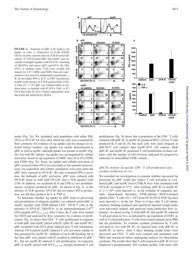
nodes (Fig. 2a). We incubated each population with either DQ-OVA or OVA-IC for 24 h, after which the cells were examined byflow cytometry for evidence of Ag uptake and for changes in se-lected surface markers. Ag uptake was clearly demonstrated inpDC as well as myDC, although uptake was greater in myDC (Fig.2b). For both DC subsets, Ag uptake was accompanied by cellularactivation, based on up-regulation of MHC class II (I-Ab), CD80,and CD40 (Fig. 2b). Some Ag uptake and cellular activation ofpDC occurred when OVA was provided as free protein; however,more Ag acquisition and cellular stimulation were seen when thepDC were exposed to OVA-IC. We also examined IFN-� secre-tion, the hallmark of pDC activation. pDC were cultured withOVA-IC alone or with both OVA-IC and a TLR agonist, CpG2336. In addition, we examined IL-6 and TNF-�, two proinflam-matory cytokines produced by pDC. As shown in Fig. 2c, in theabsence of TLR agonists, OVA-IC did not induce IFN-� produc-tion, not did they produce IL-6 or TNF-�.
To determine whether Ag uptake by pDC leads to processingand presentation of antigenic peptides, we cultured sorted pDC ormyDC together with CFSE-labeled CD4� OT-II T cells in thepresence of OVA-IC, DQ-OVA, or an immunodominant I-Ab/d
OVA-peptide (OVA323–339). After 4 days, the T cells were stainedfor CD25 and analyzed by flow cytometry for evidence of prolif-eration. Fig. 3a shows that CD4� T cells proliferated in responseto both pDC and myDC pulsed with IC (pDC-IC and myDC-IC).pDC incubated with OVA alone induced poor T cell stimulation,whereas OVA-pulsed myDC induced T cell activation similar tothat generated by myDC-IC. Addition of an Fc�R-blocking Ab atthe time pDC were pulsed with IC resulted in inhibition of pDC-IC-, but not myDC-IC-induced T cell proliferation. As expected,pDC or myDC pulsed with OVA323–339 strongly promoted T cell
proliferation. Fig. 3b shows that a proportion of the CD4� T cellscultured with pDC-IC or myDC-IC produced IFN-�. Fewer T cellsproduced IL-4 and IL-10, but such cells were more frequent inpDC-IC/T cell cultures than myDC-IC/T cell cultures. BothpDC-IC and myDC-IC promoted T cell proliferation in these cul-tures, with the number of cell divisions indicated by progressivereduction in intracellular CFSE content.
pDC-IC promote Ag-specific CD4� T cell proliferation andcytokine production in vivo
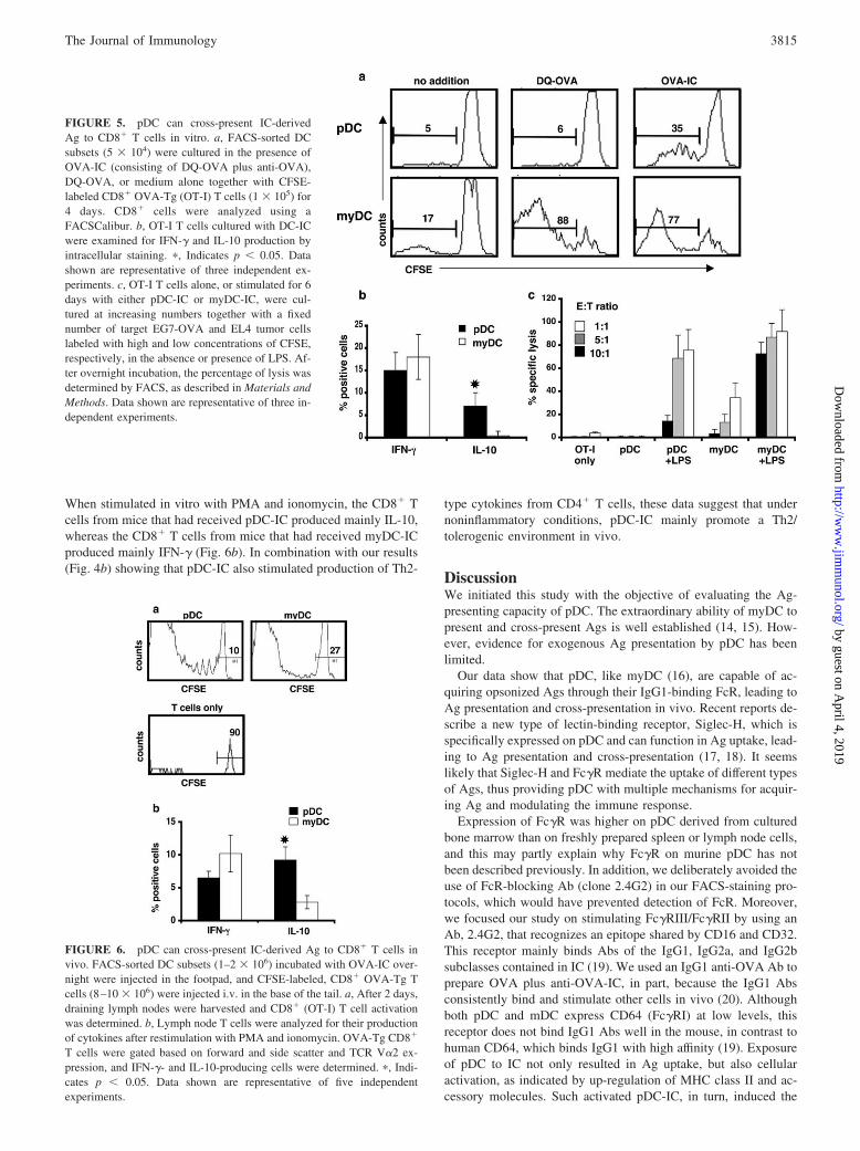
We extended our investigations to examine whether opsonized Agprocessed by pDC could also induce T cell activation in vivo.Sorted pDC and myDC from C57BL/6 mice were incubated withOVA-IC overnight at 37°C. After washing, pDC-IC or myDC-IC(1–2 � 106) were injected s.c. in the footpads of syngeneic ani-mals. Immediately thereafter, CFSE-labeled, MACS-isolatedsplenic CD4� T cells (10 � 106) from OT-II (OVA-TCR Tg) micewere injected i.v. in the tail. Three to 4 days after T cell admin-istration, draining popliteal and superficial inguinal lymph nodeswere harvested, stained, and analyzed using multicolor flow cy-tometry. As shown in Fig. 4, both pDC-IC and myDC-IC inducedT cell activation in vivo, as indicated by up-regulation of CCR7, aswell as T cell proliferation. T cells from control animals given PBSdid not proliferate. To examine cytokine production by T cellsactivated in vivo with DC-IC, we injected mice with pDC-IC ormyDC-IC, as above. After 4 days, draining lymph nodes wereharvested and CD4� T cells were isolated and stimulated withPMA and ionomycin before undergoing analysis for intracellularcytokines. The results show that T cells exposed to pDC-IC in vivodisplayed a predominantly Th2 cytokine profile, with more cells
FIGURE 2. Exposure of pDC to IC leads to Aguptake in vitro. a, Expression of Fc�R (CD16/CD32) on bone marrow-derived, FACS-sorted DCsubsets. b, FACS-sorted pDC and myDC were in-cubated overnight together with OVA-IC consistingof DQ-OVA and mouse IgG1 anti-OVA Ab, DQ-OVA, or medium alone. Cells were washed andstained for I-Ab, CD80, and CD40. Data are repre-sentative of at least five independent experiments. c,IC do not induce IFN-�, IL-6, or TNF-� productionin pDC in the absence of a TLR agonist (CpG 2336).A total of 1 � 105 pDC was cultured either in me-dium alone, or together with IC-OVA, CpG, or IC-OVA plus CpG for 48 h. Culture supernatants wereharvested and analyzed by ELISA.
3813The Journal of Immunology
by guest on April 4, 2019
http://ww
w.jim
munol.org/
Dow
nloaded from
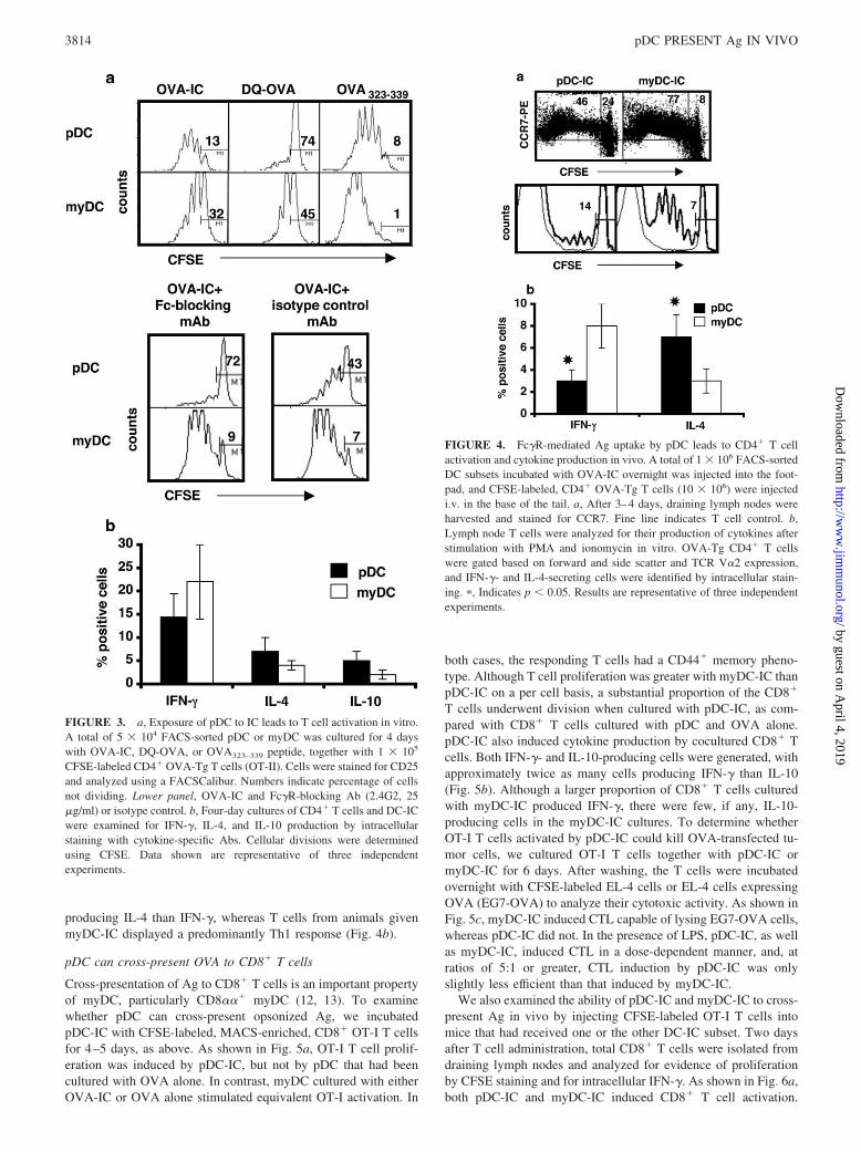
producing IL-4 than IFN-�, whereas T cells from animals givenmyDC-IC displayed a predominantly Th1 response (Fig. 4b).
pDC can cross-present OVA to CD8� T cells
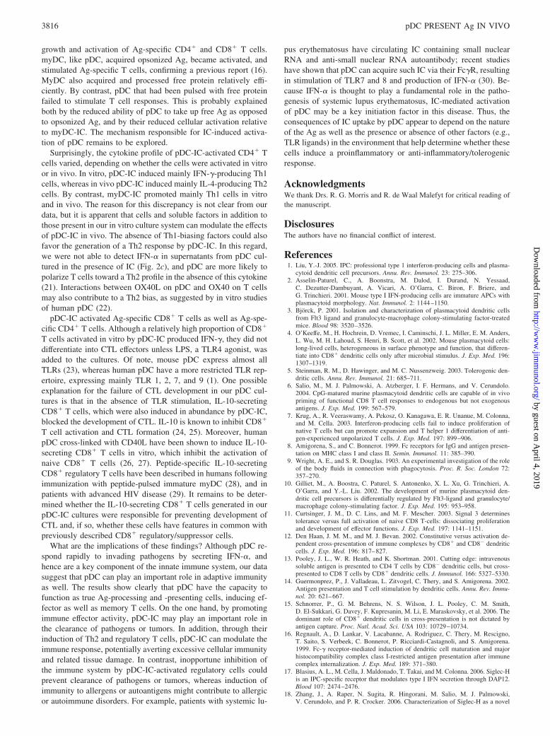
Cross-presentation of Ag to CD8� T cells is an important propertyof myDC, particularly CD8��� myDC (12, 13). To examinewhether pDC can cross-present opsonized Ag, we incubatedpDC-IC with CFSE-labeled, MACS-enriched, CD8� OT-I T cellsfor 4–5 days, as above. As shown in Fig. 5a, OT-I T cell prolif-eration was induced by pDC-IC, but not by pDC that had beencultured with OVA alone. In contrast, myDC cultured with eitherOVA-IC or OVA alone stimulated equivalent OT-I activation. In
both cases, the responding T cells had a CD44� memory pheno-type. Although T cell proliferation was greater with myDC-IC thanpDC-IC on a per cell basis, a substantial proportion of the CD8�
T cells underwent division when cultured with pDC-IC, as com-pared with CD8� T cells cultured with pDC and OVA alone.pDC-IC also induced cytokine production by cocultured CD8� Tcells. Both IFN-�- and IL-10-producing cells were generated, withapproximately twice as many cells producing IFN-� than IL-10(Fig. 5b). Although a larger proportion of CD8� T cells culturedwith myDC-IC produced IFN-�, there were few, if any, IL-10-producing cells in the myDC-IC cultures. To determine whetherOT-I T cells activated by pDC-IC could kill OVA-transfected tu-mor cells, we cultured OT-I T cells together with pDC-IC ormyDC-IC for 6 days. After washing, the T cells were incubatedovernight with CFSE-labeled EL-4 cells or EL-4 cells expressingOVA (EG7-OVA) to analyze their cytotoxic activity. As shown inFig. 5c, myDC-IC induced CTL capable of lysing EG7-OVA cells,whereas pDC-IC did not. In the presence of LPS, pDC-IC, as wellas myDC-IC, induced CTL in a dose-dependent manner, and, atratios of 5:1 or greater, CTL induction by pDC-IC was onlyslightly less efficient than that induced by myDC-IC.
We also examined the ability of pDC-IC and myDC-IC to cross-present Ag in vivo by injecting CFSE-labeled OT-I T cells intomice that had received one or the other DC-IC subset. Two daysafter T cell administration, total CD8� T cells were isolated fromdraining lymph nodes and analyzed for evidence of proliferationby CFSE staining and for intracellular IFN-�. As shown in Fig. 6a,both pDC-IC and myDC-IC induced CD8� T cell activation.
FIGURE 3. a, Exposure of pDC to IC leads to T cell activation in vitro.A total of 5 � 104 FACS-sorted pDC or myDC was cultured for 4 dayswith OVA-IC, DQ-OVA, or OVA323–339 peptide, together with 1 � 105
CFSE-labeled CD4� OVA-Tg T cells (OT-II). Cells were stained for CD25and analyzed using a FACSCalibur. Numbers indicate percentage of cellsnot dividing. Lower panel, OVA-IC and Fc�R-blocking Ab (2.4G2, 25�g/ml) or isotype control. b, Four-day cultures of CD4� T cells and DC-ICwere examined for IFN-�, IL-4, and IL-10 production by intracellularstaining with cytokine-specific Abs. Cellular divisions were determinedusing CFSE. Data shown are representative of three independentexperiments.
FIGURE 4. Fc�R-mediated Ag uptake by pDC leads to CD4� T cellactivation and cytokine production in vivo. A total of 1 � 106 FACS-sortedDC subsets incubated with OVA-IC overnight was injected into the foot-pad, and CFSE-labeled, CD4� OVA-Tg T cells (10 � 106) were injectedi.v. in the base of the tail. a, After 3–4 days, draining lymph nodes wereharvested and stained for CCR7. Fine line indicates T cell control. b,Lymph node T cells were analyzed for their production of cytokines afterstimulation with PMA and ionomycin in vitro. OVA-Tg CD4� T cellswere gated based on forward and side scatter and TCR V�2 expression,and IFN-�- and IL-4-secreting cells were identified by intracellular stain-ing. �, Indicates p � 0.05. Results are representative of three independentexperiments.
3814 pDC PRESENT Ag IN VIVO
by guest on April 4, 2019
http://ww
w.jim
munol.org/
Dow
nloaded from
When stimulated in vitro with PMA and ionomycin, the CD8� Tcells from mice that had received pDC-IC produced mainly IL-10,whereas the CD8� T cells from mice that had received myDC-ICproduced mainly IFN-� (Fig. 6b). In combination with our results(Fig. 4b) showing that pDC-IC also stimulated production of Th2-
type cytokines from CD4� T cells, these data suggest that undernoninflammatory conditions, pDC-IC mainly promote a Th2/tolerogenic environment in vivo.
DiscussionWe initiated this study with the objective of evaluating the Ag-presenting capacity of pDC. The extraordinary ability of myDC topresent and cross-present Ags is well established (14, 15). How-ever, evidence for exogenous Ag presentation by pDC has beenlimited.
Our data show that pDC, like myDC (16), are capable of ac-quiring opsonized Ags through their IgG1-binding FcR, leading toAg presentation and cross-presentation in vivo. Recent reports de-scribe a new type of lectin-binding receptor, Siglec-H, which isspecifically expressed on pDC and can function in Ag uptake, lead-ing to Ag presentation and cross-presentation (17, 18). It seemslikely that Siglec-H and Fc�R mediate the uptake of different typesof Ags, thus providing pDC with multiple mechanisms for acquir-ing Ag and modulating the immune response.
Expression of Fc�R was higher on pDC derived from culturedbone marrow than on freshly prepared spleen or lymph node cells,and this may partly explain why Fc�R on murine pDC has notbeen described previously. In addition, we deliberately avoided theuse of FcR-blocking Ab (clone 2.4G2) in our FACS-staining pro-tocols, which would have prevented detection of FcR. Moreover,we focused our study on stimulating Fc�RIII/Fc�RII by using anAb, 2.4G2, that recognizes an epitope shared by CD16 and CD32.This receptor mainly binds Abs of the IgG1, IgG2a, and IgG2bsubclasses contained in IC (19). We used an IgG1 anti-OVA Ab toprepare OVA plus anti-OVA-IC, in part, because the IgG1 Absconsistently bind and stimulate other cells in vivo (20). Althoughboth pDC and mDC express CD64 (Fc�RI) at low levels, thisreceptor does not bind IgG1 Abs well in the mouse, in contrast tohuman CD64, which binds IgG1 with high affinity (19). Exposureof pDC to IC not only resulted in Ag uptake, but also cellularactivation, as indicated by up-regulation of MHC class II and ac-cessory molecules. Such activated pDC-IC, in turn, induced the
FIGURE 5. pDC can cross-present IC-derivedAg to CD8� T cells in vitro. a, FACS-sorted DCsubsets (5 � 104) were cultured in the presence ofOVA-IC (consisting of DQ-OVA plus anti-OVA),DQ-OVA, or medium alone together with CFSE-labeled CD8� OVA-Tg (OT-I) T cells (1 � 105) for4 days. CD8� cells were analyzed using aFACSCalibur. b, OT-I T cells cultured with DC-ICwere examined for IFN-� and IL-10 production byintracellular staining. �, Indicates p � 0.05. Datashown are representative of three independent ex-periments. c, OT-I T cells alone, or stimulated for 6days with either pDC-IC or myDC-IC, were cul-tured at increasing numbers together with a fixednumber of target EG7-OVA and EL4 tumor cellslabeled with high and low concentrations of CFSE,respectively, in the absence or presence of LPS. Af-ter overnight incubation, the percentage of lysis wasdetermined by FACS, as described in Materials andMethods. Data shown are representative of three in-dependent experiments.
FIGURE 6. pDC can cross-present IC-derived Ag to CD8� T cells invivo. FACS-sorted DC subsets (1–2 � 106) incubated with OVA-IC over-night were injected in the footpad, and CFSE-labeled, CD8� OVA-Tg Tcells (8–10 � 106) were injected i.v. in the base of the tail. a, After 2 days,draining lymph nodes were harvested and CD8� (OT-I) T cell activationwas determined. b, Lymph node T cells were analyzed for their productionof cytokines after restimulation with PMA and ionomycin. OVA-Tg CD8�
T cells were gated based on forward and side scatter and TCR V�2 ex-pression, and IFN-�- and IL-10-producing cells were determined. �, Indi-cates p � 0.05. Data shown are representative of five independentexperiments.
3815The Journal of Immunology
by guest on April 4, 2019
http://ww
w.jim
munol.org/
Dow
nloaded from
growth and activation of Ag-specific CD4� and CD8� T cells.myDC, like pDC, acquired opsonized Ag, became activated, andstimulated Ag-specific T cells, confirming a previous report (16).MyDC also acquired and processed free protein relatively effi-ciently. By contrast, pDC that had been pulsed with free proteinfailed to stimulate T cell responses. This is probably explainedboth by the reduced ability of pDC to take up free Ag as opposedto opsonized Ag, and by their reduced cellular activation relativeto myDC-IC. The mechanism responsible for IC-induced activa-tion of pDC remains to be explored.
Surprisingly, the cytokine profile of pDC-IC-activated CD4� Tcells varied, depending on whether the cells were activated in vitroor in vivo. In vitro, pDC-IC induced mainly IFN-�-producing Th1cells, whereas in vivo pDC-IC induced mainly IL-4-producing Th2cells. By contrast, myDC-IC promoted mainly Th1 cells in vitroand in vivo. The reason for this discrepancy is not clear from ourdata, but it is apparent that cells and soluble factors in addition tothose present in our in vitro culture system can modulate the effectsof pDC-IC in vivo. The absence of Th1-biasing factors could alsofavor the generation of a Th2 response by pDC-IC. In this regard,we were not able to detect IFN-� in supernatants from pDC cul-tured in the presence of IC (Fig. 2c), and pDC are more likely topolarize T cells toward a Th2 profile in the absence of this cytokine(21). Interactions between OX40L on pDC and OX40 on T cellsmay also contribute to a Th2 bias, as suggested by in vitro studiesof human pDC (22).
pDC-IC activated Ag-specific CD8� T cells as well as Ag-spe-cific CD4� T cells. Although a relatively high proportion of CD8�
T cells activated in vitro by pDC-IC produced IFN-�, they did notdifferentiate into CTL effectors unless LPS, a TLR4 agonist, wasadded to the cultures. Of note, mouse pDC express almost allTLRs (23), whereas human pDC have a more restricted TLR rep-ertoire, expressing mainly TLR 1, 2, 7, and 9 (1). One possibleexplanation for the failure of CTL development in our pDC cul-tures is that in the absence of TLR stimulation, IL-10-secretingCD8� T cells, which were also induced in abundance by pDC-IC,blocked the development of CTL. IL-10 is known to inhibit CD8�
T cell activation and CTL formation (24, 25). Moreover, humanpDC cross-linked with CD40L have been shown to induce IL-10-secreting CD8� T cells in vitro, which inhibit the activation ofnaive CD8� T cells (26, 27). Peptide-specific IL-10-secretingCD8� regulatory T cells have been described in humans followingimmunization with peptide-pulsed immature myDC (28), and inpatients with advanced HIV disease (29). It remains to be deter-mined whether the IL-10-secreting CD8� T cells generated in ourpDC-IC cultures were responsible for preventing development ofCTL and, if so, whether these cells have features in common withpreviously described CD8� regulatory/suppressor cells.
What are the implications of these findings? Although pDC re-spond rapidly to invading pathogens by secreting IFN-�, andhence are a key component of the innate immune system, our datasuggest that pDC can play an important role in adaptive immunityas well. The results show clearly that pDC have the capacity tofunction as true Ag-processing and -presenting cells, inducing ef-fector as well as memory T cells. On the one hand, by promotingimmune effector activity, pDC-IC may play an important role inthe clearance of pathogens or tumors. In addition, through theirinduction of Th2 and regulatory T cells, pDC-IC can modulate theimmune response, potentially averting excessive cellular immunityand related tissue damage. In contrast, inopportune inhibition ofthe immune system by pDC-IC-activated regulatory cells couldprevent clearance of pathogens or tumors, whereas induction ofimmunity to allergens or autoantigens might contribute to allergicor autoimmune disorders. For example, patients with systemic lu-
pus erythematosus have circulating IC containing small nuclearRNA and anti-small nuclear RNA autoantibody; recent studieshave shown that pDC can acquire such IC via their Fc�R, resultingin stimulation of TLR7 and 8 and production of IFN-� (30). Be-cause IFN-� is thought to play a fundamental role in the patho-genesis of systemic lupus erythematosus, IC-mediated activationof pDC may be a key initiation factor in this disease. Thus, theconsequences of IC uptake by pDC appear to depend on the natureof the Ag as well as the presence or absence of other factors (e.g.,TLR ligands) in the environment that help determine whether thesecells induce a proinflammatory or anti-inflammatory/tolerogenicresponse.
AcknowledgmentsWe thank Drs. R. G. Morris and R. de Waal Malefyt for critical reading ofthe manuscript.
DisclosuresThe authors have no financial conflict of interest.
References1. Liu, Y.-J. 2005. IPC: professional type 1 interferon-producing cells and plasma-
cytoid dendritic cell precursors. Annu. Rev. Immunol. 23: 275–306.2. Asselin-Paturel, C., A. Boonstra, M. Dalod, I. Durand, N. Yessaad,
C. Dezutter-Dambuyant, A. Vicari, A. O’Garra, C. Biron, F. Briere, andG. Trinchieri. 2001. Mouse type I IFN-producing cells are immature APCs withplasmacytoid morphology. Nat. Immunol. 2: 1144–1150.
3. Bjorck, P. 2001. Isolation and characterization of plasmacytoid dendritic cellsfrom Flt3 ligand and granulocyte-macrophage colony-stimulating factor-treatedmice. Blood 98: 3520–3526.
4. O’Keeffe, M., H. Hochrein, D. Vremec, I. Caminschi, J. L. Miller, E. M. Anders,L. Wu, M. H. Lahoud, S. Henri, B. Scott, et al. 2002. Mouse plasmacytoid cells:long-lived cells, heterogeneous in surface phenotype and function, that differen-tiate into CD8� dendritic cells only after microbial stimulus. J. Exp. Med. 196:1307–1319.
5. Steinman, R. M., D. Hawinger, and M. C. Nussenzweig. 2003. Tolerogenic den-dritic cells. Annu. Rev. Immunol. 21: 685–711.
6. Salio, M., M. J. Palmowski, A. Atzberger, I. F. Hermans, and V. Cerundolo.2004. CpG-matured murine plasmacytoid dendritic cells are capable of in vivopriming of functional CD8 T cell responses to endogenous but not exogenousantigens. J. Exp. Med. 199: 567–579.
7. Krug, A., R. Veeraswamy, A. Pekosz, O. Kanagawa, E. R. Unanue, M. Colonna,and M. Cella. 2003. Interferon-producing cells fail to induce proliferation ofnative T cells but can promote expansion and T helper 1 differentiation of anti-gen-experienced unpolarized T cells. J. Exp. Med. 197: 899–906.
8. Amigorena, S., and C. Bonnerot. 1999. Fc receptors for IgG and antigen presen-tation on MHC class I and class II. Semin. Immunol. 11: 385–390.
9. Wright, A. E., and S. R. Douglas. 1903. An experimental investigation of the roleof the body fluids in connection with phagocytosis. Proc. R. Soc. London 72:357–270.
10. Gilliet, M., A. Boostra, C. Paturel, S. Antonenko, X. L. Xu, G. Trinchieri, A.O’Garra, and Y.-L. Liu. 2002. The development of murine plasmacytoid den-dritic cell precursors is differentially regulated by Flt3-ligand and granulocyte/macrophage colony-stimulating factor. J. Exp. Med. 195: 953–958.
11. Curtsinger, J. M., D. C. Lins, and M. F. Mescher. 2003. Signal 3 determinestolerance versus full activation of naive CD8 T-cells: dissociating proliferationand development of effector functions. J. Exp. Med. 197: 1141–1151.
12. Den Haan, J. M. M., and M. J. Bevan. 2002. Constitutive versus activation de-pendent cross-presentation of immune complexes by CD8� and CD8� dendriticcells. J. Exp. Med. 196: 817–827.
13. Pooley, J. L., W. R. Heath, and K. Shortman. 2001. Cutting edge: intravenoussoluble antigen is presented to CD4 T cells by CD8� dendritic cells, but cross-presented to CD8 T cells by CD8� dendritic cells. J. Immunol. 166: 5327–5330.
14. Guermonprez, P., J. Valladeau, L. Zitvogel, C. Thery, and S. Amigorena. 2002.Antigen presentation and T cell stimulation by dendritic cells. Annu. Rev. Immu-nol. 20: 621–667.
15. Schnorrer, P., G. M. Behrens, N. S. Wilson, J. L. Pooley, C. M. Smith,D. El-Sukkari, G. Davey, F. Kupresanin, M. Li, E. Maraskovsky, et al. 2006. Thedominant role of CD8� dendritic cells in cross-presentation is not dictated byantigen capture. Proc. Natl. Acad. Sci. USA 103: 10729–10734.
16. Regnault, A., D. Lankar, V. Lacabanne, A. Rodriguez, C. Thery, M. Rescigno,T. Saito, S. Verbeek, C. Bonnerot, P. Ricciardi-Castagnoli, and S. Amigorena.1999. Fc-� receptor-mediated induction of dendritic cell maturation and majorhistocompatibility complex class I-restricted antigen presentation after immunecomplex internalization. J. Exp. Med. 189: 371–380.
17. Blasius, A. L., M. Cella, J. Maldonado, T. Takai, and M. Colonna. 2006. Siglec-His an IPC-specific receptor that modulates type I IFN secretion through DAP12.Blood 107: 2474–2476.
18. Zhang, J., A. Raper, N. Sugita, R. Hingorani, M. Salio, M. J. Palmowski,V. Cerundolo, and P. R. Crocker. 2006. Characterization of Siglec-H as a novel
3816 pDC PRESENT Ag IN VIVO
by guest on April 4, 2019
http://ww
w.jim
munol.org/
Dow
nloaded from

endocytic receptor expressed on murine plasmacytoid dendritic cell precursors.Blood 107: 3600–3608.
19. Gessner, J. E., H. Heiken, A. Tamm, and R. E. Schmidt. 1998. The IgG Fcreceptor family. Ann. Hematol. 76: 231–248.
20. Nimmerjahn, F., and J. V. Ravetch. 2006. Fc� receptors: old friends and newfamily members. Immunity 24: 19–28.
21. Bjorck, P. 2004. Dendritic cells exposed to herpes simplex virus in vivo do notproduce IFN-� after rechallenge with virus in vitro and exhibit decreased T cellalloreactivity. J. Immunol. 172: 5396–5404.
22. Ito, T., T. Amakawa, M. Inaba, T. Hori, M. Ota, K. Nakamura, M. Takebayashi,M. Miyaji, T. Yoshimura, K. Inaba, and S. Fukuhara. 2004. Plasmacytoid den-dritic cells regulate Th1 cell responses through OX40 ligand and type I IFNs.J. Immunol. 172: 4253–4259.
23. Edwards, A. D., S. S. Diebold, E. M. Slack, H. Tomizawa, H. Hemmi, T. Kaisho,S. Akira, and C. Reis e Sousa. 2003. Toll-like receptor expression in murine DCsubsets: lack of TLR7 expression by CD8� DC correlates with unresponsivenessto imidazoquinolines. Eur. J. Immunol. 33: 827–833.
24. Steinbrink, K., H. Jonuleit, G. Muller, G. Schuler, J. Knop, and A. H. Enk. 1999.Interleukin-10 treated human dendritic cells induce a melanoma-antigen-specific
anergy in CD8� T cells resulting in a failure to lyse tumor cells. Blood 93:1634–1642.
25. Steinbrink, K., E. Graulich, S. Kunsch, J. Knop, and A. H. Enk. 2002. CD4� andCD8� anergic T cells induced by interleukin-10-treated human dendritic cellsdisplay antigen-specific suppressor activity. Blood 99: 2468–2476.
26. Gilliet, M., and Y.-J. Liu. 2002. Generation of human CD8 T regulatory cells byCD40 ligand-activated plasmacytoid dendritic cells. J. Exp. Med. 195: 695–704.
27. Gilliet, M., and Y.-J. Liu. 2002. Human plasmacytoid dendritic cells and theinduction of T-regulatory cells. Hum. Immunol. 63: 1149–1155.
28. Dhodakpar, M. D., and R. M. Steinman. 2002. Antigen-bearing immature den-dritic cells induce peptide-specific CD8� regulatory T cells in vivo in humans.Blood 100: 174–177.
29. Elrefaei, M., F. L. Ventura, C. A. Baker, C. R. Clark, D. R. Bangsberg, andH. Cao. 2007. HIV-specific IL-10-positive CD8� T cells suppress cytolysis andIL-2 production by CD8� T cells. J. Immunol. 178: 3265–3271.
30. Vollmer, J., S. Tluk, S. Schmitz, S. Hamm, M. Jurk, A. Forsbach, S. Akira,K. M. Kelly, W. H. Reeves, S. Bauer, and A. M. Krieg. 2005. Immune stimu-lation mediated by autoantigen binding sites within small nuclear RNAs involvesToll-like receptors 7 and 8. J. Exp. Med. 202: 1575–1585.
3817The Journal of Immunology
by guest on April 4, 2019
http://ww
w.jim
munol.org/
Dow
nloaded from