Embed Size (px)
Citation preview

RESEARCH Open Access
Targeting of nicotinamidephosphoribosyltransferase enzymaticactivity ameliorates lung damage inducedby ischemia/reperfusion in ratsGeng-Chin Wu1,2, Wen-I Liao3, Shu-Yu Wu4, Hsin-Ping Pao1, Shih-En Tang5, Min-Hui Li6, Kun-Lun Huang4,5*
and Shi-Jye Chu7*
Abstract
Background: Emerging evidence reveals that nicotinamide phosphoribosyltransferase (NAMPT) has a significantrole in the pathophysiology of the inflammatory process. NAMPT inhibition has a beneficial effect in the treatmentof a variety of inflammatory diseases. However, it remains unclear whether NAMPT inhibition has an impact onischemia-reperfusion (I/R)-induced acute lung injury. In this study, we examined whether NAMPT inhibition providedprotection against I/R lung injury in rats.
Methods: Isolated perfused rat lungs were subjected to 40 min of ischemia followed by 60 min of reperfusion. The ratswere randomly allotted to the control, control + FK866 (NAMPT inhibitor, 10 mg/kg), I/R, or I/R + FK866 groups (n = 6per group). The effects of FK866 on human alveolar epithelial cells exposed to hypoxia-reoxygenation (H/R) were alsoinvestigated.
Results: Treatment with FK866 significantly attenuated the increases in lung edema, pulmonary arterial pressure,lung injury scores, and TNF-α, CINC-1, and IL-6 concentrations in bronchoalveolar lavage fluid in the I/R group.Malondialdehyde levels, carbonyl contents and MPO-positive cells in lung tissue were also significantly reducedby FK866. Additionally, FK866 mitigated I/R-stimulated degradation of IκB-α, nuclear translocation of NF-κB, Aktphosphorylation, activation of mitogen-activated protein kinase, and downregulated MKP-1 activity in the injuredlung tissue. Furthermore, FK866 increased Bcl-2 and decreased caspase-3 activity in the I/R rat lungs. Comparably,the in vitro experiments showed that FK866 also inhibited IL-8 production and NF-κB activation in human alveolarepithelial cells exposed to H/R.
Conclusions: Our findings suggest that NAMPT inhibition may be a novel therapeutic approach for I/R-induced lunginjury. The protective effects involve the suppression of multiple signal pathways.
Keywords: Acute lung injury, Ischemia-reperfusion, Visfatin, Nicotinamide phosphoribosyltransferase, pre-B cellcolony-enhancing factor
* Correspondence: [email protected];[email protected] Graduate Institute of Aerospace and Undersea Medicine, NationalDefense Medical Center, Taipei, Taiwan7Department of Internal Medicine, Tri-Service General Hospital, NationalDefense Medical Center, No. 325, Section 2, Chenggong Road, Neihu 114,Taipei, TaiwanFull list of author information is available at the end of the article
© The Author(s). 2017 Open Access This article is distributed under the terms of the Creative Commons Attribution 4.0International License (http://creativecommons.org/licenses/by/4.0/), which permits unrestricted use, distribution, andreproduction in any medium, provided you give appropriate credit to the original author(s) and the source, provide a link tothe Creative Commons license, and indicate if changes were made. The Creative Commons Public Domain Dedication waiver(http://creativecommons.org/publicdomain/zero/1.0/) applies to the data made available in this article, unless otherwise stated.
Wu et al. Respiratory Research (2017) 18:71 DOI 10.1186/s12931-017-0557-2

BackgroundIntracellular nicotinamide phosphoribosyltransferase(NAMPT), also called visfatin or pre-B cell colony-enhancing factor, is the rate-limiting enzyme in the sal-vage pathway for nicotinamide adenine dinucleotide(NAD) biosynthesis. It influences the function of NAD-dependent enzymes such as sirtuins and poly (ADP-ribose)polymerases, which regulate cellular signaling, insulinresistance, apoptosis, oxidative stress response, and in-flammation [1]. All tissues and cells contain NAMPT.The ubiquitous expression of NAMPT implies thepleiotropic actions of proteins in cellular events [1].Moreover, NAMPT can also be found as an extracellu-lar secreted form under inflammatory conditions; it is apotent extracellular proinflammatory inducer of theNF-kB pathway, toll-like receptor signaling, apoptosis,and leukocyte extravasation signaling [2]. Because ofthe upregulation of NAMPT expression in a variety ofinflammatory responses, it is implicated in the pathogen-esis of various inflammatory disorders, such as atheroscler-osis, psoriasis, inflammatory bowel disease, rheumatoidarthritis, and myocardial failure [1]. Recent reports indicatethat a low molecular-weight inhibitor of NAMPT, FK866,improves inflammation-related diseases in an animalmodel, including spinal cord injury, lipopolysaccharide(LPS)-induced myocardial impairment, myocardial in-farction, inflammatory arthritis, endotoxic shock, andautoimmune encephalitis [3–6]. These findings suggestthat NAMPT could be a novel therapeutic target in vari-ous disorders, but a better understanding of its mecha-nisms of action is a prerequisite for the use of a NAMPTinhibitor as a therapeutic option in relevant diseases.Despite advances in research in acute lung injury/acute
respiratory distress syndrome (ALI/ARDS), the fundamentalbasis for ischemia-reperfusion (I/R)-evoked pathophysiologyremains unclear. Recently, NAMPT was demonstrated as apotential novel biomarker in ALI/ARDS via genomic andgenetic studies [7, 8]. The expression of NAMPT in thelungs is markedly increased in human and animal models ofALI, and NAMPT levels are significantly increased in serumand bronchoalveolar lavage fluid (BALF) [7]. Variations inNAMPT polymorphisms were also significantly associatedwith susceptibility to sepsis and ALI [7, 8]. Further-more, heterozygous NAMPT +/- mice were significantlyprotected from the development and severity of ventilator-induced lung injury (VILI) [9]. Moreno-Vinasco et al. re-cently reported that FK866, an inhibitor of NAMPT en-zymatic function, had beneficial effects in VILI and LPS-induced lung injury [10]. Matsuda et al. demonstrated thatFK866 protected against intestinal I/R-associated ALI inmice. The protective effect of FK866 occurred via modula-tion of the NF-κB pathway [11]. These investigationsstrongly support a potentially important role for NAMPTin the inflammatory processes observed in ALI/ARDS.
I/R in the lungs can lead to ALI that was obviouslydifferent from the study of Matsuda et al. that ALI wasassociated with I/R at distant, nonpulmonary sites [11].I/R-induced ALI is the major cause of primary graftdysfunction in the early stages after lung transplant-ation. The morbidity and mortality associated with I/R-induced ALI is still high [12]. Therefore, it is importantto explore the molecular mechanisms of I/R-inducedALI and develop an effective therapy. In this study, wefurther determined the role of NAMPT enzymatic ac-tivity in the pathogenesis of I/R-induced acute lung in-jury using an inhibitor of NAMPT enzymatic function,FK-866.
MethodsIsolated perfused rat lung modelCare of the rats used in this experiment met the guidelinesset forth by the National Institutes of Health (NationalAcademy Press, 1996). The Animal Review Committee ofNational Defense Medical Center approved the studyprotocol. Rat lungs were isolated and perfused as previ-ously described [13–15]. Briefly, Sprague-Dawley male rats(350 ± 20 g) were ventilated with humidified air containing5% CO2 at a tidal volume of 3 ml, a positive end-expiratory pressure of 1 cm H2O, and a rate of 60 breaths/min. After a sternotomy, heparin (1 U/g of body weight,[BW]) was injected into the right ventricle, and 10 mL ofintracardiac blood was withdrawn. The pulmonary arteryand the left ventricle were cannulated and perfused with aphysiological salt solution (119 mM NaCl, 4.7 mM KCl,1.17 mM MgSO4, 22.6 mM NaHCO3, 1.18 mM KH2PO4,1.6 mM CaCl2, 5.5 mM glucose, and 50 mM sucrose) con-taining 4% bovine serum albumin. The 10 ml of collectedblood was added to the perfusate as a “half-blood” solu-tion before recirculation. The constant flow rate of theroller pump was maintained at 8–10 ml/min. The recircu-lating perfusate with the isolated lungs in situ was placedon an electronic balance to record real-time changes inlung weight (LW). The left atrial pressure, representingthe pulmonary venous pressure (PVP), and the pulmonaryarterial pressure (PAP) were continuously monitored fromthe side arm of the cannula.
Vascular filtration coefficientThe vascular filtration coefficient (Kf) was calculated fromthe change in lung weight caused by elevation of venouspressure as described previously [14–16]. Kf was definedas the y-intercept of the plot (g min−1) divided by the PVP(10 cmH2O) and lung weight, and expressed in wholeunits of g · min−1 · cmH2O
−1 × 100 g [14–16].
Lung weight/body weight and wet/dry (W/D) weight ratiosThe right lung was removed after the experiments in thehilar region. The wet lung weight was then determined,
Wu et al. Respiratory Research (2017) 18:71 Page 2 of 12

and the LW/BW ratio was calculated. For the dryweight, a part of the right upper lung lobe was dried for48 h at 60 °C in an oven, and the W/D weight ratio wascalculated.
Assessment of total cell counts, BALF protein, cytokine-induced neutrophil chemoattractant-1(CINC-1), interleukin-6(IL-6), and tumor necrosis factor-α (TNF-α) levelsBALF was obtained by lavaging the left lung twice with2.5 ml of saline after the experiment. The lavage fluidwas centrifuged at 200 × g for 10 min. The protein con-centration in the supernatant was determined using abicinchoninic acid protein assay kit (Pierce, Rockford, IL,USA). The levels of TNF-α, IL-6 and cytokine-inducedneutrophil chemoattractant (CINC)-1 in the BALF weremeasured using a commercial ELISA kit (R&D SystemsInc., Minneapolis, MN, USA). Total cell counts in theBALF were assessed as described previously [15].
Protein carbonyl contents and malondialdehyde levels inlung tissueThe lung tissue was homogenized in a 1.15% KCl aque-ous solution. A 100-μL aliquot of the homogenized lungtissue was mixed into a solution of 200 μL of 8.1%thiobarbituric acid and 700 μL of distilled water. Themixture was then boiled for 30 min at 100 °C and cen-trifuged at 3000 × g for 10 min. The malondialdehydecontent of the supernatant was measured by absorb-ance at 532 nm and was expressed as nmol/mg pro-tein. The oxidative damage to the proteins in the lungtissue was assessed by determining the carbonyl groupcontent based on a reaction with dinitrophenylhidra-zine as previously described [15]. The carbonyl contentwas determined from the absorbance at 370 nm assuminga molar absorption coefficient of 220,000 M−1 and wasexpressed as the concentration of carbonyl derivatives inthe protein (nmol carbonyl/mg protein) [15].
Western blottingLung and cell culture protein lysates (30 μg/lane) wereseparated by 10–12% sodium dodecyl sulfate-polyacryl-amide gel electrophoresis and immunoblots were devel-oped as previously described [14, 15]. The blots wereprobed with primary antibodies against NAMPT (1:2000,Thermo Fisher Scientific, Rockford, IL, USA), B-cell lymph-oma (Bcl)-2 (1:200, Santa Cruz Biotechnology, Dallas,Texas, USA), NF-κB p65, phospho-NF-κB p65, inhibitor ofNF-κB (IκB)-α, extracellular signal-related protein kin-ase 1/2 (ERK1/2), phosho-ERK1/2, c-Jun N-terminalkinase (JNK), phospho-JNK, p38 protein kinase (p38),phospho-p38, and mitogen-activated protein kinasephosphatase-1 (MKP-1) (1:1000, Cell Signaling Technol-ogy, Danvers, MA, USA), proliferating cell nuclear antigen(PCNA) (1:1000, Abcam, Cambridge, MA, USA), and
β-actin (1:10000, Sigma Chemical Company, St. Louis,MO, USA). The data are presented as the relative ratioof the target protein to the reference protein.
Immunohistochemical analysesImmunohistochemical staining to identify myeloperoxi-dase (MPO) and caspase-3 was performed as describedpreviously [15, 17]. Briefly, paraffin-embedded lung tissuesections were deparaffinized before antigen retrieval. Theslides were immersed in 3% H2O2 and 100% methanol for15 min to quench endogenous peroxidase. Immunostain-ing of lung sections was done using a rabbit polyclonalantibody to MPO (1:100, Cell Signaling Technology) andthe large activated fragment (17/19 kD) of caspase-3(1:200; Cell Signaling Technology). The slides werewashed and then incubated with rat-specific horseradishperoxidase polymer anti-rabbit antibody (Nichirei Corpor-ation, Tokyo, Japan) for 30 min. Then, horseradish perox-idase substrate was added and reacted for 3 min, and thesections were counterstained with hematoxylin.
HistopathologyThe lung tissue was histologically prepared and stainedwith hematoxylin and eosin. The numbers of polymorpho-nuclear neutrophils in the interstitium were counted in 10high-power fields (×400) and averaged. Two pathologistsexamined a minimum of 10 randomly selected fields in amasked fashion. Semiquantitative grading of lung injuryon hematoxylin and eosin sections was done as previouslydescribed [15].
Experimental designThe rat lungs were randomly assigned to receive normalsaline (control, n = 6), FK866 (10 mg/kg, drug control,n = 6), I/R (n = 6), or I/R with FK866 (10 mg/kg, n = 6).FK866 (Alexis Biochemicals, San Diego, CA, USA) wasadded to the reservoir (containing 20 mL of perfusate).The doses of FK866 in this study were chosen accord-ing to previous investigations [4, 11]. The isolated lungswere allowed to equilibrate for 20 min before starting.The baseline PAP, PVP, weight change, and the initialKf for 7 min were then measured. All parameters wereequilibrated to baseline for 10 min after the measurements.In the I/R group, the lung preparations were kept at 25 °C.After all of the parameters had returned to the baselinestate, the lungs were deflated by stopping ventilation andperfusion to cause ischemia. They were maintained in thedeflated state for 40 min. Perfusion and ventilation wereresumed, and the Kf was measured 60 min later.
Hypoxia-reoxygenation (H/R) of A549 cellsHuman type II alveolar epithelial cells (A549) were ob-tained from the Food Industry Research and DevelopmentInstitute (BCRC 60074, Hsinchu, Taiwan) and maintained
Wu et al. Respiratory Research (2017) 18:71 Page 3 of 12

in F-12 K medium (Hyclone, Logan, UT, USA) containing10% fetal bovine serum (Hyclone), penicillin, and strepto-mycin in a humidified atmosphere of 5% CO2-95% air.A549 cells were subjected to 24 h of hypoxia (1% O2-5%CO2-94% N2) followed by 4 h of reoxygenation (5%CO2-95% air) at 37 °C [14]. The cells were pretreatedwith vehicle or FK866 (50 nM) [4, 11]. The controlgroup was continued in the reoxygenated state withoutthe hypoxic stimulus. The supernatant was collectedand assayed for IL-8 using a human IL-8 ELISA kit(R&D, Inc., Minneapolis, MN, USA).
Data analysisThe data analysis was performed using GraphPad Prism5 statistical software (GraphPad Software, San Diego, CA,USA). Data are expressed as means ± SD. The comparisonsamong the groups were conducted using one-way ANOVAfollowed by a post-hoc Bonferroni test. Two-way ANOVAfor repeated measurements followed by the post-hocBonferroni test was using for comparisons of lung weightgain and PAP between groups. The significance level wasdefined as P < 0.05.
ResultsEffect of FK866 on indices of lung edemaI/R significantly raised lung weight gain (Fig. 1a). TheFK866 treatment reduced this increase in the lung weightgain. I/R significantly increased Kf, LW/BW and W/Dweight ratios, and protein concentrations in the BALF(p < 0.05, Fig. 1b–e); FK866 treatment significantly mit-igated these increases.
Effect of FK866 on PAPIn the control group, the PAP remained steady duringthe 100-min observation period. In the I/R group, thePAP initially rose and then declined after reperfusion.After 60 min of reperfusion, the PAP in the I/R groupwas significantly higher than at baseline and that of thecontrol group. Treatment with FK866 significantly dimin-ished the increase of PAP in the I/R group (p < 0.05; Fig. 2).
Effect of FK866 on NAMPT protein expression in lung tissueThere was faint or weak immunostaining of NAMPT inthe lung sections of the control animals. In contrast,lung tissues had strong NAMPT staining after I/R injury.FK866 treatment reduced the intensity of staining in thelungs (Fig. 3a). Furthermore, I/R significantly increasedNAMPT protein expression compared with that of thecontrol group (p < 0.05; Fig. 3b). FK866 treatment signifi-cantly decreased NAMPT protein expression in the I/Rgroup.
Effect of FK866 on CINC-1, TNF-α, and IL-6 concentrations,and total cell counts in the BALFThe concentrations of TNF-α, CINC-1 and IL-6, andtotal cell counts were significantly increased in the BALFof the I/R group compared with that of control group(p < 0.05; Fig. 4). FK866 significantly inhibited the I/R-mediated increases of TNF-α, CINC-1, IL-6 and totalcell counts in the BALF (p < 0.05; Fig. 4).
Effect of FK866 on carbonyl content, malondialdehydelevel, and MPO-positive cells in lung tissueCompared with the control group, the I/R group hadsignificantly increased malondialdehyde levels, carbonylcontents, and numbers of MPO-positive cells in the lungtissue (p < 0.05, Fig. 5a–c). Treatment with FK866 sig-nificantly mitigated these increases.
Effect of FK866 on lung pathologyThe histological studies disclosed distinct morphologicalevidence of lung injury, including thickening of the alveo-lar walls characterized by interstitial edema and leukocyteinfiltrates in the I/R group compared with the controlgroup (Fig. 6a). FK866 treatment significantly reducedhistological changes, neutrophil infiltration (Fig. 6b), andlung injury scores (Fig. 6c) in the I/R group.
Effects of FK866 on Bcl-2 and caspase-3 protein expressionin lung tissueThe intensity of activated caspase-3-immunolabelled cellswas significantly greater in the I/R group than in the con-trol group. FK866 treatment significantly decreased thenumber of caspase-3 immunolabeled cells (Fig. 7a). Bcl-2protein content in lung tissue was substantially reduced inthe I/R groups compared with the control group. FK866treatment significantly mitigated the decline of Bcl-2 levelsin the lung tissue (Fig. 7b).
Effect of FK866 on the mitogen-activated protein kinase(MAPK) signaling pathway and MKP-1 induction in lungtissueI/R significantly increased activation of the mitogen-acti-vated protein kinase (MAPK) pathway, including ERK,JNK, and p38 phosphorylation in lung tissue. All threeMAPKs activated by I/R were reduced by the administra-tion of FK866 (Fig. 8a–c). In contrast, the MKP-1 proteinlevel in lung tissue was significantly lower in the I/Rgroups than in the control groups but was significantlyenhanced with FK866 treatment (Fig. 8d).
Effect of FK866 on the NF-κB signaling pathwayThe levels of NF-κB p65 in the nucleolus and Akt phos-phorylation were significantly increased (Fig. 9a, c), whereasthe level of IκB-α in the cytoplasm was significantly de-creased in the I/R group compared with the control
Wu et al. Respiratory Research (2017) 18:71 Page 4 of 12

group (Fig. 9b). FK866 treatment significantly increasedIκB-α levels, and attenuated NF-κB p65 levels and Aktphosphorylation.
Effect of FK866 in A549 epithelial cells subjected to H/RFK866 significantly reduced the increase of phospho-NF-κB p65 and the decrease of IκB-α at 2 h and 4 h afterH/R in A549 cells (Fig. 10a-c). Furthermore, FK-866 sig-nificantly reduced the levels of IL-8 at 4 h in the H/Rgroup (Fig. 10d).
DiscussionThe results of our study demonstrated that FK866, aninhibitor of NAMPT enzymatic function, significantly
ameliorated multiple indices of I/R-induced acute lunginjury, including vascular barrier dysfunction, PAP, pul-monary neutrophil influx, BALF TNF-α, CINC-1, andIL-6 production, oxidative stress, apoptosis, and tissuedamage compared with control animals. In addition,FK866 inhibited I/R-induced MAPK and Akt activation,IκB-α degradation, and nuclear translocation of NF-κB.Moreover, FK866 treatment had a similar beneficial effecton A549 epithelial cells subjected to H/R, corroboratingthe observations in the rat lung tissues. This indicates thatFK-866 exerts its protective effects through multiple sig-naling cascades. Our experiments revealed the importanceof NAMPT in the pathophysiology of I/R-induced lunginjury.
Fig. 1 Effect of FK866 on pulmonary edema. Lung weight gain (a), Kf (b), lung wet/dry (W/D) weight ratios (c), lung weight/body weight (LW/BW)ratios (d), and protein concentrations in the bronchoalveolar lavage fluid (BALF) (e) significantly increased in the ischemia-reperfusion (IR)group. Treatment with FK866 significantly attenuated the increase in these parameters. Data are expressed as mean ± SD (n = 6 per group).***p < 0.001, compared with the control group; +++p < 0.001, compared with the IR group
Wu et al. Respiratory Research (2017) 18:71 Page 5 of 12

After I/R injury, there was a significant increase in theNAMPT protein expression in the lung tissue, similar tothe results of previous studies showing that NAMPTprotein was highly upregulated in VILI and intestinalischemia-reperfusion [7, 11]. This could be due to infil-tration of activated inflammatory cells with upregulatedNAMPT expression into injured lung tissue [9]. Upregu-lation of NAMPT expression is also observed in cyto-kine, LPS, mechanical stress–challenged human lungendothelial cells and human alveolar epithelial cells, andLPS-stimulated RAW264.7 cells [7, 11, 18]. These resultsstrongly support a significant role for NAMPT in the
inflammatory events observed in ALI/ARDS. Moreover,we showed that NAMPT inhibition by FK-866 attenuatedI/R-induced increases in NAMPT protein. This attenu-ation may block various I/R-associated inflammatory re-sponses, thereby leading to a significant reduction of lungdamage. This finding is consistent with a previous studyshowing that FK866 decreased the degree of positivestaining of NAMPT in the spinal cord of mice subjectedto spinal cord injury [19].Vast evidence has demonstrated that oxidative stress
has a significant contribution in the pathogenesis ofALI/ARDS [20]. In addition, neutrophil-derived oxygenradicals disrupt endothelial barrier function and integ-rity, and increase plasma leakage and lung tissue edema.Our data demonstrated that FK-866 suppressed oxida-tive stress as reflected by attenuating the protein carbon-ylation and peroxidation of membrane lipids in I/R lungtissue. In addition, FK-866 attenuated I/R-evoked increasedneutrophil infiltration in the lung tissue, as evidenced bydiminishing numbers of neutrophils and MPO-positivecells. This attenuation blocks the interaction between neu-trophils and the endothelium, and reduces the productionof proinflammatory cytokines and free radicals by acti-vated neutrophils. Therefore, the anti-oxidative andanti-inflammatory effects of FK-866 appeared to attenu-ate lung edema as shown by the lower W/D and LW/BW ratios, reduced Kf, and decreased protein concen-tration in the BALF. These results agreed with those ofother investigations showing that NAMPT inhibitorshave the ability to attenuate vascular permeability andneutrophil infiltration in VILI, intestinal I/R, and LPS-induced lung injuries [10, 11].
Fig. 2 Effect of FK866 on pulmonary artery pressure (ΔPAP). PAPincreased significantly in the ischemia-reperfusion (IR) group. Theincrease in PAP was attenuated significantly by treatment with FK866.Data are expressed as mean ± SD (n = 6 per group). ***p < 0.001,compared with the control group; +++p < 0.001, compared withthe IR group
Fig. 3 Effect of FK866 on NAMPT protein expression in lung tissue. a Immunohistochemistry for NAMPT in the lung (indicated with arrowhead)(200× magnification). b Western blot and densitometry analysis of NAMPT protein in the lung tissue. β-actin served as loading control forcytoplasmic proteins. Representative blots are shown. Ischemia-reperfusion (IR) significantly increased positive staining and protein expression ofNAMPT in the lung tissue. FK866 significantly decreased the degree of NAMPT positive staining and protein expression. Data are expressed asmean ± SD (n = 6 per group). **p < 0.01, compared with the control group; +p < 0.05, compared with the IR group
Wu et al. Respiratory Research (2017) 18:71 Page 6 of 12

Previous investigations have implicated a complexnetwork of inflammatory cytokines and chemokines inmediating, amplifying, and perpetuating the lung injuryprocess [12]. Our experiment showed that FK-866 signifi-cantly attenuated the increased levels of inflammatory me-diators such as proinflammatory TNF-α, CINC-1 and IL-6
in the BALF after I/R-induced lung injury. Our findingswere also comparable with those in previous investigationsshowing that FK866 alleviated TNF-α production, therebyreducing inflammation, and preventing the I/R lung injury[11, 19]. In parallel with these results, FK-866 signifi-cantly suppressed caspase-3 activation and increased
Fig. 4 Effect of FK866 on CINC-1, TNF-α, and IL-6 levels, and total cell counts in bronchoalveolar lavage fluid (BALF). CINC-1 (a), TNF-α (b) and IL-6(c) levels, and total cell counts (d) in the BALF significantly increased in the ischemia-reperfusion (IR) group. Treatment with FK866 significantlyattenuated these increases in the BALF. Data are expressed as mean ± SD (n= 6 per group). ***p< 0.001, compared with the control group; +++p< 0.001,compared with the IR group
Fig. 5 Effect of FK866 on protein carbonyl contents, MDA levels, and MPO-positive cells in lung tissue. MPO-positive cells (a), carbonyl contents(b), and MDA levels (c) in lung tissue significantly increased in the ischemia-reperfusion (IR) group. FK866 treatment significantly attenuated theseincreases. a Immunohistochemistry for MPO in the lung (indicated with arrowhead) (200× magnification). Data are expressed as mean ± SD (n = 6per group). ***p < 0.001, compared with the control group; ++p < 0.01, +++p < 0.001, compared with the IR group
Wu et al. Respiratory Research (2017) 18:71 Page 7 of 12

anti-apoptotic expression of Bcl-2 after I/R lung injury.The observations were also similar to those seen in in-testinal I/R induced lung injury and spinal cord injury[11, 19]. FK866 has been employed as an antitumoragent through NAD and subsequent ATP depletion,resulting in apoptosis in many malignant cell lines [21].TNF-α, one of the major inflammatory mediators inALI/ARDS, can initiate the apoptotic cascade throughthe death receptor/caspase pathway [22]. Because FK866inhibits TNF-α production, it is reasonable to speculatethat FK866 inhibits apoptosis in I/R-induced ALI, at leastpartly, through an indirect pathway [11]. Furthermore, thisdiscrepancy suggests that FK866 has different effectormechanisms for apoptosis of tumor cells and suppressionof inflammatory reactions in inflammatory cells [4].NF-kB is a master regulator of inflammatory responses
because it activates the transcription of a cascade of pro-inflammatory cytokines and chemokines. The activity ofNF-κB is regulated primarily by the IκB family of inhibi-tory proteins, which are conjugated with NF-κB in the
cytoplasm [23]. Inappropriate activation of NF-κB isimplicated in the pathogenesis of ALI/ARDS. Akt alsoparticipates in signaling pathways that lead to NF-κBactivation and increased NF-κB-dependent transcription[24]. Moreover, Akt-dependent events contribute to thedevelopment and perpetuation of ALI [25]. Our prior in-vestigations revealed that I/R-induced lung damage causedIκB degradation and NF-κB activation [15, 26]. Hong et al.demonstrated that NAMPT +/− mice exhibited a dramaticattenuation of the VILI-mediated NF-κB pathway in thelungs [9]. In the present experiment, FK866 suppressedthe activation of the NF-κB signaling pathway in the ratlungs exposed to I/R by inhibiting Akt phosphorylation,degradation of IκBα and nuclear translocation of NF-κB.The inhibition of NF-κB activity resulted in decreasedproduction of proinflammatory cytokines such as TNF-α,CINC-1, and IL-6. Furthermore, we conducted in vitrocell culture studies by using the A549 epithelial cell line toelucidate the direct effects of FK866 on alveolar epithelialcells. In line with the findings in the rat lungs, FK866
Fig. 6 Effect of FK866 on lung pathology. As shown by a representative micrograph of lung tissue (400× magnification) (a), neutrophil infiltrationand septal edema were increased in the ischemia-reperfusion (IR) group. FK866 treatment significantly attenuated these histopathological changes, thenumbers of neutrophils per high power field (400× magnification) (b), and the lung injury scores (c). Data are expressed as mean ± SD (n= 6 per group).***p< 0.001, compared with the control group; +++p < 0.001, compared with the IR group
Wu et al. Respiratory Research (2017) 18:71 Page 8 of 12
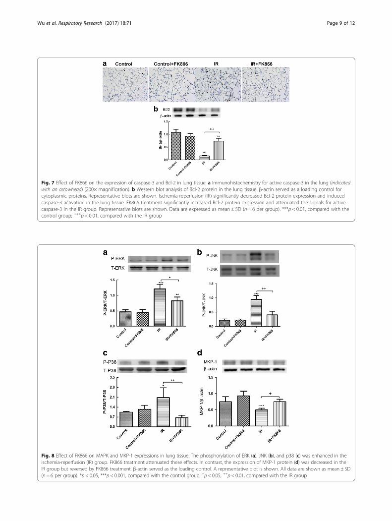
Fig. 7 Effect of FK866 on the expression of caspase-3 and Bcl-2 in lung tissue. a Immunohistochemistry for active caspase-3 in the lung (indicatedwith an arrowhead) (200× magnification). b Western blot analysis of Bcl-2 protein in the lung tissue. β-actin served as a loading control forcytoplasmic proteins. Representative blots are shown. Ischemia-reperfusion (IR) significantly decreased Bcl-2 protein expression and inducedcaspase-3 activation in the lung tissue. FK866 treatment significantly increased Bcl-2 protein expression and attenuated the signals for activecaspase-3 in the IR group. Representative blots are shown. Data are expressed as mean ± SD (n = 6 per group). ***p < 0.01, compared with thecontrol group; +++p < 0.01, compared with the IR group
Fig. 8 Effect of FK866 on MAPK and MKP-1 expressions in lung tissue. The phosphorylation of ERK (a), JNK (b), and p38 (c) was enhanced in theischemia-reperfusion (IR) group. FK866 treatment attenuated these effects. In contrast, the expression of MKP-1 protein (d) was decreased in theIR group but reversed by FK866 treatment. β-actin served as the loading control. A representative blot is shown. All data are shown as mean ± SD(n = 6 per group). *p < 0.05, ***p < 0.001, compared with the control group; +p < 0.05, ++p < 0.01, compared with the IR group
Wu et al. Respiratory Research (2017) 18:71 Page 9 of 12

significantly inhibited IκBα degradation and, consequently,NF-κB p65 phosphorylation, and the production of IL-8 inA549 cells exposed to H/R. This finding was also consist-ent with an investigation by Matsuda et al. demonstratingthat FK866 inhibited NF-κB activation in mouse lungssubjected to intestinal I/R, and LPS-stimulated RAW264.7cells [11]. However, in an experimental compressionmodel of spinal cord injury, FK866 treatment preventedthe activation of NF-κB but not IκBα degradation [19].Therefore, the precise molecular mechanisms by whichNAMPT inhibition exerts its effect in the NF-κB signalingpathway need clarification.The activation of MAPKs such as p38, ERK, and JNK
is implicated in the inflammatory process of ALI/ARDS.The inhibition of p38, ERK, and JNK MAPK, effectivelydiminishes LPS and peritonitis-induced lung inflamma-tion [27–29]. The MAPK signaling pathways are regu-lated by opposing regulatory repressors from MKP-1[30]. MKP-1 knockout mice had increased inflammatoryresponses with higher levels of inflammatory mediatorsand more episodes of multiple organ failure after LPSchallenge [31]. In our previous study, I/R induced phos-phorylation of p38, ERK, and JNK; it also decreased thelevel of MKP-1 protein in lung tissue [17]. In contrast,FK-866 treatment activated MKP-1 expression and inter-rupted I/R-induced activation of MAPK. This may conse-quently restrain widespread inflammation in I/R-inducedlung injury. Extracellular NAMPT is reported to triggerp38, ERK, and JNK phosphorylation and stimulates di-verse biological processes in various types of cells [1, 32].However, whether intracellular NAMPT triggers the sameintracellular pathways remains unknown.The function of NAMPT in various diseases is not
completely recognized. One reason for this lack of claritycould be the inability to discriminate between the intra-cellular and extracellular actions of NAMPT. NAMPT isenzymatically active both intracellularly and extracellu-larly [32]. Extracellular NAMPT may also act in a non-enzymatic way to regulate activation of inflammatorycells by increasing surface expression of costimulatorymolecules and inducing IL-1β, IL-6, and TNF-α productionthrough a currently unidentified membrane receptor [32].The fundamental pathological event of ischemic stroke isthe loss of blood supply and subsequent oxygen/nutritionshortage, which are similar in key pathophysiological
Fig. 9 Effect of FK866 on NF-κB activation and Akt phosphorylationin lung tissues. FK866 reduced Akt phosphorylation (a) and nuclearNF-κB p65 levels (b), and increased IκB-α levels (c) in ischemia-reperfusion (IR)-induced lung injury. PCNA and β-actin served asloading controls for nuclear and cytoplasmic proteins, respectively.Representative blots are shown. Data are expressed as mean ± SD(n = 6 per group). *p < 0.05, ***p < 0.001, compared with the controlgroup; ++p < 0.01, +++p < 0.001, compared with the IR group
Wu et al. Respiratory Research (2017) 18:71 Page 10 of 12

processes to I/R lung injury. Accumulating evidence fromin vitro and in vivo experiments reveals that NAMPT pro-vides cerebral protection in ischemic stroke [3]. NAMPTinhibition exacerbated brain infarction in a rat model of is-chemic stroke, whereas local NAMPT overexpression inthe brain and NAMPT enzymatic action protected againstischemia-induced cerebral strokes [3]. Therefore, fur-ther investigations are needed to explore these contra-dictory findings.
ConclusionsCollectively, we demonstrated that FK866, a NAMPTinhibitor attenuated lung I/R injury by decreasing lungedema, production of inflammatory cytokines, reactiveoxygen species, apoptosis, and NFκB and MAPK signaling.The protective actions of FK866 in this study displayed apleiotropic manner. Therefore, the pharmacological inhib-ition of NAMPT might serve as an effective approach forthe treatment of I/R-induced lung injury. A better under-standing of its physiological action is a prerequisite for theuse of NAMPT inhibitors such as FK866 as a therapeuticoption in these inflammatory diseases.
AbbreviationsALI: Acute lung injury; AnxA1: Annexin A1; ARDS: Acute respiratory distresssyndrome; BALF: Bronchoalveolar lavage fluid; Bcl-2: B-cell lymphoma-2;CINC-1: Cytokine-induced neutrophil chemoattractant-1; ERK: Extracellularsignal-related protein kinase; H/R: Hypoxia-reoxygenation; I/R: Ischemia/reperfusion; IκB-α: Inhibitor of NF-κB-α; JNK: c-Jun N-terminal kinase;Kf: Vascular filtration coefficient; LPS: Lipopolysaccharide; LW/BW: Lung weight tobody weight ratio; MKP-1: Mitogen activated protein kinase phoshphotases-1;MPO: Myeloperoxidase; NAD: Nicotinamide adenine dinucleotide;NAMPT: Nicotinamide phosphoribosyltransferase; p38: p38 protein kinase;PAP: Pulmonary arterial pressure; PVP: Pulmonary venous pressure; VILI: Ventilator-induced lung injury; W/D: Wet weight to dry weight ratio
AcknowledgementsNot applicable.
FundingThis study was supported, in part, by grants MOST 103-2314-B-016-029-MY3,MOST 105-2314-B-282-001, and NSC-102-2314-B-016-034 from Ministry ofScience and Technology, Taiwan, TSGH-C106-53, TSGH-C105-062 andTSGH-C104-067 from Tri-Service General Hospital, grants 10401 and 10501from Taoyuan Armed Forces General Hospital, and MAB-104-052 from theNational Defense Medical Center, Taiwan.
Availability of data and materialsPlease contact author for data requests.
Authors’ contributionsConceived and designed the experiments: GCW, SYW, MHL, SJC. Performedthe experiments: GCW, SYW, Analyzed the data: HPP, SET. Contributed
Fig. 10 Effect of FK866 on A549 cells subjected to hypoxia-reoxygenation (H/R). a A representative Western blot of NF-κB nuclear translocationin the lung tissue. β-actin served as the loading control. FK866 significantly reduced the increase of degradation of IκB-α (b), phosphorylatedNF-κB p65 (c) at 2 h and 4 h, and IL-8 production (d) at 4 h in A549 cells exposed to H/R. Data are expressed as mean ± SD (n = 6). *p < 0.05,**p < 0.01, ***p < 0.001 compared with the control group. +p < 0.05, +++p < 0.001, compared with the H/R group
Wu et al. Respiratory Research (2017) 18:71 Page 11 of 12

reagents/analysis tools: GCW. Wrote the manuscript: KLH, SJC. All authorsread and approved the final manuscript.
Competing interestsAll authors declare that they have no competing interests.
Consent for publicationNot applicable.
Ethics approvalNo human participants, human data or human tissue involved in this study.All animal studies were approved by the Animal Review Committee ofNational Defense Medical Center.
Publisher’s NoteSpringer Nature remains neutral with regard to jurisdictional claims inpublished maps and institutional affiliations.
Author details1The Graduate Institute of Medical Sciences, National Defense MedicalCenter, Taipei, Taiwan. 2Department of Internal Medicine, Taoyuan ArmedForces General Hospital, Taoyuan, Taiwan. 3Department of EmergencyMedicine, Tri-Service General Hospital, Taipei, Taiwan. 4The Graduate Instituteof Aerospace and Undersea Medicine, National Defense Medical Center,Taipei, Taiwan. 5Division of Pulmonary and Critical Care Medicine,Department of Internal Medicine, Tri-Service General Hospital, Taipei, Taiwan.6Department of Physical Medicine and Rehabilitation, Kaohsiung VeteransGeneral Hospital, Kaohsiung, Taiwan. 7Department of Internal Medicine,Tri-Service General Hospital, National Defense Medical Center, No. 325,Section 2, Chenggong Road, Neihu 114, Taipei, Taiwan.
Received: 6 September 2016 Accepted: 18 April 2017
References1. Dahl TB, Holm S, Aukrust P, Halvorsen B. Visfatin/NAMPT: a multifaceted
molecule with diverse roles in physiology and pathophysiology. Annu RevNutr. 2012;32:229–43.
2. Sun Z, Lei H, Zhang Z. Pre-B cell colony enhancing factor (PBEF), acytokine with multiple physiological functions. Cytokine Growth Factor Rev.2013;24:433–42.
3. Wang P, Miao CY. NAMPT as a Therapeutic Target against Stroke. TrendsPharmacol Sci. 2015;36:891–905.
4. Busso N, Karababa M, Nobile M, Rolaz A, Van Gool F, Galli M, Leo O, So A,De Smedt T. Pharmacological inhibition of nicotinamidephosphoribosyltransferase/visfatin enzymatic activity identifies a newinflammatory pathway linked to NAD. PLoS One. 2008;3:e2267.
5. Montecucco F, Bauer I, Braunersreuther V, Bruzzone S, Akhmedov A, Luscher TF,Speer T, Poggi A, Mannino E, Pelli G, et al. Inhibition of nicotinamidephosphoribosyltransferase reduces neutrophil-mediated injury in myocardialinfarction. Antioxid Redox Signal. 2013;18:630–41.
6. Van Gool F, Galli M, Gueydan C, Kruys V, Prevot PP, Bedalov A, Mostoslavsky R, AltFW, De Smedt T, Leo O. Intracellular NAD levels regulate tumor necrosis factorprotein synthesis in a sirtuin-dependent manner. Nat Med. 2009;15:206–10.
7. Ye SQ, Simon BA, Maloney JP, Zambelli-Weiner A, Gao L, Grant A, Easley RB,McVerry BJ, Tuder RM, Standiford T, et al. Pre-B-cell colony-enhancing factoras a potential novel biomarker in acute lung injury. Am J Respir Crit CareMed. 2005;171:361–70.
8. Bajwa EK, Yu CL, Gong MN, Thompson BT, Christiani DC. Pre-B-cell colony-enhancing factor gene polymorphisms and risk of acute respiratory distresssyndrome. Crit Care Med. 2007;35:1290–5.
9. Hong SB, Huang Y, Moreno-Vinasco L, Sammani S, Moitra J, Barnard JW, MaSF, Mirzapoiazova T, Evenoski C, Reeves RR, et al. Essential role of pre-B-cellcolony enhancing factor in ventilator-induced lung injury. Am J Respir CritCare Med. 2008;178:605–17.
10. Moreno-Vinasco L, Quijada H, Sammani S, Siegler J, Letsiou E, Deaton R,Saadat L, Zaidi RS, Messana J, Gann PH, et al. Nicotinamidephosphoribosyltransferase inhibitor is a novel therapeutic candidate inmurine models of inflammatory lung injury. Am J Respir Cell Mol Biol.2014;51:223–8.
11. Matsuda A, Yang WL, Jacob A, Aziz M, Matsuo S, Matsutani T, Uchida E,Wang P. FK866, a visfatin inhibitor, protects against acute lung injury afterintestinal ischemia-reperfusion in mice via NF-kappaB pathway. Ann Surg.2014;259:1007–17.
12. de Perrot M, Liu M, Waddell TK, Keshavjee S. Ischemia-reperfusion-inducedlung injury. Am J Respir Crit Care Med. 2003;167:490–511.
13. Chu SJ, Chang DM, Wang D, Chen YH, Hsu CW, Hsu K. Fructose-1,6-diphosphate attenuates acute lung injury induced by ischemia-reperfusionin rats. Crit Care Med. 2002;30:1605–9.
14. Wu SY, Li MH, Ko FC, Wu GC, Huang KL, Chu SJ. Protective effect ofhypercapnic acidosis in ischemia-reperfusion lung injury is attributable toupregulation of heme oxygenase-1. PLoS One. 2013;8:e74742.
15. Wu SY, Tang SE, Ko FC, Wu GC, Huang KL, Chu SJ. Valproic acid attenuatesacute lung injury induced by ischemia-reperfusion in rats. Anesthesiology.2015;122:1327–37.
16. Wu SY, Wu CP, Kang BH, Li MH, Chu SJ, Huang KL. Hypercapnic acidosisattenuates reperfusion injury in isolated and perfused rat lungs. Crit CareMed. 2012;40:553–9.
17. Hsu HH, Wu SY, Tang SE, Wu GC, Li MH, Huang KL, Chu SJ. Protectionagainst reperfusion lung injury via aborgating multiple signaling cascadesby trichostatin A. Int Immunopharmacol. 2015;25:267–75.
18. Liu P, Li H, Cepeda J, Zhang LQ, Cui X, Garcia JG, Ye SQ. Critical role of PBEFexpression in pulmonary cell inflammation and permeability. Cell Biol Int.2009;33:19–30.
19. Esposito E, Impellizzeri D, Mazzon E, Fakhfouri G, Rahimian R, Travelli C, Tron GC,Genazzani AA, Cuzzocrea S. The NAMPT inhibitor FK866 reverts the damage inspinal cord injury. J Neuroinflammation. 2012;9:66.
20. McCord JM. Oxygen-derived free radicals in postischemic tissue injury. NEngl J Med. 1985;312:159–63.
21. Galli U, Travelli C, Massarotti A, Fakhfouri G, Rahimian R, Tron GC, Genazzani AA.Medicinal chemistry of nicotinamide phosphoribosyltransferase (NAMPT)inhibitors. J Med Chem. 2013;56:6279–96.
22. Tang PS, Mura M, Seth R, Liu M. Acute lung injury and cell death: how manyways can cells die? Am J Physiol Lung Cell Mol Physiol. 2008;294:L632–641.
23. Liu SF, Malik AB. NF-kappa B activation as a pathological mechanism of septicshock and inflammation. Am J Physiol Lung Cell Mol Physiol. 2006;290:L622–45.
24. Romashkova JA, Makarov SS. NF-kappaB is a target of AKT in anti-apoptoticPDGF signalling. Nature. 1999;401:86–90.
25. Yum HK, Arcaroli J, Kupfner J, Shenkar R, Penninger JM, Sasaki T, Yang KY,Park JS, Abraham E. Involvement of phosphoinositide 3-kinases in neutrophilactivation and the development of acute lung injury. J Immunol.2001;167:6601–8.
26. Peng CK, Huang KL, Wu CP, Li MH, Hu YT, Hsu CW, Tsai SH, Chu SJ. Glutamineprotects ischemia-reperfusion induced acute lung injury in isolated rat lungs.Pulm Pharmacol Ther. 2011;24:153–61.
27. Asaduzzaman M, Wang Y, Thorlacius H. Critical role of p38 mitogen-activatedprotein kinase signaling in septic lung injury. Crit Care Med. 2008;36:482–8.
28. Chen LW, Tseng HT, Chen PH, Hsu CM. Peritonitis-induced peroxynitrite andlung damage depends on c-Jun NH2-terminal kinase signaling of hematopoieticcells. Crit Care Med. 2010;38:1168–78.
29. Schuh K, Pahl A. Inhibition of the MAP kinase ERK protects fromlipopolysaccharide-induced lung injury. Biochem Pharmacol. 2009;77:1827–34.
30. Cao W, Bao C, Padalko E, Lowenstein CJ. Acetylation of mitogen-activatedprotein kinase phosphatase-1 inhibits Toll-like receptor signaling. J Exp Med.2008;205:1491–503.
31. Zhao Q, Wang X, Nelin LD, Yao Y, Matta R, Manson ME, Baliga RS, Meng X,Smith CV, Bauer JA, et al. MAP kinase phosphatase 1 controls innate immuneresponses and suppresses endotoxic shock. J Exp Med. 2006;203:131–40.
32. Grolla AA, Travelli C, Genazzani AA, Sethi JK. Extracellular nicotinamidephosphoribosyltransferase, a new cancer metabokine. Br J Pharmacol.2016;173:2182–94.
Wu et al. Respiratory Research (2017) 18:71 Page 12 of 12


![Nicotinamide phosphoribosyltransferase postpones rat bone ... · entering a senescent state during aging [1]. Accordingly, the aging of SCs is crucially implicated in individual aging](https://img.pdfslide.net/doc/110x75/5f1daac5b2c2c1053d52d773/nicotinamide-phosphoribosyltransferase-postpones-rat-bone-entering-a-senescent.jpg)