Embed Size (px)
Citation preview

INVITED REVIEW
TASK channels in arterial chemoreceptors and their rolein oxygen and acid sensing
Keith J. Buckler
Received: 1 October 2014 /Revised: 6 January 2015 /Accepted: 7 January 2015 /Published online: 28 January 2015# The Author(s) 2015. This article is published with open access at Springerlink.com
Abstract Arterial chemoreceptors play a vital role in cardio-respiratory control by providing the brain with informationregarding blood oxygen, carbon dioxide, and pH. The mainchemoreceptor, the carotid body, is composed of sensory (type1) cells which respond to hypoxia or acidosis with adepolarising receptor potential which in turn activatesvoltage-gated calcium entry, neurosecretion and excitation ofadjacent afferent nerves. The receptor potential is generatedby inhibition of Twik-related acid-sensitive K+ channel 1 and3 (TASK1/TASK3) heterodimeric channels which normallymaintain the cells’ resting membrane potential. These chan-nels are thought to be directly inhibited by acidosis. Oxygensensitivity, however, probably derives from a metabolicsignalling pathway. The carotid body, isolated type 1cells, and all forms of TASK channel found in the type1 cell, are highly sensitive to inhibitors of mitochondrialmetabolism. Moreover, type1 cell TASK channels areactivated by millimolar levels of MgATP. In additionto their role in the transduction of chemostimuli, type1 cell TASK channels have also been implicated in themodulation of chemoreceptor function by a number ofneurocrine/paracrine signalling molecules includingadenosine, GABA, and serotonin. They may also beinstrumental in mediating the depression of the acutehypoxic ventilatory response that occurs with some generalanaesthetics. Modulation of TASK channel activity is there-fore a key mechanism by which the excitability of chemore-ceptors can be controlled. This is not only of physiologicalimportance but may also offer a therapeutic strategy for the
treatment of cardiorespiratory disorders that are associatedwith chemoreceptor dysfunction.
Keywords K2P channel . Potassium channel . TASKchannel . Carotid body . Hypoxia . Oxygen sensing
Arterial chemoreceptors: background to structureand function
Arterial chemoreceptors are among the smallest organs in ourbodies. Their function is to serve both cardiovascular andrespiratory control centres in the brainstem with vital informa-tion concerning the chemical composition of arterial blood.There are two main clusters of chemoreceptor tissue, that inthe vicinity of the common carotid artery, which normallyforms well-defined bilateral organs called carotid bodies,and that associated with the underside of the aortic arch (andsometimes in the vicinity of the pulmonary and subclavianarteries), which form less well-defined aggregates of tissuecalled aortic bodies. Of these, it is the carotid bodies whichprobably have the most important physiological role andwhich have been the most extensively studied. The carotidbody is composed of small clusters of type 1 (glomus) cellspartially enveloped by glial-like type 2 (sustentacular) cells.Type 1 cells are innervated by afferent neurons from the ca-rotid sinus nerve which originate in the petrosal ganglion andproject to the nucleus tractus solitarii. The carotid body isrichly vascularised and is believed to have the highest restingblood flow per unit weight of any organ in the body (forfurther background to these organs, the reader is directed toa number of extensive reviews [31, 41, 45]).
The carotid body has a number of sensory modalities. Per-haps the most striking is the ability to sense changes in oxygen
This article is part of the special issue on BK2P channels^.
K. J. Buckler (*)Department of Physiology Anatomy and Genetics, University ofOxford, Parks Rd, Oxford, OX1 3PT, Englande-mail: [email protected]
Pflugers Arch - Eur J Physiol (2015) 467:1013–1025DOI 10.1007/s00424-015-1689-1

levels in the blood. The response of an isolated carotid body orchemoreceptor cell to a fall in oxygen levels is manifestedwithin seconds. Acute oxygen sensing of this form is onlyrecognised in a few other tissues, i.e. pulmonary arterioles,neuroepithelial bodies of the lung and neonatal adrenal med-ullary chromaffin cells [135]. The carotid body is also sensi-tive to acidosis and CO2 (probably via changes in pHi, see[41]). In addition to natural stimuli, the carotid body is alsonotable for being excited by mitochondrial poisons [3, 48, 87,117] which may reflect a metabolic sensing capacity. In recentyears, the carotid body has also been proposed to act as ablood glucose sensor [97, 144]. This is a controversial areawith different groups and experimental approaches providingconflicting evidence [5, 25, 43]. It is possible that glucosesensitivity may again derive from some form of metabolicsensing pathway that is dependent upon both extracellularglucose availability and glycogen reserves [49].
Chemotransduction mechanisms are best described for ox-ygen sensing and acid/CO2 sensing. In both cases, the basicmechanism is a classical sequence of electrical excitation ofthe primary sensory cell, the type 1 cell, which leads to aninflux of calcium and neurosecretion [13–15]. The type 1 cellcontains a rich diversity of neurotransmitters which fulfil avariety of roles [41, 45, 89]. For the sake of brevity, however,current evidence suggests that ATP is probably the main ex-citatory transmitter released from the type 1 cell. This excitesafferent nerve endings via P2X2/P2X3 receptors [114, 147,148].
Role of background K-currents and other channelsin mediating electrical signalling
From the above description, it is clear that electrical signallingplays a central role in the transduction of chemostimuli. Thebasic model of electrical signalling is that hypoxia and acido-sis modulate specific ion channels in the type 1 cell to elicit adepolarising receptor potential which is often accompanied byspontaneous action potentials. These electrical events incor-porate the activation of voltage-gated calcium channels whichmediate a large influx of calcium ions resulting in a substan-tive elevation of the intracellular calcium ion concentration [8,14, 15]. Type 1 cells express a number of different ion chan-nels including Twik-related acid-sensitive K+ channels(TASK channels)+, an uncharacterised background Na+ con-ductance, Cl− channels, a calcium-activated cation channel, L-and N-type voltage-gated calcium channels, voltage-gatedNa+ channels (in some species), voltage-gated (delayed recti-fier) K+ channels and large conductance calcium-activated K+
channels (maxi K channels, BKCa) [8, 17, 19–21, 35, 38, 53,72, 74, 101, 104, 113, 120, 121, 130]. The functional role ofall of these channels however is not necessarily wellunderstood.
The resting potential of acutely isolated type 1 cells in vitroin physiological media at 35–37 C as recorded using the per-forated patch technique is around −50 to −60 mV [9, 14, 15,113, 139]. As in other cells, this resting potential represents abalance between inward currents, most likely carried by Na+
ions, and outward currents carried by K+ ions and the Na+/K+
pump. The resting (background) K+ conductance of the type 1cell was estimated at around 340 pS [8] which, at a membranepotential of −50 mV, would generate an outward K+ current ofaround 14 pA. This resting potassium conductance is largelyvoltage insensitive and appears to be mediated primarily byTASK channels [17]. The channels responsible for resting Na+
ion influx have not yet been identified although preliminaryestimates suggest that this is not an insignificant current(approx. −20 pA) [8, 20]. In the steady state, the correspond-ing Na+ influx would have to be balanced by Na+/K+ pumpactivity which, with a coupling ratio of 3Na+/2 K+, wouldgenerate an additional outward current of about 7 pA. Thus,the charge movements through these three pathways areroughly in balance. It is probable that other resting membranecurrents also exist, we know little about Cl− currents in type 1cells for instance.
The resting background K+ conductance is inhibited inresponse to hypoxia and acidosis [8, 15, 17]. Under typicalexperimental hypoxic conditions, the background K+ conduc-tance may be reduced by between 50 and 80 % [17, 56]. A50 % reduction in background K conductance would result ina decline in the outward resting K-current from 14 to 7 pA,thus creating an imbalance between resting inward and out-ward currents. The resultant net inward current (−7 pA), car-ried predominantly by Na ions, will then bring about a rapidmembrane depolarisation. Thus, the initial membranedepolarisation is primarily a consequence of background(TASK) K+ channel inhibition but is probably driven by Na+
ion influx.Whilst we can account for the onset of membrane
depolarisation in response to hypoxia through TASK channelinhibition, we do not yet know what defines the magnitude ofthe resulting receptor potential and any consequential electri-cal activity. A number of other channels are likely to play anincreasingly significant role as the cell depolarises. Of partic-ular relevance are high threshold voltage-gated Ca2+ channelswhich become active at potentials positive to −50 mV [15, 38,130] and voltage-gated Na+ channels (in those species whosetype 1 cells express them, e.g. rabbit [35]). This would add afurther drive to depolarisation with positive feedback betweendepolarisation and Ca2+ and Na+ channel activation resultingin an action potential. In addition, a calcium-activated cationchannel has recently been found in the type 1 cell. This chan-nel becomes active during hypoxia due to the elevation ofintracellular calcium (via voltage-gated Ca channels) andwould thus contribute to a positive feedback driving furthermembrane depo l a r i s a t i on and Ca in f l ux [53 ] .
1014 Pflugers Arch - Eur J Physiol (2015) 467:1013–1025

Counterbalancing these inward currents, we would expectdepolarisation to activate voltage-gated K channels andelevation of cytosolic calcium to activate calcium-activated potassium channels both of which would gener-ate outward repolarising current (and thus attenuate Cainflux). It is notable in this context that the large conduc-tance calcium-activated potassium channels (maxi-Kchannels) in type 1 cells are also inhibited by hypoxiaand acidosis [101, 102]. Inhibition of maxi-K channelsmay therefore facilitate electrical and calcium signallingin response to physiological stimuli. The exact role ofthese channels in controlling cellular electrical activityand calcium influx during excitation however requires fur-ther investigation. There may also be other chemosensitivecurrents present, e.g. acid-sensing ion channels (ASICs)can be activated under severe acidic conditions and maycontribute a very brief depolarising current [126] in re-sponse to a rapid fall in pH. In addition, there may beother oxygen-sensitive currents present in type 1 cells[129]. Figure 1 shows a schematic of the main ion chan-nels present in rat type 1 cells and their proposed role inelectrical signalling.
Calcium signalling in type 1 cells
Calcium influx and elevation of calcium concentration inthe type 1 cell is thought to be a prerequisite for stimulusevoked neurosecretion [18, 106, 108]. The mechanisms ofintracellular calcium regulation in type 1 cells have how-ever received relatively little attention. The primary causeof the rise in [Ca2+]i in response to hypoxia or acidosis iscalcium influx through voltage-gated channels as describedabove [14, 15]. Calcium induced calcium release viaryanodine receptors is thought to be of minor significancesince caffeine releasable stores seem small in type 1 cells[15]. Type 1 cells do however have intracellular calciumstores from which Ca2+ release may be evoked byreceptor-activated signalling pathways (e.g. muscarinic re-ceptors [32]), and it is possible that these have an impor-tant role to play in calcium signalling in vivo where thetype 1 cell will be exposed to a variety of autocrine,paracrine and neurocrine factors [89]. Na+/Ca2+ exchangeseems to have minimal role in mediating either calciuminflux or efflux in type 1 cells [15], so we presume thatthe primary mechanism of Ca2+ extrusion from the cell isby a plasma membrane Ca2+/H+ ATPase although thisremains to be confirmed. The role of mitochondria incalcium buffering has also not yet been studied, but giventhe exceptional sensitivity of mitochondrial membrane po-tential to hypoxia, it is possible that mitochondrial calciumuptake may be constrained in hypoxia which could facil-itate Ca2+ signalling.
Molecular and biophysical properties of TASK channelsin type 1 cells
Notwithstanding the importance of numerous channels,pumps and receptors involved in type 1 cell signalling; it isclear that TASK channels play a key role in initiating thereceptor potential in response to chemostimuli. This event ispivotal to the chemotransduction process.
It was the acid sensitivity of the background K+ current intype 1 cells, amongst other features, that led to the initialidentification of this current as being TASK-like [17]. OtherTASK-like features include little voltage sensitivity, other thanthat which may be ascribed to Goldman-Hodgkin-Katz typerectification, and a resistance to the classical K channelblockers TEA and 4-AP [8, 17]. Cell attached patch record-ings from type 1 cells show abundant potassium channel ac-tivity at the resting potential. These channels have fast flickerykinetics with short bursts of rapid openings with estimatedmean open time constant of 0.3 ms, a short closed time con-stant of 0.1 ms and a burst duration of 1.7 ms [137]. Longclosed times are more difficult to measure as most patchescontain a number of channels. Kinetic analysis of the cleanestrecordings from patches with relatively low channel activitysuggest the existence of only one long closed state but with ahighly variable closed time of between 2 and 30ms [137]. Themean single channel conductance was initially estimated atabout 14 pS [17], but it was subsequently discovered that thisis dependent upon extracellular magnesium. Early measure-ments were made presence of 4 mM extracellular (pipette)Mg2+. In the absence of extracellular Mg2+, mean singlechannel amplitude is about 28 pS [137]. Whilst thesebiophysical properties were broadly consistent with aTASK-like channel, it was notable that single channelconductance, and its dependence upon extracellularMg2+, was distinct from that of both TASK1 and TASK3.This led to the suggestion that the native TASK-likechannel might be a heterodimer [137] as TASK1 andTASK3 had been reported to heterodimerise in some ex-pression systems [28].
Subsequent analysis of single channel records in rat type 1cells, with particular emphasis on single channel conductanceand its sensitivity to extracellular magnesium, led Kim et al. topropose that type 1 cells contain a mixture of homomericTASK1 and TASK3 channels together with a third channeltype that was intermediate in size having a conductance equiv-alent to that of a TASK1-TASK3 concatameric channel [56]. Itwas proposed that this third, and most abundant, form ofchannel activity arose from a native heterodimeric assemblyof both TASK1 and TASK3 subunits [56]. Further studies intransgenic mice have confirmed that the predominant form ofchannel activity in type 1 cells is indeed a TASK1/TASK3heterodimer [129]. In type 1 cells from wild-type mice, theprincipal form of single channel activity in cell-attached
Pflugers Arch - Eur J Physiol (2015) 467:1013–1025 1015

patches was found to have a single channel conductance of33 pS (in 1 mMMg2+). This channel was absent in type 1 cellsobtained from both Task1 knockout and Task3 knockout mice,indicating that both genes are necessary for the formation ofthe wild-type channel. In its place however was observed alarger conductance TASK3-like channel in Task1−/−mice anda smaller conductance TASK1-like channel in Task3−/− mice.Neither of these channels nor the wild-type channel were ob-served in type 1 cells from double-knockout mice (Task1−/−/Task3−/−) nor indeed were TASK1-like channels seen inTask1−/− cells or TASK3-like channels seen in Task3−/− cells,thus confirming that the channels present in Task1−/− andTask3−/− were indeed homomeric TASK3 and TASK1, re-spectively [129].
In a small number of patches from normal mouse type 1cells, a TASK1-like channel was also observed, suggesting thatboth homomeric TASK1 channels and heteromeric TASK1/TASK3 are present in mouse type 1 cells as is observed in rattype 1 cells [56, 129]. The main form of channel activity pres-ent however is the TASK1/TASK3 heteromultimer (in bothspecies) which suggests that heterodimerisation is preferredto homodimerisation. Indeed, it was noted that the prevalenceof heteromeric TASK1/TASK3 channels in normal type 1 cellswas much greater than that of the homomeric TASK1 andTASK3 forms seen in the single knockout animals [129]. Thissuggests that heteromeric channels might be more readilyformed or trafficked to the membrane, or more stable, or moreactive than homomeric channels.
Fig. 1 Ion channels and electrical signalling in type 1 cells. Putativesummary model of key ion channels/currents in rat type 1 cells.Background channels and currents setting the resting potential includeTASK, predominantly TASK1/TASK3 heterodimers plus some contributionfrom TASK1 and TASK3 homodimers; Na-leak, an uncharacterisedbackground Na+-leak conductance; and Na/K ATPase, an Na/K pumpcurrent presumed to be present in order to maintain intracellular Na+
homeostasis. Voltage-gated channels mediating electrical activity(action potentials) in the rat type 1 cell include CaV, voltage-gatedcalcium channels including L-type and N-type channels; KV, voltage-gated, delayed rectifier type, potassium channels (other species mayalso have voltage gated Na+ channels). Calcium-activated channelsinclude BKCa, a large conductance calcium-activated potassium channel(note this channel is also voltage sensitive) andCatCa, a calcium-activated
cation channel permeable to Na+ ions. Signalling pathway for bothhypoxia and acidosis involves inhibition of TASK channels (all threeforms) and maxi-K+ channels. Inhibition of TASK leads to membranedepolarisation, followed by activation of voltage-gated Ca+ channelsgenerating Ca2+ influx and upstroke of action potentials; KV is assumedto mediate action potential repolarisation. The resulting rise in [Ca2+]i notonly promotes neurosecretion but also activates a non-selective cationchannel which reinforces the depolarising effect of TASK channelinhibition. Ca2+-dependent activation of maxi-K channels would beexpected to repolarise the type 1 cell and limit Ca2+ influx were it notalso inhibited by hypoxia/acidosis [102, 103]. Channels in red mediateinward (depolarising) current, those in blue mediate outward(repolarising/hyperpolarising) current
1016 Pflugers Arch - Eur J Physiol (2015) 467:1013–1025

Consequences of constitutive genetic deletion of Task1and Task3
Deletion/knockout (KO) of Task3 in mice has been reported tohave no effect upon the ventilatory response to hypoxia. Incontrast, deletion of Task1 blunted the ventilatory response tohypoxia and depressed the chemoafferent (carotid sinusnerve) response to hypoxia in vitro [127]. Double knockout(DKO) of both Task1 and Task3 similarly reduced both venti-latory and chemoreceptor nerve responses to hypoxia but didnot completely abolish them [127]. In studies conducted ontype 1 cells in vitro, however, constitutive deletion of Task3and/or Task1 appeared to have little effect upon either calciumsignalling or neurosecretion in response to hypoxia (a smallincrease in basal neurosecretion was noted in the DKO) [17,92].
Given the importance of TASK1/TASK3 in setting restingmembrane potential, the loss of either or both of Task1 andTask3 would be expected to result in a sustaineddepolarisation of type 1 cells and an increase in resting[Ca2+]i. Resting [Ca2+]i was however similar in all knockoutanimals [17], and resting membrane potential was only slight-ly depolarised in DKO (Task1−/−;Task3−/−) mice [92]. Thisindicates that the loss of Task1 and/or Task3 must be largelycompensated for in some way. Since K+ channel activity atnegative membrane potentials was much reduced in all ofthese knockouts [17], and resting membrane conductancewas much reduced in the DKO [92], the upregulation of an-other type of background potassium channel would seem notto be the principal means of compensation. Other possibilitiesinclude the downregulation of voltage-gated Ca2+ currents[92] which might help stabilise resting membrane potentialand/or changes in the magnitude of other background cur-rents, e.g. the inward (leak) Na+ current.
Irrespective of the means by which type 1 cells from KOand DKO animals avoid sustained depolarisation, what ismore interesting is that they retain chemosensitivity to hypox-ia. In single KO animals, this can be explained by the fact thatthe homomeric forms of TASK3 and TASK1 are also oxygensensitive. In the case of the double knockout, however, it isclear that neither TASK1 nor TASK3 can be involved.Calcium-activated potassium channels (BKCa) and somevoltage-activated potassium channels (KV) also display oxy-gen sensitivity in type 1 cells [73, 74, 102, 104, 107, 143], butthey tend to be already closed at potentials negative to−30mV [92, 107]. BKCa and/or KV channels would thus seemunlikely to be able to account for excitation of DKO type 1cells. The implication of this data therefore is that type 1 cellsmust possess other, as yet unknown, oxygen-sensitive ionchannels. Task1−/−;Task3−/− mice could therefore be a usefultool to aid the discovery of novel oxygen-sensitive channels. Itshould be noted however that the use of constitutive knock-outs to infer the functional role of specific ion channels in
normal animals is likely to be fraught with uncertainty. Natu-rally occurring variations in gene transcription from cell to cellcombined with a mutation that has the potential to affect cellviability could well lead to the selective survival of an atypicalcell population.
Regulation of type 1 cell TASK channels by acidosis
Peripheral chemoreceptors are important sensors of blood acidbase chemistry. Whilst they may contribute little more than20–30 % of the respiratory drive in response to hypercapnicacidosis, the rest being mediated by central chemoreceptors,they are able to respond to an acute hypercapnia more rapidlythan the central chemoreceptors. They are also able to detectmetabolic acidosis (a fall in blood pH at constant CO2, e.g. asin diabetic ketoacidosis) which central chemoreceptors can-not. They can therefore provide a respiratory compensationfor metabolic acid base disturbances. The transduction of acidstimuli, both hypercapnic and metabolic acidosis, involves theinhibition of background (TASK1/3) K+ currents much as forhypoxia [15, 17]. These channels are so named for their acidsensitivity with TASK1 being slightly more sensitive withinthe physiological range (pK=7.2–7.3) than TASK3 (pK=6.0–6.7) [36, 54, 55, 69, 79, 112]. The acid sensitivity of TASK1and TASK3 is largely attributable to the protonation of a his-tidine residue in the large extracellular loop/helical cap region[55, 71, 86, 112]. Outside-out patch recordings of TASK-likechannels in type 1 cells also show an external pH sensitivitywith channel inhibition at pH less than 7.3 [56].
There is also evidence that the carotid body and type 1 cellscan be excited by isohydric hypercapnia [13, 46, 65, 147].Since this stimulus does not present an external acidosis, theremust be another sensory mechanism probably linked to theintracellular acidosis caused by CO2 influx. This could be adirect effect of internal pH on TASK channels, or it mightinvolve the metabolic signalling pathway (see below) sincephosphofructokinase, a key enzyme and control point in gly-colysis, is highly pH sensitive [128]. The effects of intracellu-lar acidosis on TASK1/3 in these cells remain to bedetermined.
Effects of hypoxia on TASK channels in type 1 cells
As noted above, the primary effect of hypoxia is to reducebackground K+ currents in the type 1 cell, thus evoking mem-brane depolarisation, neurosecretion and excitation of the ca-rotid sinus nerve. Studies conducted at the single channel levelsuggest that hypoxia reduces channel open probability mainlyby stabilising the long closed state [17, 137]. Studies in knock-out mice have further demonstrated that hypoxia can inhibitall three forms of these channels, i.e. TASK1/TASK3
Pflugers Arch - Eur J Physiol (2015) 467:1013–1025 1017

heterodimers as well as homodimers of both TASK1 andTASK3 [129]. Although the effects of hypoxia on TASK3appeared to be less dramatic than its effects on TASK1 andTASK1/TASK3, this data nonetheless indicates that the oxy-gen sensing pathway, whatever it may be, can couple to eitherTASK channel subunit.
Oxygen sensitivity in both TASK1 and the related channelTREK-1 has also been reported in heterologous expression sys-tems (HEK293) [67, 81]. We however have never been able toreplicate these findings [10, 11]. It may be that other cells lackthe appropriate oxygen sensing pathway, or that some additionalchannel subunit is required, or that uncontrolled over expressionof these channels dilutes/titrates out some other important co-factor. Whatever the reason for this failure, it has been a majorimpediment to the investigation of putative oxygen sensingpathways regulating these channels. Studies conducted in type1 cells have however provided uswith some interesting insights.
The first, and most important, observation is that oxygensensing is not seen in excised patch recordings [17]. Nor dowe see hypoxic inhibition of background K+ current in con-ventional whole cell recordings. Indeed, these experimentaltechniques result in very rapid and extensive rundown in K+
current/channel activity (an approximate 90% reduction with-in 10–20 s) [137]. This demonstrates that cellular constituentsare essential to maintain normal levels of channel activityunder normoxic conditions and to confer oxygen sensitivityon the channel. Thus, although often colloquially referred toas an Boxygen-sensitive^ ion channel, there is no evidence yetthat TASK channels are directly oxygen sensitive.
One of the cellular constituents that is likely to play a keyrole in maintaining TASK channel activity is MgATP. Studiesusing inside-out patches have shown that, post rundown,5 mM MgATP can increase channel activity by around five-fold [132]. Whilst this is not sufficient to fully restore channelactivity to the levels seen in cell-attached patches, it is none-theless likely to be a highly significant influence on channelactivity. This effect requires the magnesium chelated form ofATP and can be mimicked by other Mg nucleotides includingMgADP and MgGTP but requires millimolar levels of nucle-otides, so that significant activation by MgADP or MgGTP isnot anticipated in the intact cell. The Km for channel activationby MgATP, 2.3 mM, on the other hand, is within the normalphysiological range for cytoplasmic MgATP concentration[132]. This observation is of particular relevance to one of theoldest theories of chemoreception, the metabolic hypothesis.
Mechanisms of oxygen sensing in TASK channels:the metabolic hypothesis
Themetabolic hypothesis posits that oxygen is sensed throughchanges in mitochondrial energy metabolism. This is support-ed by a number of key observations. The first is that inhibitors
of mitochondrial respiration are all potent chemostimulants [3,48, 87, 117]. The second is that the oxygen sensitivity ofmitochondrial function in type 1 cells appears to be specifical-ly adapted to the role of oxygen sensing [33, 34, 83, 84]. Thislatter observation has been particularly contentious over theyears, but a recent study confirms that the apparent Km foroxygen utilisation by cytochrome oxidase is extraordinarilyhigh in these cells, possibly uniquely so [12]. Early observa-tions that mitochondrial inhibitors excite chemosensorynerves and increase ventilation have proven remarkably ro-bust and are supported by more recent studies, demonstratingthat a wide variety of metabolic inhibitors depolarise type 1cells and evoke a rise in intracellular calcium [16, 140]. For allinhibitors studied thus far, excitation results from the inhibitionof background/TASK channel current. Studies in TASK KOmice have further shown that metabolic inhibitors modulate allthree forms of TASK channel in these cells (i.e., TASK1,TASK3 and TASK1/TASK3 heterodimers), see Fig. 2 [129].This data provides compelling evidence that a metabolicsensing/signalling pathway couples to TASK channels in type1 cells. It has further been observed that the effects of metabol-ic inhibitors and hypoxia upon background (TASK) K+ currentare mutually exclusive [140], suggesting that both signallingpathways converge at the level of mitochondrial metabolism.
Thus, the metabolic hypothesis of oxygen sensing is almostcomplete. Hypoxia inhibits mitochondrial electron transport[12]; this leads to a fall in cytosolic MgATP (which has beenindirectly observed using Mg-sensitive dyes, see [132]), andthis leads to TASK channel inhibition. Leaving aside the issueof why type 1 cell mitochondria behave thus, there is another,channel centric, conundrum. Whilst MgATP may stimulatethe native TASK1/TASK3 heterodimers in type 1 cells, thereis no recognisable nucleotide binding site in either channelsprimary sequence. Moreover, there have been no reports ofMgATP sensitivity for cloned TASK1 or TASK3. It maytherefore be that another intermediary, possibly an accessorysubunit to the channel, is required to conferMgATP sensitivity.
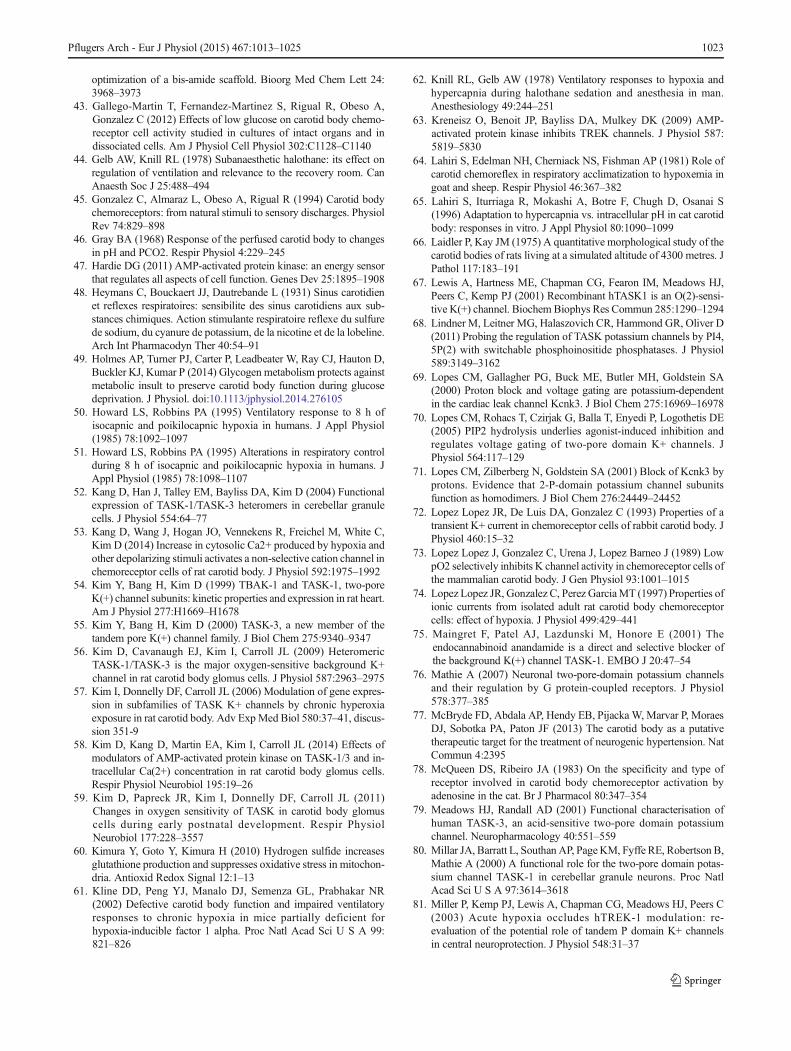
Mechanisms of oxygen sensing in TASK channels:the AMP kinase hypothesis
The AMP kinase (AMPK) hypothesis of oxygen sensing is, inessence, another variant of the metabolic hypothesis. It pro-poses that various potassium channels (including TASK chan-nels) are regulated by phosphorylation via AMP-activated ki-nase. AMPK is a well-known metabolic energy sensor whichpays a key role in coordinating metabolism and cellular ener-gy demand in other cell types. The basic model is that whenATP demand begins to exceed supply, there is a net conver-sion of ATP to ADP, followed by the conversion of ADP toAMP. AMP then activates the kinase by both displacing ATPwhich is normally bound to the AMP kinase and in the process
1018 Pflugers Arch - Eur J Physiol (2015) 467:1013–1025

making AMPK susceptible to activation by phosphorylationvia an upstream kinase, e.g. LKB1 (see [47]). Hence, there isan implied failure of type 1 cell metabolism to maintain cel-lular ATP levels during hypoxia.
Initial evidence for a role of AMPK in oxygen sensingincluded the observations that 1) AICAR, an activator ofAMPK,stimulated both isolated chemoreceptors and type 1cells; and 2) that compound C, an inhibitor of AMPK, atten-uated the excitatory effects of both AICAR and hypoxia [39,141]. This was followed by conflicting evidence as to theability of AMPK to phosphorylate and/or inhibit TASK chan-nels. AICARwas reported to inhibitTASK3 but not TASK1 inH293 cells by Dallas et al. [30]. In contrast, Kréneisz et al.reported that AICAR had no effect upon TASK1, TASK3 orthe TASK1/3 concatamer in HEK293 [63]. A more recentstudy has also failed to find any effect of the AMPK activatorsAICAR or A769662 on either TASK channel activity or Ca2+
signalling in isolated rat type 1 cells or any effect of compoundC upon TASK channel inhibition by hypoxia [58]. We haveobtained similar results in respect of the lack of effects ofAICAR and A769662 on Ca2+ signalling in type 1 cells(Buckler unpublished). The role of AMPK in regulating TASKchannels in the type 1 cell is therefore currently in doubt.
Mechanisms of oxygen sensing in TASK channels: roleof hydrogen sulfide and carbon monoxide
There has recently been much interest in the role of the puta-tive gaseous signalling molecules H2S and CO in oxygensensing both in the carotid body and in vascular smooth mus-cle. The reader is directed to a number of recent reviews forthe relevant background [91, 110]. Suffice to say that theeffects of exogenous sulfide have been tested in the carotidbody and isolated type 1 cells, and it appears to mimic theeffects of hypoxia in all major respects including thedepolarisation of type 1 cells through the inhibition of
background K+ currents and TASK channel activity [9, 105].The effects of exogenous H2S are manifest at micromolarlevels, however, which are sufficient to also inhibit mitochon-drial metabolism (as measured in the type 1 cell). Moreover,H2S had no additional effect upon TASK channel activity inthe presence of cyanide [9]. This suggests that modulation ofTASK channels by H2S is simply another manifestation ofmetabolic sensing in type 1 cells. A similar argument maybe applied to the effects of carbon monoxide. High levels ofCO excite the carotid body, depolarise type 1 cells and inhibitthe background K+ conductance [4]. At these levels, however,CO will also inhibit cytochrome oxidase, suggesting that theactions of CO on background (TASK) current is again mostlikely to be due to metabolic signalling.
Mechanisms of oxygen sensing in TASK channels: reactiveoxygen species
Reactive oxygen species have also been implicated in acuteoxygen sensing, most notably in the pulmonary vasculature(see [123] for a review) which also expresses TASK-like K-currents [90]. Kim et al. [96] have investigated the effects ofhydrogen peroxide on TASK1, TASK3 and the TASK1/TASK3 concatamer expressed in HeLa cells, as well as theTASK channels in type 1 cells. Peroxide was found to evokean irreversible stimulation of all of these channels in the ex-cised patch configuration but only at exceptionally high con-centrations (≥16 mM). This suggests that H2O2 at least isunlikely to directly modulate TASK channels physiologically.
Neurotransmitter and autocrine regulation of TASKchannels in type 1 cells
The carotid body has a rich diversity of neurotransmitters andneurotransmitter receptors which provide for a complex
Fig. 2 Metabolic regulation of TASK channels. Effects of cyanide(2 mM) on TASK channel activity in cell-attached patches from mousetype 1 cells. Wild-type cells show primarily TASK1/TASK3 heterodimerchannel activity. KCNK9−/− cells (Task3 KO) show TASK1-like activity.
KCNK3−/− cells (Task1 KO) show TASK3-like activity. Note that allforms of TASK channel activity are suppressed by cyanide. Similareffects were also seen for hypoxia and the mitochondrial uncouplerFCCP, see [129]
Pflugers Arch - Eur J Physiol (2015) 467:1013–1025 1019

regulation by neurocrine, autocrine and paracrine signallingevents involving type 1 cells, nerve endings and type 2 cells.There have, as yet, been relatively few studies of the impact ofsuch signalling pathways on TASK channels in type 1 cellsalthough this would be a prime target for regulating chemore-ceptor excitability. Three substances of likely physiologicalsignificance have thus far been identified that can regulatethe background TASK channels in type 1 cells.
Adenosine is a powerful stimulant of the carotid body [78,115, 131] and is released in response to hypoxia. Extracellularadenosine can be derived from two sources: (1) the break-down of ATP that has been released as a neurotransmitter bythe type 1 cell and (2) adenosine efflux from the cell viatransporters in response to increased adenosine generation fol-lowing cytosolic ATP breakdown [24]. Adenosine causes amarked increase in intracellular calcium and membranedepolarisation in isolated type 1 cells, an effect that was mim-icked by the TASK1 inhibitor anandamide (see below). Priorexposure to anandamide prevented any further effect of aden-osine from which it was concluded that the effects of adeno-sine were likely to be mediated by inhibition of TASK chan-nels [142]. Although no direct studies of the effects of aden-osine on TASK channels have yet been reported in the carotidbody, the second messenger pathways implicated, activationof A2A receptors Gs increase in cAMP and protein kinase A(PKA) activation, have also been implicated in the modulationof TASK current by GABA. In contrast to the proposed effectsof adenosine, GABA is reported to activate TASK current intype 1 cells through activation of GABAB receptors Gi andinhibition of adenylate cyclase and PKA activity [40].
Serotonin is another neurotransmitter released from type 1cells [146]. Serotonin is reported to inhibit the resting K-conductance in type 1 cells leading to depolarisation and cellexcitation. These effects are thought to be mediated via 5-HT2
receptors and activation of another classical signalling path-way protein kinase C [145].
Pharmacological modulation of TASK channels in type 1cells
A number of substances have been observed to inhibit TASKchannels in the type 1 cell. Of these, anandamide/methanandamide are probably among the more selective.Anandamide was originally reported to inhibit TASK1 in het-erologous expression systems [75] but was subsequentlyfound to also inhibit both TASK3 and the TASK1/3concatamer [56, 133]. It has been observed to inhibit bothbackground currents [138] and TASK channels in type 1 cells[56]. Ruthenium red, an inhibitor of TASK3 (but not ofTASK1, TASK1/3 concatamers or TASK1/TASK3 heterodi-mers) [52, 60] has only a small (20 %) effect upon TASKchannel activity in type 1 cells, supporting the view that
TASK3 contributes little to the background TASK-like currentoverall [56]. Other less selective inhibitors of the TASK-likecurrent in type 1 cells include zinc, barium and quinidine [17].The effects of recently described selective inhibitors ofTASK1 and TASK3, e.g. PK-THPP, A1899 and A293 [7,42, 82], on type 1 cell TASK channels have not yet beenreported. Some of these agents (PK-THPP and A1899) havehowever been found to act as respiratory stimulants [26]. Thisis consistent with inhibition of type 1 cell TASK channels andperipheral chemoreceptor excitation. Conversely, the widelyused respiratory stimulant doxapram, which is known to ex-cite the carotid body [85, 88], has been found to inhibit clonedTASK1, TASK3 and the TASK1/3 heterodimer [27]. It istherefore anticipated that doxapram will also directly excitethe type 1 cell through the inhibition of TASK channels al-though this has yet to be formally demonstrated. There is littleother known pharmacology for TASK in type 1 cells save forthe observation that it is strongly activated by the gaseousanaesthetic halothane [17] and weakly activated by isoflurane[95]. The effects of these anaesthetics on channel activity mir-ror their ability to blunt the acute hypoxic ventilatory responseand antagonise type 1 cell responses to hypoxia [93, 94]. An-aesthetic activation of TASK channels is thought to involvetwo regions of the molecule: a potential binding site locatedin/near the cytoplasmic end of the second transmembrane do-main and another region at the beginning of the c-terminaldomain, referred to as the halothane response element, whichis important in transducing the effects of anaesthetic [2, 124].
Fig. 3 Schematic of TASK channel regulation in type 1 cells of thecarotid body. Cartoon depicts key regions of the TASK channelsincluding transmembrane spanning domains Tm1, Tm2, Tm3 and Tm4,pore loops P1+P2, the extracellular helical cap between Tm1 and P1, andN- and C-terminal domains. Hypoxia/metabolic inhibition/cytosolic ATPsignalling is presumed to involve some unknown intermediary, e.g. anaccessory subunit (?), see text. Modulation by extracellular acidosisprobably involves a histidine residue (H98) in the helical cap region(HC). Gaseous inhalational anaesthetics, e.g. halothane, may bind to aregion in/near the C-terminal end of the M2 segment [2]. Site of action ofthe protein kinases PKA and PKC is unknown but is assumed to involvecytoplasmic domains
1020 Pflugers Arch - Eur J Physiol (2015) 467:1013–1025

Future perspectives
One of the most pressing problems is in identifying the sig-nalling mechanisms involved in mediating the effects of hyp-oxia, metabolic inhibition and cytosolic ATP. Is ATP the onlysignalling molecule involved in oxygen/metabolic signalling?It certainly has a substantive effect upon TASK channel activ-ity in the type 1 cell, but so to do other unknown cytosolicconstituents judging by the degree of channel rundownfollowing patch excision. PIP2 depletion is a commoncause of channel rundown and has been implicated inTASK channel regulation in some cells/expression sys-tems [22, 70] but not others [23, 68]. The effects ofPIP2 on type 1 cell TASK channels are unknown. Beyondthis, it is difficult to know where to start, but metabolicprofiling of other tissues subject to hypoxia or mitochon-drial poisons may provide clues.
Another general area of investigation is in the many otherways in which TASK channel activity could be influenced(see Fig. 3). Given their role in setting the type 1 cells restingmembrane potential, TASK channels are likely to be highlyeffective targets for processes regulating the excitability ofchemoreceptors overall. This has a number of implications,e.g. (1) short-term neuromodulation of chemoreceptor func-tion by neurotransmitters, (2) long-term plasticity of chemo-receptor responses, (3) carotid body dysfunction (pathology),(4) therapeutic intervention, and (5) unwanted pharmacologi-cal side effects.
Neuromodulation A role for TASK channels in theneuromodulation of type 1 cell excitability has already beendiscussed with respect to adenosine, GABA and serotonin.There are however many other important neurotransmitterspresent in the carotid body including acetylcholine, dopamineand NO [89]. The effects of these pathways on TASK channelactivity are all awaiting investigation. It is notable that TASKchannels are strongly regulated by Gq-coupled receptors inother tissues [22, 29, 37, 76, 80, 118, 125], possibly throughincrease in diacylglycerol [136] and/or by direct interactionwith Gq α-subunits [23]. Possible direct effects of Gq α-subunits and diacylglycerol have not yet been studied in thetype 1 cell.
Plasticity of chemoreceptor responses There are two physio-logical processes involving chemoreceptor plasticity. One is inthe resetting of chemoreceptor oxygen sensitivity in the earlypostnatal period. This has been attributed to changes in calci-um signalling in type 1 cells [122] and is known to be accom-panied by changes in expression patterns and oxygen sensi-tivity of TASK channels in the type 1 cell [57, 59]. The secondform of plasticity is in ventilatory acclimatisation to hypoxia.This is a classical adaptation to life at altitude in which che-moreceptor response to hypoxia is augmented over a period of
hours to days [50, 51, 64]. It is likely to be a multifac-torial event probably involving short-term plasticity andlonger term hyperplasia of type 1 cells which may bedriven by oxygen-sensitive gene transcription and/or asa consequence of local signalling [6, 61, 66, 98, 109,119]. The role of TASK channels in these processesawaits investigation.
Carotid body dysfunction A number of clinical conditionshave been associated with chemoreceptor dysfunction includ-ing sleep apnoea, sympathetic over activity in heart failure andhypertension [1, 77, 99, 100, 111, 116]. These are all complexphenomena in which the role of the carotid body is still at anearly stage of investigation. The availability of drugs or otherinterventions that could modulate TASK channel activity (andthus chemoreceptor excitability) could prove to be of particu-lar value in evaluating the role of chemoreceptor excitation inthese situations and, possibly in the long term, in theirtreatment.
Pharmacological side effects Depression of ventilation andinhibition of the ventilatory response to hypoxia is awell-known side effect of a number of general anaestheticagents which can result in hypoxemia, a significant causeof postoperative morbidity and mortality [44, 62, 134]. Abetter understanding of how such agents work, particular-ly their effects on TASK channels, could lead to improve-ments in anaesthetic design. Moreover, the developmentof TASK channel inhibitors may provide for a new genera-tion of respiratory stimulants [26] to combat the effects ofanaesthetic induced respiratory depression. In summary,there is still a great deal to be learnt about the functionalrole and regulation of TASK channels in arterial chemore-ceptors and their potential value as targets for therapeuticintervention.
Acknowledgments This work was supported in part by grants from theBHF (PG/08/086/25849) and the MRC (project grant G101134). I amgrateful for the assistance of Dr. Philip Turner for reading andcommenting upon earlier versions of this manuscript and in the prepara-tion of Fig. 2.
Open Access This article is distributed under the terms of the CreativeCommons Attribution License which permits any use, distribution, andreproduction in any medium, provided the original author(s) and thesource are credited.
References
1. Abdala AP, McBryde FD, Marina N, Hendy EB, Engelman ZJ,Fudim M, Sobotka PA, Gourine AV, Paton JF (2012)Hypertension is critically dependent on the carotid body input inthe spontaneously hypertensive rat. J Physiol 590:4269–4277
Pflugers Arch - Eur J Physiol (2015) 467:1013–1025 1021

2. Andres-Enguix I, Caley A, Yustos R, Schumacher MA, Spanu PD,Dickinson R, Maze M, Franks NP (2007) Determinants of the an-esthetic sensitivity of two-pore domain acid-sensitive potassiumchannels: molecular cloning of an anesthetic-activated potassiumchannel from Lymnaea stagnalis. J Biol Chem 282:20977–20990
3. Anichkov S, Belen'kii M (1963) Pharmacology of the carotid bodychemoreceptors. Pergamon, Oxford
4. Barbe C, Al-Hashem F, Conway AF, Dubuis E, Vandier C, Kumar P(2002) A possible dual site of action for carbon monoxide-mediatedchemoexcitation in the rat carotid body. J Physiol 543:933–945
5. Bin Jaliah I, Maskell PD, Kumar P (2004) Indirect sensing ofinsulin-induced hypoglycaemia by the carotid body in the rat. JPhysiol 556:255–266
6. Bishop T, Talbot NP, Turner PJ, Nicholls LG, Pascual A, HodsonEJ, Douglas G, Fielding JW, Smith TG, Demetriades M, SchofieldCJ, Robbins PA, Pugh CW, Buckler KJ, Ratcliffe PJ (2013) Carotidbody hyperplasia and enhanced ventilatory responses to hypoxia inmice with heterozygous deficiency of PHD2. J Physiol 591:3565–3577
7. Bittner S, Bauer MA, Ehling P, Bobak N, Breuer J, Herrmann AM,Golfels M, Wiendl H, Budde T, Meuth SG (2012) The TASK1channel inhibitor A293 shows efficacy in a mouse model of multi-ple sclerosis. Exp Neurol 238:149–155
8. Buckler KJ (1997) A novel oxygen-sensitive potassium current inrat carotid body type I cells. J Physiol 498:649–662
9. Buckler KJ (2012) Effects of exogenous hydrogen sulphide on cal-cium signalling, background (TASK) K channel activity andmitochondrial function in chemoreceptor cells. Pflugers Arch463:743–754
10. Buckler K, Honore E (2004) Molecular strategies for studyingoxygen-sensitive K+ channels. Methods Enzymol 381:233–256
11. Buckler KJ, Honore E (2005) The lipid-activated two-pore domainK+ channel TREK-1 is resistant to hypoxia: implication for ischae-mic neuroprotection. J Physiol 562:213–222
12. Buckler KJ, Turner PJ (2013) Oxygen sensitivity of mitochondrialfunction in rat arterial chemoreceptor cells. J Physiol 591:3549–3563
13. Buckler KJ, Vaughan Jones RD (1993) Effects of acidic stimuli onintracellular calcium in isolated type I cells of the neonatal rat ca-rotid body. Pflugers Arch 425:22–27
14. Buckler KJ, Vaughan Jones RD (1994) Effects of hypoxia on mem-brane potential and intracellular calcium in rat neonatal carotid bodytype I cells. J Physiol 476:423–428
15. Buckler KJ, Vaughan Jones RD (1994) Effects of hypercapnia onmembrane potential and intracellular calcium in rat carotid bodytype I cells. J Physiol 478:157–171
16. Buckler KJ, Vaughan-Jones RD (1998) Effects of mitochondrialuncouplers on intracellular calcium, pH and membrane potentialin rat carotid body type I cells. J Physiol 513(Pt 3):819–833
17. Buckler KJ, Williams BA, Honore E (2000) An oxygen-, acid- andanaesthetic-sensitive TASK-like background potassium channel inrat arterial chemoreceptor cells. J Physiol 525(Pt 1):135–142
18. Buttigieg J, Nurse CA (2004) Detection of hypoxia-evoked ATPrelease from chemoreceptor cells of the rat carotid body. BiochemBiophys Res Commun 322:82–87
19. Carpenter E, Peers C (1997) Swelling- and cAMP-activated Cl-currents in isolated rat carotid body type I cells. J Physiol 503:497–511
20. Carpenter E, Peers C (2001) A standing Na+ conductance in ratcarotid body type I cells. Neuroreport 12:1421–1425
21. Carpenter E, Wyatt CN, Hatton CJ, Bee D, Peers C (1996) Ca2+channel currents in type I carotid body cells from normoxic andchronically hypoxic rats. Adv Exp Med Biol 410:105–108
22. Chemin J, Girard C, Duprat F, Lesage F, Romey G, Lazdunski M(2003) Mechanisms underlying excitatory effects of group I
metabotropic glutamate receptors via inhibition of 2P domain K+channels. EMBO J 22:5403–5411
23. Chen X, Talley EM, Patel N, Gomis A, McIntire WE, Dong B,Viana F, Garrison JC, Bayliss DA (2006) Inhibition of a backgroundpotassium channel by Gq protein alpha-subunits. Proc Natl AcadSci U S A 103:3422–3427
24. Conde SV, Monteiro EC (2004) Hypoxia induces adenosine releasefrom the rat carotid body. J Neurochem 89:1148–1156
25. Conde SV, Obeso A, Gonzalez C (2007) Low glucose effects on ratcarotid body chemoreceptor cells’ secretory responses and actionpotential frequency in the carotid sinus nerve. J Physiol 585:721–730
26. Cotten JF (2013) TASK-1 (KCNK3) and TASK-3 (KCNK9) tan-dem pore potassium channel antagonists stimulate breathing inisoflurane-anesthetized rats. Anesth Analg 116:810–816
27. Cotten JF, Keshavaprasad B, Laster MJ, Eger EI 2nd, Yost CS(2006) The ventilatory stimulant doxapram inhibits TASK tandempore (K2P) potassium channel function but does not affect mini-mum alveolar anesthetic concentration. Anesth Analg 102:779–785
28. Czirjak G, Enyedi P (2002) Formation of functional heterodimersbetween the TASK-1 and TASK-3 two-pore domain potassiumchannel subunits. J Biol Chem 277:5426–5432
29. Czirjak G, Fischer T, Spat A, Lesage F, Enyedi P (2000) TASK(TWIK-related acid-sensitive K+ channel) is expressed inglomerulosa cells of rat adrenal cortex and inhibited by angiotensinII. Mol Endocrinol 14:863–874
30. Dallas ML, Scragg JL, Wyatt CN, Ross F, Hardie DG, Evans AM,Peers C (2009) Modulation of O(2) sensitive K (+) channels byAMP-activated protein kinase. Adv Exp Med Biol 648:57–63
31. DalyMB (1997) Peripheral arterial chemoreceptors and respiratory-cardiovascular integration. Clarendon, Oxford
32. Dasso LL, Buckler KJ, Vaughan Jones RD (1997) Muscarinic andnicotinic receptors raise intracellular Ca2+ levels in rat carotid bodytype I cells. J Physiol 498:327–338
33. Duchen MR, Biscoe TJ (1992) Relative mitochondrial membranepotential and [Ca2+]i in type I cells isolated from the rabbit carotidbody. J Physiol 450:33–61
34. Duchen MR, Biscoe TJ (1992) Mitochondrial function in type Icells isolated from rabbit arterial chemoreceptors. J Physiol 450:13–31
35. Duchen MR, Caddy KW, Kirby GC, Patterson DL, Ponte J, BiscoeTJ (1988) Biophysical studies of the cellular elements of the rabbitcarotid body. Neuroscience 26:291–311
36. Duprat F, Lesage F, Fink M, Reyes R, Heurteaux C, Lazdunski M(1997) TASK, a human background K+ channel to sense externalpH variations near physiological pH. EMBO J 16:5464–5471
37. Enyedi P, Czirjak G (2010) Molecular background of leak K+ cur-rents: two-pore domain potassium channels. Physiol Rev 90:559–605
38. Esilva MJ, Lewis DL (1995) L- and N-type Ca2+ channels in adultrat carotid body chemoreceptor type I cells. J Physiol 489:689–699
39. Evans AM, Mustard KJ, Wyatt CN, Peers C, Dipp M, Kumar P,Kinnear NP, Hardie DG (2005) Does AMP-activated protein kinasecouple inhibition of mitochondrial oxidative phosphorylation byhypoxia to calcium signaling in O2-sensing cells? J Biol Chem280:41504–41511
40. Fearon IM, Zhang M, Vollmer C, Nurse CA (2003) GABA medi-ates autoreceptor feedback inhibition in the rat carotid body viapresynaptic GABAB receptors and TASK-1. J Physiol 553:83–94
41. Fidone SJ, Gonzalez C (1986) Initiation and control of chemorecep-tor activity in the carotid body. American Physiological Society,Bethesda
42. Flaherty DP, SimpsonDS,MillerM,Maki BE, Zou B, Shi J, WuM,McManus OB, Aube J, LiM, Golden JE (2014) Potent and selectiveinhibitors of the TASK-1 potassium channel through chemical
1022 Pflugers Arch - Eur J Physiol (2015) 467:1013–1025
optimization of a bis-amide scaffold. Bioorg Med Chem Lett 24:3968–3973
43. Gallego-Martin T, Fernandez-Martinez S, Rigual R, Obeso A,Gonzalez C (2012) Effects of low glucose on carotid body chemo-receptor cell activity studied in cultures of intact organs and indissociated cells. Am J Physiol Cell Physiol 302:C1128–C1140
44. Gelb AW, Knill RL (1978) Subanaesthetic halothane: its effect onregulation of ventilation and relevance to the recovery room. CanAnaesth Soc J 25:488–494
45. Gonzalez C, Almaraz L, Obeso A, Rigual R (1994) Carotid bodychemoreceptors: from natural stimuli to sensory discharges. PhysiolRev 74:829–898
46. Gray BA (1968) Response of the perfused carotid body to changesin pH and PCO2. Respir Physiol 4:229–245
47. Hardie DG (2011) AMP-activated protein kinase: an energy sensorthat regulates all aspects of cell function. Genes Dev 25:1895–1908
48. Heymans C, Bouckaert JJ, Dautrebande L (1931) Sinus carotidienet reflexes respiratoires: sensibilite des sinus carotidiens aux sub-stances chimiques. Action stimulante respiratoire reflexe du sulfurede sodium, du cyanure de potassium, de la nicotine et de la lobeline.Arch Int Pharmacodyn Ther 40:54–91
49. Holmes AP, Turner PJ, Carter P, Leadbeater W, Ray CJ, Hauton D,Buckler KJ, Kumar P (2014) Glycogen metabolism protects againstmetabolic insult to preserve carotid body function during glucosedeprivation. J Physiol. doi:10.1113/jphysiol.2014.276105
50. Howard LS, Robbins PA (1995) Ventilatory response to 8 h ofisocapnic and poikilocapnic hypoxia in humans. J Appl Physiol(1985) 78:1092–1097
51. Howard LS, Robbins PA (1995) Alterations in respiratory controlduring 8 h of isocapnic and poikilocapnic hypoxia in humans. JAppl Physiol (1985) 78:1098–1107
52. Kang D, Han J, Talley EM, Bayliss DA, Kim D (2004) Functionalexpression of TASK-1/TASK-3 heteromers in cerebellar granulecells. J Physiol 554:64–77
53. Kang D, Wang J, Hogan JO, Vennekens R, Freichel M, White C,Kim D (2014) Increase in cytosolic Ca2+ produced by hypoxia andother depolarizing stimuli activates a non-selective cation channel inchemoreceptor cells of rat carotid body. J Physiol 592:1975–1992
54. Kim Y, Bang H, Kim D (1999) TBAK-1 and TASK-1, two-poreK(+) channel subunits: kinetic properties and expression in rat heart.Am J Physiol 277:H1669–H1678
55. Kim Y, Bang H, Kim D (2000) TASK-3, a new member of thetandem pore K(+) channel family. J Biol Chem 275:9340–9347
56. Kim D, Cavanaugh EJ, Kim I, Carroll JL (2009) HeteromericTASK-1/TASK-3 is the major oxygen-sensitive background K+channel in rat carotid body glomus cells. J Physiol 587:2963–2975
57. Kim I, Donnelly DF, Carroll JL (2006) Modulation of gene expres-sion in subfamilies of TASK K+ channels by chronic hyperoxiaexposure in rat carotid body. Adv ExpMed Biol 580:37–41, discus-sion 351-9
58. Kim D, Kang D, Martin EA, Kim I, Carroll JL (2014) Effects ofmodulators of AMP-activated protein kinase on TASK-1/3 and in-tracellular Ca(2+) concentration in rat carotid body glomus cells.Respir Physiol Neurobiol 195:19–26
59. Kim D, Papreck JR, Kim I, Donnelly DF, Carroll JL (2011)Changes in oxygen sensitivity of TASK in carotid body glomuscells during early postnatal development. Respir PhysiolNeurobiol 177:228–3557
60. Kimura Y, Goto Y, Kimura H (2010) Hydrogen sulfide increasesglutathione production and suppresses oxidative stress in mitochon-dria. Antioxid Redox Signal 12:1–13
61. Kline DD, Peng YJ, Manalo DJ, Semenza GL, Prabhakar NR(2002) Defective carotid body function and impaired ventilatoryresponses to chronic hypoxia in mice partially deficient forhypoxia-inducible factor 1 alpha. Proc Natl Acad Sci U S A 99:821–826
62. Knill RL, Gelb AW (1978) Ventilatory responses to hypoxia andhypercapnia during halothane sedation and anesthesia in man.Anesthesiology 49:244–251
63. Kreneisz O, Benoit JP, Bayliss DA, Mulkey DK (2009) AMP-activated protein kinase inhibits TREK channels. J Physiol 587:5819–5830
64. Lahiri S, Edelman NH, Cherniack NS, Fishman AP (1981) Role ofcarotid chemoreflex in respiratory acclimatization to hypoxemia ingoat and sheep. Respir Physiol 46:367–382
65. Lahiri S, Iturriaga R, Mokashi A, Botre F, Chugh D, Osanai S(1996) Adaptation to hypercapnia vs. intracellular pH in cat carotidbody: responses in vitro. J Appl Physiol 80:1090–1099
66. Laidler P, Kay JM (1975) A quantitative morphological study of thecarotid bodies of rats living at a simulated altitude of 4300 metres. JPathol 117:183–191
67. Lewis A, Hartness ME, Chapman CG, Fearon IM, Meadows HJ,Peers C, Kemp PJ (2001) Recombinant hTASK1 is an O(2)-sensi-tive K(+) channel. Biochem Biophys Res Commun 285:1290–1294
68. Lindner M, Leitner MG, Halaszovich CR, Hammond GR, Oliver D(2011) Probing the regulation of TASK potassium channels by PI4,5P(2) with switchable phosphoinositide phosphatases. J Physiol589:3149–3162
69. Lopes CM, Gallagher PG, Buck ME, Butler MH, Goldstein SA(2000) Proton block and voltage gating are potassium-dependentin the cardiac leak channel Kcnk3. J Biol Chem 275:16969–16978
70. Lopes CM, Rohacs T, Czirjak G, Balla T, Enyedi P, Logothetis DE(2005) PIP2 hydrolysis underlies agonist-induced inhibition andregulates voltage gating of two-pore domain K+ channels. JPhysiol 564:117–129
71. Lopes CM, Zilberberg N, Goldstein SA (2001) Block of Kcnk3 byprotons. Evidence that 2-P-domain potassium channel subunitsfunction as homodimers. J Biol Chem 276:24449–24452
72. Lopez Lopez JR, De Luis DA, Gonzalez C (1993) Properties of atransient K+ current in chemoreceptor cells of rabbit carotid body. JPhysiol 460:15–32
73. Lopez Lopez J, Gonzalez C, Urena J, Lopez Barneo J (1989) LowpO2 selectively inhibits K channel activity in chemoreceptor cells ofthe mammalian carotid body. J Gen Physiol 93:1001–1015
74. Lopez Lopez JR, Gonzalez C, Perez GarciaMT (1997) Properties ofionic currents from isolated adult rat carotid body chemoreceptorcells: effect of hypoxia. J Physiol 499:429–441
75. Maingret F, Patel AJ, Lazdunski M, Honore E (2001) Theendocannabinoid anandamide is a direct and selective blocker ofthe background K(+) channel TASK-1. EMBO J 20:47–54
76. Mathie A (2007) Neuronal two-pore-domain potassium channelsand their regulation by G protein-coupled receptors. J Physiol578:377–385
77. McBryde FD, Abdala AP, Hendy EB, Pijacka W, Marvar P, MoraesDJ, Sobotka PA, Paton JF (2013) The carotid body as a putativetherapeutic target for the treatment of neurogenic hypertension. NatCommun 4:2395
78. McQueen DS, Ribeiro JA (1983) On the specificity and type ofreceptor involved in carotid body chemoreceptor activation byadenosine in the cat. Br J Pharmacol 80:347–354
79. Meadows HJ, Randall AD (2001) Functional characterisation ofhuman TASK-3, an acid-sensitive two-pore domain potassiumchannel. Neuropharmacology 40:551–559
80. Millar JA, Barratt L, Southan AP, PageKM, Fyffe RE, Robertson B,Mathie A (2000) A functional role for the two-pore domain potas-sium channel TASK-1 in cerebellar granule neurons. Proc NatlAcad Sci U S A 97:3614–3618
81. Miller P, Kemp PJ, Lewis A, Chapman CG, Meadows HJ, Peers C(2003) Acute hypoxia occludes hTREK-1 modulation: re-evaluation of the potential role of tandem P domain K+ channelsin central neuroprotection. J Physiol 548:31–37
Pflugers Arch - Eur J Physiol (2015) 467:1013–1025 1023

82. Miller MR, Zou B, Shi J, Flaherty DP, Simpson DS, Yao T, MakiBE, Day VW, Douglas JT, WuM, McManus OB, Golden JE, AubeJ, Li M (2010) Development of a selective chemical inhibitor for thetwo-pore potassium channel, KCNK9 Probe Reports from the NIHMolecular Libraries Program. Bethesda
83. Mills E, Jobsis FF (1970) Simultaneous measurement of cyto-chrome a3 reduction and chemoreceptor afferent activity in the ca-rotid body. Nature 225:1147–1149
84. Mills E, Jobsis FF (1972)Mitochondrial respiratory chain of carotidbody and chemoreceptor response to changes in oxygen tension. JNeurophysiol 35:405–428
85. Mitchell RA, Herbert DA (1975) Potencies of doxapram and hyp-oxia in stimulating carotid-body chemoreceptors and ventilation inanesthetized cats. Anesthesiology 42:559–566
86. Morton MJ, O'Connell AD, Sivaprasadarao A, Hunter M (2003)Determinants of pH sensing in the two-pore domain K(+) channelsTASK-1 and -2. Pflugers Arch 445:577–583
87. Mulligan E, Lahiri S, Storey BT (1981) Carotid body O2 chemore-ception and mitochondrial oxidative phosphorylation. J ApplPhysiol 51:438–446
88. Nishino T, Mokashi A, Lahiri S (1982) Stimulation of carotid che-moreceptors and ventilation by doxapram in the cat. J Appl Physiol52:1261–1265
89. Nurse CA (2014) Synaptic and paracrine mechanisms at carotidbody arterial chemoreceptors. J Physiol 592:3419–3426
90. Olschewski A, Li Y, Tang B, Hanze J, Eul B, Bohle RM,Wilhelm J,Morty RE, Brau ME, Weir EK, Kwapiszewska G, Klepetko W,Seeger W, Olschewski H (2006) Impact of TASK-1 in human pul-monary artery smooth muscle cells. Circ Res 98:1072–1080
91. Olson KR,Whitfield NL (2010) Hydrogen sulfide and oxygen sens-ing in the cardiovascular system. Antioxid Redox Signal 12:1219–1234
92. Ortega-Saenz P, Levitsky KL, Marcos-Almaraz MT, Bonilla-HenaoV, Pascual A, Lopez-Barneo J (2010) Carotid body chemosensoryresponses in mice deficient of TASK channels. J Gen Physiol 135:379–392
93. Pandit JJ (2002) The variable effect of low-dose volatile anaes-thetics on the acute ventilatory response to hypoxia in humans: aquantitative review. Anaesthesia 57:632–643
94. Pandit JJ, Buckler KJ (2009) Differential effects of halothane andsevoflurane on hypoxia-induced intracellular calcium transients ofneonatal rat carotid body type I cells. Br J Anaesth 103:701–710
95. Pandit JJ, Winter V, Bayliss R, Buckler KJ (2010) Differential ef-fects of halothane and isoflurane on carotid body glomus cell intra-cellular Ca2+ and background K+ channel responses to hypoxia.Adv Exp Med Biol 669:205–208
96. Papreck JR, Martin EA, Lazzarini P, Kang D, Kim D (2012)Modulation of K2P3.1 (TASK-1), K2P9.1 (TASK-3), and TASK-1/3 heteromer by reactive oxygen species. Pflugers Arch 464:471–480
97. Pardal R, Lopez Barneo J (2002) Low glucose-sensing cells in thecarotid body. Nat Neurosci 5:197–198
98. Pardal R, Ortega-Saenz P, Duran R, Lopez-Barneo J (2007) Glia-like stem cells sustain physiologic neurogenesis in the adult mam-malian carotid body. Cell 131:364–377
99. Paton JF, Ratcliffe L, Hering D, Wolf J, Sobotka PA, Narkiewicz K(2013) Revelations about carotid body function through its patho-logical role in resistant hypertension. Curr Hypertens Rep 15:273–280
100. Paton JF, Sobotka PA, Fudim M, Engelman ZJ, Hart EC, McBrydeFD, Abdala AP, Marina N, Gourine AV, Lobo M, Patel N, BurchellA, Ratcliffe L, Nightingale A (2013) The carotid body as a thera-peutic target for the treatment of sympathetically mediated diseases.Hypertension 61:5–13
101. Peers C (1990) Effect of lowered extracellular pH on Ca2(+)-depen-dent K+ currents in type I cells from the neonatal rat carotid body. JPhysiol 422:381–395
102. Peers C (1990) Hypoxic suppression of K+ currents in type I carotidbody cells: selective effect on the Ca2(+)-activated K+ current.Neurosci Lett 119:253–256
103. Peers C, Green FK (1991) Inhibition of Ca(2+)-activated K+ cur-rents by intracellular acidosis in isolated type I cells of the neonatalrat carotid body. J Physiol 437:589–602
104. Peers C, O'Donnell J (1990) Potassium currents recorded in type Icarotid body cells from the neonatal rat and their modulation bychemoexcitatory agents. Brain Res 522:259–266
105. PengYJ, Nanduri J, RaghuramanG, Souvannakitti D, Gadalla MM,Kumar GK, Snyder SH, Prabhakar NR (2010) H2S mediates O2sensing in the carotid body. Proc Natl Acad Sci U S A 107:10719–10724
106. Perez Garcia MT, Almaraz L, Gonzalez C (1991) Cyclic AMPmodulates differentially the release of dopamine induced by hypox-ia and other stimuli and increases dopamine synthesis in the rabbitcarotid body. J Neurochem 57:1992–2000
107. Perez Garcia MT, Colinas O, Miguel Velado E, Moreno DominguezA, Lopez Lopez JR (2004) Characterization of the Kv channels ofmouse carotid body chemoreceptor cells and their role in oxygensensing. J Physiol 557:457–471
108. Perez Garcia MT, Obeso A, Lopez Lopez JR, Herreros B, GonzalezC (1992) Characterization of cultured chemoreceptor cells dissoci-ated from adult rabbit carotid body. Am J Physiol 263:C1152–C1159
109. Platero-Luengo A, Gonzalez-Granero S, Duran R, Diaz-Castro B,Piruat JI, Garcia-Verdugo JM, Pardal R, Lopez-Barneo J (2014) AnO2-sensitive glomus cell-stem cell synapse induces carotid bodygrowth in chronic hypoxia. Cell 156:291–303
110. Prabhakar NR (2012) Carbon monoxide (CO) and hydrogen sulfide(H(2)S) in hypoxic sensing by the carotid body. Respir PhysiolNeurobiol 184:165–169
111. Prabhakar NR, Peng YJ, Jacono FJ, Kumar GK, Dick TE (2005)Cardiovascular alterations by chronic intermittent hypoxia: impor-tance of carotid body chemoreflexes. Clin Exp Pharmacol Physiol32:447–449
112. Rajan S, Wischmeyer E, Xin Liu G, Preisig Muller R, Daut J,Karschin A, Derst C (2000) TASK-3, a novel tandem pore domainacid-sensitive K+ channel. An extracellular histiding as pH sensor. JBiol Chem 275:16650–16657
113. Rocher A, Geijo Barrientos E, Caceres AI, Rigual R, Gonzalez C,Almaraz L (2005) Role of voltage-dependent calcium channels instimulus-secretion coupling in rabbit carotid body chemoreceptorcells. J Physiol 562:407–420
114. RongW, Gourine AV, Cockayne DA, Xiang Z, Ford AP, Spyer KM,Burnstock G (2003) Pivotal role of nucleotide P2X2 receptor sub-unit of the ATP-gated ion channel mediating ventilatory responsesto hypoxia. J Neurosci 23:11315–11321
115. RunoldM, Cherniack NS, Prabhakar NR (1990) Effect of adenosineon isolated and superfused cat carotid body activity. Neurosci Lett113:111–114
116. Schultz HD, Li YL (2007) Carotid body function in heart failure.Respir Physiol Neurobiol 157:171–185
117. Shen TCR, Hauss WH (1939) Influence of dinitrophenol,dinitroortocresol and paranitrophenol upon the carotid sinus che-moreceptors of the dog. Arch Int Pharmacodyn Ther 63:251–258
118. Sirois JE, Lynch C 3rd, Bayliss DA (2002) Convergent and recip-rocal modulation of a leak K+ current and I(h) by an inhalationalanaesthetic and neurotransmitters in rat brainstem motoneurones. JPhysiol 541:717–729
119. Slingo ME, Turner PJ, Christian HC, Buckler KJ (1985) RobbinsPA (2014) The von Hippel-Lindau Chuvash mutation in mice
1024 Pflugers Arch - Eur J Physiol (2015) 467:1013–1025

causes carotid-body hyperplasia and enhanced ventilatory sensitiv-ity to hypoxia. J Appl Physiol 116:885–892
120. Stea A, Nurse CA (1989) Chloride channels in cultured glomus cellsof the rat carotid body. Am J Physiol 257:C174–C181
121. Stea A, Nurse CA (1991) Whole-cell and perforated-patch record-ings from O2-sensitive rat carotid body cells grown in short- andlong-term culture. Pflugers Arch 418:93–101
122. Sterni LM, Bamford OS, Tomares SM, Montrose MH, Carroll JL(1995) Developmental changes in intracellular Ca2+ response ofcarotid chemoreceptor cells to hypoxia. Am J Physiol 268:L801–L808
123. Sylvester JT, Shimoda LA, Aaronson PI, Ward JP (2012) Hypoxicpulmonary vasoconstriction. Physiol Rev 92:367–520
124. Talley EM, Bayliss DA (2002) Modulation of TASK-1 (Kcnk3) andTASK-3 (Kcnk9) potassium channels: volatile anesthetics and neu-rotransmitters share a molecular site of action. J Biol Chem 277:17733–17742
125. Talley EM, Lei Q, Sirois JE, Bayliss DA (2000) TASK-1, a two-pore domain K+ channel, is modulated by multiple neurotransmit-ters in motoneurons. Neuron 25:399–410
126. Tan ZY, Lu Y,Whiteis CA, Benson CJ, ChapleauMW, Abboud FM(2007) Acid-sensing ion channels contribute to transduction of ex-tracellular acidosis in rat carotid body glomus cells. Circ Res 101:1009–1019
127. Trapp S, Aller MI,WisdenW, Gourine AV (2008) A role for TASK-1 (KCNK3) channels in the chemosensory control of breathing. JNeurosci 28:8844–8850
128. Trivedi B, Danforth WH (1966) Effect of pH on the kinetics of frogmuscle phosphofructokinase. J Biol Chem 241:4110–4112
129. Turner PJ, Buckler KJ (2013) Oxygen and mitochondrial inhibitorsmodulate both monomeric and heteromeric TASK-1 and TASK-3channels in mouse carotid body type-1 cells. J Physiol 591:5977–5998
130. Urena J, Lopez Lopez J, Gonzalez C, Lopez Barneo J (1989) Ioniccurrents in dispersed chemoreceptor cells of the mammalian carotidbody. J Gen Physiol 93:979–999
131. Vandier C, Conway AF, Landauer RC, Kumar P (1999) Presynapticaction of adenosine on a 4-aminopyridine-sensitive current in the ratcarotid body. J Physiol 515:419–429
132. Varas R, Wyatt CN, Buckler KJ (2007) Modulation of TASK-likebackground potassium channels in rat arterial chemoreceptor cellsby intracellular ATP and other nucleotides. J Physiol 583:521–536
133. Veale EL, Buswell R, Clarke CE, Mathie A (2007) Identification ofa region in the TASK3 two pore domain potassium channel that iscritical for its blockade by methanandamide. Br J Pharmacol 152:778–786
134. Ward DS, Karan SB, Pandit JJ (2011) Hypoxia: developments inbasic science, physiology and clinical studies. Anaesthesia 66(Suppl2):19–26
135. Weir EK, Lopez-Barneo J, Buckler KJ, Archer SL (2005) Acuteoxygen-sensing mechanisms. N Engl J Med 353:2042–2055
136. Wilke BU, Lindner M, Greifenberg L, Albus A, Kronimus Y,Bunemann M, Leitner MG, Oliver D (2014) Diacylglycerol medi-ates regulation of TASK potassium channels by Gq-coupled recep-tors. Nat Commun 5:5540
137. Williams BA, Buckler KJ (2004) Biophysical properties and meta-bolic regulation of a TASK-like potassium channel in rat carotidbody type 1 cells. Am J Physiol Lung Cell Mol Physiol 286:L221–L230
138. Wyatt CN, Buckler KJ (2003) The effect of methanandamide onisolated type I cells. Adv Exp Med Biol 536:123–127
139. Wyatt CN, Buckler KJ (2003) Effect of mitochondrial inhibitors ontype I cells. Adv Exp Med Biol 536:55–58
140. Wyatt CN, Buckler KJ (2004) The effect of mitochondrial inhibitorson membrane currents in isolated neonatal rat carotid body type Icells. J Physiol 556:175–191
141. Wyatt CN, Mustard KJ, Pearson SA, Dallas ML, Atkinson L,Kumar P, Peers C, Hardie DG, Evans AM (2007) AMP-activatedprotein kinase mediates carotid body excitation by hypoxia. J BiolChem 282:8092–8098
142. Xu F, Xu J, Tse FW, Tse A (2006) Adenosine stimulatesdepolarization and rise in cytoplasmic [Ca2+] in type I cellsof rat carotid bodies. Am J Physiol Cell Physiol 290:C1592–C1598
143. Yamaguchi S, Lande B, Kitajima T, Hori Y, Shirahata M (2004)Patch clamp study of mouse glomus cells using a whole carotidbody. Neurosci Lett 357:155–157
144. Zhang M, Buttigieg J, Nurse CA (2007) Neurotransmitter mecha-nisms mediating low-glucose signalling in cocultures and fresh tis-sue slices of rat carotid body. J Physiol 578:735–750
145. Zhang M, Fearon IM, Zhong H, Nurse CA (2003)Presynaptic modulation of rat arterial chemoreceptor func-tion by 5-HT: role of K+ channel inhibition via proteinkinase C. J Physiol 551:825–842
146. Zhang M, Nurse CA (2000) Does endogenous 5-HT mediate spon-taneous rhythmic activity in chemoreceptor clusters of rat carotidbody? Brain Res 872:199–203
147. ZhangM, Nurse CA (2004) CO2/pH chemosensory signaling in co-cultures of rat carotid body receptors and petrosal neurons: role ofATP and ACh. J Neurophysiol 92:3433–3445
148. Zhang M, Zhong H, Vollmer C, Nurse CA (2000) Co-release ofATP and ACh mediates hypoxic signalling at rat carotid body che-moreceptors. J Physiol 525(Pt 1):143–158
Pflugers Arch - Eur J Physiol (2015) 467:1013–1025 1025