Embed Size (px)
Citation preview

ASCRS ♦ ASOA Symposium & Congress
Technicians & Nurses Program
May 6-10, 2016 – New Orleans

4/16/2016
1
Sunday, May 8, 2016, 10:30-11:30amAmy Jost, BS, COMT, CCRC
Cincinnati Eye Institute
I have none
Identify the tech work-up components of 3 common surgeries
(plastics, glaucoma, cataract) before and after surgery
Connect how the accuracy of the pre-op measurements are
important to the surgery and post-operative outcomes
Provide a general overview of what the patient goes through in
surgery
Describe common post-operative exam procedures ordered
after surgery to measure post-operative outcomes
Ptosis Repair and Blepharoplasty
Trabeculectomy (and other glaucoma surgeries)
Phacoemulsification with Intraocular Lens Implant
Protect the eyes from debris and injury; maintain a healthy tear film and
corneal surface
4/16/2016
2
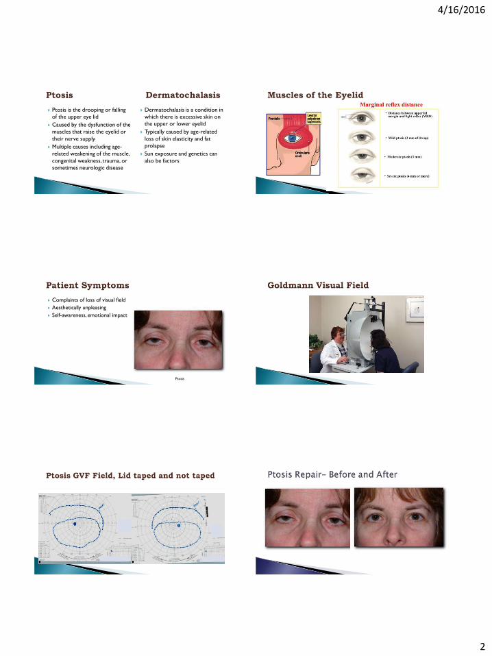
Ptosis is the drooping or falling
of the upper eye lid
Caused by the dysfunction of the
muscles that raise the eyelid or
their nerve supply
Multiple causes including age-
related weakening of the muscle,
congenital weakness, trauma, or
sometimes neurologic disease
Dermatochalasis is a condition in
which there is excessive skin on
the upper or lower eyelid
Typically caused by age-related
loss of skin elasticity and fat
prolapse
Sun exposure and genetics can
also be factors
Complaints of loss of visual field
Aesthetically unpleasing
Self-awareness, emotional impact
Ptosis

4/16/2016
3
Blepharoplasty Upper and Lower Blepharoplasty, CO2 laser
Upper and Lower Blepharoplasty, CO2 laser
Browplasty, Blepharoplasty
and Ptosis Repair

4/16/2016
4
Patient complains of:◦ Sometimes nothing!
◦ Decreased Peripheral Field
◦ Nuisance of taking eye drops
Dosing schedule
Don’t seem to help
Expensive
Vision with moderate glaucoma Normal Vision
Primary Open Angle Glaucoma (POAG)
Normal Tension Glaucoma (NTG)
Narrow/Closed Angle Glaucoma (ACG)
Congenital Glaucoma
Types of Secondary Glaucoma◦ Pigmentary Dispersion Glaucoma
◦ Pseudoexfoliative glaucoma
◦ Inflammatory glaucoma
◦ Traumatic glaucoma
◦ Neovascular Glaucoma
◦ Herpes Simplex Virus
◦ Herpes Zoster Virus
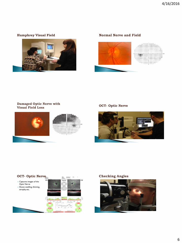
The optic nerve, also called Cranial
Nerve II (CNII), transmits visual
information from the retina to the
brain.
~30mm long from posterior globe to
orbital apex

4/16/2016
5
The amount of pressure inside the eye
◦ Normal range 10-21mmHg
The pressure is necessary to maintain the eye’s shape, nourishment, and
function
Increased IOP (ocular hypertension)
◦ Can cause ON damage
◦ One sign of possible glaucoma
◦ Medical and surgical treatment
Special Testing Required:◦ IOP (Intraocular Pressure)
◦ Manifest Refraction for Best Corrected Visual Acuity
◦ Visual Field Test
◦ Optic Nerve Analysis - OCT (Ophthalmic Coherence Tomography)
◦ Pachymetry: Corneal thickness can affect IOP results
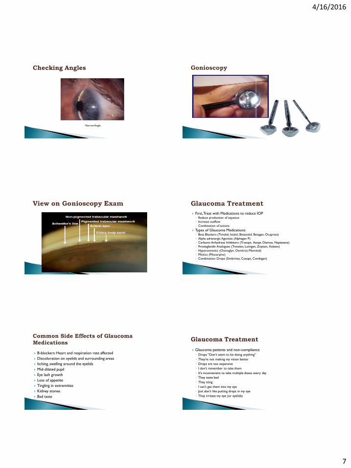
◦ Slit Lamp Exam: Check angles
◦ Gonioscopy
◦ Optic Nerve Assessment: Dilated Fundus Exam
IOP Measured on the Cornea
Normal thickness around 550m (1/2 mm)
Thickness matters…
◦ Compare a balloon to a basketball… thicker material, less indentation
◦ Thicker cornea (≥600m) allows less indentation, false high IOP readings
◦ Thinner cornea (≤500 m ) allows more indentation, false low IOP readings
4/16/2016
6
Captures images of the
Optic Nerve
Shows swelling, thinning,
atrophy, etc.
4/16/2016
7
Narrow Angle
First, Treat with Medications to reduce IOP◦ Reduce production of aqueous
◦ Increase outflow
◦ Combination of actions
Types of Glaucoma Medications: ◦ Beta Blockers (Timolol, Istalol, Betaxolol, Betagan, Ocupress)
◦ Alpha adrenergic Agonists (Alphagan P)
◦ Carbonic Anhydrase Inhibitors (Trusopt, Azopt, Diamox, Naptazane)
◦ Prostaglandin Analogues (Travatan, Lumigan, Zioptan, Xalatan)
◦ Hyperosmotics (Osmoglyn, Osmitrol, Mannitol)
◦ Miotics (Pilocarpine)
◦ Combination Drops (Simbrinza, Cosopt, Combigan)
B-blockers: Heart and respiration rate affected
Discoloration on eyelids and surrounding areas
Itching, swelling around the eyelids
Mid-dilated pupil
Eye lash growth
Loss of appetite
Tingling in extremities
Kidney stones
Bad taste
Glaucoma patients and non-compliance
◦ Drops “Don’t seem to be doing anything”
◦ They’re not making my vision better
◦ Drops are too expensive
◦ I don’t remember to take them
◦ It’s inconvenient to take multiple doses every day
◦ They taste bad
◦ They sting
◦ I can’t get them into my eye
◦ Just don’t like putting drops in my eye
◦ They irritate my eye (or eyelids)

4/16/2016
8
If medications are ineffective or if patient becomes intolerant of the drops, Laser Surgery may be scheduled
Selective Laser Trabeculoplasty (SLT)◦ Now the treatment of choice in most glaucoma patients
◦ Uses a non-thermal “cool” laser Does not cause scarring of the trabecular meshwork
Can be repeated several times
Argon Laser Trabeculoplasty (ALT)◦ Uses a thermal laser which does create a hole in the TM
◦ Can only be treated inferior and superior 180°
Laser Peripheral Iridotomy (LPI)- for angle closure
Cycloablation- Destroys the ciliary body, thereby destroying the mechanism for producing aqueous
If Medications and Laser Surgery are not effective, then conventional
surgery may be scheduled
Common types of conventional glaucoma surgery:
◦ MIGS (Micro-invasive glaucoma surgery)
◦ Trabeculectomy (“Trab”)
◦ Drainage Devices (Molteno, Baerveldt, Ahmed Valves)
Glaukos iStent(FDA approved with Cataract Surgery)
Reduces IOP by a few points, not very aggressive, but
able to use earlier in treatment course
Trabectome
4/16/2016
9
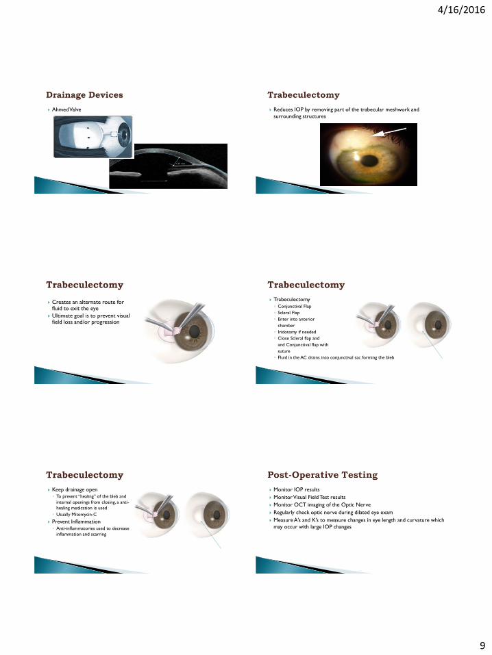
Ahmed Valve Reduces IOP by removing part of the trabecular meshwork and
surrounding structures
Creates an alternate route for fluid to exit the eye
Ultimate goal is to prevent visual field loss and/or progression
Trabeculectomy
◦ Conjunctival Flap
◦ Scleral Flap
◦ Enter into anterior
chamber
◦ Iridotomy if needed
◦ Close Scleral flap and
and Conjunctival flap with
suture
◦ Fluid in the AC drains into conjunctival sac forming the bleb
Keep drainage open
◦ To prevent “healing” of the bleb and
internal openings from closing, a anti-
healing medication is used
◦ Usually Mitomycin-C
Prevent Inflammation
◦ Anti-inflammatories used to decrease
inflammation and scarring
Monitor IOP results
Monitor Visual Field Test results
Monitor OCT imaging of the Optic Nerve
Regularly check optic nerve during dilated eye exam
Measure A’s and K’s to measure changes in eye length and curvature which
may occur with large IOP changes

4/16/2016
10
IOP too high or too low Conjunctival redness, irritation Conjunctival scarring (closes off the drainage port) Discomfort from the bleb Corneal edema Late infection, blebitis Bleeding inside the eye Choroidal detachments Hypotony maculopathy Cataracts CME Iris abnormalities Retinal tear or detachment
If IOP is high, could be related to
conjunctival healing process
Small gauged needle used to
break up conjunctival scarring
Patient complains of:◦ Decreased Vision
◦ Decreased confidence in driving at night/in the rain
◦ Glare/Halos
◦ Decreased Contrast Sensitivity
◦ Needs more light to read
May also be worried about: ◦ Ability to function in daily activities
◦ Losing their driver’s license
◦ “Going blind”
◦ Scared of eye surgery
◦ Cost of care
◦ Transportation and other perceived inconveniences
Vision with cataracts Normal Vision

4/16/2016
11
Vision with Glare and Halos as compare to normal Special Testing Required:◦ Axial Length
IOL Master, LenStar, Aladdin
◦ Keratometry
Manual K’s, Topography, Pentacam, IOL Master, LenStar, Aladdin
◦ Refraction
◦ Glare Testing
◦ Contrast Sensitivity Testing
◦ White-to-White (Corneal Diameter)
Manual WTW, IOL Master, LenStar, Aladdin
◦ Lens Thickness
Manual A-scans, LenStar, Aladdin
• Optical Biometers
• IOL Master
• LenStar
• Aladdin
• Pentacam
Contact A-Scan Immersion A-Scan
Axial length (AL)
Keratometry (K)
Anterior chamber depth (ACD)
White- to- white (WTW)
NOTE: Formula type and Target
Refraction
IOL Options listed:
EXAMPLE:
• Standard IOL
• Multi-piece IOL
• Alcon Toric IOL
• ReSTOR IOL (+3.0D add power)

4/16/2016
12
Check for best corrected visual acuity
100% Contrast 63% Contrast
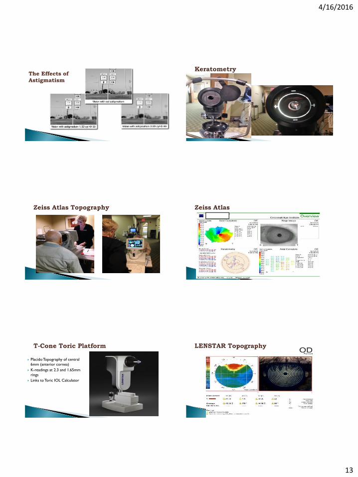
Various ways to measure K’s:
◦ Manual Keratometry
◦ Topography (Zeiss Atlas, Pentacam, iTrace,
etc.)
◦ Optical Biometry (IOL Master, LenStar,
Aladdin, etc.)
4/16/2016
13
The Effects of
Astigmatism
PlacidoTopography of central
6mm (anterior cornea)
K-readings at 2.3 and 1.65mm
rings
Links to Toric IOL Calculator
4/16/2016
14
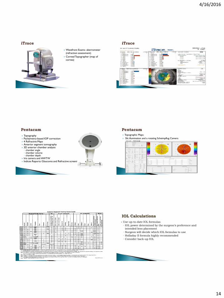
Wavefront Exams- aberrometer
(refraction assessment)
Corneal Topographer (map of
cornea)
Topography Pachymetry-based IOP correction 4 Refractive Maps Anterior segment tomography 3D anterior chamber analysis◦ chamber angle◦ chamber volume◦ chamber depth
Iris camera and HWTW Indices Reports: Glaucoma and Refractive screenings
Topographic Maps:
Slit illumination and a rotating Scheimpflug Camera
Use up-to-date IOL formulas
◦ IOL power determined by the surgeon’s preference and
intended lens placement
◦ Surgeon will decide which IOL formulas to use
◦ Holladay II formula highly recommended
◦ Consider back-up IOL

4/16/2016
15
Cataract Surgery planned for
both eyes with Toric IOL
◦ IOL Power measurement
◦ Toric IOL Power Measurement
◦ Toric IOL Axis Placement
Monovision planned◦ Distance for dominant eye (OD),
aim for plano
◦ Near vision for non-dominant eye
(OS),
◦ Aim for between -2.00 and -2.50D
91
VERION™ Reference Unit
VERION™ Microscope Integrated Display

4/16/2016
16
Video
Refraction
IOP
Slit Lamp exam
Posterior exam
Corneal edema
Cell & Flare
CME
IOP increase
Any Questions?