Embed Size (px)
Citation preview
205Oral & Craniofacial Tissue Engineering
Temporomandibular Disorders:
A Review of Etiology, Clinical Management,
and Tissue Engineering Strategies
Meghan K. Murphy, BE1/Regina F. MacBarb, BS1/Mark E. Wong, DDS2/Kyriacos A. Athanasiou, PhD, PE3
Temporomandibular disorders (TMD) are a class of degenerative musculoskeletal conditions associated with morphologic and functional deformities that affect up to 25% of the population, but their etiology and progression are poorly understood and, as a result, treatment options are limited. In up to 70% of cases, TMD are accompanied by malpositioning of the temporomandibular joint (TMJ) disc, termed “internal derangement.” Although the onset is not well characterized, correlations between internal derangement and osteoarthritic change have been identified. Because of the complex and unique nature of each TMD case, diagnosis requires patient-specific analysis accompanied by various diagnostic modalities. Likewise, treatment requires customized plans to address the specific characteristics of each patient’s disease. In the mechanically demanding and biochemically active environment of the TMJ, therapeutic approaches that can restore joint functionality while responding to changes in the joint have become a necessity. One such approach, tissue engineering, which may be capable of integration and adaptation in the TMJ, carries significant potential for the development of repair and replacement tissues. The following review presents a synopsis of etiology, current treatment methods, and the future of tissue engineering for repairing and/or replacing diseased joint components, specifically the mandibular condyle and TMJ disc. An analysis of native tissue characterization to assist clinicians in identifying tissue engineering objectives and validation metrics for restoring healthy and functional structures of the TMJ is followed by a discussion of current trends in tissue engineering. Oral Craniofac Tissue Eng 2011;1:205–226
Key words: cartilage, condyle, temporomandibular disorders, temporomandibular joint, temporomandibular joint disc, tissue engineering
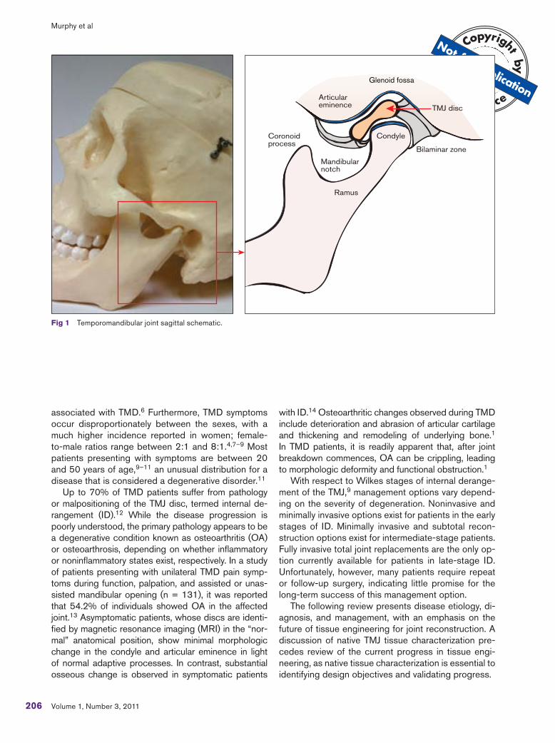
The temporomandibular articulation is composed of bilateral, diarthrodial temporomandibular joints (TMJs). Each joint is made up of a man-
dibular condyle and its corresponding temporal cavity (glenoid fossa and articular eminence) (Fig 1). The TMJ
and its associated structures play an essential role in guiding mandibular motion and distributing stresses produced by such everyday tasks as chewing, swal-lowing, and speaking. Temporomandibular disorders (TMD) are a class of degenerative musculoskeletal conditions associated with morphologic and func-tional deformities.1,2 TMD include abnormalities of the intra-articular discal position and/or structure as well as dysfunction of the associated musculature.3 Symp-toms and signs include painful joint sounds, restricted or deviating range of motion, and cranial and/or mus-cular pain, known as orofacial pain.
While up to 25% of the population may experience symptoms of TMD,4 only a small percentage of afflict-ed individuals seek treatment. For instance, studies in the 1980s detected TMD symptoms in 16% to 59% of the population,5 but only 3% to 7% of the adult popu-lation actually sought care for pain and dysfunction
1 Graduate Student Researcher, Department of Biomedical Engineering, University of California at Davis, Davis, CA, USA.
2 Chairman and Program Director, Department of Oral and Maxillofacial Surgery, The University of Texas School of Dentistry at Houston; Professor, Division of Oral and Maxillofacial Surgery, The University of Texas Medical School at Houston, Houston, TX, USA.
3 Distinguished Professor and Chair, Department of Biomedical Engineering, University of California at Davis, Davis, CA, USA.
Correspondence to: Dr K. A. Athanasiou, UC Davis, Department of Biomedical Engineering, One Shields Avenue, Davis, CA 95616. Fax: +530-754-5739. Email: [email protected]
Murphy et al
206 Volume 1, Number 3, 2011
associated with TMD.6 Furthermore, TMD symptoms occur disproportionately between the sexes, with a much higher incidence reported in women; female-to-male ratios range between 2:1 and 8:1.4,7–9 Most patients presenting with symptoms are between 20 and 50 years of age,9–11 an unusual distribution for a disease that is considered a degenerative disorder.11
Up to 70% of TMD patients suffer from pathology or malpositioning of the TMJ disc, termed internal de-rangement (ID).12 While the disease progression is poorly understood, the primary pathology appears to be a degenerative condition known as osteoarthritis (OA) or osteoarthrosis, depending on whether inflammatory or noninflammatory states exist, respectively. In a study of patients presenting with unilateral TMD pain symp-toms during function, palpation, and assisted or unas-sisted mandibular opening (n = 131), it was reported that 54.2% of individuals showed OA in the affected joint.13 Asymptomatic patients, whose discs are identi-fied by magnetic resonance imaging (MRI) in the “nor-mal” anatomical position, show minimal morphologic change in the condyle and articular eminence in light of normal adaptive processes. In contrast, substantial osseous change is observed in symptomatic patients
with ID.14 Osteoarthritic changes observed during TMD include deterioration and abrasion of articular cartilage and thickening and remodeling of underlying bone.1 In TMD patients, it is readily apparent that, after joint breakdown commences, OA can be crippling, leading to morphologic deformity and functional obstruction.1
With respect to Wilkes stages of internal derange-ment of the TMJ,9 management options vary depend-ing on the severity of degeneration. Noninvasive and minimally invasive options exist for patients in the early stages of ID. Minimally invasive and subtotal recon-struction options exist for intermediate-stage patients. Fully invasive total joint replacements are the only op-tion currently available for patients in late-stage ID. Unfortunately, however, many patients require repeat or follow-up surgery, indicating little promise for the long-term success of this management option.
The following review presents disease etiology, di-agnosis, and management, with an emphasis on the future of tissue engineering for joint reconstruction. A discussion of native TMJ tissue characterization pre-cedes review of the current progress in tissue engi-neering, as native tissue characterization is essential to identifying design objectives and validating progress.
Mandibularnotch
Coronoidprocess
Bilaminar zone
Condyle
Ramus
Articulareminence TMJ disc
Glenoid fossa
Fig 1 Temporomandibular joint sagittal schematic.
Murphy et al
207Oral & Craniofacial Tissue Engineering
DISEASE ETIOLOGY AND DIAGNOSIS
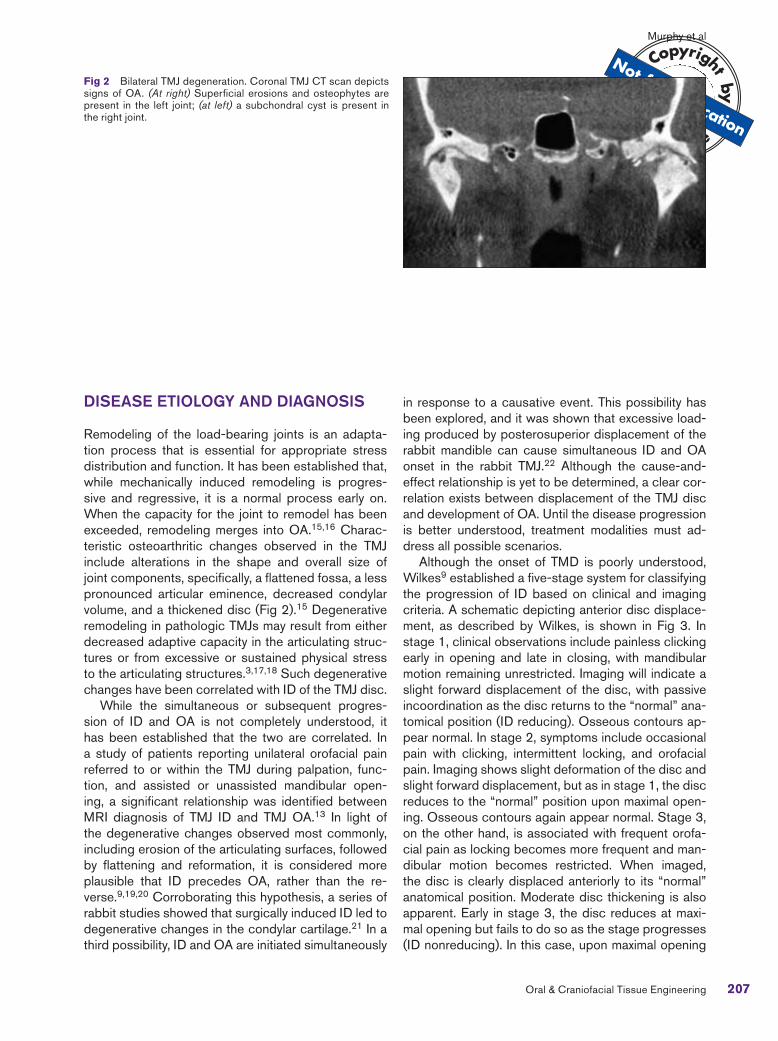
Remodeling of the load-bearing joints is an adapta-tion process that is essential for appropriate stress distribution and function. It has been established that, while mechanically induced remodeling is progres-sive and regressive, it is a normal process early on. When the capacity for the joint to remodel has been exceeded, remodeling merges into OA.15,16 Charac-teristic osteoarthritic changes observed in the TMJ include alterations in the shape and overall size of joint components, specifically, a flattened fossa, a less pronounced articular eminence, decreased condylar volume, and a thickened disc (Fig 2).15 Degenerative remodeling in pathologic TMJs may result from either decreased adaptive capacity in the articulating struc-tures or from excessive or sustained physical stress to the articulating structures.3,17,18 Such degenerative changes have been correlated with ID of the TMJ disc.
While the simultaneous or subsequent progres-sion of ID and OA is not completely understood, it has been established that the two are correlated. In a study of patients reporting unilateral orofacial pain referred to or within the TMJ during palpation, func-tion, and assisted or unassisted mandibular open-ing, a significant relationship was identified between MRI diagnosis of TMJ ID and TMJ OA.13 In light of the degenerative changes observed most commonly, including erosion of the articulating surfaces, followed by flattening and reformation, it is considered more plausible that ID precedes OA, rather than the re-verse.9,19,20 Corroborating this hypothesis, a series of rabbit studies showed that surgically induced ID led to degenerative changes in the condylar cartilage.21 In a third possibility, ID and OA are initiated simultaneously
in response to a causative event. This possibility has been explored, and it was shown that excessive load-ing produced by posterosuperior displacement of the rabbit mandible can cause simultaneous ID and OA onset in the rabbit TMJ.22 Although the cause-and-effect relationship is yet to be determined, a clear cor-relation exists between displacement of the TMJ disc and development of OA. Until the disease progression is better understood, treatment modalities must ad-dress all possible scenarios.
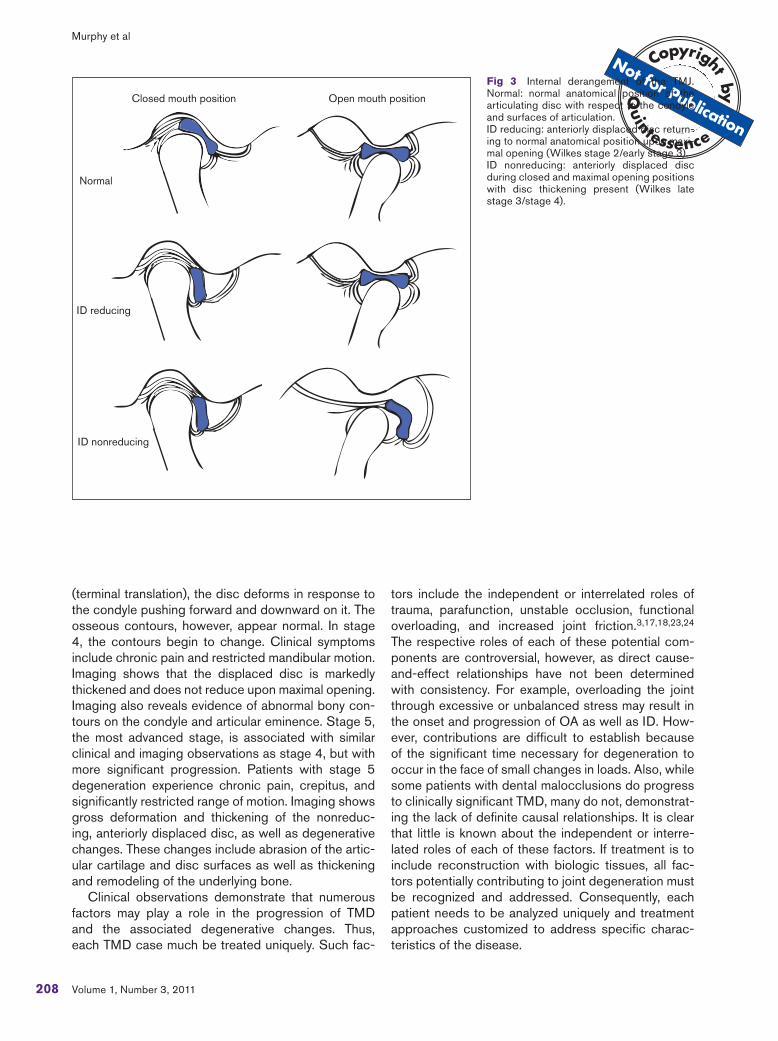
Although the onset of TMD is poorly understood, Wilkes9 established a five-stage system for classifying the progression of ID based on clinical and imaging criteria. A schematic depicting anterior disc displace-ment, as described by Wilkes, is shown in Fig 3. In stage 1, clinical observations include painless clicking early in opening and late in closing, with mandibular motion remaining unrestricted. Imaging will indicate a slight forward displacement of the disc, with passive incoordination as the disc returns to the “normal” ana-tomical position (ID reducing). Osseous contours ap-pear normal. In stage 2, symptoms include occasional pain with clicking, intermittent locking, and orofacial pain. Imaging shows slight deformation of the disc and slight forward displacement, but as in stage 1, the disc reduces to the “normal” position upon maximal open-ing. Osseous contours again appear normal. Stage 3, on the other hand, is associated with frequent orofa-cial pain as locking becomes more frequent and man-dibular motion becomes restricted. When imaged, the disc is clearly displaced anteriorly to its “normal” anatomical position. Moderate disc thickening is also apparent. Early in stage 3, the disc reduces at maxi-mal opening but fails to do so as the stage progresses (ID nonreducing). In this case, upon maximal opening
Fig 2 Bilateral TMJ degeneration. Coronal TMJ CT scan depicts signs of OA. (At right) Superficial erosions and osteophytes are present in the left joint; (at left) a subchondral cyst is present in the right joint.
Murphy et al
208 Volume 1, Number 3, 2011
(terminal translation), the disc deforms in response to the condyle pushing forward and downward on it. The osseous contours, however, appear normal. In stage 4, the contours begin to change. Clinical symptoms include chronic pain and restricted mandibular motion. Imaging shows that the displaced disc is markedly thickened and does not reduce upon maximal opening. Imaging also reveals evidence of abnormal bony con-tours on the condyle and articular eminence. Stage 5, the most advanced stage, is associated with similar clinical and imaging observations as stage 4, but with more significant progression. Patients with stage 5 degeneration experience chronic pain, crepitus, and significantly restricted range of motion. Imaging shows gross deformation and thickening of the nonreduc-ing, anteriorly displaced disc, as well as degenerative changes. These changes include abrasion of the artic-ular cartilage and disc surfaces as well as thickening and remodeling of the underlying bone.
Clinical observations demonstrate that numerous factors may play a role in the progression of TMD and the associated degenerative changes. Thus, each TMD case much be treated uniquely. Such fac-
tors include the independent or interrelated roles of trauma, parafunction, unstable occlusion, functional overloading, and increased joint friction.3,17,18,23,24 The respective roles of each of these potential com-ponents are controversial, however, as direct cause-and-effect relationships have not been determined with consistency. For example, overloading the joint through excessive or unbalanced stress may result in the onset and progression of OA as well as ID. How-ever, contributions are difficult to establish because of the significant time necessary for degeneration to occur in the face of small changes in loads. Also, while some patients with dental malocclusions do progress to clinically significant TMD, many do not, demonstrat-ing the lack of definite causal relationships. It is clear that little is known about the independent or interre-lated roles of each of these factors. If treatment is to include reconstruction with biologic tissues, all fac-tors potentially contributing to joint degeneration must be recognized and addressed. Consequently, each patient needs to be analyzed uniquely and treatment approaches customized to address specific charac-teristics of the disease.
Open mouth position
ID reducing
Normal
Closed mouth position
ID nonreducing
Fig 3 Internal derangement of the TMJ. Normal: normal anatomical position of the articulating disc with respect to the condyle and surfaces of articulation. ID reducing: anteriorly displaced disc return-ing to normal anatomical position upon maxi-mal opening (Wilkes stage 2/early stage 3). ID nonreducing: anteriorly displaced disc during closed and maximal opening positions with disc thickening present (Wilkes late stage 3/stage 4).
Murphy et al
209Oral & Craniofacial Tissue Engineering
Resulting from the diverse nature of TMD symp-toms, patient evaluation often requires a physical examination along with various imaging modalities. As previously mentioned, there is a population of in-dividuals who experience unilateral or bilateral disc displacement (presence or absence of joint noises) and minimal osseous change but who have not pro-gressed to clinically relevant TMD.14,25–27 Therefore, various diagnostic modalities, including clinical and radiologic examination, may be necessary to identify the stage of degeneration in patients presenting with possible TMD symptoms. Steadfast rules remain to be established regarding imaging for TMD diagnosis under the current Research Diagnostic Criteria for TMD. As result, TMD identification may involve any combination of the following modalities: MRI, con-ventional and computed tomography (CT), plain and panoramic radiography, arthrography, a thorough his-tory, and physical examination. CT is considered more beneficial for imaging bone and OA, while MRI is considered helpful for imaging soft tissues, including the disc and its joint relation.26,28 Clinical evaluation, together with various imaging modalities, may help to elucidate a patient’s stage of degeneration, aiding in diagnosis and treatment planning.
CLINICAL MANAGEMENT
For patients seeking to manage their TMD symptoms, it has been established that noninvasive modalities should first be explored. However, the complicated nature of the TMJ, along with the debilitating nature of late-stage disease, has created a demand for more invasive solutions. An analysis of current noninvasive, minimally invasive, and fully invasive management op-tions now follows. The ultimate goals of the presented modalities are to: (1) increase the mandibular range of motion, (2) decrease joint and masticatory muscle pain and inflammation, and (3) prevent further degen-erative change to articulating tissues, including direct or indirect joint damage.3
Noninvasive TechniquesThe noninvasive modalities implemented most com-monly include physical therapy, occlusal splints and/or adjustments, and pharmacologics. Physical therapy uses electrophysical modalities and manual/exercise techniques to relieve pain in the joint and mastica-tory muscles and improve range of motion.27 Physi-cal therapists may complement these techniques with behavioral changes by drawing awareness to the pa-tient’s posture, diet, and stress-related habits. Elec-trophysical modalities include transcutaneous electric nerve stimulation (TENS), ultrasound, and laser.28
Such modalities are implemented to reduce inflamma-tion, increase local blood flow, and promote muscle relaxation.28 Current research does not point to any significant decrease in pain in electrophysically treated patients. In fact, one study of 23 patients with bruxism showed a significant increase in range of motion and a decrease in muscular activity with muscular awareness relaxation training versus a TENS treatment group.29 Manual therapies designed to increase mobility and reduce pain have shown promise and are often used in conjunction with exercise techniques. Such exercise techniques work to strengthen and improve mobility in the masticatory and cervical spine muscles.30 Further-more, these techniques offer the potential to “reteach” and rehabilitate the musculature. This observation is especially noted in patients who exhibit stress-related habits.31 Along with exercise techniques, postural ex-ercises may aid in alignment of the craniomandibular system. Intended to relieve pain associated with TMD and improve range of motion, physical therapy treat-ment plans must be patient-specific and may involve a combination of modalities.
Occlusal splints and occlusal adjustments, another noninvasive mode of treatment, work to establish bal-ance in the occlusion and TMJs. The occlusion, or bite position, is a third and important element in the joint system and is the element often addressed by gen-eral dentists. Adjustments and splints may be used to achieve the most stable and least joint-traumatizing bite position. The ultimate goal of splints and adjustments is to minimize pain in the joint and masticatory muscles by establishing stability. Furthermore, as reviewed by In-gawale and Goswami,32 splints may be used to control bruxism, which has been associated with tooth attrition, malocclusion, myofacial pain, and masticatory muscle strain, fatigue, and fibrosis. The literature has shown mixed results for splint use. These results are not sur-prising, since the role of malocclusion in TMD progres-sion remains poorly understood. Occlusal splints and adjustments may be suggested to reestablish balance in the joint system, but the long-term effectiveness of this therapy remains controversial.32
With respect to pharmacologic agents, common-ly prescribed nonsteroidal anti-inflammatory drugs (NSAIDs) offer advantages in reducing inflamma-tion. However, research is needed to exploit long-term use and to identify whether the advantages in the management of pain and inflammation outweigh the negative side effects.33 Muscle relaxants may also be prescribed for treatment of muscle pain and/or spasm.34 However, studies have failed to demon-strate that muscle relaxants are any more effective for pain relief than NSAIDs.35 To increase their benefit, muscle relaxants are often used in combination with NSAIDs. NSAIDs may therefore be recommended for
Murphy et al
210 Volume 1, Number 3, 2011
their anti-inflammatory and analgesic benefits, but fur-ther research is needed to elucidate the benefits and risks of both short- and long-term use.
Minimally Invasive StrategiesMinimally invasive modalities for management of TMD symptoms include injections of sodium hyaluronate and/or corticosteroids, arthrocentesis, and arthrosco-py. Injections of corticosteroids and high-molecular-weight sodium hyaluronate in the superior joint space are intended to treat osteoarthritic symptoms. With research indicating both regenerative and degenera-tive responses to such injections, their use remains controversial.34 The pathophysiology of the disease indicates that there may be more significant potential for these injections in the early stages of degenera-tion, when inflammation first begins to exacerbate tis-sue catabolism.3,36
Similar to intra-articular injections, arthrocentesis and arthroscopic surgery are minimally invasive tech-niques requiring entrance into the joint capsule to lubricate articulating surfaces and reduce inflamma-tion. During arthrocentesis, a sterile needle is used to drain fluid from the joint.37 After it is drained, the joint is flushed of debris and inflammatory cytokines using a sterile solution.37 During the procedure, the physi-cian may also attempt to restore some range of motion with mandibular manipulation.38 Through arthroscopic surgery, a slightly more invasive procedure, the sur-geon may break intra-articular adhesions that may be preventing disc reduction in ID patients.39 With joint visualization during surgery, arthroscopy offers the ad-vantages of TMD stage diagnosis and identification of OA. While arthroscopic surgery and arthrocentesis may be used to lubricate joint surfaces and reduce in-flammation, further research is needed to identify long-term advantages of these approaches, especially in the absence of disc repositioning or replacement.38,40
Invasive ManagementFor the 5% of TMD patients who fail to improve with non-surgical treatment approaches, open joint surgery may be necessary to restore mandibular motion and mitigate orofacial pain.41 Most commonly, open joint surgery may include discectomy, reshaping or reconstruction of the articulating surfaces, and implantation of autolo-gous or alloplastic materials.42 Total joint replacement, the most invasive option, may become necessary when joint degeneration and pain exceed the potentials of the less invasive surgical methods. Condylar replacements in clinical use include autologous costochondral grafts, but autologous full joint replacements are not currently available. Alloplastic joint replacement systems, includ-ing total joint prostheses and hemiarthroplasties, have been in development since the 1960s. However, the
currently available systems have seen substantial modi-fications since their inception.
Discectomy and Disc Replacement. In TMD pa-tients presenting with limited range of motion, discec-tomy offers one means of regaining mandibular motion and reducing orofacial pain, and it may be followed by disc replacement. Discectomy has been shown in 5- and 10-year postoperative follow-ups to increase mandibular motion in patients previously showing no improvement with noninvasive management modali-ties.43,44 Radiographic changes in these long-term studies indicate evidence of osteophytes and flatten-ing of articular surfaces in such joints.43–45 Although the mechanism is poorly understood, some authors conclude that such changes are indicators of adap-tive change rather than degenerative disorders.43–45 In some patients, however, OA-like changes continue to exacerbate their condition, necessitating the de-velopment of autologous and alloplastic disc substi-tutes. Such substitutes, which include subcutaneous fat grafts and alloplasts, are aimed at providing a pro-tective cushion for the articulating surfaces of the joint during rotation and translation. Unfortunately, previous attempts at treatment with alloplastic disc replace-ments have often failed.46,47 Likewise, fat grafts may not sufficiently protect the articulating surfaces. Often, following implantation, the graft is displaced posterior to the condyle.48 The lack of clinical success associ-ated with disc replacement therapies may be the result of varying responses to the respective materials used. For example, with certain alloplasts, most notably the composite Teflon-Proplast implant (Vitek), degrada-tion of the implant material led to particulate debris that stimulated an osteolytic local foreign-body reac-tion. It was observed that this response eventually led to resorption of the condylar head and fossa, produc-ing perforations in the middle cranial fossa. Other more inert materials, such as silicone-based disc implants, produced a fibrotic response, resulting in capsule for-mation around the implant. Progression of this reaction led to restricted movement of the joint resulting from the development of an intra-articular scar band. A similar response was also noted with the use of interpositional fat grafts. If the fat becomes devitalized, it undergoes replacement with fibrous tissue, and the resulting scar reduces movement of the joint. Patient experience with disc replacement demonstrates the unanswered need for autologous tissue replacements that are ca-pable of function in the complex loading environment of the TMJ. While discectomy may be implemented to improve mandibular range of motion, patients experi-encing continued joint degeneration need a functional, nonpathogenic disc replacement.
Joint Reconstruction. Several techniques have been proposed for reconstruction of portions of the
Murphy et al
211Oral & Craniofacial Tissue Engineering
joint or the entire joint itself. For subtotal reconstruc-tion, a hemiarthroplasty may be used to replace the superior articulating joint surface.47 During recon-struction, joint adhesions are lysed and a Vitallium alloy fossa-eminence prosthesis, manufactured by TMJ Implants, is implanted to replace the temporal component of the joint. As noted by McLeod et al,49 a hemiarthroplasty can produce successful results in patients in whom the condyle is unaffected by severe degenerative changes. Importantly, though, condylar change often accompanies degenerative change in the temporal component of articulation. In this case, total joint reconstruction may be necessary.
Total Joint Reconstruction. Reconstruction of the entire joint is indicated when a substantial portion of the joint is lost. Such loss can result from joint removal resulting from pathology, joint destruction caused by trauma, or significant degeneration in the articulating surfaces of the joint that has led to skeletal changes and malocclusion. Severe degeneration is seen in acute, local OA and in patients with systemic condi-tions such as rheumatoid disease, where progressive loss of bone and cartilage occurs. If immune-mediated processes are not present, a costochondral graft per-mits a comprehensive reconstructive option in which autologous costochondral segments replace the con-dyle with a biologic graft. The costochondral graft has histologic and morphologic similarities to the condyle. Further, as a native tissue, its inherent adaptability and lack of immunogenic potential offer significant advan-tages over alloplastic materials.50–52 However, the results of costochondral grafting vary. When used to treat defects caused by pathology or trauma, excellent functional results are seen, even in the presence of significant long-term resorption of the graft. It appears that compensatory changes in the associated muscu-lature and the dentition accommodate for loss of the graft. On the other hand, when costochondral grafts are used to reconstruct patients with TMD, the results are less than ideal. Loss of vertical height produced by graft resorption leads to a recurrence of both joint and muscle pain. Alloplastic alternatives appear to be better suited for the treatment of these patients and those with immune-mediated degenerative processes. The currently available U.S. Food and Drug Adminis-tration–approved alloplastic total joint replacement systems include The Christensen Total Joint System (TMJ Implants), the TMJ Concepts System (TMJ Con-cepts), and the Biomet Microfixation prosthetic total joint (Biomet Microfixation). A review of the history and current use of alloplastic devices is available in the literature.53 Implant lifetimes are in the range of 10 to 15 years32; considering the average age of TMD patients, secondary surgery is often necessary. Spe-cifically, early degradation and local debris may require
follow-up or repeat surgery. When a substantial por-tion of the joint has been lost, costochondral or allo-plastic systems may be used for reconstruction, but the relative youth of the patient population and the dynamic environment of the TMJ necessitate improved treatment options. Based on previous experiences, an ideal replacement system will meet the functional de-mands of the joint system and maintain its integrity and function throughout the patient’s lifetime.
Currently, the repair and replacement of pathologic TMJ tissues remains an unmet need, and tissue engi-neering shows long-term promise for meeting this de-mand. Considering the absence of symptoms in some ID patients and the success of costochondral grafts despite graft resorption in certain patients, it is clear that the TMJ and associated musculature represent an adaptive environment that is capable of constant remodeling. While in the past 10 years significant strides have been taken in the development of joint reconstruction systems, the need remains for tissue replacements that are capable of adaptation and that possess the biochemical, biomechanical, and geo-metric properties of healthy TMJ tissues. This chal-lenge may be met using tissue engineering techniques to produce joint components that have the ability to adapt to mechanical and chemical stimuli produced by functional articulation.
CURRENT PROGRESS IN TISSUE ENGINEERING
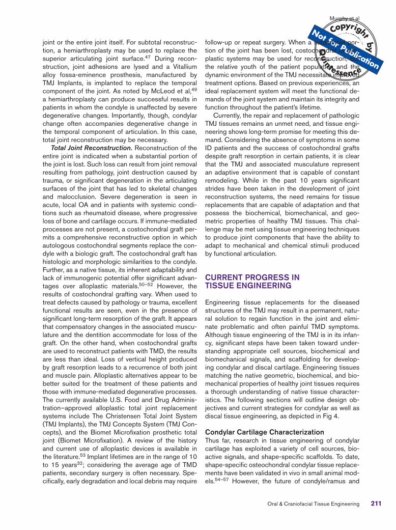
Engineering tissue replacements for the diseased structures of the TMJ may result in a permanent, natu-ral solution to regain function in the joint and elimi-nate problematic and often painful TMD symptoms. Although tissue engineering of the TMJ is in its infan-cy, significant steps have been taken toward under-standing appropriate cell sources, biochemical and biomechanical signals, and scaffolding for develop-ing condylar and discal cartilage. Engineering tissues matching the native geometric, biochemical, and bio-mechanical properties of healthy joint tissues requires a thorough understanding of native tissue character-istics. The following sections will outline design ob-jectives and current strategies for condylar as well as discal tissue engineering, as depicted in Fig 4.
Condylar Cartilage CharacterizationThus far, research in tissue engineering of condylar cartilage has exploited a variety of cell sources, bio-active signals, and shape-specific scaffolds. To date, shape-specific osteochondral condylar tissue replace-ments have been validated in vivo in small animal mod-els.54–57 However, the future of condyle/ramus and
Murphy et al
212 Volume 1, Number 3, 2011
osteochondral tissue replacements will require dem-onstrations of long-term efficacy in large animal mod-els. As reflected by the literature, validation of such engineered replacement tissues is based upon com-parison with native biochemical and biomechanical tis-sue properties.
From an anatomical standpoint, the condyle is lon-ger mediolaterally than anteroposteriorly, forming an ellipse in the transverse plane. Fibrous connective tissue extends from the periphery of the disc, secur-ing it to the condyle inferiorly and to the temporal bone superiorly. This arrangement of connective tis-sue forms a fluid-filled joint capsule with two discrete compartments. Anteriorly and posteriorly, the condyle connects to the TMJ disc via the capsular ligaments, while mediolaterally, the condyle connects to the disc via the collateral ligaments. This arrangement ensures close contact between the disc and condyle during joint movement. The condyle is formed by the con-dylar process of the mandibular bone and is covered superiorly by a layer of zonal cartilage. The mandibular
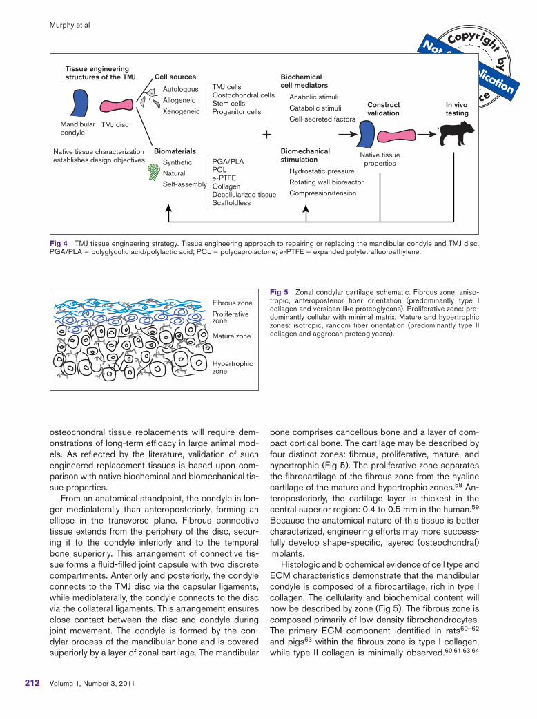
bone comprises cancellous bone and a layer of com-pact cortical bone. The cartilage may be described by four distinct zones: fibrous, proliferative, mature, and hypertrophic (Fig 5). The proliferative zone separates the fibrocartilage of the fibrous zone from the hyaline cartilage of the mature and hypertrophic zones.58 An-teroposteriorly, the cartilage layer is thickest in the central superior region: 0.4 to 0.5 mm in the human.59 Because the anatomical nature of this tissue is better characterized, engineering efforts may more success-fully develop shape-specific, layered (osteochondral) implants.
Histologic and biochemical evidence of cell type and ECM characteristics demonstrate that the mandibular condyle is composed of a fibrocartilage, rich in type I collagen. The cellularity and biochemical content will now be described by zone (Fig 5). The fibrous zone is composed primarily of low-density fibrochondrocytes. The primary ECM component identified in rats60–62 and pigs63 within the fibrous zone is type I collagen, while type II collagen is minimally observed.60,61,63,64
Fig 5 Zonal condylar cartilage schematic. Fibrous zone: aniso-tropic, anteroposterior fiber orientation (predominantly type I collagen and versican-like proteoglycans). Proliferative zone: pre-dominantly cellular with minimal matrix. Mature and hypertrophic zones: isotropic, random fiber orientation (predominantly type II collagen and aggrecan proteoglycans).
Fig 4 TMJ tissue engineering strategy. Tissue engineering approach to repairing or replacing the mandibular condyle and TMJ disc. PGA/PLA = polyglycolic acid/polylactic acid; PCL = polycaprolactone; e-PTFE = expanded polytetrafluoroethylene.
Tissue engineering
structures of the TMJ
Mandibular condyle
TMJ disc
Cell sources
Biomaterials
Synthetic
Natural
Self-assembly
Autologous
Allogeneic
Xenogeneic
TMJ cellsCostochondral cellsStem cells Progenitor cells
PGA/PLAPCLe-PTFECollagenDecellularized tissueScaffoldless
Biochemical
cell mediators
Biomechanical
stimulation
Anabolic stimuli
Catabolic stimuli
Cell-secreted factors
Hydrostatic pressure
Rotating wall bioreactor
Compression/tension
Construct
validation
Native tissueproperties
In vivo
testing
Native tissue characterization establishes design objectives
Fibrous zone
Proliferative zone
Mature zone
Hypertrophic zone
Murphy et al
213Oral & Craniofacial Tissue Engineering
An anisotropic, anteroposterior organization of fibers has been observed, similar to that of the disc.65,66 Porcine67 and rat68 studies have shown that the pri-mary proteoglycan comprising this zone is similar in nature to versican, consisting almost exclusively of chondroitin sulfate glycosaminoglycans (GAGs).
Inferior to the fibrous zone is the proliferative zone. This zone acts as a cell reservoir and contains mesen-chymal chondrocyte precursor cells. To this effect, the proliferative zone is highly cellularized and the matrix is minimally developed. Type I collagen has been de-tected in this zone, observed most often as scattered fibers.62,69 Similar to the fibrous zone, immunohisto-chemistry has identified versican-like proteoglycans in the proliferative zone.67
The mature and hypertrophic zones are similar to each other in their cellularity and ECM composition. These two layers are cellularized by mature chondro-cytes. Chondrocytes of the hypertrophic zone, how-ever, are larger. The ECM in both zones comprises primarily type II collagen,60 but types 1 and 10 have also been identified.64 Collagen organization in the mature and hypertrophic zones is isotropic, showing random bundle orientation.62,70 Furthermore, aggre-can has been identified as the primary proteoglycan in these zones in porcine67 and rat64 models. Sig-nificantly, the articulating surface of the mandibular condyles is largely fibrous (rich in type I collagen), in contrast to the hyaline nature of other articulating sur-faces, such as those found in the knee and hip.
Illustrating a structure-function relationship, biome-chanical evidence suggests that the condyle is stiffer under tension in the anteroposterior direction than in the mediolateral direction. In the porcine model, Young’s modulus has been measured as 9.0 ± 1.7 MPa in the anteroposterior direction and 6.6 ± 1.2 MPa in the mediolateral direction under axial tension to failure (n = 8).71 This mechanical behavior agrees with the work of Singh and Detamore,66 which identi-fied anisotropic collagen alignment. This group also obtained moduli ranging from 22 to 29 MPa in the anteroposterior direction and 8 to 11 MPa in the me-diolateral direction.66 Shear studies have likewise con-firmed the anisotropy of mechanical behavior. Storage moduli in dynamic shear experiments at 2 Hz frequen-cy range from 1.50 to 2.03 MPa in the anteroposterior direction, yet they range from 0.33 to 0.55 MPa in the mediolateral direction (n = 17).72 The anisotropic col-lagen orientation and tensile and shear properties of the mandibular condyle suggest anteroposterior load-ing, matching the loading patterns observed during translation and rotation of the mandible in vivo.
Although compressive structure-function relation-ships have yet to be revealed for the condyle, regional variability has been established and likely contributes
to specific condylar function. Compressive properties of the condyle have been examined via atomic force mi-croscopy (AFM), indentation testing, and unconfined compression. In one study of regional variability, rabbit condylar cartilage was divided into four regions and tested in compression using AFM.73 Young’s modulus and the Poisson ratio were both revealed to decrease in magnitude, in order, from the anteromedial region to the anterolateral, posteromedial, and posterolateral regions. Notably, these results suggest the condylar cartilage is stiffer medially than laterally.73 It has also been shown that porcine condylar cartilage deforms significantly less under intermittent compression than sustained compression,74 an expected result in light of the dynamic nature of the joint. In two other stud-ies, aggregate moduli from in situ creep testing75 and equilibrium moduli from unconfined compression test-ing76 were reported. Creep testing demonstrated the greatest aggregate moduli in the central and medial positions, with the aggregate modulus of the medial position significantly greater than that of the lateral and anterior positions.75 Equilibrium moduli obtained during unconfined testing demonstrated the greatest stiffness in the posterior region and the greatest com-pliance in the anterior region.76 Although a consensus regarding the specific regional biomechanical variabil-ity remains to be established, these data suggest that the joint sustains significant load in the medial and posterior regions in vivo and more successfully resists cyclic rather than sustained loading, a factor that may contribute to TMD progression.
Tissue Engineering of Condylar CartilageTissue engineering initiatives attempting to recreate the native condylar cartilage follow a three-part ap-proach that considers cell sourcing, biomaterials for scaffolding, and bioactive stimuli. With respect to cell sourcing, adult condylar cartilage cells have been explored in the greatest detail. However, it is impor-tant to note the significant donor site morbidity and potential pathology in TMD patients associated with this cell source. As research progresses, it is expect-ed that alternative primary and stem cells will receive more significant attention. Nonetheless, as a result of their appropriate phenotype, condylar chondrocytes offer an effective starting point for condylar cartilage engineering strategies. Among others, two distinct strategies have been established for acquiring pri-mary condylar cartilage cells. The more common tech-nique for obtaining primary cells involves harvesting, mincing, and isolating condylar cells via a collagenase treatment.77 In contrast, a second procedure allows the cells to migrate out of the fibrous zone of condy-lar tissue onto surgical sponges, yielding fibroblastlike cells upon isolation.78 With regard to alternative cell

Murphy et al
214 Volume 1, Number 3, 2011
sources, ankle hyaline cartilage cells were recently shown to outperform condylar cartilage cells in terms of biosynthesis and cell proliferation when seeded in three-dimensional nonwoven polyglycolic acid (PGA) meshes,63 although the authors cited nonadherence of condylar cells as a possible factor in their relative-ly poor performance. The hyaline cartilage–seeded scaffolds yielded a more fibrocartilaginous tissue with both type I and type II collagens. In contrast, condylar cartilage–seeded scaffolds yielded a more fibrous tis-sue, which predominantly stained positive for type I collagen.63 This is not a surprising result considering the hyaline nature of the articulating cartilage of the ankle, in contrast to the fibrous nature of the cartilage of the TMJ condyle. Prior to this work, the same group explored human umbilical cord matrix stem cells. Hu-man umbilical cord matrix stem cell constructs were found to yield 55% and 200% higher cellularity at 0 and 4 weeks, respectively, as well as higher GAG content over condylar cartilage constructs.79 Because of donor site morbidity and tissue engineering chal-lenges associated with condylar cartilage cell sourc-ing, it is apparent that researchers have begun to turn their attention toward alternative sources. More work is needed to exploit these potential sources, but promise exists in the arenas of progenitor, mesenchy-mal, embryonic, and induced pluripotent stem cells.
Research regarding scaffold selection has primarily involved the idea of developing shape-specific scaf-folds. For example, Williams et al80 demonstrated that polycaprolactone (PCL) bioresorbable scaffolds may be constructed by solid free-form fabrication tech-niques based on CT (or alternatively MRI) imaging data to generate an anatomically shaped mandibu-lar condyle scaffold that attaches to the ramus via a collar. Seeded with bone morphogenetic protein-7–transformed fibroblasts, the group obtained compres-sive moduli and yield strengths in the lower range of reports for human trabecular bone.80 A second study from the same group demonstrated that bipha-sic PCL scaffolds may be differentially seeded with transformed fibroblasts and fully differentiated chon-drocytes.57 This strategy yielded differential tissues with a mineralized interface when the scaffolds were implanted subcutaneously.57 More recently, the pres-ence of blood vessels, marrow stroma, and adipose tissue was demonstrated in the ceramic phase of these scaffolds, representing the region seeded with trans-formed fibroblasts.56 In an alternative strategy for de-veloping shape-specific scaffolds, Alhadlaq et al54,55 demonstrated the potential of sequential photopoly-merization of poly(ethylene glycol) (PEG) hydrogels. This strategy was used to create osteochondral con-structs with shapes and dimensions matching those of a human cadaveric mandibular condyle model.55
Importantly, this group demonstrated the potential for inducing differentiation of primary bone marrow– derived mesenchymal stem cells into chondrocyte and bone lineages for the development of stratified bone and cartilage layers.54,55 As can be seen, there are many biomaterials that may be implemented for con-dylar tissue engineering, some of which offer patient-specific morphology.
In vitro culture techniques may include the applica-tion of biomechanical stimulation, which is intended to mimic physiologic loading conditions and there-fore influence ECM architecture. Current efforts with bioreactors and direct stimulation have attempted to do so, specifically with the intention of encouraging cell growth and recreating the ECM architecture of healthy condylar cartilage. For example, mass transfer bioreactors can be used in culture toward obtaining a homogenous cell distribution and improved nutri-ent and waste transport over static cultures. Rotating wall bioreactors stimulate cell proliferation and bio-synthesis without causing cell damage by exposing cells to a low shear force via laminar flow. Similarly, spinner flasks accelerate the exchange of oxygen and nutrients in the interior of scaffolds, improving cell proliferation and matrix synthesis. Hydrostatic and direct- compression loading schemes may potentially be used to stimulate matrix deposition, improving the mechanical properties of engineered condylar carti-lage.81 With in vitro characterization showing that the tissue deforms significantly less under intermittent compression than sustained compression74 and in consideration of the native, dynamic loading patterns in the TMJ, Nicodemus et al82 obtained surprising results in response to dynamic compressive strains. Bovine condylar chondrocytes were encapsulated in photopolymerized PEG hydrogels, and these con-structs were exposed to dynamic loading at 0.3 Hz and 15% amplitude. Dynamic stimulation led to sup-pression of gene expression, cell proliferation, and proteoglycan synthesis versus unloaded controls.82 This work recognizes the need to further investigate the potential role of mechanical stimulation, via various loading schemes, in the development of constructs.
Bioactive signals may also be used to encourage cell proliferation and biosynthesis with cellular responses, depending on the specific signal or combination of sig-nals. Addressing first the role of proliferative agents for condylar cartilage cells, basic fibroblast growth factor (bFGF) has been found to have the greatest stimula-tory effect on the proliferation of second-passage hu-man mandibular condylar chondrocytes, in comparison to insulin-like growth factor-1 (IGF-1) and transform-ing growth factor-β1 (TGF-β1) treatments in mono-layer culture.83 Studies exploring biosynthesis as well as proliferation have observed an inhibitory effect of
Murphy et al
215Oral & Craniofacial Tissue Engineering
bFGF on GAG and collagen synthesis,84 in contrast to the enhancing effect of IGF-I on biosynthesis.85,86 Specifically, an inhibition of GAG and collagen synthe-sis in rat condylar cartilage explants was observed in the presence of bFGF following 2 weeks of culture.84 An increase in GAG and collagen synthesis, on the other hand, was observed in explants treated with IGF-I alone or in combination with bFGF, with bFGF down-regulating the biosynthetic effects of IGF-I when used in combination. With respect to epidermal growth fac-tor (EGF), Tsubai et al,78 whose isolation technique was previously mentioned, explored EGF treatment in fibroblast-like condylar cells obtained from fetal rats. EGF was shown to bring cells into the s-phase of the cell cycle more quickly and to increase cell number over controls; both these measures indicate an increase in cellular proliferation.78 The authors also noted the role of EGF in matrix deposition, with tissue volume increas-ing toward the end of the culture period (21 days).78 As research moves toward alternative cell sources, the understanding of bioactive signals must be translatable. Notably, early work by Copray et al87 demonstrated that most of the factors enhancing proliferation explored in their study, including EGF, similarly enhanced prolifera-tion in secondary mandibular condylar cartilage as well as in primary costal chondrocytes. However, these re-sults must be validated in the specific culture system under review, considering not only the cell source but also the type of scaffold and mechanical stimulation.
Combined mechanical and bioactive stimulation has revealed the interrelated roles of biochemical and biomechanical effectors. A study of rat condylar car-tilage cells explored the effects of TGF-β1 and static tension-stress (5 kPa) on cellular proliferation.88 It was demonstrated that TGF-β1 had a mitogenic effect at all concentrations under review (0.1, 1, and 10 ng/mL), but an additive effect was observed in a group treated with both TGF-β1 and static tension-stress. As vari-ous cell sources and culture systems are explored, this result illustrates the need for continued exploration of exogenous stimulation, both chemical and mechani-cal, throughout cell culture toward the development of shape-specific condylar replacements.
Glenoid Fossa and Articular EminenceIn attempts to repair or replace pathologic TMJ tis-sues, it is essential to continue with a discussion of the superior articulating surface of the joint, including the articular eminence and glenoid fossa. Together, the superior and inferior surfaces transmit loads ex-perienced by the joint through the TMJ disc. It is im-portant to note the incongruence existing between the superior and inferior surfaces. The TMJ disc and the synovial fluid contained within the joint capsule fill this gap, ensuring smooth articulation. As previously
mentioned, joint pathology, including OA and ID, can significantly affect this structure-function relationship.
Of the salient tissues in the joint, the glenoid fossa and articular eminence are the least characterized in terms of biochemical and biomechanical proper-ties. The surface of the fossa has been described as a dense, fibrous tissue,89 but more specific charac-terization is needed. As expected, the primary com-ponent of this fibrous tissue has been identified as collagen.90 Biomechanical evaluation of the glenoid fossa and articular eminence has demonstrated that the aggregate moduli are higher in the medial and posterior regions (42.6 and 58.9 kPa, respectively) and lower in the anterior, central, and lateral regions, all in the range of 35 kPa.89
With limited information available regarding charac-terization of these areas, design criteria and validation metrics have yet to be established for the engineering of tissue replacements for the superior articulating sur-faces. To the authors’ knowledge, tissue engineering efforts have not yet addressed this tissue. However, as research progresses toward the development of con-dylar and TMJ discal tissue replacements, the glenoid fossa and articular eminence must also be considered.
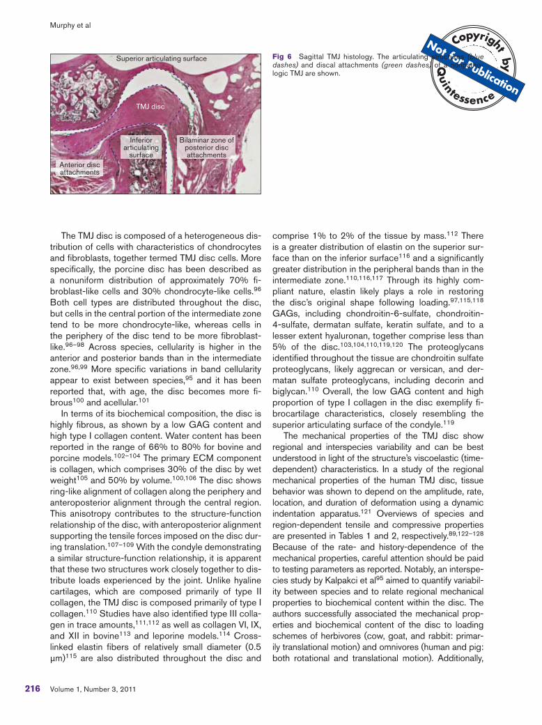
TMJ Disc CharacterizationThe following section briefly outlines the anatomy, structure, and function of the TMJ disc. More detailed reports may be found in the literature.91–94 From a superior view, the human disc takes on a biconcave, elliptic shape, and it is longer mediolaterally (~23 mm) than anteroposteriorly (~14 mm),95 similar to the shape of the condyle. The disc may be divided into three zones: the anterior band, the intermediate zone, and the posterior band.91 In the sagittal view of a hu-man TMJ (Fig 6), the posterior band is thicker than the anterior band, and the intermediate zone is the thinnest region. As described previously, the disc is attached along its periphery to the condyle and tem-poral bone via fibrous connective tissue. Anteriorly, the disc is attached to the articular eminence and to the condyle at the pterygoid fovea via capsular ligaments. Posteriorly, the disc blends with the bilaminar zone, a network of fibroelastic tissue, connecting superiorly to the glenoid fossa and inferiorly to the condyle. When the joint is in the neutral position, the disc is situated between the condyle and the glenoid fossa. With joint motion, less tenuous superior attachments allow the superior surface of the disc to translate anteroposteri-orly, and to a lesser extent mediolaterally, with respect to the fossa. The inferior surface of the disc, in con-trast, remains in close proximity to the condyle. The shape and motion of the disc impart its function: to separate the incongruent articulating surfaces and to transmit force between them.
Murphy et al
216 Volume 1, Number 3, 2011
The TMJ disc is composed of a heterogeneous dis-tribution of cells with characteristics of chondrocytes and fibroblasts, together termed TMJ disc cells. More specifically, the porcine disc has been described as a nonuniform distribution of approximately 70% fi-broblast-like cells and 30% chondrocyte-like cells.96
Both cell types are distributed throughout the disc, but cells in the central portion of the intermediate zone tend to be more chondrocyte-like, whereas cells in the periphery of the disc tend to be more fibroblast-like.96–98 Across species, cellularity is higher in the anterior and posterior bands than in the intermediate zone.96,99 More specific variations in band cellularity appear to exist between species,95 and it has been reported that, with age, the disc becomes more fi-brous100 and acellular.101
In terms of its biochemical composition, the disc is highly fibrous, as shown by a low GAG content and high type I collagen content. Water content has been reported in the range of 66% to 80% for bovine and porcine models.102–104 The primary ECM component is collagen, which comprises 30% of the disc by wet weight105 and 50% by volume.100,106 The disc shows ring-like alignment of collagen along the periphery and anteroposterior alignment through the central region. This anisotropy contributes to the structure-function relationship of the disc, with anteroposterior alignment supporting the tensile forces imposed on the disc dur-ing translation.107–109 With the condyle demonstrating a similar structure-function relationship, it is apparent that these two structures work closely together to dis-tribute loads experienced by the joint. Unlike hyaline cartilages, which are composed primarily of type II collagen, the TMJ disc is composed primarily of type I collagen.110 Studies have also identified type III colla-gen in trace amounts,111,112 as well as collagen VI, IX, and XII in bovine113 and leporine models.114 Cross-linked elastin fibers of relatively small diameter (0.5 μm)115 are also distributed throughout the disc and
comprise 1% to 2% of the tissue by mass.112 There is a greater distribution of elastin on the superior sur-face than on the inferior surface116 and a significantly greater distribution in the peripheral bands than in the intermediate zone.110,116,117 Through its highly com-pliant nature, elastin likely plays a role in restoring the disc’s original shape following loading.97,115,118
GAGs, including chondroitin-6-sulfate, chondroitin-4-sulfate, dermatan sulfate, keratin sulfate, and to a lesser extent hyaluronan, together comprise less than 5% of the disc.103,104,110,119,120 The proteoglycans identified throughout the tissue are chondroitin sulfate proteoglycans, likely aggrecan or versican, and der-matan sulfate proteoglycans, including decorin and biglycan.110 Overall, the low GAG content and high proportion of type I collagen in the disc exemplify fi-brocartilage characteristics, closely resembling the superior articulating surface of the condyle.119
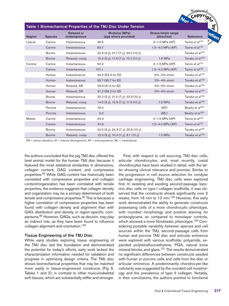
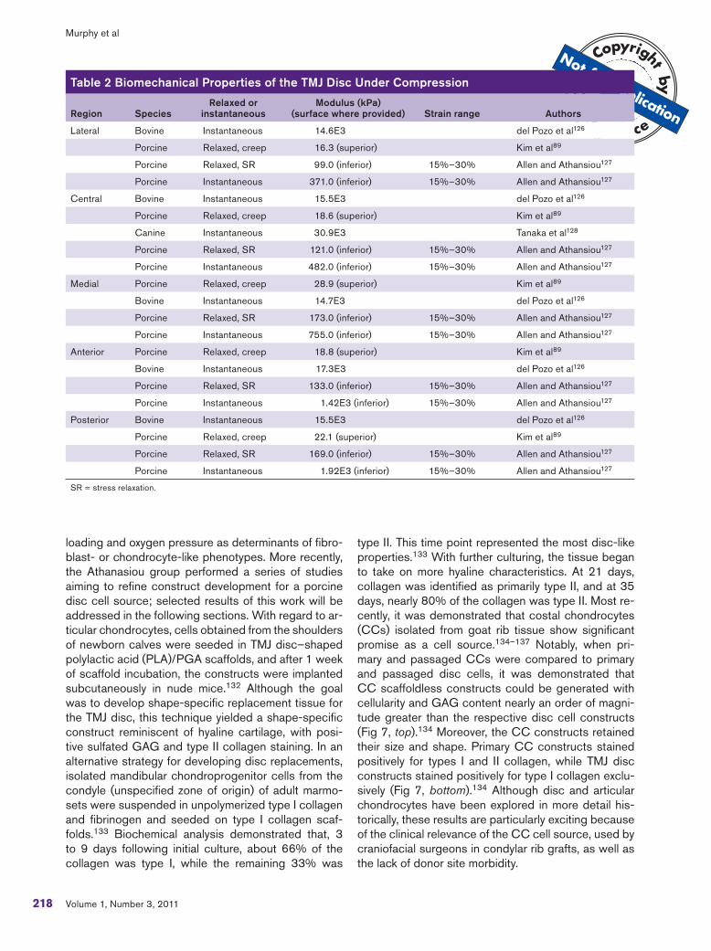
The mechanical properties of the TMJ disc show regional and interspecies variability and can be best understood in light of the structure’s viscoelastic (time-dependent) characteristics. In a study of the regional mechanical properties of the human TMJ disc, tissue behavior was shown to depend on the amplitude, rate, location, and duration of deformation using a dynamic indentation apparatus.121 Overviews of species and region-dependent tensile and compressive properties are presented in Tables 1 and 2, respectively.89,122–128
Because of the rate- and history-dependence of the mechanical properties, careful attention should be paid to testing parameters as reported. Notably, an interspe-cies study by Kalpakci et al95 aimed to quantify variabil-ity between species and to relate regional mechanical properties to biochemical content within the disc. The authors successfully associated the mechanical prop-erties and biochemical content of the disc to loading schemes of herbivores (cow, goat, and rabbit: primar-ily translational motion) and omnivores (human and pig: both rotational and translational motion). Additionally,
TMJ disc
Bilaminar zone of posterior disc attachments
Inferior articulating
surfaceAnterior disc attachments
Superior articulating surface Fig 6 Sagittal TMJ histology. The articulating structures (blue dashes) and discal attachments (green dashes) of a nonpatho-logic TMJ are shown.
Murphy et al
217Oral & Craniofacial Tissue Engineering
the authors concluded that the pig TMJ disc offered the best animal model for the human TMJ disc because it featured the most statistical similarities in dimensions, collagen content, GAG content, and compressive properties.95 While GAG content has historically been correlated with compressive properties and collagen content/organization has been correlated with tensile properties, the evidence suggests that collagen density and organization may be a primary determinant of both tensile and compressive properties.95 This is because a higher correlation of compressive properties has been found with collagen density and alignment than with GAG distribution and density in region-specific com-parisons.95 However, GAGs, such as decorin, may play an indirect role, as they have been found to influence collagen alignment and orientation.129
Tissue Engineering of the TMJ DiscWhile early studies exploring tissue engineering of the TMJ disc laid the foundation and demonstrated the potential for today’s efforts, early work lacked the characterization information needed for validation and progress in optimizing design criteria. The TMJ disc shows biomechanical properties that may be matched more easily in tissue-engineered constructs (Fig 6, Tables 1 and 2), in contrast to other musculoskeletal soft tissues, which are substantially stiffer and stronger.
First, with respect to cell sourcing, TMJ disc cells, articular chondrocytes, and, most recently, costal chondrocytes have been studied in detail, with the lat-ter showing clinical relevance and promise. Similar to the progression in cell source selection for condylar cartilage engineering, TMJ disc cells were explored first. In isolating and seeding second-passage lepo-rine disc cells on type I collagen scaffolds, it was ob-served that the constructs shrank significantly over 2 weeks, from 16 mm to 12 mm.130 However, this early work demonstrated the ability to generate constructs possessing cells of a more chondrocytic phenotype, with rounded morphology and positive staining for proteoglycans, as compared to monolayer controls, which showed a more fibroblastic phenotype.130 Con-sidering possible variability between species and cell sources within the TMJ, second-passage cells from human and porcine TMJ disc and articular eminence were explored with various scaffolds: polyamide, ex-panded polytetrafluoroethylene, PGA, natural bone mineral blocks, and glass.131 The results demonstrated no significant differences between constructs seeded with human or porcine cells and cells from the disc or articular eminence. A predominantly chondrocyte-like cellularity was suggested by the rounded cell morphol-ogy and the prevalence of type II collagen. Notably, in their conclusions, the authors pointed to functional
Table 1 Biomechanical Properties of the TMJ Disc Under Tension
Region Species
Relaxed or
instantaneous
Modulus (MPa)
(age where provided)
Stress/strain range
(direction) Reference
Lateral Canine Instantaneous 39.5 0–1.5 MPa (AP) Tanne et al122
Canine Instantaneous 83.7 1.5–4.0 MPa (AP) Tanne et al122
Bovine Instantaneous 22.5 (3 y), 21.7 (7 y), 24.0 (10 y) Tanaka et al123
Bovine Relaxed, creep 12.4 (3 y), 11.9 (7 y), 10.2 (10 y) 1.5 MPa Tanaka et al123
Central Canine Instantaneous 50.2 0–1.5 MPa (AP) Tanne et al122
Canine Instantaneous 101.1 1.5–4.0 MPa (AP) Tanne et al122
Human Instantaneous 44.0 (53.3 for ID) 0%–2% strain Tanaka et al124
Human Instantaneous 95.7 (95.7 for ID) 2%–4% strain Tanaka et al124
Human Relaxed, SR 29.9 (41.0 for ID) 0%–2% strain Tanaka et al124
Human Relaxed, SR 61.2 (59.2 for ID) 2%–4% strain Tanaka et al124
Bovine Instantaneous 20.2 (3 y), 21.0 (7 y), 22.9 (10 y) Tanaka et al123
Bovine Relaxed, creep 14.5 (3 y), 12.9 (7 y), 11.5 (10 y) 1.5 MPa Tanaka et al123
Porcine Instantaneous 76.4 (AP) Beatty et al125
Porcine Instantaneous 3.2 (ML) Beatty et al125
Medial Canine Instantaneous 43.3 0–1.5 MPa (AP) Tanne et al122
Canine Instantaneous 91.9 1.5–4.0 MPa (AP) Tanne et al122
Bovine Instantaneous 24.0 (3 y), 24.3 (7 y), 25.9 (10 y) Tanaka et al123
Bovine Relaxed, creep 12.4 (3 y), 10.4 (7 y), 9.1 (10 y) 1.5 MPa Tanaka et al123
SR = stress relaxation; ID = internal derangement; AP = anteroposterior; ML = mediolateral.
Murphy et al
218 Volume 1, Number 3, 2011
loading and oxygen pressure as determinants of fibro-blast- or chondrocyte-like phenotypes. More recently, the Athanasiou group performed a series of studies aiming to refine construct development for a porcine disc cell source; selected results of this work will be addressed in the following sections. With regard to ar-ticular chondrocytes, cells obtained from the shoulders of newborn calves were seeded in TMJ disc–shaped polylactic acid (PLA)/PGA scaffolds, and after 1 week of scaffold incubation, the constructs were implanted subcutaneously in nude mice.132 Although the goal was to develop shape-specific replacement tissue for the TMJ disc, this technique yielded a shape-specific construct reminiscent of hyaline cartilage, with posi-tive sulfated GAG and type II collagen staining. In an alternative strategy for developing disc replacements, isolated mandibular chondroprogenitor cells from the condyle (unspecified zone of origin) of adult marmo-sets were suspended in unpolymerized type I collagen and fibrinogen and seeded on type I collagen scaf-folds.133 Biochemical analysis demonstrated that, 3 to 9 days following initial culture, about 66% of the collagen was type I, while the remaining 33% was
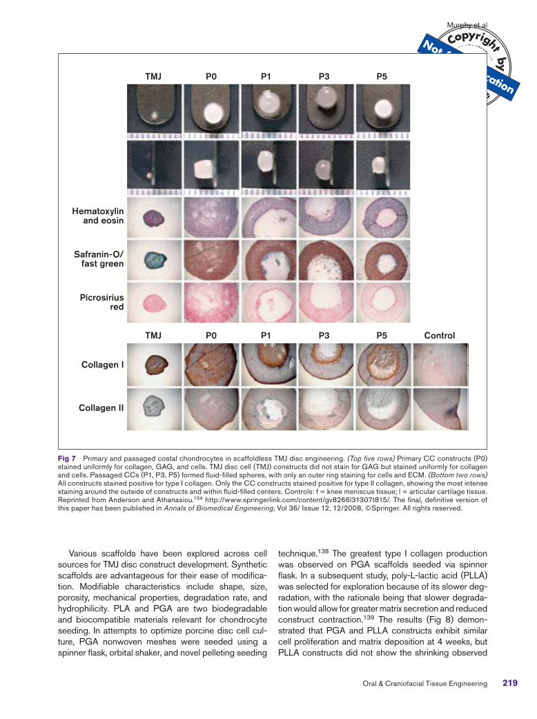
type II. This time point represented the most disc-like properties.133 With further culturing, the tissue began to take on more hyaline characteristics. At 21 days, collagen was identified as primarily type II, and at 35 days, nearly 80% of the collagen was type II. Most re-cently, it was demonstrated that costal chondrocytes (CCs) isolated from goat rib tissue show significant promise as a cell source.134–137 Notably, when pri-mary and passaged CCs were compared to primary and passaged disc cells, it was demonstrated that CC scaffoldless constructs could be generated with cellularity and GAG content nearly an order of magni-tude greater than the respective disc cell constructs (Fig 7, top).134 Moreover, the CC constructs retained their size and shape. Primary CC constructs stained positively for types I and II collagen, while TMJ disc constructs stained positively for type I collagen exclu-sively (Fig 7, bottom).134 Although disc and articular chondrocytes have been explored in more detail his-torically, these results are particularly exciting because of the clinical relevance of the CC cell source, used by craniofacial surgeons in condylar rib grafts, as well as the lack of donor site morbidity.
Table 2 Biomechanical Properties of the TMJ Disc Under Compression
Region Species
Relaxed or
instantaneous
Modulus (kPa)
(surface where provided) Strain range Authors
Lateral Bovine Instantaneous 14.6E3 del Pozo et al126
Porcine Relaxed, creep 16.3 (superior) Kim et al89
Porcine Relaxed, SR 99.0 (inferior) 15%–30% Allen and Athansiou127
Porcine Instantaneous 371.0 (inferior) 15%–30% Allen and Athansiou127
Central Bovine Instantaneous 15.5E3 del Pozo et al126
Porcine Relaxed, creep 18.6 (superior) Kim et al89
Canine Instantaneous 30.9E3 Tanaka et al128
Porcine Relaxed, SR 121.0 (inferior) 15%–30% Allen and Athansiou127
Porcine Instantaneous 482.0 (inferior) 15%–30% Allen and Athansiou127
Medial Porcine Relaxed, creep 28.9 (superior) Kim et al89
Bovine Instantaneous 14.7E3 del Pozo et al126
Porcine Relaxed, SR 173.0 (inferior) 15%–30% Allen and Athansiou127
Porcine Instantaneous 755.0 (inferior) 15%–30% Allen and Athansiou127
Anterior Porcine Relaxed, creep 18.8 (superior) Kim et al89
Bovine Instantaneous 17.3E3 del Pozo et al126
Porcine Relaxed, SR 133.0 (inferior) 15%–30% Allen and Athansiou127
Porcine Instantaneous 1.42E3 (inferior) 15%–30% Allen and Athansiou127
Posterior Bovine Instantaneous 15.5E3 del Pozo et al126
Porcine Relaxed, creep 22.1 (superior) Kim et al89
Porcine Relaxed, SR 169.0 (inferior) 15%–30% Allen and Athansiou127
Porcine Instantaneous 1.92E3 (inferior) 15%–30% Allen and Athansiou127
SR = stress relaxation.
Murphy et al
219Oral & Craniofacial Tissue Engineering
Various scaffolds have been explored across cell sources for TMJ disc construct development. Synthetic scaffolds are advantageous for their ease of modifica-tion. Modifiable characteristics include shape, size, porosity, mechanical properties, degradation rate, and hydrophilicity. PLA and PGA are two biodegradable and biocompatible materials relevant for chondrocyte seeding. In attempts to optimize porcine disc cell cul-ture, PGA nonwoven meshes were seeded using a spinner flask, orbital shaker, and novel pelleting seeding
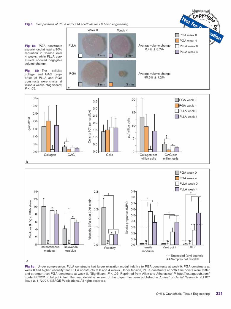
technique.138 The greatest type I collagen production was observed on PGA scaffolds seeded via spinner flask. In a subsequent study, poly-L-lactic acid (PLLA) was selected for exploration because of its slower deg-radation, with the rationale being that slower degrada-tion would allow for greater matrix secretion and reduced construct contraction.139 The results (Fig 8) demon-strated that PGA and PLLA constructs exhibit similar cell proliferation and matrix deposition at 4 weeks, but PLLA constructs did not show the shrinking observed
Fig 7 Primary and passaged costal chondrocytes in scaffoldless TMJ disc engineering. (Top five rows) Primary CC constructs (P0) stained uniformly for collagen, GAG, and cells. TMJ disc cell (TMJ) constructs did not stain for GAG but stained uniformly for collagen and cells. Passaged CCs (P1, P3, P5) formed fluid-filled spheres, with only an outer ring staining for cells and ECM. (Bottom two rows) All constructs stained positive for type I collagen. Only the CC constructs stained positive for type II collagen, showing the most intense staining around the outside of constructs and within fluid-filled centers. Controls: f = knee meniscus tissue; l = articular cartilage tissue. Reprinted from Anderson and Athanasiou.134 http://www.springerlink.com/content/gv8266l31307t815/. The final, definitive version of this paper has been published in Annals of Biomedical Engineering, Vol 36/ Issue 12, 12/2008, ©Springer. All rights reserved.
TMJ P0 P1 P3 P5
TMJ P0 P1 P3 P5 Control
Hematoxylin and eosin
Safranin-O/fast green
Picrosirius red
Collagen I
Collagen II
Murphy et al
220 Volume 1, Number 3, 2011
in PGA constructs.139 Considering native biomateri-als, type I collagen is an extensively studied scaffold material for cartilage tissue engineering. Collagen may be used as a seeding vehicle, either intact or following proteolytic digestion for gel encapsulation. Importantly, it has been demonstrated that collagen synthesis is en-hanced in constructs seeded on collagen scaffolds.140 Electrospinning collagen scaffolds may potentially be used to encourage collagen synthesis and organiza-tion toward recapitulating aforementioned native tissue characteristics. In attempting to develop disc replace-ments, it is likely that a type I collagen sponge would yield constructs with morphology more similar to that of the disc, as compared to gel encapsulation. Gels, how-ever, may be better suited for filling defects.
Decellularized tissues present another scaffold op-tion. For example, the porcine disc has been explored as a xenogeneic scaffold.141 With respect to the me-chanical integrity of scaffolds following various decel-lularizing treatments, dodecyl sulfate–treated tissues have been identified as potential seeding vehicles for TMJ disc engineering.141 However, aside from their inherent potential immunogenicity, several disadvan-tages exist for decellularized tissues, including the in-ability to control scaffold size/shape and difficulty in reseeding the tissue.
A novel and promising method for tissue engineer-ing the TMJ soft tissues involves self-assembly of constructs using a scaffoldless approach. It has been demonstrated that self-assembled articular cartilage constructs may be developed, with aggregate mod-uli approaching those of native tissue with clinically relevant dimensions.142–144 Scaffoldless constructs eliminate the problem of scaffold-induced stress shielding, permitting important mechanotransductive events during tissue development and biosynthesis. Furthermore, self-assembled constructs circumvent some disadvantages of scaffold use: hindrance of cell-to-cell communication, immunogenicity, and the potentially deleterious effects of by-products of deg-radation. Thus, while numerous seeding vehicles have been explored, a scaffoldless technique holds signifi-cant potential for engineered TMJ disc replacements.
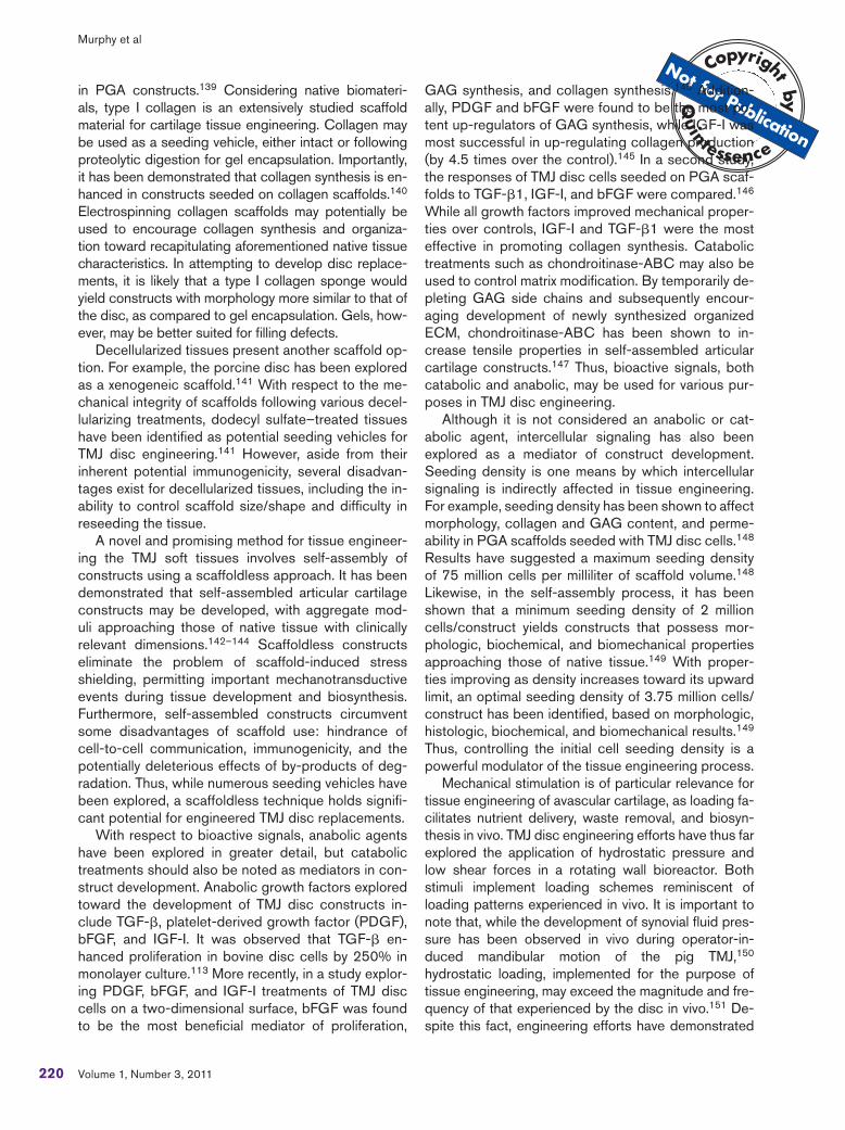
With respect to bioactive signals, anabolic agents have been explored in greater detail, but catabolic treatments should also be noted as mediators in con-struct development. Anabolic growth factors explored toward the development of TMJ disc constructs in-clude TGF-β, platelet-derived growth factor (PDGF), bFGF, and IGF-I. It was observed that TGF-β en-hanced proliferation in bovine disc cells by 250% in monolayer culture.113 More recently, in a study explor-ing PDGF, bFGF, and IGF-I treatments of TMJ disc cells on a two-dimensional surface, bFGF was found to be the most beneficial mediator of proliferation,
GAG synthesis, and collagen synthesis.145 Addition-ally, PDGF and bFGF were found to be the most po-tent up-regulators of GAG synthesis, while IGF-I was most successful in up-regulating collagen production (by 4.5 times over the control).145 In a second study, the responses of TMJ disc cells seeded on PGA scaf-folds to TGF-β1, IGF-I, and bFGF were compared.146 While all growth factors improved mechanical proper-ties over controls, IGF-I and TGF-β1 were the most effective in promoting collagen synthesis. Catabolic treatments such as chondroitinase-ABC may also be used to control matrix modification. By temporarily de-pleting GAG side chains and subsequently encour-aging development of newly synthesized organized ECM, chondroitinase-ABC has been shown to in-crease tensile properties in self-assembled articular cartilage constructs.147 Thus, bioactive signals, both catabolic and anabolic, may be used for various pur-poses in TMJ disc engineering.
Although it is not considered an anabolic or cat-abolic agent, intercellular signaling has also been explored as a mediator of construct development. Seeding density is one means by which intercellular signaling is indirectly affected in tissue engineering. For example, seeding density has been shown to affect morphology, collagen and GAG content, and perme-ability in PGA scaffolds seeded with TMJ disc cells.148 Results have suggested a maximum seeding density of 75 million cells per milliliter of scaffold volume.148 Likewise, in the self-assembly process, it has been shown that a minimum seeding density of 2 million cells/construct yields constructs that possess mor-phologic, biochemical, and biomechanical properties approaching those of native tissue.149 With proper-ties improving as density increases toward its upward limit, an optimal seeding density of 3.75 million cells/construct has been identified, based on morphologic, histologic, biochemical, and biomechanical results.149 Thus, controlling the initial cell seeding density is a powerful modulator of the tissue engineering process.
Mechanical stimulation is of particular relevance for tissue engineering of avascular cartilage, as loading fa-cilitates nutrient delivery, waste removal, and biosyn-thesis in vivo. TMJ disc engineering efforts have thus far explored the application of hydrostatic pressure and low shear forces in a rotating wall bioreactor. Both stimuli implement loading schemes reminiscent of loading patterns experienced in vivo. It is important to note that, while the development of synovial fluid pres-sure has been observed in vivo during operator-in-duced mandibular motion of the pig TMJ,150 hydrostatic loading, implemented for the purpose of tissue engineering, may exceed the magnitude and fre-quency of that experienced by the disc in vivo.151 De-spite this fact, engineering efforts have demonstrated
Murphy et al
221Oral & Craniofacial Tissue Engineering
Fig 8 Comparisons of PLLA and PGA scaffolds for TMJ disc engineering.
Fig 8a PGA constructs experienced at least a 90% reduction in volume over 4 weeks, while PLLA con-structs showed negligible volume change.
Fig 8b The cellular, collage, and GAG prop-erties of PLLA and PGA constructs were similar at 0 and 4 weeks. *Significant; P < .05.
Fig 8c Under compression, PLLA constructs had larger relaxation moduli relative to PGA constructs at week 0. PGA constructs at week 0 had higher viscosity than PLLA constructs at 0 and 4 weeks. Under tension, PLLA constructs at both time points were stiffer and stronger than PGA constructs at week 0. *Significant; P < .05. Reprinted from Allen and Athanasiou.139 http://jdr.sagepub.com/content/87/2/180.full.pdf+html. The final, definitive version of this paper has been published in Journal of Dental Research, Vol 87/ Issue 2, 11/2007, ©SAGE Publications. All rights reserved.
Week 0 Week 4
PLLA
PGA
3 mm 3 mm
3 mm 3 mm
Average volume change0.4% ± 8.7%
Average volume change95.5% ± 1.2%
PGA week 0
PGA week 4
PLLA week 0
PLLA week 4
0
5
10
15
20
μg/m
illio
n ce
lls
Collagen permillion cells
GAG permillion cells
0.0
0.5
1.0
1.5
2.0
2.5
3.0
3.5
Cel
ls (x
105 )
per
sca
ffold
Cells0.0
0.5
1.0
1.5
2.0
2.5
3.0
3.5
μg/s
caffo
ld
Collagen GAG
**
PGA week 0
PGA week 4
PLLA week 0
PLLA week 4
PGA week 0
PGA week 4
PLLA week 0
PLLA week 4
Tensilemodulus
Yield point UTS
##
##
##
Instantaneousmodulus
Relaxationmodulus
Viscosity
##
##
##
*
*
*
*
*
Samples not testableUnseeded (dry) scaffold
##
0.0
0.1
0.2
0.3
0.4
0.5
0.6
0.7
0.8
0.9
Tens
ile p
rope
rtie
s (M
Pa)
0.0
0.1
0.2
0.3
Visc
osity
(kP
a-s)
at 3
0% s
trai
n
0
2
4
6
8
10
12
14
Mod
ulus
(kP
a) a
t 30%
str
ain
a
b
c
Murphy et al
222 Volume 1, Number 3, 2011
that static hydrostatic pressure increases the collagen content over unloaded controls, improving the me-chanical integrity of constructs.151 Specifically, in ex-ploring the role of hydrostatic pressure in monolayer culture and on three-dimensional PGA scaffolds seed-ed with porcine TMJ disc cells, static loading at 10 MPa for 4 hours was found to be most beneficial in promoting biosynthesis. In monolayer culture, and simi-larly on three-dimensional scaffolds, static loading yielded the highest amount of collagen—and specifi-cally, more type I than type II collagen—compared to no loading and cyclic loading.151 In light of the biochemi-cal content of the native disc, this result demonstrates that static loading may be a suitable regimen.
With respect to shear stimulation, shear stress is experienced in vivo by the disc during joint rota-tion and translation and may be simulated in culture via a rotating wall bioreactor. Toward this end, TMJ disc cells were seeded in a spinner flask on nonwo-ven PGA scaffolds, and constructs were cultured ei-ther statically or in a low-shear rotating bioreactor.152 Scaffolds cultured in the bioreactor contracted earlier, yielding a denser matrix with higher type II collagen content versus static controls. Overall, however, the authors found no notable benefit to using bioreactor culture, as no significant differences were observed in matrix composition and construct stiffness compared to static culture.152 Although counterintuitive, these results seem to corroborate the results obtained by Nicodemus et al82 demonstrating the beneficial ap-plication of static over dynamic compressive loading for condylar tissue engineering. Further investigation is needed to elucidate the potential independent ben-efit of mechanical stimulation and the interrelated ben-efits of mechanical and biochemical stimuli for both discal and condylar cartilage engineering. With further comprehension of the in vivo loading environment in healthy joints, bioreactors may potentially be designed to more accurately recapitulate the native mechanical environment experienced during tissue development.
CONCLUSIONS
To address the mechanically demanding and bio-chemically active environment of the temporoman-dibular joint (TMJ), tissue engineering is emerging as a suitable option for replacing diseased, displaced, or degenerated tissues. Characterization of the bio-chemical and biomechanical properties of the joint structures, including the condyle, TMJ disc, superior articulating surface, and disc attachments, in both healthy and diseased cases, continues to facilitate the development and validation of tissue engineering strategies. Simultaneously, characterization efforts are
aiding researchers and clinicians in developing their understanding of temporomandibular disorder etiol-ogy and progression. To date, studies to character-ize native tissues have identified distinct differences in the biochemical and biomechanical properties of the TMJ disc and condyle, thus calling for concur-rent, yet independent, tissue engineering strategies. With refined design objectives and validation metrics, and with a growing awareness of temporomandibular disorders as a pathology in need of clinical action, it can be expected that tissue engineering for both the mandibular condyle and TMJ disc will progress signifi-cantly over the next decade.
ACKNOWLEDGMENT
This work was supported by grant R01DE019666 from the National Institute of Health.
REFERENCES
1. Zarb GA, Carlsson GE. Temporomandibular disorders: Osteoarthritis. J Orofac Pain 1999;13:295–306.
2. Laskin DM, Greenfield W, Gale E. The President’s Confer-ence on the Examination, Diagnosis, and Management of Temporomandibular Disorders. Chicago: American Dental Association, 1983.
3. Tanaka E, Detamore MS, Mercuri LG. Degenerative disorders of the temporomandibular joint: Etiology, diagno-sis, and treatment. J Dent Res 2008;87:296–307.
4. Solberg WK, Woo MW, Houston JB. Prevalence of mandibular dysfunction in young adults. J Am Dent Assoc 1979;98:25–34.
5. Carlsson GE, LeResche L. Epidemiology of temporoman-dibular disorders. In: Sessle BJ, Bryant P, Dionne R (eds). Temporomandibular Disorders and Related Pain Conditions. Seattle: IASP, 1995:497–506.
6. Carlsson GE. Epidemiology and treatment need for temporo-mandibular disorders. J Orofac Pain 1999;13:232–237.
7. Martins-Junior RL, Palma AJ, Marquardt EJ, Gondin TM, Kerber Fde C. Temporomandibular disorders: A report of 124 patients. J Contemp Dent Pract 2010;11:71–78.
8. Goncalves DA, Dal Fabbro AL, Campos JA, Bigal ME, Speciali JG. Symptoms of temporomandibular disorders in the population: An epidemiological study. J Orofac Pain 2010;24:270–278.
9. Wilkes CH. Internal derangements of the temporomandibular joint. Pathological variations. Arch Otolaryngol Head Neck Surg 1989;115:469–477.
10. Warren MP, Fried JL. Temporomandibular disorders and hormones in women. Cells Tissues Organs 2001;169: 187–192.
11. van Loon JP, de Bont LG, Stegenga B, Spijkervet FK, Verkerke GJ. Groningen temporomandibular joint prosthesis. Development and first clinical application. Int J Oral Maxillofac Surg 2002;31:44–52.
12. Farrar WB, McCarty WL Jr. The TMJ dilemma. J Ala Dent Assoc 1979;63:19–26.
Murphy et al
223Oral & Craniofacial Tissue Engineering
13. Bertram S, Rudisch A, Innerhofer K, Pumpel E, Grubwieser G, Emshoff R. Diagnosing TMJ internal derangement and osteoarthritis with magnetic resonance imaging. J Am Dent Assoc 2001;132:753–761.
14. Brooks SL, Westesson PL, Eriksson L, Hansson LG, Barsotti JB. Prevalence of osseous changes in the temporomandibu-lar joint of asymptomatic persons without internal derange-ment. Oral Surg Oral Med Oral Pathol 1992;73:118–122.
15. de Bont LG, Boering G, Liem RS, Eulderink F, Westesson PL. Osteoarthritis and internal derangement of the temporo-mandibular joint: A light microscopic study. J Oral Maxillofac Surg 1986;44:634–643.
16. Moffett BC Jr, Johnson LC, McCabe JB, Askew HC. Articular remodeling in the adult human temporomandibular joint. Am J Anat 1964;115:119–141.
17. Arnett GW, Milam SB, Gottesman L. Progressive mandibular retrusion—Idiopathic condylar resorption. Part II. Am J Orthod Dentofacial Orthop 1996;110:117–127.
18. Arnett GW, Milam SB, Gottesman L. Progressive mandibular retrusion—Idiopathic condylar resorption. Part I. Am J Orthod Dentofacial Orthop 1996;110:8–15.
19. Kai S, Kai H, Tabata O, Shiratsuchi Y, Ohishi M. Long-term outcomes of nonsurgical treatment in nonreducing anteriorly displaced disk of the temporomandibular joint. Oral Surg Oral Med Oral Pathol Oral Radiol Endod 1998;85:258–267.
20. Ogus H. The mandibular joint: Internal rearrangement. Br J Oral Maxillofac Surg 1987;25:218–226.
21. Sharawy M, Ali AM, Choi WS. Experimental induction of anterior disk displacement of the rabbit craniomandibular joint: An immuno-electron microscopic study of collagen and proteoglycan occurrence in the condylar cartilage. J Oral Pathol Med 2003;32:176–184.
22. Imai H, Sakamoto I, Yoda T, Yamashita Y. A model for internal derangement and osteoarthritis of the temporomandibular joint with experimental traction of the mandibular ramus in rabbit. Oral Dis 2001;7:185–191.
23. Stegenga B, de Bont LG, Boering G. Osteoarthrosis as the cause of craniomandibular pain and dysfunction: A unifying concept. J Oral Maxillofac Surg 1989;47:249–256.
24. Nitzan DW. The process of lubrication impairment and its involvement in temporomandibular joint disc displacement: A theoretical concept. J Oral Maxillofac Surg 2001;59:36–45.
25. Ahmad M, Hollender L, Anderson Q, et al. Research diagnostic criteria for temporomandibular disorders (RDC/TMD): Development of image analysis criteria and examiner reliability for image analysis. Oral Surg Oral Med Oral Pathol Oral Radiol Endod 2009;107:844–860.
26. McNeely ML, Armijo Olivo S, Magee DJ. A systematic review of the effectiveness of physical therapy interventions for temporomandibular disorders. Phys Ther 2006;86:710–725.
27. Gray RJ, Quayle AA, Hall CA, Schofield MA. Physiotherapy in the treatment of temporomandibular joint disorders: A comparative study of four treatment methods. Br Dent J 1994;176:257–261.
28. Petersson A. What you can and cannot see in TMJ imag-ing—An overview related to the RDC/TMD diagnostic system. J Oral Rehabil 2010;37:771–778.
29. Treacy K. Awareness/relaxation training and transcutaneous electrical neural stimulation in the treatment of bruxism. J Oral Rehabil 1999;26:280–287.
30. Rocabado M. The importance of soft tissue mechanics in stability and instability of the cervical spine: A functional diagnosis for treatment planning. Cranio 1987;5:130–138.
31. Fricton JR. Management of masticatory myofascial pain. Semin Orthod 1995;1:229–243.
32. Ingawale S, Goswami T. Temporomandibular joint: Disorders, treatments, and biomechanics. Ann Biomed Eng 2009;37: 976–996.
33. Manjanna KM, Shivakumar B, Pramod Kumar TM. Microen-capsulation: An acclaimed novel drug-delivery system for NSAIDs in arthritis. Crit Rev Ther Drug Carrier Syst 2010;27: 509–545.
34. Dionne RA. Pharmacologic treatments for temporomandibu-lar disorders. Oral Surg Oral Med Oral Pathol Oral Radiol Endod 1997;83:134–142.
35. Dimitroulis G, Gremillion HA, Dolwick MF, Walter JH. Temporomandibular disorders. 2. Non-surgical treatment. Aust Dent J 1995;40:372–376.
36. Mountziaris PM, Kramer PR, Mikos AG. Emerging intra-articular drug delivery systems for the temporomandibular joint. Methods 2009;47:134–140.
37. Nitzan DW, Price A. The use of arthrocentesis for the treatment of osteoarthritic temporomandibular joints. J Oral Maxillofac Surg 2001;59:1154–1159.
38. Laskin DM. Surgical management of internal derangements. In: Laskin DM, Greene CS, Hylander WL (eds). TMDs: An Evidence-Based Approach to Diagnosis and Treatment. Chicago: Quintessence, 2006:476.
39. Holmlund A, Hellsing G, Bang G. Arthroscopy of the rabbit temporomandibular joint. Int J Oral Maxillofac Surg 1986;15: 170–175.
40. Holmlund A. Diagnostic TMJ arthroscopy. Oral Surg Oral Diagn 1992;3:13–18.
41. Dolwick MF, Dimitroulis G. Is there a role for temporoman-dibular joint surgery? Br J Oral Maxillofac Surg 1994;32: 307–313.
42. Dolwick MF. The role of temporomandibular joint surgery in the treatment of patients with internal derangement. Oral Surg Oral Med Oral Pathol Oral Radiol Endod 1997;83: 150–155.
43. Bjornland T, Larheim TA. Discectomy of the temporoman-dibular joint: 3-year follow-up as a predictor of the 10-year outcome. J Oral Maxillofac Surg 2003;61:55–60.
44. Eriksson L, Westesson PL. Discectomy as an effective treatment for painful temporomandibular joint internal derangement: A 5-year clinical and radiographic follow-up. J Oral Maxillofac Surg 2001;59:750–758.
45. Takaku S, Toyoda T. Long-term evaluation of discectomy of the temporomandibular joint. J Oral Maxillofac Surg 1994;52: 722–726.
46. Henry CH, Wolford LM. Treatment outcomes for temporo-mandibular joint reconstruction after Proplast-Teflon implant failure. J Oral Maxillofac Surg 1993;51:352–358.
47. Wolford LM. Factors to consider in joint prosthesis systems. Proc (Bayl Univ Med Cent) 2006;19:232–238.
48. Dimitroulis G. Condylar morphology after temporomandibu-lar joint discectomy with interpositional abdominal dermis-fat graft. J Oral Maxillofac Surg 2011;69:439–446.
49. McLeod NM, Saeed NR, Hensher R. Internal derangement of the temporomandibular joint treated by discectomy and hemi-arthroplasty with a Christensen fossa-eminence prosthesis. Br J Oral Maxillofac Surg 2001;39:63–66.
50. Troulis MJ, Tayebaty FT, Papadaki M, Williams WB, Kaban LB. Condylectomy and costochondral graft reconstruction for treatment of active idiopathic condylar resorption. J Oral Maxillofac Surg 2008;66:65–72.
51. Qiu YT, Yang C, Chen MJ. Endoscopically assisted reconstruction of the mandibular condyle with a costochon-dral graft through a modified preauricular approach. Br J Oral Maxillofac Surg 2010;48:443–447.
Murphy et al
224 Volume 1, Number 3, 2011
52. Wang YL, Yang C, Qiu YT, Chen MJ, Zhang SY. The clinical application of arthroscope-assisted reconstruction of the mandibular condyle with costochondral graft. Hua Xi Kou Qiang Yi Xue Za Zhi 2008;26:534–540.
53. Wong ME, Allen KD, Athanasiou KA. Tissue engineering of the temporomandibular joint. In: Brozino JD (ed). Tissue Engineering and Artificial Organs. Boca Raton: CRC, 2006.
54. Alhadlaq A, Elisseeff JH, Hong L, et al. Adult stem cell driven genesis of human-shaped articular condyle. Ann Biomed Eng 2004;32:911–923.
55. Alhadlaq A, Mao JJ. Tissue-engineered osteochondral constructs in the shape of an articular condyle. J Bone Joint Surg Am 2005;87:936–944.
56. Schek RM, Taboas JM, Hollister SJ, Krebsbach PH. Tissue engineering osteochondral implants for temporomandibular joint repair. Orthod Craniofac Res 2005;8:313–319.
57. Schek RM, Taboas JM, Segvich SJ, Hollister SJ, Krebsbach PH. Engineered osteochondral grafts using biphasic composite solid free-form fabricated scaffolds. Tissue Eng 2004;10:1376–1385.
58. Wang L, Detamore MS. Tissue engineering the mandibular condyle. Tissue Eng 2007;13:1955–1971.
59. Hansson T, Oberg T, Carlsson GE, Kopp S. Thickness of the soft tissue layers and the articular disk in the temporoman-dibular joint. Acta Odontol Scand 1977;35:77–83.
60. Mizoguchi I, Takahashi I, Nakamura M, et al. An immunohisto-chemical study of regional differences in the distribution of type I and type II collagens in rat mandibular condylar cartilage. Arch Oral Biol 1996;41:863–869.
61. Delatte M, Von den Hoff JW, van Rheden RE, Kuijpers-Jagt-man AM. Primary and secondary cartilages of the neonatal rat: The femoral head and the mandibular condyle. Eur J Oral Sci 2004;112:156–162.
62. Shibata S, Baba O, Ohsako M, Suzuki S, Yamashita Y, Ichijo T. Ultrastructural observation on matrix fibers in the condylar cartilage of the adult rat mandible. Bull Tokyo Med Dent Univ 1991;38:53–61.
63. Wang L, Lazebnik M, Detamore MS. Hyaline cartilage cells outperform mandibular condylar cartilage cells in a TMJ fibrocartilage tissue engineering application. Osteoarthritis Cartilage 2009;17:346–353.
64. Teramoto M, Kaneko S, Shibata S, Yanagishita M, Soma K. Effect of compressive forces on extracellular matrix in rat mandibular condylar cartilage. J Bone Miner Metab 2003; 21:276–286.
65. Detamore MS, Athanasiou KA. Tensile properties of the porcine temporomandibular joint disc. J Biomech Eng 2003; 125:558–565.
66. Singh M, Detamore MS. Tensile properties of the mandibular condylar cartilage. J Biomech Eng 2008;130:011009.
67. Roth S, Muller K, Fischer DC, Dannhauer KH. Specific properties of the extracellular chondroitin sulphate proteogly-cans in the mandibular condylar growth centre in pigs. Arch Oral Biol 1997;42:63–76.
68. Mao JJ, Rahemtulla F, Scott PG. Proteoglycan expression in the rat temporomandibular joint in response to unilateral bite raise. J Dent Res 1998;77:1520–1528.
69. Klinge RF. The structure of the mandibular condyle in the monkey (Macaca mulatta). Micron 1996;27:381–387.
70. de Bont LG, Boering G, Havinga P, Liem RS. Spatial arrangement of collagen fibrils in the articular cartilage of the mandibular condyle: A light microscopic and scanning electron microscopic study. J Oral Maxillofac Surg 1984; 42:306–313.
71. Kang H, Bao G, Dong Y, Yi X, Chao Y, Chen M. Tensile mechanics of mandibular condylar cartilage. Hua Xi Kou Qiang Yi Xue Za Zhi 2000;18:85–87.
72. Tanaka E, Iwabuchi Y, Rego EB, et al. Dynamic shear behavior of mandibular condylar cartilage is dependent on testing direction. J Biomech 2008;41:1119–1123.
73. Hu K, Radhakrishnan P, Patel RV, Mao JJ. Regional structural and viscoelastic properties of fibrocartilage upon dynamic nanoindentation of the articular condyle. J Struct Biol 2001; 136:46–52.
74. Kuboki T, Shinoda M, Orsini MG, Yamashita A. Viscoelastic properties of the pig temporomandibular joint articular soft tissues of the condyle and disc. J Dent Res 1997;76: 1760–1769.
75. Lu XL, Mow VC, Guo XE. Proteoglycans and mechanical behavior of condylar cartilage. J Dent Res 2009;88: 244–248.
76. Singh M, Detamore MS. Stress relaxation behavior of mandibular condylar cartilage under high-strain compres-sion. J Biomech Eng 2009;131:061008.
77. Takigawa M, Okada M, Takano T, Ohmae H, Sakuda M, Suzuki F. Studies on chondrocytes from mandibular condylar cartilage, nasal septal cartilage, and spheno-occipital synchondrosis in culture. I. Morphology, growth, glycosami-noglycan synthesis, and responsiveness to bovine parathy-roid hormone (1-34). J Dent Res 1984;63:19–22.
78. Tsubai T, Higashi Y, Scott JE. The effect of epidermal growth factor on the fetal rabbit mandibular condyle and isolated condylar fibroblasts. Arch Oral Biol 2000;45:507–515.
79. Bailey MM, Wang L, Bode CJ, Mitchell KE, Detamore MS. A comparison of human umbilical cord matrix stem cells and temporomandibular joint condylar chondrocytes for tissue engineering temporomandibular joint condylar cartilage. Tissue Eng 2007;13:2003–2010.
80. Williams JM, Adewunmi A, Schek RM, et al. Bone tissue engineering using polycaprolactone scaffolds fabricated via selective laser sintering. Biomaterials 2005;26:4817–4827.
81. Darling EM, Athanasiou KA. Articular cartilage bioreactors and bioprocesses. Tissue Eng 2003;9:9–26.
82. Nicodemus GD, Villanueva I, Bryant SJ. Mechanical stimulation of TMJ condylar chondrocytes encapsulated in PEG hydrogels. J Biomed Mater Res A 2007;83:323–331.
83. Jiao Y, Wang D, Han WL. Effects of various growth factors on human mandibular condylar cartilage cell proliferation. Zhonghua Kou Qiang Yi Xue Za Zhi 2000;35:346–349.
84. Delatte ML, Von den Hoff JW, Nottet SJ, De Clerck HJ, Kuijpers-Jagtman AM. Growth regulation of the rat mandibu-lar condyle and femoral head by transforming growth factor-beta1, fibroblast growth factor-2 and insulin-like growth factor-I. Eur J Orthod 2005;27:17–26.
85. Delatte M, Von den Hoff JW, Maltha JC, Kuijpers-Jagtman AM. Growth stimulation of mandibular condyles and femoral heads of newborn rats by IGF-I. Arch Oral Biol 2004;49:165–175.
86. Maor G, Hochberg Z, Silbermann M. Insulin-like growth factor I accelerates proliferation and differentiation of cartilage progenitor cells in cultures of neonatal mandibular condyles. Acta Endocrinol (Copenh) 1993;128:56–64.
87. Copray JC, Brouwer N, Prins AP, Jansen HW. Effects of polypeptide growth factors on mandibular condylar cartilage of the rat in vitro. J Biol Buccale 1988;16:109–117.
88. Song J, Luo S, Fan Y. Effects of static tension-stress and TGF-beta 1 on proliferation of mandibular condylar chondro-cytes. Hua Xi Kou Qiang Yi Xue Za Zhi 2003;21:61–63,73.
Murphy et al
225Oral & Craniofacial Tissue Engineering
89. Kim KW, Wong ME, Helfrick JF, Thomas JB, Athanasiou KA. Biomechanical tissue characterization of the superior joint space of the porcine temporomandibular joint. Ann Biomed Eng 2003;31:924–930.
90. Berkovitz BK. Collagen crimping in the intra-articular disc and articular surfaces of the human temporomandibular joint. Arch Oral Biol 2000;45:749–756.
91. Rees L. The structure and function of the mandibular joint. Br Dent J 1954;96:125–133.
92. Dolwick M. The temporomandibular joint: Normal and abnormal anatomy. In: Helms CA KR, Dolwick MF (eds). Internal Derangements of the Temporomandibular Joint. San Francisco: Radiology Research and Education Foundation, 1983:1–14.
93. Piette E. Anatomy of the human temporomandibular joint. An updated comprehensive review. Acta Stomatol Belg 1993; 90:103–127.
94. Werner JA, Tillmann B, Schleicher A. Functional anatomy of the temporomandibular joint. A morphologic study on human autopsy material. Anat Embryol (Berl) 1991;183:89–95.
95. Kalpakci KN, Willard VP, Wong ME, Athanasiou KA. An interspecies comparison of the temporomandibular joint disc. J Dent Res 2011;90:193–198.
96. Detamore MS, Hegde JN, Wagle RR, et al. Cell type and distribution in the porcine temporomandibular joint disc. J Oral Maxillofac Surg 2006;64:243–248.
97. Mills DK, Fiandaca DJ, Scapino RP. Morphologic, micro-scopic, and immunohistochemical investigation into the function of the primate TMJ disc. J Orofac Pain 1994;8: 136–154.
98. Milam SB, Klebe RJ, Triplett RG, Herbert D. Characterization of the extracellular matrix of the primate temporomandibular joint. J Oral Maxillofac Surg 1991;49:381–391.
99. Mah J. Histochemistry of the foetal human temporomandibu-lar joint articular disc. Eur J Orthod 2004;26:359–365.
100. Berkovitz BK, Pacy J. Ultrastructure of the human intra-artic-ular disc of the temporomandibular joint. Eur J Orthod 2002; 24:151–158.
101. Minarelli AM, Liberti EA. A microscopic survey of the human temporomandibular joint disc. J Oral Rehabil 1997;24: 835–840.
102. Sindelar BJ, Evanko SP, Alonzo T, Herring SW, Wight T. Effects of intraoral splint wear on proteoglycans in the temporomandibular joint disc. Arch Biochem Biophys 2000; 379:64–70.
103. Nakano T, Scott PG. A quantitative chemical study of glycos-aminoglycans in the articular disc of the bovine temporoman-dibular joint. Arch Oral Biol 1989;34:749–757.
104. Nakano T, Scott PG. Changes in the chemical composition of the bovine temporomandibular joint disc with age. Arch Oral Biol 1996;41:845–853.
105. Gage JP, Shaw RM, Moloney FB. Collagen type in dysfunc-tional temporomandibular joint disks. J Prosthet Dent 1995; 74:517–520.
106. Almarza AJ, Athanasiou KA. Design characteristics for the tissue engineering of cartilaginous tissues. Ann Biomed Eng 2004;32:2–17.
107. Scapino RP, Obrez A, Greising D. Organization and function of the collagen fiber system in the human temporomandibular joint disk and its attachments. Cells Tissues Organs 2006;182:201–225.
108. Scapino RP, Canham PB, Finlay HM, Mills DK. The behaviour of collagen fibres in stress relaxation and stress distribution in the jaw-joint disc of rabbits. Arch Oral Biol 1996;41:1039–1052.
109. de Bont LG, Liem RS, Havinga P, Boering G. Fibrous component of the temporomandibular joint disk. Cranio 1985;3:368–373.
110. Detamore MS, Orfanos JG, Almarza AJ, French MM, Wong ME, Athanasiou KA. Quantitative analysis and comparative regional investigation of the extracellular matrix of the porcine temporomandibular joint disc. Matrix Biol 2005;24:45–57.
111. Hirschmann PN, Shuttleworth CA. The collagen composition of the mandibular joint of the foetal calf. Arch Oral Biol 1976; 21:771–773.
112. Carvalho RS, Yen EH, Suga DM. The effect of growth on collagen and glycosaminoglycans in the articular disc of the rat temporomandibular joint. Arch Oral Biol 1993;38: 457–466.
113. Landesberg R, Takeuchi E, Puzas JE. Cellular, biochemical and molecular characterization of the bovine temporoman-dibular joint disc. Arch Oral Biol 1996;41:761–767.
114. Ali AM, Sharawy MM. An immunohistochemical study of collagen types III, VI and IX in rabbit craniomandibular joint tissues following surgical induction of anterior disk displace-ment. J Oral Pathol Med 1996;25:78–85.
115. Keith DA. Elastin in the bovine mandibular joint. Arch Oral Biol 1979;24:211–215.
116. Gross A, Bumann A, Hoffmeister B. Elastic fibers in the human temporo-mandibular joint disc. Int J Oral Maxillofac Surg 1999;28:464–468.
117. O’Dell NL, Starcher BC, Wilson JT, Pennington CB, Jones GA. Morphological and biochemical evidence for elastic fibres in the Syrian hamster temporomandibular joint disc. Arch Oral Biol 1990;35:807–811.
118. Christensen L. Elastic tissue in the temporomandibular disc of miniature swine. J Oral Rehabil 1975;2:373–377.
119. Almarza AJ, Bean AC, Baggett LS, Athanasiou KA. Biochemical analysis of the porcine temporomandibular joint disc. Br J Oral Maxillofac Surg 2006;44:124–128.
120. Axelsson S, Holmlund A, Hjerpe A. Glycosaminoglycans in normal and osteoarthrotic human temporomandibular joint disks. Acta Odontol Scand 1992;50:113–119.
121. Beek M, Aarnts MP, Koolstra JH, Feilzer AJ, van Eijden TM. Dynamic properties of the human temporomandibular joint disc. J Dent Res 2001;80:876–880.
122. Tanne K, Tanaka E, Sakuda M. The elastic modulus of the temporomandibular joint disc from adult dogs. J Dent Res 1991;70:1545–1548.
123. Tanaka E, Tanaka M, Hattori Y, et al. Biomechanical behaviour of bovine temporomandibular articular discs with age. Arch Oral Biol 2001;46:997–1003.
124. Tanaka E, Shibaguchi T, Tanaka M, Tanne K. Viscoelastic properties of the human temporomandibular joint disc in patients with internal derangement. J Oral Maxillofac Surg 2000;58:997–1002.
125. Beatty MW, Bruno MJ, Iwasaki LR, Nickel JC. Strain rate dependent orthotropic properties of pristine and impulsively loaded porcine temporomandibular joint disk. J Biomed Mater Res 2001;57:25–34.
126. del Pozo R, Tanaka E, Tanaka M, Okazaki M, Tanne K. The regional difference of viscoelastic property of bovine temporomandibular joint disc in compressive stress-relax-ation. Med Eng Phys 2002;24:165–171.
127. Allen KD, Athanasiou KA. Viscoelastic characterization of the porcine temporomandibular joint disc under unconfined compression. J Biomech 2006;39:312–322.
128. Tanaka E, Tanaka M, Miyawaki Y, Tanne K. Viscoelastic properties of canine temporomandibular joint disc in compressive load-relaxation. Arch Oral Biol 1999;44: 1021–1026.
Murphy et al
226 Volume 1, Number 3, 2011
129. Scott JE, Orford CR, Hughes EW. Proteoglycan-collagen arrangements in developing rat tail tendon. An electron microscopical and biochemical investigation. Biochem J 1981;195:573–581.
130. Thomas M, Grande D, Haug RH. Development of an in vitro temporomandibular joint cartilage analog. J Oral Maxillofac Surg 1991;49:854–856.
131. Springer IN, Fleiner B, Jepsen S, Acil Y. Culture of cells gained from temporomandibular joint cartilage on non-ab-sorbable scaffolds. Biomaterials 2001;22:2569–2577.
132. Puelacher WC, Wisser J, Vacanti CA, Ferraro NF, Jaramillo D, Vacanti JP. Temporomandibular joint disc replacement made by tissue-engineered growth of cartilage. J Oral Maxillofac Surg 1994;52:1172–1177.
133. Girdler NM. In vitro synthesis and characterization of a cartilaginous meniscus grown from isolated temporoman-dibular chondroprogenitor cells. Scand J Rheumatol 1998; 27:446–453.
134. Anderson DE, Athanasiou KA. Passaged goat costal chondrocytes provide a feasible cell source for temporoman-dibular joint tissue engineering. Ann Biomed Eng 2008;36: 1992–2001.
135. Anderson DE, Athanasiou KA. A comparison of primary and passaged chondrocytes for use in engineering the temporo-mandibular joint. Arch Oral Biol 2009;54:138–145.
136. Johns DE, Athanasiou KA. Growth factor effects on costal chondrocytes for tissue engineering fibrocartilage. Cell Tissue Res 2008;333:439–447.
137. Johns DE, Wong ME, Athanasiou KA. Clinically relevant cell sources for TMJ disc engineering. J Dent Res 2008;87: 548–552.
138. Almarza AJ, Athanasiou KA. Seeding techniques and scaffolding choice for tissue engineering of the temporoman-dibular joint disk. Tissue Eng 2004;10:1787–1795.
139. Allen KD, Athanasiou KA. Scaffold and growth factor selection in temporomandibular joint disc engineering. J Dent Res 2008;87:180–185.
140. Grande DA, Halberstadt C, Naughton G, Schwartz R, Manji R. Evaluation of matrix scaffolds for tissue engineering of articular cartilage grafts. J Biomed Mater Res 1997;34: 211–220.
141. Lumpkins SB, Pierre N, McFetridge PS. A mechanical evaluation of three decellularization methods in the design of a xenogeneic scaffold for tissue engineering the temporo-mandibular joint disc. Acta Biomater 2008;4:808–816.
142. Hu JC, Athanasiou KA. A self-assembling process in articular cartilage tissue engineering. Tissue Eng 2006;12: 969–979.
143. Elder BD, Athanasiou KA. Systematic assessment of growth factor treatment on biochemical and biomechanical properties of engineered articular cartilage constructs. Osteoarthritis Cartilage 2009;17:114–123.
144. Elder BD, Athanasiou KA. Synergistic and additive effects of hydrostatic pressure and growth factors on tissue formation. PLoS One 2008;3:e2341.
145. Detamore MS, Athanasiou KA. Effects of growth factors on temporomandibular joint disc cells. Arch Oral Biol 2004;49: 577–583.
146. Detamore MS, Athanasiou KA. Evaluation of three growth factors for TMJ disc tissue engineering. Ann Biomed Eng 2005;33:383–390.
147. Natoli RM, Responte DJ, Lu BY, Athanasiou KA. Effects of multiple chondroitinase ABC applications on tissue engineered articular cartilage. J Orthop Res 2009;27: 949–956.
148. Almarza AJ, Athanasiou KA. Effects of initial cell seeding density for the tissue engineering of the temporomandibular joint disc. Ann Biomed Eng 2005;33:943–950.
149. Revell CM, Reynolds CE, Athanasiou KA. Effects of initial cell seeding in self assembly of articular cartilage. Ann Biomed Eng 2008;36:1441–1448.
150. Roth TE, Goldberg JS, Behrents RG. Synovial fluid pressure determination in the temporomandibular joint. Oral Surg Oral Med Oral Pathol 1984;57:583–588.
151. Almarza AJ, Athanasiou KA. Effects of hydrostatic pressure on TMJ disc cells. Tissue Eng 2006;12:1285–1294.
152. Detamore MS, Athanasiou KA. Use of a rotating bioreactor toward tissue engineering the temporomandibular joint disc. Tissue Eng 2005;11:1188–1197.