Embed Size (px)
Citation preview

1
Slide 1JSOMTC, SWMG(A)
SOCMEAP‐The Skeletal System
PFN: SOMAPL16
Hours: 3.0
Slide 2JSOMTC, SWMG(A)
Terminal Learning Objective
Action: Communicate knowledge of “The Skeletal System”
Condition: Given a lecture in a classroom environment
Standard: Received a minimum score of 75% on the written exam IAW course standards
Slide 3JSOMTC, SWMG(A)
References
Essentials of Anatomy and Physiology (6th edition; 2013; Martini; Bartholomew)

2
Slide 4JSOMTC, SWMG(A)
Reason
In addition to supporting the body and providing movement, the Skeletal System plays a major role in homeostasis of the human body.
As a SOCM Medic / Corpsman your knowledge of this system will enhance your patient treatment skills.
Slide 5JSOMTC, SWMG(A)
Agenda
Define the medical vocabulary components related to the skeletal system
Communicate the functions of the skeletal system
Identify the structures and functions of compact and spongy bone
Communicate bone growth, development, and variations in the internal structure of specific bones
Slide 6JSOMTC, SWMG(A)
Agenda
Communicate the remodeling and repair of the skeleton and homeostatic mechanisms responsible for regulating mineral deposition and turnover
Identify the components and functions of the axial and appendicular skeletons
Identify the bones of the skull

3
Slide 7JSOMTC, SWMG(A)
Agenda
Communicate the differences in structure and function of the various vertebrae
Identify the structural differences between the pectoral and pelvic girdles to their various functional roles
Identify among different types of joints, and link structural features to joint functions
Slide 8JSOMTC, SWMG(A)
Agenda
Identify the dynamic movements of the skeleton and the structure of representative articulations
Communicate the relationship between joint structure and mobility, using specific examples
Communicate the functional relationships between the skeletal system and other body systems
Slide 9JSOMTC, SWMG(A)
The Medical Vocabulary Components Related to the Skeletal
System

4
Slide 10JSOMTC, SWMG(A)
Vocabulary Development
ab‐ from; abduction
acetabulum a vinegar cup; acetabulum of the hip joint
ad‐ toward, to; adduction
amphi‐ on both sides; amphiarthrosis
arthros joint; synarthrosis
blast precursor; osteoblast
circum‐ around; circumduction
Slide 11JSOMTC, SWMG(A)
Vocabulary Development
clast break; osteoclast
clavius clavicle; clavicle
concha shell; middle concha
corona crown; coronoid fossa
cranio‐ skull; cranium
cribrum sieve; cribrifrom plate
dens tooth; dens
dia‐ through; diarthrosis
Slide 12JSOMTC, SWMG(A)
Vocabulary Development
duco to lead; adduction
e‐ out; eversion
gennan to produce; osteogenesis
gomphosis a bolting together; gomphosis
in‐ into; inversion
infra‐ beneath; infraspinous fossa
lacrimae tears; lacrimal bones
lamella thin plate; lamellae of bone

5
Slide 13JSOMTC, SWMG(A)
Vocabulary Development
malleolus little hammer; medial malleolus
meniscus crescent; menisci
osteon bone; osteocytes
penia lacking; osteopenia
planta sole; plantar
porosus porous; osteoporosis
septum wall; nasal septum
stylos pillar; styloid process
Slide 14JSOMTC, SWMG(A)
Vocabulary Development
supra‐ above; supraspinous fossa
sutura a sewing together; suture
teres cylindrical; ligamentum teres
trabecula wall; trabeculae in spongy bone
trochlea pulley; trochlea
vertere to turn; inversion
Slide 15JSOMTC, SWMG(A)
The Functions of the Skeletal System

6
Slide 16JSOMTC, SWMG(A)
The Skeletal System
Functions of the Skeletal System
Support against gravity
Storage
•Mineral reserve – calcium salts maintain normal concentrations of calcium and phosphate ions in body fluids
• Lipids – yellow lipids = energy reserve
Blood cell production
Protection of soft internal organs
Leverage for muscle action
Slide 17JSOMTC, SWMG(A)
Check on Learning
What are the five major functions of the skeletal system?
A. Protection / Maintain posture and body position / Support soft tissue / Guard entrances and exits / Maintain body temperature.
B. Support / Storage / Blood production / Protection / Leverage.
C. Protection / Temperature maintenance / Synthesis and storage of nutrients / Sensory reception / Excretion and secretion.
D. None of the above.
Slide 18JSOMTC, SWMG(A)
The Structures and Functions of Compact and Spongy Bone

7
Slide 19JSOMTC, SWMG(A)
The Structure of Bone
Bone (Osseous Tissue)
Specialized cells
• 2% of bone weight
Strong flexible matrix
• Calcium phosphate crystals
Two‐thirds of bone weight
• Collagen fibers
Slide 20JSOMTC, SWMG(A)
The Structure of Bone
Macroscopic Features of Bone
General shapes of bones
• Long bones (e.g., humerus)
• Short bones (e.g., carpal bones)
• Flat bones (e.g., scapulae)
• Irregular bones (e.g., vertebra)
Slide 21JSOMTC, SWMG(A)
The Structure of BoneShapes of Bones

8
Slide 22JSOMTC, SWMG(A)
The Structure of Bone
Features in a Long Bone
Diaphysis (shaft)
• Compact (dense) bone
•Marrow cavity
Epiphyses (ends)
• Spongy (cancellous) bone
Articular cartilage
Periosteum (covering)
Endosteum (lining)
Slide 23JSOMTC, SWMG(A)
The Structure of Bone
Slide 24JSOMTC, SWMG(A)
The Structure of Bone
Microscopic Features of Bone
Periosteum
•Outer fibrous layer
• Inner cellular layer
Osteocytes
•Within lacunae (holes) in matrix
• Between lamellae of matrix
• Branches within canaliculi
9
Slide 25JSOMTC, SWMG(A)
The Structure of Bone
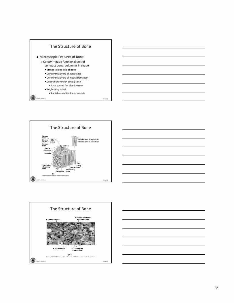
Microscopic Features of Bone
Osteon—Basic functional unit of compact bone; columnar in shape
• Strong in long axis of bone
• Concentric layers of osteocytes
• Concentric layers of matrix (lamellae)
• Central (Haversian canal) canal
Axial tunnel for blood vessels
• Perforating canal
Radial tunnel for blood vessels
Slide 26JSOMTC, SWMG(A)
The Structure of Bone
Slide 27JSOMTC, SWMG(A)
The Structure of Bone
10
Slide 28JSOMTC, SWMG(A)
The Structure of Bone
Microscopic Features of Spongy Bone
No osteons
Lamellae as trabeculae
• Arches, rods, plates of bone
• Branching network of bony tissue
• Strong in many directions
• Red marrow (blood forming) spaces
Slide 29JSOMTC, SWMG(A)
The Structure of Bone
Cells in Bone
Osteocytes
•Mature bone cells between lamellae
Osteoclasts
• Source of acid, enzymes for osteolysis
• Calcium homeostasis
Osteoblasts
• Responsible for osteogenesis (new bone)
• Source of collagen, calcium salts
Slide 30JSOMTC, SWMG(A)
Check on Learning
Which type of bone is found where stress comes from many directions?
A. Spongy bone.
B. Periosteum.
C. Endosteum.
D. Compact bone.
11
Slide 31JSOMTC, SWMG(A)
Bone Growth, Development, and Variations in the Internal Structure
of Specific Bones
Slide 32JSOMTC, SWMG(A)
Bone Formation and Growth
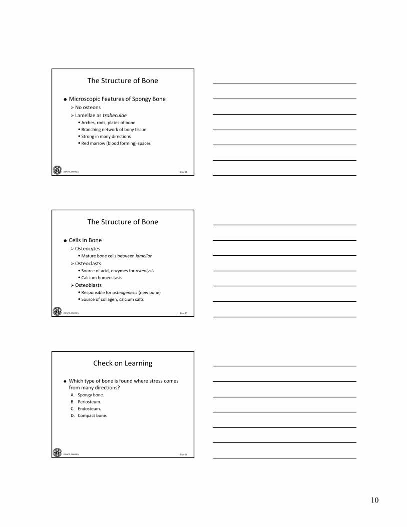
Intramembranous Ossification
Ossification—Process of converting other tissues to bone
Forms flat bones of skull, mandible, clavicle
Stem cells differentiate to osteoblasts
Produces spongy bone, then compact bone
Slide 33JSOMTC, SWMG(A)
Bone Formation and GrowthBone Formation in 16‐Week‐Old Fetus

12
Slide 34JSOMTC, SWMG(A)
Bone Formation and Growth
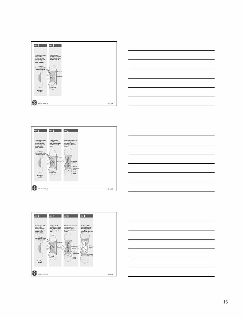
Endochondral Ossification
Most bones formed this way
Cartilage model replaced by bone
Replacement begins in middle (diaphysis)
Replacement follows in ends (epiphyses)
Slide 35JSOMTC, SWMG(A)
Enlargingchondrocytes within
calcifying matrix
Chondrocytes at the center of the growing cartilage model enlarge and then die as the matrix calicifies.
Newly derived osteoblasts cover the shaft of the cartilage in a thin layer of bone.
Blood vessels penetrate the cartilage. New osteoblasts form a primary ossification center.
The bone of the shaft thickens, and the cartilage near each epiphysis is replaced by shafts of bone.
Blood vessels invade the epiphyses and osteo-blasts form secondary centers of ossification.
Cartilagemodel
Boneformation
Epiphysis
Diaphysis Marrowcavity
Primaryossificationcenter
Bloodvessel
Marrowcavity
Bloodvessel
Secondaryossificationcenter
Epiphysealcartilage
Articularcartilage
Slide 36JSOMTC, SWMG(A)
Enlargingchondrocytes within
calcifying matrix
Chondrocytes at the center of the growing cartilage model enlarge and then die as the matrix calicifies.
Cartilagemodel
13
Slide 37JSOMTC, SWMG(A)
Enlargingchondrocytes within
calcifying matrix
Chondrocytes at the center of the growing cartilage model enlarge and then die as the matrix calicifies.
Newly derived osteoblasts cover the shaft of the cartilage in a thin layer of bone.
Cartilagemodel
Boneformation
Epiphysis
Diaphysis
Slide 38JSOMTC, SWMG(A)
Enlargingchondrocytes within
calcifying matrix
Chondrocytes at the center of the growing cartilage model enlarge and then die as the matrix calicifies.
Newly derived osteoblasts cover the shaft of the cartilage in a thin layer of bone.
Blood vessels penetrate the cartilage. New osteoblasts form a primary ossification center.
Cartilagemodel
Boneformation
Epiphysis
Diaphysis Marrowcavity
Primaryossificationcenter
Bloodvessel
Slide 39JSOMTC, SWMG(A)
Enlargingchondrocytes within
calcifying matrix
Chondrocytes at the center of the growing cartilage model enlarge and then die as the matrix calicifies.
Newly derived osteoblasts cover the shaft of the cartilage in a thin layer of bone.
Blood vessels penetrate the cartilage. New osteoblasts form a primary ossification center.
The bone of the shaft thickens, and the cartilage near each epiphysis is replaced by shafts of bone.
Cartilagemodel
Boneformation
Epiphysis
Diaphysis Marrowcavity
Primaryossificationcenter
Bloodvessel
Marrowcavity
Bloodvessel

14
Slide 40JSOMTC, SWMG(A)
Enlargingchondrocytes within
calcifying matrix
Chondrocytes at the center of the growing cartilage model enlarge and then die as the matrix calicifies.
Newly derived osteoblasts cover the shaft of the cartilage in a thin layer of bone.
Blood vessels penetrate the cartilage. New osteoblasts form a primary ossification center.
The bone of the shaft thickens, and the cartilage near each epiphysis is replaced by shafts of bone.
Blood vessels invade the epiphyses and osteo-blasts form secondary centers of ossification.
Cartilagemodel
Boneformation
Epiphysis
Diaphysis Marrowcavity
Primaryossificationcenter
Bloodvessel
Marrowcavity
Bloodvessel
Secondaryossificationcenter
Epiphysealcartilage
Articularcartilage
Slide 41JSOMTC, SWMG(A)
Bone Formation and GrowthAppositional Bone Growth
Slide 42JSOMTC, SWMG(A)
Bone Formation and GrowthRequirements for Normal Bone Growth
Minerals
Calcium, phosphate
Vitamins
Vitamin D3• Deficiency = Rickets
Vitamin C• Deficiency = Scurvy
Vitamin A• Deficiency = Vision Loss
Hormones
Growth Hormone
Sex hormones, thyroid hormone, others

15
Slide 43JSOMTC, SWMG(A)
Check on Learning
How could an X‐ray be used to determine if a person is at his full height?
A. If the diameter of the diaphysis is greater then the diameter of the proximal and distal epiphysis the person has not reached full height.
B. If the diameter of the diaphysis is less then the diameter of the proximal and distal epiphysis the person has not reached full height.
C. If you can still see the epiphyseal plate they have not reached their full height.
D. None of the above.
Slide 44JSOMTC, SWMG(A)
The Remodeling and Repair of the Skeleton and Homeostatic Mechanisms Responsible for
Regulating Mineral Deposition and Turnover
Slide 45JSOMTC, SWMG(A)
Bone Remodeling/Homeostasis
Role of Remodeling in Support
Remodeling—Continuous breakdown and reforming of bone tissue
Shapes reflect applied loads
Mineral turnover enables adapting to new stresses

16
Slide 46JSOMTC, SWMG(A)
Bone Remodeling/Homeostasis
Key Note
What you don’t use, you lose. The stresses applied to bones during exercise are essential to maintaining bone strength and bone mass. STRESS IS GOOD!
Slide 47JSOMTC, SWMG(A)
Bone Remodeling/Homeostasis
Homeostasis and Mineral Storage
Bones store calcium
• Contain 99% of body calcium
• Store up to two kgs of calcium
• Hormones control storage/release
PTH, calcitriol release bone calcium
Calcitonin stores bone calcium
• Blood levels kept constant
Slide 48JSOMTC, SWMG(A)
Bone Remodeling/Homeostasis
Injury and Repair
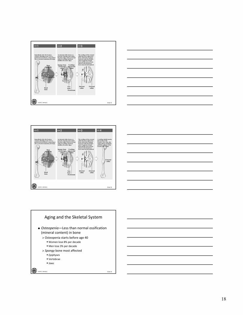
Fracture—A crack or break in a bone
Steps in fracture repair
• Fracture hematoma
•Mitoses in periosteum, endosteum
Internal callus
External callus
• Bone remodeling

17
Slide 49JSOMTC, SWMG(A)
Bonefragments
Immediately after the fracture, extensive bleeding occurs. Over a period of several hours, a large blood clot, or fracture hematoma, develops.
Fracturehematoma
Deadbone
NewbonePeriosteum
Spongy boneof internal
callus
Cartilageof external
callus
An internal callus forms as a network of spongy bone unites the inner edges, and an external callus of cartilage and bone stabilizes the outer edges.
The cartilage of the external callus has been replaced by bone, and struts of spongy bone now unite the broken ends. Fragments of dead bone and the areas of bone closest to the break have been removed and replaced.
A swelling initially marks the location of the fracture. Over time, this region will be remodeled, and little evidence of the fracture will remain.
Internalcallus
Externalcallus
Externalcallus
Slide 50JSOMTC, SWMG(A)
Bonefragments
Immediately after the fracture, extensive bleeding occurs. Over a period of several hours, a large blood clot, or fracture hematoma, develops.
Fracturehematoma
Deadbone
Slide 51JSOMTC, SWMG(A)
Bonefragments
Immediately after the fracture, extensive bleeding occurs. Over a period of several hours, a large blood clot, or fracture hematoma, develops.
Fracturehematoma
Deadbone
NewbonePeriosteum
Spongy boneof internal
callus
Cartilageof external
callus
An internal callus forms as a network of spongy bone unites the inner edges, and an external callus of cartilage and bone stabilizes the outer edges.
18
Slide 52JSOMTC, SWMG(A)
Bonefragments
Immediately after the fracture, extensive bleeding occurs. Over a period of several hours, a large blood clot, or fracture hematoma, develops.
Fracturehematoma
Deadbone
NewbonePeriosteum
Spongy boneof internal
callus
Cartilageof external
callus
An internal callus forms as a network of spongy bone unites the inner edges, and an external callus of cartilage and bone stabilizes the outer edges.
The cartilage of the external callus has been replaced by bone, and struts of spongy bone now unite the broken ends. Fragments of dead bone and the areas of bone closest to the break have been removed and replaced.
Internalcallus
Externalcallus
Slide 53JSOMTC, SWMG(A)
Bonefragments
Immediately after the fracture, extensive bleeding occurs. Over a period of several hours, a large blood clot, or fracture hematoma, develops.
Fracturehematoma
Deadbone
NewbonePeriosteum
Spongy boneof internal
callus
Cartilageof external
callus
An internal callus forms as a network of spongy bone unites the inner edges, and an external callus of cartilage and bone stabilizes the outer edges.
The cartilage of the external callus has been replaced by bone, and struts of spongy bone now unite the broken ends. Fragments of dead bone and the areas of bone closest to the break have been removed and replaced.
A swelling initially marks the location of the fracture. Over time, this region will be remodeled, and little evidence of the fracture will remain.
Internalcallus
Externalcallus
Externalcallus
Slide 54JSOMTC, SWMG(A)
Aging and the Skeletal System
Osteopenia—Less than normal ossification (mineral content) in bone
Osteopenia starts before age 40
•Women lose 8% per decade
•Men lose 3% per decade
Spongy bone most affected
• Epiphyses
• Vertebrae
• Jaws
19
Slide 55JSOMTC, SWMG(A)
Check on Learning
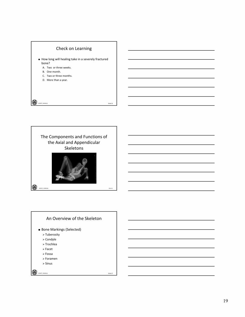
How long will healing take in a severely fractured bone?
A. Two or three weeks.
B. One month.
C. Two or three months.
D. More than a year.
Slide 56JSOMTC, SWMG(A)
The Components and Functions of the Axial and Appendicular
Skeletons
Slide 57JSOMTC, SWMG(A)
An Overview of the Skeleton
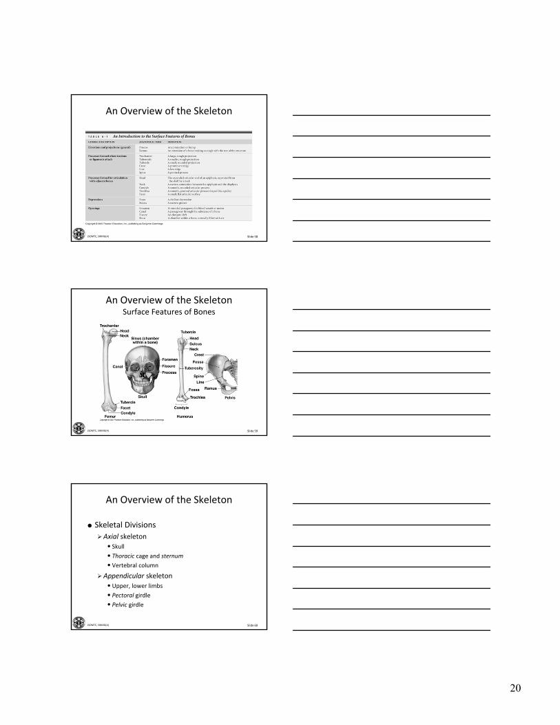
Bone Markings (Selected)
Tuberosity
Condyle
Trochlea
Facet
Fossa
Foramen
Sinus
20
Slide 58JSOMTC, SWMG(A)
An Overview of the Skeleton
Slide 59JSOMTC, SWMG(A)
An Overview of the SkeletonSurface Features of Bones
Slide 60JSOMTC, SWMG(A)
An Overview of the Skeleton
Skeletal Divisions
Axial skeleton
• Skull
• Thoracic cage and sternum
• Vertebral column
Appendicular skeleton
• Upper, lower limbs
• Pectoral girdle
• Pelvic girdle

21
Slide 61JSOMTC, SWMG(A)
An Overview of the SkeletonThe Anterior Skeleton
Slide 62JSOMTC, SWMG(A)
An Overview of the SkeletonThe Posterior Skeleton
Slide 63JSOMTC, SWMG(A)

22
Slide 64JSOMTC, SWMG(A)
Check on Learning Which of the following statements about the axial skeleton and appendicular skeleton are true?
A. The sacrum, clavicles and scapula are part of the axial skeleton and the humerus radius and patella are part of the appendicular skeleton.
B. The femur, tibia and fibula are part of the axial skeleton and the sacrum, sternum and coccyx are part of the appendicular skeleton.
C. The patella, tibia and radius are part of the axial skeleton and the sternum, sacrum and the vertebrae are part of the appendicular skeleton.
D. The bones of the cranium, sternum and coccyx are part of the axial skeleton and the clavicles, scapula and the phalanges are part of the appendicular skeleton.
Slide 65JSOMTC, SWMG(A)
The Bones of the Skull
Slide 66JSOMTC, SWMG(A)
The Axial Division: The Skull
Bones of the Cranium
Frontal bone
• Forehead, superior surface of orbits
Parietal bones
• Sides, roof
Occipital bone
• Foramen magnum
Temporal bones
• Sides, base

23
Slide 67JSOMTC, SWMG(A)
The Axial Division: The Skull
Bones of the Cranium (continued)
Sphenoid bone
• Bridge between cranial and facial bones
Ethmoid bone
• Cribriform plate
• Nasal septum
Slide 68JSOMTC, SWMG(A)
The Axial Division: The SkullThe Adult Skull (Part I)
Figure 6-10
Slide 69JSOMTC, SWMG(A)
The Axial Division: The Skull
Bones of the Face
Maxillary bones
Palatine bones
The Vomer
Zygomatic bones
• Zygomatic arch (with temporal bones)

24
Slide 70JSOMTC, SWMG(A)
The Axial Division: The Skull
Bones of the Face (continued)
Nasal bones
Lacrimal bones
Inferior nasal conchae
Slide 71JSOMTC, SWMG(A)
The Axial Division: The Skull
Bones of the Face (continued)
Nasal complex
• Nasal septum
Paranasal sinuses
• Frontal
• Sphenoidal
• Ethmoidal
• Palatine
•Maxillary
Mandible
Slide 72JSOMTC, SWMG(A)
The Axial Division: The SkullThe Adult Skull (Part II)
25
Slide 73JSOMTC, SWMG(A)
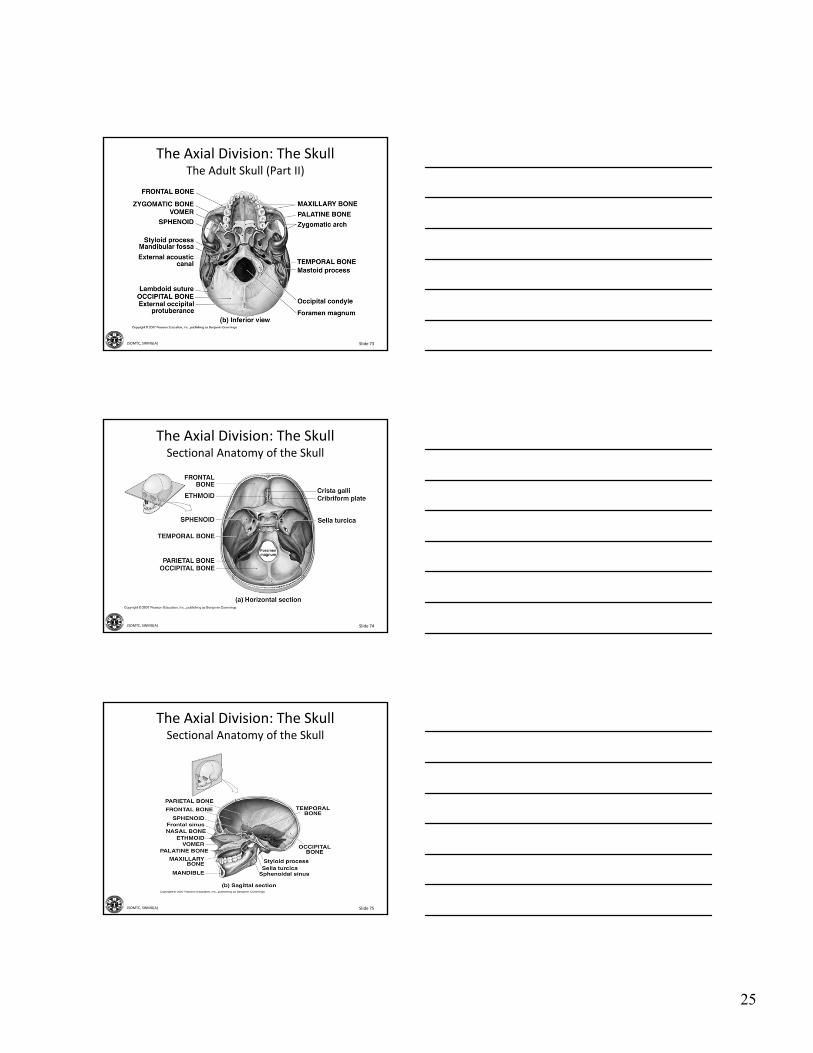
The Axial Division: The SkullThe Adult Skull (Part II)
Slide 74JSOMTC, SWMG(A)
The Axial Division: The SkullSectional Anatomy of the Skull
Slide 75JSOMTC, SWMG(A)
The Axial Division: The SkullSectional Anatomy of the Skull

26
Slide 76JSOMTC, SWMG(A)
The Axial Division: The SkullSectional Anatomy of the Skull
Slide 77JSOMTC, SWMG(A)
The Axial Division: The SkullThe Paranasal Sinuses
Slide 78JSOMTC, SWMG(A)
The Axial Division: The SkullThe Hyoid Bone
27
Slide 79JSOMTC, SWMG(A)
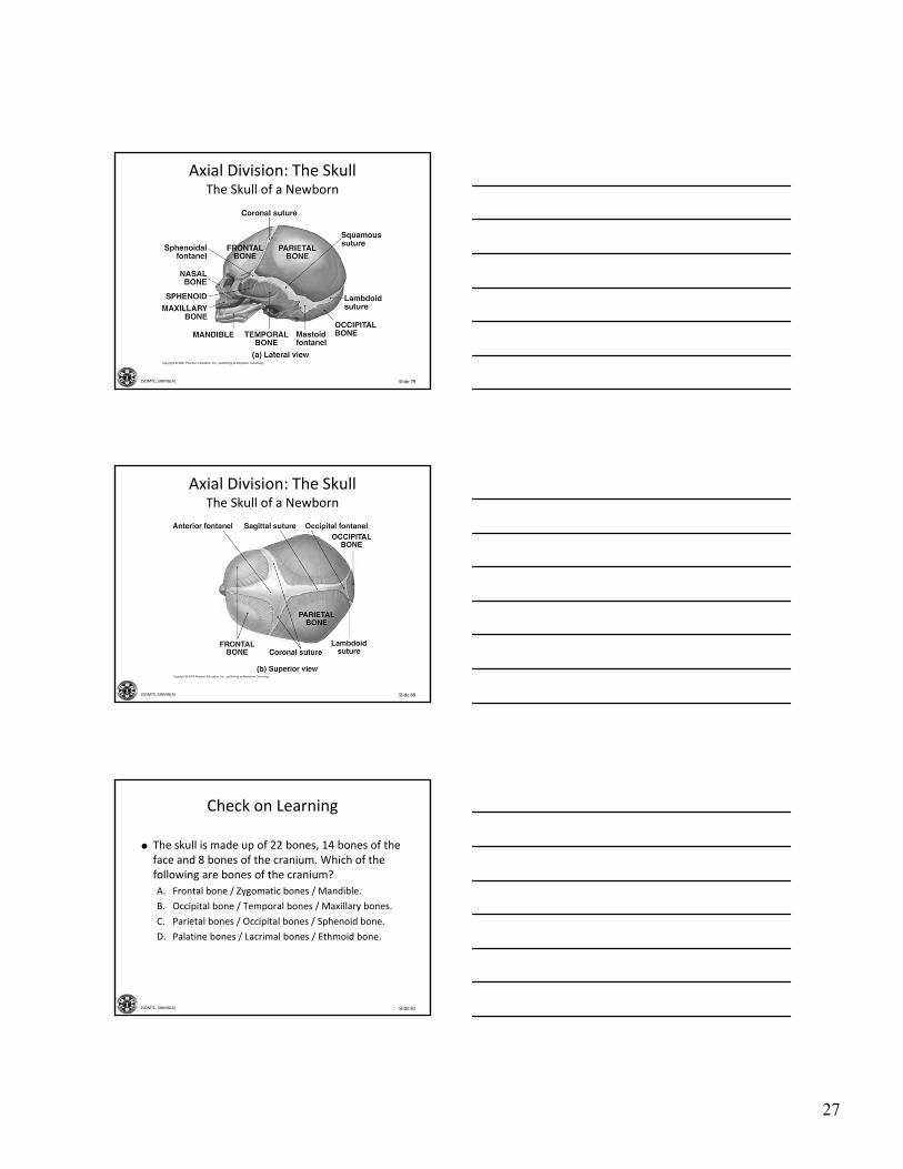
Axial Division: The SkullThe Skull of a Newborn
Slide 80JSOMTC, SWMG(A)
Axial Division: The SkullThe Skull of a Newborn
Slide 81JSOMTC, SWMG(A)
Check on Learning
The skull is made up of 22 bones, 14 bones of the face and 8 bones of the cranium. Which of the following are bones of the cranium?
A. Frontal bone / Zygomatic bones / Mandible.
B. Occipital bone / Temporal bones / Maxillary bones.
C. Parietal bones / Occipital bones / Sphenoid bone.
D. Palatine bones / Lacrimal bones / Ethmoid bone.
28
Slide 82JSOMTC, SWMG(A)
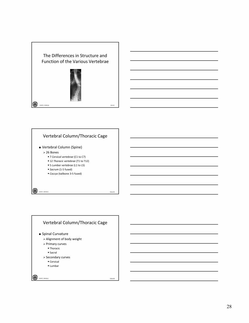
The Differences in Structure and Function of the Various Vertebrae
Slide 83JSOMTC, SWMG(A)
Vertebral Column/Thoracic Cage
Vertebral Column (Spine)
26 Bones
• 7 Cervical vertebrae (C1 to C7)
• 12 Thoracic vertebrae (T1 to T12)
• 5 Lumbar vertebrae (L1 to L5)
• Sacrum (1‐5 fused)
• Coccyx (tailbone 3‐5 fused)
Slide 84JSOMTC, SWMG(A)
Vertebral Column/Thoracic Cage
Spinal Curvature
Alignment of body weight
Primary curves
• Thoracic
• Sacral
Secondary curves
• Cervical
• Lumbar
29
Slide 85JSOMTC, SWMG(A)
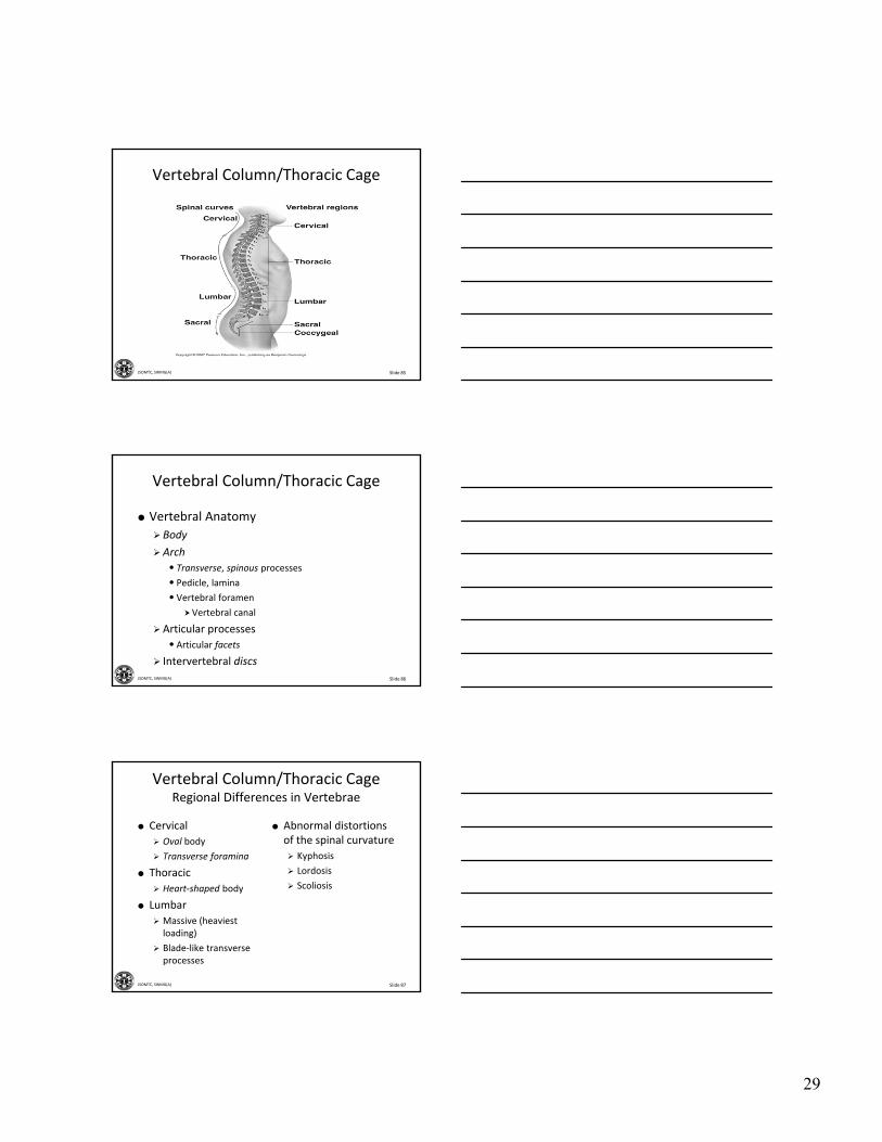
Vertebral Column/Thoracic Cage
Slide 86JSOMTC, SWMG(A)
Vertebral Column/Thoracic Cage
Vertebral Anatomy
Body
Arch
• Transverse, spinous processes
• Pedicle, lamina
• Vertebral foramen
Vertebral canal
Articular processes
• Articular facets
Intervertebral discs
Slide 87JSOMTC, SWMG(A)
Vertebral Column/Thoracic CageRegional Differences in Vertebrae
Cervical
Oval body
Transverse foramina
Thoracic
Heart‐shaped body
Lumbar
Massive (heaviest loading)
Blade‐like transverse processes
Abnormal distortions of the spinal curvature
Kyphosis
Lordosis
Scoliosis
30
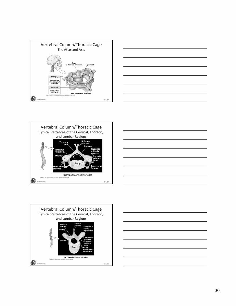
Slide 88JSOMTC, SWMG(A)
Vertebral Column/Thoracic CageThe Atlas and Axis
Slide 89JSOMTC, SWMG(A)
Vertebral Column/Thoracic CageTypical Vertebrae of the Cervical, Thoracic,
and Lumbar Regions
Slide 90JSOMTC, SWMG(A)
Vertebral Column/Thoracic CageTypical Vertebrae of the Cervical, Thoracic,
and Lumbar Regions

31
Slide 91JSOMTC, SWMG(A)
Vertebral Column/Thoracic CageTypical Vertebrae of the Cervical, Thoracic,
and Lumbar Regions
Slide 92JSOMTC, SWMG(A)
Vertebral Column/Thoracic Cage
Functions of Sacrum
Protects pelvic organs
Base articulates with lumbar vertebra
Apex articulates with coccyx
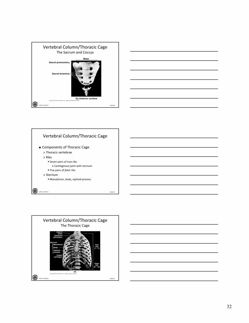
Slide 93JSOMTC, SWMG(A)
Vertebral Column/Thoracic CageThe Sacrum and Coccyx
32
Slide 94JSOMTC, SWMG(A)
Vertebral Column/Thoracic CageThe Sacrum and Coccyx
Slide 95JSOMTC, SWMG(A)
Vertebral Column/Thoracic Cage
Components of Thoracic Cage
Thoracic vertebrae
Ribs
• Seven pairs of true ribs
Cartilaginous joint with sternum
• Five pairs of false ribs
Sternum
•Manubrium, body, xiphoid process
Slide 96JSOMTC, SWMG(A)
Vertebral Column/Thoracic CageThe Thoracic Cage

33
Slide 97JSOMTC, SWMG(A)
Vertebral Column/Thoracic Cage
Slide 98JSOMTC, SWMG(A)
Check on Learning
The vertebral column is divided into five major regions with four spinal curves. Which of the following statements are correct?
A. The five major regions are the; Cervical / Thoracic / Lumbar / Sacral / Coccygeal.
B. The four spinal curves are the; Cervical / Thoracic / Lumbar / Sacral.
C. The Lumbar vertebra are larger then the cervical vertebra.
D. All of the above.
Slide 99JSOMTC, SWMG(A)
The Structural Differences Between the Pectoral and Pelvic Girdles to Their Various Functional Roles
34
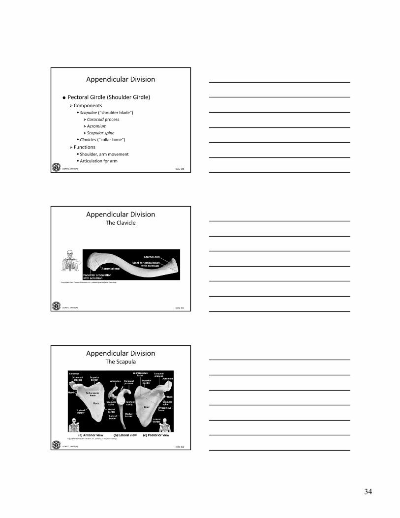
Slide 100JSOMTC, SWMG(A)
Appendicular Division
Pectoral Girdle (Shoulder Girdle)
Components
• Scapulae (“shoulder blade”)
Coracoid process
Acromium
Scapular spine
• Clavicles (“collar bone”)
Functions
• Shoulder, arm movement
• Articulation for arm
Slide 101JSOMTC, SWMG(A)
Appendicular DivisionThe Clavicle
Slide 102JSOMTC, SWMG(A)
Appendicular DivisionThe Scapula

35
Slide 103JSOMTC, SWMG(A)
Appendicular Division
Upper Limb
Humerus
•Head articulates with scapula
•Muscles attach to
Greater, lesser tubercles
Deltoid tuberosity
Medial, lateral epicondyles
• Distal condyle articulates with forearm
Slide 104JSOMTC, SWMG(A)
Appendicular Division
Upper Limb Anatomy
Distal articulation of humerus
• Coronoid fossa
• Olecranon fossa
• Trochlea
Slide 105JSOMTC, SWMG(A)
Appendicular DivisionThe Humerus
36
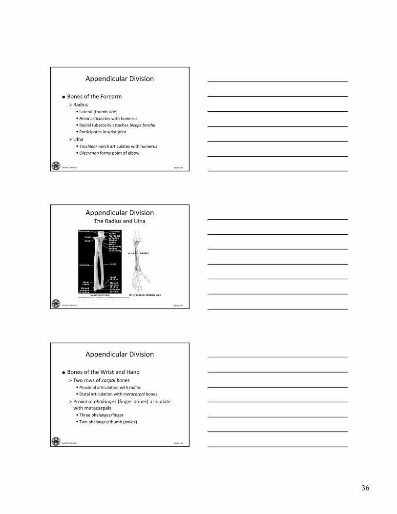
Slide 106JSOMTC, SWMG(A)
Appendicular Division
Bones of the Forearm
Radius
• Lateral (thumb side)
•Head articulates with humerus
• Radial tuberosity attaches biceps brachii
• Participates in wrist joint
Ulna
• Trochlear notch articulates with humerus
•Olecranon forms point of elbow
Slide 107JSOMTC, SWMG(A)
Appendicular DivisionThe Radius and Ulna
Slide 108JSOMTC, SWMG(A)
Appendicular Division
Bones of the Wrist and Hand
Two rows of carpal bones
• Proximal articulation with radius
• Distal articulation with metacarpal bones
Proximal phalanges (finger bones) articulate with metacarpals
• Three phalanges/finger
• Two phalanges/thumb (pollex)
37
Slide 109JSOMTC, SWMG(A)
Appendicular DivisionBones of the Wrist and Hand
Slide 110JSOMTC, SWMG(A)
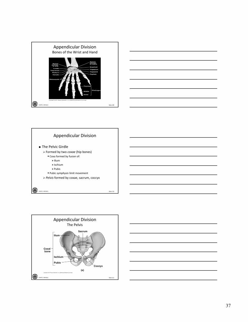
Appendicular Division
The Pelvic Girdle
Formed by two coxae (hip bones)
• Coxa formed by fusion of:
Ilium
Ischium
Pubis
• Pubic symphysis limit movement
Pelvis formed by coxae, sacrum, coccyx
Slide 111JSOMTC, SWMG(A)
Appendicular DivisionThe Pelvis

38
Slide 112JSOMTC, SWMG(A)
Appendicular DivisionThe Pelvis
Slide 113JSOMTC, SWMG(A)
Appendicular DivisionThe Pelvis
Slide 114JSOMTC, SWMG(A)
Appendicular DivisionDifferences in the Anatomy of the Pelvis in
Males and Females
39
Slide 115JSOMTC, SWMG(A)
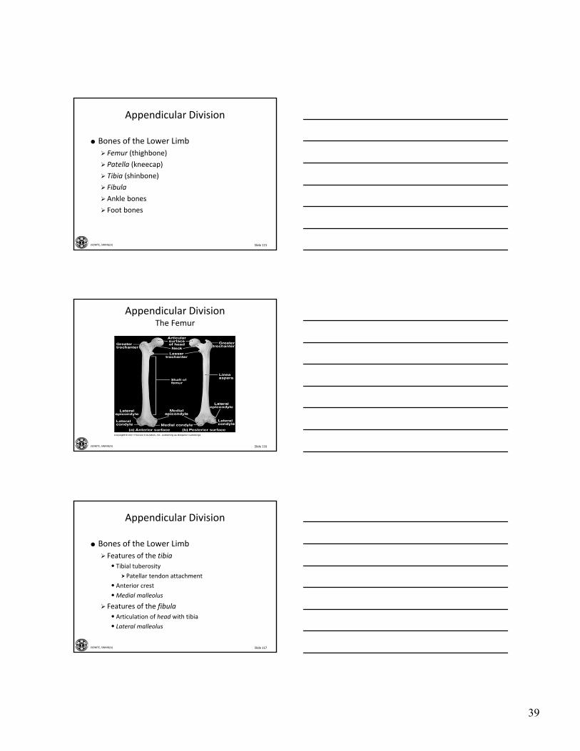
Appendicular Division
Bones of the Lower Limb
Femur (thighbone)
Patella (kneecap)
Tibia (shinbone)
Fibula
Ankle bones
Foot bones
Slide 116JSOMTC, SWMG(A)
Appendicular DivisionThe Femur
Slide 117JSOMTC, SWMG(A)
Appendicular Division
Bones of the Lower Limb
Features of the tibia
• Tibial tuberosity
Patellar tendon attachment
• Anterior crest
•Medial malleolus
Features of the fibula
• Articulation of head with tibia
• Lateral malleolus

40
Slide 118JSOMTC, SWMG(A)
Appendicular DivisionThe Right Tibia and Fibula
Slide 119JSOMTC, SWMG(A)
Appendicular Division
The Bones of the Ankle and Foot
Ankle
• Seven tarsal bones
Talus
– Joint with tibia, fibula
Foot
• Calcaneus (heel bone)
Major load‐bearing bone
•Metatarsal bones
• Five phalanges (toes)
Slide 120JSOMTC, SWMG(A)
Appendicular DivisionThe Bones of the Ankle and Foot

41
Slide 121JSOMTC, SWMG(A)
Appendicular DivisionThe Bones of the Ankle and Foot
Slide 122JSOMTC, SWMG(A)
Check on Learning
The pelvic and pectoral girdles articulates with the bones of the lower and upper extremities. Which of the following statements are correct?
A. Each upper limb articulates with the trunk at the pelvic girdle.
B. The bones of the pectoral girdle are more massive then the bones of the pelvic girdle.
C. The pelvic girdle is more firmly attached to the axial skeleton.
D. All of the above.
Slide 123JSOMTC, SWMG(A)
The Different Types of Joints and Their Structural Features Relative to
Joint Function

42
Slide 124JSOMTC, SWMG(A)
Articulations
Classification of Joints (Articulations)
Joint—Where two bones interact
Three functional classes of joint
• Synarthroses
Immovable
• Amphiarthroses
Slightly movable
•Diarthroses
Freely movable
Slide 125JSOMTC, SWMG(A)
Articulations
Examples of Joints
Synarthroses
• Suture – sagittal, lamdoidal, etc
•Gomphosis – tooth within the aveolus
• Synchondrosis – epiphiseal plate
Amphiarthroses
• Syndesmosis – distal articulation between the tibia and fibula
• Symphysis – pubic symphysis
Diarthroses
• Synovial joints – shoulder, knee
Slide 126JSOMTC, SWMG(A)
Articulations
Synovial Joints (Diarthroses)
Epiphyses covered by articular cartilage
Lubricated by synovial fluid
Enclosed within joint capsule
Other synovial structures include:
•Menisci
• Bursae
• Fat pads
• Ligaments

43
Slide 127JSOMTC, SWMG(A)
ArticulationsThe Structure of Synovial Joints
Slide 128JSOMTC, SWMG(A)
ArticulationsThe Structure of Synovial Joints
Slide 129JSOMTC, SWMG(A)
Check on Learning
Joints can be classified according to their structure or function. What are the structuralclassifications of joints?
A. Synarthroses / Amphiarthroses / Diathroses.
B. Fibrous / Cartilaginous / Synovial.
C. Gliding / Angular motion / Rotation.
D. All of the above.

44
Slide 130JSOMTC, SWMG(A)
The Dynamic Movements of the Skeleton and the Structure of Representative Articulations
Slide 131JSOMTC, SWMG(A)
Articulations
Synovial Joints: Movements
Flexion
Extension
Hyperextension
Abduction
Adduction
Circumduction
Rotation
• Pronation, supination
Slide 132JSOMTC, SWMG(A)
ArticulationsAngular Movements

45
Slide 133JSOMTC, SWMG(A)
ArticulationsAngular Movements
Slide 134JSOMTC, SWMG(A)
ArticulationsAngular Movements
Slide 135JSOMTC, SWMG(A)
ArticulationsAngular Movements

46
Slide 136JSOMTC, SWMG(A)
ArticulationsRotational Movements
Slide 137JSOMTC, SWMG(A)
ArticulationsRotational Movements
Slide 138JSOMTC, SWMG(A)
Articulations
Special Movements
Foot and ankle
• Inversion, eversion
•Dorsiflexion, plantar flexion
Hand
•Opposition of thumb, palm
Head
• Protraction, retraction
•Depression, elevation (jaw)

47
Slide 139JSOMTC, SWMG(A)
ArticulationsSpecial Movements
Slide 140JSOMTC, SWMG(A)
Check on Learning
Which of the following movements are examples of abduction?
A. In the frontal plane, swinging the upper limb to the side (away from the body).
B. In the anterior–posterior plane, bringing the head toward the chest.
C. Bring the fingers or toes together from a position where they were spread apart.
D. All of the above.
Slide 141JSOMTC, SWMG(A)
The Relationship Between Joint Structure and Mobility

48
Slide 142JSOMTC, SWMG(A)
Articulations
Structural Classification of Synovial Joints
Gliding (e.g., vertebra–vertebra)
Hinge (e.g., knee)
Pivot (e.g., atlas–axis)
Ellipsoidal (e.g., distal radius)
Saddle (e.g., thumb)
Ball‐and‐Socket (e.g., hip)
Slide 143JSOMTC, SWMG(A)
ArticulationsStructural Classification of Synovial Joints
Slide 144JSOMTC, SWMG(A)
ArticulationsStructural Classification of Synovial Joints

49
Slide 145JSOMTC, SWMG(A)
ArticulationsStructural Classification of Synovial Joints
Slide 146JSOMTC, SWMG(A)
ArticulationsStructural Classification of Synovial Joints
Slide 147JSOMTC, SWMG(A)
ArticulationsStructural Classification of Synovial Joints

50
Slide 148JSOMTC, SWMG(A)
ArticulationsStructural Classification of Synovial Joints
Slide 149JSOMTC, SWMG(A)
Articulations
Key Note
A joint cannot be both highly mobile and very strong. The greater the mobility, the weaker the joint, because mobile joints rely on support from muscles and ligaments rather than solid bone‐to‐bone connections.
Slide 150JSOMTC, SWMG(A)
Articulations
Intervertebral Articulations
Two kinds join adjacent vertebrae
• Gliding joints
Between superior and inferior articularprocesses
Permit small movements
• Symphyseal joints
Intervertebral discs composed of fibrocartilage
Cushion and connect

51
Slide 151JSOMTC, SWMG(A)
ArticulationsIntervertebral Articulations
Slide 152JSOMTC, SWMG(A)
Articulations
The Shoulder Joint
Ball‐and‐socket design frees movement
• Humerus head mates with glenoid cavity
Joint capsule extends from scapular neck to humerus
Joint dislocates easily
Bursae reduce friction
• Bursitis restricts motion, causes pain
Slide 153JSOMTC, SWMG(A)
ArticulationsThe Shoulder Joint

52
Slide 154JSOMTC, SWMG(A)
Articulations
The Elbow Joint
Two articulations
• Humerus–radius
• Humerus–ulna
Interlocking hinge design
Limited movement
Flexion and extension only
Strong ligaments
Slide 155JSOMTC, SWMG(A)
ArticulationsThe Elbow Joint
Slide 156JSOMTC, SWMG(A)
Articulations
Articulations of the Lower Limb
Joints of the hip, ankle, and foot
• Sturdier than corresponding locations in the upper limb
• Smaller range of motion
Knee and elbow range of motion is comparable, but knee is subjected to much greater forces, therefore is less stable

53
Slide 157JSOMTC, SWMG(A)
Articulations
The Hip Joint
Acetabulum and head of femur
Extremely strong, stable joint
•Many strong ligaments
• Tough joint capsule
• Bulky muscles
Versatile movements
• Flexion, extension, adduction, abduction, circumduction, rotation
Slide 158JSOMTC, SWMG(A)
ArticulationsThe Hip Joint
Slide 159JSOMTC, SWMG(A)
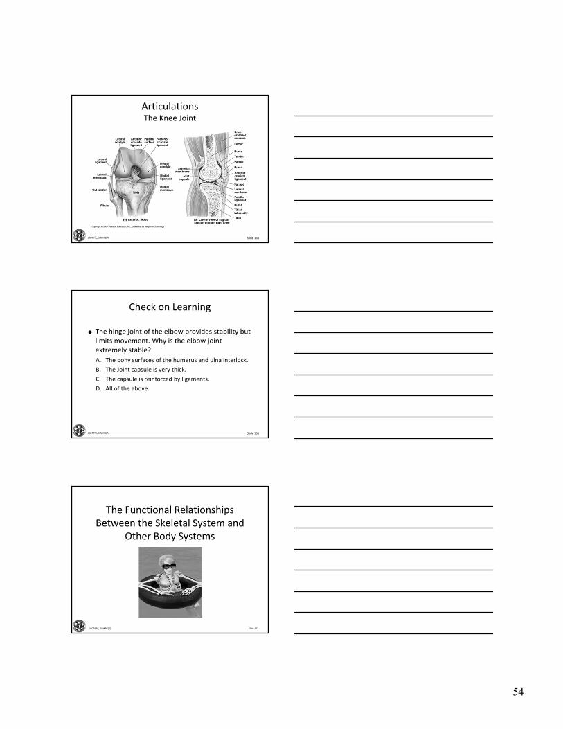
Articulations
The Knee Joint
Complex hinge joint
• Three separate articulations
Femur‐tibia (between condyles—lateral and medial)
Femur‐patella
Fibrocartilage pads
•Medial and lateral menisci
Ligaments
• Cruciate ligaments inside joint
54
Slide 160JSOMTC, SWMG(A)
ArticulationsThe Knee Joint
Slide 161JSOMTC, SWMG(A)
Check on Learning
The hinge joint of the elbow provides stability but limits movement. Why is the elbow joint extremely stable?
A. The bony surfaces of the humerus and ulna interlock.
B. The Joint capsule is very thick.
C. The capsule is reinforced by ligaments.
D. All of the above.
Slide 162JSOMTC, SWMG(A)
The Functional Relationships Between the Skeletal System and
Other Body Systems

55
Slide 163JSOMTC, SWMG(A)
The Integumentary System
Synthesizes vitamin D3, essential for calcium and phosphorus absorption (bone maintenance and growth)
Provides structural support
Slide 164JSOMTC, SWMG(A)
The Muscular System
Stabilizes bone positions; tension in tendons stimulates bone growth and maintenance
Provides calcium needed for normal muscle contraction; bones act as levers to produce body movements
Slide 165JSOMTC, SWMG(A)
The Nervous System
Regulates bone position by controlling muscle contractions
Provides calcium for neural function; protects brain, spinal cord; receptors at joints provide information about body position

56
Slide 166JSOMTC, SWMG(A)
The Endocrine System
Skeletal growth regulated by growth hormone, thyroid hormones, and sex hormones; calcium mobilization regulated by parathyroid hormone and calcitonin
Protects endocrine organs, especially in brain, chest, and pelvic cavity
Slide 167JSOMTC, SWMG(A)
The Cardiovascular System
Provides oxygen, nutrients, hormones, blood cells; removes waste products and carbon dioxide
Provides calcium needed for cardiac muscle contraction, blood cells produced in bone marrow
Slide 168JSOMTC, SWMG(A)
The Lymphatic System
Lymphocytes assist in the defense and repair of bone following injuries
Lymphocytes and other cells of the immune response are produced and stored in bone marrow

57
Slide 169JSOMTC, SWMG(A)
The Respiratory System
Provides oxygen and eliminates carbon dioxide
Movements of ribs important in breathing; axial skeleton surrounds and protects lungs
Slide 170JSOMTC, SWMG(A)
The Digestive System
Provides nutrients, calcium, and phosphate
Ribs protect portions of liver, stomach, and intestines
Slide 171JSOMTC, SWMG(A)
The Urinary System
Conserves calcium and phosphate needed for bone growth; disposes of waste products
Axial skeleton provides some protection for kidneys and ureters; pelvis protects urinary bladder and proximal urethra
58
Slide 172JSOMTC, SWMG(A)
The Reproductive System
Sex hormones stimulate growth and maintenance of bones; surge of sex hormones at puberty causes acceleration of growth and closure of epiphyseal cartilages
Pelvis protects reproductive organs of female, protects portion of ductus deferens and accessory glands in males
Slide 173JSOMTC, SWMG(A)
Check on Learning
Which of the body systems synthesizes vitamin D3
essential for calcium and phosphorus absorption necessary for bone growth and maintenance?
A. Endocrine system.
B. Lymphatic system.
C. Integumentary system.
D. Cardiovascular system.
Slide 174JSOMTC, SWMG(A)
Questions?

59
Slide 175JSOMTC, SWMG(A)
Terminal Learning Objective
Action: Communicate knowledge of “The Skeletal System”
Condition: Given a lecture in a classroom environment
Standard: Received a minimum score of 75% on the written exam IAW course standards
Slide 176JSOMTC, SWMG(A)
Agenda
Define the medical vocabulary components related to the skeletal system
Communicate the functions of the skeletal system
Identify the structures and functions of compact and spongy bone
Communicate bone growth, development, and variations in the internal structure of specific bones
Slide 177JSOMTC, SWMG(A)
Agenda
Communicate the remodeling and repair of the skeleton and homeostatic mechanisms responsible for regulating mineral deposition and turnover
Identify the components and functions of the axial and appendicular skeletons
Identify the bones of the skull
60
Slide 178JSOMTC, SWMG(A)
Agenda
Communicate the differences in structure and function of the various vertebrae
Identify the structural differences between the pectoral and pelvic girdles to their various functional roles
Identify among different types of joints, and link structural features to joint functions
Slide 179JSOMTC, SWMG(A)
Agenda
Identify the dynamic movements of the skeleton and the structure of representative articulations
Communicate the relationship between joint structure and mobility, using specific examples
Communicate the functional relationships between the skeletal system and other body systems
Slide 180JSOMTC, SWMG(A)
Reason
In addition to supporting the body and providing movement, the Skeletal System plays a major role in homeostasis of the human body.
As a SOCM Medic / Corpsman your knowledge of this system will enhance your patient treatment skills.

61
Slide 181JSOMTC, SWMG(A)
Break


![Development Objective Agreement and Bilateral Project ... · The Development Objective ("Objective") is: [state objective]. Section 2.2. Results. In order to achieve that Objective,](https://img.pdfslide.net/doc/110x75/5f056b8e7e708231d412dfe2/development-objective-agreement-and-bilateral-project-the-development-objective.jpg)


![[Objective-C] Objective-C의 메모리 관리 방법](https://img.pdfslide.net/doc/110x75/55a397691a28ab9e7a8b47de/objective-c-objective-c-.jpg)