Embed Size (px)
Citation preview
UNIVERSITA’ DEGLI STUDI DI MILANO – BICOCCA
FACOLTA’ DI MEDICINA E CHIRURGIA
DOTTORATO DI RICERCA IN EMATOLOGIA SPERIMENTALE
(XXII ciclo)
Coordinatore: Prof. ENRICO MARIA POGLIANI
TESI DI DOTTORATO DI RICERCA
NUTRIENT-GENE INTERACTIONS
WITHIN ONE-CARBON METABOLISM AND EFFECTS ON
EPIGENETIC REGULATION THROUGH DNA METHYLATION
IN PERIPHERAL BLOOD MONONUCLEAR CELLS
Relatore:
Dott.ssa SIMONETTA FRISO
Università degli Studi di Verona
Dottorando in Ricerca:
Dott.ssa VALENTINA LOTTO
ANNO ACCADEMICO 2009-2010
1
TABLE OF CONTENTS
ABSTRACT 3
INTRODUCTION 6
CHAPTER 1: ONE-CARBON METABOLISM BETWEEN GENETICS
AND EPIGENETICS 8
FOLATE AND EPIGENETICS 8
One-carbon metabolism 8
Epigenetic mechanisms 12
Genomic DNA methylation: effect of nutrients 14
Gene-specific DNA methylation at the promoter region 15
Gene-specific DNA methylation at the coding region 17
GENE-NUTRIENT INTERACTIONS IN ONE-CARBON METABOLISM:
ROLE OF CYCLE FOLATE POLYMORPHISMS 24
The 677C>T polymorphism of MTHFR gene: a clear model of gene-
nutrition interaction 24
Methionine synthase and role of MS polymorphism 2756 A>G on
epigenetic modulation 31
Thymidylate synthase and role of polymorphism 2rpt-3rpt on plasma
folate levels 34
Serine hydroxy-methyl-transferase and role of polymorphism
1420 C>T on epigenetic modulation 38
Dihydrofolate reductase (DHFR) and role of polymorphism
19bp-ins/del on epigenetic modulation. 43
CHAPTER 2: A POLYMORPHIC VARIANT OF DIHYDROFOLATE
REDUCTASE (DHFR 19BP-INS/DEL) IN CHRONIC DISEASE
RATHER THAN CANCER. 54
2
CHAPTER 3: STUDY OF A FUNCTIONAL MODEL OF INTER-
RELATIONSHIP BETWEEN GENETICS AND EPIGENETICS IN
FACTOR VII GENE PROMOTER: EVALUATION OF TWO FUNCTIONAL
POLYMORPHISMS ON F7 PROMOTER GENE AND INTERACTION
WITH METHYLATION INDEX IN THE F7 PROMOTER REGION. 71
PROMOTER METHYLATION IN COAGULATION F7 GENE
INFLUENCES PLASMA FVII CONCENTRATIONS AND RELATES
TO CORONARY ARTERY DISEASE 72
CHAPTER 4: ONE-CARBON METABOLITES AND ROLE OF B
VITAMINS BETWEEN PHYSIOLOGY AND PATHOLOGY AND ITS
CORRELATION WITH EPIGENETIC MECHANISMS 97
PLASMA VITAMIN B6: A CHALLENGING LINK BETWEEN NUTRITION
AND INFLAMMATION IN CARDIOVASCULAR DISEASE 97
Plasma vitamin B6 and coronary artery disease, myocardial infarction
and ischemic stroke 102
Plasma vitamin B6 supplementation, inflammation and cardiovascular
disease prevention 106
Inflammation and vitamin B6-related atherogenesis 110
ONE-CARBON METABOLISM, ADMA PLASMA CONCENTRATIONS
AND CARDIOVASCULAR RISK 137
CHAPTER 5: FUTURE PERSPECTIVES 163
GENETICS AND EPIGENETICS RELATIONSHIP IN CANCER TYPE
WITH A LINK TO ONE-CARBON METABOLISM: A POSSIBLE MODEL
FOR THE ROLE OF EPIGENETIC MARKERS IN PERIPHERAL
BLOOD MONONUCLEAR CELLS 163
CONCLUSIONS 173
3
ABSTRACT
Epigenetics is a field of molecular biology that copes with the study of gene
function regulation without variations in DNA structure or nucleotide
sequences.
Among the main epigenetic phenomema in eukaryotic cells there are DNA
methylation and post-traslational mechanisms among which the major are
histone methylation and acetylation.
Epigenetic changes are potentially reversible phenomena that are controlled
also by nutritional factors as the methyl-donors involved in the folate cycle.
Plasma levels of B vitamins, among which “in primis” plasma folate
concentrations, are implicated in epigenetic modulation so that it can be
hypothesized that they may affect the modulation of gene expression through
epigenetic mechanisms.
Epigenetic modifications represent one of the earliest events in the genesis
of some complex pathologies, therefore the study of the interaction between
epigenetics and nutritional status is of great interest either to define the
physiopathological mechanisms of development of some illnesses, and for
possible personalized strategies of prevention.
The present work has been articulated, at first, on the analysis of gene-
nutritional interaction mechanisms within the folate cycle through the study of
polymorphisms of enzymes involved in the metabolism of methyl-group
donors; the aim was to study their possible role on the modulation of genomic
DNA methylation in relationship to different plasma levels of idrosoluble B
4
vitamins. In this regard, the most important functional polymorfisms known on
the genes of one-carbon metabolism and their relationship with methylation
status of polymorphonuclear cells DNA have been analyzed from a cohort of
around 800 subjects within a clinical study, underlining the role of the key
folate-related enzymes in the modulation of DNA methylation.
Besides the function of genomic DNA methylation, the methylation status at
specific sites has been also approached with the specific intent of
considering a possible interrelationship between the role of promoter
methylation and the co-presence of functional polymorphisms in the same
genic site for a gene for which a precise functional effect is well-known. To
address this issue the promoter region of coagulation factor VII gene was
evaluated for both genetic and epigenetic modifications as a possible model
of genetic-epigenetic interaction in the modulation of gene product regulation.
The results showed the key importance of genetic-epigenetic interactions, so
far unknowm, in modulating gene-expression at promoter gene sites.
The role of other vitamins involved in one-carbon metabolism in major
chronic diseases, and specifically the emerging role of B6 vitamin, have been
also studied.
Furthermore, a clinical study is now in progress to evaluate the function of
gene-specific methylation in liver tissue where most of the folate cycle
functions take place. The aim of this project is the evaluation of both
genome-wide and gene-specific methylation status in the liver in comparison
to that observed in peripheral blood mononuclear cells DNA to define
5
whether methylation status of peripheral blood DNA may be regarded as a
good systemic biomarker for this epigenetic feature of DNA in relation to B
vitamins nutritional status in cancer disease. Results from this study may
help to define possible functional markers of gene-nutrients interactions with
effects on epigenetic modulation for future preventive or therapeutic
strategies. With that purpose, a novel high-throughput array-based technique
for the detection of gene-specific methylation at promoter sites has been
optimized in our laboratory.
6
INTRODUCTION
Epigenetics is defined by heritable changes in gene function that occur
without changes in DNA sequence and it transmits non-coded information in
DNA sequence from mother cell to daughter cell, from generation to
generation, through mitosis. The main epigenetic phenomena are DNA
methylation and histones modification, the DNA packaging proteins. When
DNA methylation occurs within genes or gene regulatory elements, such as
exons and promoters, generally suppresses the transcription of the gene,
while when it takes place within gene deficient regions, as heterochromatin,
assumes a crucial role in maintaining the conformation and integrity of the
chromosome. Methylation of the mammalian genome undergoes major
changes during early development and appears to be an integral mechanism
for rapid differentiation and formation of various tissues and organs. When
the differentiation is to be completed, the methylation pattern shows a tissue-
specificity while the model remains essentially stable during later life.
However, these models are not immutable and they can be influenced by
nutritional status.
Nutrients involved in one-carbon metabolism, such as methionine, choline,
betaine, folate, vitamin B12, vitamin B6, riboflavin, zinc and selenium, can
alter the methylation status by interfering with the cellular levels of S-
adenosylmethionine and S-adenosylhomocysteine. Those nutrients, in fact,
play a crucial role in the epigenetic control of gene expression and genome

7
integrity. Therefore the study of the interaction between epigenetics and
nutritional factors is critical both for a better understanding of the
physiological mechanisms and for the assessment of possible underlying
mechanisms of disease, especially those leading to carcinogenesis. Studies
concerning the modulation of epigenetics by nutritional factors are needed to
understand the molecular mechanisms underlying these interactions and, at
the same time, to find new ways for disease prevention and treatment.
8
CHAPTER 1
ONE-CARBON METABOLISM BETWEEN GENETICS AND
EPIGENETICS
FOLATE AND EPIGENETICS
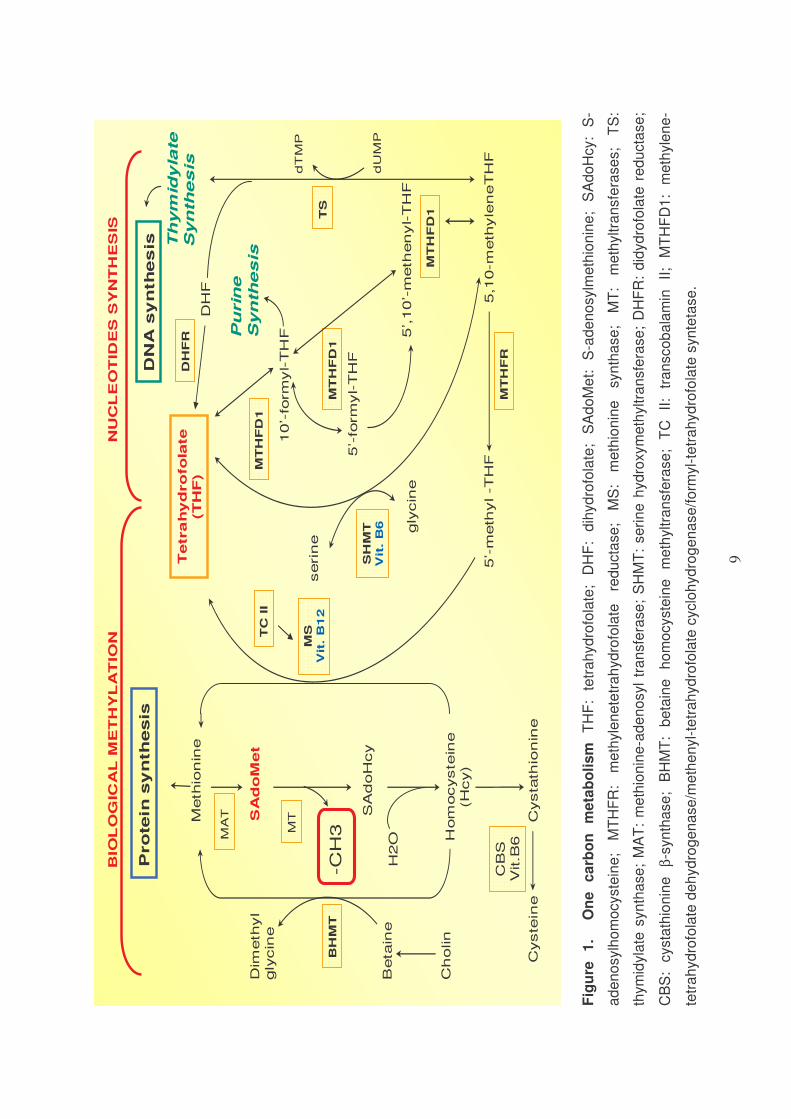
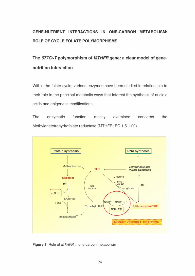
One-carbon metabolism
One-carbon metabolism is a network of interconnected biochemical reactions
in which a one-carbon unit is transferred from a donor to tetrahydrofolate
(THF) for subsequent reduction or oxidation and/or transfer into biochemical
pathways essential for DNA synthesis (thymidylate and purine synthesis) and
biological methylation (methionine synthesis or serine-glycine
interconversion) (Figure 1).
Folate coenzymes act as acceptors or donors of one-carbon units in a
multiplicity of reactions involved in amino acid and nucleotide metabolism in
mammalian tissue1.
One-carbon metabolism is essential mainly for the methylation reaction and
for the synthesis of nucleotides.
9
Meth
ion
ine
SA
do
Met
SA
doH
cy
Hom
ocyste
ine
(H
cy)
5’-
meth
yl -T
HF
Tetra
hy
dro
fo
late
(T
HF
)
5,1
0-m
eth
yle
neT
HF
MT
HF
R
Cysta
thio
nin
eC
yste
ine
Pro
tein
sy
nth
esis
MS
Vit
. B
12
CB
SV
it.B
6
-CH
3
H2O
DN
A s
yn
th
es
is
serin
e
gly
cin
e
SH
MT
Vit
. B
6
TS
MT
5’,10’-
meth
en
yl-T
HF
5’-
form
yl-T
HF
10’-
form
yl-T
HF
MT
HF
D1
MT
HF
D1
MT
HF
D1
TC
II
Beta
ine
Ch
olin
Dim
eth
yl
gly
cin
e
BH
MT
BIO
LO
GIC
AL
ME
TH
YL
AT
ION
NU
CL
EO
TID
ES
SY
NT
HE
SIS
dU
MP
dT
MP
Th
ym
idy
late
Syn
thes
is
MA
T
Pu
rin
e
Syn
thes
is
DH
F
DH
FR
Fig
ure
1
.
On
e
ca
rbo
n
me
tab
oli
sm
T
HF
: te
tra
hyd
rofo
late
; D
HF
: d
ihyd
rofo
late
; S
Ad
oM
et:
S
-ad
en
osylm
eth
ion
ine
; S
Ad
oH
cy:
S-
ad
en
osylh
om
ocyste
ine
; M
TH
FR
: m
eth
yle
ne
tetr
ah
yd
rofo
late
re
du
cta
se
; M
S:
me
thio
nin
e
syn
tha
se
; M
T:
me
thyltra
nsfe
rase
s;
TS
:
thym
idyla
te s
yn
tha
se
; M
AT
: m
eth
ion
ine
-ad
en
osyl
tra
nsfe
rase
; S
HM
T:
se
rin
e h
yd
roxym
eth
yltra
nsfe
rase
; D
HF
R:
did
yd
rofo
late
re
ducta
se
;
CB
S:
cysta
thio
nin
e
β-s
yn
tha
se
; B
HM
T:
be
tain
e
ho
mo
cyste
ine
m
eth
yltra
nsfe
rase
; T
C
II:
tra
nsco
ba
lam
in
II;
MT
HF
D1
: m
eth
yle
ne
-
tetr
ah
yd
rofo
late
de
hyd
rog
en
ase
/me
the
nyl-
tetr
ah
yd
rofo
late
cyclo
hyd
rog
en
ase
/fo
rmyl-
tetr
ah
yd
rofo
late
syn
teta
se
.
10
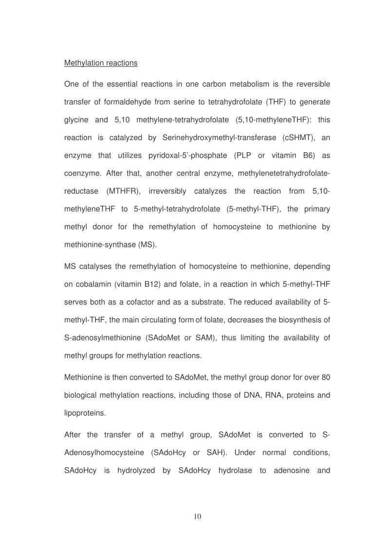
Methylation reactions
One of the essential reactions in one carbon metabolism is the reversible
transfer of formaldehyde from serine to tetrahydrofolate (THF) to generate
glycine and 5,10 methylene-tetrahydrofolate (5,10-methyleneTHF): this
reaction is catalyzed by Serinehydroxymethyl-transferase (cSHMT), an
enzyme that utilizes pyridoxal-5’-phosphate (PLP or vitamin B6) as
coenzyme. After that, another central enzyme, methylenetetrahydrofolate-
reductase (MTHFR), irreversibly catalyzes the reaction from 5,10-
methyleneTHF to 5-methyl-tetrahydrofolate (5-methyl-THF), the primary
methyl donor for the remethylation of homocysteine to methionine by
methionine-synthase (MS).
MS catalyses the remethylation of homocysteine to methionine, depending
on cobalamin (vitamin B12) and folate, in a reaction in which 5-methyl-THF
serves both as a cofactor and as a substrate. The reduced availability of 5-
methyl-THF, the main circulating form of folate, decreases the biosynthesis of
S-adenosylmethionine (SAdoMet or SAM), thus limiting the availability of
methyl groups for methylation reactions.
Methionine is then converted to SAdoMet, the methyl group donor for over 80
biological methylation reactions, including those of DNA, RNA, proteins and
lipoproteins.
After the transfer of a methyl group, SAdoMet is converted to S-
Adenosylhomocysteine (SAdoHcy or SAH). Under normal conditions,
SAdoHcy is hydrolyzed by SAdoHcy hydrolase to adenosine and

11
homocysteine. However, this reaction is rapidly reversible with equilibrium
dynamics that strongly favor SAdoHcy synthesis rather than its hydrolysis.
Thus, chronic elevation in plasma homocysteine levels may have an indirect
and negative effect on cellular methylation reactions through a concomitant
increase in intracellular SAdoHcy concentrations2.
Homocysteine can be methylated to regenerate methionine by folate/vitamin
B12 dependent MS reaction and additionally by the betaine-homocysteine
methyltransferase (BHMT) reaction using choline and betaine pathway in the
liver and kidney. Homocysteine also condenses with serine to form
cystathionine in an irreversible reaction catalyzed by cystathionine-β-
synthase (CBS), using vitamin B6 as a coenzyme.
Nucleotides synthesis
Thymidylate synthase catalyzes the transfer of methyl groups from folate to
deoxyuridylate, producing thymidylate, via a rate-limiting reaction for DNA
synthesis1. Since folate-derived one–carbon groups are essential for the de
novo synthesis of thymidylate, inhibition of folate metabolism in mammalian
cells results in uracil misincorporation into DNA due to an imbalance in the
deoxyribonucleotide pool. It has also been shown that folate deficiency
results in excess uracil incorporation into human DNA3; similarly, a moderate,
chronic folate depletion in rats has been shown to result in excess uracil
incorporation in colonic DNA4. One-carbon moieties from folate are also
utilized in de novo purine biosynthesis.
12
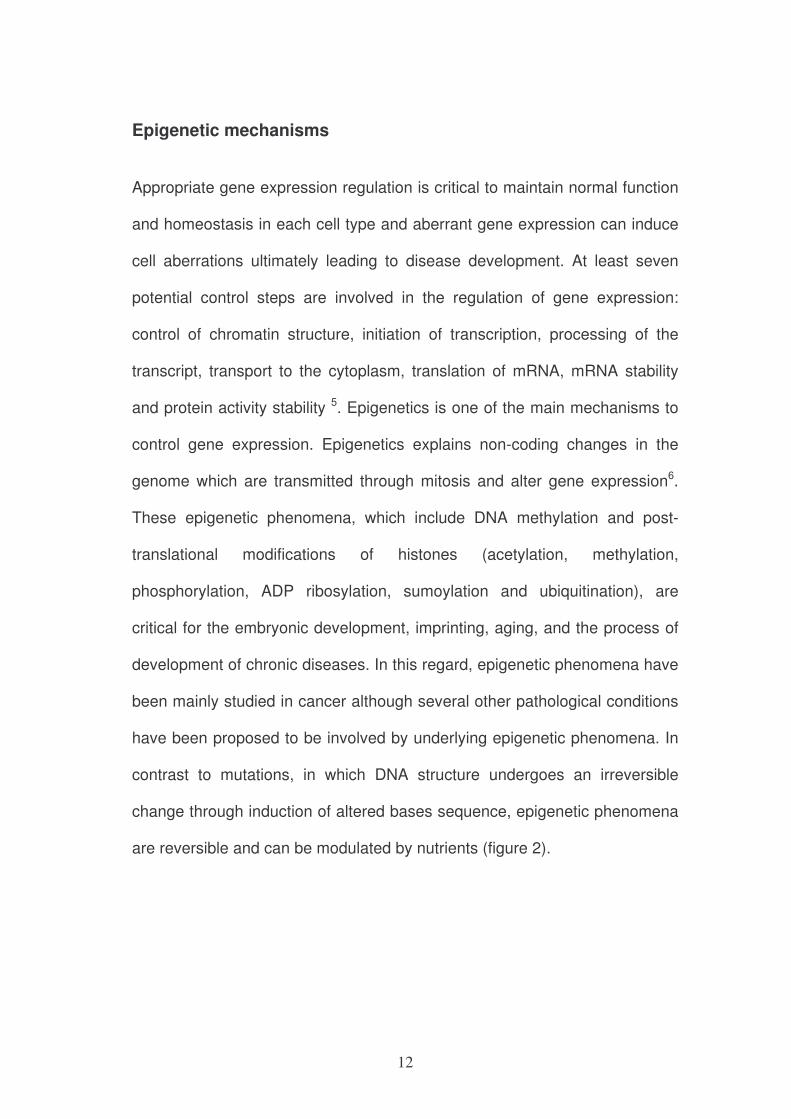
Epigenetic mechanisms
Appropriate gene expression regulation is critical to maintain normal function
and homeostasis in each cell type and aberrant gene expression can induce
cell aberrations ultimately leading to disease development. At least seven
potential control steps are involved in the regulation of gene expression:
control of chromatin structure, initiation of transcription, processing of the
transcript, transport to the cytoplasm, translation of mRNA, mRNA stability
and protein activity stability 5. Epigenetics is one of the main mechanisms to
control gene expression. Epigenetics explains non-coding changes in the
genome which are transmitted through mitosis and alter gene expression6.
These epigenetic phenomena, which include DNA methylation and post-
translational modifications of histones (acetylation, methylation,
phosphorylation, ADP ribosylation, sumoylation and ubiquitination), are
critical for the embryonic development, imprinting, aging, and the process of
development of chronic diseases. In this regard, epigenetic phenomena have
been mainly studied in cancer although several other pathological conditions
have been proposed to be involved by underlying epigenetic phenomena. In
contrast to mutations, in which DNA structure undergoes an irreversible
change through induction of altered bases sequence, epigenetic phenomena
are reversible and can be modulated by nutrients (figure 2).
13
IMBALANCE OF NUTRIENTS BY DIETFolate, dietary
methyl group, zinc, selenium,
…
Alteration of gene specific
DNA methylationAlteration of genomic DNA
methylation
Methylation on promoter
site
Methylation
on exon site
Gene expression
Gene transcription
Cancer,
diseases, cellgrowth, tissue
differentiation
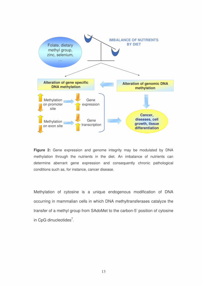
Figure 2: Gene expression and genome integrity may be modulated by DNA
methylation through the nutrients in the diet. An imbalance of nutrients can
determine aberrant gene expression and consequently chronic pathological
conditions such as, for instance, cancer disease.
Methylation of cytosine is a unique endogenous modification of DNA
occurring in mammalian cells in which DNA methyltransferases catalyze the
transfer of a methyl group from SAdoMet to the carbon-5’ position of cytosine
in CpG dinucleotides7.
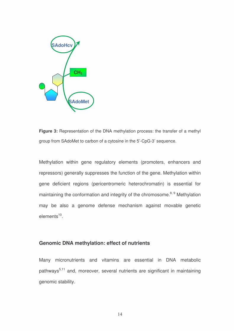
14
Figure 3: Representation of the DNA methylation process: the transfer of a methyl
group from SAdoMet to carbon of a cytosine in the 5’-CpG-3’ sequence.
Methylation within gene regulatory elements (promoters, enhancers and
repressors) generally suppresses the function of the gene. Methylation within
gene deficient regions (pericentromeric heterochromatin) is essential for
maintaining the conformation and integrity of the chromosome.8, 9 Methylation
may be also a genome defense mechanism against movable genetic
elements10.
Genomic DNA methylation: effect of nutrients
Many micronutrients and vitamins are essential in DNA metabolic
pathways5,11 and, moreover, several nutrients are significant in maintaining
genomic stability.
CH3
SAdoMet
SAdoHcy
15
DNA methylation state, especially in genome-wide methylation, can be
regulated through the availability of methyl groups, by aminoacids as
methionine, choline, betaine and serine, contained in the diet, and also by
bioactive nutrients, as zinc, selenium and retinoic acid in modifying the
methyltransferases or the enzymes of one-carbon metabolism.12, 13, 14, 15, 16,17
Nevertheless, folate and/or methyl group dietary supply provides the most
convincing data for the interaction of nutrients and DNA methylation, because
these dietary elements are directly involved in DNA methylation via one-
carbon metabolism. The metabolic function of all coenzymatic forms of folate
is to transfer one-carbon units for the synthesis of SAdoMet, the universal
methyl donor for several biological methylation reactions, and the de novo
deoxynucleoside triphosphate synthesis. Therefore, a dietary folate depletion
may decrease genomic DNA methylation in humans18, 19 and in animal
models20 and, on the other hand, a folate replete diet may restore the DNA
methylation status19.
Gene-specific DNA methylation at the promoter region
The 5'-promoter regions or the first exons of around one-half of human genes
are areas very rich in CpG sequences called CpG islands21. At the promoter
region, CpG-rich sequences usually are unmethylated22. The methylation of
these CpG islands usually induces inhibition of their expression. Although the
exact molecular mechanism by which DNA methylation represses the
transcription is not yet clear, several data demonstrate an active role of
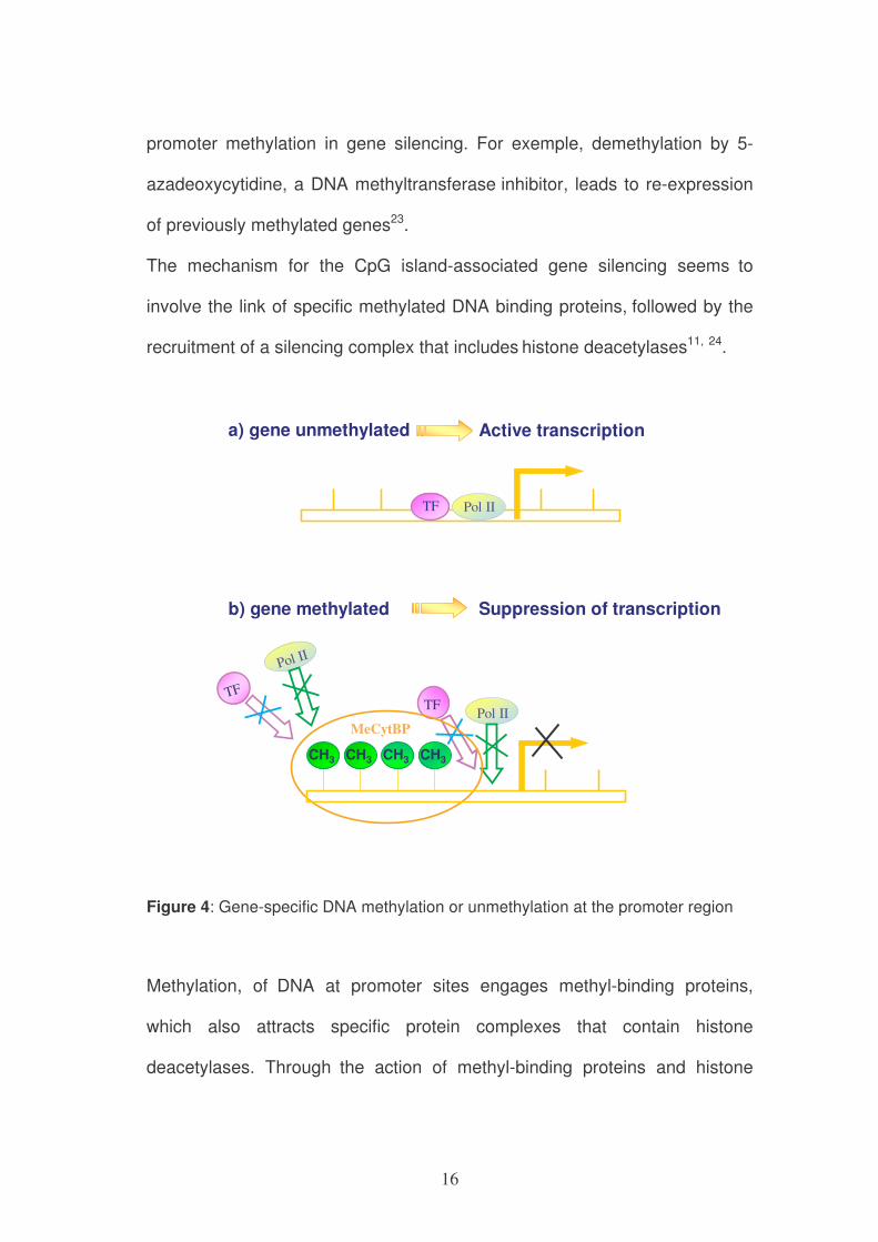
16
promoter methylation in gene silencing. For exemple, demethylation by 5-
azadeoxycytidine, a DNA methyltransferase inhibitor, leads to re-expression
of previously methylated genes23.
The mechanism for the CpG island-associated gene silencing seems to
involve the link of specific methylated DNA binding proteins, followed by the
recruitment of a silencing complex that includes histone deacetylases11, 24.
Pol IITF
CH3 CH3 CH3 CH3
Pol IITF
MeCytBP
Pol II
TF
a) gene unmethylated Active transcription
b) gene methylated Suppression of transcription
Figure 4: Gene-specific DNA methylation or unmethylation at the promoter region
Methylation, of DNA at promoter sites engages methyl-binding proteins,
which also attracts specific protein complexes that contain histone
deacetylases. Through the action of methyl-binding proteins and histone
17
deacetylases, the DNA structure changes to a compact, condensed
chromatin configuration that results in permanent inhibition of messenger
RNA and protein production11. In carcinogenesis, hypermethylation of CpG
islands in promoter region is mainly associated with transcriptional silencing
of gene expression, which has an important role as an alternative mechanism
by which tumor suppressor genes are inactivated without mutation or allele
deletion. On the other hand, hypomethylation of CpG islands is associated
with the gene activation, which also is an important mechanism by which
proto-oncogenes are activated.
Gene-specific DNA methylation at the coding region
Hypermethylation of the coding region can decrease the gene transcription
since local cytosine methylation of a specific sequence can directly interfere
with the binding of certain transcriptional factors25. On the other hand, the
gene transcription of the coding region can be increased by hypomethylation,
enhancing the binding of transcription factors.
Some observations propose that nutrients may affect gene transcription by
gene-specific alteration of DNA methylation.
In hepatocarcinogenesis, where a chronic dietary methyl deficiency has been
demonstrated, the progressive loss of methyl groups at most CpG sites on
both coding and noncoding strands in hepatic DNA during the early phase of
folate/methyl deficiency was observed26. After tumor formation, the majority
18
of cytosines became remethylated. In the preneoplastic nodules, the level of
p53 mRNA was increased and associated with hypomethylation in the coding
region, whereas in cancer tissue, p53 mRNA is decreased and associated
with relative hypermethylation. This observation suggests that a folate/methyl-
deficient diet induces liver cancer by affecting the methylation status of the
p53 gene coding region and by consequent alteration of p53 gene
transcription26. In other methyl-deficient animal studies, increased levels of
mRNA for c-fos, c-Ha-ras and c-myc were correlated with hypomethylation at
specific sites within these genes27, 28.
Hypomethylation of the coding regions of critical genes can lead to genome
instability either because this region becomes more susceptible to
endogenous nucleases29 or because the site of hypomethylation is likely to
undergo enzymatic deamination to uracil30, 31. The second situation is more
frequently observed in conditions where intracellular SAdoMet levels are low,
such as in a folate depletion status31.
Several animal studies and cell culture studies confirm that folate deficiency
might induce DNA strand breaks and subsequent mutations through exon
site hypomethylation30, 32, 33, 34, 35.
At the other hand, in an animal model of chemical carcinogenesis, folate
supplementation reduces gene disruption by reversing the site-specific DNA
hypomethylation33.
19
References
1. Friso S, Choi SW. Gene-nutrient interactions in one-carbon
metabolism. Curr Drug Metab. Feb 2005;6(1):37-46.
2. Yi P, Melnyk S, Pogribna M, Pogribny IP, Hine RJ, James SJ.
Increase in plasma homocysteine associated with parallel increases in
plasma S-adenosylhomocysteine and lymphocyte DNA
hypomethylation. J Biol Chem. Sep 22 2000;275(38):29318-29323.
3. Fenech M. The role of folic acid and Vitamin B12 in genomic stability
of human cells. Mutat Res. Apr 18 2001;475(1-2):57-67.
4. Choi SW, Friso S, Dolnikowski GG, et al. Biochemical and molecular
aberrations in the rat colon due to folate depletion are age-specific. J
Nutr. Apr 2003;133(4):1206-1212.
5. Villard J. Transcription regulation and human diseases. Swiss Med
Wkly. Oct 2 2004;134(39-40):571-579.
6. Mathers JC. Reversal of DNA hypomethylation by folic acid
supplements: possible role in colorectal cancer prevention. Gut. May
2005;54(5):579-581.
7. Costello JF, Plass C. Methylation matters. J Med Genet. May
2001;38(5):285-303.
8. Robertson KD, Jones PA. DNA methylation: past, present and future
directions. Carcinogenesis. Mar 2000;21(3):461-467.
9. Friso S, Choi SW. Gene-nutrient interactions and DNA methylation. J
Nutr. Aug 2002;132(8 Suppl):2382S-2387S.
20
10. O'Neill RJ, O'Neill MJ, Graves JA. Undermethylation associated with
retroelement activation and chromosome remodelling in an
interspecific mammalian hybrid. Nature. May 7 1998;393(6680):68-72.
11. Jones PL, Veenstra GJ, Wade PA, et al. Methylated DNA and MeCP2
recruit histone deacetylase to repress transcription. Nat Genet. Jun
1998;19(2):187-191.
12. Ono T, Uehara Y, Kurishita A, Tawa R, Sakurai H. Biological
significance of DNA methylation in the ageing process. Age Ageing.
Jan 1993;22(1):S34-43.
13. Issa JP. CpG-island methylation in aging and cancer. Curr Top
Microbiol Immunol. 2000;249:101-118.
14. Issa JP, Ottaviano YL, Celano P, Hamilton SR, Davidson NE, Baylin
SB. Methylation of the oestrogen receptor CpG island links ageing and
neoplasia in human colon. Nat Genet. Aug 1994;7(4):536-540.
15. Goelz SE, Vogelstein B, Hamilton SR, Feinberg AP. Hypomethylation
of DNA from benign and malignant human colon neoplasms. Science.
Apr 12 1985;228(4696):187-190.
16. Counts JL, Goodman JI. Hypomethylation of DNA: an epigenetic
mechanism involved in tumor promotion. Mol Carcinog. Dec
1994;11(4):185-188.
17. Gaudet F, Hodgson JG, Eden A, et al. Induction of tumors in mice by
genomic hypomethylation. Science. Apr 18 2003;300(5618):489-492.
21
18. Wainfan E, Poirier LA. Methyl groups in carcinogenesis: effects on
DNA methylation and gene expression. Cancer Res. Apr 1 1992;52(7
Suppl):2071s-2077s.
19. Pogribny IP, Miller BJ, James SJ. Alterations in hepatic p53 gene
methylation patterns during tumor progression with folate/methyl
deficiency in the rat. Cancer Lett. May 1 1997;115(1):31-38.
20. Herman JG, Merlo A, Mao L, et al. Inactivation of the
CDKN2/p16/MTS1 gene is frequently associated with aberrant DNA
methylation in all common human cancers. Cancer Res. Oct 15
1995;55(20):4525-4530.
21. Carrozza MJ, Utley RT, Workman JL, Cote J. The diverse functions of
histone acetyltransferase complexes. Trends Genet. Jun
2003;19(6):321-329.
22. Roth SY, Denu JM, Allis CD. Histone acetyltransferases. Annu Rev
Biochem. 2001;70:81-120.
23. Johnstone RW. Histone-deacetylase inhibitors: novel drugs for the
treatment of cancer. Nat Rev Drug Discov. Apr 2002;1(4):287-299.
24. Nan X, Ng HH, Johnson CA, et al. Transcriptional repression by the
methyl-CpG-binding protein MeCP2 involves a histone deacetylase
complex. Nature. May 28 1998;393(6683):386-389.
25. Tate PH, Bird AP. Effects of DNA methylation on DNA-binding
proteins and gene expression. Curr Opin Genet Dev. Apr
1993;3(2):226-231.
22
26. Pogribny IP, Basnakian AG, Miller BJ, Lopatina NG, Poirier LA, James
SJ. Breaks in genomic DNA and within the p53 gene are associated
with hypomethylation in livers of folate/methyl-deficient rats. Cancer
Res. May 1 1995;55(9):1894-1901.
27. Zapisek WF, Cronin GM, Lyn-Cook BD, Poirier LA. The onset of
oncogene hypomethylation in the livers of rats fed methyl-deficient,
amino acid-defined diets. Carcinogenesis. Oct 1992;13(10):1869-
1872.
28. Dizik M, Christman JK, Wainfan E. Alterations in expression and
methylation of specific genes in livers of rats fed a cancer promoting
methyl-deficient diet. Carcinogenesis. Jul 1991;12(7):1307-1312.
29. Hansen RS, Ellis NA, Gartler SM. Demethylation of specific sites in
the 5' region of the inactive X-linked human phosphoglycerate kinase
gene correlates with the appearance of nuclease sensitivity and gene
expression. Mol Cell Biol. Nov 1988;8(11):4692-4699.
30. Shen JC, Rideout WM, 3rd, Jones PA. High frequency mutagenesis
by a DNA methyltransferase. Cell. Dec 24 1992;71(7):1073-1080.
31. Yang AS, Shen JC, Zingg JM, Mi S, Jones PA. HhaI and HpaII DNA
methyltransferases bind DNA mismatches, methylate uracil and block
DNA repair. Nucleic Acids Res. Apr 25 1995;23(8):1380-1387.
32. Kim YI, Pogribny IP, Basnakian AG, et al. Folate deficiency in rats
induces DNA strand breaks and hypomethylation within the p53 tumor
suppressor gene. Am J Clin Nutr. Jan 1997;65(1):46-52.
23
33. Kim YI, Pogribny IP, Salomon RN, et al. Exon-specific DNA
hypomethylation of the p53 gene of rat colon induced by
dimethylhydrazine. Modulation by dietary folate. Am J Pathol. Oct
1996;149(4):1129-1137.
34. Woodson K, Mason J, Choi SW, et al. Hypomethylation of p53 in
peripheral blood DNA is associated with the development of lung
cancer. Cancer Epidemiol Biomarkers Prev. Jan 2001;10(1):69-74.
35. Blount BC, Mack MM, Wehr CM, et al. Folate deficiency causes uracil
misincorporation into human DNA and chromosome breakage:
implications for cancer and neuronal damage. Proc Natl Acad Sci U S
A. Apr 1 1997;94(7):3290-3295.
24
GENE-NUTRIENT INTERACTIONS IN ONE-CARBON METABOLISM:
ROLE OF CYCLE FOLATE POLYMORPHISMS
The 677C>T polymorphism of MTHFR gene: a clear model of gene-
nutrition interaction
Within the folate cycle, various enzymes have been studied in relationship to
their role in the principal metabolic ways that interest the synthesis of nucleic
acids and epigenetic modifications.
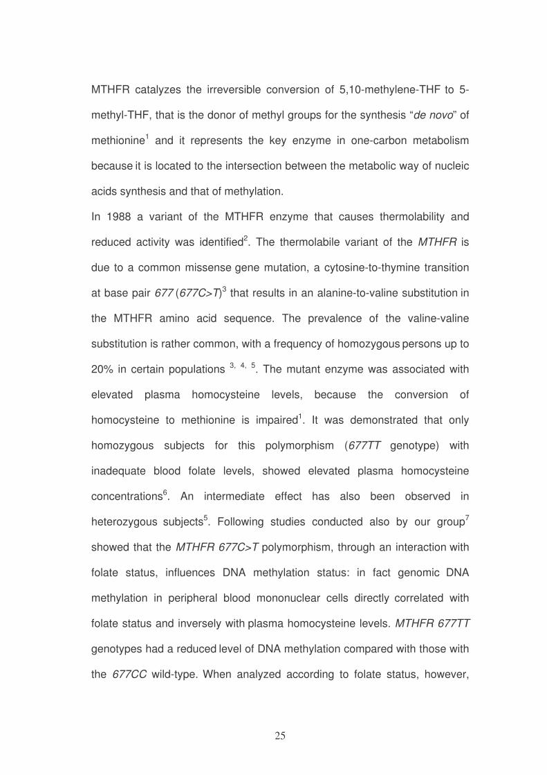
The enzymatic function mostly examined concerns the
Methylenetetrahydrofolate reductase (MTHFR; EC 1.5.1.20).
homocysteine
Methionine
SAdoMet
SAdoHcy
MT
5’-methyl -THF
THF
5,10-methyleneTHF
MTHFR
Thymidylate and Purine Synthesis
MSVit.B12
-CH3
H2O
serine
glicine
SHMTVit. B6 TS
NADPH+H+NADP+
NON-REVERSIBLE REACTION
DNA synthesisProtein synthesis
Figure 1: Role of MTHFR in one-carbon metabolism
25
MTHFR catalyzes the irreversible conversion of 5,10-methylene-THF to 5-
methyl-THF, that is the donor of methyl groups for the synthesis “de novo” of
methionine1 and it represents the key enzyme in one-carbon metabolism
because it is located to the intersection between the metabolic way of nucleic
acids synthesis and that of methylation.
In 1988 a variant of the MTHFR enzyme that causes thermolability and
reduced activity was identified2. The thermolabile variant of the MTHFR is
due to a common missense gene mutation, a cytosine-to-thymine transition
at base pair 677 (677C>T)3 that results in an alanine-to-valine substitution in
the MTHFR amino acid sequence. The prevalence of the valine-valine
substitution is rather common, with a frequency of homozygous persons up to
20% in certain populations 3, 4, 5. The mutant enzyme was associated with
elevated plasma homocysteine levels, because the conversion of
homocysteine to methionine is impaired1. It was demonstrated that only
homozygous subjects for this polymorphism (677TT genotype) with
inadequate blood folate levels, showed elevated plasma homocysteine
concentrations6. An intermediate effect has also been observed in
heterozygous subjects5. Following studies conducted also by our group7
showed that the MTHFR 677C>T polymorphism, through an interaction with
folate status, influences DNA methylation status: in fact genomic DNA
methylation in peripheral blood mononuclear cells directly correlated with
folate status and inversely with plasma homocysteine levels. MTHFR 677TT
genotypes had a reduced level of DNA methylation compared with those with
the 677CC wild-type. When analyzed according to folate status, however,
26
only the TT subjects with low levels of folate accounted for the reduced DNA
methylation.
An interesting model of gene-nutrient interaction in phenotypic expression is
clearly shaped in this model5, 6. Moreover, the MTHFR 677C>T
polymorphism is related to the reduced availability of methyl-THF and it has
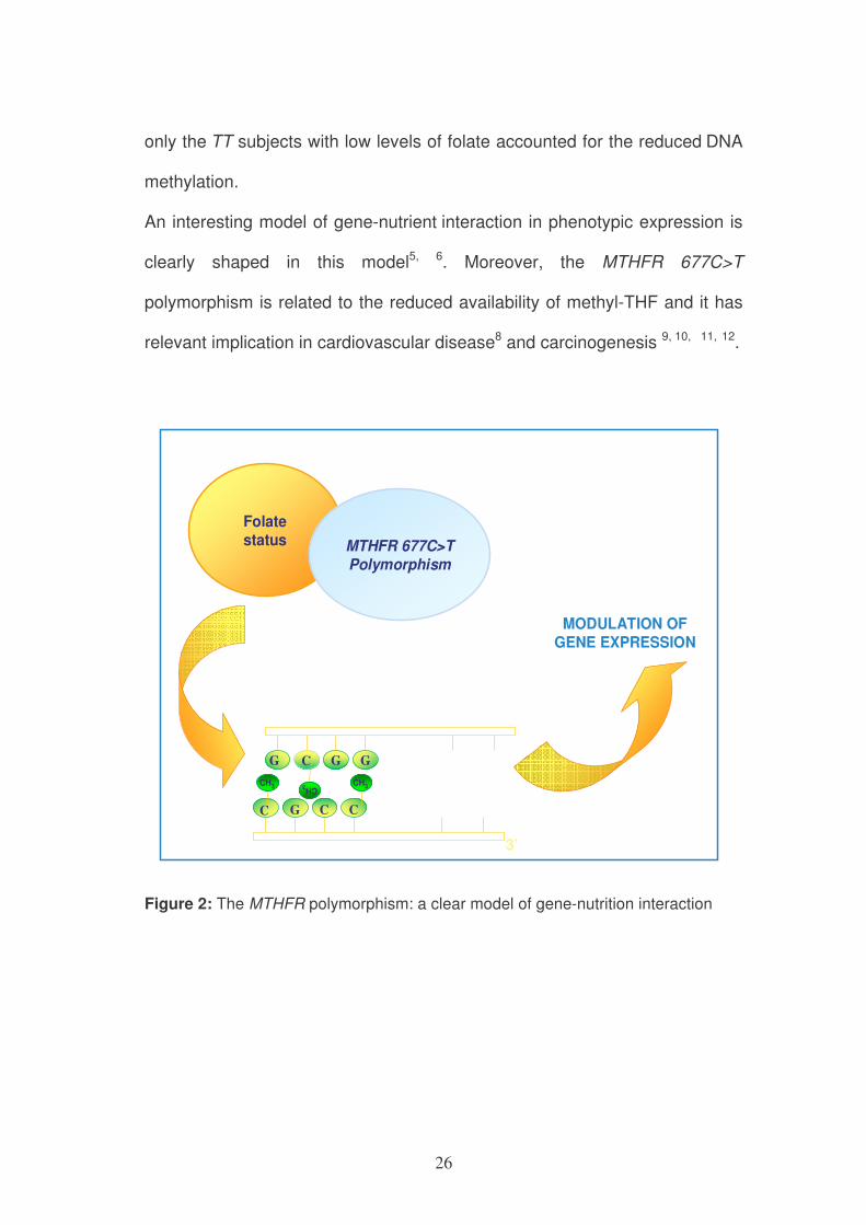
relevant implication in cardiovascular disease8 and carcinogenesis 9, 10, 11, 12.
Folate
status MTHFR 677C>T
Polymorphism
G C G G
C G C C
CH3 CH3CH3
3’
MODULATION OF
GENE EXPRESSION
Figure 2: The MTHFR polymorphism: a clear model of gene-nutrition interaction
27
Aim
Starting from the observations on the role of MTHFR enzyme for gene-
nutrition interaction models, we intended to appraise the principal
polymorphisms of the other enzymes involved in folate metabolic cycle, to
highlight possible similar examples of interactions among genetics,
epigenetics and nutrition and therefore to study the possible effects of gene-
nutrition interactions on the main epigenetic feature of DNA, DNA
methylation.
Materials and methods
Around eight hundred subjects, sex and age-matched, have been recruited .
They were for the greatest part native of the Veneto Region in Italy or born in
neighbouring regions in Northern Italy. For each selected subject,
biochemical data concerning one-carbon metabolism (plasma concentrations
of folate, vitamin B12, vitamin B6 and homocysteine) and a complete clinical
history were collected. Subjects with known conditions that could affect one-
carbon metabolism were excluded from the study, such as subjects who
used multivitamins supplements and/or with acute illnesses and/or making
use of drugs interfering with folate metabolism (i.e. penicillinamine,
anticonvulsants, methotrexate). The allelic frequencies and the genotypes
were studied; moreover,the relationship between the presence or absence of
each polymorphism and genomic DNA methylation status were analyzed.
28
DNA was extracted from peripheral blood mononuclear cells. Plasma
homocysteine levels were measured by HPLC (High performance liquid
chromatography), according to the method described by Araki and Sako13.
Folate plasma levels and vitamin B12 were determined using an automatic
method based on chemiluminescence technique (Chiron Diagnostics, East
Walpole, MA).
The levels of vitamin B6 were measured as plasma pyridoxal phosphate
(PLP), the active form of vitamin B6, by HPLC using a method described by
Kimura et al14.
Genotyping was performed using a standard PCR method followed by
digestion with site-specific restriction enzyme or by allelic discrimination
method through Real-Time PCR AB 7500.
Genomic DNA methylation was performed by a method that uses a combined
technology with liquid chromatography and mass spectrometry (LC/MS)15.
Briefly, DNA was denatured by heat at 100°C for three minutes then rapidly
cooled in ice and digested sequentially by three enzymes. Approximately 20
µL of solution containing DNA was hydrolyzed and instilled into the analytical
column protected by a pre-column and a separation of four major DNA bases
and the 5’-methyl-2’desossicytidine was obtained by an isocratic elution. The
quantification of non-methylated and methylated cytosine bases were made
through isotopic isomers as internal standards of known concentration (stable
isotopes). DNA genomic methylation was defined as the amount of 5-methyl-
29
cytosine to total bases cytosine (methylated and non methylated) in the DNA
analyzed.
Results and comments
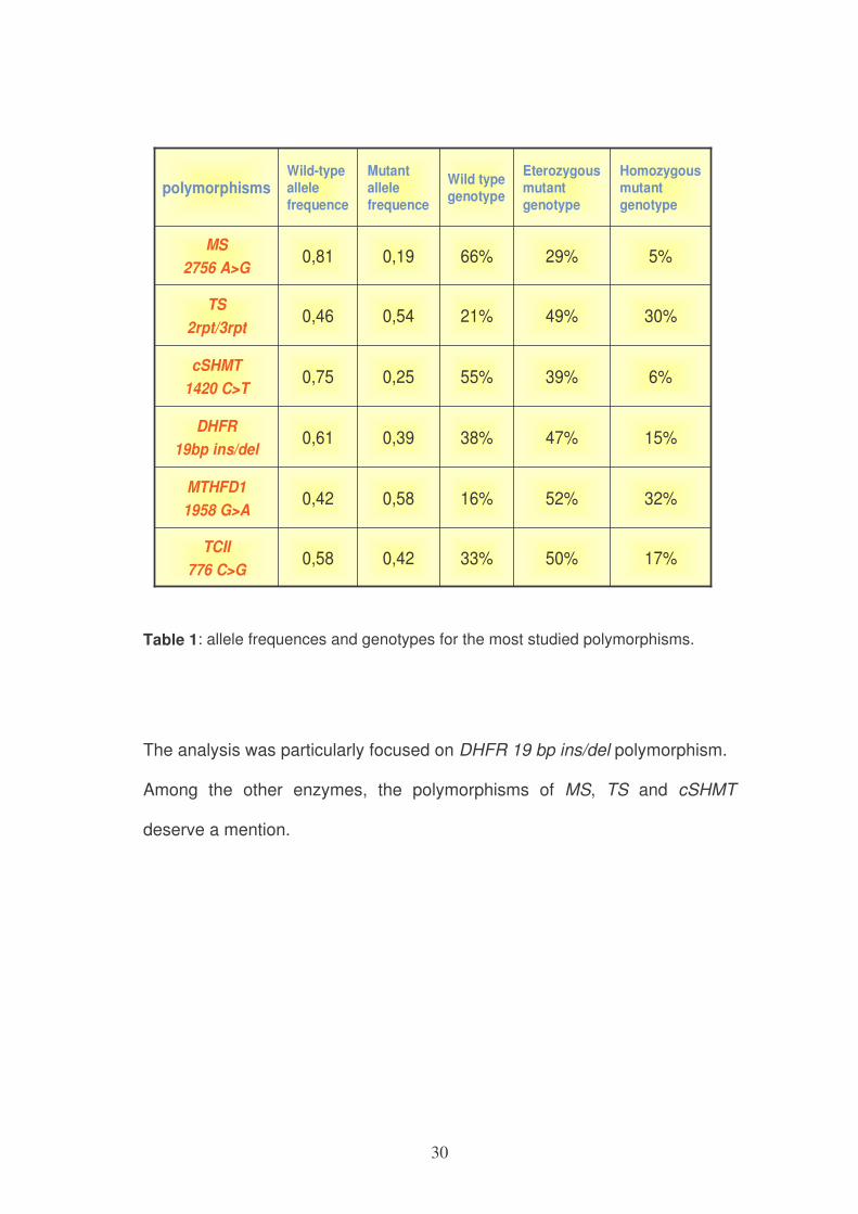
The following polymorphisms were studied:
• DHFR – Dihydrofolate-reductase (EC 1.5.1.3.): polymorphism 19 pb
ins/del
• MS - methionine synthase (EC 2.1.1.13) polymorphism 2756 A>G
• TS - thymidylate synthase (EC 2.1.1.45) polymorphism 2rpt-3rpt
• cSHMT - Serine hydroxy-methyl-transferase (EC 2.1.2.1): polymorphism
1420 C>T
• MTHFD1 - Methylene-THF dehydrogenase (EC 1.5.1.5) / metenil-THF-
cicloidrogenasi (EC 3.5.4.9) / formyl-THF synthetase (EC 6.3.4.3)
polymorphism 1958 G> A
• TCII - transcobalamin II polymorphism 776 C> G
• BHMT - Betaine-homocysteine methyl-transferase: polymorphism 742
A>G
Of each polymorphism we set out to assess the allele frequencies and
genotypes in our population.
30
17%50%33%0,420,58TCII
776 C>G
32%52%16%0,580,42MTHFD1
1958 G>A
15%47%38%0,390,61DHFR
19bp ins/del
6%39%55%0,250,75cSHMT
1420 C>T
30%49%21%0,540,46TS
2rpt/3rpt
5%29%66%0,190,81MS
2756 A>G
Homozygous mutant
genotype
Eterozygousmutant
genotype
Wild type genotype
Mutant allele
frequence
Wild-type allele
frequencepolymorphisms
Table 1: allele frequences and genotypes for the most studied polymorphisms.
The analysis was particularly focused on DHFR 19 bp ins/del polymorphism.
Among the other enzymes, the polymorphisms of MS, TS and cSHMT
deserve a mention.
31
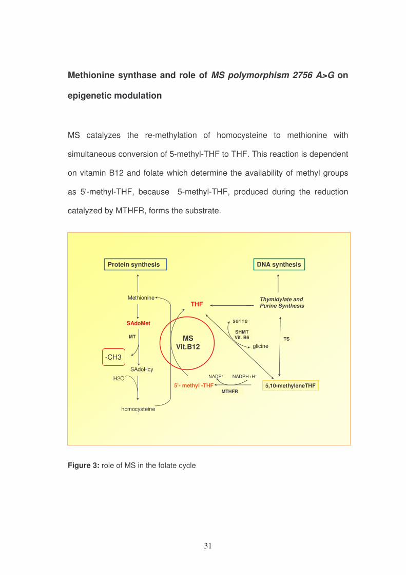
Methionine synthase and role of MS polymorphism 2756 A>G on
epigenetic modulation
MS catalyzes the re-methylation of homocysteine to methionine with
simultaneous conversion of 5-methyl-THF to THF. This reaction is dependent
on vitamin B12 and folate which determine the availability of methyl groups
as 5'-methyl-THF, because 5-methyl-THF, produced during the reduction
catalyzed by MTHFR, forms the substrate.
homocysteine
Methionine
SAdoMet
SAdoHcy
MT
5’- methyl -THF
THF
5,10-methyleneTHFMTHFR
Thymidylate and Purine Synthesis
MSVit.B12
-CH3
H2O
serine
glicine
SHMTVit. B6 TS
NADPH+H+NADP+
DNA synthesisProtein synthesis
Figure 3: role of MS in the folate cycle
32
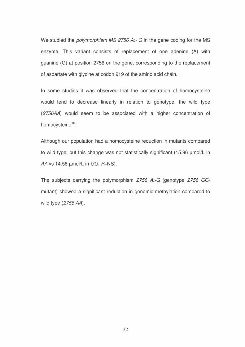
We studied the polymorphism MS 2756 A> G in the gene coding for the MS
enzyme. This variant consists of replacement of one adenine (A) with
guanine (G) at position 2756 on the gene, corresponding to the replacement
of aspartate with glycine at codon 919 of the amino acid chain.
In some studies it was observed that the concentration of homocysteine
would tend to decrease linearly in relation to genotype: the wild type
(2756AA) would seem to be associated with a higher concentration of
homocysteine16.
Although our population had a homocysteine reduction in mutants compared
to wild type, but this change was not statistically significant (15.96 µmol/L in
AA vs 14.58 µmol/L in GG, P=NS).
The subjects carrying the polymorphism 2756 A>G (genotype 2756 GG-
mutant) showed a significant reduction in genomic methylation compared to
wild type (2756 AA).
33
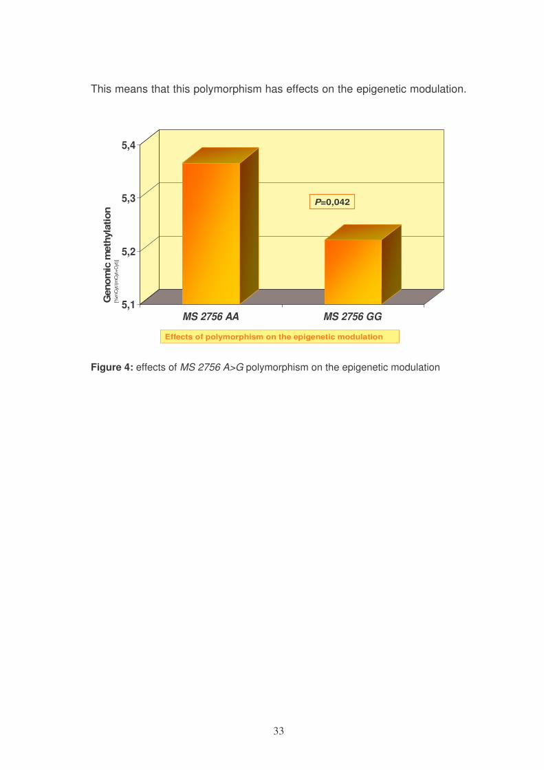
This means that this polymorphism has effects on the epigenetic modulation.
5,1
5,2
5,3
5,4
MS 2756 AA MS 2756 GG
P=0,042
Genom
icm
eth
yla
tion
[%m
Cyt
/(m
Cyt
+C
yt)]
Effects of polymorphism on the epigenetic modulation
Figure 4: effects of MS 2756 A>G polymorphism on the epigenetic modulation
34
Thymidylate synthase and role of polymorphism 2rpt-3rpt on
plasma folate levels
One-carbon metabolism is also essential for the synthesis of deoxy-
ribonucleotides.
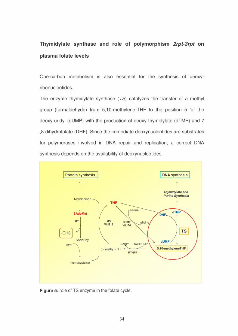
The enzyme thymidylate synthase (TS) catalyzes the transfer of a methyl
group (formaldehyde) from 5,10-methylene-THF to the position 5 'of the
deoxy-uridyl (dUMP) with the production of deoxy-thymidylate (dTMP) and 7
,8-dihydrofolate (DHF). Since the immediate deoxynucleotides are substrates
for polymerases involved in DNA repair and replication, a correct DNA
synthesis depends on the availability of deoxynucleotides.
homocysteine
Methionine
SAdoMet
SAdoHcy
MT
5’- methyl -THF
THF
5,10-methyleneTHF
MTHFR
Thymidylate and
Purine Synthesis
MSVit.B12
-CH3
H2O
serine
glicineSHMTVit. B6
NADPH+H+NADP+
DNA synthesisProtein synthesis
TS
dUMP
DHFdTMP
Figure 5: role of TS enzyme in the folate cycle.
35
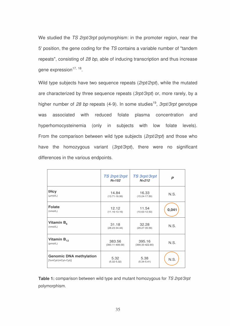
We studied the TS 2rpt/3rpt polymorphism: in the promoter region, near the
5' position, the gene coding for the TS contains a variable number of "tandem
repeats", consisting of 28 bp, able of inducing transcription and thus increase
gene expression17, 18.
Wild type subjects have two sequence repeats (2rpt/2rpt), while the mutated
are characterized by three sequence repeats (3rpt/3rpt) or, more rarely, by a
higher number of 28 bp repeats (4-9). In some studies19, 3rpt/3rpt genotype
was associated with reduced folate plasma concentration and
hyperhomocysteinemia (only in subjects with low folate levels).
From the comparison between wild type subjects (2rpt/2rpt) and those who
have the homozygous variant (3rpt/3rpt), there were no significant
differences in the various endpoints.
N.S.5.38(5.34-5.41)
5.32 (5.32-5.32)
Genomic DNA methylation [%mCyt/(mCyt+Cyt)]
N.S.395.16(369.33-422.80)
383.56(360.11-408.58)
Vitamin B12
(pmol/L)
N.S.32.28(29.27-35.59)
31.18(28.23-34.44)
Vitamin B6(nmol/L)
0,04111.54(10.63-12.53)
12.12(11.16-13.16)
Folate (nmol/L)
N.S.16.33(15.24-17.50)
14.84(13.71-16.06)
tHcy(µmol/L)
PTS 3rpt/3rpt
N=212
TS 2rpt/2rptN=152
Table 1: comparison between wild type and mutant homozygous for TS 2rpt/3rpt
polymorphism.
36
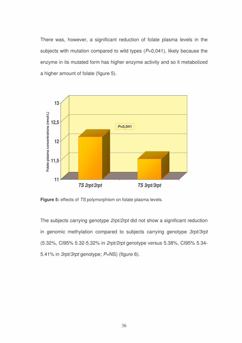
There was, however, a significant reduction of folate plasma levels in the
subjects with mutation compared to wild types (P=0,041), likely because the
enzyme in its mutated form has higher enzyme activity and so it metabolized
a higher amount of folate (figure 5).
11
11,5
12
12,5
13
TS 2rpt/2rpt TS 3rpt/3rpt
P=0,041
Fo
late
pla
sm
a c
oncen
trati
on
s (
nm
ol/L
)
Figure 5: effects of TS polymorphism on folate plasma levels.
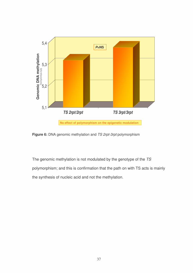
The subjects carrying genotype 2rpt/2rpt did not show a significant reduction
in genomic methylation compared to subjects carrying genotype 3rpt/3rpt
(5.32%, CI95% 5.32-5.32% in 2rpt/2rpt genotype versus 5.38%, CI95% 5.34-
5.41% in 3rpt/3rpt genotype; P=NS) (figure 6).
37
5,1
5,2
5,3
5,4
TS 2rpt/2rpt TS 3rpt/3rpt
P=NS
Ge
no
mic
DN
A m
eth
yla
tio
n
[%m
Cyt
/(m
Cyt
+C
yt)]
No effect of polymorphism on the epigenetic modulation
Figure 6: DNA genomic methylation and TS 2rpt-3rpt polymorphism
The genomic methylation is not modulated by the genotype of the TS
polymorphism; and this is confirmation that the path on with TS acts is mainly
the synthesis of nucleic acid and not the methylation.
38
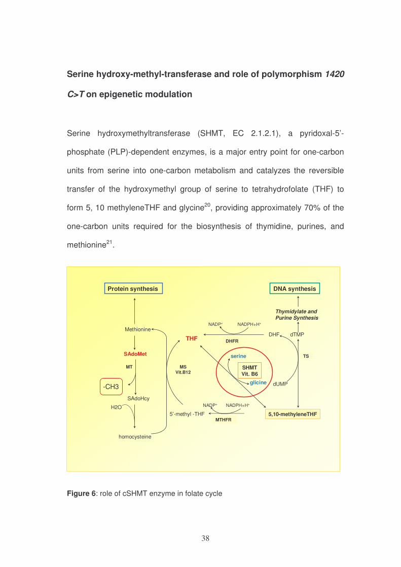
Serine hydroxy-methyl-transferase and role of polymorphism 1420
C>T on epigenetic modulation
Serine hydroxymethyltransferase (SHMT, EC 2.1.2.1), a pyridoxal-5’-
phosphate (PLP)-dependent enzymes, is a major entry point for one-carbon
units from serine into one-carbon metabolism and catalyzes the reversible
transfer of the hydroxymethyl group of serine to tetrahydrofolate (THF) to
form 5, 10 methyleneTHF and glycine20, providing approximately 70% of the
one-carbon units required for the biosynthesis of thymidine, purines, and
methionine21.
homocysteine
Methionine
SAdoMet
SAdoHcy
MT
5’-methyl -THF
THF
5,10-methyleneTHFMTHFR
Thymidylate and
Purine Synthesis
MSVit.B12
-CH3
H2O
serine
glicine
SHMT
Vit. B6
NADPH+H+NADP+
DNA synthesisProtein synthesis
TS
dUMP
DHF dTMP
NADP+ NADPH+H+
DHFR
Figure 6: role of cSHMT enzyme in folate cycle
39
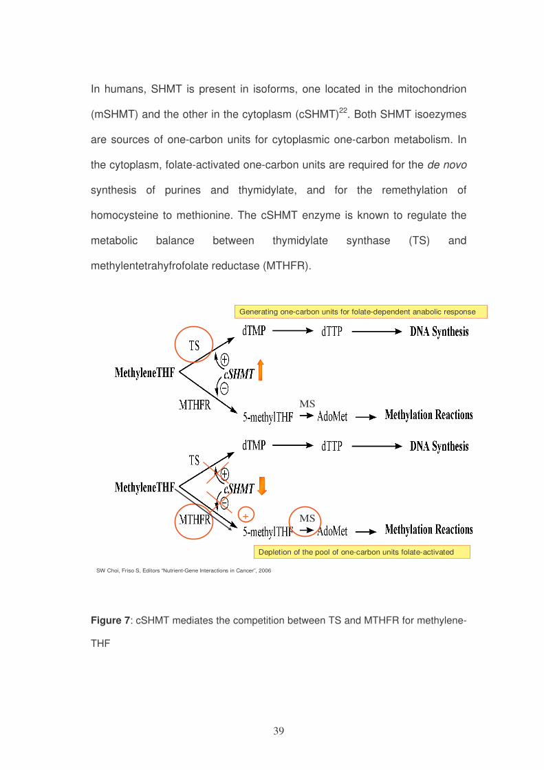
In humans, SHMT is present in isoforms, one located in the mitochondrion
(mSHMT) and the other in the cytoplasm (cSHMT)22. Both SHMT isoezymes
are sources of one-carbon units for cytoplasmic one-carbon metabolism. In
the cytoplasm, folate-activated one-carbon units are required for the de novo
synthesis of purines and thymidylate, and for the remethylation of
homocysteine to methionine. The cSHMT enzyme is known to regulate the
metabolic balance between thymidylate synthase (TS) and
methylentetrahyfrofolate reductase (MTHFR).
SW Choi, Friso S, Editors “Nutrient-Gene Interactions in Cancer”, 2006
MS
Generating one-carbon units for folate-dependent anabolic response
+
Depletion of the pool of one-carbon units folate-activated
MS
Figure 7: cSHMT mediates the competition between TS and MTHFR for methylene-
THF
40
Previous studies have indicated that a limited methyl group availability
caused by folate depletion shifts the flux of one-carbon units to S-
Adenosylmethionine (SAdoMet) synthesis and thereby suppresses DNA
synthesis23, 24. However, recently Herbig et al. proposed that under certain
conditions cSHMT acts as a switch to increase DNA synthesis at the
expense of homocysteine remethylation25. Once this enzyme is activated, it
preferentially supplies one carbon units for thymidylate synthesis, depletes
MTHFR pools for SAdoMet synthesis by synthesizing serine and sequesters
5-methylTHF that is needed for SAdoMet synthesis. Collectively, these
observations strongly suggest that alteration of cSHMT expression can
influence DNA methylation status which needs SAdoMet as a one carbon
donor.
In 2001 a genetic polymorphism of cSHMT was identified22. This variant
consists in the substitution of a cytosine with a tymine in the nucleotide at
position 1420 of the gene (chromosome 17p11.2) and consequently of a
leucine (Leu) with a phenylalanine (Phe) at position 474 of the corresponding
amino acid chain (Leu474Phe).
In our study, when we compare wild type (1420CC) and homozygous
variants (1420TT), there was no significant difference at all endpoints.
However, homozygous mutant subjects (1420TT) subjects showed
significantly decreased tendency in genomic DNA methylation compared with
1420CC subjects. (P=0,005)
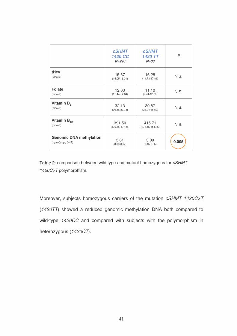
41
0.0053.09(2.45-3.85)
3.81 (3.63-3.97)
Genomic DNA methylation(ng mCyt/µg DNA)
N.S.415.71(376.15-454.86)
391.50(376.15-407.48)
Vitamin B12
(pmol/L)
N.S.30.87(26.04-36.59)
32.13(30.56-33.78)
Vitamin B6
(nmol/L)
N.S.11.10(9.74-12.78)
12,03(11.44-12.64)
Folate (nmol/L)
N.S.16.28(14.73-17.81)
15.67(15.05-16.31)
tHcy(µmol/L)
PcSHMT1420 TT
N=33
cSHMT1420 CC
N=290
Table 2: comparison between wild type and mutant homozygous for cSHMT
1420C>T polymorphism.
Moreover, subjects homozygous carriers of the mutation cSHMT 1420C>T
(1420TT) showed a reduced genomic methylation DNA both compared to
wild-type 1420CC and compared with subjects with the polymorphism in
heterozygous (1420CT).
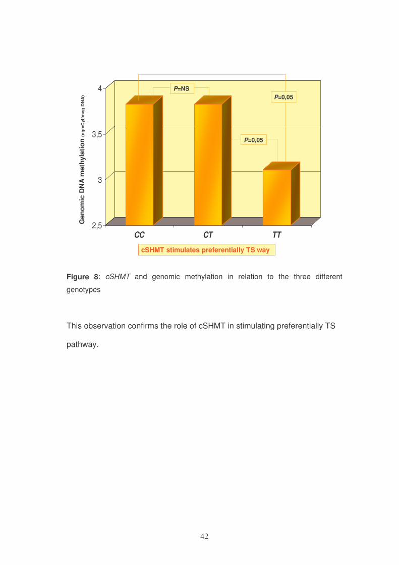
42
2,5
3
3,5
4
CC CT TT
P=NS
P=0,05
P=0,05
cSHMT stimulates preferentially TS way
Gen
om
ic D
NA
me
thy
lati
on
(ng
mC
yt/
mc
g D
NA
)
Figure 8: cSHMT and genomic methylation in relation to the three different
genotypes
This observation confirms the role of cSHMT in stimulating preferentially TS
pathway.
43
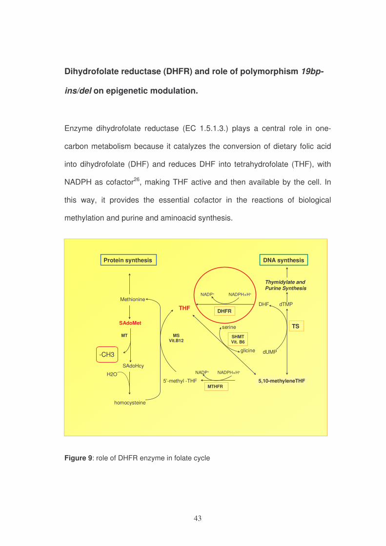
Dihydrofolate reductase (DHFR) and role of polymorphism 19bp-
ins/del on epigenetic modulation.
Enzyme dihydrofolate reductase (EC 1.5.1.3.) plays a central role in one-
carbon metabolism because it catalyzes the conversion of dietary folic acid
into dihydrofolate (DHF) and reduces DHF into tetrahydrofolate (THF), with
NADPH as cofactor26, making THF active and then available by the cell. In
this way, it provides the essential cofactor in the reactions of biological
methylation and purine and aminoacid synthesis.
homocysteine
Methionine
SAdoMet
SAdoHcy
MT
5’-methyl -THF
THF
5,10-methyleneTHFMTHFR
Thymidylate and
Purine Synthesis
MSVit.B12
-CH3
H2O
serine
glicine
SHMTVit. B6
NADPH+H+NADP+
DNA synthesisProtein synthesis
TS
dUMP
DHF dTMP
NADP+ NADPH+H+
DHFR
Figure 9: role of DHFR enzyme in folate cycle
44
Between the allelic variants identified on the DHFR gene, the polymorphism
of interest from the point of view of the impact on enzyme activity is
represented by a deletion of 19 bp to the first intron. The mechanisms by
which this deletion may cause a reduced activity of DHFR are manifold. First,
it eliminates the binding site for a transcriptional factor (Sp1); second, the
intron itself has a role in gene regulation, acting as a binding site for
transcription factors or altering the splicing.
Some studies evaluated the role of DHFR 19bp ins/del in determining the
onset of cancer. Gemmati and Collegues27 examined the association
between DHFR 19bp ins/del polymorphism and acute lymphoblastic
leukemia (ALL) in adult subjects (245 patients and 245 control cases) and the
possible interaction with the MTHFR 677C>T polymorphism. The deletion,
removing a binding site for a transcription factor, induces an increase of
enzyme expression, thus increasing the level of methylene-THF that favors
the intracellular synthesis of DNA pathway. Polymorphism MTHFR677C>T
has a synergistic effect by reducing enzymatic activity and the conversion of
methylene-THF into methyl-THF leading to a build-up inside the cell. Both
polymorphisms therefore play a protective role by reducing the risk of ALL27.
The protection against the development of ALL appears to be attributable to
a DNA synthesis more faithful and efficient thanks to a lot of THF reduced.
Specifically, a better synthesis of thymidylate reduces the risk of DNA uracil
misincorporation and prevents genomic instability. On the other hand, the
reduction of methyl-THF, required for methylation, makes it less likely hyper-
methylation and gene silencing of tumor suppressor genes.
45
In our study, we proposed not only to evaluate the possible role of DHFR
19bp ins/del polymorphism in modulating DNA genomic methylation but also
to analyze the function of this polymorphism on gene-specific methylation in
some genes involved in carcinogenesis, as E-cadherin, RAR-beta, APC,
RASSF1a, p15.
Moreover, we evaluated the effect of co-presence of DHFR 19bp ins/del
polymorphism and MTHFR 677C>T mutation in modulating DNA genomic
methylation.
In our study, there was no statistically significant difference between wild type
subjects and carrier of the mutant allele, for age, gender, folate
concentration, plasma homocysteine, vitamins B6 and B12 levels. There
were no significant differences also with regard to DNA genomic methylation.
However, when the DNA genomic methylation was compared between
subjects with or without the DHFR 19bp del mutant allele in agreement with
plasma folate levels, there was a significant reduction of DNA methylation in
the carriers of DHFR 19bp del mutant allele del and low folate
concentrations. The subjects carrying the wild type DHFR 19bp ins allele
showed no statistically significant differences in DNA methylation in relation
to folate status (Figure 10).
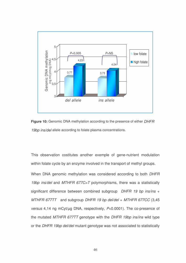
46
3,77
4,23
3,73
4,04
3
3,5
4
4,5
5G
en
om
ic D
NA
me
thy
latio
n
(ng
mC
yt/m
cg
DN
A)
del allele ins allele
low folate
high folate
P=NSP=0,005
Figure 10: Genomic DNA methylation according to the presence of either DHFR
19bp ins/del allele according to folate plasma concentrations.
This observation costitutes another exemple of gene-nutrient modulation
within folate cycle by an enzyme involved in the transport of methyl groups.
When DNA genomic methylation was considered according to both DHFR
19bp ins/del and MTHFR 677C>T polymorphisms, there was a statistically
significant difference between combined subgroup DHFR 19 bp ins/ins +
MTHFR 677TT and subgroup DHFR 19 bp del/del + MTHFR 677CC (3,45
versus 4,14 ng mCyt/µg DNA, respectively, P<0.0001). The co-presence of
the mutated MTHFR 677TT genotype with the DHFR 19bp ins/ins wild type
or the DHFR 19bp del/del mutant genotype was not associated to statistically
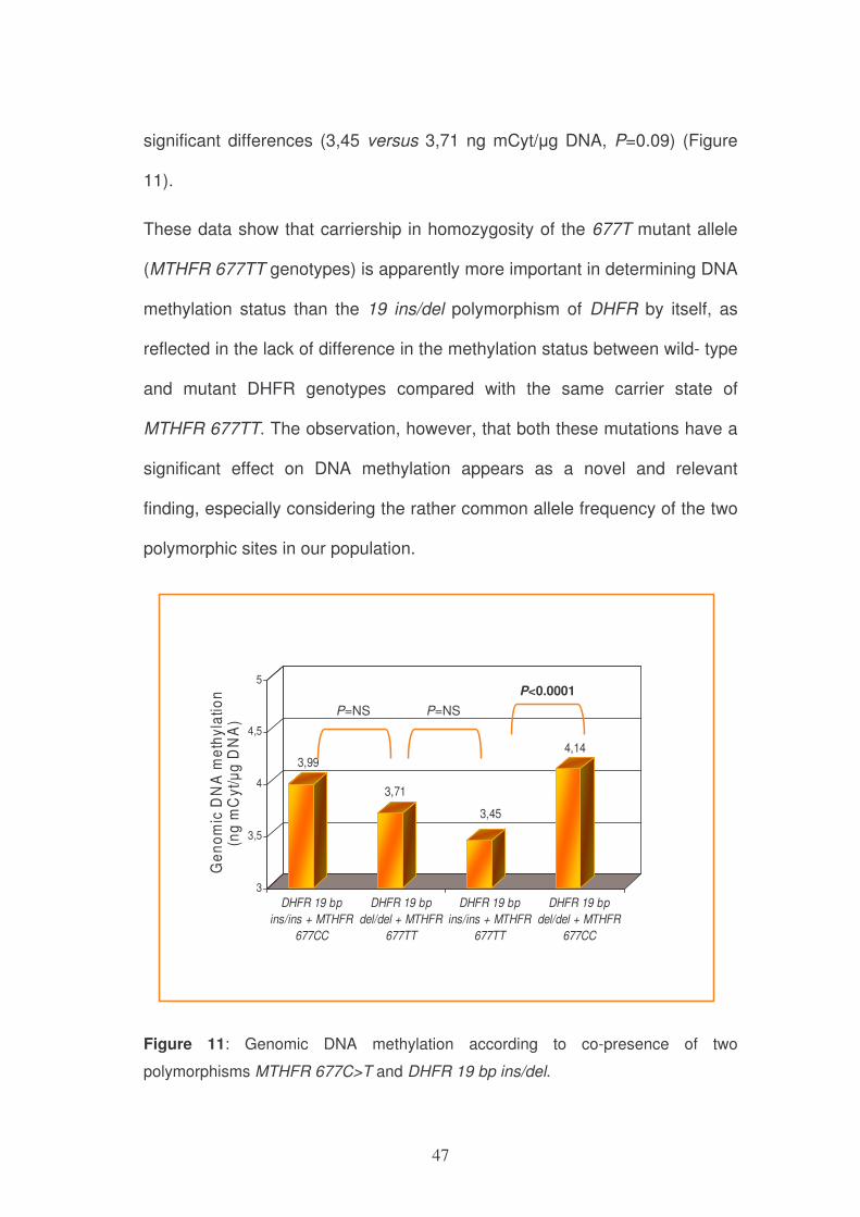
47
significant differences (3,45 versus 3,71 ng mCyt/µg DNA, P=0.09) (Figure
11).
These data show that carriership in homozygosity of the 677T mutant allele
(MTHFR 677TT genotypes) is apparently more important in determining DNA
methylation status than the 19 ins/del polymorphism of DHFR by itself, as
reflected in the lack of difference in the methylation status between wild- type
and mutant DHFR genotypes compared with the same carrier state of
MTHFR 677TT. The observation, however, that both these mutations have a
significant effect on DNA methylation appears as a novel and relevant
finding, especially considering the rather common allele frequency of the two
polymorphic sites in our population.
3,99
3,71
3,45
4,14
3
3,5
4
4,5
5
Ge
no
mic
DN
A m
eth
yla
tio
n
(n
g m
Cy
t/µ
g D
NA
)
DHFR 19 bp
ins/ins + MTHFR
677CC
DHFR 19 bp
del/del + MTHFR
677TT
DHFR 19 bp
ins/ins + MTHFR
677TT
DHFR 19 bp
del/del + MTHFR
677CC
P=NS
P<0.0001
P=NS
Figure 11: Genomic DNA methylation according to co-presence of two
polymorphisms MTHFR 677C>T and DHFR 19 bp ins/del.
48
The study of DNA gene-specific methylation of E-cadherin, RAR-beta, APC,
RASSF1a, p15 promoter genes according to DHFR 19bp ins/del genotype,
however, did not show any significant difference in the different genotypes.
This finding can be due to different reasons being the most likely that in
absence of a clear pathologic condition the methylation status at this gene
sites in peripheral blood mononuclear cells may not be so striking to allow a
significant difference. Another reason may be due to the relatively small
number of subjects analyzed, therefore a larger study may be auscpicable in
the future to consent to potentially identify differencies in genomic compared
to gene-specific methylation in such cell model.
49
References
1. Selhub J. Homocysteine metabolism. Annu Rev Nutr. 1999;19:217-
246.
2. Kang SS, Zhou J, Wong PW, Kowalisyn J, Strokosch G. Intermediate
homocysteinemia: a thermolabile variant of methylenetetrahydrofolate
reductase. Am J Hum Genet. Oct 1988;43(4):414-421.
3. Frosst P, Blom HJ, Milos R, et al. A candidate genetic risk factor for
vascular disease: a common mutation in methylenetetrahydrofolate
reductase. Nat Genet. May 1995;10(1):111-113.
4. Ma J, Stampfer MJ, Hennekens CH, et al. Methylenetetrahydrofolate
reductase polymorphism, plasma folate, homocysteine, and risk of
myocardial infarction in US physicians. Circulation. Nov 15
1996;94(10):2410-2416.
5. Girelli D, Friso S, Trabetti E, et al. Methylenetetrahydrofolate
reductase C677T mutation, plasma homocysteine, and folate in
subjects from northern Italy with or without angiographically
documented severe coronary atherosclerotic disease: evidence for an
important genetic-environmental interaction. Blood. Jun 1
1998;91(11):4158-4163.
6. Jacques PF, Bostom AG, Williams RR, et al. Relation between folate
status, a common mutation in methylenetetrahydrofolate reductase,
and plasma homocysteine concentrations. Circulation. Jan 1
1996;93(1):7-9.
50
7. Friso S, Choi SW, Girelli D, et al. A common mutation in the 5,10-
methylenetetrahydrofolate reductase gene affects genomic DNA
methylation through an interaction with folate status. Proc Natl Acad
Sci U S A. Apr 16 2002;99(8):5606-5611.
8. Ueland PM, Refsum H, Beresford SA, Vollset SE. The controversy
over homocysteine and cardiovascular risk. Am J Clin Nutr. Aug
2000;72(2):324-332.
9. Kim YI. Methylenetetrahydrofolate reductase polymorphisms, folate,
and cancer risk: a paradigm of gene-nutrient interactions in
carcinogenesis. Nutr Rev. Jul 2000;58(7):205-209.
10. Choi SW, Mason JB. Folate and carcinogenesis: an integrated
scheme. J Nutr. Feb 2000;130(2):129-132.
11. Ma J, Stampfer MJ, Giovannucci E, et al. Methylenetetrahydrofolate
reductase polymorphism, dietary interactions, and risk of colorectal
cancer. Cancer Res. Mar 15 1997;57(6):1098-1102.
12. Ulrich CM, Kampman E, Bigler J, et al. Lack of association between
the C677T MTHFR polymorphism and colorectal hyperplastic polyps.
Cancer Epidemiol Biomarkers Prev. Apr 2000;9(4):427-433.
13. Araki A, Sako Y. Determination of free and total homocysteine in
human plasma by high-performance liquid chromatography with
fluorescence detection. J Chromatogr. Nov 27 1987;422:43-52.
14. Kimura M, Kanehira K, Yokoi K. Highly sensitive and simple liquid
chromatographic determination in plasma of B6 vitamers, especially
51
pyridoxal 5′-phosphate. Journal of Chromatography A. 1996;722(1-
2):295-301.
15. Friso S, Choi SW, Dolnikowski GG, Selhub J. A method to assess
genomic DNA methylation using high-performance liquid
chromatography/electrospray ionization mass spectrometry. Anal
Chem. Sep 1 2002;74(17):4526-4531.
16. Tsai MY, Bignell M, Yang F, Welge BG, Graham KJ, Hanson NQ.
Polygenic influence on plasma homocysteine: association of two
prevalent mutations, the 844ins68 of cystathionine beta-synthase and
A(2756)G of methionine synthase, with lowered plasma homocysteine
levels. Atherosclerosis. Mar 2000;149(1):131-137.
17. Horie N, Aiba H, Oguro K, Hojo H, Takeishi K. Functional analysis and
DNA polymorphism of the tandemly repeated sequences in the 5'-
terminal regulatory region of the human gene for thymidylate
synthase. Cell Struct Funct. Jun 1995;20(3):191-197.
18. Kawakami K, Omura K, Kanehira E, Watanabe Y. Polymorphic
tandem repeats in the thymidylate synthase gene is associated with its
protein expression in human gastrointestinal cancers. Anticancer Res.
Jul-Aug 1999;19(4B):3249-3252.
19. Trinh BN, Ong CN, Coetzee GA, Yu MC, Laird PW. Thymidylate
synthase: a novel genetic determinant of plasma homocysteine and
folate levels. Hum Genet. Sep 2002;111(3):299-302.
52
20. Schirch V, Strong WB. Interaction of folylpolyglutamates with enzymes
in one-carbon metabolism. Arch Biochem Biophys. Mar
1989;269(2):371-380.
21. Stover P, Schirch V. 5-Formyltetrahydrofolate polyglutamates are slow
tight binding inhibitors of serine hydroxymethyltransferase. J Biol
Chem. Jan 25 1991;266(3):1543-1550.
22. Heil SG, Van der Put NM, Waas ET, den Heijer M, Trijbels FJ, Blom
HJ. Is mutated serine hydroxymethyltransferase (SHMT) involved in
the etiology of neural tube defects? Mol Genet Metab. Jun
2001;73(2):164-172.
23. Scott JM, Dinn JJ, Wilson P, Weir DG. Pathogenesis of subacute
combined degeneration: a result of methyl group deficiency. Lancet.
Aug 15 1981;2(8242):334-337.
24. Green JM, MacKenzie RE, Matthews RG. Substrate flux through
methylenetetrahydrofolate dehydrogenase: predicted effects of the
concentration of methylenetetrahydrofolate on its partitioning into
pathways leading to nucleotide biosynthesis or methionine
regeneration. Biochemistry. Oct 18 1988;27(21):8014-8022.
25. Herbig K, Chiang EP, Lee LR, Hills J, Shane B, Stover PJ.
Cytoplasmic serine hydroxymethyltransferase mediates competition
between folate-dependent deoxyribonucleotide and S-
adenosylmethionine biosyntheses. J Biol Chem. Oct 11
2002;277(41):38381-38389.
53
26. Gellekink H, Blom HJ, van der Linden IJ, den Heijer M. Molecular
genetic analysis of the human dihydrofolate reductase gene: relation
with plasma total homocysteine, serum and red blood cell folate levels.
Eur J Hum Genet. Jan 2007;15(1):103-109.
27. Gemmati D, De Mattei M, Catozzi L, et al. DHFR 19-bp
insertion/deletion polymorphism and MTHFR C677T in adult acute
lymphoblastic leukaemia: is the risk reduction due to intracellular folate
unbalancing? Am J Hematol. Aug 2009;84(8):526-529.
54
Among the polymorphisms examined, the analysis was particularly focused
on the latest polymorphism. DNA methylation has been mainly studied in
cancer, but probably it is involved also in other pathological condition. Indeed
we have observed, for example, that the DHFR 19bp-ins/del variant appears
associated with coronary artery disease.
CHAPTER 2
A POLYMORPHIC VARIANT OF DIHYDROFOLATE
REDUCTASE (DHFR 19BP-INS/DEL) IN CHRONIC DISEASE
RATHER THAN CANCER.
Abstract
Folate metabolism disorders are associated with higher risk of cancer, neural
congenital defects and cardiovascular diseases. The dihydrofolate reductase
enzyme (DHFR) converts dietary folic acid into dihydrofolate (DHF) and
reduces this latter into tetrahydrofolate (THF), essential cofactor in the
reactions of biological methylation and purine and aminoacids synthesis.
Recently a 19-bp deletion polymorphism has been described in intron 1,
likely related with the risk of spina bifida, preterm delivery, autism, breast
cancer and cardiovascular disease. We studied a group of 681 subjects
enrolled in the Verona Heart project, subdivided into two groups according
with the presence or not of coronary artery disease (CAD) and the
prevalence of the polymorphism was determined by allele specific
55
polymerase chain reaction. A significantly higher frequency of the DHFR
19bp del/del genotype was found in the group of affected subjects as
compared with healthy controls. Moreover, the presence of del allele was
associated with higher risk of being affected by coronary artery disease in a
fashion dependent on the number of allele (OD: 1.56 for ins/del and 1.85 for
del/del respectively).
Introduction
Folic acid is a water soluble B-group vitamin and plays a crucial role in DNA
synthesis and repair and in DNA methylation. The NADPH dependent
enzyme dihydrofolate reductase (DHFR) catalyzes the hydrogenation of
dihydrofolate (DHF) to tetrahydrofolate (THF). This latter molecule and its
one carbon derivatives are essential cofactors for purine, thymidylate and
aminoacids biosynthesis, through the so called “one-carbon transfer
reactions”1. Moreover tetrahydrofolate is necessary for DNA methylation,
since 5-methyltetrahydrofolate arising through the action of methionine
sinthetase, is essential for synthesis of S-adenosylmethionine (SAdoMet),
universal donor of methyl groups2, 3 .
In presence of low availability of THF, the synthesis of 5,10 methylene-THF
and 5 methyl-THF is impaired, as well as the reconversion of homocysteine
to methionine.
56
Plasma total homocysteine levels are a strong predictor of mortality in
patients with angiographically confirmed coronary artery disease4. Moreover
a meta-analysis study5 demonstrated a causal association between
homocysteine and cardiovascular disease and in a more recent work the
same authors could show that the severity of angiographically detected
coronary artery disease was positively associated with total homocysteine
levels6.
A close relationship exists between folate and homocysteine in circulating
blood, with homocysteine levels being inversely related to folate both intake
and plasma levels7.
In 2004 Johnson and Colleagues8 described for the first time a polymorphic
variant of DHFR consisting in a 19bp deletion in the first intron of the gene,
and reported an association between the presence of the polymorphism in a
group of women and the increased risk of having a child affected by spina
bifida.
More recently the presence of DHFR 19bp-del has been associated with low
levels of plasma homocysteine9 and the decrease correlated with both
condition of heterozygosis (ins/del) and homozygosis (del/del); in the same
year Xu and colleagues studied 2000 women and reported that the presence
of the variant in homozygotic condition correlated with higher plasma and
erythrocyte folate levels10. In 2008 the results of Framingham Offspring
Study (Kalmbach 2008) suggested that, regardless of folate intake, the
del/del polymorphism in DHFR was functional, since it was associated with
57
both higher levels of unmetabolized plasma folate and decreased red blood
cell folate store.
The presence of allele del has been associated with increased DHFR gene
expression: the raise was 2.4-fold in heterozygosis and 4.8-fold in
homozygotic subjects, in comparison with wild type group10. This evidence is
contrasting with a report where it was hypothesized that DHFR 19-bp
deletion might decrease enzyme expression through the removal of a strong
transcription factor binding site for Sp18.
Variants of DHFR that might affect plasma homocysteine and the status of
folate in blood and erythrocytes were examined in a study9 where the gene
for DHFR was sequenced in twenty individuals and the presence of several
SNPs and the 19-bp deletion were described. In 330 subjects the 19-bp
del/del genotype was found to be associated with lower plasma Hcy
compared with the wild genotype. No association was found with plasma and
red blood cell folate levels.
No data are available in literature about the possible association between
DHFR deletion polymorphism and coronary artery disease; on the other hand
the role of plasma homocysteine is well established as risk factor for
coronary heart disease.
In this study we accomplished several targets: a. to determine the frequency
of DHFR 19-bp del polymorphisms in a group of patients affected by
coronary artery disease compared with control subjects; b. to ascertain the
58
possible association between DHFR genetic variants, B-vitamin status and
homocysteine in coronary artery disease; c. to verify if the presence of the
mutant polymorphism might represent an additional and independent risk
factor for coronary artery disease.
Materials and methods
Study population. This study was performed within the framework of the
Verona Heart Study, a regional survey aimed at searching for new risk factors
for CAD in subjects with angiographic documentation of their coronary
vessels. Details of the enrolment criteria were given elsewhere11, 12. A total of
681 subject were included in the present study. Two hundred forty-two
subjects had completely normal coronary arteries, having undergone
coronary angiography for reasons other than CAD, mainly valvular heart
disease (CAD-free group), and they served as controls. These subjects were
also required to have neither a positive history nor clinical or instrumental
evidence of atherosclerosis in vascular districts beyond the coronary bed.
Four hundred thirty-nine subjects had angiographically proven CAD (most of
them were candidates for coronary artery bypass grafting).
All participants came from the same geographical area (Northern Italy) and
were of similar socioeconomic status. At the time of blood sampling, a
complete clinical history, including the assessment of cardiovascular risk
factors such as obesity, smoking, hypertension, and diabetes, was collected.
59
Written informed consent was obtained from all participants after a full
explanation of the study. The study was approved by our Institutional Ethical
Committee.
Biochemical analysis. Samples of venous blood were drawn from each
subject after an overnight fast. Plasma folate and vitamin B-12 levels were
determined by a chemiluminescence technique (Chiron Diagnostics, East
Walpole, MA, USA). Homocysteine was measured in plasma with HPLC
following the method by Araki and Sako13, with slight modifications. Levels of
vitamin B-6 were determined as plasma concentrations of pyridoxal-5'-
phosphate (PLP), the active metabolite of vitamin B-6 with a HPLC
method14,15. Erythrocyte folate content was assayed by affinity method,
followed by reversed-phase chromatography with electrical detection
according to Bagley and Selhub16.
Serum lipids were measured with an automatic analyzer (Technicon DAX96)
and LDL cholesterol was calculated with Friedwald formula.
DNA extraction and DHFR 19bp ins/del determination. Genomic DNA was
extracted from mononucleated blood cells with Wizard® Genomic DNA
purification kit (PROMEGA Corporation, Madison, WI, USA) and the
polymorphic variant 19-bp ins/del was analyzed by allele specific polymerase
chain reaction, which was set up by modification of published methods9,
followed by agarose gel electrophoresis of amplification products. Briefly, the
reaction was performed in 25 µl volume, containing 100 ng of genomic DNA,
0.4 µM each of the following primers: forward 1 (5’-
60
CCACGGTCGGGGTACCTGGG -3’), forward 2 (5’-
ACGGTCGGGGTGGCCGACTC-3’), reverse (5’-
AAAAGGGGAATCCAGTCGG-3’), 0.25 mM dNTPs, 1.5 mM MgCl2, 0.75 U
SuperTaq (AB-Analitica, Padova, Italy) and 8% DMSO. Thermocycler
conditions were 4 min at 94° followed by 35 cycles of 94° 1 min, 62° 1 min,
72° 1 min, with the final extension step of 72° for 7 min.
PCR products were loaded on 4% agarose gel containing ethidium bromide,
run at 100 V and analyzed on an image detector GelDoc (BioRad
Laboratories). The insertion allele product was represented by a 113 bp
band, whereas the deletion allele generated a 92 bp band.
Statistical methods. All statistical analyses were performed with SPSS 17.0
statistical package (SPSS Inc., Chicago, IL). Distribution of continuous
variables in groups was expressed as means ± standard deviation.
Logarithmic transformation was performed on all skewed variables, including
homocysteine, folate, vitamin B12, vitamin B6. Therefore, the statistical
differences concerning these parameters were also computed on the
corresponding log-transformed values, although, for the sake of clearness,
non-transformed data were reported in the Results. Quantitative data were
assessed using the Student's t-test or by ANOVA and Tuckey’s post-hoc test
when indicated. Covariance analysis (ANCOVA) was performed to evaluate
the possible confounding effects of classic risk factors for CAD, on the
assessment of risk associated with each polymorphic variant.
61
Associations between qualitative variables were analyzed with the χ2-test,
and linear trend analysis when indicated. The median of plasma folate levels
in the control group (12 nmol/L) was used as cut-off value for defining low
and high folate concentrations. A value of p < 0.05 was considered
significant.
To assess the extent to which genetic polymorphisms were associated with
coronary artery disease, odds ratios with 95% CIs were estimated by
univariate logistic regression analysis. Adjustment for classical
cardiovascular risk factors (i.e. sex, age, smoke, diabetes, cholesterol,
triglyceride) was performed by adding those covariates in a multiple logistic-
regression model.
Results
The clinical and biochemical characteristics of the subjects are summarized
in table 1. As expected the CAD group presented significant differences in
most parameters acknowledged as risk factors for cardiovascular pathology:
higher proportion of males and smokers and significantly elevated plasma
levels of total cholesterol, LDL-cholesterol and triglycerides. Inversely HDL-
cholesterol was lower in cases as compared with controls. Moreover plasma
homocysteine concentrations were higher in CAD patients.
62
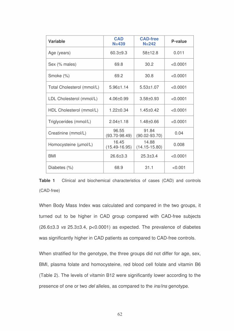
Variable CAD
N=439 CAD-free
N=242 P-value
Age (years) 60.3±9.3 58±12.8 0.011
Sex (% males) 69.8 30.2 <0.0001
Smoke (%) 69.2 30.8 <0.0001
Total Cholesterol (mmol/L) 5.96±1.14 5.53±1.07 <0.0001
LDL Cholesterol (mmol/L) 4.06±0.99 3.58±0.93 <0.0001
HDL Cholesterol (mmol/L) 1.22±0.34 1.45±0.42 <0.0001
Triglycerides (mmol/L) 2.04±1.18 1.48±0.66 <0.0001
Creatinine (mmol/L) 96.55
(93.70-98.49) 91.84
(90.02-93.70) 0.04
Homocysteine (µmol/L) 16.45
(15.49-16.95) 14.88
(14.15-15.80) 0.008
BMI 26.6±3.3 25.3±3.4 <0.0001
Diabetes (%) 68.9 31.1 <0.001
Table 1 Clinical and biochemical characteristics of cases (CAD) and controls
(CAD-free)
When Body Mass Index was calculated and compared in the two groups, it
turned out to be higher in CAD group compared with CAD-free subjects
(26.6±3.3 vs 25.3±3.4, p<0.0001) as expected. The prevalence of diabetes
was significantly higher in CAD patients as compared to CAD-free controls.
When stratified for the genotype, the three groups did not differ for age, sex,
BMI, plasma folate and homocysteine, red blood cell folate and vitamin B6
(Table 2). The levels of vitamin B12 were significantly lower according to the
presence of one or two del alleles, as compared to the ins/ins genotype.
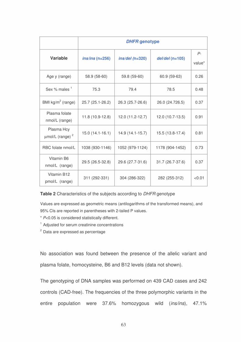
63
DHFR genotype
Variable ins/ins (n=256) ins/del (n=320) del/del (n=105) P-
value*
Age y (range) 58.9 (58-60) 59.8 (59-60) 60.9 (59-63) 0.26
Sex % males 1 75.3 79.4 78.5 0.48
BMI kg/m2 (range) 25.7 (25.1-26.2) 26.3 (25.7-26.6) 26.0 (24.726.5) 0.37
Plasma folate
nmol/L (range) 11.8 (10.9-12.8) 12.0 (11.2-12.7) 12.0 (10.7-13.5) 0.91
Plasma Hcy
µmol/L (range) 2
15.0 (14.1-16.1) 14.9 (14.1-15.7) 15.5 (13.8-17.4) 0.81
RBC folate nmol/L 1038 (930-1146) 1052 (979-1124) 1178 (904-1452) 0.73
Vitamin B6
nmol/L (range) 29.5 (26.5-32.8) 29.6 (27.7-31.6) 31.7 (26.7-37.6) 0.37
Vitamin B12
pmol/L (range) 311 (292-331) 304 (286-322) 282 (255-312) <0.01
Table 2 Characteristics of the subjects according to DHFR genotype
Values are expressed as geometric means (antilogarithms of the transformed means), and
95% CIs are reported in parentheses with 2-tailed P values.
* P<0.05 is considered statistically different.
1 Adjusted for serum creatinine concentrations
2 Data are expressed as percentage
No association was found between the presence of the allelic variant and
plasma folate, homocysteine, B6 and B12 levels (data not shown).
The genotyping of DNA samples was performed on 439 CAD cases and 242
controls (CAD-free). The frequencies of the three polymorphic variants in the
entire population were 37.6% homozygous wild (ins/ins), 47.1%
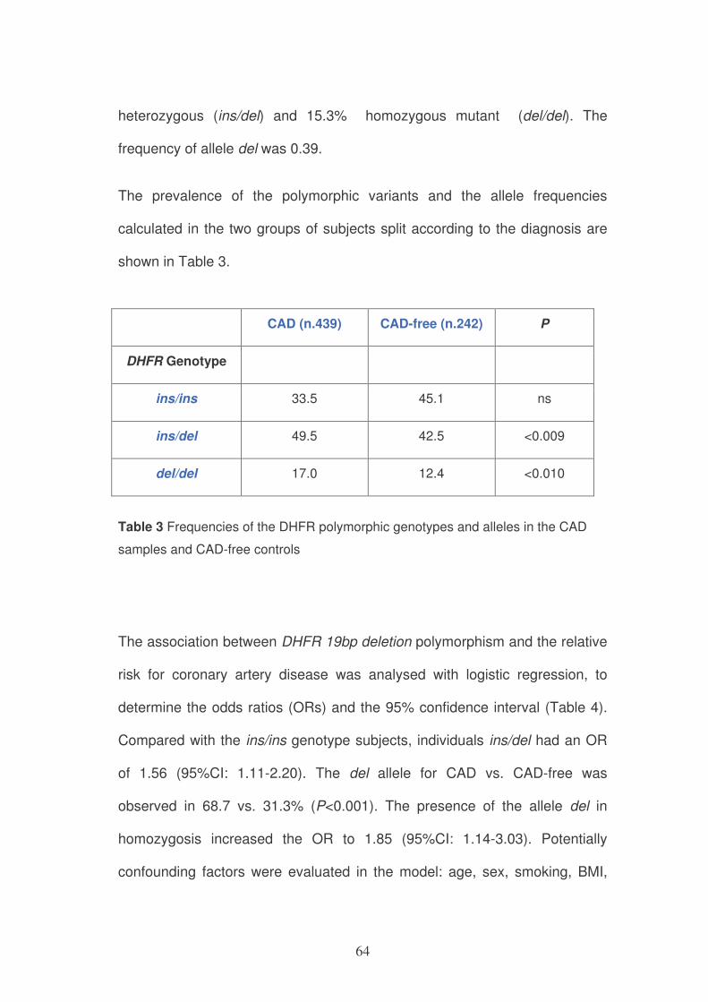
64
heterozygous (ins/del) and 15.3% homozygous mutant (del/del). The
frequency of allele del was 0.39.
The prevalence of the polymorphic variants and the allele frequencies
calculated in the two groups of subjects split according to the diagnosis are
shown in Table 3.
CAD (n.439) CAD-free (n.242) P
DHFR Genotype
ins/ins 33.5 45.1 ns
ins/del 49.5 42.5 <0.009
del/del 17.0 12.4 <0.010
Table 3 Frequencies of the DHFR polymorphic genotypes and alleles in the CAD
samples and CAD-free controls
The association between DHFR 19bp deletion polymorphism and the relative
risk for coronary artery disease was analysed with logistic regression, to
determine the odds ratios (ORs) and the 95% confidence interval (Table 4).
Compared with the ins/ins genotype subjects, individuals ins/del had an OR
of 1.56 (95%CI: 1.11-2.20). The del allele for CAD vs. CAD-free was
observed in 68.7 vs. 31.3% (P<0.001). The presence of the allele del in
homozygosis increased the OR to 1.85 (95%CI: 1.14-3.03). Potentially
confounding factors were evaluated in the model: age, sex, smoking, BMI,
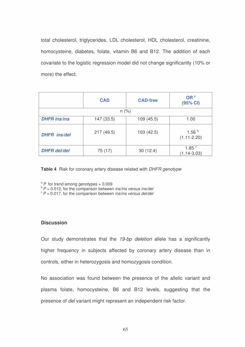
65
total cholesterol, triglycerides, LDL cholesterol, HDL cholesterol, creatinine,
homocysteine, diabetes, folate, vitamin B6 and B12. The addition of each
covariate to the logistic regression model did not change significantly (10% or
more) the effect.
CAD
CAD-free OR a
(95% CI)
n (%)
DHFR ins/ins 147 (33.5) 109 (45.5) 1.00
DHFR ins/del
217 (49.5)
103 (42.5)
1.56 b (1.11-2.20)
DHFR del/del 75 (17) 30 (12.4) 1.85 c
(1.14-3.03)
Table 4 Risk for coronary artery disease related with DHFR genotype
a P for trend among genotypes = 0.009
b P = 0.012, for the comparison between ins/ins versus ins/del
c P = 0.017, for the comparison between ins/ins versus del/del
Discussion
Our study demonstrates that the 19-bp deletion allele has a significantly
higher frequency in subjects affected by coronary artery disease than in
controls, either in heterozygosis and homozygosis condition.
No association was found between the presence of the allelic variant and
plasma folate, homocysteine, B6 and B12 levels, suggesting that the
presence of del variant might represent an independent risk factor.
66
We analysed with logistic regression the association between DHFR 19bp
deletion polymorphism and the relative risk for coronary artery disease, to
determine the odds ratios (ORs) and the 95% confidence interval. Compared
with the ins/ins genotype subjects, individuals ins/del had an OR of 1.56 and
the presence of the allele del in homozygosis increased the OR to 1.85.
Potentially confounding factors were evaluated in the model: age, sex,
smoking, BMI, total cholesterol, triglycerides, LDL cholesterol, HDL
cholesterol, creatinine, homocysteine, diabetes, folate, vitamin B6 and B12,
but the addition of each covariate to the logistic regression model did not
change significantly (10% or more) the effect.
Plasma total homocysteine level is a strong predictor of mortality in patients
with angiographically confirmed coronary artery disease4. Moreover, a meta-
analysis study5 demonstrated a causal association between homocysteine
and cardiovascular disease and in a more recent work the same group
showed a positive association between the severity of angiographically
detected coronary artery disease and total homocysteine levels6.
A close relationship exists between folate and homocysteine in circulating
blood, with homocysteine levels inversely related to both folate intake and
plasma levels7.
Very scarce are the data from scientific literature about the correlation
between DHFR polymorphic variants and cardiovascular disease, while other
enzymes involved in one-carbon metabolism have been investigated for their
impact on homocysteine levels in blood. A recent review17 presented an
67
overview of the association among metabolism of homocysteine,
polymorphic variants of genes encoding for enzymes involved in the folate-
homocysteine metabolic pathways and influence of nutrients on
homocysteine levels and the risk for cardiovascular disease. The genes
examined were methylenetetrahydrofolate reductase (MTHFR), methionine
synthase (MTR), methionine synthase reductase (MTRR) and cystathionine
beta-synthase (CBS). The authors suggested that in future studies other
genes should be considered because genetic modifications of enzymes and
changes of products depending on the B-group vitamins could alter Hcy
status and modify disease risk . Our results lines up in this direction,
demonstrating for the first time that the DHFR 19-bp deletion allele shows in
subjects affected by coronary artery disease a significantly higher frequency
than in controls, either in heterozygosis and homozygosis condition, in
association with a higher risk of being affected by the disease.
The presence of DHFR 19bp-del has been associated with low levels of
plasma homocysteine9 and the decrease correlated with heterozygosis
(ins/del) or homozygosis (del/del); in the same year Xu and colleagues
studied 2000 women and reported that the presence of the variant in
homozygotic condition correlated with higher plasma and erythrocyte folate
levels10. Our results showed no association between DHFR 19bp-del variant
and plasma homocysteine and B-group vitamins levels, moreover the
addition of these covariate to the logistic regression model did not change
significantly (10% or more) the relative risk for CAD.
68
Rather surprising is the finding that the levels of plasma B12 correlates with
the presence of the polymorphic variant of DHFR (table 2). On the other hand
the assay of plasma B12 is not a reliable biomarker of possible metabolism
alterations, since the measurement of holotranscobalamin or methylmalonic
acid are more sensible indicators of vitamin B12 status and subclinical
deficit18.
References
1. Scott JM, Weir DG. Folic acid, homocysteine and one-carbon
metabolism: a review of the essential biochemistry. J Cardiovasc Risk.
Aug 1998;5(4):223-227.
2. Choi SW, Mason JB. Folate and carcinogenesis: an integrated
scheme. J Nutr. Feb 2000;130(2):129-132.
3. Friso S, Choi SW. Gene-nutrient interactions and DNA methylation. J
Nutr. Aug 2002;132(8 Suppl):2382S-2387S.
4. Nygard O, Nordrehaug JE, Refsum H, Ueland PM, Farstad M, Vollset
SE. Plasma homocysteine levels and mortality in patients with
coronary artery disease. N Engl J Med. Jul 24 1997;337(4):230-236.
5. Wald DS, Law M, Morris JK. Homocysteine and cardiovascular
disease: evidence on causality from a meta-analysis. BMJ. Nov 23
2002;325(7374):1202.
69
6. Wald DS, Law M, Morris J. Serum homocysteine and the severity of
coronary artery disease. Thromb Res. 2003;111(1-2):55-57.
7. Graham IM, O'Callaghan P. The role of folic acid in the prevention of
cardiovascular disease. Curr Opin Lipidol. Dec 2000;11(6):577-587.
8. Johnson WG, Stenroos ES, Spychala JR, Chatkupt S, Ming SX,
Buyske S. New 19 bp deletion polymorphism in intron-1 of
dihydrofolate reductase (DHFR): a risk factor for spina bifida acting in
mothers during pregnancy? Am J Med Genet A. Feb 1
2004;124A(4):339-345.
9. Gellekink H, Blom HJ, van der Linden IJ, den Heijer M. Molecular
genetic analysis of the human dihydrofolate reductase gene: relation
with plasma total homocysteine, serum and red blood cell folate levels.
Eur J Hum Genet. Jan 2007;15(1):103-109.
10. Xu X, Gammon MD, Wetmur JG, et al. A functional 19-base pair
deletion polymorphism of dihydrofolate reductase (DHFR) and risk of
breast cancer in multivitamin users. Am J Clin Nutr. Apr
2007;85(4):1098-1102.
11. Girelli D, Russo C, Ferraresi P, et al. Polymorphisms in the factor VII
gene and the risk of myocardial infarction in patients with coronary
artery disease. N Engl J Med. Sep 14 2000;343(11):774-780.
12. Olivieri O, Martinelli N, Sandri M, et al. Apolipoprotein C-III, n-3
polyunsaturated fatty acids, and "insulin-resistant" T-455C APOC3
gene polymorphism in heart disease patients: example of gene-diet
interaction. Clin Chem. Feb 2005;51(2):360-367.
70
13. Araki A, Sako Y. Determination of free and total homocysteine in
human plasma by high-performance liquid chromatography with
fluorescence detection. J Chromatogr. Nov 27 1987;422:43-52.
14. Kimura M, Kanehira K, Yokoi K. Highly sensitive and simple liquid
chromatographic determination in plasma of B6 vitamers, especially
pyridoxal 5'-phosphate. J Chromatogr A. Jan 26 1996;722(1-2):296-
301.
15. Friso S, Girelli D, Martinelli N, et al. Low plasma vitamin B-6
concentrations and modulation of coronary artery disease risk. Am J
Clin Nutr. Jun 2004;79(6):992-998.
16. Bagley PJ, Selhub J. A common mutation in the
methylenetetrahydrofolate reductase gene is associated with an
accumulation of formylated tetrahydrofolates in red blood cells. Proc
Natl Acad Sci U S A. Oct 27 1998;95(22):13217-13220.
17. Malinowska A, Chmurzynska A. Polymorphism of genes encoding
homocysteine metabolism-related enzymes and risk for cardiovascular
disease. Nutr Res. Oct 2009;29(10):685-695.
18. von Castel-Roberts KM, Morkbak AL, Nexo E, et al. Holo-
transcobalamin is an indicator of vitamin B-12 absorption in healthy
adults with adequate vitamin B-12 status. Am J Clin Nutr. Apr
2007;85(4):1057-1061.
71
CHAPTER 3
STUDY OF A FUNCTIONAL MODEL OF INTER-
RELATIONSHIP BETWEEN GENETICS AND EPIGENETICS IN
FACTOR VII GENE PROMOTER: EVALUATION OF TWO
FUNCTIONAL POLYMORPHISMS ON F7 PROMOTER GENE
AND INTERACTION WITH METHYLATION INDEX IN THE F7
PROMOTER REGION.
DNA methylation, the main epigenetic mechanism that controls gene
expression, occurs in position 5 'only of cytosine dinucleotide sequences
within 5'-CpG-3'. As known, the distribution of CpG dinucleotides in the
genome is not homogeneous: they have high concentration especially in
promoter regions and in the first exons of genes, where they are called "CpG
islands", namely areas rich in these dinucleotide CpG sequences (70% of
CpG sequences).
Based on the observations made on folic acid polymorphisms we chose to
study the F7 gene because in its promoter region, near the transcription start
sites, polymorphisms with different functional effects, including 5'F7 and -
402G>A were identified. Our aim was to assess the epigenetic control on the
expression of a specific gene in presence/absence of functional
polymorphisms.
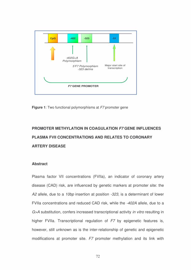
72
-323
5’F7 Polymorphism-323 del/ins
F7 GENE PROMOTER
-51CpG -402
-402G>A Polymorphism
Major start site oftranscription
Figure 1: Two functional polymorphisms at F7 promoter gene
PROMOTER METHYLATION IN COAGULATION F7 GENE INFLUENCES
PLASMA FVII CONCENTRATIONS AND RELATES TO CORONARY
ARTERY DISEASE
Abstract
Plasma factor VII concentrations (FVIIa), an indicator of coronary artery
disease (CAD) risk, are influenced by genetic markers at promoter site: the
A2 allele, due to a 10bp insertion at position -323, is a determinant of lower
FVIIa concentrations and reduced CAD risk, while the -402A allele, due to a
G>A substitution, confers increased transcriptional activity in vitro resulting in
higher FVIIa. Transcriptional regulation of F7 by epigenetic features is,
however, still unknown as is the inter-relationship of genetic and epigenetic
modifications at promoter site. F7 promoter methylation and its link with
73
promoter polymorphisms in modulating FVIIa and CAD risk was evaluated in
253 CAD and CAD-free subjects. Plasma FVIIa was inversely related to
methylation in A1A1 and -402GG, i.e. in the absence of the rare A2 and -
402A allele. The higher FVIIa paralleled with lower methylation in A1A1
compared to A2A2 (P=0.035) while no variation in methylation was
associated with the different -402G>A genotypes. The methylation-induced
FVIIa concentrations modulation was observed only in A1A1 where the
higher methylation resulting in lower FVIIa was prevalent within CAD-free
compare to CAD subgroup (P=0.011).
These findings identify the role of F7 promoter methylation in regulating
plasma FVIIa and its association with CAD.
1. Introduction
Factor VII, for its key function in coagulation pathway [1], is related to
atherothrombosis [2] and elevated concentrations of plasma activated factor
VII (FVIIa) are recognized as a significant risk factor for coronary artery
disease (CAD) and its main thrombotic complication, myocardial infarction [3,
4]. Several polymorphisms have been identified in F7 gene with functional
effects in modulating plasma concentrations of the gene product [5-8].
Among the major genetic determinants of FVII concentrations, an important
role has been defined for a decamer insertion at position –323 in the 5’
promoter region from the start of translation (0/10bp), where allele A1
corresponds to the absence of the decamer and allele A2 to its insertion [5,
9, 10]. We previously observed that the functional -323 10bp del/ins
74
polymorphism in the 5’ promoter region affects the risk of atherothrombosis
[4]. The F7 gene promoter activity is reduced by 33% in carriers of the A2
allele [4, 11] so that subjects with the A2A2 genotype have the lowest FVIIa
concentrations and are more likely not to develop myocardial infarction
compared to patients with the wild-type genotype [4]. Another polymorphism
at F7 gene promoter (–402G>A) has been also described having opposite
effect in vitro [12]. The rare -402A allele confers increased transcriptional
activity and is associated with higher plasma FVII concentrations [12],
although with a gender specificity in a selected population subset [7].
While the characterization of F7 genotype is indeed of help to predict FVIIa
phenotype as well as the risk of CAD, genetic mechanisms alone may not
fully explain the different FVIIa concentrations and the incidence of CAD.
Besides gene sequence variations, epigenetic modulation is also a major
player in gene expression regulation and phenotype characterization [13].
DNA methylation is the main epigenetic feature of mammalian DNA [14, 15]
with a critical role for gene expression regulation which has been extensively
described mainly in cancer disease [16, 17]. The methylation-related gene-
silencing phenomenon involves specific CpG–rich regions, the so-called CpG
islands [18, 19] that preferentially reside in promoters of genes [18]. CpG
islands hypermethylation within promoter sites is usually associated with
gene transcriptional silencing [18] in cancer-related genes [19], therefore
providing an important alternative path by which genes are modulated
without structural changes.
75
Mechanisms of epigenetic control of genes related to atherothrombotic
disease have not been extensively investigated, thus far. We recently
described, however, the epigenetic regulation through repression via
methylation at the 11 beta-hydroxysteroid dehydrogenase 2 gene promoter
site, a hypertension-disease associated gene, therefore suggesting a novel
link between regulation through epigenetic mechanism in diseases other than
cancer [20].
As for atherothrombosis-linked genes, a distinctive high interest has been
given to the role of F7 gene [2, 3] for its key role in activating the extrinsic
pathway by the exposure of tissue factor after plaque disruption [21] and for a
strong genotype-phenotype correlation that defines a singular link between
various genotypes at promoter site as predictor of plasma FVIIa
concentrations and CAD risk [4, 7, 22].
While certain polymorphisms at F7 promoter are well-recognized
determinants of FVIIa concentrations and CAD risk [4], little is known about
F7 promoter DNA methylation as a determinant of plasma FVIIa
concentrations by influencing gene product concentrations and possibly CAD
risk, either in the presence or absence of known functional polymorphisms at
promoter site.
The present study was therefore designed to evaluate: i) the role of DNA
methylation at F7 promoter site and its functional effects in phenotype
characterization, i.e. FVIIa concentrations; ii) the significance of F7 promoter
76
methylation relative to the presence of functional polymorphisms in affecting
FVIIa concentrations; iii) the relationship between F7 promoter methylation
and risk of CAD, with regard to the presence/absence of F7 promoter
polymorphic site.
2. Materials and Methods
We studied 253 unrelated, age- and sex-matched subjects recruited from a
single geographical area (Northern Italy) who were selected from
consecutively recruited subjects among those referred to the Department of
Clinical and Experimental Medicine and the Institute of Cardiovascular
Surgery of the University of Verona in Italy and participating to the Verona
Heart Project. Criteria for selection of the study population have been
previously described in details.[4, 23]
All subjects underwent coronary artery angiography which was performed by
two cardiologists unaware that the patients were participating to the study.
According to coronary angiography documentation, the patients were
classified as either being or not affected by severe coronary atherosclerotic
disease (CAD and CAD-free, respectively).
Of these 253 patients, 165 CAD had angiographically documented, severe
multivessel coronary atherosclerosis. Subjects with non significant coronary
stenosis (i.e. <50%) were excluded from the study. As a control group, 88
CAD-free patients who were examined with coronary angiography for
reasons other than possible coronary artery disease (mostly valvular heart
77
disease) were enrolled. They were required to have normal coronary arteries
as documented by angiography and to have neither a history of
atherosclerosis nor clinical or laboratory evidence of atherosclerosis in other
vascular beds. This control group was included so that the atherosclerotic
phenotype could be clearly defined and any association between methylation
status of the F7 gene promoter and coronary atherosclerosis itself could be
identified.
A complete clinical history was collected from each subject. Patients who
were taking an anticoagulant drug, vitamin supplements or other drugs that
could potentially interact with one-carbon metabolism known to affect
methylation status were excluded from this study.
The study was approved by our Institutional review boards. Written informed
consent was obtained from all the participants after a full explanation of the
study.
2.1. Biochemical analysis
After an overnight fast, samples of venous blood for FVII assay were drawn
into Vacutainer® tubes containing 0.1 part of 0.129 M buffered sodium citrate
per 10 parts blood. FVIIa was assayed as previously described[4] with a kit
utilizing a soluble recombinant truncated tissue factor that is selectively
deficient in promoting FVII activation but retains FVIIa cofactor function, thus
allowing quantitative plasma FVIIa assessment (Staclot VIIa-rTF, Diagnostica
Stago, Asnières-sur-Seine, France). The results were expressed in mU/ml,
where 30 such units are equivalent to 1 ng of FVIIa. The within-run, and
78
between-run coefficients of variation were 7.8% and 6.4%, respectively.
Routine biochemical parameters including those to assess major CAD risk
factors were measured by standard established methods as previously
described. [4, 23]
2.2. Genetic analysis: polymorphisms detection
DNA was extracted from peripheral-blood mononuclear cells (PBMC) using a
phenol/chloroform protocol and subsequently stored at –80°C until analysis.
The detection of the 5’F7 -323 del/ins and the -402G>A polymorphisms was
performed as previously described [10]. Polymorphism 5’F7 involves a
decamer insertion at position -323 in the 5’promoter region, where allele A1
corresponds to the absence of the decamer and allele A2 to its insertion. The
-402G>A is due to the substitution of a guanine to an alanine at position -402
at promoter site where the -402G represents the rare allele [12].
2.3. Epigenetic analysis: methylation at F7 gene promoter
In the F7 gene promoter[24, 25] CpG-dinucleotide-rich regions were localized
upstream to known functional polymorphisms including the 10bp insertion at -
323 and the G>A substitution at -402 before setting for F7 specific
methylation primers design (Fig. 1).
A methylation-specific PCR assay [26] was set up to detect F7 promoter
methylation using bisulfite-treated DNA by designing ad hoc primers and
after optimization of PCR conditions. Bisulfite treatment was performed using
79
the CpG Genome™ DNA modification kit (Chemicon International, Inc.
Temecula, CA, USA).
Briefly, in the bisulfite reaction, all unmethylated cytosines are deaminated
and sulphonated to be converted into uracil bases, while 5-methylcytosine
remain unaltered.
Thus, the sequence of the treated DNA will differ depending on whether the
DNA is originally methylated or unmethylated and, consequently, primer sets
were designed to specifically amplify either a bisulfite-sensitive or a bisulfite-
resistant sequence. In short, 1 µg genomic DNA was denatured by
incubation for 10 minutes at 37°C with 7 µl 3M NaOH and subsequently
bisulfite treated by overnight incubation at 55°C. Desalting procedure
followed in sequential steps and desulphonation was then performed with 50
µl of 20 mM NaOH/90% ethanol before final elution. DNA was immediately
used for PCR reaction or stored at -80°C until use. After sodium bisulfite
treatment, enzymatic amplification of DNA was performed by PCR using hot
start Taq polymerase (by AB Analitica, Padova, Italy) after detection of
optimal conditions that allowed to perform the same protocol for the two sets
of forward and reverse primers specifically designed to identify either
methylated or unmethylated sites on the F7 gene promoter region.
Primers set for unmethylated sites were
TGTGGATTGTTGTTAGTTGGGTATAGT (forward) and
CAAAAATAAAATTTCACCATATTAACCAAAC (reverse); for methylated sites
were ACGTGGATTGTCGTTAGTCGG (forward) and AAATAAAATTTCACC
80
GTATTAACCAAACT (reverse). In order to verify the efficiency of the newly
designed primers, both unmethylated and methylated primer sets were tested
with bisulfite-treated fully unmethylated and fully methylated DNA (CpG
Genome™ Universal Unmethylated or Universal Methylated DNA,
respectively by Chemicon International, Inc. Temecula, CA, USA) which were
both run during each PCR reaction simultaneously with the samples.
Amplification products were resolved by gel electrophoresis and stained with
ethidium bromide. The index of methylation is obtained by calculating the
density (intensity/mm2) of each band specific for methyl- and unmethyl-
primers and expressed as the percent density of [methyl-band/(methyl-band
+ unmethyl-band)] measured under UV light by a Gel Doc XR 170-8170 (Bio-
Rad, Laboratories, Hercules, CA, USA).
2.4. Statistical Analysis
All the calculations were performed using the SPSS 17.0 statistical software
(SPSS Inc., Chicago, IL, U.S.A). Distributions of continuous variables in
groups was expressed as means ± SD. Logarithmic transformation was
performed on all the skewed variables, as is the case for FVIIa; therefore,
geometric means with 95% confidence intervals are given for these variables.
To investigate for significant associations between FVIIa concentrations and
other continuous or categorical variables, multiple regression models
estimating r2 and standardized β-coefficients were used. Categorical
variables were analysed after transformation into binary variables (i.e. for F7
gene polymorphisms, patients were considered for either being carriers or
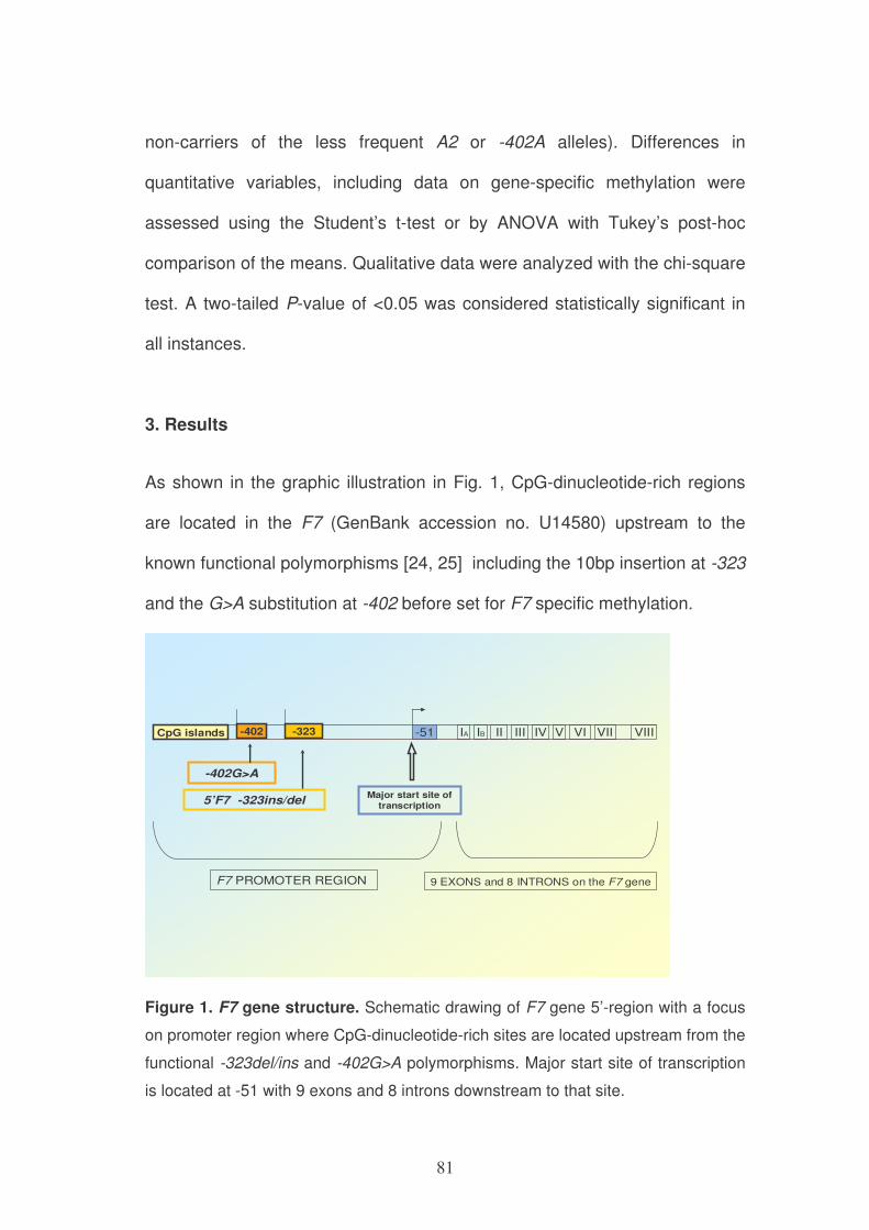
81
non-carriers of the less frequent A2 or -402A alleles). Differences in
quantitative variables, including data on gene-specific methylation were
assessed using the Student’s t-test or by ANOVA with Tukey’s post-hoc
comparison of the means. Qualitative data were analyzed with the chi-square
test. A two-tailed P-value of <0.05 was considered statistically significant in
all instances.
3. Results
As shown in the graphic illustration in Fig. 1, CpG-dinucleotide-rich regions
are located in the F7 (GenBank accession no. U14580) upstream to the
known functional polymorphisms [24, 25] including the 10bp insertion at -323
and the G>A substitution at -402 before set for F7 specific methylation.
-323
5’F7 -323ins/delMajor start site of
transcription
F7 PROMOTER REGION
-51 IA IB II III IV V VI VII VIII
9 EXONS and 8 INTRONS on the F7 gene
CpG islands -402
-402G>A
Figure 1. F7 gene structure. Schematic drawing of F7 gene 5’-region with a focus
on promoter region where CpG-dinucleotide-rich sites are located upstream from the
functional -323del/ins and -402G>A polymorphisms. Major start site of transcription
is located at -51 with 9 exons and 8 introns downstream to that site.
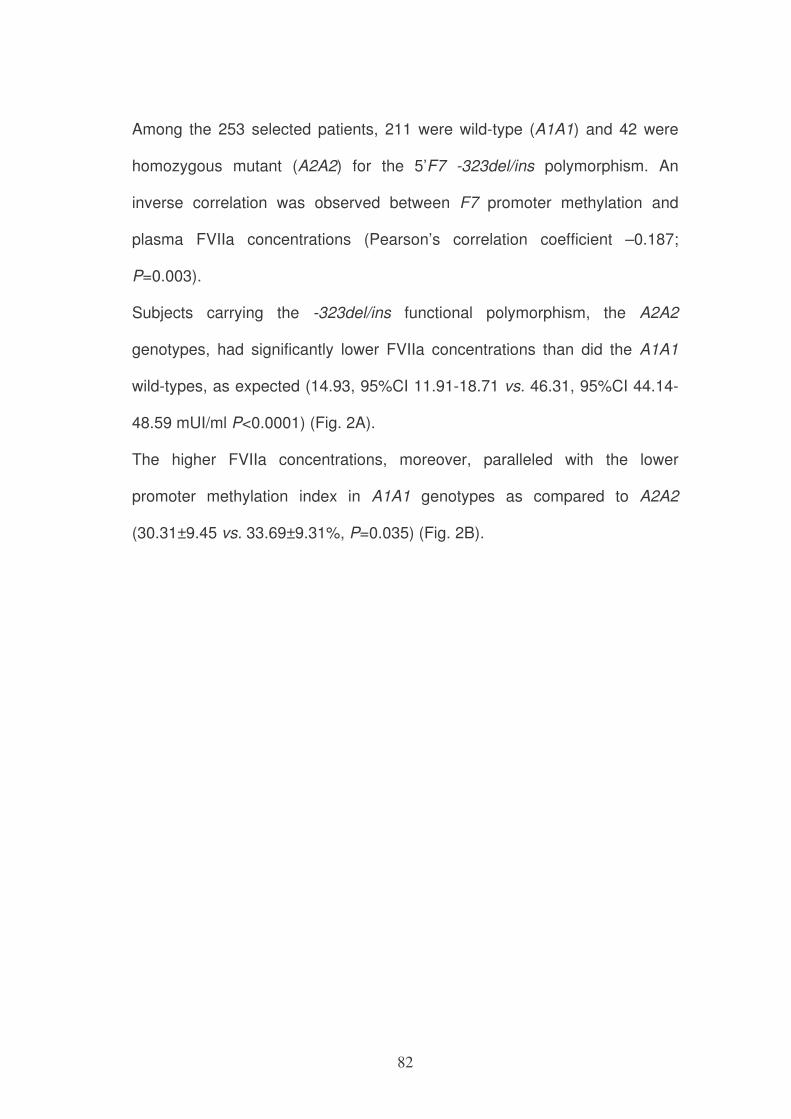
82
Among the 253 selected patients, 211 were wild-type (A1A1) and 42 were
homozygous mutant (A2A2) for the 5’F7 -323del/ins polymorphism. An
inverse correlation was observed between F7 promoter methylation and
plasma FVIIa concentrations (Pearson’s correlation coefficient –0.187;
P=0.003).
Subjects carrying the -323del/ins functional polymorphism, the A2A2
genotypes, had significantly lower FVIIa concentrations than did the A1A1
wild-types, as expected (14.93, 95%CI 11.91-18.71 vs. 46.31, 95%CI 44.14-
48.59 mUI/ml P<0.0001) (Fig. 2A).
The higher FVIIa concentrations, moreover, paralleled with the lower
promoter methylation index in A1A1 genotypes as compared to A2A2
(30.31±9.45 vs. 33.69±9.31%, P=0.035) (Fig. 2B).
83
B
A
F7
pro
mo
ter
meth
yla
tio
n i
nd
ex
(%)
Pla
sm
a F
VIIa (
mU
/ml)
0
20
40
60
A1A1 A2A2
0
5
10
15
20
25
30
35
40
45
50
A1A1 A2A2
**
N=211 N=42
N=211 N=42
*
Figure 2. F7 promoter methylation index according to -323del/ins
polymorphism genotypes. The methylation index at F7 gene promoter is shown
accordingly with -323del/ins polymorphism genotypes at 5’F7 site, A2A2 mutants
(n=42) and A21A1 wild types (n=211), respectively. (A) Subjects carrying the -
323del/ins functional polymorphism, the A2A2, show significantly lower
concentrations of gene end-product, protein FVIIa than do the A1A1 wild-types
(14.93, 95%CI 11.91-18.71 vs. 46.31, 95%CI 44.14-48.59 mUI/ml P<0.0001). As
shown in panel (B), the lower FVIIa concentrations in A2A2 paralleled with the
higher promoter methylation index as compared to A1A1 (33.69±9.31 vs.
30.31±9.45 %, P=0.035). Results are expressed as mean plus or minus SD for
continuous variables such as F7 promoter methylation index. Error bars in panel (B)
therefore indicate SD. Logarithmic transformation was performed on all the skewed
variables, as in the case of FVIIa; therefore, geometric means with 95% confidence
intervals are given for this variable. *P <0.0001; **P=0.035.
84
The effect of methylation at promoter site was diverse if considering carriers
or not carriers of the functional -323del/ins polymorphism so that only the
A1A1 wild-types accounted for the inverse correlation between methylation
and plasma FVIIa concentrations (Pearson correlation coefficient –0.176;
P=0.011); no such relationship was detected in A2A2 genotypes (P=0.548).
When considering carriers of the -402G>A variation at promoter site only the
-402GG wild-types showed an inverse correlation between promoter
methylation and plasma FVIIa concentrations (Pearson correlation coefficient
–0.201; P=0.012) while there was no such relationship in -402AA
homozygous mutants (P=0.30). No effect of the -402G>A polymorphism was
detected in terms of FVIIa concentrations (47.01 vs. 50.6 mUI/ml, P=0.52)
with no difference in promoter methylation index (30.74 vs. 23.71% for -
402GG vs. -402AA genotypes, P=0.054).
As shown in Fig. 3, when analysis was conducted evaluating haplotypes, a
higher F7 promoter methylation index was detected in carriers of the A2A2/-
402GG vs. both A1A1/-402GG and A1A1/-402AA (33.8 vs. 29.0%, P= 0.034
and 33.8 vs. 23.0%, P=0.016, respectively).
The higher methylation at F7 promoter region paralleled with the lower
concentration of FVIIa in the A2A2/-402GG compared to both A1A1/-402GG
and A1A1/-402AA (P=0.0001). No carriers of the rare A2A2/-402AA were
detected in the present population.
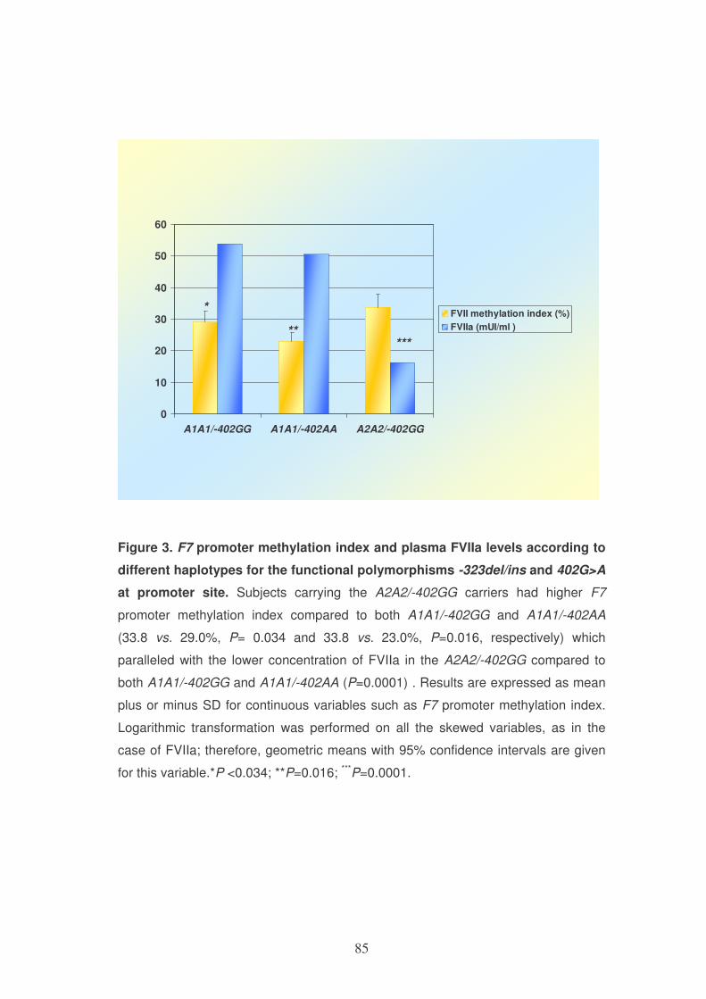
85
0
10
20
30
40
50
60
A1A1/-402GG A1A1/-402AA A2A2/-402GG
FVII methylation index (%)
FVIIa (mUI/ml )
*
*****
Figure 3. F7 promoter methylation index and plasma FVIIa levels according to
different haplotypes for the functional polymorphisms -323del/ins and 402G>A
at promoter site. Subjects carrying the A2A2/-402GG carriers had higher F7
promoter methylation index compared to both A1A1/-402GG and A1A1/-402AA
(33.8 vs. 29.0%, P= 0.034 and 33.8 vs. 23.0%, P=0.016, respectively) which
paralleled with the lower concentration of FVIIa in the A2A2/-402GG compared to
both A1A1/-402GG and A1A1/-402AA (P=0.0001) . Results are expressed as mean
plus or minus SD for continuous variables such as F7 promoter methylation index.
Logarithmic transformation was performed on all the skewed variables, as in the
case of FVIIa; therefore, geometric means with 95% confidence intervals are given
for this variable.*P <0.034; **P=0.016; ***P=0.0001.
86
Moreover CAD-free subjects showed a higher F7 methylation index
compared to CAD patients (32.12±9.80 vs.30.22±9.23, P=0.012).
In a genotype-based analysis only the A1A1 group accounted for such a
difference in F7 promoter methylation between CAD and CAD-free subjects,
so that the CAD-free group showed higher methylation associated with lower
FVIIa concentrations compared to CAD group, in a multiple logistic
regression model analysis adjusted for the major risk factors for CAD
(hypertension, diabetes, smoking habit, BMI, homocysteine) (31.65±10.29 vs.
29.60±8.93, P=0.011). The presence of the -323del/ins polymorphism was
instead responsible for the lower plasma FVIIa concentrations and reduced
CAD risk in A2A2 genotypes where no difference was detected in F7
promoter methylation (34.04±7.32 vs. 33.49±10.38, P=NS).
4. Discussion
The present study shows that epigenetic regulation by means of CpG sites
methylation is a fundamental phenomenon at F7 promoter region that
significantly affects plasma FVIIa concentrations. An inverse correlation was
identified between F7 promoter methylation index and plasma FVIIa
concentrations, therefore highlighting a transcriptional-silencing function of
methylation at F7 promoter site which affects the gene product.
The role of methylation appears significant in absence of known functional F7
gene variations at promoter site and is associated to coronary artery disease.
87
To the best of our knowledge, this is the first report which demonstrates the
function of methylation at F7 promoter site in modulating plasma FVIIa
concentrations and its association with coronary artery disease. The present
data show also a relationship between F7 methylation index with known
functional polymorphisms at gene promoter region, emphasizing the role of
this epigenetic feature of DNA as a main player for the gene product
regulation specifically for A1A1 genotypes.
Genotype variations at F7 promoter are indeed well-assessed determinants
of plasma FVIIa concentrations [5, 9, 10, 12]. The -323del/ins polymorphism
is associated with lower FVIIa concentrations in homozygous mutants [5, 9,
10] and the G>A substitution at position -402 confers instead a higher
transcriptional activity associated with increased plasma FVIIa concentrations
in vitro [12] and in selected populations [7].
As for epigenetic marks, not only is the role of methylation largely unexplored
for atherothrombosis-related genes but the relationship between promoter
methylation and the simultaneous presence of gene variations is also mostly
unknown.
Coagulation F7 promoter site polymorphisms have a definite functional effect
for the protein end-product concentrations [5, 9, 10, 12]. Therefore, the study
of this gene appears of peculiar interest to evaluate the role of methylation in
the case of conserved gene sequence at the promoter site.
88
DNA methylation is the major epigenetic feature in mammalian DNA [14].
This epigenetic phenomenon has been mainly studied in cancer disease,
thus far. Hypermethylation of CpG islands at promoter regions is generally
associated with gene transcriptional silencing [18, 19, 27, 28] and represents,
therefore, an unconventional mechanism through which tumor suppressor
genes are inactivated without the presence of sequence anomalies such as
mutations or allele deletions [19, 28]. Hypomethylation of CpG islands,
instead, is usually associated with gene activation and represents an
important mechanism by which proto-oncogenes are activated [28].
The epigenetic regulation of genes is still not entirely known for a large
number of genes. Furthermore, the role of epigenetic modifications in DNA
related to diseases other than cancer is yet mostly unexplored.
The present study shows that F7 methylation index is inversely related to
plasma FVIIa concentrations, therefore confirming earlier data referred to
cancer-related genes on the prominent transcriptional repression function of
methylation at promoter site [18, 19]. The silencing effect of methylation at F7
promoter region appears certainly significant for this crucial atherothrombosis
disease-related gene [2, 3].
Not only is the role of epigenetic mechanisms yet incompletely defined for
many genes but the inter-relationship between regulation through methylation
and the simultaneous presence of functional genetic variations at promoter
site is yet unclear.
89
The present results highlight a substantial role for methylation in absence of
the rare A2 allele, along with the substantiation of previous results on the
significant functional effect of the F7 10bp del/ins polymorphism at position -
323 in determining plasma FVIIa [4]. Subjects carrying the A2 allele showed,
as expected, lower FVIIa concentrations compared to A1A1. There was,
however, no difference in FVIIa concentrations between carriers and wild
types of the -402G>A polymorphism in the present setting of subjects,
therefore being these findings partly different from previous studies in which
a functional role of the rare -402A genotype was observed [7, 29]. However,
an inverse correlation between promoter methylation and FVIIa
concentrations was detected within the -402GG wild-type group, then
indicating once more a possible role for methylation in the absence of this
important functional polymorphic site at F7 promoter. The limited number of
subjects may be a reason for not reaching a statistically significant difference
in the methylation index for the -402GG versus the-402AA genotype group
(P=0.054).
The methylation score is appreciably lower in A1A1 wild-types compared to
the A2A2 genotypes and corresponds to the higher plasma FVIIa
concentrations in those not carriers of the rare functional A2 allele.
Furthermore, F7 promoter methylation index is inversely related to plasma
FVIIa concentrations only in A1A1 genotypes, highlighting a link between an
epigenetic marker and the biochemical phenotype in absence of the well-
defined genetic variation, namely the A2 allele. Modulation of FVIIa
90
concentrations by promoter methylation was apparently critical in carriers of
the A1A1 genotype.
Although, further studies are certainly needed to reveal the role of genetic-
epigenetic relationships at promoter site, through these findings one can
speculate that epigenetic regulation through methylation may exert a stronger
effect in absence of functionally significant genetic variations at promoter site
for the control of a gene product. Furthermore, for the unequivocal effect of
the rare A2 allele on regulating FVIIa levels, i.e. the gene end product, F7
may in fact represent a paradigmatic example of different roles of genetic
variations comparatively to epigenetic modifications in transcriptional
regulation at promoter region. A correlation between methylation and plasma
FVIIa concentrations was indeed observed only in A1A1 wild type subjects,
while DNA methylation was not associated with A2 allele carriership which
may be related to the localization of -323del/ins at the start of translation
region which may also give reason for the minor effect of the genetic-
epigenetic relationship observed according to the presence of the -402G>A
genotype.
It is, in truth, interesting to point out that the higher F7 promoter methylation
index corresponding to lower FVIIa concentrations, was higher in CAD-free
compared to CAD subjects and related to lower risk in A1A1 subjects while
no effect in terms of CAD risk was related to methylation in A2A2 genotypes.
Our clear definitions of phenotypes should have reduced the chance of
spurious results, a problem inherent in studies of allelic association [30].
91
This observation may allow, therefore, for considering F7 promoter
methylation in PBMC DNA as a possible novel protective marker for coronary
disease risk in a subgroup of patients not-carriers of the functional -
323del/ins polymorphism. Because epigenetic mechanisms, differently from
genetic aberrations, are potentially reversible, the observations of the present
study are of possible high interest for opening up new insights for preventive
strategies particularly in complex diseases such as CAD where several
factors are into place.
A major strength of our study is its fairly large number of subjects for the
evaluation of promoter methylation and its relationship with CAD disease in a
case-control clinical study. The method used in this study allowed to detect
significant differences and appeared the most suitable approach for the
rather large number of cases and controls. The measurement of FVIIa in
plasma is a highly accurate index of the gene end-product, as previously
shown, for its correlation with genotype markers and CAD risk [4].
One possible limitation is to be seen in the use of PBMC DNA for methylation
analysis, although the significant association between methylation index and
FVIIa concentrations as well as CAD risk may emphasize a potential role of
methylation at F7 promoter site as a molecular biomarker related to CAD
disease in an easily accessible tissue DNA such as that from peripheral
blood mononuclear cells.
92
In conclusion, methylation at F7 promoter region is a key mechanism through
which the gene regulates FVII protein production via an epigenetic modality
that acts differently from that related to the presence of polymorphic variants
at the same site and is associated to modulation of coronary artery disease
risk. Additional studies are definitely warranted to better understand the
significance of the genetic-epigenetic cross-talks with effects on gene
transcription, although the present findings certainly outlines the essential
role of promoter methylation for gene transcriptional regulation of F7 gene
product, which appears to exert its prime function in the absence of functional
polymorphisms in the promoter region.
Considering that, unlike sequence variants, epigenetic features of DNA are
potentially reversible, this previously unknown mechanism of F7 gene
regulation at promoter site may represent a fundamental mechanism through
which atherothrombotic disease risk may be modified, therefore opening up a
fascinating prospective for future studies related to both pathogenesis and
prevention of such a high impact disease for public health issues.
References
[1] Furie B, Furie BC. Mechanisms of thrombus formation. New Engl J
Med 2008;359: 938-949.
[2] Meade TW, Mellows S, Brozovic M, Miller GJ, Chakrabarti RR, North
WR, Haines AP, Stirling Y, Imeson JD, Thompson SG. Haemostatic function
93
and ischaemic heart disease: principal results of the Northwick Park Heart
Study. Lancet 1986;2: 533-7.
[3] Heinrich J, Balleisen L, Schulte H, Assmann G, van de Loo J.
Fibrinogen and factor VII in the prediction of coronary risk. Results from the
PROCAM study in healthy men. Arterioscler Thromb 1994;14:54-9.
[4] Girelli D, Russo C, Ferraresi P, Olivieri O, Pinotti M, Friso S, Manzato
F, Mazzucco A, Bernardi F, Corrocher R. Polymorphisms in the factor VII
gene and the risk of myocardial infarction in patients with coronary artery
disease. N Engl J Med 2000;343:774-80.
[5] Marchetti G, Patracchini P, Papacchini M, Ferrati M, Bernardi F. A
polymorphism in the 5' region of coagulation factor VII gene (F7) caused by
an inserted decanucleotide. Hum Genet 1993;90:575-6.
[6] Bernardi F, Marchetti G, Pinotti M, Arcieri P, Baroncini C, Papacchini
M, Zepponi E, Ursicino N, Chiarotti F, Mariani G. Factor VII gene
polymorphisms contribute about one third of the factor VII level variation in
plasma. Arterioscler Thromb Vasc Biol 1996;16:72-6.
[7] Bozzini C, Girelli D, Bernardi F, Ferraresi P, Olivieri O, Pinotti M,
Martinelli N, Manzato F, Friso S, Villa G, Pizzolo F, Beltrame F, Corrocher R.
Influence of polymorphisms in the factor VII gene promoter on activated
factor VII levels and on the risk of myocardial infarction in advanced coronary
atherosclerosis. Thromb Haemost 2004;92:541-9.
[8] Iacoviello L, Di Castelnuovo A, De Knijff P, D'Orazio A, Amore C,
Arboretti R, Kluft C, Benedetta Donati M. Polymorphisms in the coagulation
94
factor VII gene and the risk of myocardial infarction. N Engl J Med
1998;338:79-85.
[9] Green F, Kelleher C, Wilkes H, Temple A, Meade T, Humphries S. A
common genetic polymorphism associated with lower coagulation factor VII
levels in healthy individuals. Arterioscler Thromb 1991;11:540-6.
[10] Bernardi F, Arcieri P, Bertina RM, Chiarotti F, Corral J, Pinotti M,
Prydz H, Samama M, Sandset PM, Strom R, Garcia VV, Mariani G.
Contribution of factor VII genotype to activated FVII levels. Differences in
genotype frequencies between northern and southern European populations.
Arterioscler Thromb Vasc Biol 1997;17:2548-53.
[11] Pollak ES, Hung HL, Godin W, Overton GC, High KA. Functional
characterization of the human factor VII 5'-flanking region. J Biol Chem
1996;271:1738-47.
[12] van 't Hooft FM, Silveira A, Tornvall P, Iliadou A, Ehrenborg E,
Eriksson P, Hamsten A. Two common functional polymorphisms in the
promoter region of the coagulation factor VII gene determining plasma factor
VII activity and mass concentration. Blood 1999;93:3432-41.
[13] Jaenisch R, Bird A. Epigenetic regulation of gene expression: how the
genome integrates intrinsic and environmental signals. Nat Genet 2003;33
Suppl: 245-54.
[14] Robertson KD, Wolffe AP. DNA methylation in health and disease. Nat
Rev Genet 2000;1:11-9.
[15] Bernstein BE, Meissner A, Lander ES. The mammalian epigenome.
Cell 2007;128: 669-81.

95
[16] Esteller M. Epigenetics in cancer. N Engl J Med 2008;358:1148-59.
[17] Jones PA, Baylin SB. The epigenomics of cancer. Cell 2007;128:683-
92.
[18] Jones PA. The DNA methylation paradox. Trends Genet 1999;15:34-
7.
[19] Herman JG, Baylin SB. Gene silencing in cancer in association with
promoter hypermethylation. N Engl J Med 2003;349:2042-54.
[20] Friso S, Pizzolo F, Choi SW, Guarini P, Castagna A, Ravagnani V,
Carletto A, Pattini P, Corrocher R, Olivieri O. Epigenetic control of 11 beta-
hydroxysteroid dehydrogenase 2 gene promoter is related to human
hypertension. Atherosclerosis 2008;199:323-7.
[21] Wilcox JN, Smith KM, Schwartz SM, Gordon D. Localization of tissue
factor in the normal vessel wall and in the atherosclerotic plaque. Proc Natl
Acad Sci U S A 1989;86:2839-43.
[22] Campo G, Valgimigli M, Ferraresi P, Malagutti P, Baroni M, Arcozzi C,
Gemmati D, Percoco G, Parrinello G, Ferrari R, Bernardi F. Tissue factor and
coagulation factor VII levels during acute myocardial infarction: association
with genotype and adverse events. Arterioscler Thromb Vasc Biol
2006;26:2800-6.
[23] Girelli D, Friso S, Trabetti E, Olivieri O, Russo C, Pessotto R, Faccini
G, Pignatti PF, Mazzucco A, Corrocher R. Methylenetetrahydrofolate
reductase C677T mutation, plasma homocysteine, and folate in subjects from
northern Italy with or without angiographically documented severe coronary
96
atherosclerotic disease: evidence for an important genetic-environmental
interaction. Blood 1998;91:4158-63.
[24] O'Hara PJ, Grant FJ, Haldeman BA, Gray CL, Insley MY, Hagen FS,
Murray MJ. Nucleotide sequence of the gene coding for human factor VII, a
vitamin K-dependent protein participating in blood coagulation. Proc Natl
Acad Sci U S A 1987;84:5158-62.
[25] Erdmann D, Heim J. Orphan nuclear receptor HNF-4 binds to the
human coagulation factor VII promoter. J Biol Chem 1995;270: 22988-96.
[26] Herman JG, Graff JR, Myohanen S, Nelkin BD, Baylin SB.
Methylation-specific PCR: a novel PCR assay for methylation status of CpG
islands. Proc Natl Acad Sci U S A 1996;93:9821-6.
[27] Wolffe AP, Matzke MA. Epigenetics: regulation through repression.
Science 1999;286:481-6.
[28] Ehrlich M. DNA methylation in cancer: too much, but also too little.
Oncogene 2002;21:5400-13.
[29] Di Bitondo R, Hall AJ, Peake IR, Iacoviello L, Winship PR. Oestrogenic
repression of human coagulation factor VII expression mediated through an
oestrogen response element sequence motif in the promoter region. Hum
Mol Genet 2002;11: 723-31.
[30] Lander ES, Schork NJ. Genetic dissection of complex traits. Science
1994;265: 2037-48.
97
CHAPTER 4
ONE-CARBON METABOLITES AND ROLE OF B VITAMINS
BETWEEN PHYSIOLOGY AND PATHOLOGY AND ITS
CORRELATION WITH EPIGENETIC MECHANISMS
Among the essential cofactors that interact with the enzymes of the folate
cycle, there is vitamin B6. The relationship between vitamin B6, inflammation
and cardiovascular disease still remains a debated topic and there are not
recent works that allow to point out the scientific research in this field.
For this reason we have been concerned to review the available literature
focusing on the relationship between nutrition and inflammation in
cardiovascular disease.
PLASMA VITAMIN B6: A CHALLENGING LINK BETWEEN NUTRITION
AND INFLAMMATION IN CARDIOVASCULAR DISEASE
Abstract
The objective of the present review is to highlight the relationship between
low vitamin B6 status and cardiovascular disease (CVD) through its link with
inflammation.
While overt vitamin B6 deficiency is uncommon in clinical practice, increasing
evidence suggests that mild vitamin B6 deficiency is rather frequent in a
98
consistent portion of the population and is related to an increased risk of
inflammation-related diseases.
Ample evidence substantiates the theory of atherosclerosis as an
inflammatory disease, and low plasma vitamin B6 concentrations have been
related to increased cardiovascular disease risk. Several studies have also
shown that low vitamin B6 status is associated with rheumatoid arthritis and
chronic inflammatory bowel diseases, both of which hold an underlying
chronic inflammatory condition. Furthermore, the inverse association
observed between inflammation markers and vitamin B6 supports the notion
that inflammation may represent the common link between low vitamin B6
status and CVD risk. In addition to the epidemiological evidence, there are a
number of cell culture and animal studies that suggest several possible
mechanisms relating impaired vitamin B6 status with chronic inflammation.
A mild vitamin B6 deficiency characterizes, in most cases, a subclinical at-risk
condition in inflammatory-linked diseases which should be addressed for an
appropriate individually-tailored nutritional preventive or therapeutic strategy.
Introduction
The two major derivates of vitamin B6 are the coenzyme species
pyridoxamine 5’-phosphate and pyridoxal 5’-phosphate (PLP)(1). PLP is both
the major coenzyme form of vitamin B6 in plasma and the metabolically
active coenzyme produced by the phosphorylation of the pyridoxal compound
following the oxidation of other vitamin B6 vitamers in the liver. Plasma PLP is
99
considered the most sensitive indicator available of tissue vitamin B6 status
because it reflects liver PLP concentrations and stores(2-6), although other
measures of vitamin B6 such as erythrocyte PLP, total plasma B6, and urinary
excretion of 4-pyridoxic acid are also useful marker of human vitamin B6
status even if less commonly available in clinical practice (7,8). Overt vitamin
B6 deficiency is a rare condition that is mainly defined by the appearance of
specific clinical signs or symptoms whereas also a suboptimal vitamin B6
status may influence the risk of development of several diseases. It has been
previously suggested that vitamin B6 deficiency corresponds to plasma PLP
concentrations below 20 nmol/L while a borderline, marginal impairment of
vitamin B6 status may be observed for plasma PLP level < 30 nmol/L(7, 9),
although a clear-cut definition for deficiency of this nutrient seems a notion to
be considered in evolution for novel at-risk pathologic conditions due to a
grade of impairment that fails to meet the magnitude of deficit classically
defined as a deficiency state(8, 9).
Several authors have indicated a role for low plasma PLP concentrations in a
number of pathological conditions. This suggests that, not only does overt
vitamin B6 deficiency increase the risk of certain chronic diseases, but even
mild vitamin B6 deficiency could be associated with increased risk of certain
chronic diseases(10-12).
It has been reported that low plasma PLP concentrations are inversely
related to C-reactive protein (CRP), a major marker of inflammation and a
risk factor for atherosclerotic disease(13-14). A number of studies have shown
100
that low plasma vitamin B6 levels are associated with typical inflammatory
chronic diseases, such as rheumatoid arthritis (RA)(15) and inflammatory
bowel diseases (IBD)(16), and are inversely related to markers of
inflammation(17,18). These studies suggest that impaired vitamin B6 status in
RA patients is not solely caused by either lower intake, malabsorption or
excessive catabolism of the vitamin, but is rather the result of metabolic
mechanisms caused by the inflammatory process(18,19). Among other possible
hypotheses, it has also been suggested that the underlying inflammatory
condition itself may reduce circulating and hepatic concentrations of PLP by
mobilizing PLP from the liver and peripheral tissues to the sites of
inflammation(18,19). Chiang et al. observed that plasma PLP concentration in
rats with adjuvant arthritis was about 53% of the controls at acme of
inflammation and related to the content of PLP in the liver, thus suggesting
that the lower circulating PLP levels observed in rheumatoid arthritis could be
a sign of a decrease in hepatic PLP pools, and that plasma PLP is a valuable
indicator of liver B6 status during inflammation(18). Further data showed that
PLP in plasma seems a more relevant metabolic marker than PLP in the
erythrocytes during inflammation(19) as also confirmed by the observation that
plasma, but not erythrocyte PLP concentrations, inversely relates to both
clinical and biochemical indices of disease activity and severity in patients
affected by RA(19).
For decades, several studies have demonstrated that patients with RA or
other inflammatory diseases have a higher risk of developing premature
coronary artery disease (CAD)(20-21) and traditional CVD risk factors did not,
101
by themselves, clarify the increased prevalence of CAD in such patients(22).
Through studying the relationship between RA, inflammation, and CAD,
some CVD prevention strategies in RA patients have been proposed,
including anti-inflammatory therapy with cyclooxygenase-2-specific inhibitors
and statins(23) as well as pyridoxine hydrochloride supplementation(24).
Several studies, moreover, confirmed the theory of atherosclerosis as an
inflammatory disease, thus demonstrating that the link between
atherosclerosis, RA and other inflammatory chronic diseases is through
inflammatory processes. Inflammation may therefore be considered a major
pathogenic mediator underlying atherosclerosis and its complications(25-26).
The chronic inflammation in atherosclerotic-related disease, such as CAD
and stroke, and its major clinical complications, namely myocardial infarction
(MI), have also been associated with low plasma vitamin B6
concentrations(11,27-28, 29).
The relationship between atherosclerosis and vitamin B6-linked inflammatory
mechanisms is indeed of great interest. Does low plasma PLP indicate the
sole effect of inflammation? Or could it be a cofactor that promotes the
development of inflammation, potentially contributing to a sustained chronic
inflammatory response? The answer to these questions may open way to
novel and interesting approaches for dietary prevention and therapy.
102
Plasma vitamin B6 and coronary artery disease, myocardial
infarction and ischemic stroke
Vitamin B6 and coronary artery disease
Table 1 summarizes the human studies which addressed the relationship
between vitamin B6 status and coronary artery disease. Low plasma vitamin
B6 concentrations are not only associated with an increased risk for
atherosclerotic diseases(30) and more specifically with higher CAD
incidence(27,31,32) but the higher risk also appears to be independent of other
recognised risk factors for CAD, including homocysteine(27). Moreover,
adequate vitamin B6 levels emerged as a protective factor for CAD(31). In a
prospective design study, Folsom et al.,(33) demonstrated the risk of
developing coronary heart disease within five years significantly decreased
with consensual increasing concentrations of the plasma PLP33. The
association between low PLP and CAD was, however, not confirmed in all
studies(34,35) (Table 1).
In addition to several epidemiological studies demonstrating a role for low
PLP as an independent risk factor for CAD, some studies have shown that
low PLP may be linked to CAD through inflammation. In an observational
study conducted in an Italian population, a cohort of patients with
angiography-defined, severe, multivessel CAD were compared to CAD-free
subjects to evaluate the relationship between CAD risk and both plasma PLP
concentrations and major markers of the acute-phase reaction(11). An inverse
103
relationship between plasma PLP and both hs-CRP and fibrinogen was
found, confirming previous findings of an inverse association between
inflammatory markers and vitamin B6(13). The prevalence of low PLP (defined
as PLP concentrations below 36.3 nmol/L, which was the median value in
control group) was significantly higher among CAD patients compared to
controls(11). The association between low plasma PLP concentrations and
increased CAD risk was also independent of the major classical risk factors
for atherosclerosis, including total plasma homocysteine. This association
continued to be significant even when hs-CRP and fibrinogen were included
in the multiple logistic regression models. The strength of this independent
relationship was confirmed even after adjustments for a number of other
conditions known to be associated with low concentrations of plasma PLP,
including aging, smoking status, and impaired renal function. Results from
this study also showed that the combined presence of low PLP along with
other major risk factors for CAD, such as higher hs-CRP and elevated
LDL/HDL ratio, further increased the risk for CAD in a graded manner(11).
These plasma PLP levels could be described as a mild PLP impairment
compared to previous studies that had defined PLP impairment by
concentrations as low as 20 nmol/L(27), suggesting that even a moderate
impairment of this vitamin is sufficient to confer a higher risk for CAD. In a
case-control study performed by Lin and Colleagues(36), low PLP
concentrations (defined as PLP below 30 nmol/L) were associated with a
significantly increased risk of CAD for angiography-documented patients
compared to healthy controls, even after adjustments for hs-CRP(36). In a
104
case-control study evaluating whether plasma PLP exerts an independent or
a synergic effect with inflammation in elevating the risk of CAD, Cheng and
colleagues confirmed that low PLP levels (below 20 nmol/L) are
independently associated with higher CAD risk. The inverse relationship
between PLP and hs-CRP was observed only within the control group(37).
Vitamin B6 and myocardial infarction
A specific association between low plasma PLP and incidence of MI, the
main thrombotic complication of CAD, is supported by early reports(38-40-41)
and by subsequent case-control and prospective studies(42,43,44,45), as shown
in Table 2. Moreover, an association between lower dietary vitamin B6 intake
and higher risk of MI has also been suggested(42).
A number of studies observed an inverse association between plasma
concentrations of vitamin B6 and risk of MI, independent of known CAD risk
factors(44,46), though not all studies confirmed this independent association(47).
In a study evaluating heart transplant recipient, it was observed that about
21% of the patients showed PLP lower concentrations and that only 9% of
those with lower plasma PLP levels did not have CVD complications(48).
Subjects in the highest quintile of PLP had a significantly reduced risk of MI
but adjustment for either low-grade inflammation or smoking diminished this
association(47).
With regard to the relationship between vitamin B6 and major markers of
inflammation, results from early studies have shown that low plasma PLP
105
levels are related to acute phase reactants in patients with MI who are in the
initial phase of the disease(41, 49).
Vitamin B6 and stroke
In 1995, Selhub et al. described a relationship between lower vitamin B6
concentrations and extracranial carotid artery stenosis through its role in
homocysteine metabolism(28). Moreover, recent case-control studies have
described a possible relationship between low plasma vitamin B6
concentrations and the onset of cerebrovascular disease, specifically
ischemic stroke and transient ischemic attack(29,50). A strong association
between stroke/TIA and low PLP (defined as levels less than 20 nmol/L) was
independent of other well-established vascular risk factors, including total
plasma homocysteine concentrations. Furthermore, results from that study
identified a possible protective effect for higher PLP concentrations(29).
In the context of the Health Professional Follow-up Study, He et al.,
evaluated dietary intakes of vitamin B6 together with the intake of other B
vitamins by a semiquantitative food frequency questionnaire(51) in relation to
risk of ischemic and hemorrhagic stroke(52). Unlike data related to other B
vitamins, the intake of vitamin B6 was not associated with risk of ischemic
stroke after adjustments for lifestyle and dietary factors(52).
Data from the Spanish National Nutrition Survey, which was designed to
assess the association between dietary vitamin B6 intake and CVD mortality,
106
also showed no association between vitamin B6 impairment and
cardiovascular mortality(53).
Vitamin B6 and peripheral artery disease
Reports on the relationship between vitamin B6 and peripheral artery disease
are few(53). Wilmink et al., reported that daily vitamin B6 intake is lower in
patients with peripheral artery disease, as defined by an ankle-brachial
pressure index below 0.9, and appears as an independent predictor of
peripheral artery occlusive disease. An increase in daily vitamin B6 intake by
one standard deviation significantly decreased the risk of peripheral artery
disease by 29%(53).
Plasma vitamin B6 supplementation, inflammation and
cardiovascular disease prevention
An early observation of patients given vitamin B6 for inflammatory diseases
or degenerative diseases found that subjects taking vitamin supplementation
had a lower risk of developing MI compared with patients who had not taken
vitamin B6(55). Table 3 summarizes the main studies in which B vitamin
supplementation, including supplementation with B6 vitamin, have been
performed. In the Nurses' Health Study, among women with no prior history
107
of CAD, users of multivitamins containing folate and vitamin B6 had a
reduced risk of CAD(56).
Very few human studies have been performed to evaluate the modifications
of major markers of inflammation during supplementation with B vitamins,
including vitamin B6, despite the association with either folate and/or vitamin
B12(57,58). Furthermore, results from those studies showed that CRP and pro-
inflammatory interleukins levels are unchanged after vitamin
supplementation(57,58). Antioxidant activity of vitamin B6 supplementation has
been observed in a study performed on rats, but the exact mechanism is
unclear(59).
Several large, prospective trials have been conducted in recent years with
the principal aim of studying the effects of lowering serum homocysteine
concentrations with the use of B vitamins, including vitamin B6 on
cardiovascular events(60-61).
Overall, the most compelling data from vitamin supplementation studies have
demonstrated that vitamin B6 is not effective for preventing the recurrence of
cardiovascular events, including CAD, peripheral vascular disease and
stroke(54,62-63).
Only a few small trials, performed with renal transplant patients that have
hyperhomocysteinemia and subjects at risk for cerebral ischemia have
demonstrated the effectiveness of vitamin B6 supplementation with folate and
vitamin B12 on carotid artery intima-media thickness progression(64,65). This
108
marker for subclinical atherosclerosis was also evaluated in another recent
double-blind, placebo-controlled, randomized clinical trial(66). Vitamin
supplementation significantly reduced subclinical atherosclerosis progression
only in subjects at low risk for cardiovascular disease with total plasma
homocysteine concentrations equal to or above 9.1 µmol/L(66). Other trials
evaluated CAD patients for the effects of B vitamin supplementation, though
the interpretation of the results was ambiguous. Supplementation with
vitamin B6, folate and vitamin B12 after coronary angioplasty decreased the
rate of restenosis and the need for revascularization(67), while
supplementation after coronary stenting increased the risk of in-stent
restenosis and the need for target-vessel revascularization(68).
Despite the advantage of large randomized intervention studies, wisdom from
studies of cancer chemoprevention with folate clearly suggest that two critical
factors should be taken into account in the above mentioned trials: time and
dose of B vitamin supplementation. Nutritional intervention is indeed
considered to be a two-edged sword, where a beneficial effect may be
observed with nutritional support in the prevention phase and a disease
aggravating effect may be observed after the onset of illness, with nutritional
support actually fueling the disease process. This observation may certainly
apply to folate supplementation, which can effectively prevent the onset or
progression of disease in the early phase, while it may accelerate the
progression of disease in the late phase. It can therefore be speculated that
109
continuous supplementation with a single non-physiologic form of vitamin
supplements might contribute to unexpected or even harmful outcomes, even
in the at-risk condition of lower vitamin B6 status. Furthermore, the major
endpoints of these studies were those of evaluating the effect of lowering
homocysteine on the recurrence of established disease, mainly by the
simultaneous use of various B-vitamins that have many functions other than
lowering total plasma homocysteine. Very little information is available,
however, regarding the effects of B vitamin supplementation on inflammatory
markers and more so for what pertain to the dose and timing issue as well as
possible harmful effect for a possible primary or secondary preventive
approach with vitamin B6.
Even if there is consistently a lack of benefit in secondary prevention of
cardiovascular disease with B vitamins supplementation, with or without
vitamin B6, (WENBIT(69), WAFACS(62), VISP(63), NORVIT(61), HOPE-2(70)), it
should be also considered that all the above-mentioned studies are quite
diverse from one another in terms of the time period of supplementation. In
fact, one may raise the point that also the time period of supplementation is
extremely critical to reach positive outcomes or avoid possible harmful effects
in terms of the rate of CVD events(71). However, despite the apparently clear
outcomes(71), it could be argued that a potential benefit that modifies
secondary prevention outcomes may not be observed over a period of
moderate duration (between two to five years for the NORVIT, HOPE-2 and
VISP studies or of about seven years for the WAFACS study).
110
Furthermore, the negative results of vitamin supplementation trials, including
vitamin B6 use, does not preclude the possibility of a protective effect in
primary prevention. It could be difficult, however, to demonstrate that vitamin
B supplementation is ineffective in patients who have had a clinical vascular
event, while effective in those without a clinical event or with subclinical
atherosclerosis. Clinical trials for primary prevention may require a longer
duration and larger population studies in order to answer the key question on
whether vitamin B6 supplementation is effective in preventing CAD before the
first vascular event or in younger life.
Studies are needed to find the specific time and optimal dose of vitamin B6 in
order to maximize efficacy, minimize adverse effects, and identify targets for
vitamin B6 interventions on the basis of genetic susceptibility and
environmental factors. A better understanding of the mechanisms underlying
the relationship between cardiovascular disease and vitamin B6 may indeed
be extremely helpful in designing the most accurate preventive strategies.
Inflammation and vitamin B6-related atherogenesis
PLP functions as a coenzyme in more than one-hundred reactions that are
involved in the metabolic pathways of neurotransmitters as well as the
metabolism of amino acids, lipids, and carbohydrates(72). PLP also takes part
in other significant pathways related to immune function(73), thrombosis(74-75),
111
and inflammation(17), all of which are crucial mechanisms in every stage of
the atherosclerotic process.
Furthermore, PLP is implicated in the synthesis and repair of both nucleic
acids and proteins. Low vitamin B6 concentrations could thus reflect an
increased consumption of PLP in the accelerated synthesis of cytokines(76)
and in the activation and proliferation of lymphocytes, both of which are key
events in the inflammatory process(77,78).
Considering the epidemiological evidence of a role of vitamin B6 in
inflammatory-related diseases and the observed relationship with
inflammatory markers(11,16,18,79), it is plausible that vitamin B6 plays a role in
CVD pathogenesis through mechanisms linked to inflammation.
Therefore, with the knowledge that systemic acute-phase markers are solid
and independent risk factors for CAD(80, 81), that inflammation exerts an
essential role in all stages of the atherosclerotic process(82), and the
possibility that vitamin B6 has a role in inflammatory processes, several
mechanisms were proposed linking low vitamin B6 and CVD using cell culture
studies, animal studies(72), and clinical trials.
Animal studies reported that inflammation reduces circulating and hepatic
concentrations of vitamin B6. Plasma PLP is considered a sensitive indicator
of tissue vitamin B6 status(3-7, 15, 18). As shown in Fig. 1, it is thus possible that,
in patients in an inflammatory state, PLP is mobilized from the liver and
peripheral tissues to the sites of inflammation(19). Plasma PLP levels are
112
known to be inversely related to tumour necrosis factor alpha production and
other inflammatory cytokines in rheumatoid arthritis (17). It has been also
reported that low plasma PLP concentrations are inversely related to both
plasma fibrinogen(83) and CRP (11, 13) with a robust and independent
association of other major biomarkers related to vitamin B6 metabolism(11).
Moreover, inflammatory status increases oxidant stress, which results from
an imbalance between oxidant production and antioxidant defenses (Fig.1).
All these conditions characterized by the increase of inflammatory cytokines,
fibrinogen, CRP and superoxide radicals may induce the consumption of
vitamin B6 with a consequent reduction of PLP plasma levels and its
antioxidant effect and, at the same time, they may favour a thrombogenic
effect thus triggering impairment of endothelial function a key event in the
pathogenesis of atherosclerotic processes (Fig. 1)(84). PLP has been also
described to have an inhibitory effect on endothelial cell proliferation (85, 86).
The persistence of a chronic inflammatory condition may result in the
depletion of vitamin B6, which might then contribute to a sustained chronic
inflammatory response (Fig. 1).
Most of the evidence demonstrates a potential role for vitamin B6 in
inflammatory processes where the low vitamin B6 status in inflammation-
related illnesses appears to result not from lower intake or excessive
catabolism of PLP but from the inflammatory process underlying the disease
itself(79).
113
Although vitamin B6 represents an important coenzyme in the metabolism of
homocysteine, a recognized risk factor for thrombosis, the role of PLP in
atherosclerosis is only partly related to its function in the one-carbon
pathway. Several studies thus supported a role for vitamin B6 in the risk of
CVD(27, 31, 42, 46) and stroke(29), independently of homocysteine and other risk
factors. A hypothesis is that vitamin B6 could be directly implicated as a
cofactor in an anti-inflammatory mechanism where the utilization of the
vitamin results in its consumption, and the consequent low vitamin B6
concentrations could support and amplify the inflammatory process thereby
leading to chronic progression of inflammatory disease. Indeed, a mild
deficiency of vitamin B6 may be associated with an increased risk, not only of
atherosclerosis, but also of other chronic inflammatory diseases such as
rheumatoid arthritis (RA)(18,87), and inflammatory bowel diseases (IBD)(16). A
number of studies highlight the relationship between PLP and inflammation,
showing an inverse association between vitamin B6 and major markers of
inflammation, including plasma fibrinogen concentration(83), erythrocyte
sedimentation rate (ESR), and CRP(13). In the population-based Framingham
Heart Study cohort, the association between PLP and CRP was strong and
independent of other major biomarkers related to vitamin B6 metabolism(13),
supporting a possible role for plasma PLP in inflammatory processes.
Some reports did not support this observation(88), as the study by Folsom et
al., conducted among healthy middle-aged adults in the Atherosclerosis Risk
in Communities (ARIC) Study, which did not find an inverse association
between CRP and PLP. However, the authors did observe that lower plasma
114
PLP and dietary vitamin B6 were associated with a higher white blood cell
count, though this association was not found with the use of vitamin
supplements(88). It should be also taken into account that some diversity in
the observations reported by the studies may be due to the different methods
utilized to measure plasma PLP, although the methods used by most studies
are considered highly reliable for the assessment of vitamin B6 status(11, 15, 88-
90). The majority of studies, however, confirmed the inverse correlation
between PLP and major markers of inflammation in various inflammation-
related diseases. In subjects affected by RA(19,24), PLP was associated with
ESR, CRP levels, and other markers of disease activity and severity,
suggesting that impaired vitamin B6 status is a result of inflammation. It also
appeared that the relationship between lower vitamin B6 and increased
inflammation in these RA patients could be tissue-specific(24).
Plasma PLP concentrations are also altered in subjects who have acute
diseases with an evident underlying inflammatory condition. Vasilaki et al,
observed that, in patients admitted to the Intensive Therapy Unit with high
concentrations of CRP, PLP and intracellular pyridoxal levels were
significantly lower in those critically ill patients than in the group of subjects
taken as controls(91).
Further support for the hypothesis of a link between vitamin B6 and
inflammation can be found in an analysis of data from a large population-
based survey from participants in the 2003–2004 NHANES. Results showed
115
that higher vitamin B6 intakes are protective against inflammation, as
indicated by hs-CRP concentrations. Moreover, the level of vitamin B6 intake
that was associated with maximum protection against vitamin B6 inadequacy
was elevated in the presence of inflammation compared to its absence(79).
A strong inverse association between vitamin B6 status, as measured by
plasma PLP concentration, and the inflammatory marker CRP was also
observed recently in a cohort of elderly Puerto Ricans living in
Massachusetts(92). In this study, chronic inflammatory conditions, such as
metabolic syndrome, diabetes, and obesity, were significantly associated with
lower plasma PLP. The patients affected by such diseases were also
significantly more likely to demonstrate vitamin B6 inadequacy. Furthermore,
lower PLP plasma concentrations were associated with oxidative stress, as
indicated by higher urinary concentrations of 8-hydroxydeoxyguanosine, a
marker of DNA damage and oxidative stress(92). Authors concluded that
vitamin B6 status may influence CAD risk through mechanisms that link
vitamin B6 to inflammatory processes rather than mechanisms related to the
role of vitamin B6 in homocysteine metabolism(92).
116
Conclusions
Several studies demonstrated an association between mild vitamin B6
deficiency with inflammation-related diseases, including CVD(11,16,18,79), by
highlighting an inverse relationship between vitamin B6 and inflammatory
markers(11,19,79). In a consistent number of studies, this association between
impaired vitamin B6 status and higher risk of CVD is independent of other
major traditional atherosclerosis risk factors(13,27,37,46,44) and is inversely
related to major markers of inflammation(11). This evidence suggests a link
between impaired vitamin B6 and CVD through inflammation. Because
vitamin B6 is involved in a large number of physiological reactions, it could be
essential to design appropriate studies to define the exact mechanisms
underlying the inter-relationships among suboptimal vitamin B6 status, as
defined by both plasma and tissue PLP concentrations, and biochemical-
molecular alterations leading to development of inflammation-related
diseases. A research priority may be that of investigating the kinetics and
regulation of B6 vitamers and enzymes in different body compartments
during inflammatory processes.
Mild vitamin B6 deficiency is not a rare occurrence in population-based
studies(12). This issue, therefore, deserves further investigation, especially in
terms of prevention strategies, for the purpose of promoting specific public
health policies. Current clinical trials indicate that vitamin B6 supplementation
seems not to be effective for the prevention of recurrence of CVD, although
remains still to be discussed the appropriate dosage and timing for possible
117
beneficial effects through the use of vitamin supplements. Other crucial
issues pertain also to the evaluation of the actual effect of supplementation
with vitamin B6 in synthetic form, as well as to whether such approach may
be as effective as an adequate dietary vitamin B6 intake. The question of
whether supplementation with vitamin B6 may be useful for primary
prevention of CVD is yet another key issue to be defined. The importance of
considering vitamin B6 status in relation to the risk for CVD may nevertheless
open new insights for the potential identification of innovative preventive and
therapeutic strategies. In order to help tailor an adequate nutritional approach
on an individual basis, both dose and timing as well as possible harmful
effect of vitamin B6 supplementation should be carefully considered in the
design of future ad hoc clinical trials that are aimed at identifying appropriate
vitamin B6 supplementation.
The notion of a definite deficient status for this nutrient is, as a consequence,
a concept to be considered in fieri because the risk of certain diseases
seems to result from degrees of vitamin B6 reduction that fall short of the
extent of depletion that has been classically defined as a deficiency state.
118
References
1. Shane B (2000) Folate, Vitamin B12, and Vitamin B6. In: MH S, ed.
Biochemical and physiological aspects of human nutrition.
Philadelphia: WB Saunders:483-518.
2. Stickel F, Choi SW, Kim YI et al. (2000) Effect of chronic alcohol
consumption on total plasma homocysteine level in rats. Alcohol Clin
Exp Res 24, 259-264.
3. Parker TH, Marshall JP, 2nd, Roberts RK et al. (1979) Effect of acute
alcohol ingestion on plasma pyridoxal 5'-phosphate. Am J Clin Nutr
32, 1246-1252.
4. Lui A, Lumeng L, Aronoff GR et al. (1985) Relationship between body
store of vitamin B6 and plasma pyridoxal-P clearance: metabolic
balance studies in humans. J Lab Clin Med 106, 491-497.
5. Bode W, Mocking JA & van den Berg H (1991) Influence of age and
sex on vitamin B-6 vitamer distribution and on vitamin B-6
metabolizing enzymes in Wistar rats. J Nutr 121, 318-329.
6. Lumeng L, Lui A & Li TK (1980) Plasma content of B6 vitamers and its
relationship to hepatic vitamin B6 metabolism. J Clin Invest 66, 688-
695.
7. Leklem JE (1990) Vitamin B-6: a status report. J Nutr 120 Suppl.
11,1503-1507.
8. Mason JB (2003) Biomarkers of nutrient exposure and status in one-
carbon (methyl) metabolism. J Nutr 133 Suppl. 3, 941S-947S.
119
9. Food and Nutrition Board-Institute of Medicine. Dietary reference
intakes. Thiamin, riboflavin, niacin, vitamin B-6, folate, vitamin B-12,
pantothenic acid, biotin, and choline. . Washington, DC: National
Academy Press; 1998.
10. Pasceri V & Yeh ET (1999) A tale of two diseases: atherosclerosis and
rheumatoid arthritis. Circulation 100, 2124-2126.
11. Friso S, Girelli D, Martinelli N, et al. (2004) Low plasma vitamin B-6
concentrations and modulation of coronary artery disease risk. Am J
Clin Nutr 79, 992-998.
12. Fairfield KM & Fletcher RH (2002) Vitamins for chronic disease
prevention in adults: scientific review. JAMA 287, 3116-3126.
13. Friso S, Jacques PF, Wilson PW et al. (2001) Low circulating vitamin
B(6) is associated with elevation of the inflammation marker C-reactive
protein independently of plasma homocysteine levels. Circulation 103,
2788-2791.
14. Willerson JT & Ridker PM (2004) Inflammation as a cardiovascular
risk factor. Circulation 109 Suppl. 1, II2-10.
15. Chiang EP, Bagley PJ, Roubenoff R et al. (2003) Plasma pyridoxal 5'-
phosphate concentration is correlated with functional vitamin B-6
indices in patients with rheumatoid arthritis and marginal vitamin B-6
status. J Nutr 133, 1056-1059.
16. Saibeni S, Cattaneo M, Vecchi M, et al. (2003) Low vitamin B(6)
plasma levels, a risk factor for thrombosis, in inflammatory bowel
120
disease: role of inflammation and correlation with acute phase
reactants. Am J Gastroenterol 98, 112-117.
17. Roubenoff R, Roubenoff RA, Selhub J, et al. (1995) Abnormal vitamin
B6 status in rheumatoid cachexia. Association with spontaneous
tumor necrosis factor alpha production and markers of inflammation.
Arthritis Rheum 38, 105-109.
18. Chiang EP, Smith DE, Selhub J, et al. (2005) Inflammation causes
tissue-specific depletion of vitamin B6. Arthritis Res Ther 7, R1254-
1262.
19. Chiang EP, Bagley PJ, Selhub J et al. (2003) Abnormal vitamin B(6)
status is associated with severity of symptoms in patients with
rheumatoid arthritis. Am J Med 114, 283-287.
20. Monson RR & Hall AP (1976) Mortality among arthritics. J Chronic Dis
29, 459-467.
21. Mikuls TR & Saag KG (2001) Comorbidity in rheumatoid arthritis.
Rheum Dis Clin North Am 27, 283-303.
22. del Rincon ID, Williams K, Stern MP et al. (2001) High incidence of
cardiovascular events in a rheumatoid arthritis cohort not explained by
traditional cardiac risk factors. Arthritis Rheum 44, 2737-2745.
23. Snow MH & Mikuls TR (2005) Rheumatoid arthritis and cardiovascular
disease: the role of systemic inflammation and evolving strategies of
prevention. Curr Opin Rheumatol 17, 234-241.
24. Chiang EP, Selhub J, Bagley PJ et al. (2005) Pyridoxine
supplementation corrects vitamin B6 deficiency but does not improve
121
inflammation in patients with rheumatoid arthritis. Arthritis Res Ther 7,
R1404-1411.
25. Ross R (1999) Atherosclerosis--an inflammatory disease. N Engl J
Med 340, 115-126.
26. Libby P, Ridker PM & Hansson GK (2009) Inflammation in
atherosclerosis: from pathophysiology to practice. J Am Coll Cardiol
54, 2129-2138.
27. Robinson K, Mayer EL, Miller DP, et al. (1995) Hyperhomocysteinemia
and low pyridoxal phosphate. Common and independent reversible
risk factors for coronary artery disease. Circulation 92, 2825-2830.
28. Selhub J, Jacques PF, Bostom AG, et al. (1995) Association between
plasma homocysteine concentrations and extracranial carotid-artery
stenosis. N Engl J Med 332, 286-291.
29. Kelly PJ, Shih VE, Kistler JP, et al. (2003) Low vitamin B6 but not
homocyst(e)ine is associated with increased risk of stroke and
transient ischemic attack in the era of folic acid grain fortification.
Stroke 34, e51-54.
30. Robinson K, Arheart K, Refsum H, et al. (1998) Low circulating folate
and vitamin B6 concentrations: risk factors for stroke, peripheral
vascular disease, and coronary artery disease. European COMAC
Group. Circulation 97, 437-443.
31. Folsom AR, Nieto FJ, McGovern PG, et al. (1998) Prospective study
of coronary heart disease incidence in relation to fasting total
homocysteine, related genetic polymorphisms, and B vitamins: the
122
Atherosclerosis Risk in Communities (ARIC) study. Circulation 98,
204-210.
32. Dalery K, Lussier-Cacan S, Selhub J et al. (1995) Homocysteine and
coronary artery disease in French Canadian subjects: relation with
vitamins B12, B6, pyridoxal phosphate, and folate. Am J Cardiol 75,
1107-1111.
33. Folsom AR, Chambless LE, Ballantyne CM, et al. (2006) An
assessment of incremental coronary risk prediction using C-reactive
protein and other novel risk markers: the atherosclerosis risk in
communities study. Arch Intern Med 166, 1368-1373.
34. Verhoef P, Kok FJ, Kruyssen DA, et al. (1997) Plasma total
homocysteine, B vitamins, and risk of coronary atherosclerosis.
Arterioscler Thromb Vasc Biol 17, 989-995.
35. Siri PW, Verhoef P, Kok FJ et al. (1998) Vitamins B6, B12, and folate:
association with plasma total homocysteine and risk of coronary
atherosclerosis. J Am Coll Nutr 17, 435-441.
36. Lin PT, Cheng CH, Liaw YP et al. (2006) Low pyridoxal 5'-phosphate
is associated with increased risk of coronary artery disease. Nutrition
22, 1146-1151.
37. Cheng CH, Lin PT, Liaw YP, et al. (2008) Plasma pyridoxal 5'-
phosphate and high-sensitivity C-reactive protein are independently
associated with an increased risk of coronary artery disease. Nutrition
24, 239-244.
123
38. Kok FJ, Schrijver J, Hofman A, et al. (1989) Low vitamin B6 status in
patients with acute myocardial infarction. Am J Cardiol 63, 513-516.
39. Labadarios D & Weich HF (1987) Vitamin B6--in search of a clinical
perspective. S Afr Med J 71, 549-550.
40. Iqbal MP, Ishaq M, Kazmi KA, et al. (2005) Role of vitamins B6, B12
and folic acid on hyperhomocysteinemia in a Pakistani population of
patients with acute myocardial infarction. Nutr Metab Cardiovasc Dis
15, 100-108.
41. Vermaak WJ, Barnard HC, Potgieter GM et al. (1986) Plasma
pyridoxal-5'-phosphate levels in myocardial infarction. S Afr Med J 70,
195-196.
42. Chasan-Taber L, Selhub J, Rosenberg IH, et al. (1996) A prospective
study of folate and vitamin B6 and risk of myocardial infarction in US
physicians. J Am Coll Nutr 15, 136-143.
43. Tavani A, Pelucchi C, Parpinel M et al. (2004) Folate and vitamin B(6)
intake and risk of acute myocardial infarction in Italy. Eur J Clin Nutr
58, 1266-1272.
44. Page JH, Ma J, Chiuve SE, et al. (2009) Plasma vitamin B(6) and risk
of myocardial infarction in women. Circulation 120, 649-655.
45. Gallant JE, Staszewski S, Pozniak AL, et al. (2004) Efficacy and
safety of tenofovir DF vs stavudine in combination therapy in
antiretroviral-naive patients: a 3-year randomized trial. JAMA 292,
191-201.
124
46. Verhoef P, Stampfer MJ, Buring JE, et al. (1996) Homocysteine
metabolism and risk of myocardial infarction: relation with vitamins B6,
B12, and folate. Am J Epidemiol 143, 845-859.
47. Dierkes J, Weikert C, Klipstein-Grobusch K, et al. (2007) Plasma
pyridoxal-5-phosphate and future risk of myocardial infarction in the
European Prospective Investigation into Cancer and Nutrition
Potsdam cohort. Am J Clin Nutr 86, 214-220.
48. Nahlawi M, Seshadri N, Boparai N, et al. (2002) Usefulness of plasma
vitamin B(6), B(12), folate, homocysteine, and creatinine in predicting
outcomes in heart transplant recipients. Am J Cardiol 89, 834-837.
49. Vermaak WJ, Barnard HC, Van Dalen EM et al. (1988)
Compartmentalization of pyridoxal-5'-phosphate during the acute
phase of myocardial infarction. Klin Wochenschr 66, 428-433.
50. Kelly PJ, Kistler JP, Shih VE, et al. (2004) Inflammation,
homocysteine, and vitamin B6 status after ischemic stroke. Stroke 35,
12-15.
51. Willett W, ed Nutritional epidemiology.New York, NY: Oxford
University Press; 1998.
52. He K, Merchant A, Rimm EB, et al. (2004) Folate, vitamin B6, and B12
intakes in relation to risk of stroke among men. Stroke 35, 169-174.
53. Medrano MJ, Sierra MJ, Almazan J et al. (2000) The association of
dietary folate, B6, and B12 with cardiovascular mortality in Spain: an
ecological analysis. Am J Public Health 90, 1636-1638.
125
54. Wilmink AB, Welch AA, Quick CR, et al. (2004) Dietary folate and
vitamin B6 are independent predictors of peripheral arterial occlusive
disease. J Vasc Surg 39, 513-516.
55. Ellis JM & McCully KS (1995) Prevention of myocardial infarction by
vitamin B6. Res Commun Mol Pathol Pharmacol 89, 208-220.
56. Rimm EB, Willett WC, Hu FB, et al. (1998) Folate and vitamin B6 from
diet and supplements in relation to risk of coronary heart disease
among women. JAMA 279, 359-364.
57. Peeters AC, van Aken BE, Blom HJ et al. (2007) The effect of
homocysteine reduction by B-vitamin supplementation on
inflammatory markers. Clin Chem Lab Med 45, 54-58.
58. Palmas W (2006) Effects of short-term supplementation with
ascorbate, folate, and vitamins B6 and B12 on inflammatory factors
and estrogen levels in obese postmenopausal women. Int J Vitam
Nutr Res 76, 34-38.
59. Mahfouz MM & Kummerow FA (2004) Vitamin C or Vitamin B6
supplementation prevent the oxidative stress and decrease of
prostacyclin generation in homocysteinemic rats. Int J Biochem Cell
Biol 36, 1919-1932.
60. Toole JF, Sane DC & Bettermann K (2004) Stroke prevention:
optimizing the response to a common threat. JAMA 292, 1885-1887.
61. Bonaa KH, Njolstad I, Ueland PM, et al. (2006) Homocysteine
lowering and cardiovascular events after acute myocardial infarction.
N Engl J Med 354, 1578-1588.
126
62. Albert CM, Cook NR, Gaziano JM, et al. (2008) Effect of folic acid and
B vitamins on risk of cardiovascular events and total mortality among
women at high risk for cardiovascular disease: a randomized trial.
JAMA 299, 2027-2036.
63. Toole JF, Malinow MR, Chambless LE, et al. (2004) Lowering
homocysteine in patients with ischemic stroke to prevent recurrent
stroke, myocardial infarction, and death: the Vitamin Intervention for
Stroke Prevention (VISP) randomized controlled trial. JAMA 291, 565-
575.
64. Marcucci R, Zanazzi M, Bertoni E, et al. (2003) Vitamin
supplementation reduces the progression of atherosclerosis in
hyperhomocysteinemic renal-transplant recipients. Transplantation 75,
1551-1555.
65. Till U, Rohl P, Jentsch A, et al. (2005) Decrease of carotid intima-
media thickness in patients at risk to cerebral ischemia after
supplementation with folic acid, Vitamins B6 and B12. Atherosclerosis
181, 131-135.
66. Hodis HN, Mack WJ, Dustin L, et al. (2009) High-dose B vitamin
supplementation and progression of subclinical atherosclerosis: a
randomized controlled trial. Stroke 40, 730-736.
67. Schnyder G, Roffi M, Pin R, et al. (2001) Decreased rate of coronary
restenosis after lowering of plasma homocysteine levels. N Engl J
Med 345, 1593-1600.
127
68. Lange H, Suryapranata H, De Luca G, et al. (2004) Folate therapy and
in-stent restenosis after coronary stenting. N Engl J Med 350, 2673-
2681.
69. Ebbing M, Bleie O, Ueland PM, et al. (2008) Mortality and
cardiovascular events in patients treated with homocysteine-lowering
B vitamins after coronary angiography: a randomized controlled trial.
JAMA 300, 795-804.
70. Lonn E, Yusuf S, Arnold MJ, et al. (2006) Homocysteine lowering with
folic acid and B vitamins in vascular disease. N Engl J Med 354, 1567-
1577.
71. Loscalzo J (2006) Homocysteine trials--clear outcomes for complex
reasons. N Engl J Med 354, 1629-1632.
72. Endo N, Nishiyama K, Otsuka A et al. (2006) Antioxidant activity of
vitamin B6 delays homocysteine-induced atherosclerosis in rats. Br J
Nutr 95, 1088-1093.
73. Meydani SN, Hayek M & Coleman L (1992) Influence of vitamins E
and B6 on immune response. Ann N Y Acad Sci 669, 125-139;
discussion 139-140.
74. Is vitamin B6 an antithrombotic agent? (1981) Lancet 1, 1299-1300.
75. Schoene NW, Chanmugam P & Reynolds RD (1986) Effect of oral
vitamin B6 supplementation on in vitro platelet aggregation. Am J Clin
Nutr 43, 825-830.
128
76. Doke S, Inagaki N, Hayakawa T et al. (1998) Effects of vitamin B6
deficiency on cytokine levels and lymphocytes in mice. Biosci
Biotechnol Biochem 62, 1008-1010.
77. Meydani SN, Ribaya-Mercado JD, Russell RM et al. (1991) Vitamin B-
6 deficiency impairs interleukin 2 production and lymphocyte
proliferation in elderly adults. Am J Clin Nutr 53, 1275-1280.
78. Kwak HK, Hansen CM, Leklem JE et al. (2002) Improved vitamin B-6
status is positively related to lymphocyte proliferation in young women
consuming a controlled diet. J Nutr 132, 3308-3313.
79. Morris MS, Sakakeeny L, Jacques PF et al. (2010) Vitamin B-6 intake
is inversely related to, and the requirement is affected by,
inflammation status. J Nutr 140, 103-110.
80. Ridker PM (2001) Role of inflammatory biomarkers in prediction of
coronary heart disease. Lancet 358, 946-948.
81. Ridker PM, Hennekens CH, Buring JE et al. (2000) C-reactive protein
and other markers of inflammation in the prediction of cardiovascular
disease in women. N Engl J Med 342, 836-843.
82. Grundy SM (1999) Primary prevention of coronary heart disease:
integrating risk assessment with intervention. Circulation 100, 988-
998.
83. James S, Vorster HH, Venter CS, et al. (2000) Nutritional status
influences plasma fibrinogen concentration: evidence from the THUSA
survey. Thromb Res 98, 383-394.
129
84. Heitzer T, Schlinzig T, Krohn K et al. (2001) Endothelial dysfunction,
oxidative stress, and risk of cardiovascular events in patients with
coronary artery disease. Circulation 104, 2673-2678.
85. Matsubara K, Matsumoto H, Mizushina Y et al. (2003) Inhibitory effect
of pyridoxal 5'-phosphate on endothelial cell proliferation, replicative
DNA polymerase and DNA topoisomerase. Int J Mol Med 12, 51-55.
86. Matsubara K, Mori M, Akagi R et al. (2004) Anti-angiogenic effect of
pyridoxal 5'-phosphate, pyridoxal and pyridoxamine on embryoid
bodies derived from mouse embryonic stem cells. Int J Mol Med 14,
819-823.
87. Woolf K & Manore MM (2008) Elevated plasma homocysteine and low
vitamin B-6 status in nonsupplementing older women with rheumatoid
arthritis. J Am Diet Assoc 108, 443-453; discussion 454.
88. Folsom AR, Desvarieux M, Nieto FJ et al. (2003) B vitamin status and
inflammatory markers. Atherosclerosis 169, 169-174.
89. Camp VM, Chipponi J & Faraj BA (1983) Radioenzymatic assay for
direct measurement of plasma pyridoxal 5'-phosphate. Clin Chem 29,
642-644.
90. Kimura M, Kanehira K & Yokoi K (1996) Highly sensitive and simple
liquid chromatographic determination in plasma of B6 vitamers,
especially pyridoxal 5'-phosphate. J Chromatogr A 722, 296-301.
91. Vasilaki AT, McMillan DC, Kinsella J et al. (2008) Relation between
pyridoxal and pyridoxal phosphate concentrations in plasma, red cells,

130
and white cells in patients with critical illness. Am J Clin Nutr 88, 140-
146.
92. Shen J, Lai CQ, Mattei J et al. (2010) Association of vitamin B-6 status
with inflammation, oxidative stress, and chronic inflammatory
conditions: the Boston Puerto Rican Health Study. Am J Clin Nutr 91,
337-342.
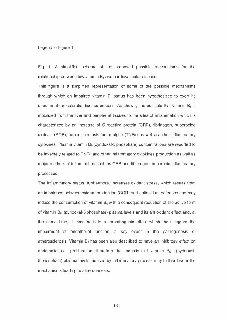
131
Legend to Figure 1
Fig. 1. A simplified scheme of the proposed possible mechanisms for the
relationship between low vitamin B6 and cardiovascular disease.
This figure is a simplified representation of some of the possible mechanisms
through which an impaired vitamin B6 status has been hypothesized to exert its
effect in atherosclerotic disease process. As shown, it is possible that vitamin B6 is
mobilized from the liver and peripheral tissues to the sites of inflammation which is
characterized by an increase of C-reactive protein (CRP), fibrinogen, superoxide
radicals (SOR), tumour necrosis factor alpha (TNFα) as well as other inflammatory
cytokines. Plasma vitamin B6 (pyridoxal-5’phosphate) concentrations are reported to
be inversely related to TNFα and other inflammatory cytokines production as well as
major markers of inflammation such as CRP and fibrinogen, in chronic inflammatory
processes.
The inflammatory status, furthermore, increases oxidant stress, which results from
an imbalance between oxidant production (SOR) and antioxidant defenses and may
induce the consumption of vitamin B6 with a consequent reduction of the active form
of vitamin B6 (pyridoxal-5’phosphate) plasma levels and its antioxidant effect and, at
the same time, it may facilitate a thrombogenic effect which then triggers the
impairment of endothelial function, a key event in the pathogenesis of
atherosclerosis. Vitamin B6 has been also described to have an inhibitory effect on
endothelial cell proliferation, therefore the reduction of vitamin B6 (pyridoxal-
5’phosphate) plasma levels induced by inflammatory process may further favour the
mechanisms leading to atherogenesis.
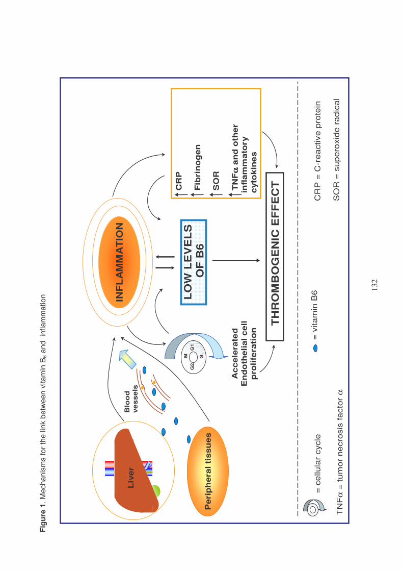
132
Fig
ure
1.
Me
ch
an
ism
s fo
r th
e lin
k b
etw
ee
n v
ita
min
B6 a
nd
in
fla
mm
atio
n
LO
W L
EV
EL
S
OF
B6
TH
RO
MB
OG
EN
IC E
FF
EC
T
= c
ellula
r cycle
CR
P =
C-r
eactive p
rote
in
SO
R =
sup
ero
xid
e r
adic
al
INF
LA
MM
AT
ION
Peri
ph
era
lti
ssu
es
CR
P
Fib
rin
og
en
SO
R
TN
Fα ααα
an
d o
ther
infl
am
mato
ry
cyto
kin
es
Blo
od
ve
ss
els
G1
G2
SM
Ac
cele
rate
d
En
do
theli
al c
ell
pro
life
rati
on
TN
Fα
= t
um
or
necro
sis
facto
r α
= v
itam
in B
6
Liv
er
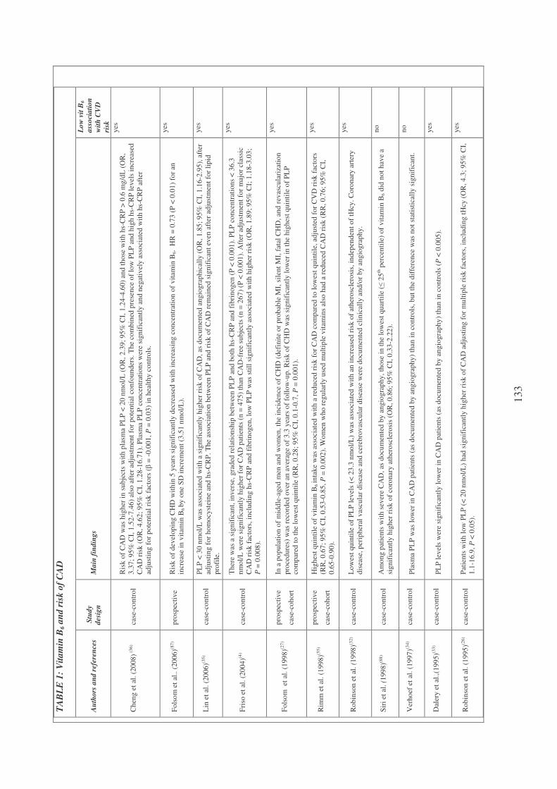
133
TA
BL
E 1
: V
ita
min
B6 a
nd
ris
k o
f C
AD
Au
tho
rs a
nd
ref
eren
ces
Stu
dy
des
ign
M
ain
fin
din
gs
Lo
w v
it B
6
ass
oci
ati
on
wit
h C
VD
risk
Ch
eng
et
al. (2
00
8) (
36
) ca
se-c
on
tro
l
Ris
k o
f C
AD
was
hig
her
in
su
bje
cts
wit
h p
lasm
a P
LP
< 2
0 n
mo
l/L
(O
R, 2
.39
; 9
5%
CI,
1.2
4-4
.60
) an
d t
ho
se w
ith
hs-
CR
P >
0.6
mg
/dL
(O
R,
3.3
7;
95
% C
I, 1
.52
-7.4
6)
also
aft
er a
dju
stm
ent
for
po
ten
tial
co
nfo
un
der
s. T
he
com
bin
ed p
rese
nce
of
low
PL
P a
nd
hig
h h
s-C
RP
lev
els
incr
ease
d
CA
D r
isk
(O
R, 4
.62
; 9
5%
CI,
1.2
8-1
6.7
1).
Pla
sma
PL
P c
on
cen
trat
ion
s w
ere
sig
nif
ican
tly a
nd
neg
ativ
ely a
sso
ciat
ed w
ith
hs-
CR
P a
fter
adju
stin
g f
or
po
tenti
al r
isk
fac
tors
(β
= -
0.0
01
, P
= 0
.03
) in
hea
lth
y c
on
tro
ls.
yes
Fo
lso
m e
t al
.. (
20
06
)(87
) p
rosp
ecti
ve
Ris
k o
f d
evel
op
ing C
HD
wit
hin
5 y
ears
sig
nif
ican
tly d
ecre
ased
wit
h i
ncr
easi
ng
co
nce
ntr
atio
n o
f v
itam
in B
6.
HR
= 0
.73
(P
< 0
.01
) fo
r an
incr
ease
in
vit
am
in B
6 b
y o
ne
SD
in
crem
ent
(3.5
1 n
mo
l/L
).
yes
Lin
et
al. (2
00
6)(3
5)
case
-co
ntr
ol
PL
P <
30
nm
ol/
L w
as a
sso
ciat
ed w
ith
a s
ign
ific
antl
y h
igh
er r
isk
of
CA
D,
as d
ocu
men
ted
an
gio
gra
ph
ical
ly (
OR
, 1
.85
; 9
5%
CI,
1.1
6-2
.95
), a
fter
adju
stin
g f
or
ho
mo
cyst
ein
e an
d h
s-C
RP
. T
he
asso
ciat
ion
bet
wee
n P
LP
an
d r
isk
of
CA
D r
em
ain
ed s
ign
ific
ant
even
aft
er a
dju
stm
ent
for
lip
id
pro
file
.
yes
Fri
so e
t al
. (2
00
4)(4
) ca
se-c
on
tro
l
Th
ere
was
a s
ign
ific
ant,
in
ver
se, g
rad
ed r
elat
ion
ship
bet
wee
n P
LP
an
d b
oth
hs-
CR
P a
nd
fib
rin
og
en (
P <
0.0
01
). P
LP
co
nce
ntr
atio
ns
< 3
6.3
nm
ol/
L w
ere
sig
nif
ican
tly h
igh
er f
or
CA
D p
atie
nts
(n
= 4
75
) th
an C
AD
-fre
e su
bje
cts
(n =
26
7)
(P <
0.0
01
). A
fter
ad
just
men
t fo
r m
ajo
r cl
assi
c
CA
D r
isk
fac
tors
, in
clu
din
g h
s-C
RP
an
d f
ibri
nog
en, lo
w P
LP
was
sti
ll s
ign
ific
antl
y a
sso
ciat
ed w
ith
hig
her
ris
k (
OR
, 1
.89
; 9
5%
CI;
1.1
8-3
.03
;
P =
0.0
08
).
yes
Fo
lso
m
et a
l. (
19
98
)(27
) p
rosp
ecti
ve
case
-co
ho
rt
In a
po
pu
lati
on
of
mid
dle
-ag
ed m
en a
nd
wo
men
, th
e in
cid
ence
of
CH
D (
def
init
e o
r p
rob
able
MI,
sil
ent
MI,
fat
al C
HD
, an
d r
evas
cula
riza
tio
n
pro
ced
ure
s) w
as r
eco
rded
ov
er a
n a
ver
age
of
3.3
yea
rs o
f fo
llo
w-u
p. R
isk
of
CH
D w
as s
ign
ific
antl
y l
ow
er i
n t
he
hig
hes
t q
uin
tile
of
PL
P
com
par
ed t
o t
he
low
est
qu
inti
le (
RR
, 0
.28
; 9
5%
CI,
0.1
-0.7
, P
= 0
.00
1).
yes
Rim
m e
t al
. (1
99
8)(5
5)
pro
spec
tiv
e
case
-co
ho
rt
Hig
hes
t q
uin
tile
of
vit
amin
B6
inta
ke
was
ass
oci
ated
wit
h a
red
uce
d r
isk
fo
r C
AD
co
mp
ared
to
lo
wes
t q
uin
tile
, ad
just
ed f
or
CV
D r
isk
fac
tors
(RR
, 0
.67
; 9
5%
CI,
0.5
3-0
.85
, P
= 0
.00
2).
Wo
men
wh
o r
egu
larl
y u
sed
mu
ltip
le v
itam
ins
also
had
a r
edu
ced
CA
D r
isk
(R
R, 0
.76
; 9
5%
CI,
0.6
5-0
.90
).
yes
Ro
bin
son
et
al.
(19
98
)(32
) ca
se-c
on
tro
l L
ow
est
qu
inti
le o
f P
LP
lev
els
(< 2
3.3
nm
ol/
L)
was
ass
oci
ated
wit
h a
n i
ncr
ease
d r
isk
of
ath
ero
scle
rosi
s, i
nd
epen
den
t o
f tH
cy. C
oro
nar
y a
rter
y
dis
ease
, p
erip
her
al v
ascu
lar
dis
ease
an
d c
ereb
rov
ascu
lar
dis
ease
wer
e d
ocu
men
ted
cli
nic
ally
an
d/o
r b
y a
ng
iog
rap
hy.
yes
Sir
i et
al.
(1
99
8)(8
8)
case
-co
ntr
ol
Am
on
g p
atie
nts
wit
h s
ever
e C
AD
, as
do
cum
ente
d b
y a
ng
iog
rap
hy, th
ose
in
th
e lo
wes
t q
uar
tile
(≤
25
th p
erce
nti
le)
of
vit
amin
B6 d
id n
ot
hav
e a
sig
nif
ican
tly h
igh
er r
isk
of
coro
nar
y a
ther
osc
lero
sis
(OR
, 0
.86
; 9
5%
CI,
0.3
3-2
.22
).
no
Ver
ho
ef e
t al
. (1
99
7)(3
4)
case
-co
ntr
ol
Pla
sma
PL
P w
as l
ow
er i
n C
AD
pat
ien
ts (
as d
ocu
men
ted
by a
ng
iog
rap
hy)
than
in
co
ntr
ols
, b
ut
the
dif
fere
nce
was
no
t st
atis
tica
lly
sig
nif
ican
t.
no
Dal
ery e
t al
.,(1
99
5)(3
3)
case
-co
ntr
ol
PL
P l
evel
s w
ere
sig
nif
ican
tly l
ow
er i
n C
AD
pat
ien
ts (
as d
ocu
men
ted
by a
ng
iog
rap
hy)
than
in
co
ntr
ols
(P
< 0
.00
5).
yes
Ro
bin
son
et
al. (1
99
5)(2
6)
case
-co
ntr
ol
Pat
ien
ts w
ith
lo
w P
LP
(<
20
nm
ol/
L)
had
sig
nif
ican
tly h
igh
er r
isk
of
CA
D a
dju
stin
g f
or
mu
ltip
le r
isk
fac
tors
, in
clu
din
g t
Hcy
(O
R, 4
.3;
95
% C
I,
1.1
-16
.9, P
< 0
.05
).
yes

134
TA
BL
E 2
: V
ita
min
B6 a
nd
ris
k o
f M
I
Au
tho
r a
nd
Ref
eren
ces
Stu
dy
des
ign
M
ain
fin
din
gs
Lo
w v
it B
6
ind
epen
den
t
ass
oci
ati
on
wit
h M
I ri
sk
Pag
e (2
00
9)(4
4)
Nes
ted
case
-
con
tro
l
Fas
tin
g c
on
cen
trat
ion
s o
f P
LP
wer
e si
gn
ific
antl
y i
nv
erse
ly a
sso
ciat
ed w
ith
su
bse
qu
ent
risk
of
MI:
hig
hes
t v
ersu
s lo
wes
t q
uar
tile
RR
= 0
.22
(9
5%
CI,
0.0
9-0
.55
; P
= 0
.04
7).
In
a c
on
dit
ion
al l
og
isti
c re
gre
ssio
n m
od
el,
the
effe
ct o
f ad
din
g P
LP
wit
h c
on
ven
tio
nal
MI
risk
fac
tors
was
sta
tist
ical
ly s
ign
ific
ant
(χ²
= 1
5.8
; P
= 0
.00
1).
Th
e re
lati
on
ship
bet
wee
n P
LP
an
d r
isk
of
MI
was
str
on
ger
am
on
g w
om
en u
nd
er 6
0 y
ears
old
th
an a
mo
ng
old
er w
om
en:
hig
hes
t v
ersu
s lo
wes
t q
uar
tile
RR
= 0
.05
(95
% C
I, 0
.00
4-0
.61
).
yes
Die
rkes
(2
00
7)(4
7)
Nes
ted
case
-co
ho
rt
Su
bje
cts
wer
e re
cru
ited
at
ran
do
m f
rom
th
e g
ener
al p
op
ula
tio
n a
nd
ex
clud
ed i
f th
ey h
ad a
his
tory
of
MI
and
str
ok
e at
bas
elin
e. T
he
hig
hes
t P
LP
qu
inti
le h
ad a
sig
nif
ican
tly r
edu
ced
ris
k o
f M
I (H
R,
0.5
0;
95
% C
I, 0
.29
-0.8
3).
Ad
just
men
t fo
r ei
ther
lo
w-g
rad
e in
flam
mat
ion
or
smo
kin
g d
imin
ish
ed t
his
ass
oci
atio
n. W
hen
adju
sted
fo
r b
oth
lo
w-g
rad
e in
flam
mat
ion
an
d s
mo
kin
g, , th
e as
soci
atio
n b
etw
een
PL
P a
nd
ris
k o
f M
I w
as a
bo
lish
ed. A
dju
stm
ent
for
esta
bli
shed
ris
k f
acto
rs a
lso
abo
lish
ed t
he
asso
ciat
ion
.
no
Tav
ani
(20
04
)(41
) ca
se-
con
tro
l R
isk
of
acu
te M
I w
as s
ign
ific
antl
y l
ow
er f
or
the
hig
hes
t te
rtil
e o
f v
itam
in B
6 i
nta
ke
com
par
ed t
o t
he
low
est
tert
ile
of
inta
ke
(OR
, 0
.34
; 9
5%
CI,
0.1
9-0
.60
).
yes
Nah
law
i (2
00
2)(8
9)
foll
ow
-up
2
1%
of
hea
rt t
ran
spla
nt
reci
pie
nts
had
PL
P d
efic
ien
cy.
On
ly 9
% o
f th
e p
atie
nts
wit
h l
ow
PL
P d
id n
ot
hav
e C
VD
co
mp
lica
tio
ns/
dea
th (
P =
0.0
5).
Ris
k f
or
CV
D
even
ts, in
clu
din
g d
eath
, w
as 2
.7 t
imes
hig
her
fo
r p
atie
nts
wit
h l
ow
vit
am
in B
6 (P
LP
≤ 2
0 n
mo
l/L
) (9
5%
CI,
1.2
-5.9
, P
= 0
.02
).
yes
Fo
lso
m (
19
98
)(27
) p
rosp
ecti
ve
case
-co
ho
rt
In a
po
pu
lati
on
of
mid
dle
-ag
ed m
en a
nd
wo
men
, in
cid
ence
of
CH
D (
def
init
e o
r p
rob
able
MI,
sil
ent
MI,
fat
al C
HD
, an
d r
evas
cula
riza
tio
n p
roce
du
res)
was
rec
ord
ed
ov
er a
n a
ver
age
of
3.3
yea
rs o
f fo
llo
w-u
p. R
isk
of
CH
D w
as l
ow
er i
n t
he
hig
hes
t q
uin
tile
of
PL
P v
ersu
s th
e lo
wes
t q
uin
tile
(R
R, 0
.28
; 9
5%
CI,
0.1
-0.7
; P
= 0
.00
1).
yes
Ch
asan
-Tab
er
(19
96
)(29)
case
-
con
tro
l
Th
e lo
wes
t q
uin
tile
of
PL
P (
< 2
8.9
nm
ol/
L)
had
a h
igh
er r
isk
of
MI
wh
en a
dju
stin
g f
or
mu
ltip
le v
aria
ble
s (R
R,
1.5
; 9
5%
CI,
1.0
-2.2
). I
f ad
just
ed f
or
fola
te, R
R w
as
1.3
(9
5%
CI,
0.9
-2.1
).
yes
Ver
ho
ef (
19
96
)(46)
case
-
con
tro
l
Pat
ien
ts w
ith
fir
st M
I h
ad l
ow
er d
ieta
ry a
nd
pla
sma
vit
amin
B6 t
han
co
ntr
ols
. O
R w
as 0
.97
fo
r th
e lo
wes
t q
uin
tile
of
pla
sma
PL
P (≤
29
.7 n
mo
l/L
) w
hen
ad
just
ed f
or
age
and
sex
an
d 0
.97
(9
5%
CI,
0.4
0-2
.33
) w
hen
ad
just
ed f
or
mu
ltip
le v
aria
ble
s,.O
R w
as 0
.32
fo
r th
e h
igh
est
qu
inti
le o
f p
lasm
a P
LP
(>
88
.9 n
mo
l/L
) w
hen
adju
stin
g f
or
age
and
sex
an
d 0
.51
(95
% C
I, 0
.19
-1.3
6)
wh
en a
dju
stin
g f
or
mu
ltip
le v
aria
ble
s.
yes
Ko
k (
19
89
)(28
) ca
se-
con
tro
l
Pat
ien
ts w
ith
MI
(n =
84
) w
ere
com
par
ed t
o c
on
tro
l su
bje
cts
(n =
84
). M
I w
as m
ore
th
an 5
tim
es m
ore
lik
ely a
mo
ng
su
bje
cts
in t
he
low
est
qu
arti
le o
f p
lasm
a P
LP
(< 2
0 n
mo
l/L
) (O
R, 5
.2;
95
% C
I, 1
.4-1
8.9
).
yes
Leg
end
: M
I =
myo
card
ial
infa
rcti
on
; C
HD
= c
oro
nar
y h
eart
dis
ease
; C
VD
= c
ard
iov
ascu
lar
dis
ease
; 9
5%
CI
= 9
5%
co
nfi
den
ce i
nte
rval
; R
R =
rel
ativ
e ri
sk;
OR
= o
dd
s ra
tio
; H
R =
haz
ard
rat
io
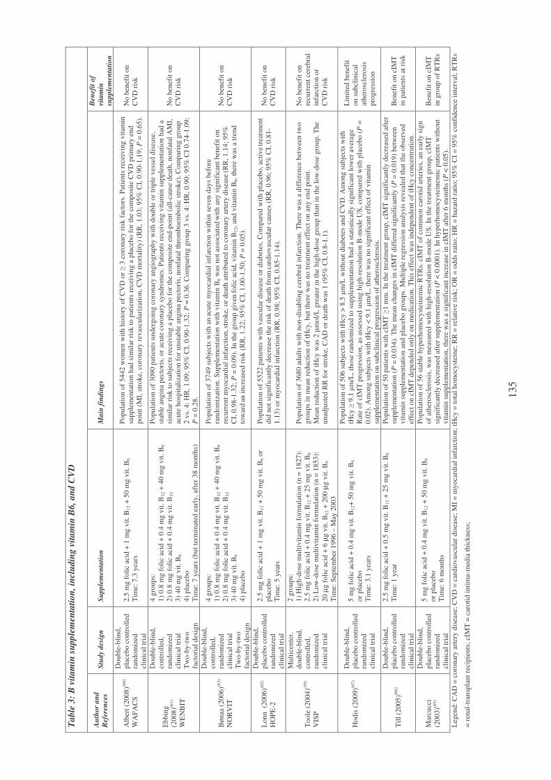
135
Tab
le 3
: B
vit
am
in s
upp
lem
enta
tio
n, in
clu
din
g v
ita
min
B6,
an
d C
VD
Au
tho
r a
nd
Ref
eren
ces
Stu
dy
des
ign
S
up
ple
men
tati
on
M
ain
fin
din
gs
Ben
efit
of
vita
min
sup
ple
men
tati
on
Alb
ert
(20
08
)(60
)
WA
FA
CS
Do
ub
le-b
lin
d,
pla
ceb
o c
on
tro
lled
ran
do
miz
ed
clin
ical
tri
al
2.5
mg
fo
lic
acid
+ 1
mg
vit
. B
12 +
50
mg
vit
. B
6
Tim
e: 7
.3 y
ears
Po
pu
lati
on
of
544
2 w
om
en w
ith
his
tory
of
CV
D o
r ≥
3 c
oro
nar
y r
isk
fac
tors
. P
atie
nts
rec
eiv
ing
vit
amin
sup
ple
men
tati
on
had
sim
ilar
ris
k t
o p
atie
nts
rec
eiv
ing
a p
lace
bo
fo
r th
e co
mp
osi
te C
VD
pri
mar
y e
nd
po
int
(MI,
str
ok
e, c
oro
nar
y r
evas
cula
riza
tio
n, C
VD
mo
rtal
ity)
(RR
, 1
.03
; 9
5%
CI,
0.9
0-1
.19
; P
= 0
.65
).
No
ben
efit
on
CV
D r
isk
Eb
bin
g
(20
08
)(61)
WE
NB
IT
Do
ub
le-b
lin
d,
con
tro
lled
,
ran
do
miz
ed
clin
ical
tri
al
Tw
o-b
y-t
wo
fact
ori
al d
esig
n
4 g
rou
ps:
1)
0.8
mg
fo
lic
acid
+ 0
.4 m
g v
it.
B1
2 +
40
mg
vit
. B
6
2)
0.8
mg
fo
lic
acid
+ 0
.4 m
g v
it.
B1
2
3)
40
mg
vit
. B
6
4)
pla
ceb
o
Tim
e: 7
yea
rs (
bu
t te
rmin
ated
ear
ly,
afte
r 3
8 m
on
ths)
Po
pu
lati
on
of
309
0 p
atie
nts
un
der
go
ing
co
ron
ary a
ng
iog
rap
hy w
ith
do
ub
le o
r tr
iple
ves
sel
dis
ease
,
stab
le a
ng
ina
pec
tori
s, o
r ac
ute
co
ron
ary s
yn
dro
mes
. P
atie
nts
rec
eiv
ing
vit
amin
su
pp
lem
enta
tio
n h
ad a
sim
ilar
ris
k t
o s
ub
ject
s re
ceiv
ing
a p
lace
bo
fo
r th
e co
mp
osi
te e
nd
-po
int
(all
-cau
se d
eath
, n
on
fata
l A
MI,
acu
te h
osp
ital
izat
ion
fo
r u
nst
able
ang
ina
pec
tori
s, n
on
fata
l th
rom
bo
emb
oli
c st
rok
e). C
om
par
ing
gro
up
2 v
s. 4
: H
R, 1
.09
; 9
5%
CI,
0.9
0-1
.32
; P
= 0
.36
. C
om
par
ing
gro
up
3 v
s. 4
: H
R, 0
.90
; 9
5%
CI
0.7
4-1
.09
;
P =
0.2
8.
No
ben
efit
on
CV
D r
isk
Bø
naa
(2
00
6)(6
3)
NO
RV
IT
Do
ub
le-b
lin
d,
con
tro
lled
,
ran
do
miz
ed
clin
ical
tri
al
Tw
o-b
y-t
wo
fact
ori
al d
esig
n
4 g
rou
ps:
1)
0.8
mg
fo
lic
acid
+ 0
.4 m
g v
it.
B1
2 +
40
mg
vit
. B
6
2)
0.8
mg
fo
lic
acid
+ 0
.4 m
g v
it.
B1
2
3)
40
mg
vit
. B
6
4)
pla
ceb
o
Po
pu
lati
on
of
374
9 s
ub
ject
s w
ith
an
acu
te m
yo
card
ial
infa
rcti
on
wit
hin
sev
en d
ays
bef
ore
ran
do
miz
atio
n. S
up
ple
men
tati
on
wit
h v
itam
in B
6 w
as n
ot
asso
ciat
ed w
ith
an
y s
ign
ific
ant
ben
efit
on
recu
rren
t m
yo
card
ial
infa
rcti
on
, stro
ke,
or
dea
th a
ttri
bu
ted
to
co
ron
ary a
rter
y d
isea
se (R
R, 1
.14
; 9
5%
CI,
0.9
8-1
.32
; P
= 0
.09
). I
n t
he
gro
up
giv
en fo
lic
acid
, v
itam
in B
12, an
d v
itam
in B
6, th
ere
was
a t
ren
d
tow
ard
an i
ncr
ease
d r
isk
(R
R, 1
.22
; 9
5%
CI,
1.0
0-1
.50
; P
= 0
.05
).
No
ben
efit
on
CV
D r
isk
Lo
nn
(2
00
6)(6
2)
HO
PE
-2
Do
ub
le-b
lin
d,
pla
ceb
o c
on
tro
lled
ran
do
miz
ed
clin
ical
tri
al
2.5
mg
fo
lic
acid
+ 1
mg
vit
. B
12 +
50
mg
vit
. B
6 o
r
pla
ceb
o
Tim
e: 5
yea
rs
Po
pu
lati
on
of
552
2 p
atie
nts
wit
h v
ascu
lar
dis
ease
or
dia
bet
es. C
om
par
ed w
ith
pla
ceb
o, ac
tiv
e trea
tmen
t
did
no
t si
gn
ific
antl
y d
ecre
ase
the
risk
of
dea
th f
rom
card
iov
ascu
lar
cau
ses
(RR
, 0
.96
; 9
5%
CI,
0.8
1-
1.1
3)
or
myo
card
ial
infa
rcti
on
(R
R, 0
.98
; 9
5%
CI,
0.8
5-1
.14
).
No
ben
efit
on
CV
D r
isk
To
ole
(2
00
4)(5
9)
VIS
P
Mu
ltic
ente
r,
do
ub
le-b
lind
,
con
tro
lled
,
ran
do
miz
ed
clin
ical
tri
al
2 g
rou
ps:
1)
Hig
h-d
ose
mu
ltiv
itam
in f
orm
ula
tio
n (
n =
18
27
):
2.5
mg
fo
lic
acid
+ 0
.4 m
g v
it. B
12 +
25
mg
vit
. B
6
2)
Lo
w-d
ose
mu
ltiv
itam
in f
orm
ula
tio
n (
n =
18
53
):
20
µg
fo
lic
acid
+ 6
µg
vit
. B
12 +
20
0 µ
g v
it.
B6
Tim
e: S
epte
mb
er 1
99
6 –
May
20
03
Po
pu
lati
on
of
368
0 a
du
lts
wit
h n
on
-dis
abli
ng
cer
ebra
l in
farc
tio
n. T
her
e w
as a
dif
fere
nce
bet
wee
n t
wo
gro
up
s in
mea
n r
edu
ctio
n o
f tH
cy, b
ut
ther
e w
as n
o t
reat
men
t ef
fect
on
an
y e
nd
po
int.
Mea
n r
edu
ctio
n o
f tH
cy w
as 2
µm
ol/
L g
reat
er i
n t
he
hig
h-d
ose
gro
up
th
an i
n t
he
low
-do
se g
rou
p. T
he
un
adju
sted
RR
fo
r st
rok
e, C
AD
or
dea
th w
as 1
(9
5%
CI,
0.8
-1.1
).
No
ben
efit
on
recu
rren
t ce
reb
ral
infa
rcti
on
or
CV
D r
isk
Ho
dis
(2
00
9)(6
7)
Do
ub
le-b
lin
d,
pla
ceb
o c
on
tro
lled
ran
do
miz
ed
clin
ical
tri
al
5 m
g f
oli
c ac
id +
0.4
mg
vit
. B
12+
50
mg
vit
. B
6
or
pla
ceb
o
Tim
e: 3
.1 y
ears
Po
pu
lati
on
of
506
su
bje
cts
wit
h t
Hcy
> 8
.5 µ
m/L
wit
ho
ut
dia
bet
es a
nd
CV
D. A
mo
ng
su
bje
cts
wit
h
tHcy
≥ 9
.1 µ
m/L
, th
ose
ran
do
miz
ed t
o s
up
ple
men
tati
on
had
a s
tati
stic
ally
sig
nif
ican
t lo
wer
av
erag
e
Rat
e o
f cI
MT
pro
gre
ssio
n, as
ass
esse
d u
sin
g h
igh
-res
olu
tio
n B
-mo
de
US
, co
mp
ared
wit
h p
lace
bo
(P
=
0.0
2).
Am
on
g s
ub
ject
s w
ith
tH
cy <
9.1
µm
/L, th
ere
was
no
sig
nif
ican
t ef
fect
of
vit
amin
sup
ple
men
tati
on
on
su
bcl
inic
al p
rog
ress
ion
of
ath
ero
scle
rosi
s.
Lim
ited
ben
efit
on
su
bcl
inic
al
ath
ero
scle
rosi
s
pro
gre
ssio
n
Til
l (2
00
5)(6
6)
Do
ub
le-b
lin
d,
pla
ceb
o c
on
tro
lled
ran
do
miz
ed
clin
ical
tri
al
2.5
mg
fo
lic
acid
+ 0
.5 m
g v
it. B
12 +
25
mg
vit
. B
6
Tim
e: 1
yea
r
Po
pu
lati
on
of
50
pat
ien
ts w
ith
cIM
T ≥
1 m
m.
In t
he
trea
tmen
t g
rou
p, cI
MT
sig
nif
ican
tly d
ecre
ased
aft
er
sup
ple
men
tati
on
(P
= 0
.03
4).
Th
e m
ean
ch
ang
es i
n c
IMT
dif
fere
d s
ign
ific
antl
y (
P =
0.0
19
) b
etw
een
vit
amin
su
pp
lem
enta
tio
n a
nd
pla
ceb
o g
rou
ps.
Mu
ltip
le r
egre
ssio
n a
nal
ysi
s re
vea
led
th
at t
he
ob
serv
ed
effe
ct o
n c
IMT
dep
end
ed o
nly
on
med
icat
ion
. T
his
eff
ect
was
in
dep
end
ent
of
tHcy
co
nce
ntr
atio
n.
Ben
efit
on
cIM
T
in p
atie
nts
at
risk
Mar
cucc
i
(20
03
)(65)
Do
ub
le-b
lin
d,
pla
ceb
o c
on
tro
lled
ran
do
miz
ed
clin
ical
tri
al
5 m
g f
oli
c ac
id +
0.4
mg
vit
. B
12 +
50
mg
vit
. B
6
or
pla
ceb
o
Tim
e: 6
mo
nth
s
Po
pu
lati
on
of
56
sta
ble
hyp
erh
om
ocy
stei
nem
ic R
TR
s. c
IMT
of
com
mo
n c
aro
tid
art
erie
s, a
n e
arly
sig
n
of
ath
ero
scle
rosi
s, w
as m
easu
red
wit
h h
igh
-res
olu
tio
n B
-mo
de
US
. In
th
e tr
eatm
ent
gro
up
, cI
MT
sig
nif
ican
tly d
ecre
ased
aft
er s
up
ple
men
tati
on
(P
< 0
.00
01
). I
n h
yp
erh
om
ocy
stei
nem
ic p
atie
nts
wit
ho
ut
vit
amin
su
pp
lem
enta
tio
n, th
ere
was
a s
ign
ific
ant
incr
ease
in
cIM
T a
fter
6 m
on
ths
(P <
0.0
5).
Ben
efit
on
cIM
T
in g
rou
p o
f R
TR
s
Leg
end
: C
AD
= c
oro
nar
y a
rter
y d
isea
se;
CV
D =
car
dio
vas
cula
r d
isea
se;
MI
= m
yo
card
ial
infa
rcti
on
; tH
cy =
to
tal
ho
mo
cyst
ein
e; R
R =
rel
ativ
e ri
sk;
OR
= o
dd
s ra
tio
; H
R =
haz
ard
rat
io;
95
% C
I =
95
% c
on
fid
ence
in
terv
al;
RT
Rs
= r
enal
-tra
nsp
lan
t re
cip
ien
ts;
cIM
T =
car
oti
d i
nti
ma-
med
ia t
hic
kn
ess;
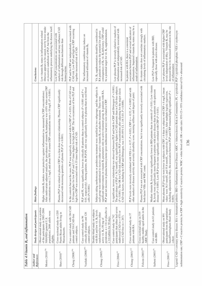
136
Tab
le 4
:Vit
am
in B
6 a
nd
in
flam
ma
tion
Au
tho
r a
nd
Ref
eren
ces
Stu
dy
des
ign
an
d p
art
icip
an
ts
Ma
in f
ind
ing
s C
on
clu
sio
ns
Mo
rris
(2
01
0)(7
8)
Ob
serv
atio
nal
stu
dy o
n a
po
rtio
n
of
the
par
tici
pan
ts i
n 2
00
3-2
00
4
NH
AN
ES
(g
ener
al U
.S.
po
pu
lati
on
); 2
68
6 a
du
lts
wer
e
elig
ible
.
Hig
her
vit
amin
B6 i
nta
kes
wer
e p
rote
ctiv
e ag
ain
st i
nfl
amm
atio
n (
as m
easu
red
by C
RP
co
nce
ntr
atio
ns)
.
Aft
er m
ult
ivar
iate
-ad
just
ed a
nal
ysi
s, t
he
pre
val
ence
of
vit
amin
B6
inad
equ
acy w
as <
10
% w
hen
ser
um
CR
P
con
cen
trat
ion
s w
ere ≤
3 m
g/L
an
d a
bo
ut
50
% i
f se
rum
CR
P c
on
cen
trat
ion
s w
ere
> 1
0 m
g/L
(P
< 0
.00
1).
Lo
w v
itam
in B
6 s
tatu
s in
in
flam
mat
ion
-rel
ated
illn
esse
s ap
pea
rs t
o b
e ca
use
d, n
ot
by l
ow
er i
nta
ke
or
exce
ssiv
e ca
tab
oli
sm o
f P
LP
, b
ut
fro
m t
he
infl
amm
ato
ry p
roce
ss u
nd
erly
ing
th
e d
isea
se i
tsel
f.
Sh
en (
20
10
)(6)
Cro
ss-s
ecti
on
al s
tud
y o
n 1
20
5
Pu
erto
Ric
an a
du
lts
liv
ing
in
Mas
sach
use
tts.
Pla
sma
PL
P w
as r
elat
ed t
o p
lasm
a C
RP
in
a c
lear
do
se-r
esp
on
se r
elat
ion
ship
. P
lasm
a C
RP
sig
nif
ican
tly
dec
reas
ed w
ith
in
crea
sin
g q
uar
tile
s o
f p
lasm
a P
LP
(P
< 0
.00
1).
Lo
w P
LP
co
nce
ntr
atio
ns
are
asso
ciat
ed w
ith
mar
ker
s o
f in
flam
mat
ion
an
d m
ay i
nfl
uen
ce C
AD
risk
th
rou
gh
dif
fere
nt
mec
han
ism
s th
an
ho
mo
cyst
ein
e.
Ch
eng
(2
008
)(36
)
Cas
e-co
ntr
ol
stu
dy w
ith
18
4 C
AD
pat
ien
ts a
nd
51
6 C
AD
-fre
e
con
tro
l su
bje
cts.
In t
he
con
tro
ls b
ut
no
t th
e ca
ses,
PL
P w
as n
egat
ivel
y a
sso
ciat
ed w
ith
hs-
CR
P (
P =
0.0
3).
Ris
k o
f C
AD
was
2.3
9 t
imes
hig
her
fo
r lo
w P
LP
an
d 3
.37
tim
es h
igh
er f
or
hig
h h
s-C
RP
. T
he
co-o
ccu
rren
ce o
f lo
w P
LP
an
d
hig
h h
s-C
RP
was
ass
oci
ated
wit
h 4
.35
tim
es h
igh
er r
isk
of
CA
D.
PL
P a
nd
hs-
CR
P a
re i
nd
epen
den
tly a
sso
ciat
ed w
ith
CA
D r
isk
. L
ow
PL
P a
nd
hig
h h
s-C
RP
occ
urr
ing
tog
eth
er i
ncr
ease
d r
isk
of
CA
D.
Vas
ilak
i (2
00
8)(8
5)
Cas
e-co
ntr
ol
stu
dy o
n 9
6
crit
ical
ly i
ll p
atie
nts
an
d 1
26
con
tro
l su
bje
cts.
PL
P a
nd
PL
wer
e si
gn
ific
antl
y l
ow
er i
n c
riti
call
y i
ll p
atie
nts
th
an i
n c
on
tro
l su
bje
cts
(P <
0.0
01
an
d P
<
0.0
1, re
spec
tiv
ely
). A
mo
ng
pat
ien
ts, th
e P
LP
:PL
rat
io w
as s
ign
ific
antl
y l
ow
er i
n p
lasm
a th
an i
n r
ed b
loo
d
cell
s (P
= 0
.00
1).
Th
e in
flam
mat
ory
co
nd
itio
n m
ay i
nd
uce
an
incr
ease
d u
tili
zati
on
of
vit
amin
B6.
Ch
ian
g (
20
05
)(20
)
Do
ub
le-b
lin
d s
tudy o
n s
ub
ject
s
wit
h R
A a
nd
lo
w v
it. B
6 l
evel
s
ran
do
miz
ed t
o r
ecei
ve
eith
er
acti
ve
vit
. B
6 (
50
mg
of
pyri
do
xin
e, n
= 1
4)
or
pla
ceb
o (
n
= 1
4)
tab
lets
fo
r 3
0 d
ays.
Su
bje
cts
tak
ing
met
ho
trex
ate
or
pre
dn
iso
ne
trea
tmen
t w
ere
stra
tifi
ed i
n t
wo
su
bg
roup
s, a
nd
th
e su
bje
cts
in
each
gro
up
wer
e ra
nd
om
ized
to
rec
eiv
e ei
ther
act
ive
vit
amin
B6 o
r p
lace
bo
tre
atm
ent.
Th
ere
wer
e
sig
nif
ican
t im
pro
vem
ents
in
vit
am
in B
6 p
aram
eter
s o
nly
in
act
ive
trea
tmen
t g
rou
p. In
pat
ien
ts w
ith
RA
,
PL
P a
nd
net
in
crea
se i
n p
lasm
a h
om
ocy
stei
ne
(po
st-m
eth
ion
ine
load
tes
t) w
ere
rela
ted
to
CR
P.
Vit
. B
6 s
up
ple
men
tati
on
sh
ou
ld b
e co
nsi
der
ed i
n
RA
pat
ien
ts t
o r
edu
ce t
he
po
ten
tial
fo
r ad
ver
se
con
seq
uen
ces
of
B6 v
it. B
6 d
efic
ien
cy. C
RP
co
uld
be
a p
ote
nti
al t
arg
et f
or
vit
. B
6 s
up
ple
men
tati
on
.
Fri
so (
20
04
)(4)
Cas
e-co
ntr
ol
stu
dy o
n 7
42
par
tici
pan
ts t
hat
eit
her
had
sev
ere
mu
ltiv
esse
l C
AD
(n
= 4
75
) o
r
wer
e C
AD
-fre
e (n
= 2
67
).
A s
ign
ific
ant,
in
ver
se r
elat
ion
ship
was
ob
serv
ed b
etw
een
PL
P a
nd
bo
th h
s-C
RP
an
d f
ibri
no
gen
(P
< 0
.00
1).
Th
e p
rev
alen
ce o
f P
LP
< 3
6.3
nm
ol/
L w
as s
ign
ific
antl
y h
igh
er i
n C
AD
pat
ien
ts t
han
in
CA
D-f
ree
sub
ject
s
(P <
0.0
01
). T
he
OR
fo
r C
AD
ris
k r
elat
ed t
o l
ow
PL
P c
on
cen
trat
ion
s af
ter
adju
stin
g f
or
the
maj
or
clas
sic
CA
D r
isk
fac
tors
, in
clu
din
g h
s-C
RP
an
d f
ibri
nog
en, w
as 1
.89
(9
5%
CI,
1.1
8-3
.03
; P
= 0
.00
8).
Lo
w p
lasm
a P
LP
is
inv
erse
ly r
elat
ed t
o m
ark
ers
of
infl
amm
atio
n a
nd
in
dep
end
entl
y a
ssoci
ated
wit
h
incr
ease
d r
isk
of
CA
D.
Ch
ian
g (
20
03
)(12
) C
ross
-sec
tio
nal
stu
dy o
n 3
7
pat
ien
ts w
ith
RA
.
PL
P l
evel
s w
ere
inv
erse
ly c
orr
elat
ed w
ith
VE
S (
r =
-0
.37
, P
= 0
.02
), C
RP
(r
= -
0.5
2, P
= 0
.00
2)
and
wit
h
oth
er m
ark
ers
of
dis
ease
act
ivit
y a
nd
sev
erit
y (
dis
abil
ity
sco
re,
mo
rnin
g s
tiff
nes
s an
d d
egre
e o
f p
ain
).
In p
atie
nts
wit
h R
A, th
ere
is a
co
nsi
sten
t
asso
ciat
ion
bet
wee
n P
LP
an
d s
ever
al i
nd
icat
ors
of
infl
amm
atio
n. Im
pai
red
vit
amin
B6 s
tatu
s m
ay b
e a
resu
lt o
f in
flam
mat
ion
.
Fo
lso
m (
20
03
)(84
)
Cro
ss-s
ecti
on
al s
tud
y o
n 5
19
hea
lth
y m
idd
le-a
ged
ad
ult
s in
th
e
AR
IC S
tud
y.
Pla
sma
PL
P w
as n
ot
asso
ciat
ed w
ith
CR
P c
on
cen
trat
ion
s, b
ut
it w
as s
ign
ific
antl
y i
nv
erse
ly a
sso
ciat
ed w
ith
bo
th f
acto
r V
IIIc
an
d W
BC
co
un
t.
B-v
itam
in s
tatu
s d
oes
no
t co
rrel
ate
stro
ng
ly w
ith
circ
ula
tin
g l
evel
s o
f in
flam
mat
ory
mark
ers.
Sai
ben
i (2
00
3)(9
) C
ase-
con
tro
l st
ud
y o
n 6
1 p
atie
nts
wit
h I
BD
.
Med
ian
vit
amin
B6 l
evel
s w
ere
sig
nif
ican
tly l
ow
er i
n I
BD
pat
ien
ts t
han
in
co
ntr
ols
(P
< 0
.01
). L
ow
vit
am
in
B6 l
evel
s w
ere
sig
nif
ican
tly m
ore
fre
qu
ent
in p
atie
nts
wit
h a
ctiv
e d
isea
se t
han
in
pat
ien
ts w
ith
qu
iesc
ent
dis
ease
(P
< 0
.00
1).
Lo
w P
LP
lev
els
wer
e si
gn
ific
antl
y c
orr
elat
ed w
ith
CR
P (
P <
0.0
1).
Lo
w P
LP
is
freq
uen
t in
pat
ien
ts w
ith
IB
D,
esp
ecia
lly t
ho
se w
ith
act
ive
dis
ease
.
Fri
so (
20
01
)(5)
Ob
serv
atio
nal
stu
dy o
n 8
91
par
tici
pan
ts f
rom
th
e p
op
ula
tion
-
bas
ed F
ram
ing
ham
Hea
rt S
tud
y
coh
ort
.
Mea
n p
lasm
a P
LP
lev
els
wer
e low
er i
n s
ub
ject
s w
ith
CR
P ≥
6 t
han
in
su
bje
cts
wit
h C
RP
< 6
mg
/L (
mea
n
val
ues
wer
e 3
6.5
nm
ol/
L a
nd
55
.8 n
mo
l/L
, re
spec
tiv
ely;
P <
0.0
01
). A
fter
mu
ltip
le l
og
isti
c re
gre
ssio
n,
incl
ud
ing
ad
just
men
t fo
r tH
cy,
the as
soci
atio
n b
etw
een
PL
P a
nd
CR
P r
emai
ned
hig
hly
sig
nif
ican
t (P
=
0.0
03
).
Lo
w p
lasm
a P
LP
is
asso
ciat
ed w
ith
hig
her
CR
P
lev
els
ind
epen
den
t o
f tH
cy.
Vit
am
in B
6 m
ay b
e
dec
reas
ed d
ue
to i
ts i
ncr
ease
d u
tili
zati
on
in
th
e si
te
of
infl
amm
atio
n
Leg
end
: C
AD
= c
oro
nar
y a
rter
y d
isea
se;
RA
= r
heu
mat
oid
art
hri
tis;
IB
D =
In
flam
mat
ory
Bo
wel
Dis
ease
; A
RIC
= A
ther
osc
lero
sis
Ris
k i
n C
om
mu
nit
ies;
PL
= p
yri
do
xal
; P
LP
= p
yri
do
xal
ph
osp
hat
e; V
ES
= e
ryth
rocy
te
sed
imen
tati
on
rat
e; C
RP
= C
-rea
ctiv
e p
rote
in;
hs-
CR
P =
hig
h-s
ensi
tiv
ity C
-rea
ctiv
e p
rote
in;
WB
C =
wh
ite
blo
od
cel
l; O
R =
od
ds
rati
o;
95
% C
I =
95
% c
on
fid
ence
in
terv
al U
S =
ult
raso
un
d
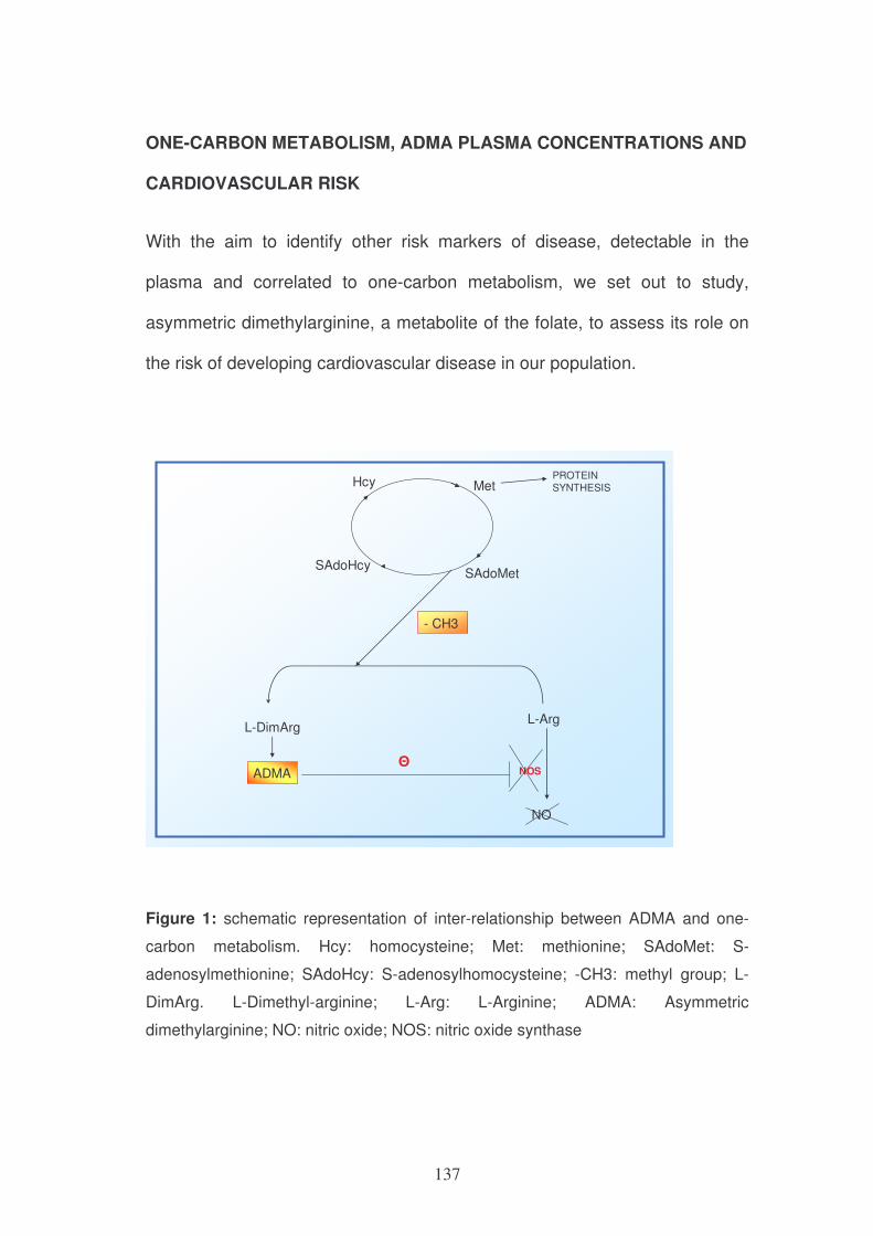
137
ONE-CARBON METABOLISM, ADMA PLASMA CONCENTRATIONS AND
CARDIOVASCULAR RISK
With the aim to identify other risk markers of disease, detectable in the
plasma and correlated to one-carbon metabolism, we set out to study,
asymmetric dimethylarginine, a metabolite of the folate, to assess its role on
the risk of developing cardiovascular disease in our population.
PROTEIN SYNTHESIS
L-ArgL-DimArg
- CH3
ADMA
NO
NOSΘ
Hcy Met
SAdoMetSAdoHcy
Figure 1: schematic representation of inter-relationship between ADMA and one-
carbon metabolism. Hcy: homocysteine; Met: methionine; SAdoMet: S-
adenosylmethionine; SAdoHcy: S-adenosylhomocysteine; -CH3: methyl group; L-
DimArg. L-Dimethyl-arginine; L-Arg: L-Arginine; ADMA: Asymmetric
dimethylarginine; NO: nitric oxide; NOS: nitric oxide synthase
138
Abstract
Asymmetric dimethylarginine (ADMA), is an endogenous competitive inhibitor
of nitric oxide synthase (NOS), enzyme responsible for the production of
nitric oxide (NO) by endothelial cells. NO is able both to induce vascular
dilation and to influence monocytes adhesion, platelets aggregation and
smooth muscle cells proliferation.
In 300 subjects of Verona Heart Project, the association between ADMA and
coronary artery disease (CAD) was evaluated. ADMA plasma concentrations
higher than 0.69 µmol/L (the highest tertile) defined a significant increase risk
of myocardial infarction (MI), the main thrombotic complication of CAD
(OR=2.14, 1.17-3.92, IC 95%, P=0.012). This association concerned only the
female subgroup (OR=2.79, 1.07-7.27, IC 95%, P=0.032), where the
increase of MI risk maintained a statistically significant difference in a
multivariate model analysis adjusted for the major risk factors for CAD
(OR=2.04, 1.09-3.82, IC 95%, P=0.026). This finding could allow to highlight
a novel markers of CAD risk more specific for female patients.
Introduction
The endothelium plays a crucial role to maintain the vascular tone and
structure. One of the major endothelium-derived vasoactive mediators is
nitric oxide (NO). Asymmetric DiMethylArginine (ADMA) is a natural
component of human blood plasma, produced by protein turnover. It is an
endogenous competitive inhibitor of all three isoforms of nitric oxide synthase
139
(NOS) and elevated ADMA plasma levels have been reported in relation with
diseases connected with an impaired endothelial L-arginine-NO pathway and
endothelial dysfunction1, 2. ADMA is relased when methylated proteins are
degraded into their amino acid components during hydrolytic protein
turnover. The majority of ADMA is eliminated by degradation to
dimethylamine and citrulline by the enzyme dimethylarginine
dimethylaminohydrolase (DDAH); the other is eliminated by renal excretion.
DDAH dysfunction may be a crucial unifying element of increased ADMA
levels as well as cardiovascular risk1, 3, 4. Moreover, DDAH is currently
investigated as a promising target for therapeutic interventions: transgenic
and knock-out mouse with altered DDAH activity have been developed to
study drug effects5.
When ADMA levels are elevated, NO production by NOS is decreased since
ADMA inhibits NOS by competitive displacement of the physiological
substrate (L-arginine), from the enzyme. This inhibition leads to a decreased
NO production in the endothelium of vessel walls with consequent increased
resting vascular tone (vasoconstriction) and enhanced pro-atherogenic
mechanisms including proliferation of vascular smooth muscle cell, platelet
aggregation and adherence of monocytes, extracellular matrix formation6
(Figure 2).
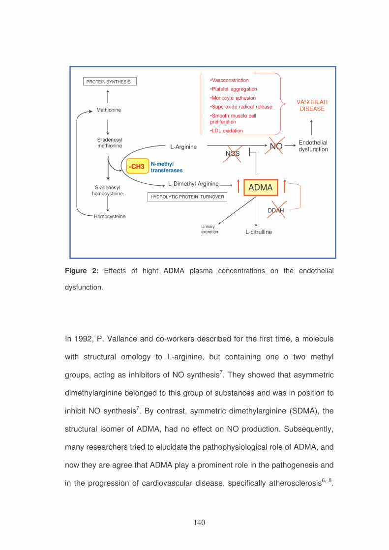
140
Methionine
Homocysteine
S-adenosylmethionine
S-adenosylhomocysteine
PROTEIN SYNTHESIS
-CH3 N-methyltransferases
L-Arginine
L-Dimethyl ArginineADMA
NONOS
Endothelialdysfunction
VASCULAR DISEASE
L-citrulline
DDAH
Urinary
excretion
•Vasoconstriction
•Platelet aggregation
•Monocyte adhesion
•Superoxide radical release
•Smooth muscle cellproliferation
•LDL oxidation
HYDROLYTIC PROTEIN TURNOVER
Figure 2: Effects of hight ADMA plasma concentrations on the endothelial
dysfunction.
In 1992, P. Vallance and co-workers described for the first time, a molecule
with structural omology to L-arginine, but containing one o two methyl
groups, acting as inhibitors of NO synthesis7. They showed that asymmetric
dimethylarginine belonged to this group of substances and was in position to
inhibit NO synthesis7. By contrast, symmetric dimethylarginine (SDMA), the
structural isomer of ADMA, had no effect on NO production. Subsequently,
many researchers tried to elucidate the pathophysiological role of ADMA, and
now they are agree that ADMA play a prominent role in the pathogenesis and
in the progression of cardiovascular disease, specifically atherosclerosis6, 8.
141
Many clinical studies demonstrated a significant and independent relationship
between ADMA levels and the incidence of major adverse cardiovascular
events9,10, death 8,11, and higher risk of disease relapse6.
Elevated ADMA concentration may play an important pathophysiological role
also in many other clinical condition, such as hypertension12,
hypercholesterolemia13, chronic renal failure14, 15, chronic heart failure16,
diabetes mellitus17, 18. Moreover, other situations have been reported to be
associated with elevated ADMA concentration as smoking, erectile
dysfunction and preeclampsia19.
The main aim of this project was to evaluate a possible association between
plasma concentration of ADMA and coronary artery disease, including its
main thrombotic complication, myocardial infarction,. The second aim was to
analyse the role of ADMA in male and female patients to evaluate a possible
risk variation with regard to gender.
Methods
Study Population
The Verona Heart Study (VHS) is a case-control study ongoing since May
1996 and designed to identify novel risk factors for CAD and MI in a
population of subjects with documented coronary angiography data. Study
design and details about enrolment/exclusion criteria have been previously
described20. In the present study, we examined a total of 300 subjects for
whom complete biochemical and clinical data were available. Of these, 200
142
had angiographically documented severe coronary atherosclerosis (CAD)
with or without previous history of MI (MI group), documented by combining
data from clinical history with a thorough review of medical records including
diagnostic electrocardiogram and biochemical data on myocardial enzymes,
and/or the typical sequelae of MI on ventricular angiography. One hundred
subjects, that underwent coronary angiography for reasons other than CAD
(mainly valvular heart disease) and showed normal coronary arteries, were
considered as controls (CAD-free group). Controls were also required to
have neither history nor clinical or instrumental evidence of atherosclerosis in
vascular districts beyond the coronary bed. At the time of blood sampling, a
complete clinical history was collected, including the assessment of
cardiovascular risk factors such as obesity, smoking, hypertension and
diabetes, as well as medical therapy.
The study was approved by our Institutional Ethical Committee. Informed
consent was obtained from all the subjects after a full explanation of the
study.
Biochemical analyses and ADMA assay
Samples of venous blood were drawn from each subject after an overnight
fast. Laboratory assessment of several biomarkers of CAD were determined
as previously described20, 21. Total plasma homocysteine levels were
determined by high-performance liquid chromatography (HPLC) with
fluorescent detection, according to Araki and Sako22. Plasma folate and
143
vitamin B12 concentrations were measured by an automated
chemiluminescence method (Chiron Diagnostics, East Walpole, MA).
Asymmetric dimethylarginine (ADMA) levels were determined in EDTA
plasma samples stored at –80°C before use and without freeze-thaw cycles
by an enzyme-linked immunosorbent assay (DLD Diagnostika, Hamburg,
Germany) according to the manufacturer’s instructions.
Statistical analysis
All statistical analyses were performed with SPSS 17.0 statistical package
(SPSS Inc., Chicago, IL). Distributions of continuous variables in groups were
expressed as means±standard deviation. Logarithmic transformation was
performed on all skewed variables, including tHcy, folate, vitamin B12 and
ADMA. Therefore, the statistical differences concerning these parameters
were also computed on the corresponding log-transformed values.
Quantitative data were assessed using the Student’s t-test or by ANOVA.
Associations between qualitative variables were analyzed with the χ2-test. A
value of P < 0.05 was considered significant.
To assess the extent to which ADMA was associated with CAD and MI, odds
ratios with 95% CIs were estimated by univariate logistic regression analysis.
Adjustment for classical cardiovascular risk factors (i.e. gender, age,
hypertension, smoke, diabetes, cholesterol and triglyceride) was performed
by adding those covariates in a multiple logistic-regression model.
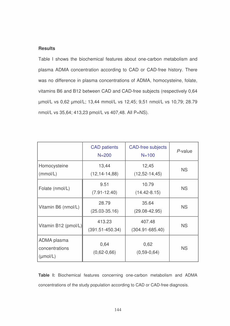
144
Results
Table I shows the biochemical features about one-carbon metabolism and
plasma ADMA concentration according to CAD or CAD-free history. There
was no difference in plasma concentrations of ADMA, homocysteine, folate,
vitamins B6 and B12 between CAD and CAD-free subjects (respectively 0,64
µmol/L vs 0,62 µmol/L; 13,44 mmol/L vs 12,45; 9,51 nmol/L vs 10,79; 28.79
nmol/L vs 35,64; 413,23 pmol/L vs 407,48. All P=NS).
CAD patients
N=200
CAD-free subjects
N=100 P-value
Homocysteine
(mmol/L)
13,44
(12,14-14,88)
12,45
(12,52-14,45) NS
Folate (nmol/L) 9.51
(7.91-12.40)
10.79
(14.42-8.15) NS
Vitamin B6 (nmol/L) 28.79
(25.03-35.16)
35.64
(29.08-42.95) NS
Vitamin B12 (pmol/L) 413.23
(391.51-450.34)
407.48
(304.91-685.40) NS
ADMA plasma
concentrations
(µmol/L)
0,64
(0,62-0,66)
0,62
(0,59-0,64) NS
Table I: Biochemical features concerning one-carbon metabolism and ADMA
concentrations of the study population according to CAD or CAD-free diagnosis.
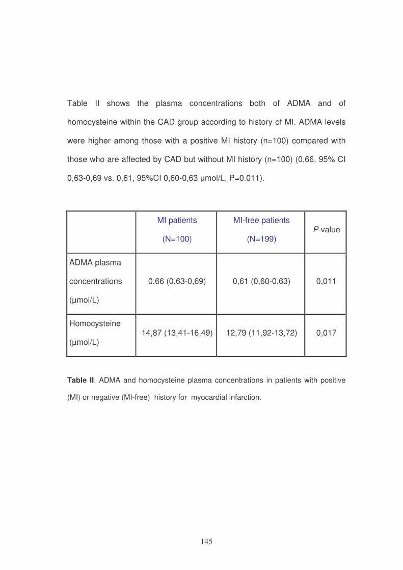
145
Table II shows the plasma concentrations both of ADMA and of
homocysteine within the CAD group according to history of MI. ADMA levels
were higher among those with a positive MI history (n=100) compared with
those who are affected by CAD but without MI history (n=100) (0,66, 95% CI
0,63-0,69 vs. 0,61, 95%CI 0,60-0,63 µmol/L, P=0.011).
MI patients
(N=100)
MI-free patients
(N=199) P-value
ADMA plasma
concentrations
(µmol/L)
0,66 (0,63-0,69) 0,61 (0,60-0,63) 0,011
Homocysteine
(µmol/L) 14,87 (13,41-16,49) 12,79 (11,92-13,72) 0,017
Table II. ADMA and homocysteine plasma concentrations in patients with positive
(MI) or negative (MI-free) history for myocardial infarction.
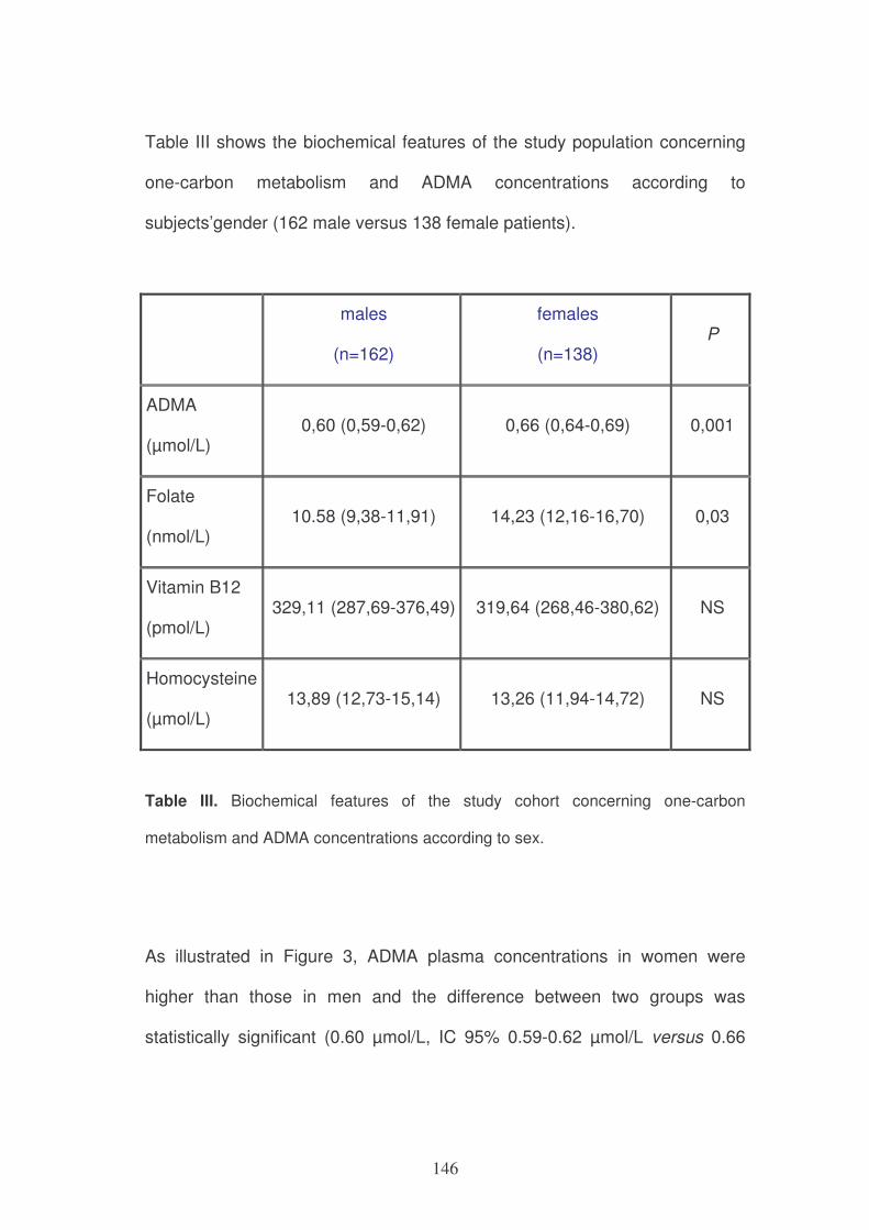
146
Table III shows the biochemical features of the study population concerning
one-carbon metabolism and ADMA concentrations according to
subjects’gender (162 male versus 138 female patients).
males
(n=162)
females
(n=138) P
ADMA
(µmol/L) 0,60 (0,59-0,62) 0,66 (0,64-0,69) 0,001
Folate
(nmol/L) 10.58 (9,38-11,91) 14,23 (12,16-16,70) 0,03
Vitamin B12
(pmol/L) 329,11 (287,69-376,49) 319,64 (268,46-380,62) NS
Homocysteine
(µmol/L) 13,89 (12,73-15,14) 13,26 (11,94-14,72) NS
Table III. Biochemical features of the study cohort concerning one-carbon
metabolism and ADMA concentrations according to sex.
As illustrated in Figure 3, ADMA plasma concentrations in women were
higher than those in men and the difference between two groups was
statistically significant (0.60 µmol/L, IC 95% 0.59-0.62 µmol/L versus 0.66
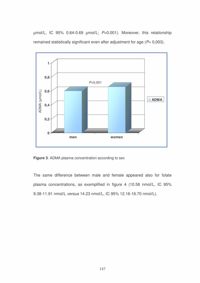
147
µmol/L, IC 95% 0.64-0.69 µmol/L; P=0.001). Moreover, this relationship
remained statistically significant even after adjustment for age (P= 0,003).
0
0,2
0,4
0,6
0,8
1
men women
ADMA
AD
MA
(µ
mo
l/L
)
P=0,001
Figure 3: ADMA plasma concentration according to sex
The same difference between male and female appeared also for folate
plasma concentrations, as exemplified in figure 4 (10.58 nmol/L, IC 95%
9.38-11.91 nmol/L versus 14.23 nmol/L, IC 95% 12.16-16.70 nmol/L).
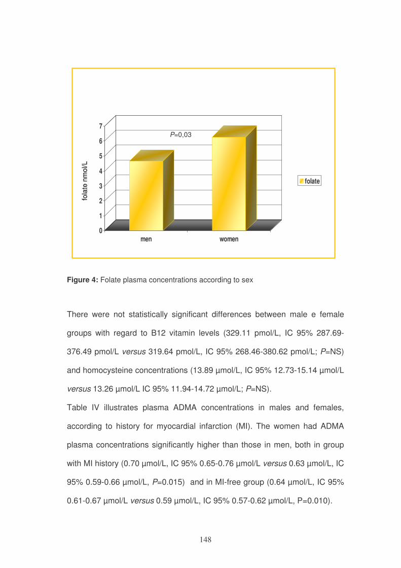
148
P=0,03
fola
to n
mo
l/L
0
1
2
3
4
5
6
7
men women
folate
fola
te n
mo
l/L
Figure 4: Folate plasma concentrations according to sex
There were not statistically significant differences between male e female
groups with regard to B12 vitamin levels (329.11 pmol/L, IC 95% 287.69-
376.49 pmol/L versus 319.64 pmol/L, IC 95% 268.46-380.62 pmol/L; P=NS)
and homocysteine concentrations (13.89 µmol/L, IC 95% 12.73-15.14 µmol/L
versus 13.26 µmol/L IC 95% 11.94-14.72 µmol/L; P=NS).
Table IV illustrates plasma ADMA concentrations in males and females,
according to history for myocardial infarction (MI). The women had ADMA
plasma concentrations significantly higher than those in men, both in group
with MI history (0.70 µmol/L, IC 95% 0.65-0.76 µmol/L versus 0.63 µmol/L, IC
95% 0.59-0.66 µmol/L, P=0.015) and in MI-free group (0.64 µmol/L, IC 95%
0.61-0.67 µmol/L versus 0.59 µmol/L, IC 95% 0.57-0.62 µmol/L, P=0.010).
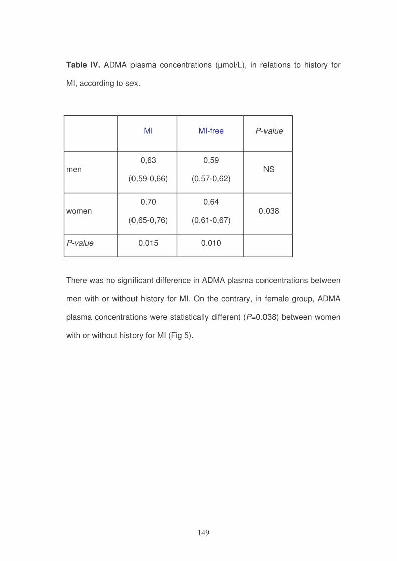
149
Table IV. ADMA plasma concentrations (µmol/L), in relations to history for
MI, according to sex.
MI MI-free P-value
men 0,63
(0,59-0,66)
0,59
(0,57-0,62) NS
women 0,70
(0,65-0,76)
0,64
(0,61-0,67) 0.038
P-value 0.015 0.010
There was no significant difference in ADMA plasma concentrations between
men with or without history for MI. On the contrary, in female group, ADMA
plasma concentrations were statistically different (P=0.038) between women
with or without history for MI (Fig 5).
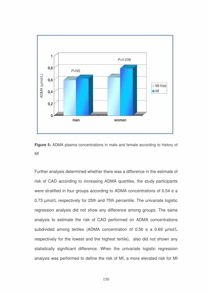
150
0
0,2
0,4
0,6
0,8
1
men women
MI-free
MI
AD
MA
(µ
mo
l/L
)
P=NS
P=0.038
Figure 5: ADMA plasma concentrations in male and female according to history of
MI
Further analysis determined whether there was a difference in the estimate of
risk of CAD according to increasing ADMA quartiles, the study participants
were stratified in four groups according to ADMA concentrations of 0.54 e a
0.73 µmol/L respectively for 25th and 75th percentile. The univariate logistic
regression analysis did not show any difference among groups. The same
analysis to estimate the risk of CAD performed on ADMA concentrations
subdivided among tertiles (ADMA concentration of 0.56 e a 0.69 µmol/L
respectively for the lowest and the highest tertile), also did not shown any
statistically significant difference. When the univariate logistic regression
analysis was performed to define the risk of MI, a more elevated risk for MI
151
according to ADMA concentrations higher than the upper tertile was
observed (OR=2.14, 1.17-3.92, IC 95%, P=0.012).
The univariate logistic regression analysis performed to define the risk of MI
according to sex, highlighted a more elevated risk depending on ADMA
concentrations higher than the superior tertile only for the female subgroup
(OR=2.79, 1.07-7.27, IC 95%, P=0.032). There was no such associations in
male subgroup of subjects (OR=1.71, 0.77-3.85, IC 95%, P=0.19). Moreover,
in the female subgroup, the higher risk for MI for ADMA concentrations
higher than 3rd tertile was maintained in a multiple logistic regression model
adjusted for menopause (OR=2.75, 1.04-7.28, IC 95%, P= 0.041) and also
for other factors such as age, family history for CAD, hypertension and BMI
(OR=2.23, 1.09-4.54, IC 95%, P=0.027).
In the multinomial regression analysis, higher risk for MI remained statistically
significant also after adjustment for smoking (OR=2.04, 1.09-3.82, IC 95%,
P=0.026) and diabetes mellitus (OR=1.86, 0.99-3.47, IC 95%, P=0.050).
When the risk of MI associated to ADMA tertiles was adjusted for
homocysteine plasma levels, the statistical analysis lost the statistically
significant value (P=0.19), since there was a correlation between the two
variables. In the multinomial regression analysis, however, also
homocysteine lost the statistically significant value (P=0.21).
152
Discussion
The cardiovascular disease has a composite genesis based on many
different factors. In the recent years, several studies attempted to identify
some new risk factors to support classic factor as diabetes, cholesterol,
obesity, smoking, age and familiarity.
Among new parameters that have been studied, ADMA may be considered a
cardiovascular risk marker in numerous diseases related to endothelial
dysfunction increased cardiovascular mortality and morbidity.
In the multicenter case-control CARDIAC study (Coronary Artery Risk
Determination investigating the Influence of ADMA Concentration) Schulze
and colleagues23 analysing 131 CAD patients and 131 controls from the
general population, found that cases had higher ADMA plasma levels than
controls. This relationship remained statistically significant (p=0,001) also
after multivariare logistic regression model including laboratory parameters
and traditional risk factors (homocysteine was not take into consideration), so
they concluded that ADMA is an independent risk factors for coronary heart
disease23. Some Researchers report that ADMA is significantly increased in
patients with coronary artery disease, especially in presence of chronic renal
failure24.
The present study suggests the hypothesis that a marker within one-carbon
metabolism, ADMA plasma concentration, may be a link a risk factor for MI to
be considered more carefully in female gender.
153
One of the main peculiarities of this study is the angiographic method to
enlist the population in case-group and control-group for coronary
atherosclerosis.
These data highlighted that ADMA plasma concentrations did not affect the
risk of CAD, but its main thrombotic complication, myocardial infarction. That
result may be of particular interest because, to date, the factor influencing the
evolution of CAD in MI is yet not completely known. Moreover, ADMA plasma
concentrations higher than 0.69 µmol/L, that corresponding to 3th tertile in
studied population, may be considered an index for elevated MI risk only in
female subgroup (OR=2.79, P=0.032), on the contrary in male subgroup a
higher risk of MI associated to elevated ADMA levels did not appear
(OR=1.71, P=0.19). Higher risk of MI in women remains statistically
significant also after adjustment for smoking and diabetes; furthermore after
adjustment for age, familiarity, coronary artery disease, hypertension and
BMI. The degree of CAD risk increase for high plasma ADMA concentrations
is little but this cardiovascular disease has a composite genesis with a lot of
genetic and biochemical-enviromental factors.
Other Authors identified an association between ADMA concentrations and
increase of CAD risk2, hypothesizing that high baseline ADMA levels may
have prognostic value and independently predict future cardiovascular
risk10.Therefore, our study lines up with previous studies. A Moreover, the
population studied presented the same social, economical and environmental
conditions and for each subject there were much knowledge about its history,
154
its clinical and biochemical data and the other main classical risk factors, to
define more precisely the risk of cardiovascular disease.
In 2003, Lu e Coll.9 observed that high ADMA levels in patients undergoing
percutaneous coronary intervention (PCI) predicted an increased risk of
restenosis. In 2005 Schnabel and Coll. related that, in 1874 patients with
CAD, high concentrations of baseline ADMA were independently correlated
with future death from cardiovascular causes or non-fatal myocardial
infarction10.
The risk of future cardiovascular event was also associated with increasing
thirds of baseline ADMA (p for trend <0,001) and this relationship remained
nearly unchanged after adjustment for traditional risk factors (homocysteine
didn’t take into consideration).
Other observations pointed out that ADMA plasma concentrations were
positively correlated with age25 in 1126 non-smoking healthy individuals from
the Framingham Offspring Study; in that same study, ADMA levels were
higher both in women than in men and in post-menopausal women than in
pre-menopausal women.
In our study there were not statistically significant differences between male
and female for age.
Bae and colleagues26 observed that in patients with acute coronary
syndrome ADMA is significantly higher than in controls and it rapidly
decreases after short-term medical therapy. Analogously, other studies have
demonstrated that ADMA in patients with angina instable is higher than in
155
patients with stable angina and there is a significant reduction of ADMA
levels after percutaneous coronary intervention in patients with unstable
angina6., High ADMA levels may independently predict major adverse
cardiovascular events also in subjects with advanced peripheral artery
disease (PAD)27.
Korandji and Colleagues28 investigated the relationship between increased
levels of ADMA and plasma homocysteinemia (tHcy), an independent risk
factors for atherosclerotic vascular disease, in patients with acute myocardial
infarction. In the 138 patients with MI studied, ADMA was positively
associated with SDMA (p<0,001) and tHcy (p=0,03). By multiple linear
regression, SDMA but not ADMA was independently associated with tHcy
(p=0,005; vs 0,74). They concluded that in AMI patients, ADMA levels are
independent of traditional cardiovascular risk factors and high levels of tHcy
are indirectly related to ADMA.
With regards to the relationship among ADMA levels and homocysteinemia,
Antoniades and colleagues29 observed that ADMA levels increased rapidly in
acute homocysteinemia. Increased oxidative stress is the main probable
mechanism underlying endothelial dysfunction homocysteinemia-induced,
with increased concentration of pro-inflammatory cytokines.30,13,31
In our study, ADMA correlates with homocysteine and the latter correlates
with folate. So ADMA may have significance in risk definition and prevention
in relation to biochemical-nutritional data. Other studies are needed to
confirm this hypothesis.
156
In conclusion, ADMA may represent an important prognostic factor to
developing thrombotic complications in CAD patients. According to previous
results reported in other studies, ADMA may be considered a risk factor for
myocardial infarction, although the homocysteine have a predominant role
among these.
Many studies dealt with defining generic risk, though knowing the different
risk for atherosclerosis in woman and in man. Considering the significance to
define markers of atherosclerotic disease according to gender, the results of
this study highlights some relevant observations that contribute to identify
new factors of MI risk, the main complication of CAD, in female population.
References 1. Boger RH. Asymmetric dimethylarginine, an endogenous inhibitor of
nitric oxide synthase, explains the "L-arginine paradox" and acts as a
novel cardiovascular risk factor. J Nutr. Oct 2004;134(10
Suppl):2842S-2847S; discussion 2853S.
2. Boger RH, Maas R, Schulze F, Schwedhelm E. Asymmetric
dimethylarginine (ADMA) as a prospective marker of cardiovascular
disease and mortality-An update on patient populations with a wide
range of cardiovascular risk. Pharmacol Res. Dec 2009;60(6):481-
487.
157
3. Leiper J, Nandi M, Torondel B, et al. Disruption of methylarginine
metabolism impairs vascular homeostasis. Nat Med. Feb
2007;13(2):198-203.
4. Schulze F, Wesemann R, Schwedhelm E, et al. Determination of
asymmetric dimethylarginine (ADMA) using a novel ELISA assay. Clin
Chem Lab Med. 2004;42(12):1377-1383.
5. Maas R, Tan-Andreesen J, Schwedhelm E, Schulze F, Boger RH. A
stable-isotope based technique for the determination of
dimethylarginine dimethylaminohydrolase (DDAH) activity in mouse
tissue. J Chromatogr B Analyt Technol Biomed Life Sci. May 15
2007;851(1-2):220-228.
6. Krempl TK, Maas R, Sydow K, Meinertz T, Boger RH, Kahler J.
Elevation of asymmetric dimethylarginine in patients with unstable
angina and recurrent cardiovascular events. Eur Heart J. Sep
2005;26(18):1846-1851.
7. Vallance P, Leone A, Calver A, Collier J, Moncada S. Accumulation of
an endogenous inhibitor of nitric oxide synthesis in chronic renal
failure. Lancet. Mar 7 1992;339(8793):572-575.
8. Zoccali C, Bode-Boger S, Mallamaci F, et al. Plasma concentration of
asymmetrical dimethylarginine and mortality in patients with end-stage
renal disease: a prospective study. Lancet. Dec 22-29
2001;358(9299):2113-2117.
9. Lu TM, Ding YA, Lin SJ, Lee WS, Tai HC. Plasma levels of
asymmetrical dimethylarginine and adverse cardiovascular events
158
after percutaneous coronary intervention. Eur Heart J. Nov
2003;24(21):1912-1919.
10. Schnabel R, Blankenberg S, Lubos E, et al. Asymmetric
dimethylarginine and the risk of cardiovascular events and death in
patients with coronary artery disease: results from the AtheroGene
Study. Circ Res. Sep 2 2005;97(5):e53-59.
11. Boger RH, Sullivan LM, Schwedhelm E, et al. Plasma asymmetric
dimethylarginine and incidence of cardiovascular disease and death in
the community. Circulation. Mar 31 2009;119(12):1592-1600.
12. Surdacki A, Nowicki M, Sandmann J, et al. Reduced urinary excretion
of nitric oxide metabolites and increased plasma levels of asymmetric
dimethylarginine in men with essential hypertension. J Cardiovasc
Pharmacol. Apr 1999;33(4):652-658.
13. Ito A, Tsao PS, Adimoolam S, Kimoto M, Ogawa T, Cooke JP. Novel
mechanism for endothelial dysfunction: dysregulation of
dimethylarginine dimethylaminohydrolase. Circulation. Jun 22
1999;99(24):3092-3095.
14. Boger RH, Zoccali C. ADMA: a novel risk factor that explains excess
cardiovascular event rate in patients with end-stage renal disease.
Atheroscler Suppl. Dec 2003;4(4):23-28.
15. Zoccali C. Prediction of hypertension in hemodialysis patients. Kidney
Int. Mar 2002;61(3):1179-1181.
159
16. Zoccali C, Mallamaci F, Maas R, et al. Left ventricular hypertrophy,
cardiac remodeling and asymmetric dimethylarginine (ADMA) in
hemodialysis patients. Kidney Int. Jul 2002;62(1):339-345.
17. Abbasi F, Asagmi T, Cooke JP, et al. Plasma concentrations of
asymmetric dimethylarginine are increased in patients with type 2
diabetes mellitus. Am J Cardiol. Nov 15 2001;88(10):1201-1203.
18. Fard A, Tuck CH, Donis JA, et al. Acute elevations of plasma
asymmetric dimethylarginine and impaired endothelial function in
response to a high-fat meal in patients with type 2 diabetes.
Arterioscler Thromb Vasc Biol. Sep 2000;20(9):2039-2044.
19. Savvidou MD, Hingorani AD, Tsikas D, Frolich JC, Vallance P,
Nicolaides KH. Endothelial dysfunction and raised plasma
concentrations of asymmetric dimethylarginine in pregnant women
who subsequently develop pre-eclampsia. Lancet. May 3
2003;361(9368):1511-1517.
20. Girelli D, Russo C, Ferraresi P, et al. Polymorphisms in the factor VII
gene and the risk of myocardial infarction in patients with coronary
artery disease. N Engl J Med. Sep 14 2000;343(11):774-780.
21. Girelli D, Friso S, Trabetti E, et al. Methylenetetrahydrofolate
reductase C677T mutation, plasma homocysteine, and folate in
subjects from northern Italy with or without angiographically
documented severe coronary atherosclerotic disease: evidence for an
important genetic-environmental interaction. Blood. Jun 1
1998;91(11):4158-4163.
160
22. Araki A, Sako Y. Determination of free and total homocysteine in
human plasma by high-performance liquid chromatography with
fluorescence detection. J Chromatogr. Nov 27 1987;422:43-52.
23. Schulze F, Lenzen H, Hanefeld C, et al. Asymmetric dimethylarginine
is an independent risk factor for coronary heart disease: results from
the multicenter Coronary Artery Risk Determination investigating the
Influence of ADMA Concentration (CARDIAC) study. Am Heart J. Sep
2006;152(3):493 e491-498.
24. Wang J, Sim AS, Wang XL, Salonikas C, Naidoo D, Wilcken DE.
Relations between plasma asymmetric dimethylarginine (ADMA) and
risk factors for coronary disease. Atherosclerosis. Feb
2006;184(2):383-388.
25. Schwedhelm E, Xanthakis V, Maas R, et al. Asymmetric
dimethylarginine reference intervals determined with liquid
chromatography-tandem mass spectrometry: results from the
Framingham offspring cohort. Clin Chem. Aug 2009;55(8):1539-1545.
26. Bae SW, Stuhlinger MC, Yoo HS, et al. Plasma asymmetric
dimethylarginine concentrations in newly diagnosed patients with
acute myocardial infarction or unstable angina pectoris during two
weeks of medical treatment. Am J Cardiol. Mar 15 2005;95(6):729-
733.
27. Mittermayer F, Krzyzanowska K, Exner M, et al. Asymmetric
dimethylarginine predicts major adverse cardiovascular events in
161
patients with advanced peripheral artery disease. Arterioscler Thromb
Vasc Biol. Nov 2006;26(11):2536-2540.
28. Korandji C, Zeller M, Guilland JC, et al. Asymmetric dimethylarginine
(ADMA) and hyperhomocysteinemia in patients with acute myocardial
infarction. Clin Biochem. Jan 2007;40(1-2):66-72.
29. Antoniades C, Tousoulis D, Marinou K, et al. Asymmetrical
dimethylarginine regulates endothelial function in methionine-induced
but not in chronic homocystinemia in humans: effect of oxidative
stress and proinflammatory cytokines. Am J Clin Nutr. Oct
2006;84(4):781-788.
30. Dayoub H, Achan V, Adimoolam S, et al. Dimethylarginine
dimethylaminohydrolase regulates nitric oxide synthesis: genetic and
physiological evidence. Circulation. Dec 16 2003;108(24):3042-3047.
31. Jacobi J, Sydow K, von Degenfeld G, et al. Overexpression of
dimethylarginine dimethylaminohydrolase reduces tissue asymmetric
dimethylarginine levels and enhances angiogenesis. Circulation. Mar
22 2005;111(11):1431-1438.
162
After we attended both to study the main polymorphisms of the enzymes
involved in one-carbon metabolism and to evaluate its impact on genomic
methylation, investigating the possible existence of nutrient-gene interaction
examples, we proposed to initiate a clinical study to identify possible
functional markers of nutrient-gene interaction with effects on epigenetic
modulation.
To achieve this goal, we set out not only to explore genomic methylation but
also to compare gene-specific methylation on blood tissue with gene-specific
methylation on liver tissue, healthy and cancerous changes affected.
Our aim was to study also the co-interaction with alcohol, another dietary
factor that may determine some changes in folate metabolism with effects on
both hematopoietic tissue and the liver tissue.
The liver, in fact, is the tissue which occurs in almost all the enzymatic
functions relating to the folate cycle and it is known that epigenetic
mechanisms and alcohol play an important role in carcinogenesis and tumor
progression both in hepatocellular carcinoma and in cholangiocarcinoma
intrahepatic. However, in humans liver tissue is obviously not easily
accessible to be studied in terms of biochemical and molecular level.
Consequently, in the literature there are no demonstrations of the correlation
between epigenetic mechanisms, nutrition and liver carcinogenesis. The
search by a gene-nutrition approach of functional markers, documented both
in the liver and in the blood, we believe that it would open new frontiers in
terms of prevention and therapeutic approach of these tumors.
163
CHAPTER 5
FUTURE PERSPECTIVES
GENETICS AND EPIGENETICS RELATIONSHIP IN CANCER TYPE WITH
A LINK TO ONE-CARBON METABOLISM: A POSSIBLE MODEL FOR
THE ROLE OF EPIGENETIC MARKERS IN PERIPHERAL BLOOD
MONONUCLEAR CELLS
Abstract
Folate-dependent one-carbon pathway is related to carcinogenesis for its
function in nucleic acid synthesis and biological methylation. Alcohol
consumption is one of the most important dietary factors implicated in cancer
development and is metabolically related to folate metabolism. Specific
polymorphic genes of folate-related enzymes in association with different
folate and other B-vitamins status influence methyl transfer capacity and
DNA methylation. Such models of gene-nutrient interactions may, therefore,
modulate epigenetic mechanisms strictly linked to carcinogenesis. A causal
relationship between one-carbon metabolism and cancer. in particular
colorectal and liver cancer, has been previously demonstrated in a rodent
model. The effect of alcohol consumption in association with different genetic
patterns of folate-related enzymes and B-vitamins status has not been
studied in humans for their possible role in modifying epigenetic phenomena.
Our hypothesis is that different nutritional pattern alters genomic DNA
164
methylation status and histone epigenetic modifications via one-carbon
metabolism, therefore inducing molecular aberrations that ultimately lead to
neoplastic degeneration and can be detected in the hematopoietic tissue as a
systemic marker of cancer disease. We also hypothesize an interrelationship
between alcohol, one of the main nutrients known to affect both
carcinogenesis and one-carbon metabolism, folate-related polymorphic
genes and B-vitamins status that may alter epigenetic mechanisms and the
expression of critical cancer pathway genes in a gene-nutrient interaction
fashion. To address these issues we proposed a human study to determine
either in peripheral blood mononuclear cells and in liver tissue whether
different degrees of chronic alcohol consumption induce altered genomic
DNA methylation as well as promoter site DNA methylation profiling by an
array-based platform for single-site CpG resolution and to evaluate the
expression of genes by an array-based technology.
The study has been approved by “Comitato Etico of Azienda Ospedaliera
Universitaria Integrata of Verona with Prot. ET 001, Prot. 15295 16/4/2009,
Prog. N. 1647, and Prot. N. 5486 4/2/2010 amendment N.1 27/11/2009.
Summary and specific aims
Different B vitamins status either due to dietary factors or altered absorption
are hypothesized to be related to epigenetic phenomena. Among the most
important dietary factors for hepatocellular carcinoma and colorectal cancer
is alcohol consumption1 which is metabolically related to folate in the
complex biochemical scheme of one-carbon metabolism2. Alcohol has not,
165
however, been studied extensively in an experimental fashion in regard to
how it interacts with one-carbon metabolism in modulating the epigenetic
phenomena which are related to cancer risk and particularly that of
hepatocellular carcinoma and colorectal cancer and how this interaction can
be ameliorated by dietary intervention. In an animal study we previously
observed that chronic alcohol consumption induces hyperhomocysteinemia
and genomic DNA hypomethylation in the colon but folate supplementation
alone cannot prevent this phenomenon, therefore suggesting other possible
mechanisms involved in carcinogenesis which are potentially reversible.
Evidence from both human and animal studies has repeatedly demonstrated
that alcohol impedes one-carbon metabolism. In 1993 Hultberg et al.3 found
that chronic alcoholics were hyperhomocysteinemic. In human alcoholics
Cravo et al.4 also observed hyperhomocysteinemia which was associated
with low blood folate levels and a significant decrease in serum pyridoxal-5’-
phosphate (the active form of vitamin B6). Alcohol stimulates catabolism of
methionine to generate cysteine and replenish glutathione, but at the same
time, the cell attempts to conserve methionine through the choline and
betaine pathway. This results in a drastic wastage of methyl groups and B6.
Studies in a number of different mammals indicate that chronic alcohol
ingestion diminishes methionine synthase activity in the liver, reducing the
availability of methionine and SAdoMet, the universal methyl donor. In a
recent rodent study we observed an impairment in one-carbon metabolism in
the colonic mucosa of the alcoholic rat, leading to diminished methylation of
DNA5. It is also not surprising that chronic alcohol ingestion would cause a
166
substantial shift in the distribution of the various forms of intracellular folate.
Alcohol ingestion also causes an increase in the proportion of intracellular
folate appearing as methylated THF and THF and a decrease in the
proportion of formylated THF. Alcohol diverts formylated THF towards serine
synthesis, thus folate-mediated thymidine synthesis is also interrupted.
These alcohol-induced changes in folate form distribution are similar to what
occurs in vitamin B12 deficiency (i.e. a reduction in the proportion of those
co-enzymatic forms of folate which are used for nucleotide synthesis), a
condition which increases uracil misincorporation as well as decreased DNA
methylation status in the colon6. Vitamin B12 deficiency is thought to be less
common in chronic alcoholics. Nonetheless, tissue deficiencies of this vitamin
may occur despite subnormal, normal, or even higher circulating
concentrations, suggesting that chronic alcohol consumption may impair the
availability of B12 in tissues7. In a four-months long animal study Barak et al.
reported that chronic alcohol feeding inhibited the activity of MS throughout
the study, but increased the activity of BHMT and decreased betaine levels to
maintain vital tissue levels of SAdoMet 8.
Our long-term goal is to highlight possible markers of genetic and epigenetic
disruption that are related to one-carbon metabolism associated to alcohol
consumption and, ultimately, to find an effective strategy for possible
chemoprevention. Such models of gene-nutrient interactions may, therefore,
modulate epigenetic mechanisms strictly linked to carcinogenesis.
Considering that: a) metabolism of folate and other water-soluble B vitamins
such as vitamin B12, B6, thiamine and betaine are altered in diseases of the
167
hepatic tissue; b) such B vitamins alterations are mainly observed in hepatic
diseases due to alcohol ingestion; c) despite similar amounts of alcohol
ingestion not all people develop alcohol liver disease including hepatocellular
carcinoma, therefore suggesting the role of possible genetic factors; d) both
alcohol and B vitamins impairment alter one-carbon metabolic pathway and
may lead to epigenetic aberrations that potentially underlie neoplastic
transformation of liver tissue; e) both alcohol and B vitamins impairment are
also reflected in hematopoietic tissue aberrations. We, therefore, hypothesize
that B vitamins impairment whether or not associated with alcohol ingestion
may induce molecular aberrations in a gene-nutrient interaction fashion that
are similar in peripheral blood mononuclear cells and in hepatocellular
carcinoma secondary to alcohol-induced cirrhosis tissue and liver metastatic
tissue due to colon adenocarcinoma.
For the easier availability of hematopoietic tissue in humans, the identification
of biochemical and molecular markers in such tissue may represent an
important finding for risk stratification study and for chemopreventive
strategies. In particular, since epigenetic phenomena are potentially
reversible, their modulation may be done by dietary habits modifications in
which nutritional intervention may reduce the pro-carcinogenic effects of
alcohol consumption and prevent carcinogenesis.
The present proposal will constitute the first study to investigate the
mechanism(s) by which altered one carbon metabolism induced by alcohol
consumption alters epigenetic phenomena in hematopoietic tissue and liver
as well as liver metastatic tissue from colorectal cancer. To address these
168
issues, we propose to conduct an observational and prospective study in
human subjects. In brief, our specific aims are as follows: a) to determine
gene-nutrient interactions between B vitamins (folate, vitamin B12, B2, B6,
betaine, choline, thiamine) and polymorphic genes within one-carbon
metabolism that may affect epigenetic phenomena such as DNA genomic
and gene-specific methylation as well as histone modifications in human
peripheral blood mononuclear cells and liver tissue in a different age and
dietary setting; b) to determine B vitamins status, one-carbon pathway
biochemical markers such as homocysteine and genetic polymorphisms
within one-carbon metabolism genes in subjects either assuming or not
alcohol drinks and of different age (adults or elderly); c) to determine whether
B vitamins abnormalities either alone or in association to alcohol
consumption also induces altered expression of gene pathways related to
carcinogenesis in human peripheral blood mononuclear cells and liver tissue.
The main goal of the study is therefore to determine the ‘molecular signature’
(genetic and epigenetic markers) in peripheral blood mononuclear cells as
wells as hepatocarcinoma tissue or liver metastasis tissue from colonic
cancer compared to normal tumour-surrounding tissue, in order to focus on
those genes whose expressions most likely mediate folate metabolism-
related epigenetic phenomena.
We expect that the study of mononuclear cells from peripheral blood show
similar molecular signatures to those in primary or secondary tumour of the
liver tissue and therefore can be used as a valid biochemical marker of
folate-related carcinogenesis
169
Our main hypothesis is that of the existence of a gene-nutrient interaction
between folate-related B vitamins, polymorphisms within one-carbon
metabolism genes that can affect epigenetic phenomena, other endpoints will
be biochemical markers related to folate pathway such as plasma folate,
vitamin B12 and holotranscobalamin levels9, 10, pyridoxal phosphate as the
active form of vitamin B611 as well as homocysteine12, SAdoMet, SAdoHcy.
We, therefore, proposed to test biochemical parameters such as plasma
folate, vitamin B6 and B12 concentrations, homocysteine and the major
biochemical parameters defining the stage of disease and as well as to
determine the genotype of polymorphisms in folate-related genes.
We are planning to explore mechanistic pathway with a high-throughput
novel array technology for DNA promoter methylation, chromatin remodelling
and gene expression in a limited subset of subjects in order to define specific
pathways or genes of interest related to cell cycle control. Genomic DNA
methylation will be also determined by a precise quantitative method13.
Based on the data including new genes will be found from microarray, we
would like to pursue to find the exact mechanism(s) by which alcohol
enhances hepatocellular carcinogenesis and to find whether molecular
markers expressed in peripheral blood mononuclear cells of healthy subjects
consuming alcohol may show a biochemical-molecular signature related to
aberrant folate-related pathway that may offer novel chemopreventive
strategy including optimal dose, time and combination of B vitamins such as
mainly folic acid, vitamin B6 or betaine. We will also extend our observation
to genes involving retinoic acid metabolism and JunN-terminal kinase-
170
dependent signaling pathway, alteration of which has been demonstrated in
the liver of alcohol fed animals.
These future studies will provide us with a better mechanistic understanding
of how one-carbon nutrients modulate cancer risk and will provide much
greater insight into how to effectively prevent cancer, and which individuals
we should target for nutritional modulation.
References
1. Cho E, Smith-Warner SA, Ritz J, et al. Alcohol intake and colorectal
cancer: a pooled analysis of 8 cohort studies. Ann Intern Med. Apr 20
2004;140(8):603-613.
2. Giovannucci E. Alcohol, one-carbon metabolism, and colorectal
cancer: recent insights from molecular studies. J Nutr. Sep
2004;134(9):2475S-2481S.
3. Hultberg B, Berglund M, Andersson A, Frank A. Elevated plasma
homocysteine in alcoholics. Alcohol Clin Exp Res. Jun
1993;17(3):687-689.
4. Cravo ML, Gloria LM, Selhub J, et al. Hyperhomocysteinemia in
chronic alcoholism: correlation with folate, vitamin B-12, and vitamin
B-6 status. Am J Clin Nutr. Feb 1996;63(2):220-224.
5. Sauer J, Jang H, Zimmerly EM, et al. Ageing, chronic alcohol
consumption and folate are determinants of genomic DNA
171
methylation, p16 promoter methylation and the expression of p16 in
the mouse colon. Br J Nutr. Jul;104(1):24-30.
6. Choi SW, Friso S, Ghandour H, Bagley PJ, Selhub J, Mason JB.
Vitamin B-12 deficiency induces anomalies of base substitution and
methylation in the DNA of rat colonic epithelium. J Nutr. Apr
2004;134(4):750-755.
7. Cravo ML, Camilo ME. Hyperhomocysteinemia in chronic alcoholism:
relations to folic acid and vitamins B(6) and B(12) status. Nutrition. Apr
2000;16(4):296-302.
8. Barak AJ, Beckenhauer HC, Tuma DJ, Badakhsh S. Effects of
prolonged ethanol feeding on methionine metabolism in rat liver.
Biochem Cell Biol. Mar 1987;65(3):230-233.
9. Morkbak AL, Heimdal RM, Emmens K, et al. Evaluation of the
technical performance of novel holotranscobalamin (holoTC) assays in
a multicenter European demonstration project. Clin Chem Lab Med.
2005;43(10):1058-1064.
10. Nexo E, Christensen AL, Hvas AM, Petersen TE, Fedosov SN.
Quantification of holo-transcobalamin, a marker of vitamin B12
deficiency. Clin Chem. Mar 2002;48(3):561-562.
11. Kimura M, Kanehira K, Yokoi K. Highly sensitive and simple liquid
chromatographic determination in plasma of B6 vitamers, especially
pyridoxal 5′-phosphate. Journal of Chromatography A. 1996;722(1-
2):295-301.

172
12. Araki A, Sako Y. Determination of free and total homocysteine in
human plasma by high-performance liquid chromatography with
fluorescence detection. J Chromatogr. Nov 27 1987;422:43-52.
13. Friso S, Choi SW, Dolnikowski GG, Selhub J. A method to assess
genomic DNA methylation using high-performance liquid
chromatography/electrospray ionization mass spectrometry. Anal
Chem. Sep 1 2002;74(17):4526-4531.
173
CONCLUSIONS
In recent years, the scientific research in nutritional field has pointed out that
many vitamins and micronutrients play an important role in genomic
regulation1: they are, in fact, crucial substrates and cofactors in metabolic
pathways for the DNA synthesis/repair and for the maintenance of DNA
methylation patterns and thus, ultimately, for gene expression regulation.
It is also known that nutrient deficiencies can lead to impairment of genomic
stability by modifying the normal process of methylation. DNA methylation
represents, therefore, a fundamental mechanism for the control of gene
expression and maintainance of genomic integrity2, 3, with important
consequences for the regulation of cell growth, tissue-specific differentiation4-
6 and carcinogenesis3.
It is clear, therefore, the strong link between nutritional status and modulation
of gene expression.
Folate is one of nutrients most widely studied for its role as a methyl group
carrier for nucleotide synthesis and biological methylation. The discovery of
polymorphic variants of enzymes involved in folate-related metabolic pathway
largely contributed to new insights to better define the balance between
genetics-epigenetics and nutrition in phenotypic expression.
The answer to a particular nutritional status in many cases appears to be
specific for each genotype, and alterations of specific nutrients leads to a
different gene expression, in relation to each genotype.
174
A clear example of interaction between nutrition (folate status) and genotype
is represented by a common polymorphic variant of the key enzyme in one-
carbon metabolism, the MTHFR, that is responsible for the availability of
methyl groups for biological methylation reactions7, 8. In presence of this
polymorphism, changes in DNA methylation, appears only in the case of
impaired folate status.
Based on these premises, the first part of our research was performed, in
order to identify new patterns of gene-nutrient interaction regarding other
enzymes involved in one-carbon metabolism. Particular attention has been
directed to interactions between genes and nutrients in the modulation of
DNA methylation.
The field of nutrient-gene interactions appears a fascinating model to
potentially explain different phenotypic expression which may occur in
response to the environment and diet. It is moreover to be pointed out that
the interaction between nutrients and DNA methylation has been recently
emphasized since epigenetic mechanisms are potentially reversible
phenomena, dependent on the methyl groups availability for metabolic
processes.
Thus, the hypothesis of a possible modification of phenotypic expression, by
affecting epigenetic mechanisms through nutritional status, seems to open
new frontiers in the field of disease prevention and new therapeutic
approaches.
For this reason, in the second part of our study we evaluated some possible
175
pathological conditions due to B vitamins deficiency and to identify potential
risk markers for disease. We therefore started a novel study project focused
on carcinogenicity, aimed at evaluating either genomic DNA methylation and
gene-specific methylation, in blood mononuclear cells compared with liver
tissue, organ, in which almost all the enzymatic functions of the folate cycle
occur.
In conclusion, our work contributed to a better understanding of the
mechanisms of nutrition and DNA methylation interrelationship and allowed
to deepen our understanding on gene-nutrient interactions to propose novel
pathways involved in the development of certain chronic diseases as well as
to propose possible innovative prevention strategies by modifying epigenetic
modulation through a nutritional approach.
176
References
1. Fenech M, Ferguson LR. Vitamins/minerals and genomic stability in
humans. Mutat Res. Apr 18 2001;475(1-2):1-6.
2. Wolffe AP, Matzke MA. Epigenetics: regulation through repression.
Science. Oct 15 1999;286(5439):481-486.
3. Jones PA, Laird PW. Cancer epigenetics comes of age. Nat Genet.
Feb 1999;21(2):163-167.
4. Razin A, Riggs AD. DNA methylation and gene function. Science. Nov
7 1980;210(4470):604-610.
5. Robertson KD, Wolffe AP. DNA methylation in health and disease. Nat
Rev Genet. Oct 2000;1(1):11-19.
6. Feinberg AP. Methylation meets genomics. Nat Genet. Jan
2001;27(1):9-10.
7. Friso S, Choi SW, Girelli D, et al. A common mutation in the 5,10-
methylenetetrahydrofolate reductase gene affects genomic DNA
methylation through an interaction with folate status. Proc Natl Acad
Sci U S A. Apr 16 2002;99(8):5606-5611.
8. Stern LL, Mason JB, Selhub J, Choi SW. Genomic DNA
hypomethylation, a characteristic of most cancers, is present in
peripheral leukocytes of individuals who are homozygous for the
C677T polymorphism in the methylenetetrahydrofolate reductase
gene. Cancer Epidemiol Biomarkers Prev. Aug 2000;9(8):849-853.