Embed Size (px)
Citation preview
Tethered cord syndrome
SWISS SOCIETY OF NEONATOLOGY
April 2012
2
Burth S, Berger TM, Winiker H, Caduff JH, Schmitt-Mechelke
T, Kothbauer KF, Neonatal and Pediatric Intensive Care Unit
(BS, BTM), Department of Pediatic Surgery (WH), Department
of Pediatric Radiology (CJH), Department of Neuropediatrics
(SMT), Children’s Hospital of Lucerne, Department of Neuro-
surgery (KFK), Cantonal Hospital of Lucerne, Switzerland
© Swiss Society of Neonatology, Thomas M Berger, Webmaster
3
This female patient was born to a 40-year-old G4/
P2 at term by elective caesarean section because of
breech presentation. Pregnancy had been unevent-
ful. Maternal serologies (HBV, HCV, HIV, lues, rubella
and toxoplasmosis) as well as prenatal routine ultra-
sound scans had been unremarkable and there was
no history of illicit drug use, nicotine or alcohol con-
sumption. The father of the child has type 1 diabetes
mellitus. The two-year-old brother is healthy. There is
no family history of congenital malformations.
The baby girl adapted well with normal Apgar scores.
Her birth weight, length and head circumference were
appropriate for gestational age (all between P50 and
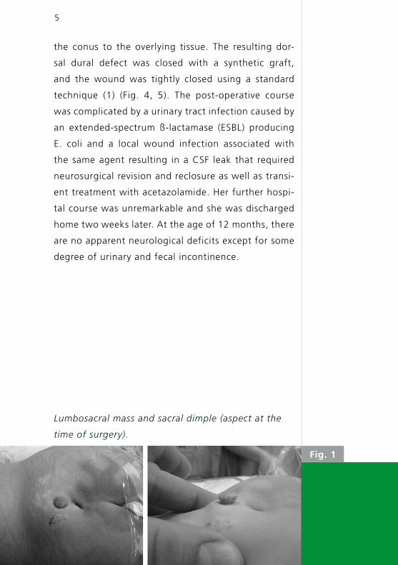
P75). On examination shortly after birth, she was no-
ticed to have a mass over the lumbosacral region and
a sacral dimple approximately 1 cm below this mass
(Fig. 1). The remainder of her physical examination
was normal.
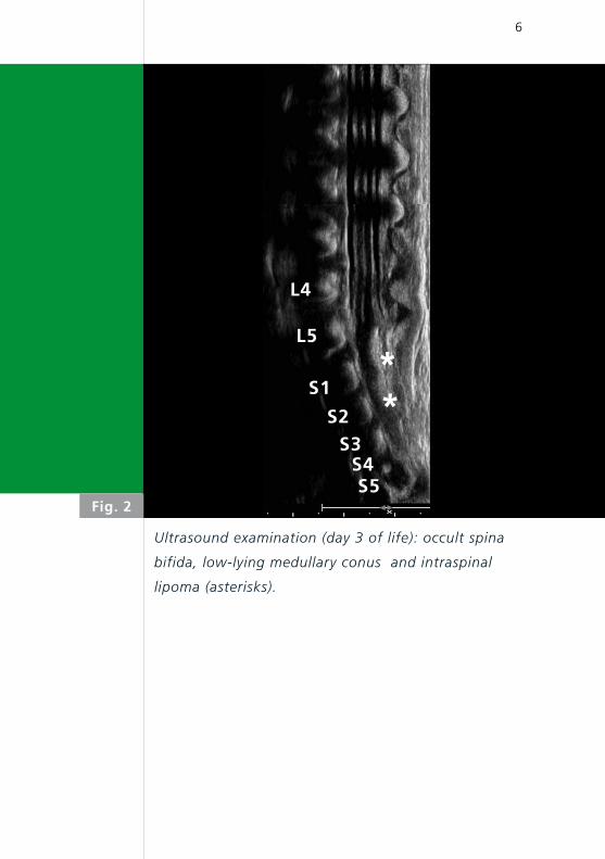
A spinal ultrasound revealed an occult sacral spina bi-
fida, a low-lying medullary conus at the level of L5/S1,
a tethered cord as well as a lipoma within the sacral
canal (Fig. 2). An intraspinal extension of the dermal
sinus could not be ruled out. Even on detailed neuro-
logical examination, muscular tone, deep tendon re-
flexes (DTRs), anal reflex and sensibility of the lower
extremities appeared to be normal. Voiding was unim-
paired without residuals after micturition.
CASE REPORT
4
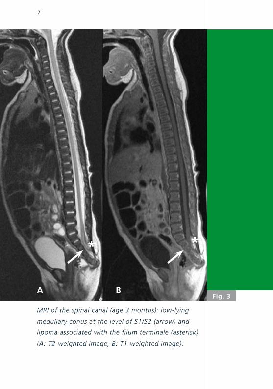
At 3 months of age, a spinal MRI confirmed the low-
lying conus with the apex at the level of S1/S2, an
intraspinal lipoma extending from S1 caudally to the
right and dorsally of the conus and the filum termi-
nale, and a tethered cord (Fig. 3). All sacral vertebrae
were bifid, and there was a sacro-cutaneous duct or
tract and a blindly ending duct beginning over the tip
of the coccyx. Cranial MR images were normal.
Clinically, the girl was thriving and her neurological ex-
amination remained without abnormality. The parents
reported normal voiding and bowel movements. They
had the impression that their daughter might be less
sensitive to painful stimuli than one might expect. A
voiding cystourethrogram (VCUG) revealed no reflux
but signs consistent with a neurogenic bladder. The
family was informed about the concept of prophylac-
tic untethering in the first year of life. Some evidence
of incipient neurogenic bladder and the uncertainty
about the patency of the sacrocutaneous duct repre-
senting an infection risk added to the argument for
surgery.
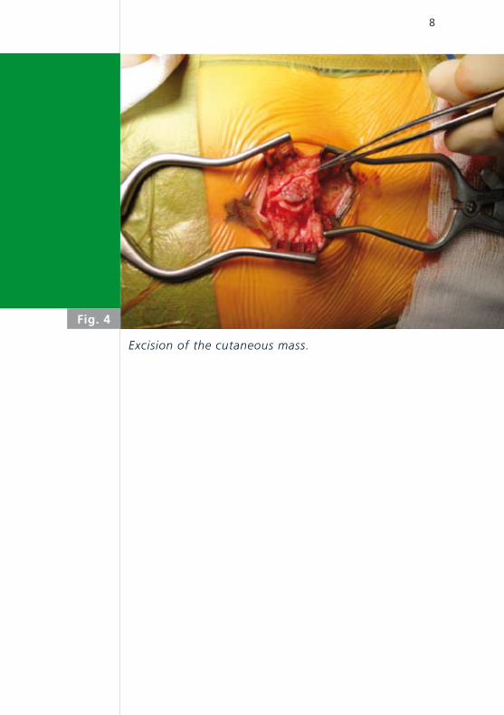
At nine months of age, she was admitted for the ope-
ration. At that time, the deep tendon reflexes appea-
red slightly reduced but otherwise the neurological
examination continued to be unremarkable. At surge-
ry, the tethered conus and the lipoma were exposed.
Untethering was accomplished through partial lipoma
resection and removal of all fibrous attachment of
5
the conus to the overlying tissue. The resulting dor-
sal dural defect was closed with a synthetic graft,
and the wound was tightly closed using a standard
technique (1) (Fig. 4, 5). The post-operative course
was complicated by a urinary tract infection caused by
an extended-spectrum ß-lactamase (ESBL) producing
E. coli and a local wound infection associated with
the same agent resulting in a CSF leak that required
neurosurgical revision and reclosure as well as transi-
ent treatment with acetazolamide. Her further hospi-
tal course was unremarkable and she was discharged
home two weeks later. At the age of 12 months, there
are no apparent neurological deficits except for some
degree of urinary and fecal incontinence.
Fig. 1
Lumbosacral mass and sacral dimple (aspect at the
time of surgery).
6
Fig. 2
Ultrasound examination (day 3 of life): occult spina
bifida, low-lying medullary conus and intraspinal
lipoma (asterisks).
L4
L5
S1 S2 S3 S4 S5
**
7
Fig. 3
MRI of the spinal canal (age 3 months): low-lying
medullary conus at the level of S1/S2 (arrow) and
lipoma associated with the filum terminale (asterisk)
(A: T2-weighted image, B: T1-weighted image).
A B
* *
8
Excision of the cutaneous mass.
Fig. 4
9
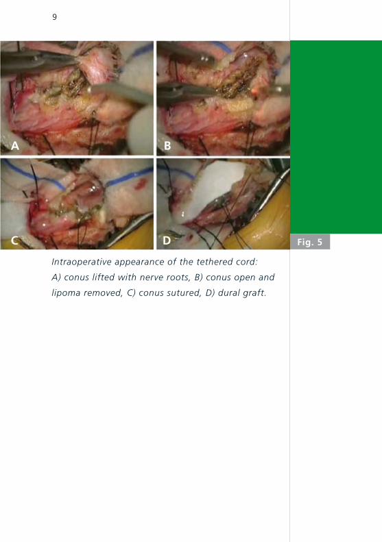
Fig. 5
Intraoperative appearance of the tethered cord:
A) conus lifted with nerve roots, B) conus open and
lipoma removed, C) conus sutured, D) dural graft.
A B
C D
10
DISCUSSION
Open or closed spinal dysraphism occurs with a fre-
quency of 0.5-8:1000 live births (2). There is no uni-
fied risk factor for all occult spinal dysraphic lesions
and depending on the stage of development of the
nervous system at which an insult occurs one or more
dysraphic lesions can develop. Folic acid deficiency has
been clearly associated with open spinal dysraphism (3)
but its relationship with closed spinal dysraphism has
not been evaluated (4). Another well-known risk fac-
tor for spina bifida is maternal exposure to valproate
or carbamazepine (5-7).
Closed spinal dysraphic anomalies can be classified
into three groups based on the developmental stage
at which they are thought to arise (Table). One or more
dysraphic lesions commonly occur in the same patient (8).
Abnormalities of notochord development
n neurenteric cystsn split notochord syndromen split spinal cord malformationsn sacral meningeal cysts or sacral meningocelen dorsal dermal sinus tracts and cysts
Abnormalities of primary neurulation
n syringohydromyelian spina bifida occultan spinal lipomas and teratomas
Anomalies of the caudal cell mass and secondary neurulation
n tethered cord syndrome (TCS)n terminal diplomyelian sacrococcygeal teratomasn caudal regression or sacral agenesis
Classification of closed spinal dysraphic lesions.
Table
11
Failure of fusion of the posterior vertebral arches
during primary neurulation results in spina bifida
occulta. Minor abnormalities of the overlying skin
(naevi, dermal sinus or dimple, underlying lipoma or a
hirsute area) are common (9).
Spinal lipomas are less common than spinal dysraphic
lesions (1:4000 live births) (10). They are thought to
arise from abnormal mesodermal cells that failed to
migrate normally and hence got trapped between the
roof plate and the ectoderm. They often contain vari-
ous types of tissue and thus can be considered com-
plex teratomas (11). They may compress the cord and
thereby cause progressive neurological dysfunction.
Most patients with spinal lipomas have cutaneous
stigmata (subcutaneous lump or dermal sinus). Spinal
lipomas frequently coexist with a tethered cord in the
same patient.
Tethered cord syndrome (TCS) is a stretch-induced
dysfunction of the caudal spinal cord and conus.
Normally, the filum terminale is elastic and does not
cause traction on the spinal cord during inclination.
In TCS, the filum terminale is caudally attached to in-
elastic structures causing a low-impact high frequen-
cy stretch injury upon the cord (12). This may cause
repeated microtrauma, which in the long run leads
to first neuronal and later axonal degeneration sub-
sequently resulting in irreversible neurological deficits
(13, 14).
12
The diagnosis of closed spinal dysraphism in a neonate
requires careful clinical examination. Symptoms lea-
ding to the diagnosis can be cutaneous abnormalities,
upper or lower motor neuron dysfunction and associ-
ated malformations. Since detection of subtle neuro-
logical disturbances and clinical evaluation of bladder
function are obviously difficult in a neonate, typically
located cutaneous stigmata (present in about 80% of
cases) are of great diagnostic value (14). Meticulous
clinical examination of the skin all along the spine is
mandatory. Ultrasonography of the spinal canal is the
initial investigation of choice in an asymptomatic in-
fant with cutaneous stigma in the lumbosacral region.
At about three months of age, an MRI of the spine and
the brain should be obtained for a definitive anatomi-
cal diagnosis. Urological assessment during the first 3
months of life is mandatory and should include micro-
scopic urinalysis, periodic urine cultures, renal func-
tion tests, a VCUG and ultrasonography of the bladder
and the kidneys.
Key elements of successful management of an infant
with closed spinal dysraphism are early detection and
treatment of a TCS, prevention of further deterioration
and treatment of associated malformations. A multi-
disciplinary team approach involving pediatric neurolo-
gists, pediatric neurosurgeons, urologists, orthopedic
surgeons, neonatologists, physiotherapists and social
workers is required. Indication for and timing of sur-
gical untethering in patients with TCS continues to

13
be a controversial issue between the here-proposed
prophylactic concept and a conservative wait-and-see
approach (15, 16). The main principles of surgery are
complete untethering and dural reconstruction with
adequate CSF space around the spinal cord to prevent
retethering (17).
14
1. Zide BM, Epstein FJ, Wisoff J. Optimal wound closure after
tethered cord correction. Technical Note. J Neurosurg 1991;
74: 673-676
2. Saitsu H, Yamada S, Uwabe C, Ishibashi M, Shiota K.
Development of the posterior neural tube in human embryos.
Anat Embryol (Berl) 2004;209:107-117
3. Betteridge KJ. Comparative aspects of conceptus growth:
a historical perspective. Reproduction 2001;122:11-19
4. Michelson DJ, Ashwal S. Tethered cord syndrome in childhood:
diagnostic features and relationship to congenital anomalies.
Neurol Res 2004;26:745-753
5. Ceylan S, Duru S, Ceylan S. Valproic acid sodium-induced
spina bifida occulta in the rat. Neurosurg Rev 2001;24:31-34
6. Rosa FW. Spina bifida in infants of women treated with
carbamazepine during pregnancy. N Eng J Med 1991;
324:674-677
7. Morrow J, Russell A, Guthrie E, et al. Malformation risks of
antiepileptic drugs in pregnancy: a prospective study from the
UK Epilepsy and Pregnancy Register. J Neurol Neurosurg
Psychiatry 2006;77:193-198
8. Schijman E. Split spinal cord malformations: report of 22 cases
and review of the literature. Childs Nerv Syst 2003;19:96-103
9. Guggisberg D, Hadj-Rabia S, Viney C, et al. Skin markers of
occult spinal dysraphism in children: a review of 54 cases.
Arch Dermatol 2004;140:1109-1115
10. Tubbs RS, Wellons JC III, Oakes WJ. Occipital encephalocele,
lipomeningomyelocele and Chiari I malformation: case report
and review of the literature. Childs Nerv Syst 2003;19:50-53
11. Pierre-Kahn A, Zerah M, Renier D, et al. Congenital
lumbosacral lipomas. Childs Nerv Syst 1997;13:298-334
REFERENCES
15
12. Yamada S, Won DJ, Siddiqi J, Yamada SM. Tethered cord
syndrome: overview of diagnosis and treatment. Neurol Res
2004;26:719-721
13. Yamada S, Zinke DE, Sanders D. Pathophysiology of tethered
cord syndrome. J Neurosurg 1981;54:494-503
14. Yamada S, Knerium DS, Mandybur GM, Schultz RL, Yamada
BS. Pathophysiology of tethered cord syndrome and other
complex factors. Neurol Res 2004;26:722-726
15. Pugh JA, Tubbs RS, et al. Lipomyelomeningocele/tethered cord
- Prophylactic untethering. Controversies in Pediatric
Neurosurgery. Jallo GI, Kothbauer KF and Pradilla G. New York,
Thieme: 204-207 (2010)
16. Thompson DNP. (2010). Lipomyelomeningocele/tethered
cord - Is surgery always necessary. Controversies in Pediatric
Neurosurgery. Jallo GI, Kothbauer KF and Pradilla G. New York,
Thieme: 204-215 (2010)
17. Gibson PJ, Britton J, Hall DM, Hill CR. Lumbosacral skin
markers and identification of occult spinal dysraphism in
neonates. Acta Pediatr 1995;84:208-209
18. Venkataramana NK. Spinal dysraphism. J Pediatr Neurosci
2011;6:S31-S40
Swiss Society of Neonatology
www.neonet.ch
CONTACT
SUPPORTED BY
con
cep
t &
des
ign
by
mes
ch.c
h


![Resume Kompilasi Skenario 2 [Coccyx]](https://img.pdfslide.net/doc/110x75/563db878550346aa9a940427/resume-kompilasi-skenario-2-coccyx.jpg)