Embed Size (px)
Citation preview

THE MECHANICSOF BREATHINGIN DIFFERENT BODYPOSITIONS.I. IN NORMALSUBJECTS
BY ERNST0. ATTINGER,1 R. GRIER MONROE,AND MAURICES. SEGAL
(From the Lung Station (Tufts) and Department of Inhalation Therapy, Boston City Hospital,and the Department of Medicine, Tufts University School of Medicine, Boston, Mass.)
(Submitted for publication July 8, 1955; accepted April 16, 1956)
In preliminary experiments some striking dif-ferences in the mechanics of breathing in dif-ferent body positions were observed. In view ofthose findings it was thought worthwhile to carryout a more extensive study with the anticipationthat the results might shed some light on the fac-tors which influence the compliance and mechani-cal resistance of the lung.
Compliance is defined as the volume changebrought about by one centimeter of water pres-sure (L. per cm. H20). The lower the compli-ance the stiffer the lung and vice versa. Mechani-cal resistance measures the amount of pressurenecessary to obtain a certain flow rate and is ex-pressed as cm. H20 per L. per sec.
In this paper, mechanical resistance includesboth the resistance to air flow per se and the resis-tance to tissue deformation. The latter resistancehas been measured to be about 30 to 40 per cent ofthe total mechanical resistance in normal lungs(1). Because the pressure flow relationships arenot linear, the mechanical resistance changes con-tinuously over the respiratory cycle especially inpatients with increased resistance. In order toget better comparative values over an entire re-spiratory cycle, values for mean mechanical re-sistance are given in this paper.
METHODS
Intraesophageal pressures were substituted for intra-pleural pressures. The method used was essentially theone described by Mead and Whittenberger (2, 3), em-ploying an esophageal balloon, 15 cm. in length and in-flated with 1 ml. of air. All pressures were measured bymechano-electrical transducers. Flow rates were meas-ured by a pneumotachograph. The volume flow rate waselectrically integrated by a condenser-resistor unit. Be-cause the integrator only maintained linearity for 10 sec-onds, an automatic switch was provided which dischargedthe condensers at determined time intervals (5 to 8 sec-onds). A controlled secondary circuit made it possible
1 Research Fellow in Medicine, Tufts University Schoolof Medicine, Boston, Mass.
to correct for volume differences caused by the respira-tory quotient and temperature and humidity changes be-tween the inspired and expired air. Consequently the baseline remained horizontal in a given midposition. Thevolumes measured are the inspired volumes at 25°C (notsaturated) and are reported as such. This would reducethe compliance values about 7 per cent as compared tovalues corrected for B.T.P.S. All electrical outputswere fed into a Sanborn four channel recording unit.
Since considerable variations may occur between in-dividual respiratory cycles caused by heart beats, at leasttwo series of ten cycles each were measured in each posi-tion and at each respiratory rate. The mean complianceof the two series was required to check within 0.03 forvalues above 0.15, within 0.02 for values above 0.1 andwithin 0.01 for values below 0.1. Resistive pressureswere measured by drawing the corrected static intra-pleural pressure and obtaining the difference between dy-namic and static intrapleural pressure at each gAo of asecond for slow respiratory rates and at each %5 of asecond for rapid rates. Tlle resistive pressure was di-vided by the flow rate existing at that particular momentand plotted on graph paper-first as pressure against flowand second, as mechanical resistance against time. Themean was measured planimetrically. All pressures werecalibrated by a water manometer and flow rates andvolumes with suitable Fischer and Porter flowmeters be-fore and after the experiment.
In two subjects with spontaneous pneumothorax intra-pleural pressures were measured through an 18-gaugeneedle and in two patients with pleural effusion with anindwelling 14-gauge polyethylene catheter and a Stathamstrain gauge whose zero point was the posterior axillaryline.
The studies were done on eight subjects who by history,clinical, and x-ray findings were free of chronic pul-
TABLE I
Physical characteristics of eight normal subjects
v.c.B.S. (erect) M.B.C.
Patient Sex Age (ml) (ml.) (L./min.)
B. D. M 35 1.84 4,300 115W. C. M 37 1.70 3,500J. R. F 21 1.56 2,900 96M.G. M 30 1.80 4,500 140J.T. M 23 1.86 4,000C. B. M 37 1.78 3,600J. C. M 49 1.65 3,835 128H. T. M 20 1.65 2,200 80
904
BODY POSITION AND THE MECHANICSOF BREATHING
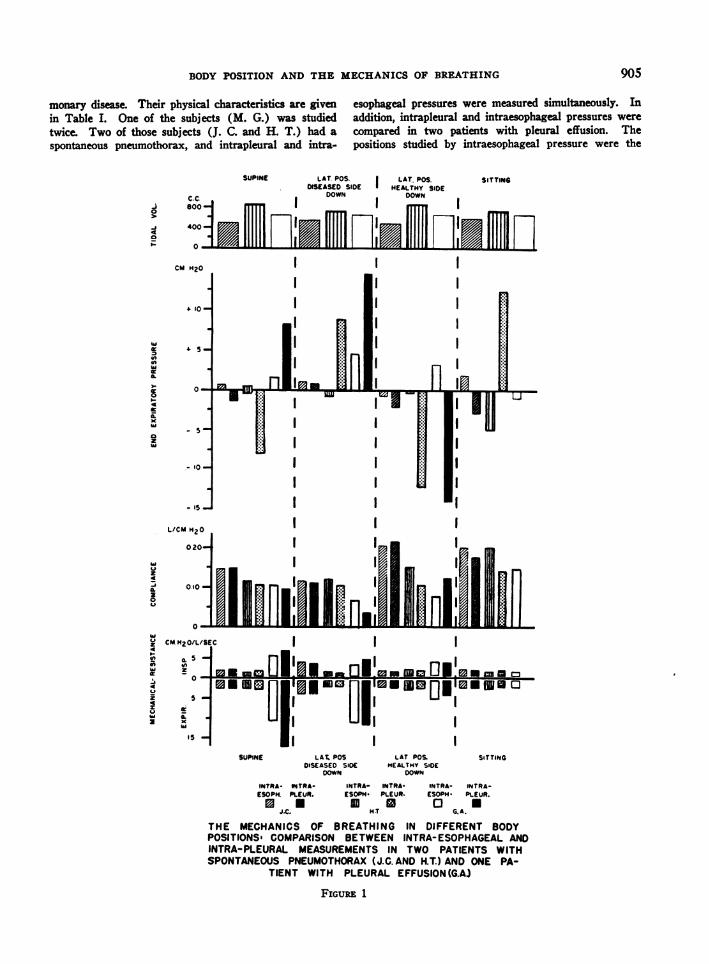
monary disease. Their physical characteristics are givenin Table I. One of the subjects (M. G.) was studiedtwice. Two of those subjects (J. C. and H. T.) had aspontaneous pneumothorax, and intrapleural and intra-
esophageal pressures were measured simultaneously. Inaddition, intrapleural and intraesophageal pressures werecompared in two patients with pleural effusion. Thepositions studied by intraesophageal pressure were the
SUPINE LAT. POS. I LAT. POS. SITTIN6DISEASED SIDE I HEALTHY SIDE
C.C. DOWN DOWN
J MSoo . I I I
CM H20 1 1 I
15
w
or
rLlo
g
SUPINE LAX POS LAT POS. SiTTINGDISEASED SIOE HEALTHY SIDE
DOWN DOWN
INTRA- W4TRA- INTRA- INTRA- INTRA- INTRA-
ESOPH. PLEUR. ESOPH. PLEUR. ESOPH PLEUR.
X III 12! 0CJJ.C. T. G.A.
THE MECHANICS OF BREATHING IN DIFFERENT BODYPOSITIONS- COMPARISON BETWEEN INTRA-ESOPHAGEALANDINTRA-PLEURAL MEASUREMENTSIN TWO PATIENTS WITHSPONTANEOUSPNEUMOTHORAX(J.C. AND H.T.) AND ONE PA-
TIENT WITH PLEURAL EFFUSION(G.A)
FIGURE 1
905
IIIIIIIII
I II II II II II I
ERNST 0. ATTINGER, R. GRIER MONROE,AND MAURICE S. SEGAL
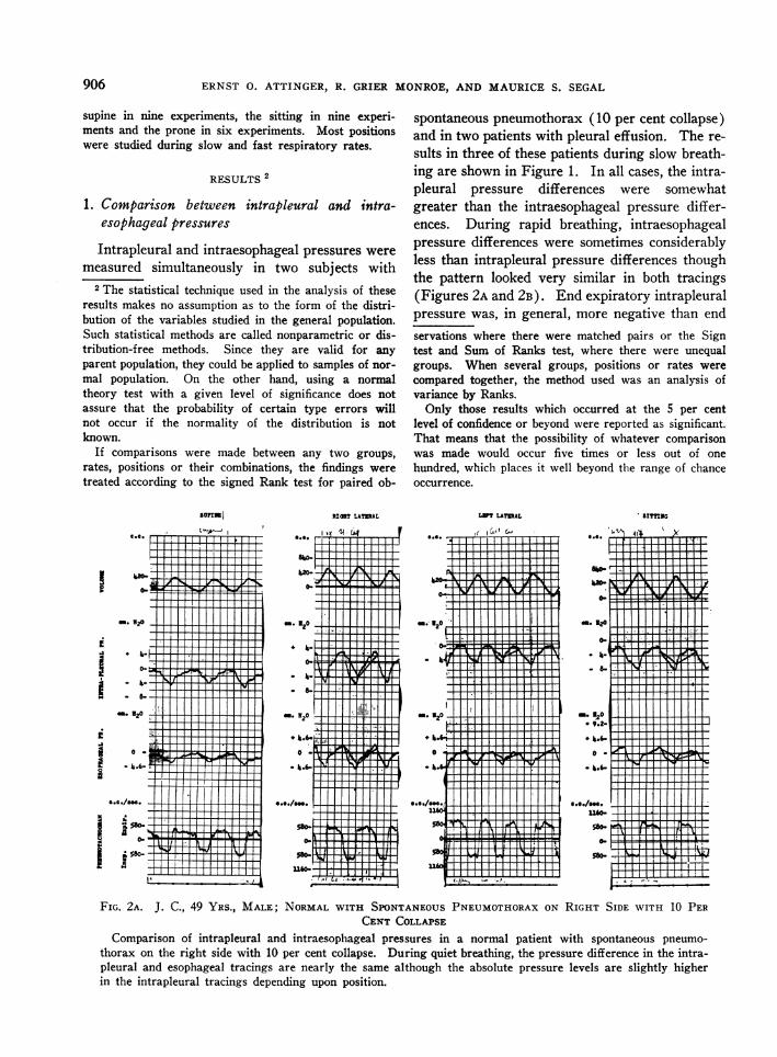
supine in nine experiments, the sitting in nine experi-ments and the prone in six experiments. Most positionswere studied during slow and fast respiratory rates.
RESULTS2
1. Comparison between intrapleural and intra-esophageal pressures
Intrapleural and intraesophageal pressures weremeasured simultaneously in two subjects with
2 The statistical technique used in the analysis of theseresults makes no assumption as to the form of the distri-bution of the variables studied in the general population.Such statistical methods are called nonparametric or dis-tribution-free methods. Since they are valid for anyparent population, they could be applied to samples of nor-mal population. On the other hand, using a normaltheory test with a given level of significance does notassure that the probability of certain type errors willnot occur if the normality of the distribution is notknown.
If comparisons were made between any two groups,rates, positions or their combinations, the findings weretreated according to the signed Rank test for paired ob-
sunul RI|MT LATERAL........I ....... ._
I
I£
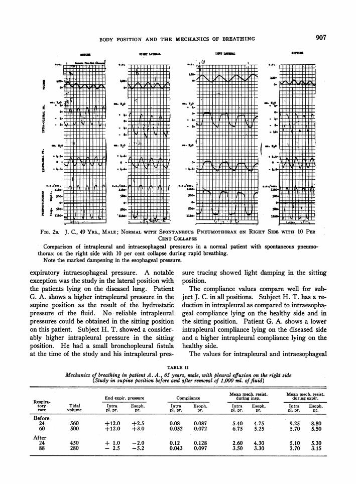
spontaneous pneumothorax (10 per cent collapse)and in two patients with pleural effusion. The re-sults in three of these patients during slow breath-ing are shown in Figure 1. In all cases, the intra-pleural pressure differences were somewhatgreater than the intraesophageal pressure differ-ences. During rapid breathing, intraesophagealpressure differences were sometimes considerablyless than intrapleural pressure differences thoughthe pattern looked very similar in both tracings(Figures 2A and 2B). End expiratory intrapleuralpressure was, in general, more negative than endservations where there were matched pairs or the Signtest and Sum of Ranks test, where there were unequalgroups. When several groups, positions or rates werecompared together, the method used was an analysis ofvariance by Ranks.
Only those results which occurred at the 5 per centlevel of confidence or beyond were reported as significant.That means that the possibility of whatever comparisonwas made would occur five times or less out of onehundred, which places it well beyond the range of chanceoccurrence.
.*.. _
13-
20-*12
IIM.C
L LATEURL * srmsI
I( a . n 41
-. 32,0
aII
FIG. 2A. J. C., 49 YRS., MALE; NORMALWITH SPONTANEOUSPNEUMOTHORAXON RIGHT SIDE WITH 10 PERCENT COLLAPSE
Comparison of intrapleural and intraesophageal pressures in a normal patient with spontaneous pneumo-thorax on the right side with 10 per cent collapse. During quiet breathing, the pressure difference in the intra-pleural and esophageal tracings are nearly the same although the absolute pressure levels are slightly higherin the intrapleural tracings depending upon position.
S.*./". 1:i
906
907BODY POSITION AND THE MECHANICSOF BREATHING
laU! LA?WL
'.'. I(
I
-. 320
0-
* I.
-. 320-
M..,"-.4° -
5160--0.110./.916
ilw
FIG. 2B. J. C., 49 YRS., MALE; NORMALWrrH SPONTANEOUSPNEUMOTHORAXON RIGHT SIDE WITH 10 PERCENTCOLLAPSE
Comparison of intrapleural and intraesophageal pressures in a normal patient with spontaneous pneumo-thorax on the right side with 10 per cent collapse during rapid breathing.
Note the marked dampening in the esophageal pressure.
expiratory intraesophageal pressure. A notableexception was the study in the lateral position withthe patients lying on the diseased lung. PatientG. A. shows a higher intrapleural pressure in thesupine position as the result of the hydrostaticpressure of the fluid. No reliable intrapleuralpressures could be obtained in the sitting positionon this patient. Subject H. T. showed a consider-ably higher intrapleural pressure in the sittingposition. He had a small bronchopleural fistulaat the time of the study and his intrapleural pres-
sure tracing showed light damping in the sittingposition.
The compliance values compare well for sub-ject J. C. in all positions. Subject H. T. has a re-
duction in intrapleural as compared to intraesopha-geal compliance lying on the healthy side and inthe sitting position. Patient G. A. shows a lowerintrapleural compliance lying on the diseased sideand a higher intrapleural compliance lying on thehealthy side.
The values for intrapleural and intraesophageal
TABLE II
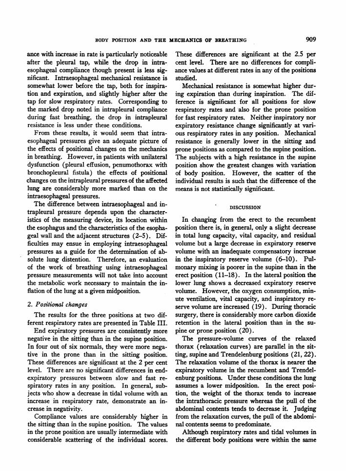
Mechanics of breathing in patient A. A., 65 years, male, with pleural effusion on the right side(Study in supine position before and after removal of 1,000 ml. of fluid)
Mean mech. resist. Mean mech. resist.End expir. pressure Compliance during insp. during expir.
Respira-tory Tidal Intra Esoph. Intra Esoph. Intra Esoph. Intra Esoph.rate volume p1. pr. pr. pl. pr. pr. p1. pr. pr. pl. pr. pr.
Before24 560 +12.0 +2.5 0.08 0.087 5.40 4.75 9.25 8.8060 500 +12.0 +3.0 0.052 0.072 6.75 5.25 5.70 5.50
After24 450 + 1.0 -2.0 0.12 0.128 2.60 4.30 5.10 5.3088 280 - 2.5 -5.2 0.043 0.097 3.50 3.30 2.70 3.15
i
I

ERNST 0. ATTINGER, R. GRIER MONROE,AND MAURICE S. SEGAL
mechanical resistance compare fairly well in sub-jects J. C. and H. T. Patient G. A. shows a some-
what higher intrapleural resistance in the supineposition and lying on the diseased side, while itis somewhat lower lying on the healthy side. Thedirection of positional changes is the same for in-traesophageal and intrapleural compliance andmechanical resistance.
In order to get some ideas about the influence ofa pleural effusion upon these differences, a patientwith pleural effusion before and after a tap of1000 ml. fluid was studied. The results are pre-
sented in Table II. Both intrapleural and intra-esophageal pressures become more negative afterthe tap. Both compliances increase for slow re-
spiratory rates. The drop in intrapleural compli-
TABLE III
Mechanics of breathing: Results in eight normal subects in supine, sitting and prone positionsduring slow and fast respiratory rates
Tidal volumeRespiratory rate (ml.)
Supine Sitting Prone Supine Sitting Prone
Patient SR FR SR FR SR FR SR FR SR FR SR FR
B. D. 10 75 13 12 60 750 570 740 740 690W. C. 1S 33 1S 44 20 50 490 850 505 1,050 607 951
J.R. iS 70 12 75 12 75 580 200 463 370 694 405k .G. (I)* 10 55 22 50 15 43 700 545 665 655 800 700M. G. (I)t 11 37 17 50 15 40 845 811 800 650 830 820J. T. 25 60 16 60 30 60 300 570 400 355 430 650C. B. 15 120 24 496 600 570
J.C. 20 100 6 100 560 245 592 270k. T. 11 80 14 80 850 350 750 435
Mean 14.7 70 15.5 66 17.3 55 620 528 610 542 682 702
Intraesophageal end expiratory presure Compliance(cm. HaG) (L./cm. HtO)
Supine Sitting Prone Supine Sitting Prone
SR FR SR FR SR FR SR FR SR FR SR FR
B. D. +2.0 +1.4 +0.25 + 2.8 + 0.6 0.125 0.120 0.240 0.136 0.146W. C. +0.5 +1.75 0 +2.2 - 0.25 + 1.4 0.223 0.225 0.205 0.225 O.1S0 0.162
].R. +2.0 +0.5 -2.2 -3.5 + O.S - 2.2 0.105 0.082 0.177 0.169 0.189 0.200.. G. (I) +0.5 -0.6 -5.0 -3.5 -13.0 -10.0 0.140 0.150 0.162 0.144 0.072 0.073
M. G. ()t +1.25 +2.6 -3.5 -4.5 - 7.5 - 6.0 0.147 0.146 0.175 0.160 0.097 0.117J. T. +0.5 -2.5 0 -1.0 - 2.8 - 5.5 0.128 0.090 0.253 0.262 0.230 0.205C.B. +1.0 +1.0 -3.5 0.107 0.085 0.174
J.C. +1.8 +0.9 +1.1 -2.0 0.143 0.162 0.161 0.161kI. T. 0 -1.0 -5.0 -5.0 0.112 0.112 0.200 0.210
Mean +1.06 +0.66 -2.0 -2.5 - 3.4 - 3.6 0.137 0.130 0.195 0.190 0.146 0.150
Mean mechanical resistance during inspirtion Mean mechanical resistance during expiration(cm. HsO/L./sec.) (cm. HsO/L./sec.)
Supine Sitting Prone Supine Sitting Prone
Patient SR FR SR FR SR FR SR FR SR FR SR FR
B. D. 2.70$ 1.60 1.34 0.40t 0.65 1.62 1.72 1.55 1.20 0.85W. C. 0.68 0.65 0.24 1.00 1.30 2.74 1.00 3.00
J.R. 5.40 5.00 3.90 4.20 3.80 3.20 7.30 5.30 4.90 5.20 4.00 3.50k.G. I) 3.00 2.67 2.25 3.20 2.30 2.80 4.35 2.22 2.60 3.30 4.40 3.00M. G. II)t 3.05 3.30 2.30 2.90 1.60 2.23 4.25 3.85 3.50 3.20 3.60 3.40J. T. 6.50 3.60 1.90 2.10 3.70 3.54 6.60 3.90 2.70 2.50 4.10 5.10C.B. 3.30 5.80 1.47 3.40 5.10 2.30
J.C. 2.18 2.83 2.34 2.60 2.38 2.80 2.32 2.22lI.T. 1.50 2.40 1.65 1.00 2.50 3.00 2.70 1.30
Mean 3.14 3.40 1.98 2.32 2.13 2.48 3.74 3.49 2.81 2.68 3.38 3.17
* First study.t Second study-two months later.$ Marked interference of heart beats.
Ong8
BODY POSITION AND THE MECHANICSOF BREATHING
ance with increase in rate is particularly noticeableafter the pleural tap, while the drop in intra-esophageal compliance though present is less sig-nificant. Intraesophageal mechanical resistance issomewhat lower before the tap, both for inspira-tion and expiration, and slightly higher after thetap for slow respiratory rates. Corresponding tothe marked drop noted in intrapleural complianceduring fast breathing, the drop in intrapleuralresistance is less under these conditions.
From these results, it would seem that intra-esophageal pressures give an adequate picture ofthe effects of positional changes on the mechanicsin breathing. However, in patients with unilateraldysfunction (pleural effusion, penumothorax withbronchopleural fistula) the effects of positionalchanges on the intrapleural pressures of the affectedlung are considerably more marked than on theintraesophageal pressures.
The difference between intraesophageal and in-trapleural pressure depends upon the character-istics of the measuring device, its location withinthe esophagus and the characteristics of the esopha-geal wall and the adjacent structures (2-5). Dif-ficulties may ensue in employing intraesophagealpressures as a guide for the determination of ab-solute lung distention. Therefore, an evaluationof the work of breathing using intraesophagealpressure measurements will not take into accountthe metabolic work necessary to maintain the in-flation of the lung at a given midposition.
2. Positional changesThe results for the three positions at two dif-
ferent respiratory rates are presented in Table III.End expiratory pressures are consistently more
negative in the sitting than in the supine position.In four out of six normals, they were more nega-tive in the prone than in the sitting position.These differences are significant at the 2 per centlevel. There are no significant differences in end-expiratory pressures between slow and fast re-spiratory rates in any position. In general, sub-jects who show a decrease in tidal volume with anincrease in respiratory rate, demonstrate an in-crease in negativity.
Compliance values are considerably higher inthe sitting than in the supine position. The valuesin the prone position are usually intermediate withconsiderable scattering of the individual scores.
These differences are significant at the 2.5 percent level. There are no differences for compli-ance values at different rates in any of the positionsstudied.
Mechanical resistance is somewhat higher dur-ing expiration than during inspiration. The dif-ference is signlificant for all positions for slowrespiratory rates and also for the prone positionfor fast respiratory rates. Neither inspiratory norexpiratory resistance change significantly at vari-ous respiratory rates in any position. Mechanicalresistance is generally lower in the sitting andprone positions as compared to the supine position.The subjects with a high resistance in the supineposition show the greatest changes with variationof body position. However, the scatter of theindividual results is such that the difference of themeans is not statistically significant.
DISCUSSION
In changing from the erect to the recumbentposition there is, in general, only a slight decreasein total lung capacity, vital capacity, and residualvolume but a large decrease in expiratory reservevolume with an inadequate compensatory increasein the inspiratory reserve volume (6-10). Pul-monary mixing is poorer in the supine than in theerect position (11-18). In the lateral position thelower lung shows a decreased expiratory reservevolume. However, the oxygen consumption, min-ute ventilation, vital capacity, and inspiratory re-serve volume are increased (19). During thoracicsurgery, there is considerably more carbon dioxideretention in the lateral position than in the su-pine or prone position (20).
The pressure-volume curves of the relaxedthorax (relaxation curves) are parallel in the sit-ting, supine and Trendelenburg positions (21, 22).The relaxation volume of the thorax is nearer theexpiratory volume in the recumbent and Trendel-enburg positions. Under these conditions the lungassumes a lower midposition. In the erect posi-tion, the weight of the thorax tends to increasethe intrathoracic pressure whereas the pull of theabdominal contents tends to decrease it. Judgingfrom the relaxation curves, the pull of the abdomi-nal contents seems to predominate.
Although respiratory rates and tidal volumes inthe different body positions were within the same
909
ERNST 0. ATTINGER, R. GRIER MONROE,AND MAURICE S. SEGAL
range in the experiments (only the prone positionshowed a considerable increase in the minute ven-tilation), changes in body position resulted in con-siderable changes of end expiratory pressure,compliance, and mechanical resistance. Thechanges in end expiratory pressures are greaterthan one would expect from changes in midposi-tion alone (23, 24). If the observed changes incompliance were only the result of changes inmidposition, they should also have been noted inchanging from slow to rapid respiratory rates,where marked changes in tidal volumes and mid-position occurred. Therefore, one has to look foradditional factors which could contribute to the ob-served changes in the mechanics of breathing.
A decrease in hydrostatic pressure by removalof intrapleural fluid leads to an increase in com-pliance and a decrease in mechanical resistance.Changes in hydrostatic pressure could influencethe physical properties of the lung directly, butthey also can affect both the distribution of pul-monary blood flow and the forces acting upon thechest wall, mediastinum and diaphragm. Thelatter in turn changes the shape of the thorax and,therefore, the midposition of the lung.
The amount and distribution of blood withinthe lungs influences pulmonary surface tensionand, therefore, compliance and mechanical re-sistance. In patients with mitral stenosis (25)studied in the sitting position, the deviation fromnormal values was more marked when the patientswere in failure. Some of the changes observed inthe supine position may be the result of increasedthoracic blood volume, thus simulating to someextent the changes observed in mitral stenosis.Oser, Ruston, and Ryan (26) showed that the in-crease in venous hydrostatic pressure in the erectposition can exceed the venous tone below thediaphragm and result in blood trapping and re-duction of right atrial pressure.
Another factor to be considered is the amountof unequal ventilation and the opening up of ad-ditional ventilatory space when the lung volume isincreased (27). Our results seem to indicatethat this is a minor factor in normal lungs, com-pliance being constant at different respiratoryrates (28).
The reported changes in the mechanics ofbreathing are probably of no practical importancein normal subjects who have a large functional re-
serve and are capable of compensating automati-cally for the occurring changes. However, theproblem is quite different for patients with cardio-pulmonary disease. Even for the normal subject,it is important to report results of pulmonaryfunction tests with the body position in which thepatient was studied. The changes with body po-sition can exceed the variation of normal valuesin a given position.
SUMMARY
1. The mechanics of breathing were studiedin eight normal subjects.
2. Intrapleural and intraesophageal pressureswere compared in four patients in different bodypositions. Intrapleural pressure differences wereusually somewhat greater than intraesophagealpressures. End expiratory intraesophageal pres-sure is usually more positive than end expiratoryintrapleural pressure. The difference varies fromsubject to subject and in each depending upon thebody position. Intraesophageal pressures seemto be adequate for the measurement of the me-chanics of breathing, but unsuitable for the de-termination of absolute lung distention (midposi-tion).
3. Compliance and mechanical resistance weremeasured during slow and rapid breathing in thesupine, sitting and prone positions.
4. Compliance was lowest in the supine andhighest in the sitting position and did not changewith change in respiratory rate.
5. Mechanical resistance was usually highest inthe supine and lowest in the sitting position, ex-piratory resistance being somewhat higher thaninspiratory resistance in all positions studied.
6. Some of the physiologic factors which mightcontribute to the observed changes are discussed.
7. The full significance of pulmonary functiontesting can only be determined after consideringthe body position during the tests.
REFERENCES
1. McIlroy, M. B., Mead, J., Selverstone, N. J., andRadford, E. P., Measurements of lung tissue vis-cous resistance using gases of equal kinematic vis-cosity. J. Applied Physiol., 1955, 7, 485.
2. Mead, J., and Whittenberger, J. L., Physical prop-erties of human lungs measured during spontane-ous respiration. J. Applied Physiol., 1953, 5, 779.
910
BODY POSITION AND THE MECHANICSOF BREATHING
3. Mead, J., McIlroy, M. B., Selverstone, N. J., andKriete, B. C., Measurement of intraesophagealpressure. J. Applied Physiol., 1955, 7, 491.
4. Cherniack, R. M., Farhi, L. E., Armstrong, B. W.,and Proctor, D. F., A comparison of esophagealand intrapleural pressure in man. J. AppliedPhysiol., 1955, 8, 203.
5. Attinger, E. O., and Segal, M. S., Comparison ofintraesophageal and intrapleural pressures. Inpreparation.
6. Anthony, A. J., Untersuchungen uber Lungenvolu-mina und Lungenventilation. Deutsche Arch. klin.Med., 1930, 167, 129.
7. Bohr, C., Die funktionellen Anderungen in der Mit-tellage und Vitalkapazitat der Lungen. Normalesund pathologisches Emphysem. Deutsche Arch.klin. Med., 1907, 88, 385.
8. Christie, C. D., and Beams, A. J., The estimation ofnormal vital capacity with especial reference tothe effect of posture. Arch. Int. Med., 1922, 30,34.
9. Hurtado, A., and Fray, W. W., Studies of total pul-monary capacity and its subdivisions. III. Changeswith body posture. J. Clin. Invest., 1933, 12, 825.
10. Briscoe, J. C., The mechanism of post-operative mas-
sive collapse of the lungs. Quart J. Med., 1920,13, 293.
11. Blair, E., and Hickam, J. B., The effect of changein body position on lung volume and intrapulmo-nary gas mixing in normal subjects. J. Clin. In-vest., 1955, 34, 383.
12. Bates, D. V., Fowler, W. S., Forster, R. E., andVan Lingen, B., Uniformity of alveolar ventila-tion at different lung volumes. J. Applied Physiol.,1954, 6, 598.
13. Martin, C. J., Cline, F., Jr., and Marshall, H., Lobaralveolar gas concentrations: Effect of body posi-tion. J. Clin. Invest., 1953, 32, 617.
14. Mundt, E., Schoedel, W., and Schwarz, H., Ober dieGleichmiissigkeit der Lungenbeluftung. Pfluger'sArch. f. d. ges. Physiol., 1940, 244, 99.
15. Rauwerda, P. E., Unequal ventilation of different partsof the lung and determination of cardiac output.Dissertation, Groningen University, 1948.
16. Roelsen, E., The composition of the alveolar air in-vestigated by fractional sampling. Comparativeinvestigations on normal persons and patients withbronchial asthma and pulmonary emphysema.Acta med. Scandinav., 1939, 98, 141.
17. Roelsen, E., Fractional analysis of alveolar air afterinspiration of hydrogen as a method for the de-termination of the distribution of inspired air in thelungs. Examination of normal persons and of pa-tients suffering from bronchial asthma and pul-monary emphysema. Acta med. Scandinav., 1938,95, 452.
18. Rohrer, F., Der Stromungswiderstand in den mensch-lichen Atemwegen und der Einfluss der unregel-massigen Verzweigung des Bronchial systems aufden Atmungsverlauf in verschiedenen Lungenbe-zirken. Pfluger's Arch. f. d. ges. Physiol., 1915,162, 225.
19. Vaccarezza, R. F., Lanari, A., Bence, A. E., andLabourt, F., Influencia del decubito lateral sobreel resposo pulmonar. Estudio funcional de cadapulmon por separado. Medicina, Buenos Aires,1942, 2, 279.
20. Beecher, H. K., in Problems in Ventilation. A paneldiscussion. Anesthesiology, 1954, 15, 416.
21. Rohrer, F., Der Zusammenhang der Atemkrafte undihre Abhangigkeit vom Dehnungszustand der At-mungsorgane. Pfluger's Arch. f. d. ges. Physiol.,1916, 165, 419.
22. Rahn, H., Otis, A. B., Chadwick, L. E., and Fenn,W. O., The pressure-volume diagram of the thoraxand lung. Am. J. Physiol., 1946, 146, 161.
23. Stead, W. W., Fry, D. L, and Ebert, R. V., Theelastic properties of the lung in normal men andin patients with chronic pulmonary emphysema.J. Lab. & Clin. Med., 1952, 40, 674.
24. Butler, J., and Arnott, W. M., The work of pulmonaryventilation at different respiratory levels. Clin.Sc., 1955, 14, 703.
25. Mead, J., Frank, N. R., Lindgren, L., Gaensler, E. A.,and Whittenberger, J. L., A technic for the meas-urement of pulmonary compliance and resistance:Its application to normal patients and patients withmitral stenosis. Clin. Res. Proc., 1953, 1, 116.
26. Oser, B. M., Ruston, J. R., and Ryan, J. M., Theeffect of tilting on the right atrial pressures ofpatients with heart failure. Clin. Res. Proc., 1955,3, 197.
27. Bernstein, L., The discontinuity in the pressure vol-ume curve of the rabbit's lung. J. Physiol., 1954,124, 35 p.
28. Attinger, E. O., Herschfus, J. A., and Segal, M. S.,The mechanics of breathing in different body posi-tions. II. In cardiopulmonary disease. J. Clin. In-vest., 1956, 35, 912.
911