Embed Size (px)
Citation preview

www.asner.org
The 6th National Conference of ASNER,
The Romanian Society of Electrodiagnostic Neurophysiology
CN2014, Bucharest, RomaniaOctober 31st –November 2nd 2014
Program & Abstract book
Biblioteca Academiei Române, Calea Victoriei Nr. 125.

ASNER CN2014 Bucharest, Oct 31-Nov 2, 2014– Program & Abstract book6th National Conference of the Romanian Society of Electrodiagnostic Neurophysiology
Scientific partners:
National Neuroscience Societyof Romania
2

ASNER CN2014 Bucharest, Oct 31-Nov 2, 2014– Program & Abstract book6th National Conference of the Romanian Society of Electrodiagnostic Neurophysiology
Dear Friends,
Dear Colleagues,
Dear Guests,
It is for me a pleasure and an honor to invite you tothe 6th National Conference of ClinicalNeurophysiology, here in Bucharest. Since 2009 wehave been dedicated to organizing on an yearly basis2 or 3 scientific events - a Symposium during theNational Congress in Neurology, a Summer School inClinical Neurophysiology, and a National Conference.It is now for the 18th time that we, the organizers,have managed to put together an interesting scientificprogram that, we hope, you will find satisfying. Itisn`t easy, but the enthusiasm of the participants,during all these years works, for us as a real drivingforce.
This year you will have the possibility to listen toLetizia Leocani from Milano, a well known expert inevoked potentials and transcranial magneticstimulation. Bjorn Falck, who has worked for manyyears in Uppsala, will share us from his knowledge inelectromyography. Our dear friend Florin Amzicafrom Montreal will take us again on a fascinatingjourney in the "insides" of electroencephalograhy.
We have prepared for you workshops in EEG andEMG. You will hear interesting reviews in importanttopics of clinical neurophysiology, and colleagueswill present cases where, how we like to say,neurophysiology made the difference.
Let us enjoy the opportunity of gathering together,learning new knowledge, connecting and makingfriends.
Participants at SNN2014 will be awarded 14Continuous Medical Education points (EMC).
Sincerely,
Tudor Lupescu M.D. Ph.D.
ASNER President
http://www.asner.org
Ioana Mindruta, M.D. Ph.D.
ASNER Vice-President
Neurology Department, “Carol Davila” University ofMedicine and Pharmacy, Bucharest, Romania
Ionela Codita, M.D. Ph.D.
ASNER Secretary
Neurology Department of Elias UniversityEmergency Hospital, Bucharest, Romania
Ana-Maria Cobzaru, M.D.
ASNER Treasurer
Neurology Department, “Carol Davila” University ofMedicine and Pharmacy, Bucharest, Romania
Mihai Moldovan, MD, PhD
ASNER Scientific director
Copenhagen University, Denmark and “Carol Davila”University of Medicine and Pharmacy, Bucharest,Romania
3

ASNER CN2014 Bucharest, Oct 31-Nov 2, 2014– Program & Abstract book6th National Conference of the Romanian Society of Electrodiagnostic Neurophysiology
Friday October 31st
12.00 – 12:30 Welcome
12.30 – 13:00 Opening Session (Chair, TudorLupescu)
From Cartesian dualism to social neurosciences (LeonZagrean)
13:00–14:00 Nerves less commonly studied (BjornFalck)
14:00- 16:00 EMG workshop 1
Case demonstrations (Bjorn Falk, Tudor Lupescu,Ana-Maria Cobzaru)
15.45 – 16.00 Coffee break
16:00- 18:00 EMG workshop 2
Practical demonstrations at the conference venue.Tutors: Ana-Maria Cobzaru, Ionela Codita, MirceaMoldovan
16:00- 18:00 EEG workshop 1
Practical EEG demonstrations at University Hospital,9th floor, Epilepsy Center (Ioana Mindruta, MihaiMalaia). Places are limited.
20:00- 22:00 EEG workshop 2
Polysomnography (PSG) demonstrations atUniversity Hospital, 9th floor Epilepsy Center(Floriana Boghez and Irina Moisei-Constantinescu).The PSG recordings will be shown during the CasePresentation Session on Sunday.
Saturday November 1st
9:00- 11:30 Plenary session 1 (Chair, TudorLupescu)
9:00-9:30 Connectomics – Ovidiu Bajenaru
9:30-10:30 Evoked Potentials in Multiple Sclerosis-Letia Leocani
10:30-11:30 Brachial plexus neuropathies -BjörnFalck
11:30-12:00 Coffee Break
12:00-12:30 TEVA symposium
12:30- 14:00 Plenary session 2 (Chair, Dan Psatta)
12:30- 13:30 EEG activity in deep sleep andanesthetic coma. What is different? -Florin Amzica,Montreal (CA)
13:30- 14:00 Evidentiation by EEG Mapping of theCentral Motor Neurons dysfunction in ALS-DanPsatta
14:00-14:30 Lunch
14:30- 15:10 Plenary session 3 (Chair, IoanaMindruta)
14:30-14:50 Patient specific topographic mapping ofelectrophysiological biomarkers to delineate theseizure onset zone in intracranial EEG recordings–Ioana Mindruta
14:50-15:10 EEG in ICU: a monitoring tool forcritically ill patients – Bogdan Florea
15:15 – 15:45 Medison symposium
15:45-16:00 Coffee Break
16:00-17:30 Plenary session 4 (Chair, MihaiMoldovan)
16:00-16:20 Pseudo-neuromuscular pathology –Tudor Lupescu
16:20- 16:40 Elbow Pain: Types, Diagnosis &Treatments – Mircea Moldovan
16:40- 17:00 Myotonia – Edith Sisak
17:00-17:30 Physiological differences betweenperipheral nerves. Does it matter? - Mihai Moldovan
18:00 Festive Cocktail
4

ASNER CN2014 Bucharest, Oct 31-Nov 2, 2014– Program & Abstract book6th National Conference of the Romanian Society of Electrodiagnostic Neurophysiology
Sunday November 2nd
9:00- 10:30 Case presentations 1 (Chair, IonelaCodita)
Approach to Patient with Myoclonus –Electrophysiological Data in a Clinical Case – IonelaCodita
Kinesiotaping in peripheral nervous systemrehabilitation - Gjorgji Nedelkoski, Mircea Moldovan
Atypical motor neuron symptoms –Izabela Popa
Etiologie rară a parezei de nerv sciatic popliteu extern,caz clinic - Epure Diana Anamaria
Polineuropatie cu afectari articulare- prezentare de caz-Vasile Daniela
Diagnostic and therapeutic features of the isolatedvasculitis of the peripheral nervous system - a casepresentation - Antonescu Florian
10:30-11:00 Coffee Break
11:00-11:30 Merk symposium
11:30- 12:30 Case presentations 2 (Chair, AnaMaria Cobzaru)
Polysomnography cases – Ioana Mindruta
Cyclic alternating sleep patterns - Floriana Boghez
Benign EEG variants: are they really benign? -Moisei-Constantinescu Irina
Utilitatea PESS in diagnosticul si managementulneuropatiei diabetice - Calugaru Lidia
Brainstem auditory evoked responses recorded withsurface electrodes in the diagnosis of centralvestibular syndrome in dogs -Stanciu GabrielaDumitrita
Rolul stimulării magnetice transcraniene în urmărireaeficienței tratamentului puseului din SclerozaMultiplă, Trofin Daniela
12:30-13:00 Closing roundtable (certificates,feedback etc)
5

ASNER CN2014 Bucharest, Oct 31-Nov 2, 2014– Program & Abstract book6th National Conference of the Romanian Society of Electrodiagnostic Neurophysiology
2011 : Director of the Department of Neurology,Neurosurgery and Psychiatry -University of Medicineand Pharmacy ” Catrol Davila” Bucharest
2013 ( since ) : Honorary President ( for longlifeduration ) of the Romanian Society of Neurology
2004-2009 : member of the Scientific Committee ofECTRIMS
2005-2009 : member of the Executive Committee ofthe European Society of Neurology
2011( since ) : member of the National Committee ofHabilitation of the Romanian Ministery of Educationfor PhD accreditation and high academic degrees inMedicine
-more than 450 scientific papers published andreported in different national and internationalscientific meetings ; 6 medical books andmonographies ( published in Romania ) -co-author in2 international medical books ; Country PrincipalInvestigator in more than 30 international,multicentric clinical trials -Principal Investigator ofthe research site – in more than 30 international andnational multicentic trials
-coordinator of the CME national program of theRomanian Society of Neurology -coordinator andauthor of the Guidelines for diagnosis and treatmentof neurological diseases ( credited by the College ofMedecins of Romania ) -coordinator of the NationalProgram for treatment of patients with multiplesclerosis of the National House of Insurance andMinistery of Health ( since 2000 ) -coordinator of thefirst medical team in Romania for treatment by DBSin Parkinson’s disease. -chief-editor of RomanianJournal of Neurology ( the official journal of theRomanian Society of Neurology )
2005, 2006, 2010, 2011: awarded by the Prize ofExcelence in Neurology for the scientific activity inRomania ( decided by a National Jury organized bythe Health Chamber of the Romanian Parliament
Ovidiu Bajenaru,
Prof. M.D., Ph.D.
University Emergency HospitalBucharest Department of Neurology,Bucharest,
The Experimental Neurophysiology Unit has basicand clinical research activities aimed at thedevelopment, validation and application of noninvasive electrophysiological methods for thefunctional assessment and neuromodulation of thenervous system, both in humans and in animalmodels. The techniques utilized allow to evaluate thenervous system function, usingelectroencephalography, stimulus-evoked sensory,motor and cognitive potentials, neuromuscular tests,and to provide with non invasive neuromodulatorytreatments through non invasive electrical or magneticstimulation of the brain, spinal cord or peripheralnerves. Moreover, the possibility of applying all thesetechniques both in humans and in animal models ofdiseases allows a translational approach, with parallelassessment of molecular mechanisms and clinicalvalidation.
Dr. Letizia Leocani, after the medical degree at theState University of Milan, took a PhD degree inHuman Physiology and specialized in Neurology atthe same University the Neurology. She was also aResearch Fellow at the Human Motor Control Sectionof the National Institutes of Health in Bethesda-USA).She is currently Senior Researcher at the INSPE-Institute of Experimental Neurology, where she isGroup Leader of the Experimental NeurophysiologyUnit and is responsible of the MAGICSIntraCerebralStimulation Center. She performspsychophysiological research using non-invasiveneurophysiological techniques of brain recording andstimulation.
Letizia Leocani,
MD, PhD
Experimental neurophysiology andMAGICS center, Istituto ScientificoSan Raffaele, Milan, Italy
6

ASNER CN2014 Bucharest, Oct 31-Nov 2, 2014– Program & Abstract book6th National Conference of the Romanian Society of Electrodiagnostic Neurophysiology
Brachial plexus neuropathies (BPN) are seen in about2% of the patients referred to EMG laboratories. Themost common causes are: Parsonage-Turnersyndrome (plexus neuritis), trauma, temporarycompression, chronic compression (thoracic outletsyndrome), iatrogenic lesions (perioperative lesions,complications of local anesthesia), cancer (Pancoasttumour) and Erb’s palsy. The presentation will coverwith (1) anatomy of the brachial plexus, (2) causes ofBPN, (3) neurophysiological methods and strategiesused in the diagnosis of BPN and (4) illustrativeexamples of patients.
M.D. University of Turku, Turku, Finland 1974
Specialist in clinical neurophysiology, Univesity ofTurku, Finland 1980
Ph.D., University of Turku, Turku, Finland 198383
Associate professor in cinical neurophysiology,University of Turku, Turku, Finland 1986
Senior consultant and head, Clinicalneurophysiology, University Hospital, Turku, Finland1986-92, 1998-2007
Senior consultant, Clinical neurophysiologyUniversity hospital, Uppsala, Sweden, 1992-98 and2007-
Publications on neurophysiological methods used forthe diagnosis of neuromuscular disorders. Specialintrest in focal peripheral neuropathies.
Brachial plexus neuropathies
Department of Clinical neurophysiology, Universityhospital, Uppsala, Sweden
Björn Falck
Björn Falck
7

ASNER CN2014 Bucharest, Oct 31-Nov 2, 2014– Program & Abstract book6th National Conference of the Romanian Society of Electrodiagnostic Neurophysiology
The necessity of understanding basic mechanisms ofsleep, both at the cellular and molecular level, hasimposed the necessity of a suitable model. Anesthesiaemerged as the most prominent one, due to a series ofcommon features such as: loss of consciousness,diminished reactivity to noxious stimulation,recuperative properties, and, above all, the ability toproduce electroencephalographic (EEG) patternssimilar to the ones recorded during sleep. The use ofanesthesia for the study of sleep elicited importantbreakthroughs in exposing the cellular mechanisms ofcerebral networks during sleep. However, the above-mentioned model has notable limitations that will bediscussed during the talk. First, sleep is a naturalprocess occurring either actively or passively as afunction of a given neuromodulating set point.Anesthesia acts through activation/blockage ofvarious receptor/transporter sites on neurons and glialcells. There are different classes of anesthetics havingdistinct effects on different receptor categories(GABA, glutamate, etc.). Each of them tends toproduce a given pattern of activity at the global levelof the EEG. This results from the intimate action ofthe anesthetic on various cerebral structures (cortex,thalamus, brainstem, spinal cord). The reactivity ofthe brain is determined by the blood concentration ofthe used anesthetic, leading to a given degree ofdeafferentation. At difference, natural sleep is a self-centered process meant to release/recuperate ahomeostatic pressure built up during the precedingwakefulness. Finally, the end of sleep occurs either atthe end of this regulatory process or at any significantsensory stimulation, while waking from anesthesia isonly allowed when blood levels of the agent dropbelow a threshold blood concentration.
Doctor Florin Amzica has a PhD in neurobiologyfrom the Laval University. He started as an engineerin electronics and informatics, and holds a masterdegree in medical electronics (1982, from thePolytechnics Institute in Bucharest, Romania). Duringhis PhD and after he studied the neuronal mechanismsof sleep and epilepsy with Doctor Mircea Steriade atthe Laval University in Quebec. He has been aprofessor researcher at the Laval University since July1999, and at the Université de Montreal since 2008.
His main research interests encompass:1. The neuron-glia dialogue. Through experimentsthat are underway, he has opened a new field ofinvestigation, studying relationships between neuronsand glia in intact brain networks during physiologicalstates such as sleep, wakefulness and duringpathological states such as epilepsy and coma.2. The study of anesthesia mechanisms. Dr. Amzica isparticularly interested in the mechanisms underlyingthe burst-suppression (BS) pattern defined as a stateduring which the brain reaches one of the lowestlevels of neuronal and metabolic activity.3. Effects of implants and electrical stimulations inpatients with Parkinson’s, dystonia and essentialtremor. The interest is related to the intra-op recordingand the electrophysiological study of thalamic,subthalamic and basal forebrain neurons in humans,as well as the development of a post-op recordingtechnique of these structures.
EEG activity in deep sleep and anesthetic coma.What is different?
Université de Montreal, CanadaAmzica F.
Florin AMZICA
Prof., PhD
8

ASNER CN2014 Bucharest, Oct 31-Nov 2, 2014– Program & Abstract book6th National Conference of the Romanian Society of Electrodiagnostic Neurophysiology
Rationale. We demonstrated in 1986 that acomparison of EEG power obtained under sensorystimulation with the EEG power at rest (%), gives riseto a Frequencies Curve that we called EEG SpectralReaction Curve (SPRC). This curve revealed twopeaks of Delta and Theta increase, two peaks of alphadecrease and 3 peaks of beta increase on stimulation.In further researches we showed that these changescan be reflected by the EEG Mapping (Psatta et al.,1996). The slow waves encountered on these mapsderived from a translation of the slow latecomponents of sensory potentials in terms of EEGfrequencies analysis. But, slow EEG components(Motor potentials) also occur in the central areasduring voluntary movements. Kornhuber and Decke(1965) called them "Readiness potentials", GrayWalter et al. (1964) “Contingent Negative Variation”(CNV). We decided to investigate whether thesepotentials can be also revealed by EEG Mapping, andwhether they are disturbed in ALS (Psatta et al.,2004).
Methods. Our EEG Source Derivation PowerMapping is done on 19 leads (10-20 Internationalrecoding system). An autoregressive model of SourceDerivation was elaborated. Recording and Mapping ofthe main frequency domains power was performedtwice: 1. at rest (with the eyes closed), 2. undertesting. Finally the ratios between testing and restpowers were plotted on a third Maps row, that wecalled Spectral Reaction or Functional EEG Maps.For the purpose, we investigated 14 subjects withdefinite ALS, 9 subjects with probable ALS, 4subjects with bulbar ALS, 11 patients with ALSSyndrome and, by comparison, 5 patients withEpilepsy and 7 patients with Multiple Sclerosis orEncephalitis.
Dan M. Psatta, Scientist, MD, PhD. Working in theInstitute of Neurology of Bucharest, he published 128original papers. As associate Professor in theEcological University he elaborated a treaty ofApplied Neurophysiology. In 1981 he was rewardedfor his scientific activity with the GheorgheMarinescu prize of the Romanian Academy. He waspresident of the Romanian Society of ClinicalNeurophysiology between 1990 and 2002. At presenthe activates in the laboratory of ClinicalNeurophysiology from the Colentina Hospital,affiliated to the Romanian Center of Neurosciences.
Evidentiation by EEG Mapping of the CentralMotor Neurons dysfunction in ALS
Center of Neurosciences Colentina, Bucharest,Romania
Psatta D.
Dan M. PSATTA
Prof. MD PhD
Results. The voluntary motor testing consisted inbilateral symmetric 1/sec finger flexions during oneminute. In these conditions the testing/rest powerMapping shows, in normal subjects a clear-cutactivation of the Rolandic area: projection areas ofthe left and right hands (C3, C4). The activationconsisted in significant Delta and Theta powerenhancement during testing, as well as the increase ofa Delta+Theta/Alpha+Beta coefficient. The motorcenters reaction totally disappeared in ALS patients.One could find a peripheral compensatory reaction inthe pre-motor and retro- rolandic areas. The absentreaction could be uni- or bilateral. This absence wasso more paradoxical, as EEG Mapping images at restwere entirely normal. The Spectral Reaction Curveswere also changed in the C3, C4 EEG sources of ALSpatients. EEG Mapping rolandic reaction images werenormal in the Spondilotic Myelopathy with ALSsyndrome. In Epilepsy the reaction was accompaniedby an activation of the cortical temporal lobe foci. InEncephalitis there were typical EEG Mappingpathological images at rest. The difference betweenthe mentioned groups EEG motor reactions washighly significant statistically, evidencing the severeupper motor neuron dysfunctions only in ALS.
Conclusion. EEG power Mapping may be used as analternative method to Transcranial brain stimulation,for the diagnosis setting in Amyotrophic LateralSclerosis.
9

ASNER CN2014 Bucharest, Oct 31-Nov 2, 2014– Program & Abstract book6th National Conference of the Romanian Society of Electrodiagnostic Neurophysiology
Tudor Lupescu obtained his medical degree from“Carol Davila” University of Medicine in Bucharest,in 1989. After 3 years of training at Colentina ClinicalHospital he became Specialist in Neurology in 1994.Since 2006 he is running the Neurology Departmental Agrippa Ionescu Hospital in Bucharest. 1998, hequalified as Consultant Neurologist. Since his earlyyears of training in Neurology, Tudor Lupescu hasshown a special interest in Clinical Neurophysiology.In 2000 he earned a Competence in ClinicalNeurophysiology (EEG, EMG, and EvokedPotentials). 1997 he was the first to use TranscranialMagnetic Stimulation in Romania. This was also thesubject of his PhD thesis presented in 2005. Since2008, Tudor Lupescu is President of ASNER –Romanian Society of ElectrodiagnosticNeurophysiology. He is also founding member andvicepresident of the the Romanian Society of DiabeticNeuropathy.
Dr Tudor Lupescu is associate member of theAmerican Academy of Neurology, and associatemember of the American Association ofNeuromuscular and Electrodiagnostic Medicine.Between 2008 and 2013 he was also member of theNeurophysiology Subcommittee of ENS
Lupescu T.,
Spitalul Clinic de Urgenta "Prof Dr Agrippa Ionescu"
Tudor Dimitrie LUPESCU
MD, Ph.D.
Pseudo-neuromuscular pathology
Frequently, in our practice, we encounter situationsin which we are dealing with musculoskeletaldisorders that mimic neuromuscular diseases. Suchcases can become a burden on our activity, manytimes the electroneuromyographic examination beinguseless or, musculoskeletal disorders can beassociated with neuromuscular disorders.
Therefore I find it useful to present the most frequentneuromuscular mimics, that involve the upper andlower limbs, among them - supraspinous tendinitis,biceps tendinitis, lateral epicondylitis, de Quervain`stendinitis, carpo-metacarpian arthritis, hiposteoarthritis, greater trochanter bursitis, "pesanserinus" bursitis, plantar fasciitis and, to showsimple clinical tests that can identify such conditions.
10

ASNER CN2014 Bucharest, Oct 31-Nov 2, 2014– Program & Abstract book6th National Conference of the Romanian Society of Electrodiagnostic Neurophysiology
This work attempts to present a brief description ofthe way the body-mind relationship knowledge fromthe Cartesian dualism, passing the mechanistic,reductionist approach to phenomenological andquantic dimension of our neuroworld. The authorpresents a short history of social neurosciences andtheir great impact upon our civilization and nature ofour present and future times.
Leon Zagrean graduated from the Faculty of Medicinein Bucharest in 1978. After a period of four years ofmedical practice he became, through competition,assistant professor at the Department of Physiology,University of Medicine and Pharmacy “Carol Davila”.He got a PhD in Medical Sciences in 1993 and, aftercompleting all academic stages, he became professorin 2002. Since 2004, he is heading the Department/Discipline of Physiology.In addition to teaching and scientific activity inphysiology, Dr. Leon Zagrean, initiated in 1982 astudent research group, which is formed gradually inthe Neuroscience Laboratory, and in 2005, togetherwith Prof. dr. Ovidiu Bajenaru, Head of theDepartment of Neurology of the University Hospital,organizes the Center of Excellence in Neuroscience,accredited by CNCSIS. In 2001 he initiates andorganizes National Neuroscience Society, whosepresident will be until 2012, when it becomesHonorary President. .Leon Zagrean developed in the Laboratory ofNeuroscience a cerebral ischemia/reperfusionexperimental model, which, then, together with agroup of young researchers was extended andexplored by electrophysiological methods, neuronalcell cultures under various conditions hypoxia,metabolic and pharmacological treatments. Thesestudies represented the objectives of several nationaland European research projects whose results havebeen presented at scientific meetings and published inmany papers in journals of international circulation.Prof. Leon Zagrean is editor/author or co-author ofseveral scientific books some of which are publishedin prestigious international publishing houses.Prof. Leon Zagrean is a member of the Academy ofMedical Sciences and a member of several nationaland international scientific societies. He was Directorof the National Biomedical Research “Viasan” in2001-2008.
From Cartesian dualism to social neurosciences
Division of Physiology and FundamentalNeuroscience, "Carol Davila" University of Medicineand Pharmacy, Bucharest, Romania
Zagrean L
Leon ZAGREAN
Prof., MD, PhD
11

ASNER CN2014 Bucharest, Oct 31-Nov 2, 2014– Program & Abstract book6th National Conference of the Romanian Society of Electrodiagnostic Neurophysiology
Seizure onset zone (SOZ) is defined in intracranialEEG recordings as an area that displays tonic lowvoltage fast activity before the occurrence of the firstclinical sign during an epileptic seizure.
Both ictal LVFA (low voltage fast activity) and DCshifts are considered to be robust biomarkers ofepileptogenicity.
We therefore combine these biomarkers in a weightedpower ratio to delineate SOZ during invasivepresurgical diagnostic work up for patients with drugresistant epilepsies.
We calculate instantaneous aEEG over severalfrequency bands of interest and we determine aninstantaneous signal power ratio (IWPR) for allcontacts and perform a 3D topographic animatedrepresentation overlapped with patient’s anatomy.
This signal analysis method helps us to delineate thearea that should be resected during surgeries to makethe patient seizure free.
Funding: UEFISCDI PN-II-ID-PCE-2011-3-0240
45-year old, neurologist, with competence inelectrophysiology and special interest in epileptology,mainly presurgical exploration for epilepsy surgery.PhD thesis on “Sleep studies in epileptic syndromes”in 2006.
Current position at the University EmergencyHospital in Bucharest in the Epilepsy and SleepMonitoring Unit and also hospital coordinator of theNational Programs for Pharmacoresistant Epilepsyand Rare Disorders.
Academic affiliation - lecturer in neurology at theUniversity of Medicine and Pharmacy “Carol Davila”of Bucharest.
Vicepresident of Romanian Association for ClinicalElectrodiagnosis (ASNER) since 2009 and member inthe board of Romanian Society of Neurology since2013.
Patient specific topographic mapping ofelectrophysiological biomarkers to delineate theseizure onset zone in intracranial EEG recordings
Ioana Mindruta (1,2) and Andrei Barborica (3,4)
1 Neurology Department, University EmergencyHospital, Bucharest, Romania
2 Neurology Department, Carol Davila University ofMedicine and Pharmacy, Bucharest, Romania
3 Physics Department, University of Bucharest,Bucharest, Romania
4 FHC Inc, Bowdoin ME, USA
Ioana Mindruta
Assoc.Prof. MD, PhD
12

ASNER CN2014 Bucharest, Oct 31-Nov 2, 2014– Program & Abstract book6th National Conference of the Romanian Society of Electrodiagnostic Neurophysiology
Electroencephalography provides dynamicinformation about the brain function that allow earlydetection of changes in neurologic status, which isespecially useful when the clinical examination islimited. Identification of ongoing electrographicseizures, non-convulsive status epilepticus (NCSE),periodic epileptogenic discharges (PED), irreversiblecerebral dysfunction i.e., isoelectric tracing wouldhelp the care providers in appropriate decision makingregarding the management. Non-convulsive seizures(NCSz) are more common than previously recognizedand are associated with worse outcome if not treatedin time. The discrete movements could bring greatimportance data for diagnosis and therapy. Majorityof seizures at the ICU are not clinically identifiedbecause of the disease phenomena or as the patientmay remain under sedation. Studies revealed the firstNCSz within 1 to 24 hours of EEG monitoring; longerperiod of monitoring is required in comatose patientand those with PED. Factors associated with anincreased risk of NCSz and NCSE include coma, priorclinical seizures, CNS infection, trauma, stroke,hypoxic ischemic encephalopathy, brain tumor, recentneurosurgery, and PED.Brain function monitoring with EEG is useful and thisis in great demand at the ICU of present time. Suchmonitoring can help to improve neurological outcomein a variety of ICU settings.
Dr. Bogdan FLOREA: UMF “Iuliu Hatieganu” Cluj-Napoca, Imogen Research Center
Bogdan Florea graduated the “Iuliu Hatieganu”University of Medicine in Cluj-Napoca in 1997 andbecame senior consulting neurologist in 2012.Clinical neurophysiology fellowships in Italy –Modena and Bologna, USA – Mayo Clinic, Sweden –Uppsala doubled by the daily activity in thecomputerized EEG department of the NeurologicalClinic and many teaching courses in this arearecommend him as a passionate in neurophysiology.His domains of interest are epilepsy andneurophysiology of coma.
EEG in ICU: a monitoring tool for critically illpatients
UMF Cluj-Napoca "Iuliu Hatieganu ", CentrulIMOGEN
Florea Bogdan
Dr. Bogdan FLOREA
13

ASNER CN2014 Bucharest, Oct 31-Nov 2, 2014– Program & Abstract book6th National Conference of the Romanian Society of Electrodiagnostic Neurophysiology
Primul diagnostic in durerea laterala la cot este deepicondilita laterala ( Moris 1882 tenis elbow)deoarece initial este greu de deosebit de sindomul desupinator sau de sindromul de tunel radial Sindromulde supinator prezinta un deficit motor in teritoriul
n interososos posterior (NIP)
Sindromul de tunel radial-este un diagnosticcontroversat ,un sindrom miofascial cu afectarea saunu a n interososs posterior si se manifesta clinicuneori numai prin dureri in partea laterala a cotului 5cm distal de capul radial, exagerate de supinatiacontrarezistenta ,prin cresterea compresiei n radial lanivelul arcadei Frose si foarte rar cu afectareasecundara NIP
Se prezinta cazul unei paciente in etate de 38 ani cudureri cronice la nivelul partii laterale a cotului ,limitarea mobilitatii ,agravare la supinatie si extensiadegetului mijlociu contrarezistenta, sensibilitatelocalala presiune distal de epicondilul lateral, absentaafectarii motorii si de sensibiltatae
Examenul neurografic a fost normal deoarece nervulinterososs posterior contine fibrele groase mielinicecare nu sunt afectate iar fibrele nemielinizate grup IVsi fibre subtiri mielinizate grup II A aferente ,sensibilela presiune, nu pot fi evaluate EMG
Am constatat inca odata dificultatea diagnostica inaceste cazuri si am optat ca localizare spre un sindromde tunel radial cu afectare inervatiei m supinator incursul miscarilor ocupationale ,orientand optiuneaterapeutica
Dr. Mircea Moldovan, graduate of the “Carol Davila”University Bucharest, Doctor of Medical Sciences,MD is a neurologist at the Hospital “Elias” Bucharestsince 1968. Throughout his career, he had acontinuous interest for clinical neurophysiology. Inthe 80s, his main interest was the EEG and evokedpotentials under the guidance of Prof Dr VVoiculescu. In the 90s, his interest expanded to theperipheral conduction studies and EMG. During hispioneering work in Romanian clinicalneurophysiology, Mircea Moldovan advocated thediagnostic importance of clinical neurophysiology forneurological practice through talks at nationalscientific meetings and scientific publications. Mostimportantly, however, through his wealth of practicalexperience and didactic spirit, he helped initiate inclinical neurophysiology generations of youngneurologists. During the last decade, with thetransformation of “Elias” hospital neurology into auniversity department and re-formalizing his skills inEMG (2003) and EEG (2004), Dr. Mircea Moldovandeveloped his preoccupation for clinicalneurophysiology teaching. Together with Dr. IonelaCodita he carries out practical demonstrations of post-graduate courses organized by Professor Dr. PaneaEMG. In addition, Dr. Mircea Moldovan contributedto re-launch of the clinical neurophysiology society inRomania as founding member of ASNER 2009.
Elbow Pain: Types, Diagnosis & Treatments
Neurology and Rehabilitation, Department of EliasUniversity Emergency Hospital, Bucharest, MedlifeClinic Bucharest
Mircea Moldovan, Ionela Codita , Uko Ema Mfon,Lavinia Grozoiu, Patricia Toboc
Mircea Moldovan
MD, PhD
14

ASNER CN2014 Bucharest, Oct 31-Nov 2, 2014– Program & Abstract book6th National Conference of the Romanian Society of Electrodiagnostic Neurophysiology
We present the case of a patient who was hospitalizedfor symptoms consisting of cervical pain andmuscular “cramps”, predominantly of the scapulargirdle. The patient reports onset of symptoms twomonths after the last session of radiotherapyperformed for laryngeal cancer.
Clinical examination reveals involuntary movementsrelevant for myoclonus affecting the axial region andproximal upper limb muscles.
Nerve conduction studies and electromyographyexcluded the brachial plexopathy and other peripheralneuropathy .
The EEG didn’t show epileptiform discharges.
Myoclonus can be classified in a number of ways,although classification based on the underlyingphysiology is the most useful from the therapeuticpoint of view.
We intend to define the presumed source of itsgeneration (cortical, subcortical, spinal, peripheral),taking into account the clinical andelectrophysiological features and also the influence ofseveral elements ( activity during which it occurs,precipitating or alleviating factors).
Ionela Codita is currently working as a SeniorNeurologist in the Neurology Department of EliasUniversity Emergency Hospital in Bucharest. She hasgraduated “Carol Davila” University of Medicine andPharmacy in 1995 and became a specialist inNeurology in 2000.
She earned a Competence in ClinicalNeurophysiology in 2005. During her practice, dr.Codita attended many courses and teaching programsin the field of Clinical Neurophysiology such as:scholarship in Neurophysiopathology field atPoliclinical Institute of San Donato Milanese, Italy(2002-2004), training Course in EMG andNeurography Uppsala , Sweden (2009), InternationalSFEMG and QEMG Course –Kobe, Japan (2010),VIREPA distance learning courses on “EEG in thediagnosis and management of epilepsy – Basic Course6th edition” (September 2011- March 2012) and“EEG SCORE course-1st edition”( November 2012-March 2013), the international educational course“Dinalund Summer School on EEG and Epilepsy”(July 2012).
She also manifests interest in Epilepsy, Motor NeuronDiseases and Movement Disorders. Dr. Ionela Coditais a member of the Romanian Society of Neurology,affiliated to the ENFS (European Federation ofNeurological Societies) and to the WFN (WorldFederation of Neurology) and since May 2013 she isthe secretary of ASNER-The Romanian Society ofElectrodiagnostic Neurophysiology.
Approach to Patient with Myoclonus –Electrophysiological Data in a Clinical Case
1. Elias University Emergency Hospital , Bucharest,RO
2. Hipomed Clinical Care , Chiajna, RO
Ionela Codita(1), Raluca Simona Gurgu(1), LianaStanescu(2), Gabriela Elena Cioara(1), Cristina AuraPanea(1)
Ionela Codita
MD, PhD
15

ASNER CN2014 Bucharest, Oct 31-Nov 2, 2014– Program & Abstract book6th National Conference of the Romanian Society of Electrodiagnostic Neurophysiology
Scleroza multiplă (SM) este una dintre cele maivenerabile boli neurologice, nu numai prin frecvențăși evoluție cronică, cât mai ales prin incidența acesteiaîn rândul adulților tineri.
Procesul de demielinizare ce duce la pierdere demielină la nivelul axonilor este procesul patologicprimar din cadrul acestei afecțiuni, la care se adaugăsuferința neuronală axonală și glială, expresia clinicăa acestui mecanism fiind manifestată prin noțiunea depuseu.
În investigarea SM, un loc tot mai important alături depotențialele evocate vizuale (PEV) și auditive (PEA),îl reprezintă stimularea magnetică transcraniană(TMS).
TMS reprezintă o metodă neinvazivă de evaluare adisfuncțiilor tractului corticospinal la pacienții cu SM,vizând observarea alungirii timpului central deconducere motorie (TCCM), creșterea pragului motorși reducerea amplitudinilor potențialului evocat motor(PEM). Stimularea ariei motorii determină un răspunscare reprezintă comportamentul electrofiziologic alfascicolului piramidal.
Scopului studiului nostru a fost evidențiereamodificărilor PEM obținut prin TMS la pacienții cuSM în puseu (manifestat atât prin deficit motor, cât șiprin manifestări tip nevrită optică, dar șisimptomatologie senzitivă).
Am investigat 37 pacienți cu SM în puseu, cu deficitmotor, din acesti pacienti 26 au prezentat ameliorareasimptomelor după corticoterapie iar 11 pacienți auprezentat agravarea simptomelor.
Pacienții care s-au prezentat în puseu au fostinvestigați prin TMS înainte de administrareacorticoterapiei, după terminarea corticoterapiei(Metiprednisolon 1000 mg/zi, 5 zile) și la o lună.
2013 - prezent
Doctorand în cadrul U.M.F. Iași cu tema „Rolulpotențialului evocat vizual și motor în investigareapuseului din Scleroza Multiplă”;
Ianuarie 2012 – prezent
Medic rezident specialitatea Neurologie SpitalulClinic de Recuperare Iași, Str. Pantelimon Halipanr.14;
2010 – 2011 Medic rezident specialitateaEpidemiologie Spitalul Sf. Spiridon, Iași.
Rolul stimulării magnetice transcraniene înurmărirea eficienței tratamentului puseului dinSclerozaMultiplă
Spitalul Clinic de Recuperare, Iași, Clinica deNeurologie
Dr. Trofin Daniela, Dr. Trofin Dan , Dr. BolboceanOrest, Prof. Dr. Popescu Dinu Cristian
Trofin Daniela
MD, PhD
16

ASNER CN2014 Bucharest, Oct 31-Nov 2, 2014– Program & Abstract book6th National Conference of the Romanian Society of Electrodiagnostic Neurophysiology
ALS is the most common of all motor neurondisorders. Yet, not any patient presenting with lowermotor neuron symptoms is ALS. When cerebellar andpsychiatric signs are associated, hexosaminidase Adeficiency should be considered in the differentialdiagnosis.
The case of a 46 years old male with atypical motorneuron symptoms is presented, where the adult-onsetform of lysosomal enzyme deficiency is suspected.
Izabela Popa graduated the University of Medicineand Pharmacy, Timisoara in 1999. She finishedinternship and residency in neurology in Timisoaraand became neurologist in 2006. Followed clinicalneurophysiology trainings at the Department ofNeurology, University of Szeged, Hungary (2004),Department of Neurology, University of Leipzig,Germany (2005) and the Department ofNeurophysiology, Uppsala University Hospital,Sweden (2008, 2011). In 2007 earned competence inelectromyography and nerve conduction studies andin 2009 received a Certification forElectrophysiological Testing from Albert EinsteinCollege of Medicine of Yeshiva University. Since2007 she works as a private practitioner with specialinterest in neurophysiology.
Atypical motor neuron symptoms
Timisoara
Izabela Popa
Izabela Popa
MD
Neurologist with competence in electrophysiologyand special interest in clinical neurophysiologyworking in the University Emergency Hospital inBucharest as general neurologist and in private sectoras neurophysiologist.
Ana-Maria Cobzaru
MD
Myotonia (clinical cases)
Edith Sisak
MD, PhD
Neurologie, Spit.Jud.Sf.-Gheorghe
Distrofia miotonica este cea mai frecventa forma dedistrofie musculara a adultului, implicand multipleorgane in lantul patologic. Studiile genetice dinultimii 20 ani au clarificat baza moleculara sidiferitele forme ale bolii. Vor fi prezentate douacazuri clinicoe cu examinarea lor electrofizilogica .
EMG cases (practical demonstrations)
17
ASNER CN2014 Bucharest, Oct 31-Nov 2, 2014– Program & Abstract book6th National Conference of the Romanian Society of Electrodiagnostic Neurophysiology
Myelinated peripheral axons are biological structuresspecialized in energy-efficient conduction of actionpotentials. Axonal conduction involves a complicatedvoltage-gated ion channel machinery comprised ofseveral types of Na+ channels (mediating the inwarddepolarizing currents), K+ channels (mediating theoutward rectifying currents) and hyperpolarization-activated cyclic nucleotide-gated channels(mediatingthe inward rectifying currents), as well as energy-dependent pumping mechanisms required to maintainthe ionic concentration gradients across themembranes. Additionally, the spatial distribution ofthese channels is tightly controlled by axon-Schwanncell interactions. In spite of this complexity, theprevailing neurophysiological message aboutconduction is linked to “nerve conduction velocity is6 fold the largest myelinated axon diameter”established nearly a century ago by cat physiologists.This proportionality between electrophysiology andmorphology leads to the oversimplification that “alow conduction velocity reflects a demyelinatingneuropathy”. Undoubtedly this is important toconsider in the clinical context because ademyelinating neuropathy is potentially treatable.
Various normative values exist for nerve conductionvelocities with slight difference between laboratoriesand the technique used. Nevertheless, a consistentobservation is that “normal” nerve conductionvelocity is different between nerves, and, to someextent, between different segments of the same nerve.Does this matter? To answer this, the presentationpleads for a more pathophysiologic approach tounderstanding the factors contributing to nerveconduction velocity and how the electrodiagnosticvalue of low conduction velocity can be helped byother electrophysiological measures such as nerveexcitability studies by “threshold-tracking”.
Mihai Moldovan obtained his medical degree from“Carol Davila” University Bucharest in 1999. Basedon his research interests as a student, after graduationhe was selected to work in the group of prof. ChristianKrarup that continues the Copenhagenneurophysiology school founded by prof. FritzBuchthal in the 60’ with the aim of translatingexperimental neurophysiology into clinicalelectrodiagnostic procedures for patients with nerveand muscle disease. Mihai Moldovan obtained hisPhD degree in neurophysiology from CopenhagenUniversity in 2004 where he continues his scientificcareer as associate professor. His research is focusedon distinguishing the contribution of voltage-gatedion channel dysfunction to pathophysiology ofneurodegenerative disorders, with particular emphasison peripheral nerve excitability testing. While basedin Copenhagen, Mihai Moldovan continued tocollaborate with prof .Leon Zagrean at “Carol Davila”University, Department of Physiology andNeuroscience. In 2011, he founded theCOMAEEG.RO international network, bringingtogether Romanian neuroscientists, clinicians andengineers dedicated to improving the monitoring ofthe comatose brain excitability. Emerging from thesewide research interests are not only originalpublications and review articles in internationaljournals but also educational chapters in neuroscienceand neurophysiology textbooks in Romanianlanguage. Mihai Moldovan has scientific duties inseveral international organizations includingInternational Brain Research Organization (IBRO)and the European Federation of NeuroscienceSocieties (FENS). He is also founder and actingpresident of the National Neuroscience Society ofRomania (SNN) and founder of the Romanian Societyof Electrodiagnostic Neurophysiology (ASNER) forwhich he now serves as scientific director. For hisactivity he received several international and nationalprizes and he was recently appointed editorial boardmember for Clinical Neurophysiology, the officialscientific journal of the International Federation ofClinical Neurophysiology (IFCN).
Physiological differences between peripheralnerves. Does it matter?
1) Copenhagen University DK; 2) Carol DavilaUniversity, Bucharest, RO
Moldovan M. (1,2)
Mihai Moldovan
Assoc. Prof., MD, Ph.D.
18

Case presentations

ASNER CN2014 Bucharest, Oct 31-Nov 2, 2014– Program & Abstract book6th National Conference of the Romanian Society of Electrodiagnostic Neurophysiology
Diagnosticarea precoce a neuropatiei diabetice (ND)prin efectuarea potentialelor evocate somato-senzoriale(PESS) la pacientii cu diabet zaharat careduce, prin tratament precoce corespunzator laschimbarea cursului bolii.
Au fost luati in studiu un numar de 25 de pacienti cuneuropatie diabetica, urmand tratament cugabapentina si vitamine B, si un grup de 20 depacienti cu neuropatie fara tratament.
Cele 2 loturi s-au comparat cu un al treilea lot formatdin 10 pacienti cu diabet, dar fara semne clinice deneuropatie diabetica.
Inregistrarile s-au efectuat in dinamica la fiecarepacient, fiind repetate la un interval in jur de 2 luni.S-au identificat undele componente N9 si N20 si s-aumasurat latentele acestora.
Concluzii
PESS poate fi valorificat in diagnosticul simanagementul neuropatiei diabetice:
-Valorile undei N9 obtinute prin inregistrarea inpunctul Erb a potentialelor sunt mai mari la pacientiicu neuropatie diabetica comparativ cu cele inregistratein lotul martor.
-Prelungirea N20 este prezenta insa la valori maireduse in comparatie cu intarzierea undei N9. UndaN20 are latenta crescuta la pacientii cu AVC ischemicin antecedente, denotand afectarea emisferuluicerebral contralateral stimularii nervului median.
-In lotul martor cu diabet zaharat fara neuropatiecresterile de latenta ale undei N9 au valori mai scazutefata de loturile cu neuropatie diabetica, indicandposibilitatea existentei unei neuropatii infraclinice.
-Datele obtinute prin inregistrarea PES la bolnavii dinlotul cu neuropatie diabetica sub tratament cugamapentina si vitamine grup B confirma partial unefect pozitiv al acestei terapii.
-Evolutia neuropatiei diabetice,inclusiv dupatratament,poate fi monitorizata prin inregistrareaPESS.
Utilitatea PESS in diagnosticul si managementulneuropatiei diabetice
Centrul Medical "Dr. Calugaru" Craiova
Calugaru Lidia, Calugaru Gabriel Tiberiu, CalugaruOvidiu Marius
OBJECTIVESWe shall be presenting the case of a 57 year old male,admitted in our clinic for a tetramelic asymmetricmotor deficit, with a subacute onset in the legs,progressing in the next 8 weeks to the upper limbs,also associated with severe neuropathic pain.
METHODThe clinical and EMG exams confirmed a severemononeuritis multiplex (overlapping type), in asubacute-chronic stage, with a ONLS score of 7/12.The patient has been thoroughly investigated, butwithout confirming neither a systemic vasculitis or aneoplastic process.The biopsy showed possible vasculitic neuropathy,with severe subacute axonal loss and subtle signs of
inflammation.The cerebral MRI revealed chronic microanyopathiclesions and an acute ischemic lesion in the rightthalamus in a patient without any vascular risk
factors.The patient was put on corticosteroids and painmedication, at 3 months the results being favorable,with clinical (5/12 ONLS) and electrophysiologicalimprovement (stabilizing chronic neurogenic lesion).
CONCLUSIONSThe clinical, electrophysiological andhistopathological data, with the exclusion of othercauses through the extensive paraclinical testing, haveled to a diagnosis of isolated vasculitis of theperipheral nervous system with mononeuritis
multiplex.Currently the diagnostic criteria and therapy of thisentity are not firmly standardized, although aguideline in this sense has been published by thePeripheral Nerves Society.The diagnosis remains one of exclusion, the core ofthe treatment being immunosuppressive therapy.We will discuss the significance of the association ofperipheral inflammatory lesions with the small vesseldisease identified in the MRI screening.
National Institute of Neurology and NeurovascularDisorders
Antonescu Florian, Tugui Alina
Diagnostic and therapeutic features of the isolatedvasculitis of the peripheral nervous system - a casepresentation
20

ASNER CN2014 Bucharest, Oct 31-Nov 2, 2014– Program & Abstract book6th National Conference of the Romanian Society of Electrodiagnostic Neurophysiology
The goal of the current study was to determine thediagnostic usefulness the brainstem auditory evokedresponses (BAER) acquired with surface electrodes(SE) in dogs with central vestibular syndrome (CVS),by assessing the prognostic value and the possibilityof this test to distinguish central from peripheralorigin of vestibular syndrome.BAER were recorded in ten dogs with CVS and in tenhealthy dogs by monaural and binaural stimulation,with stimulus intensities of 90 dBSPL. To compareears of patients with lateralized CVS with healthydogs ears, the latencies and amplitudes waves wereanalyzed in SPSS 20 with Wilcoxon Signed RanksTest for 2 paired samples, at a significance thresholdof P<0.05.BAER reflected morphological changes of waves I, II,III and V in 7/10 dogs and decreased amplitudes of allwaves at all dogs with CVS. P values obtained were =0.014 for wave I amplitude, 0.031 for waves II and IIIand 0.032 for wave V. Comparing the latency I, II, IIIand V to between left and right ear of dogs sufferingfrom CVS no statistically significant differences wereobtained. No statistical differences were observed forBAER latencies between patients with CVS andhealthy dogs, which demonstrates that the latency iskept normal and the presence of biauricular
interaction.In CVS, BAER recorded with SE were characterizedby decrease amplitudes without changes in latencieswaves, which may show an impairment of strength,but not the speed of transmission of informationbetween the nuclei of the auditory pathway.
Brainstem auditory evoked responses recordedwith surface electrodes in the diagnosis of centralvestibular syndrome in dogs
University of Agricultural Sciences and VeterinaryMedicine “Ion Ionescu de la Brad”, Iași.
Stanciu Gabriela Dumitrita
Musteata Mihai, Armasu Mihaela, Solcan Gheorghe
Polineuropatiile periferice au un polimorfismimportant atat din punct de vedere genetic, etiologic,cat si al formelor de prezentare clinica.Va prezentam cazul unei paciente in varsta de 13 ani,cu multiple APP ( epilepsie, retard psihic usor,microcefalie, sindrom dismorfic) internata in modrepetat pentru tulburari de mers, caderi frecvente,retractii tendinoase progresive care asociaza aspectclinic de polineuropatie (picior deformat, atrofiimusculare distale, dificultati de mers) cu ROTprezente, redori articulare.Investigatiile paraclinice (studiul electrofiziologic,biopsie cutanata, musculara si de n senzitiv) releva oafectare tip neuropatie senzitiva demielinizanta lanivelul MI . Data fiind simptomatologia, raman indiscutie cauze variate de atrofii musculare sideformari articulareCuvinte cheie: polineuropatie, retractii tendinoase,copil
Polineuropatie cu afectari articulare- prezentarede caz
Sp. Clinic de Copii Dr V Gomoiu Bucuresti
Vasile Daniela
Diana Epure, Magdalena Sandu, Bogdan Buse,Raluca Teleanu
21

ASNER CN2014 Bucharest, Oct 31-Nov 2, 2014– Program & Abstract book6th National Conference of the Romanian Society of Electrodiagnostic Neurophysiology
Pareza nervului sciatic popliteu extern poate avea oetiologie variată, implicand lezarea directă sauindirectă a nervului. Determinarea sediului leziuniieste foarte importantă pentru diagnostic şi tratament,cât şi pentru prognostic. Acest lucru este posibil prinasocierea examinării electroneurografice cuexaminarea electromiografică.Prezentam cazul unei paciente în vârstă de 12 ani carea fost internată în clinica noastra pentru deficit motorîn teritoriul nervului peronier stâng, insidios instalat.Examenul neurologic a relevat pareză a nervuluisciatic popliteu extern stâng, picior scobit bilateral.Examenul electrofiziologic extins – electroneurograficşi electromiografic – a decelat o afectare axonalădifuză a plexului lombosacrat bilateral, cu situareasediului leziunii nervului peronier comun stângproximal de desprinderea sa din trunchiul nervuluisciatic stâng. S-au continuat investigaţiile cuefectuarea IRM lombo-sacrat şi neurografie prinrezonanţă magnetică la nivelul plexurilorlombosacrate care au decelat o malformaţie medularărară asociată cu lipomielocel sacrat.
Cuvinte cheie: nerv sciatic popliteu extern, studiuelectrofiziologic, copil.
Etiologie rară a parezei de nerv sciatic popliteuextern – caz clinic
Sp. Clinic de Copii "Dr.V.Gomoiu" Bucuresti
Epure Diana Anamaria
Daniela Vasile, Ana-Maria Geanta, Raluca Teleanu
The significance of the interictal EEG “normalvariants” remains controversial, despite advances inEEG techniques. Overinterpretation of benign EEGvariants is a common problem that can lead tomisdiagnosis of epilepsy. Most studies point out 3“patterns of uncertain significance”, benignepileptiform transients of sleep (BETS), wicket spikesand 6-Hz spike and waves, which may be consideredeither true normal variants or associated withepilepsy, depending on topography and morphology.Long-term EEG is indicated in assessing the potentialepileptogenicity of the “normal variants”.
Benign EEG variants: are they really benign?
UMF "Grigore T.Popa" Iasi, Spitalul de Urgente"N.Oblu", Iasi
Moisei-Constantinescu Irina
Conf.Dr.D.I.CuciureanuProf. Dr. C.D.Popescu
22

ASNER CN2014 Bucharest, Oct 31-Nov 2, 2014– Program & Abstract book6th National Conference of the Romanian Society of Electrodiagnostic Neurophysiology
Kinesiotaping-ul este o tehnica de recuperaremedicala recent aparuta in Romania , dar in originedin Japonia in urma Patenttului inregistrat de catre dr-ul Kase Kenzo in urma cu 25 ani, fiind folosita lainceput
doar in competitiile sportive ulterior captand o mareatentie medicilor de recuperare medicala sifisioterapeutilor fiind folosita ca metoda benefica siajutatoare in patologie larga de afectiuni.
-effect la nivelul pielii benzile lipite stimuleazaproprioceptorii, cellule sau grupuri de cellule sensibilela miscare, presiune sau intindere sobiacenta betendoane sau muschi care trimit catre SNCcontribuind ulterior la coordonarea activitatiimusculare la mentinerea echilibrului si pozitieicorpului.
Kinesiotaping in peripheral nervous systemrehabilitation
S.U.U.Elias Sectie Neurologie
Clinica Medicala Kineto Medical Health
Gjorgji Nedelkoski
Mircea Moldovan
Somnul nu este un proces liniar, ci este uncomportament complex care implica sistemul nervoscentral la diferite niveluri in diferite etape ale sale pemasura ce se desfasoara (Steriade, 1993).
Din punct de vedere neurofiziologic, somnul poate fistudiat ca macrostructura, cu stadiile sale binecunoscute (W, N1, N2, N3 si R) sau ca microstructuracu stadiile CAP si nonCAP. Patternul ciclic alternantal somnului (cycling alternating pattern, CAP) este unfenomen fiziologic al somnului ce reprezintaactivitatea EEG din timpul somnului NREMcaracterizata prin secvente tranzitorii de evenimenteelectrocorticale care se deosebesc de activitatea debackground, aceste secvente fiind elementele fazicedin somn/oscilatii (complexele K, fusurile, undele devertex, activitatea lenta, microtrezirile). Importantapatternului ciclic alternant este ca reflectainstabilitatea somnului NREM. La fel ca sistadializarea somnului din macrostructura, simicrostructura este alcatuita din faze A (de activitate)si faze B (de background). Fazele A sunt de trei feluriin functie de tipul de activitate pe care il contin (lenta,mixta sau rapida) si suprapunerea lor pestehipnograma (macrostructura) sugereaza tendintapacientului de a migra catre veghe sau somn profund.
Cyclic alternating sleep patterns
Clinica Academica, Bucharest
Floriana Boghez
Ioana Mandruta
23

ASNER CN2014 Bucharest, Oct 31-Nov 2, 2014– Program & Abstract book6th National Conference of the Romanian Society of Electrodiagnostic Neurophysiology
Notes
24

ASNER CN2014 Bucharest, Oct 31-Nov 2, 2014– Program & Abstract book6th National Conference of the Romanian Society of Electrodiagnostic Neurophysiology
Notes
25

ASNER CN2014 Bucharest, Oct 31-Nov 2, 2014– Program & Abstract book6th National Conference of the Romanian Society of Electrodiagnostic Neurophysiology
Notes
26
ASNER CN2014 Bucharest, Oct 31-Nov 2, 2014– Program & Abstract book6th National Conference of the Romanian Society of Electrodiagnostic Neurophysiology
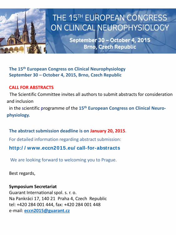
The 15th European Congress on Clinical NeurophysiologySeptember 30 – October 4, 2015, Brno, Czech Republic
CALL FOR ABSTRACTSThe Scientific Committee invites all authors to submit abstracts for consideration
and inclusion in the scientific programme of the 15th European Congress on Clinical Neuro-
physiology.
The abstract submission deadline is on January 20, 2015.
For detailed information regarding abstract submission:
http://www.eccn2015.eu/call-for-abstracts
We are looking forward to welcoming you to Prague.
Best regards,
Symposium Secretariat Guarant International spol. s. r. o. Na Pankráci 17, 140 21 Praha 4, Czech Republictel: +420 284 001 444, fax: +420 284 001 448e-mail: [email protected]

ASNER CN2014 Bucharest, Oct 31-Nov 2, 2014– Program & Abstract book6th National Conference of the Romanian Society of Electrodiagnostic Neurophysiology
Sponsors: