Embed Size (px)
Citation preview
FMD 101
Esther S.H. Kim, MD, MPH, FACC, FSVM
FMDSA Annual Meeting
18 May 2013
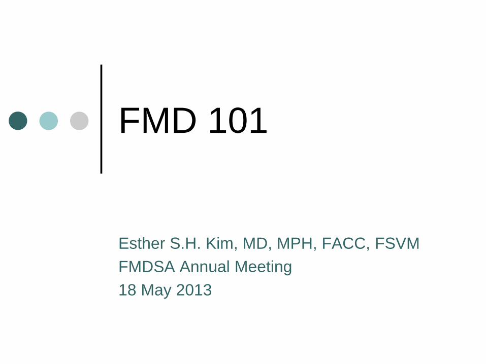
Back to basics…
Blood
Bodily fluid that transports
necessary substances
(oxygen, nutrients,
antibodies, hormones, etc)
and waste to and from cells
in the body
Blood vessels
Circulates blood to organs
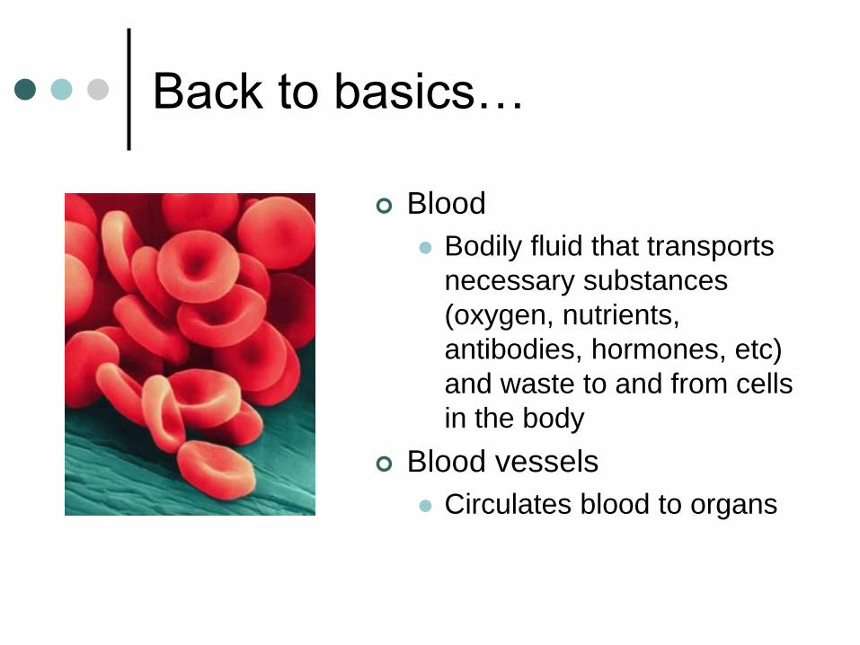
The Circulatory System
Heart
Arteries Away from the
heart
Veins Towards the
heart
Lymphatics Part of the
immune system
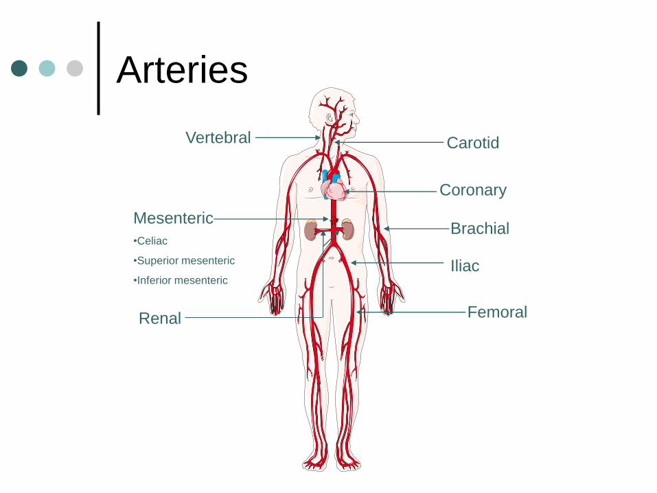
Arteries
Carotid
Coronary
Mesenteric
•Celiac
•Superior mesenteric
•Inferior mesenteric
Renal
Brachial
Iliac
Femoral
Vertebral
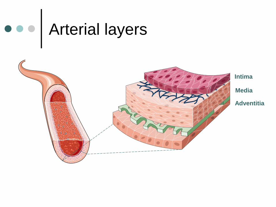
Arterial layers
Intima
Media
Adventitia
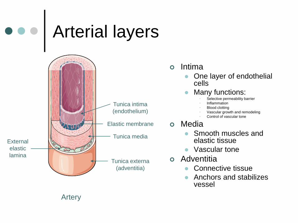
Arterial layers
Intima One layer of endothelial
cells
Many functions: • Selective permeability barrier
• Inflammation
• Blood clotting
• Vascular growth and remodeling
• Control of vascular tone
Media Smooth muscles and
elastic tissue
Vascular tone
Adventitia Connective tissue
Anchors and stabilizes vessel
Tunica externa
(adventitia)
Elastic membrane
Artery
Tunica media External
elastic
lamina
Tunica intima
(endothelium)
Arterial Pathologies
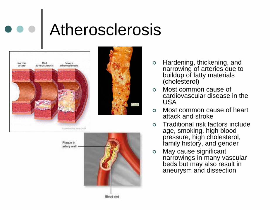
Atherosclerosis
Hardening, thickening, and narrowing of arteries due to buildup of fatty materials (cholesterol)
Most common cause of cardiovascular disease in the USA
Most common cause of heart attack and stroke
Traditional risk factors include age, smoking, high blood pressure, high cholesterol, family history, and gender
May cause significant narrowings in many vascular beds but may also result in aneurysm and dissection
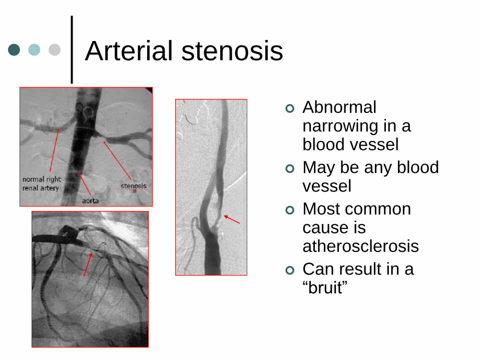
Arterial stenosis
Abnormal narrowing in a blood vessel
May be any blood vessel
Most common cause is atherosclerosis
Can result in a “bruit”
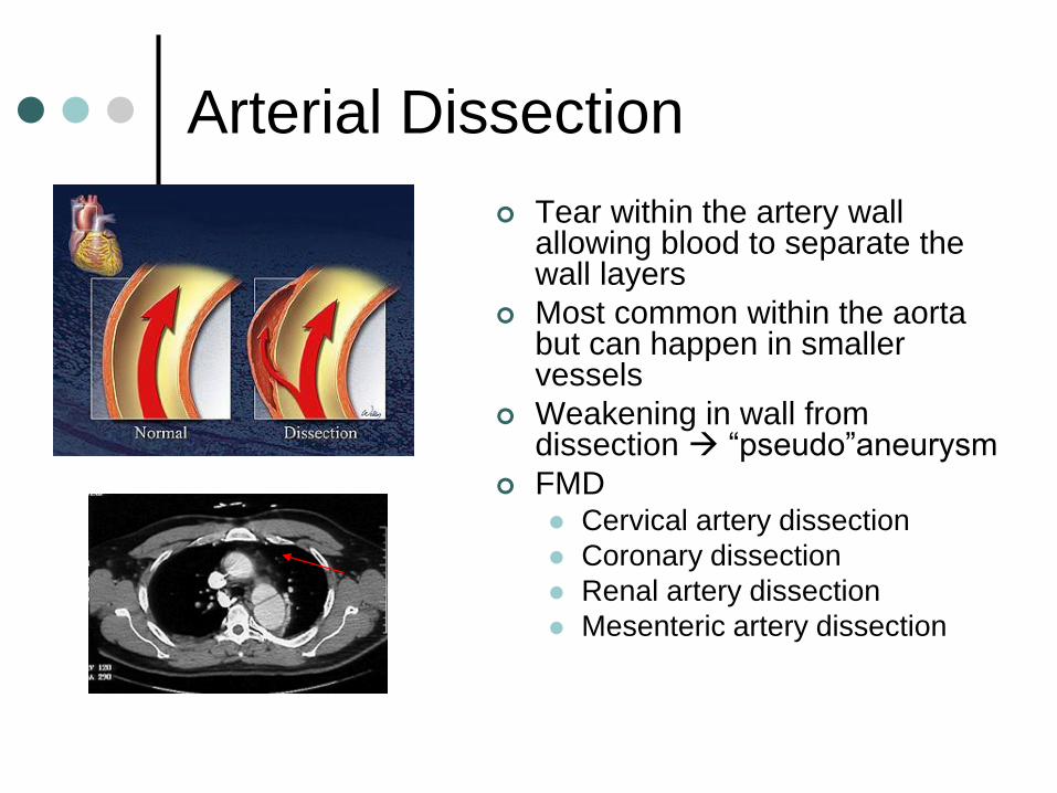
Arterial Dissection
Tear within the artery wall allowing blood to separate the wall layers
Most common within the aorta but can happen in smaller vessels
Weakening in wall from dissection “pseudo”aneurysm
FMD Cervical artery dissection
Coronary dissection
Renal artery dissection
Mesenteric artery dissection
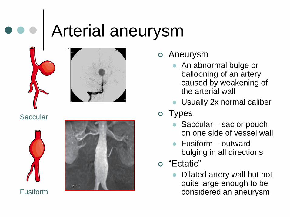
Arterial aneurysm
Aneurysm
An abnormal bulge or ballooning of an artery caused by weakening of the arterial wall
Usually 2x normal caliber
Types
Saccular – sac or pouch on one side of vessel wall
Fusiform – outward bulging in all directions
“Ectatic”
Dilated artery wall but not quite large enough to be considered an aneurysm
Saccular
Fusiform
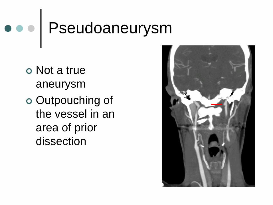
Pseudoaneurysm
Not a true
aneurysm
Outpouching of
the vessel in an
area of prior
dissection
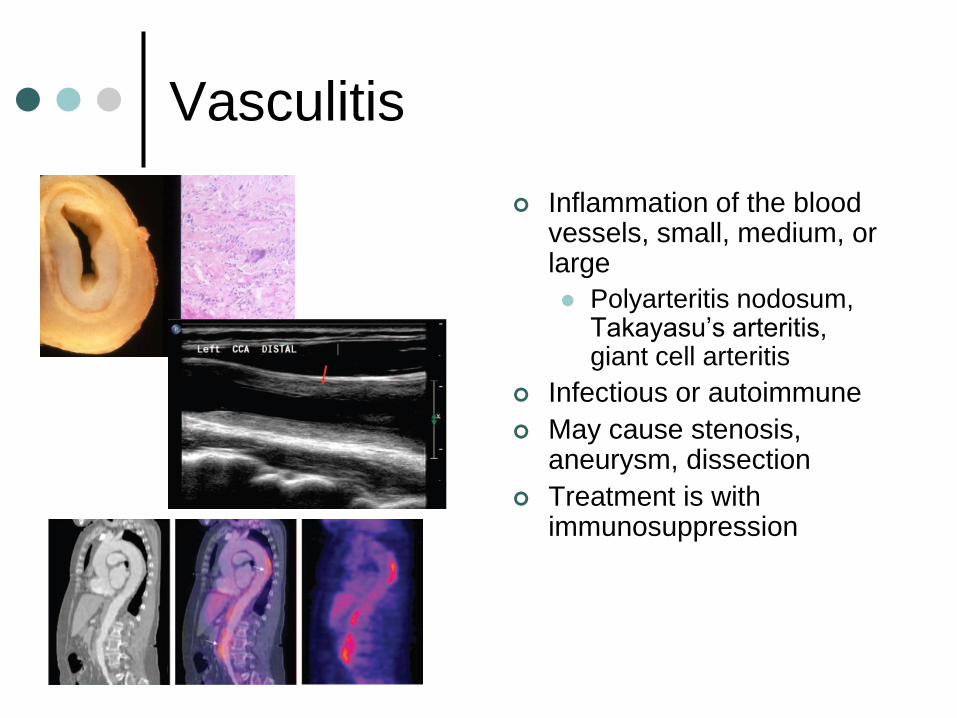
Vasculitis
Inflammation of the blood vessels, small, medium, or large
Polyarteritis nodosum, Takayasu’s arteritis, giant cell arteritis
Infectious or autoimmune
May cause stenosis, aneurysm, dissection
Treatment is with immunosuppression
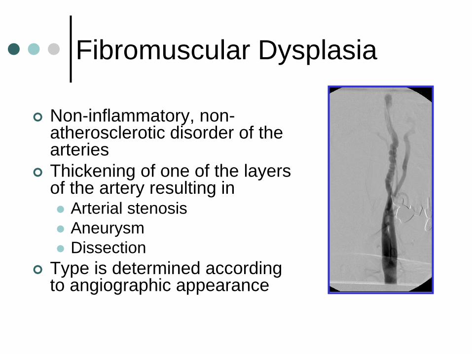
Fibromuscular Dysplasia
Non-inflammatory, non-atherosclerotic disorder of the arteries
Thickening of one of the layers of the artery resulting in Arterial stenosis
Aneurysm
Dissection
Type is determined according to angiographic appearance
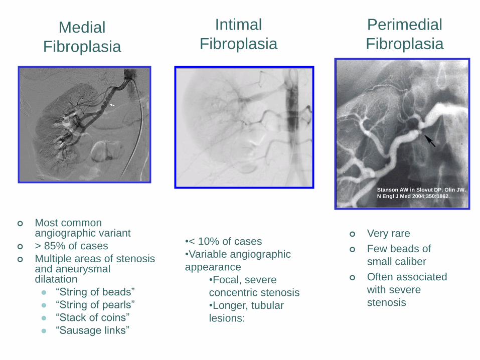
Medial
Fibroplasia
Most common angiographic variant
> 85% of cases
Multiple areas of stenosis and aneurysmal dilatation
“String of beads”
“String of pearls”
“Stack of coins”
“Sausage links”
Intimal
Fibroplasia
•< 10% of cases
•Variable angiographic
appearance
•Focal, severe
concentric stenosis
•Longer, tubular
lesions:
Very rare
Few beads of
small caliber
Often associated
with severe
stenosis
Perimedial
Fibroplasia
Stanson AW in Slovut DP. Olin JW.
N Engl J Med 2004;350:1862.
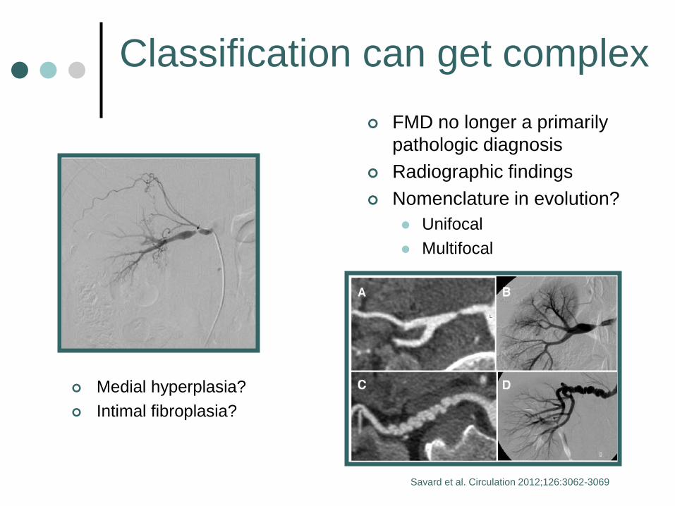
Classification can get complex
FMD no longer a primarily
pathologic diagnosis
Radiographic findings
Nomenclature in evolution?
Unifocal
Multifocal
Medial hyperplasia?
Intimal fibroplasia?
Savard et al. Circulation 2012;126:3062-3069

Physical Exam

Torus Palantinus
NEJM 2013;368:1434
Thumb and Wrist Sign
Elbow Hyperextension Knee Hyperextension
Horner’s sign
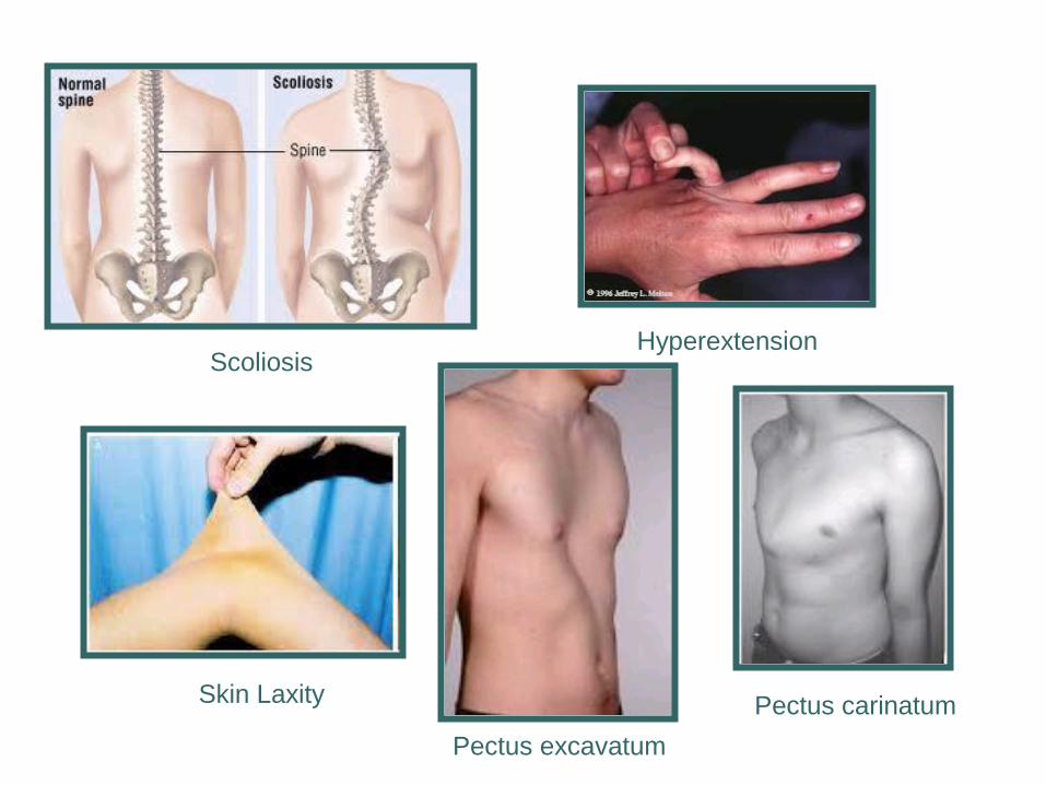
Skin Laxity
Scoliosis Hyperextension
Pectus excavatum
Pectus carinatum
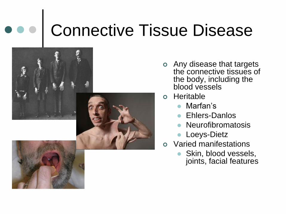
Connective Tissue Disease
Any disease that targets the connective tissues of the body, including the blood vessels
Heritable
Marfan’s
Ehlers-Danlos
Neurofibromatosis
Loeys-Dietz
Varied manifestations
Skin, blood vessels, joints, facial features
Bruit (french for “noise”)
“the unusual sound that blood makes when it rushes past an obstruction in an artery when the sound is auscultated
with a stethoscope” – Dr Wikipedia
Imaging
Ultrasound
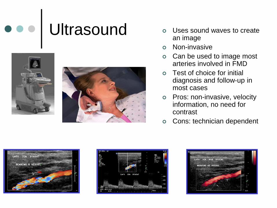
Ultrasound Uses sound waves to create an image
Non-invasive
Can be used to image most arteries involved in FMD
Test of choice for initial diagnosis and follow-up in most cases
Pros: non-invasive, velocity information, no need for contrast
Cons: technician dependent
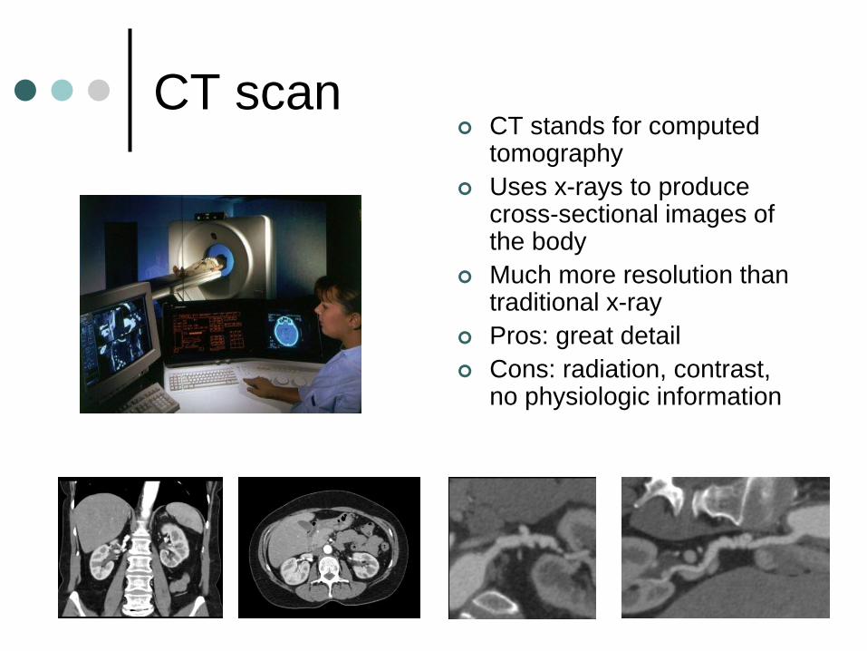
CT scan CT stands for computed
tomography
Uses x-rays to produce cross-sectional images of the body
Much more resolution than traditional x-ray
Pros: great detail
Cons: radiation, contrast, no physiologic information
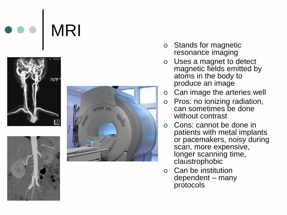
MRI Stands for magnetic
resonance imaging
Uses a magnet to detect magnetic fields emitted by atoms in the body to produce an image
Can image the arteries well
Pros: no ionizing radiation, can sometimes be done without contrast
Cons: cannot be done in patients with metal implants or pacemakers, noisy during scan, more expensive, longer scanning time, claustrophobic
Can be institution dependent – many protocols
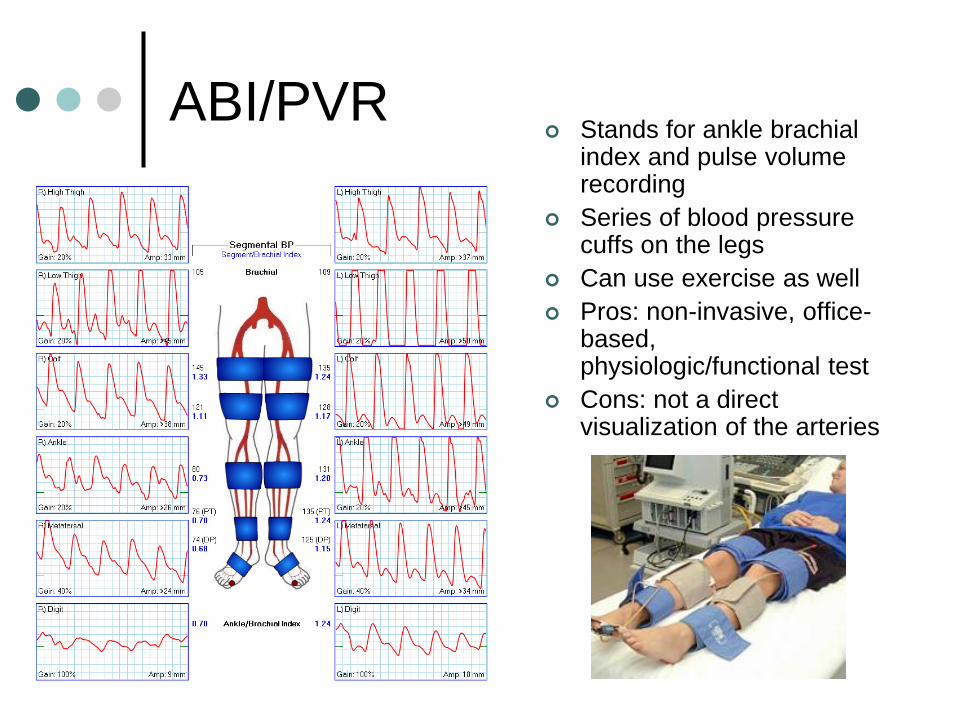
ABI/PVR Stands for ankle brachial
index and pulse volume recording
Series of blood pressure cuffs on the legs
Can use exercise as well
Pros: non-invasive, office-based, physiologic/functional test
Cons: not a direct visualization of the arteries
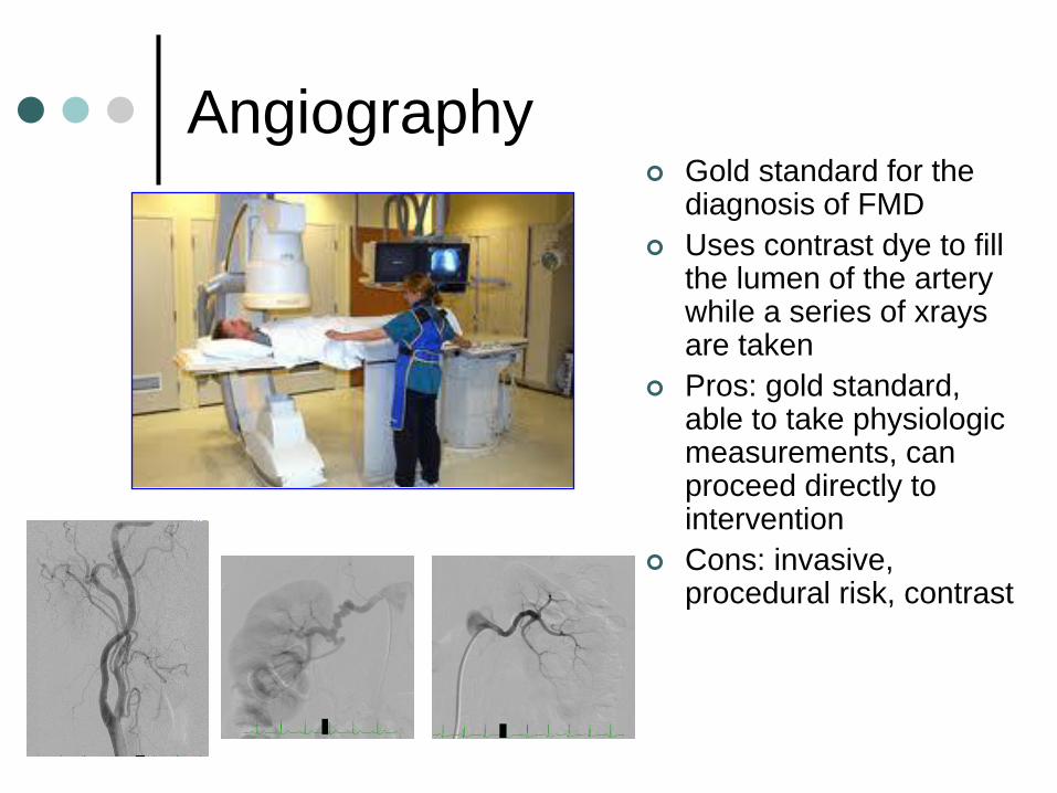
Angiography Gold standard for the
diagnosis of FMD
Uses contrast dye to fill the lumen of the artery while a series of xrays are taken
Pros: gold standard, able to take physiologic measurements, can proceed directly to intervention
Cons: invasive, procedural risk, contrast
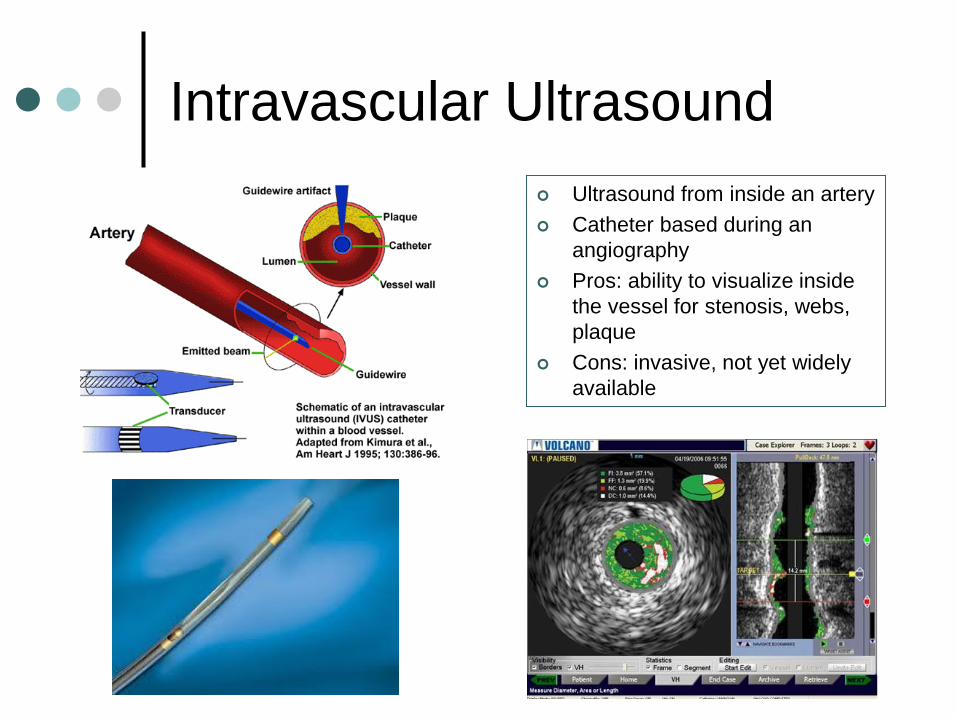
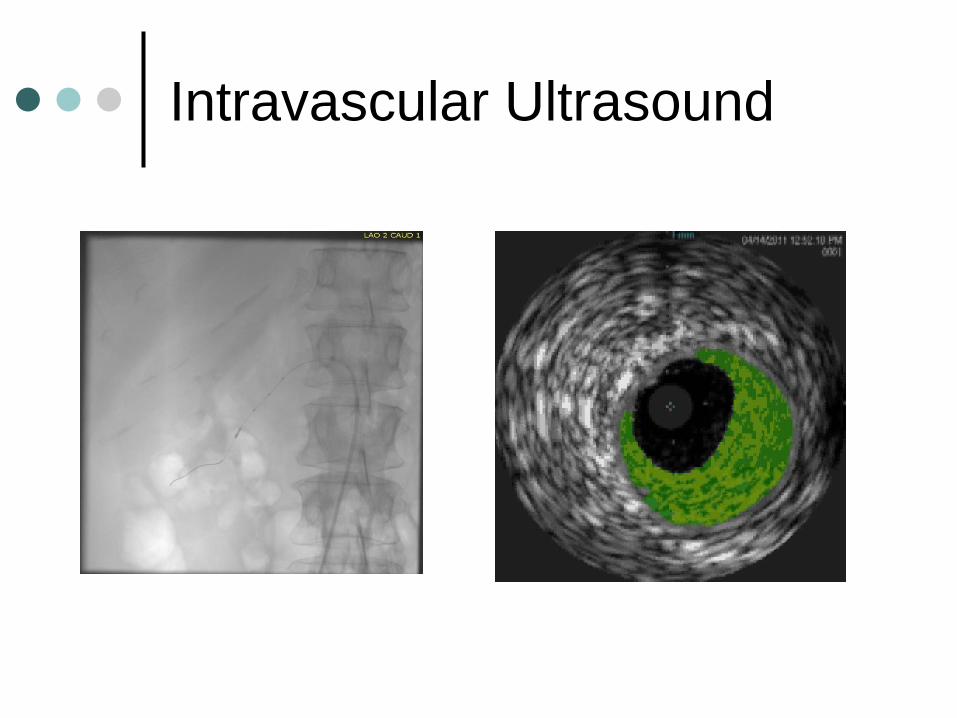
Intravascular Ultrasound
Ultrasound from inside an artery
Catheter based during an
angiography
Pros: ability to visualize inside
the vessel for stenosis, webs,
plaque
Cons: invasive, not yet widely
available
Intravascular Ultrasound
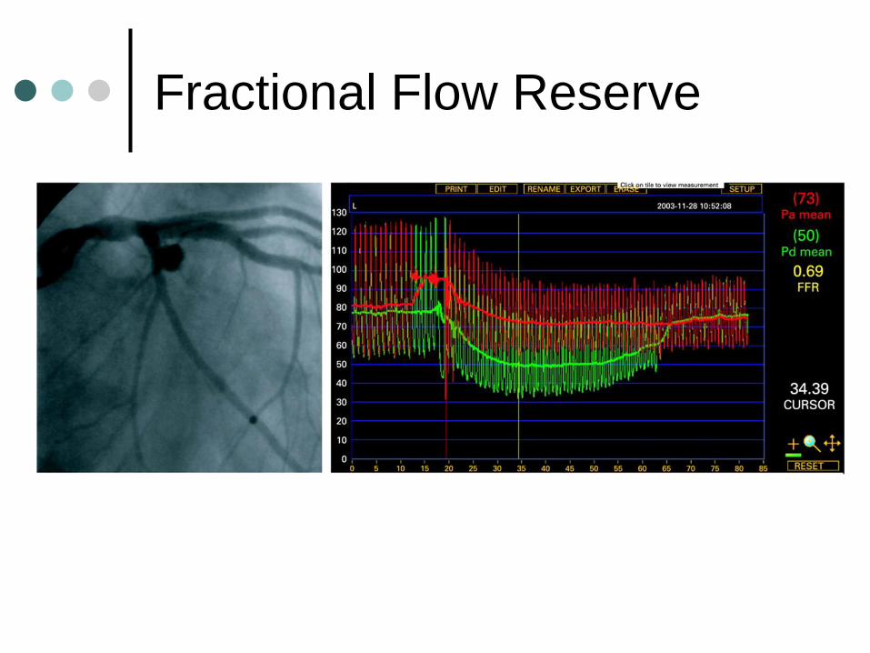
Fractional Flow Reserve

Treatment
Invasive Treatment
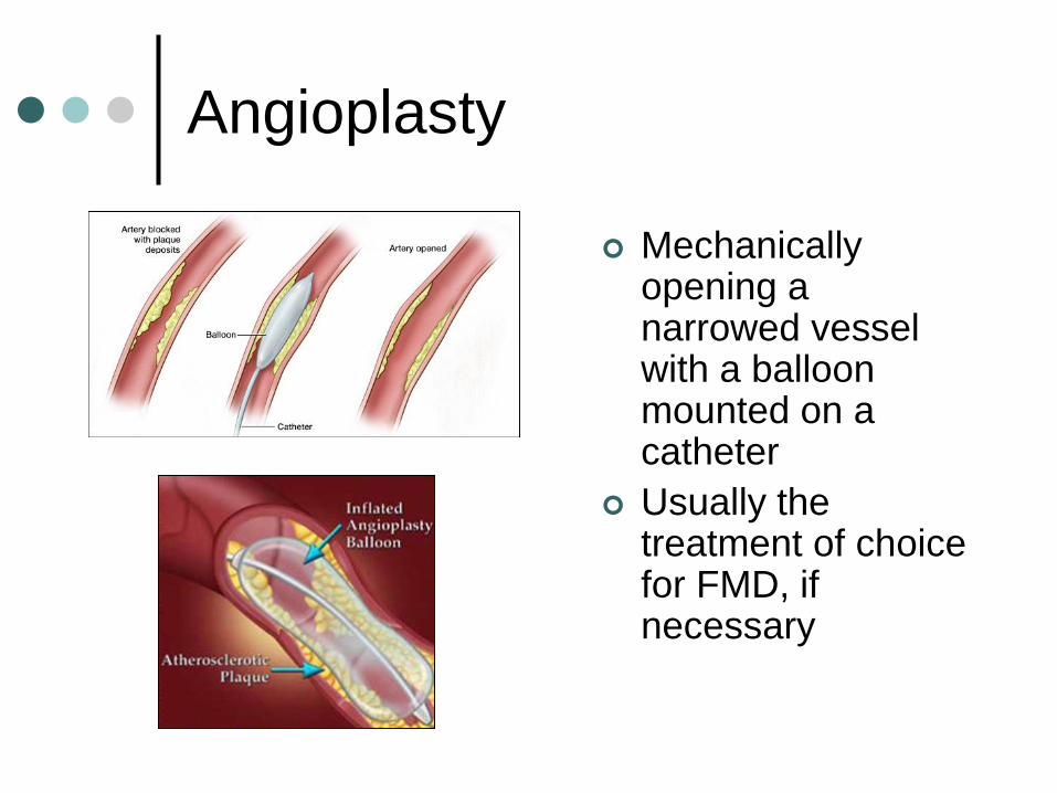
Angioplasty
Mechanically opening a narrowed vessel with a balloon mounted on a catheter
Usually the treatment of choice for FMD, if necessary
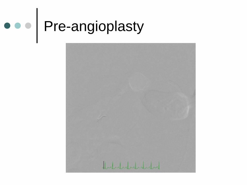
Pre-angioplasty

Angioplasty
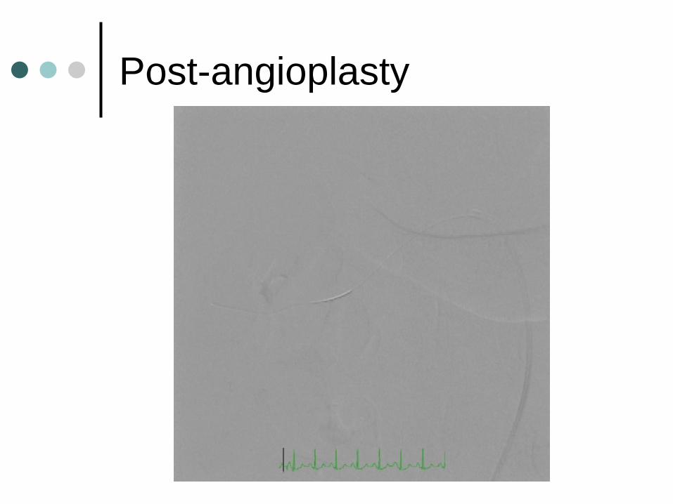
Post-angioplasty
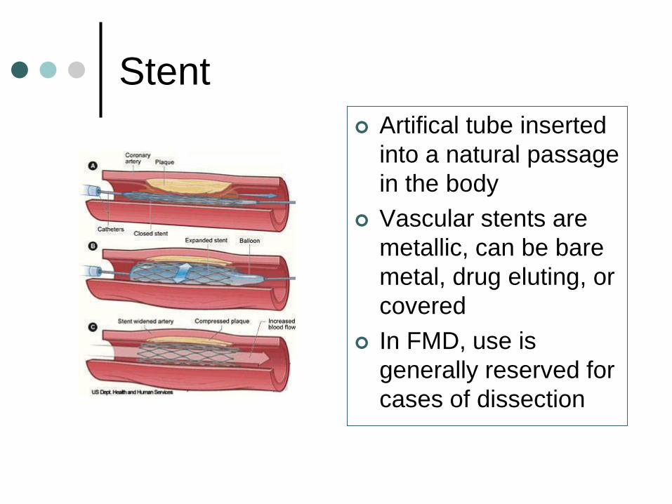
Stent
Artifical tube inserted
into a natural passage
in the body
Vascular stents are
metallic, can be bare
metal, drug eluting, or
covered
In FMD, use is
generally reserved for
cases of dissection

Bypass Surgery
Surgical procedure where an artery or vein from elsewhere in the body or an artificial graft is used the bypass a diseased artery and supply blood flow to the organ
Generally reserved for cases not amenable to PCI
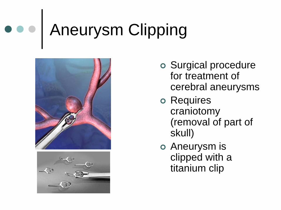
Aneurysm Clipping
Surgical procedure for treatment of cerebral aneurysms
Requires craniotomy (removal of part of skull)
Aneurysm is clipped with a titanium clip
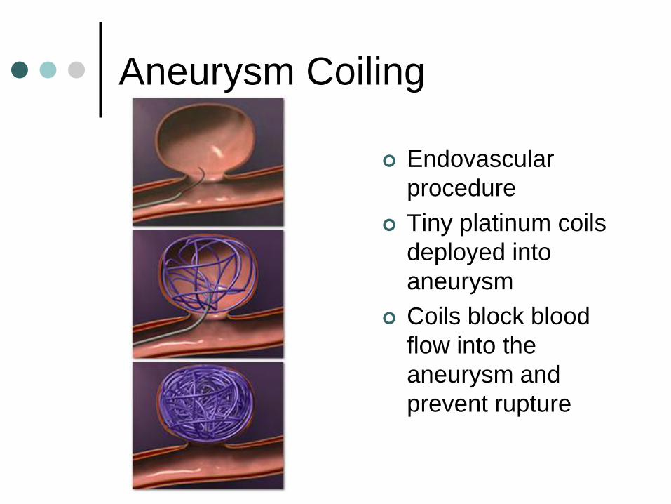
Aneurysm Coiling
Endovascular
procedure
Tiny platinum coils
deployed into
aneurysm
Coils block blood
flow into the
aneurysm and
prevent rupture

Thank you