Embed Size (px)
Citation preview
NeuroImage 13, 76–90 (2001)doi:10.1006/nimg.2000.0674, available online at http://www.idealibrary.com on
The Accuracy of Near Infrared Spectroscopy and Imagingduring Focal Changes in Cerebral Hemodynamics
David A. Boas,*,† Tom Gaudette,* Gary Strangman,* Xuefeng Cheng,†John J. A. Marota,* and Joseph B. Mandeville*
*NMR Center, Massachusetts General Hospital, Harvard Medical School, Charlestown, Massachusetts 02129; and†Electro-Optic Technology Center, Tufts University, Medford, Massachusetts 02155
Received January 21, 2000; published online November 7, 2000
Near infrared spectroscopy (NIRS) can detectchanges in the concentrations of oxy-hemoglobin([HbO]) and deoxy-hemoglobin ([Hb]) in tissue basedupon differential absorption at multiple wavelengths.The common analysis of NIRS data uses the modifiedBeer–Lambert law, which is an empirical formulationthat assumes global concentration changes. We usedsimulations to examine the errors that result whenthis analysis is applied to focal hemodynamic changes,and we performed simultaneous NIRS measurementsduring a motor task in adult humans and a neonate toevaluate the dependence of the measured changes ondetector-probe geometry. For both simulations and invivo measurements, the wide range of NIRS resultswas compared to an imaging analysis, diffuse opticaltomography (DOT). The results demonstrate that rel-ative changes in [HbO] and [Hb] cannot, in general, bequantified with NIRS. In contrast to that method, DOTanalysis was shown to accurately quantify simulatedchanges in chromophore concentrations. These re-sults and the general principles suggest that DOT canaccurately measure changes in [Hb] and [HbO], butNIRS cannot accurately determine even relative focalchanges in these chromophore concentrations. For thestandard NIRS analysis to become more accurate forfocal changes, it must account for the position of thefocal change relative to the source and detector aswell as the wavelength dependent optical propertiesof the medium. © 2001 Academic Press
INTRODUCTION
Optical methods based upon differential absorptionproportional to concentrations of oxy-hemoglobin([HbO]) and deoxy-hemoglobin ([Hb]) have long beenapplied to biological applications. Pulse oximetry non-invasively monitors arterial oxygen saturation by mea-suring arterial pulsation (1,2). Near infrared spectros-copy (NIRS) was developed to measure average tissue
hemoglobin oxygen saturation and total hemoglobin761053-8119/01 $35.00Copyright © 2001 by Academic PressAll rights of reproduction in any form reserved.
concentration (3–6). NIRS has been applied to suchdiverse applications as fundamental studies of brainfunction (7), neonate ECMO (8), and cardiac surgery(9) and we use it in this paper to refer to spectroscopicmeasurements made with a single source-detectorpair. Diffuse optical tomography (DOT), a more recenttechnique (10), reconstructs images of changes in chro-mophore concentrations using multiple light sourcesand detectors. Both NIRS and DOT measure total op-tical absorption at multiple wavelengths, rather thandifferential absorption due to pulsation, and thus aresensitive to hemoglobin in the arterial, venial, andcapillary compartments.
The ultimate promise of optical methodologies is si-multaneous quantification of concentrations of multi-ple chromophores in order to determine changes inbiologically relevant quantities like blood oxygen sat-uration and blood volume. Qualitative assessments se-verely limit the biological issues that can be addressed.For instance, a change in local [Hb] during stroke (11)or stimulus-induced brain activation (12,13), as mea-sured by fMRI, can indicate a change in blood volume,oxygen saturation, or some combination of these pos-sibilities.
Although it has generally been presumed that thebiochemical specificity of optical absorption in the nearinfrared regime translates directly into quantifiablemeasurements of chromophore concentrations, thephysics of optical methods merits increased attentionas NIRS and DOT find increased application in biolog-ical systems. It has recently been shown that the com-mon analysis of optical reflectance spectra from micro-scopic measurements introduces systematic “crosstalk”between calculated changes in [Hb] and [HbO] due towavelength-dependent changes in optical pathlengthduring global brain activation (14). Corrections for thiserror may affect basic interpretations of physiology(e.g., 15). In this report, we address another fundamen-tal issue: namely, can spectroscopic methods accu-rately determine relative changes in [Hb] and [HbO]
during focal or graded hemodynamics changes, or are
pilTq
dC
77THE ACCURACY OF NIRS DURING FOCAL HEMODYNAMIC CHANGES
imaging methods required to accurately measure thesechanges?
In order to address this issue, we illustrate the widerange of results that can be obtained by NIRS using themodified Beer–Lambert law, MBLL (4,5), to calculatechanges in [Hb] and [HbO] during focal motor activa-tion in adult humans and a neonate. We then presenta theoretical analysis of the accuracy of the modifiedBeer–Lambert law for quantifying focal concentrationchanges. We show that the wide disparity of results ininterpreting human subject data using the MBLL isconsistent with our description of the errors beingdriven by partial volume and differential sensitivityeffects. These errors can be significantly reduced byDOT (16,17).
THEORY
To set the theoretical context for our analysis, webriefly review the modified Beer–Lambert law as ap-plied to quantifying global changes in oxy- and deoxy-hemoglobin. The rest of the paper examines the valid-ity of applying this simple theoretical model toanalyzing focal changes.
Near-Infrared Spectroscopy
The theory of the modified Beer–Lambert Law(MBLL) has been explained previously (4,5). Briefly,the technique is based on the absorption of near infra-red light by oxy- and deoxyhemoglobin. Changes in theconcentrations of these chromophores are quantifiedusing a modified Beer–Lambert law, which is an em-pirical description of optical attenuation in a highlyscattering medium (4,5). The modified Beer–Lambertlaw is
OD 5 2logI
Io5 eCLB 1 G, (1)
where OD is the optical density, Io is the incident lightintensity, I is the detected light intensity, e is theextinction coefficient of the chromophore, C is the con-centration of the chromophore, L is the distance be-tween where the light enters the tissue and where thedetected light exits the tissue, B is a pathlength factor,which accounts for increases in the photon pathlengthcaused by tissue scattering, and G is a factor whichaccounts for the measurement geometry. We use theconvention of log base e.
A change in the chromophore concentration causesthe detected intensity to change. When the concentra-tion changes, the extinction coefficient e and distance Lremain constant and it is assumed that B and G re-
main constant. Thus, Eq. (1) can be rewritten asDOD 5 2logIFinal
IInitial5 eDCLB, (2)
where DOD 5 ODFinal 2 ODInitial is the change in opticaldensity Ifinal and IInitial are the measured intensitiesbefore and after the concentration change, and DC isthe change in concentration. L is specified by the probegeometry, e is an intrinsic property of the chro-mophore, and B is often referred to as the differential
athlength factor (DPF) and can be determined fromndependent measurements with ultrashort pulses ofight (18) and has been tabulated for various tissues.hus, given the extinction coefficient, it is possible touantify the change in chromophore concentration.Figure 1 plots the extinction coefficients for oxy- and
eoxyhemoglobin versus wavelength as measured byope et al. (5,19,20). The wavelength range of 600 to
900 nm in the graph is the only region in which light isable to penetrate several centimeters through tissue.The other chromophores of significance in tissue in thiswavelength range are water, lipids, and cytochromeaa3. We do not consider these chromophores in thispaper, as their contribution in general is an order ofmagnitude less significant than hemoglobin. In orderto consider the contribution of two chromophores, wemust rewrite Eq. (2) as
DOD l 5 ~e HbOl D@HbO# 1 e Hb
l D@Hb#!B lL, (3)
where l indicates a particular wavelength. Equation(3) explicitly accounts for independent concentrationchanges in oxyhemoglobin (D[HbO]) and deoxyhemo-globin (D[Hb]).
By measuring DOD at two wavelengths (l1 and l2)and using the known extinction coefficients of oxyhe-moglobin (eHbO) and deoxyhemoglobin (eHb) at thosewavelengths, we can then determine the concentration
FIG. 1. These values for the molar extinction coefficients are inunits of [cm21/(moles/liter)] and were obtained from (5,19,20).
changes of oxyhemoglobin and deoxyhemoglobin,
F
p
afi
e
78 BOAS ET AL.
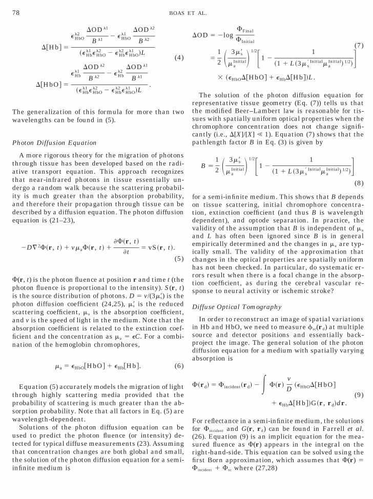
D@Hb# 5
e HbOl2
DOD l1
B l12 e HbO
l1DOD l2
B l2
~e Hbl1 e HbO
l2 2 e Hbl2 e HbO
l1 !L(4)
D@HbO# 5
e Hbl1
DOD l2
B l22 e Hb
l2DOD l1
B l1
~e Hbl1 e HbO
l2 2 e Hbl2 e HbO
l1 !L.
The generalization of this formula for more than twowavelengths can be found in (5).
Photon Diffusion Equation
A more rigorous theory for the migration of photonsthrough tissue has been developed based on the radi-ative transport equation. This approach recognizesthat near-infrared photons in tissue essentially un-dergo a random walk because the scattering probabil-ity is much greater than the absorption probability,and therefore their propagation through tissue can bedescribed by a diffusion equation. The photon diffusionequation is (21–23),
2D¹ 2F~r, t! 1 vmaF~r, t! 1F~r, t!
t5 vS~r, t!.
(5)
(r, t) is the photon fluence at position r and time t (thephoton fluence is proportional to the intensity). S(r, t)is the source distribution of photons. D 5 v/(3m9s) is the
hoton diffusion coefficient (24,25), m9s is the reducedscattering coefficient, ma is the absorption coefficient,and v is the speed of light in the medium. Note that thebsorption coefficient is related to the extinction coef-cient and the concentration as ma 5 eC. For a combi-
nation of the hemoglobin chromophores,
ma 5 eHbO@HbO# 1 eHb@Hb#. (6)
Equation (5) accurately models the migration of lightthrough highly scattering media provided that theprobability of scattering is much greater than the ab-sorption probability. Note that all factors in Eq. (5) arewavelength-dependent.
Solutions of the photon diffusion equation can beused to predict the photon fluence (or intensity) de-tected for typical diffuse measurements (23). Assumingthat concentration changes are both global and small,the solution of the photon diffusion equation for a semi-
infinite medium isDOD 5 2logFFinal
FInitial(7)
51
2 S 3m9s
m aInitialD 1/2F1 2
1
~1 1 L~3m s9 Initialm a
Initial! 1/2!G3 ~eHbOD@HbO# 1 eHbD@Hb#!L.
The solution of the photon diffusion equation forrepresentative tissue geometry (Eq. (7)) tells us thatthe modified Beer–Lambert law is reasonable for tis-sues with spatially uniform optical properties when thechromophore concentration does not change signifi-cantly (i.e., D[X]/[X] ! 1). Equation (7) shows that thepathlength factor B in Eq. (3) is given by
B 51
2 S 3m9s
m aInitialD 1/2F1 2
1
~1 1 L~3m s9 Initialm a
Initial! 1/2!G(8)
for a semi-infinite medium. This shows that B dependson tissue scattering, initial chromophore concentra-tion, extinction coefficient (and thus B is wavelengthdependent), and optode separation. In practice, thevalidity of the assumption that B is independent of ma
and L has often been ignored since B is in generalmpirically determined and the changes in ma are typ-
ically small. The validity of the approximation thatchanges in the optical properties are spatially uniformhas not been checked. In particular, do systematic er-rors result when there is a focal change in the absorp-tion coefficient, as during the cerebral vascular re-sponse to neural activity or ischemic stroke?
Diffuse Optical Tomography
In order to reconstruct an image of spatial variationsin Hb and HbO, we need to measure fsc(rd) at multiplesource and detector positions and essentially back-project the image. The general solution of the photondiffusion equation for a medium with spatially varyingabsorption is
F~rd! 5 Fincident~rd! 2 E F~r!v
D~eHbOD@HbO#
1 eHbD@Hb#!G~r, rd!dr.(9)
For reflectance in a semi-infinite medium, the solutionsfor Fincident and G(r, rd) can be found in Farrell et al.(26). Equation (9) is an implicit equation for the mea-sured fluence as F(r) appears in the integral on theright-hand-side. This equation can be solved using thefirst Born approximation, which assumes that F(r) 5
Fincident 1 Fsc where (27,28)
c
D
Bt
E
79THE ACCURACY OF NIRS DURING FOCAL HEMODYNAMIC CHANGES
Fsc~rd! 5 2E Fincident~r!v
D~eHbOD@HbO#
1 eHbD@Hb#!G~r, rd!dr.(10)
By measuring Fsc(rd) using multiple source and de-tector positions and multiple wavelengths, we can in-vert the integral equation to obtain images of D[HbO]and D[Hb]. There are numerous methods for solvingEq. (10) (29). Most rely on reducing Eq. (10) to a matrixequation by rewriting the integral as a sum overvoxels, i.e., y 5 Ax, where y is the vector of measure-ments Fsc(rd), x is the vector of image voxels, and A isthe transformation matrix obtained from the integrandof Eq. (10). The integrand of Eq. (10) essentially de-scribes the sensitivity that each measurement has tothe change in absorption within each volume elementwithin the medium. In other words, if the absorptionchanges within a localized region, then
DODBORN 5 2logSFincident~rs, rd! 1 Fsc~rs, rd!
Fincident~rs, rd!D
< 2Fsc~rs, rd!
Fincident~rs, rd!5
Fincident~rs, r!G~r, rd!
Fincident~rs, rd!
vdr
DDma,
(11)
where dr is the volume over which the absorptionhange Dma occurs and Dma
l 5 (eHbOl D[HbO] 1 eHb
l D[Hb]).
Hemodynamic Cross-Talk Resulting from aSystematic Error
When we use the modified Beer–Lambert law (Eq.(3)) to analyze the absorption change within a focalregion (Eq. (11)), we are assuming that DODMBLL
l 5DODBORN
l . This gives us
Dm a,MBLLl 5
1
B lL
Fincident~rs, r!G l~r, rd!
Fincident~rs, rd!
vdr
D
3 Dm a,BORNl 5 k lDm a,BORN
l .(12)
ma,BORNl is the real change in absorption (Dma,Real
l ) andDma,MBLL
l is the absorption change that we will measure(Dma,Meas
l ) having assumed the accuracy of the modifiedeer–Lambert Law. This indicates that we will mises-
imate the real absorption change by kl. kl will typi-cally be less than 1 due to the partial volume effect.
To explore the effect of this factor on our determina-tion of D[HbO] and D[Hb], we return to Eq. (4) whichrelated the measured concentration changes to the
measured absorption changes:D@Hb#Meas 5e HbO
l2 k l1Dm a,REALl1 2 e HbO
l1 k l2Dm a,REALl2
~e Hbl1 e HbO
l2 2 e Hbl2 e HbO
l1 !
(13)
5
e HbOl1 e HbO
l2 ~k l1 2 k l2!D@HbO#Real
2 ~k l1e Hbl1 e HbO
l2 2 k l2e Hbl2 e HbO
l1 !D@Hb#Real
~e Hbl1 e HbO
l2 2 e Hbl2 e HbO
l1 !
D@HbO#Meas 5e Hb
l1 k l2Dm a,REALl2 2 e Hb
l2 k l1Dm a,REALl1
~e Hbl1 e HbO
l2 2 e Hbl2 e HbO
l1 !
5
~k l2e Hbl1 e HbO
l2 2 k l1e Hbl2 e HbO
l1 !D@HbO#Real
2 e Hbl1 e Hb
l2 ~k l1 2 k l2!D@Hb#Real
~e Hbl1 e HbO
l2 2 e Hbl2 e HbO
l1 !.
If the measurements at different wavelengths have thesame sensitivity to the focal change, then kl1 5 kl2 and
q. (13) reduces to D[X]Meas 5 kD[X]Real such that thevalue we measure is linearly proportional to the correctvalue and the calculated ratio D[HbO]:D[Hb] equals thecorrect ratio. However, since the optical properties atdifferent wavelengths are generally different, the spa-tial sensitivity profiles are thus also expected to bedifferent with the result that kl1 Þ kl2. Therefore, ingeneral, there is cross-talk between real focal hemoglo-bin concentration changes and the determined/calcu-lated concentration changes. This cross-talk can onlybe removed if the values of kl1 and kl2 are known. Thisproblem can potentially be solved by diffuse opticaltomography, or prior knowledge of the geometry andoptical properties of the head as well as the spatiallocation and extent of the focal activation. This paperexplores the significance of this cross-talk in experi-ments and simulations.
METHODS
Adult Human Brain Activation
We compared signals recorded from neighboring de-tector fibers on an adult human during the perfor-mance of a simple motor task. The study was per-formed under a protocol approved by the MGH internalreview board. A single source location was positioned1.5 cm posterior to position C3 in the EEG 10-20 sys-tem. This source was composed of two laser diodes–780and 830 nm–which were fed into a 1 mm bifurcatedfiber. A 2-mm detector fiber was positioned 1.8 cmanterior to C3 (on the line formed by C3 and thesource). Another detector fiber was also positioned 3.3cm from the source, but more medially than the firstdetector, with a center-to-center detector separation of1.0 cm. It has been shown that C3 and C4 lie approx-
imately over the hand areas of primary motor cortex
gw
a
80 BOAS ET AL.
(30). Hence, this optode arrangement was expected tosample M1 for the right hand.
Prior to commencing the recording, the subject wasasked to simply rest quietly. When the experimentalrun began, the subject alternated between quiet restfor 15 s, and 15 s of 4-finger flexion/extension with theright hand (at approximately 3 Hz). The subject per-formed 10 blocks of the motor task, interleaved with 11blocks of rest, for a total run length of 315 s.
Adult Human Data Analysis
The resulting motor-task data was time-trigger av-eraged—separately for each wavelength and detec-tor—and triggering on the onset of motor activation.Thus, each data point in the four resulting timeseriesis an average of 10 points in the original data. The twotimeseries pairs (780 and 830 nm, for each of twodetectors) were then analyzed using the modifiedBeer–Lambert law. This resulted in four new time-series: Hb and HbO time-courses for each of the twodetectors. For the pathlength factor B, a fixed value of5.93 was used (31). To summarize the results acrossthree subjects we averaged the concentration changefor 5 s before onset of stimulation and then from 6–11s following onset of the stimulus. The differences ofthese average values are compared.
Neonate Brain Activation
We compared a MBLL and DOT analysis of diffuseoptical data obtained during a motor-sensory brainactivation in a newborn human. All clinical studieswere performed under a protocol approved by an insti-tutional review board-, and parents signed informedconsent prior to initiation of studies. More details ofthis human subject study can be found in Hintz et al.(36). For the results presented here, a 32–33 week EGAinfant underwent a passive motor stimulation protocol.The infant was quiet and asleep, but not pharmacolog-ically sedated. The patient had a normal heart rate andO2 saturation by pulse oximetry during the approxi-mately 40-min study period.
For this study, a large probe was designed and wasplaced in true midline position in the area that corre-sponded to the motor cortex (see Fig. 3). For the opticalprobe, the nine source fibers were arranged in a 3 3 3rid with a 2 3 1-cm spacing. The 16 collection fibersere arranged in a 4 3 4 grid with the same 2 3 1-cm
spacing. To improve optical coupling, the height of eachfiber was manually adjusted to match the surface con-tours of the skull. The Z positions were not measured,nor was the actual contour of the scalp. There were nocomplications from probe placement.
Baseline data were collected for 20 s, then the pa-tient’s right arm was flexed and extended at the elbowwith a 1 Hz repetition rate. This study employed a 5-s
integration time, and each stimulation period was 30 swith a rest period of 45–60 s between each stimulationperiod. Imaging data was collected at both 780 and 830nm to facilitate a spectroscopic determination of oxy-and deoxyhemoglobin.
Neonate Data Analysis
The optical data recorded from the multiple source-detector pairs on the neonate brain was analyzed usingtwo different approaches: a NIRS analysis was per-formed for each source-detector pair using the modifiedBeer–Lambert law (MBLL), and a DOT analysis com-bined the information from all such pairs to recon-struct images of optical absorption. Each analysistreated the geometry as a semi-infinite medium with ahomogeneous distribution of absorbers, except for thelocalized activation, and used the expression presentedpreviously. For the MBLL analysis, the pathlengthfactor B was calculated using Eq. (10) with m9s 5 10.0cm21, [HbT] 5 100 mM, SO2 5 85%, and the knownseparations L between various source-detector pairs.
For the DOT analysis, we used the SimultaneousIterative Reconstruction Technique (SIRT) to invertEq. (10) (17,28). SIRT is a variation of the AlgebraicReconstruction Technique (ART), which sequentiallyprojects estimates onto a hyperplane defined by a par-ticular row of the linear system. SIRT has been ob-served to generate smoother reconstructions of the ob-ject function due to the averaging over a number ofprojections, but this comes at a cost of slower conver-gence. The SIRT reconstruction can be expressedmathematically as x̂i 5 x̂i21 2 mean((x̂i21
T a j 2 b j/(a jTa j))
j), where x̂i is the ith estimate of the object function, ai
is the ith row of the transformation matrix A, and bi isthe ith measurement. For underdetermined systems,this reconstruction will converge to a point on thehyperplane satisfying the linear system that is nearestto the initial guess. Limiting the number of interac-tions controls regularization; our reconstructions wereperformed using 10 iterations of the SIRT algorithm,starting with an initial guess of zero perturbation.
DOT images were reconstructed in a 6 3 3-cm planepositioned at a depth of 1 cm, with a thickness of 3 mm,and located parallel to the source/detector plane. Thevoxel size was 3 3 1.5 3 3 mm in the x, y, and zdirections respectively, giving an image with 21 3 21 31 voxels. With this geometry, we reconstructed 2-Dimages of the absorption coefficient, and we assumedthat the out-of-plane optical properties remain un-changed. This means that any out-of-plane opticalchanges were projected into the image plane.
Simulations
In order to understand the wide range of resultsobtained by our simultaneous NIRS measurements inthe neonate, we simulated the experimental setup and
the process of light diffusion through a simplified me-
b
81THE ACCURACY OF NIRS DURING FOCAL HEMODYNAMIC CHANGES
dium containing a focal hemodynamic change. Thesimulated data, based upon the forward solution to thediffusion equation, was analyzed using the MBLL andDOT methods described previously in order to deter-mine how closely each method reconstructed theknown changes in chromophore concentrations.
To simulate changes in the diffuse intensity (i.e.,DOD) due to a focal change in total hemoglobin concen-tration, we employed the first Born approximation (Eq.(12)) to solve the photon diffusion equation. We used areduced scattering coefficient of 10 cm21 and includedabsorption coefficients at wavelengths of 780 and 830nm that depended only upon oxy- and deoxyhemoglo-bin. To calculate the absorption coefficient for differentconcentrations of hemoglobin, we used the tabulatedextinction coefficients provided by Cope et al. (5,19,20)as shown in Fig. 1. For the semi-infinite medium, weused an index of refraction n 5 1.33, and we assumedaseline values of HbT 5 100 mM and SO2 5 85% to
give ma780 5 0.175 cm21 and ma
830 5 0.214 cm21. A focalincrease in absorption (the “object”) was positionedhalf-way between the first source (S1) and sixth detec-tor (D6) at a depth of 1 cm. It had dimensions of 0.3 30.15 3 0.3 mm in x, y, and z respectively. The reducedscattering coefficient of the object was the same as thebackground, and the absorption coefficient was ma
780 50.210 cm21 and ma
830 5 0.257 cm21 corresponding to a20% increase in total hemoglobin concentration anddma
780 5 0.035 cm21 and dma830 5 0.043 cm21. The image
of the change in the absorption coefficient was recon-structed with 10 iterations of SIRT, as described abovefor the neonate data analysis.
Numerous optical instruments available for mea-surements of localized concentration changes in gen-eral, and brain function specifically, utilize more thantwo wavelengths. The Hamamatsu NIRO-500 usesfour wavelengths of 778, 813, 867, and 900 nm. Asystem developed by ISS uses 8 wavelengths of 631,670, 751, 776, 786, 813, 830, and 841 nm (personalcommunication with Franceschini). True white-lightspectroscopy systems also exist that utilize the wholewavelength spectrum from 700–900 nm for example(32). We have therefore also performed simulations onthe above geometry using these three different wave-length combinations. Furthermore, in addition to afocal change of 20% in [HbO] and [Hb], we also consid-ered a 30% decrease in [Hb] and a 29% increase in[HbO].
RESULTS
Adult NIRS
The results of the adult NIRS experiment appear inFig. 2. The dark bar atop each graph indicates the timeof motor activity (0–15 s). The oxyhemoglobin curves
for the two detectors (bottom panel) lie directly atopone another. Both exhibit an increase in [HbO] startingat approximately 3 s and peaking at approximately 8 swhich, after the cessation of motor activity, decreasesback to baseline in approximately 9 s. The deoxyhemo-globin curves, however, are not at all similar. Onedetector shows an early increase in Hb (3–10 s) thatpartially coincides with the [HbO] rise, whereas theother detector shows only a decrease in [Hb] startingaround 5 s, peaking at 10 s, and returning to baselinearound 25 s (that is, 10 s after cessation of motor
FIG. 2. Time course of D[HbO] and D[Hb] response to a motorstimulation lasting 15 s from t 5 0 to 15 s. The response at the twodifferent detectors are shown for the first subject. Notice that D[Hb]differs between the two detectors while D[HbO] remains nearly thesame. In addition, the initial increase and peak in D[Hb] at position1 is similar to that of D[HbO].
activity). It is clear from these two graphs that the

82 BOAS ET AL.
D[Hb]/D[HbO] ratio is not only different between thetwo detectors, but it is not even of the same sign.
Table 1 quantifies this lattermost phenomenon ob-served across three subjects. The first column showsD[Hb] and D[HbO] calculated by subtracting the meanof the prebaseline (25 to 0 s) from the mean at theinitial activation peaks (6 to 11 s). Using these num-bers for the first subject, notice that the D[HbO]/D[Hb]ratios for detector 1 would all turn out positive, whilethe same ratios are negative for detector 2, despite(assumed) overlapping brain sensitivity profiles giventheir relatively small (1 cm) separation. Similar largediscrepancies are seen in the other two subjects.
TABLE 1
Summary of the Peak Response in D[HbO] and D[Hb] atthe Two Different Detector Positions for Three Subjects
Subject Position D[HbO] (mM) D[Hb] (mM) D[HbO]/D[Hb]
A #1 1.4 0.31 4.5#2 1.5 20.36 24.2
B #1 0.46 8.1e-4 570#2 20.21 1.0 20.21
C #1 1.3 0.067 19#2 1.1 0.16 6.9
FIG. 3. Diffuse optical tomography images obtained from activatihead indicates the position of the sources and detectors on the scalp.of sources and detectors, red indicates an increase in the absorpti
coefficient due to the activation. Images are shown for the measuremenNeonate
DOT images of the focal changes in absorption at 780and 830 nm in response to 30 s of the sensory motorstimulation in the neonate are shown in Fig. 3. Theimages where obtained by averaging 5 different stim-ulation trials on a single neonate. The transition fromblue to green to red indicates increasing absorption ona linear scale indicated by the color bar. These imagesindicate a focal increase in the total hemoglobin con-centration, which is consistent with an increase inblood flow to the activated region in the contralateralcortex. A region of interest analysis of the activatedregion in the contralateral cortex indicated that D[Hb]5 2.8 mM and D[HbO] 5 8.5 mM.
An overlay of the sources and detectors on the DOTabsorption image obtained at 780 nm is shown in Fig.4a. The changes in oxy- and deoxyhemoglobin as de-termined by MBLL analyses for source S7 with detec-tors D1, D2, D5, and D6 are shown in Fig. 4b togetherwith the result obtained using the DOT analysis. Notethat absolute changes in [Hb] and [HbO] as determinedby the MBLL were more than an order of magnitudesmaller than the DOT result; this difference was ex-pected and is trivially explained as a partial volumeerror arising from the focal nature of the hemodynamic
of the motor-sensory cortex of a neonate. The drawing of the neonatehe images, which were reconstructed in a plane parallel to the planecoefficient while blue indicates a small decrease in the absorption
onIn ton
ts made at 780 and 830 nm.

83THE ACCURACY OF NIRS DURING FOCAL HEMODYNAMIC CHANGES
response. More significantly, however, the MBLL anal-yses for each source-detector pair determined widelydifferent relative changes in [Hb] in [HbO], eventhough the source and all detectors were in close prox-imity to the focal hemodynamic alteration as deter-mined by DOT.
Simulations
The image reconstructed from the simulation at 780
FIG. 4. (a) An overlay of the sources and detectors on the imagebetween source 7 and detectors 1, 2, 5, and 6. The calculated chanposition of the detector. The result obtained by DOT is shown of the
nm is presented in Fig. 5a with an overlay indicating
the positions of the sources and detectors. The colorscale is linear with the transition from blue to green tored indicating an increased absorption. Note that thecentroid of the reconstructed object was shifted towardsource S1 relative to the actual location, which ismarked on the figure with an “X”. The full width at halfmaximum of the reconstructed object was significantlylarger than the real object due to a point spread func-tion introduced by the probabilistic nature of photon
onstructed at 780 nm. (b) NIRS analysis of the measurements madein [HbO] and [Hb] reveal significant differences depending on theht (its magnitude has been reduced by 20-fold).
recges
rig
diffusion and the limited sampling. However, the re-

t2dT
84 BOAS ET AL.
constructed peak change in the absorption coefficientwas approximately equal to the known change. Theimage at 830 nm was similar. A region of interestanalysis using a 3 by 3 voxel square centered aroundthe peak absorption revealed that the reconstructedobject had D[Hb] 5 2.2 mM and D[HbO] 5 18 mM,which is similar to the actual changes of D[Hb] 5 3.0mM and D[HbO] 5 17 mM. As expected, the magnitudeof these changes diminished using larger summatedregions due to the focal nature of the hemodynamic
FIG. 5. (a) An overlay of the sources and detectors on the imagreveals the actual position of the absorbing object. The DOT reconsthe actual values of D[HbO] 5 17 mM and D[Hb] 5 3.0 mM. (b) NIRS, 5, and 6. The calculated changes in [HbO] and [Hb] are 3 ordersifferences depending on the position of the detector. For comparison,he DOT result agrees well with the actual result.
perturbation, but the ratio of D[Hb] to D[HbO] was less
sensitive to the region size because images of D[Hb]and D[HbO] were blurred in similar ways by the recon-struction.
Figure 5b presents the MBLL analysis of the simu-lated data as determined for source S7 with detectorsD1, D2, D5, and D6. In terms of absolute quantificationof D[Hb] and D[HbO], the MBLL results were morethan 3 orders of magnitude smaller than the knownchanges. The results obtained at detectors D1 and D2were much larger than found at detectors D5 and D6
constructed at 780 nm from simulated data. The “x” near source 1tion resulted in D[HbO] 5 17 mM and D[Hb] 5 3.0 mM compared toalysis of the measurements made between source 7 and detectors 1,magnitude smaller than the actual changes and reveal significantDOT result is shown on the right, reduced by 3 orders of magnitude.
e rerucanof
the
because a larger fraction of the light detected at the

5l
85THE ACCURACY OF NIRS DURING FOCAL HEMODYNAMIC CHANGES
former detectors sampled the object. We tested howthese results changed as D[HbT] increased from 10 to0 mM at constant oxygen saturation, and we foundinear relationships between changes in D[Hb] and
D[HbO] for each source-detector pair. However, thelinear slopes were widely different for each source-detector pair, as illustrated for the single value ofD[HbT] analyzed and presented in Fig. 5b.
Although it was expected that a MBLL analysisshould reduce the absolute magnitudes of measuredconcentration changes when applied to a focal change,the more significant result was that the MBLL did noteven preserve the relative magnitudes of D[Hb] andD[HbO]. The full extent of this problem is illustrated inFig. 6a, which displays the D[HbO]/D[Hb] ratio ob-tained by a MBLL analysis of the simulated data foreach possible combination of the 16 detectors and 9sources. While the known ratio is 5.7, the MBLL ratiosrange from less than 0 to 11. The four source-detectorpairs that agree best with the correct value are circledand correspond to source-detector pairs S4D1, S4D3,S7D3, and S9D2.
The wide range of D[HbO]/D[Hb] ratios can be un-derstood in terms of the differential sensitivities ofeach wavelength to the region of the focal object. FromEq. (10) we calculated the sensitivity to the focal ab-sorbing region for each source-detector measurementat each wavelength. Figure 6b shows the ratio of thesensitivity at 780 nm to that at 830 nm for each source-detector pair. When the sensitivity at each wavelengthwas the same (and thus the ratio of sensitivities is 1),calculated ratios of D[HbO] and D[Hb] agreed with theactual ratio. The four source-detector pairs for whichthe sensitivity ratio was closest to 1 are circled in Fig.6b; this is more clearly illustrated in the expandedcolor scale shown in Fig. 6c. Comparison with Fig. 6ademonstrates that these source-detector pairs are thesame pairs that produce the most accurate evaluationof the true [HbO]:[Hb] ratio.
Finally, Fig. 6d graphs the D[HbO]/D[Hb] ratio ver-sus the ratio of 730 nm sensitivity to 830 nm sensitiv-ity. Each data point in this figure corresponds to one ofthe 144 measurements with the 16 detectors and 9sources. This graph reveals the strong dependence ofthe calculated ratio on the differential wavelength sen-sitivity. A 620% difference in wavelength sensitivitycauses a 50 to 100% systematic error in the relativevalues of D[HbO] and D[Hb]. If the measurement at830 nm is more sensitive to the volume of the focalchange than that at 780 nm, then D[HbO] will beoverestimated relative to D[Hb]. This occurs becausethe extinction coefficient at 830 nm is more sensitive tochanges in [HbO] than [Hb]. The reverse occurs if themeasurement at 780 nm is more sensitive to the focalvolume. It is interesting to note that even the sign ofD[HbO]/D[Hb] changes if the sensitivity at 780 nm
becomes significantly greater than that at 830 nm.The results of simulations exploring the effect ofusing more wavelengths and different concentrationchanges is summarized in Fig. 7. Basically, increasingthe number of wavelengths used in the measurementcan decrease the systematic error, but it can also in-crease the error. In addition, the magnitude of thefractional error in the ratio D[HbO]/D[Hb] depends onthe real concentration changes in the focal region.
DISCUSSION
In this study, we observed a wide variance in theMBLL results for multiple simultaneous acquisitionsduring a motor task in adult humans and a neonate.MBLL and DOT results did not agree in terms of ab-solute magnitudes, relative magnitudes, or even therelative sign for changes in [HbO] and [Hb]. In order toinvestigate sources of error in chromophore quantifica-tion by using the standard modified Beer–LambertLaw (MBLL) analysis of NIRS data, we performed asimplified optical simulation using a similar geometryto the in vivo study. The in vivo data, the results of theoptical simulation, and the underlying principles sug-gest that the standard MBLL analysis cannot accu-rately quantify even relative changes in D[HbO] andD[Hb] for a hemodynamic perturbation that is focal innature, whereas DOT can produce a much more accu-rate representation of hemodynamic changes. DOT isless susceptible to such errors as it accounts for spatialvariation in the changes by proper analysis of multipleoverlapping measurements spanning the region. Itmay be possible to obtain better results from the NIRSdata by using an analysis method that accounts for thespatial variation, as opposed to the MBLL.
One standard approach to minimizing the variationin the MBLL analysis of NIRS data from brain activa-tion, has been to position the source and detector so asto maximize the measured signal. In this way the par-tial-volume effect is minimized and the results arebelieved to be accurate. However, our results show thatadjusting the optodes to maximize the signal (DOD)does not necessarily produce results with the correctratio D[HbO]/D[Hb]. Importantly, it does appear thatfollowing this procedure does insure the proper sign forthe concentration changes even though the relativemagnitudes are inaccurate.
In the human, focal brain activation was localized byrepeated placement of the optodes to maximize therecorded activation signal. Our MBLL analysis of theNIRS data obtained absolute magnitudes for D[HbO]and D[Hb] that showed a wide variance between dif-ferent source-detector pairs. This result could be ex-plained simply as a partial volume error arising fromthe focal nature of the brain activation, since the par-tial volume error will depend upon the geometry be-tween source, detector, and activated region. However,
a more surprising and significant observation was that
86 BOAS ET AL.

87THE ACCURACY OF NIRS DURING FOCAL HEMODYNAMIC CHANGES
FIG. 6—Continued
FIG. 6. A review of the NIRS results obtained for each source-detector pair compared to the calculated sensitivity to the absorbing object.(a) The ratio of D[HbO] to D[Hb], which ranges from slightly less than 0 to 13. (b) The corresponding sensitivity that each measurement hasto the absorbing object. The sensitivity is shown as a ratio of the sensitivity at 780 nm to that at 830 nm. (c) The color scale is windowed tomore clearly indicate which source-detector pairs have a sensitivity ratio near 1. (d) A plot of the ratio of D[HbO] to D[Hb] versus the
corresponding sensitivity ratio. Each point corresponds to one of the 144 measurements.
88 BOAS ET AL.
the analysis showed a wide variance in the ratio ofD[HbO] to D[Hb] obtained from different source-detec-tor pairs. This result could actually be correct andresult from real underlying spatial variation in theconcentration changes. However, our simulations ofthe human subject experiment indicate that underideal conditions the standard MBLL analysis is unableto accurately determine the relative concentrationchanges.
Although the primary purpose of the neonate studywas not to investigate the physiology of focal brainactivation in the neonate, it is interesting to note thatthe optical results from this single study would beconsidered atypical in adult brain, since an increase in[Hb] is counter to the change normally recorded usingblood oxygen level dependent (BOLD) signal obtainedusing magnetic resonance imaging. However, similarresults have been obtained in infants using MRI (33)and optical techniques (34). More DOT studies wouldbe needed in order to confirm our result. A comprehen-sive NIRS study based on the MBLL analysis, on theother hand, would be unlikely to provide accurate in-formation even with more subjects since the intrasu-bject results were so highly dependent upon source-detector geometry, an observation consistent withprevious reports by others (35).
The wide variation seen in the MBLL analysis forthe ratio of D[HbO] to D[Hb] results from differential
FIG. 7. The fractional error in the ratio D[HbO]/D[Hb] for diffprovided under Methods.
sensitivity of the 780 and 830 nm measurement to the
focal change. As we see from Eq. (13), differential sen-sitivity results in cross-talk between oxy- and deoxy-hemoglobin. That is, a real change in oxyhemoglobincan look like a change in deoxyhemoglobin and viceversa. Our time-course data presented in Fig. 2 sup-ports the cross-talk prediction as the initial increase indeoxyhemoglobin has the same characteristics as theincrease in oxyhemoglobin. Equation (13) explicitlyshows the cross-talk for a measurement with twowavelengths. A general equation can be derived tocalculate the cross-talk for measurements with Nwavelengths to prove that more wavelengths does notreduce the cross-talk error, as supported by our simu-lations.
There are a couple of approaches that can minimizethe observed cross-talk. Measurements could be madeat wavelengths for which the optical properties of themedium are nearly the same in which case the partial-volume effect will be constant for the different wave-lengths. In this case, the determined concentrationchanges will be smaller than the real changes, but therelative values will be accurate. Another approach thatcan be applied to data collected at more than twowavelengths involves examining the residuals in thespectral analysis. The parameters of a model account-ing for the spatial variation in the concentrationchanges can then be altered to minimize the residuals.This latter approach has promise and is left for future
nt numbers of wavelengths and different activations. Details are
erework.

(mTcmlcti
89THE ACCURACY OF NIRS DURING FOCAL HEMODYNAMIC CHANGES
SUMMARY
We have investigated the accuracy of using the Mod-ified Beer–Lambert Law (MBLL) to analyze measure-ments of focal changes in the absorption coefficient ofhighly scattering media. The MBLL has long been usedfor analyzing the measurements of global changes inthe absorption coefficient, but there has been no dis-cussion of the systematic errors that arise when theanalysis is applied to focal changes. This discussion isimportant now because of the increasing application ofthe MBLL for analyzing data from focal brain activa-tion which necessarily causes a focal change in theabsorption of hemoglobin within the cortex. Our re-sults show that the MBLL analysis of focal changes (1)generally underestimates the hemoglobin concentra-tion changes because of the partial volume effect and(2) because of the differential wavelength sensitivity,causes systematic errors in the concentration changeswhich depends on the position of the focal change rel-ative to the source and detector positions. This lastobservation is significant because it means that aMBLL analysis can produce just about any result dueto uncontrolled changes in source-detector positionsand background optical properties of the medium. It istherefore prudent that researchers take extreme carewhen interpreting their standard NIRS analysis of fo-cal changes caused by, for instance, brain activation.This observation has been made by Kleinschmidt et al.35) from measurements in the adult human sensori-otor cortex during a unilateral finger opposition task.his problem will potentially be solved by diffuse opti-al tomography which uses a multiplicity of measure-ents in order to deconvolve the differential wave-
ength sensitivities to reconstruct images of the focalhange. Finally, it may be possible to develop modelshat account for spatial variation when analyzing non-maging NIRS data to reduce these systematic errors.
ACKNOWLEDGMENTS
The authors gratefully acknowledge Bruce Rosen and Sean Mar-rett for useful discussions leading to this paper and Susan Hintz andDavid Benaron for their enthusiasm for collecting the neonate data.D.A.B. acknowledges financial support from NIH 1 N29 NS38842 A01 and from the Center for Minimally Invasive Therapies. Thisresearch was also funded in part by the U.S. Army, under Coopera-tive Agreement No. DAMD17-99-2-9001. This publication does notnecessarily reflect the position or the policy of the Government, andno official endorsement should be inferred. G.S. acknowledges sup-port from the NRSA under NIH-NINDS F32-NS10567.
REFERENCES
1. Neuman, M. R. 1987. Pulse oximetry: Physical principles tech-nical realization and present limitations. Adv. Exp. Med. Biol.220: 135–144.
2. Severinghaus, J. W., and Honda, Y. 1987. Hystory of blood gas
analysis. VII. Pulse oximetry. J. Clin. Monit. 3: 135–138.3. Jobsis, F. F. 1977. Noninvasive infrared monitoring of cerebraland myocardial sufficiency and circulatory parameters. Science198: 1264–1267.
4. Cope, M., and Delpy, D. T. 1988. System for long-term measure-ment of cerebral blood flow and tissue oxygenation on newborninfants by infra-red transillumination. Med. Biol. Eng. Comput.26: 289–294.
5. Cope, M. 1991. The Development of a Near-Infrared SpectroscopySystem and Its Application for Noninvasive Monitoring of Cere-bral Blood and Tissue Oxygenation in the Newborn Infant. Univ.College London, London.
6. Kurth, C. D., Steven, J. M., et al. 1993. Near-infrared monitoringof cerebral circulation. J. Clin. Monit. 9: 163–170.
7. Villringer, A., and Chance, B. 1997. Non-invasive optical spec-troscopy and imaging of human brain function. Trends Neurosci.20: 435–442.
8. du Plessis, A. J. 1995. Near-infrared spectroscopy for the in-vivostudy of cerebral hemodynamics and oxygenations. Curr. Opin.Pediatr. 7: 632–639.
9. Nollert, G., Shinoka, T., et al. 1998. Near-infrared spectropho-tometry of the brain in cardiovascular surgery. Thorac. Cardio-vasc. Surg. 46: 167–175.
10. Arridge, S. R. 1999. Optical Tomography in medical imaging.Inverse Problems 15: R41–R93.
11. Dijkhuizen, R. M., Berkelbach van der Sprenkel, J. W., et al.1997. Regional assessment of tissue oxygenation and the tempo-ral evolution of hemodynamic parameters and water diffusionduring acute focal ischemia in rat brain. Brain Res. 750: 161–170.
12. Kwong, K. K., Belliveau, J. W., et al. 1992. Dynamic magneticresonance imaging of human brain activity during primary sen-sory stimulation. Proc. Natl. Acad. Sci. USA 89: 5675–5679.
13. Ogawa, S., Tank, D., et al. 1992. Intrinsic signal changes accom-panying sensory stimulation: Functional brain mapping withmagnetic resonance imaging. Proc. Natl. Acad. Sci. USA 89:5951–5955.
14. Mayhew, J., Zheng, Y., et al. 1999. Spectroscopic analysis ofchanges in remitted illumination: The response to increasedneural activity in brain. Neuroimage 10: 304–326.
15. Malonek, D., and Grinvald, A. 1996. Interactions between elec-trical activity and cortical microcirculation revealed by imagingspectroscopy: Implications for functional brain mapping. Science272: 551–554.
16. Pogue, B. W., Patterson, M. S., et al. 1995. Initial assessment ofa simple system for frequency domain diffuse optical tomogra-phy. Phys. Med. Biol. 40: 1709–1729.
17. O’Leary, M. A., Boas, D. A., et al. 1995. Experimental images ofheterogeneous turbid media by frequency-domain diffusing-pho-ton tomography. Optics Letters 20: 426–428.
18. Delpy, D. T., Cope, M., et al. 1988. Estimation of optical path-length through tissue from direct time of flight measurement.Phys. Med. Biol. 33: 1433–1442.
19. Wray, S., Cope, M., et al. 1988. Characteristics of the nearinfrared absorption spectra of cytochrome aa3 and hemoglobinfor the noninvasive monitoring of cerebral oxygenation. Biochim.Biophys. Acta 933: 184–192.
20. Matcher, S. J., Elwell, C. E., et al. 1995. Performance comparisonof several published tissue near-infrared spectroscopy algo-rithms. Analytical Biochemistry 227: 54–68.
21. Ishimaru, A. 1978. Wave Propagation and Scattering in RandomMedia. Academic Press, San Diego.
22. Patterson, M. S., Chance, B., et al. 1989. Time resolved reflec-

2
2
2
2
2
2
3
3
3
3
3
3
3
90 BOAS ET AL.
tance and transmittance for the non-invasive measurement oftissue optical properties. Applied Optics 28: 2331–2336.
23. Haskell, R. C., Svaasand, L. O., et al. 1994. Boundary conditionsfor the diffusion equation in radiative transfer. J. Opt. Soc. ofAm. A 11: 2727–2741.
4. Furutsu, K., and Yamada, Y. 1994. Diffusion approximation fora dissipative random medium and the applications. Phys. Rev. E50: 3634.
5. Durduran, T., Chance, B., et al. 1997. Does the photon diffusioncoefficient depend on absorption? J. Opt. Soc. Am. A 14: 3358–3365.
6. Farrell, T. J., Patterson, M. S., et al. 1992. A diffusion theorymodel of spatially resolved, steady state diffuse reflectance forthe noninvasive determination of tissue optical properties invivo. Medical Physics 19: 879–888.
7. Jackson, J. D. 1975. Classical Electrodynamics. New York, JohnWiley & Sons. 2nd.
8. Kak, A. C., and Slaney, M. 1988. Principles of ComputerizedTomographic Imaging. IEEE Press, New York.
9. Arridge, S. R., and Hebden, J. C. 1997. Optical Imaging inMedicine: II. Modelling and reconstruction. Phys. Med. Biol. 42:841–854.
0. Steinmetz, H., Furst, G., et al. 1989. Craniocerebral topography
within the international 10-20 system. Electroencephalogr. Clin.Neurophysiol. 72: 499–506.
1. van der Zee, P., Cope, M., et al. 1992. Experimentally measuredoptical pathlengths for the adult’s head, calf and forearm and thehead of the newborn infant as a function of interoptode spacing.Adv. Exp. Med. Biol. 316: 143–153.
2. Heekeren, H. R., Kohl, M., et al. 1999. Noninvasive assessment ofchanges in cytochrome-c oxidase oxidation in human subjects dur-ing visual stimulation. J. Cereb. Blood Flow Metab. 19: 592–603.
3. Martin, E., Joeri, P., et al. 1999. Visual processing in infants andchildren studied using functional MRI. Pediatr. Res. 46: 135–140.
4. Meek, J. H., Firbank, M., et al. 1998. Regional hemodynamicresponses to visual stimulation in awake infants. Pediatr. Res.43: 840–843.
5. Kleinschmidt, A., Obrig, H., et al. 1996. Simultaneous recordingof cerebral blood oxygenation changes during human brain acti-vation by magnetic resonance imaging and near-infrared spec-troscopy. J. Cereb. Blood Flow Metab. 16: 817–826.
6. Hintz, S. R., Benaron, D. A., Siegel, A. M., Stevenson, D. K.,Boas, D. A. 1999. Real-time functional imaging of the prematureinfant brain during passive motor activation. Pediatr. Res. 45:
2021.