Embed Size (px)
Citation preview
1277
Arch. Biol. Sci., Belgrade, 67(4), 1277-1284, 2015 DOI:10.2298/ABS150330104S
THE ACTIVITY OF DIFFERENT EXTRACTS FROM PANAX QUINQUEFOLIUM L. CULTURES AGAINST PATHOGENIC STAPHYLOCOCCUS AUREUS WITH RESPECT TO GINSENOSIDE CONTENT
Monika Sienkiewicz1,*, Anna Głowacka1, Edward Kowalczyk2 and Ewa Kochan3
1 Environmental Biology Department, Medical University of Lodz, Poland2 Pharmacology and Toxicology Department, Medical University of Lodz, Poland3 Pharmaceutical Biotechnology Department, Medical University of Lodz, Polland
*Corresponding author: [email protected]
Abstract: Ginsenosides can be isolated from various cultures of Panax quinquefolium L., American ginseng. The aim of the study was to determine the antibacterial activity of extracts from leaves, stalks, hairy root cultures and field roots of P. quinquefolium L. containing ginsenosides against Staphylococcus aureus isolates obtained from various clinical materi-als. The agar well diffusion assay was used to evaluate microbial growth inhibition at various concentrations of extracts. The susceptibility of the clinical isolates to recommended antibiotics was determined with the disk-diffusion method. The results showed that the tested extracts inhibited the growth of all S. aureus clinical isolates, including MRSA (methicillin-resistant S. aureus) with MIC values ranging from 0.5 mg/mL to 1.7 mg/mL. The level of antimicrobial activity of extracts depends on the ginsenoside content. Both field roots and hairy root cultures represent excellent sources of these metabolites. Extracts with ginsenosides were found to inhibit multidrug-resistant staphylococci and can be a valuable complement to antistaphylococcal therapy.
Key words: antibacterial effect; minimal inhibitory concentration; plant secondary metabolites
Received: March 30, 2015; Revised: April 28, 2015; Accepted: April 28, 2015
INTRODUCTION
American ginseng (Panax quinquefolium L.) is a na-tive North American member of the Araliaceae fam-ily, and is a perennial understory herb associated with deciduous forests. Ginsenosides (triterpene saponins) present in ginseng extract are considered to be its main biologically active components. Gin-senosides are divided into three groups, depending on their type of aglycone and sugar moieties: the Rb group (including Ra1, Ra2, Rb1, Rb2, Rb3, Rc, Rd, Rg3, Rh2), the Rg group (including Rg1, Rg2, Re, Rh1), and the Ro group (oleanolic acid) (Wang et al., 2012). The most common ginsenosides are Rb1, Rb2, Rc, Rd, Rg1 and Re, and these are responsible for the majority of pharmacological effects of ginseng on the nervous, cardiovascular, reproductive and metabolic
systems (Wang et al., 2009; Ernst 2010; Cho 2012; Kim 2012). Ginsenosides also have anti-fatigue, anti-hyperglycemic, anti-obesity, anticancer, antioxidant and anti-aging properties (Wang et al., 2009; Yuan et al., 2010). The growing phenomenon of bacterial resistance to antibiotics provides a strong incentive to study the antimicrobial activity of extracts from various medicinal species, such as ginseng. However, studies of antimicrobial activity have so far been con-fined to the reports of field-grown plants and concern standard strains of bacteria (Yan-Qing, 2013, Lee et al. 2013).
Our previous studies demonstrated that the hairy root extracts from three clones containing ginsenosides at concentrations between 0.8 mg/mL and 1.4 mg/mL inhibited the growth of standard
1278
strains of bacteria (S. aureus ATCC 4330, S. aureus ATCC MR3, E. faecalis ATCC 29212 Van, E. faecalis ATCC 51299 VanB, E. faecium ATCC 35667 Van, E. coli ATCC 35218, E. coli ATCC 25922, P. aeruginosa ATCC 27853 and yeast (C. albicans) ATCC 10231) (Kochan et al., 2013). The aim of this study was to compare the antibacterial activity of extracts from ginseng leaves, stalks, hairy root cultures and field roots against clinical strains of S. aureus, MRSA and MSSA (methicillin-sensitive S. aureus) with different patterns of resistance to recommended antibiotics.
MATERIALS AND METHODS
Ground plants and hairy root cultures
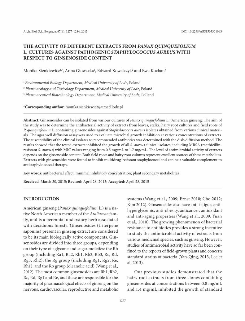
The plant material used for the study was obtained thus: (i) From experimental fields at the Agricultural University of Lublin as roots, leaves and stalks of soil-grown 3-year-old P. quinquefolium L. (Fig. 1). Organs from field cultivation were harvested in the middle of September from 3-year-old P. quinquefolium L. plants grown on dusty, light, loamy sand characterized by neutral pH, very high phosphorus content, and aver-age potassium and magnesium content; (ii) from in vitro culture as hairy root cultures. The hairy roots of P. quinquefolium L. were obtained after infection with an agropine-type strain of Agrobacterium rhi-zogenes ATCC 15834. The transformation was veri-fied by PCR analysis as described earlier (Kochan et al., 2012). Hairy root cultures were grown in 300-mL shaken Erlenmeyer flasks with 80 mL of hormone-
free, liquid, B-5 medium (Gamborg et al., 1968). The average inoculum size was about 300 mg fresh weight (f.w.) and 28.9 mg dry weight (d.w.). The cultures were maintained in the dark at 26°C on a rotary shak-er (100 rpm) and subcultured every 28 days. Fresh roots, after drying on absorbent paper, were dried at room temperature and processed for ginsenoside ex-traction and HPLC analysis.
Sample preparation
Samples of 1 g of dry raw material, weighed to 0.1 g tolerance, were placed in 250-mL flasks. They were extracted 3 times with 50 mL of 80% methanol for 30 min at boiling temperature under a reflux condenser. The combined methanol extracts were evaporated until dryness in a vacuum evaporator under lowered pressure at 60°C. The flask with dried residues was placed in a desiccator filled with a drying agent. The dried methanolic extract was weighed.
The dried methanolic extracts were dissolved in 5 mL of 50% HPLC methanol. Samples of 2 mL were placed on a solid phase extraction (SPE) column with octadecyl (C18) as reverse phase. The column was first rinsed with 10 mL of 100% HPLC methanol and 10 mL of distilled H2O to prepare the column to receive the sample. A 2-mL sample was passed through the column, which was then washed with 10 mL of dis-tilled H2O and 10 mL of 30% HPLC methanol to selectively remove impurities. Ginsenosides were se-lectively eluted using 10 mL of 100% HPLC metha-nol and were evaporated until dryness in a vacuum evaporator under lowered pressure at 60°C. Dry ex-tracts were used for HPLC analysis.
Ginsenosides Rb1, Rb2, Rc, Rd, Re and Rg1 were purchased from C. Roth GmbH+Co, Karlsruhe, Germany. A standard stock solution consisting of a mixture of the Rb1, Rb2, Rc, Rd, Re and Rg1 ginsen-osides (10 mg/mL of each ginsenoside) was prepared in methanol of HPLC grade (J.T. Baker, Netherlands). A series of standard operating solutions of different concentration were obtained by diluting the mixed standard stock solution.
Fig. 1. P. quinquefolium hairy roots; scale: 0.01 m.
Sienkiewicz et al.
1279ANTISTAPHYLOCOCCAL ACTIVITY OF GINSENG ExTRACTS
HPLC analysis of ginsenosides
Dried extracts obtained using the SPE procedure were dissolved in 2 mL of methanol (HPLC grade) and filtered through a 0.2-μm pore diameter Millex®-FG Hydrophobic Fluoropore filter (PTFE). Aliquots of 20 μL were then introduced to a liquid chromatography system consisting of LiChroART® 250-4, Waters 600 Controller pump and UV-VIS Waters 996 detector combined with a Pentium 60 PC running Millen-nium software. A reverse C18 column was employed to separate ginsenosides. Two different mixtures of acetonitrile with water were used as eluent. A 30:70 acetonitrile to water ratio was used for determination of the ginsenosides Rb1, Rb2, Rc, Rd (flow rate 2 mL/min, analysis time 45 min), and an 18:82 ratio was used for determination of the ginsenosides Rg1 and Re (flow rate 3 mL/min, analysis time 40 min.). Gin-senoside detection was performed at a wavelength of 203 nm. Quantification of ginsenosides (mg/g d.w.) was carried out by comparing retention times and peak areas between standards and samples.
Staphylococcus aureus clinical isolates
The clinical isolates of S. aureus were collected in 2011 and 2012 from a range of clinical materials recovered from patients of the Internal Medicine, Surgery and Dermatology departments of two of the Medical University hospitals in Lodz. The thirty bac-terial strains used in our investigations were isolated from the respiratory tract (n=6), abdominal cavity (n=8), postoperative wounds (n=10) and skin lesions (n=6).
S. aureus clinical isolates were cultured on Colum-bia agar (bioMerieux), on mannitol salt agar (bioM-erieux), and the ability of bacteria to produce catalase and coagulase was determined (bioMerieux). Bacteria were identified to species level using API Staph tests (bioMerieux). The tested strains were cultivated on Columbia agar medium with 5% blood and incubated at 37ºC for 24 h. Bacterial suspensions with an optical density of 0.5MF were prepared with the bioMerieux densitometer.
Susceptibility testing
Susceptibility testing was carried out with the use of the disk-diffusion method. (Jorgensen and Turnidge, 2007). The following antibiotics (Becton Dickinson) were used for testing the susceptibility of S. aureus: FOx − cefoxitin (30 μg), P − penicillin (10IU), E − erythromycin (15 μg), DA − clindamycin (2μg), TE − tetracycline (30μg), TGC − tigecycline (15 μg), C − chloramphenicol (30μg), CIP − ciprofloxacin (5 μg), RA − rifampin (5 μg), GM − gentamicin (10 μg), SxT − trimethoprim/sulfamethoxazole (1.25 μg /23.75 μg), LZD − linezolid (30 μg), FD − fusidic acid (10 μg), QDA − quinupristin/dalfopristin (15 μg), and also for VA –vancomycin (30 μg), DPC – daptomycin (15μg). Cultures were incubated at 37°C for 16-18 h under aerobic conditions on Mueller-Hinton II agar. The re-sults were interpreted according to Clinical and Labo-ratory Standard Institute guidelines (EUCAST, 2012).
Determination of minimal inhibitory concentration (MIC)
The extracts from leaves, stalks, hairy root cultures and roots of P. quinquefolium L. growing in the ground were weighed and diluted in ethanol at con-centrations from 95% to 97% w/v, which was used as a stock solution. An inoculum containing 1.5 ×10⁸ CFU (0.1 mL) per spot was applied to the surface of the Columbia agar medium at concentrations from 0.5 mg/mL to 1.6 mg/mL of tested extracts. The mini-mal inhibitory concentration (MIC) was determined by the agar well diffusion assay after 24 h of incuba-tion at 37°C under aerobic conditions on Columbia agar. Three independent analyses of the antibacterial activity of the extracts were performed. Control me-dia containing only ethanol at concentrations used in diluting the extracts did not inhibit the growth of the bacteria.
Statistical analysis
All treatments were replicated in triplicate. The re-sults were subjected to a Kruskal-Wallis test. The level of significance was set at P≤0.05. Spearman’s
1280
rank correlation coefficient between MIC value and ginsenoside content was calculated. STATISTICA ver-sion 10 (STAT Soft, Poland) software was used for all calculations.
RESULTS
Characteristics of extracts from field plants and hairy root cultures
Six ginsenosides were quantified in extracts from organs of P. quinquefolium L. taken from field-cul-tivated plants and hairy root cultures: Rb1, Rb2, Rc, Rd (protopanaxadiol derivatives), Rg1 and Re (pro-topanaxatriol derivatives). HPLC analysis of ginseng saponins confirmed that plant material from both sources contained all examined ginsenosides. The highest amount of saponins, about 35 mg/g d.w., was found in roots from field cultivation. Leaves and hairy roots contained lower amounts, and stalks the least (Table 1). Our findings also reveal that the levels of individual ginsenosides differed between the studied extracts (Table 1). An analysis of the amount of pro-topanaxatriol derivatives indicated that Re predomi-nates quantitatively: its level was about 4-times higher than Rg1 content in extracts from organs cultivated in the field and 2.5 times greater in hairy roots cul-tures. Other observations were recorded for deriva-tives of protopanaxadiol. The Rb1 saponin was seen to dominate quantitatively in both types of roots, Rb2 in leaves and Rd in stalks.
Susceptibility testing of S. aureus isolates to recommended antibiotics
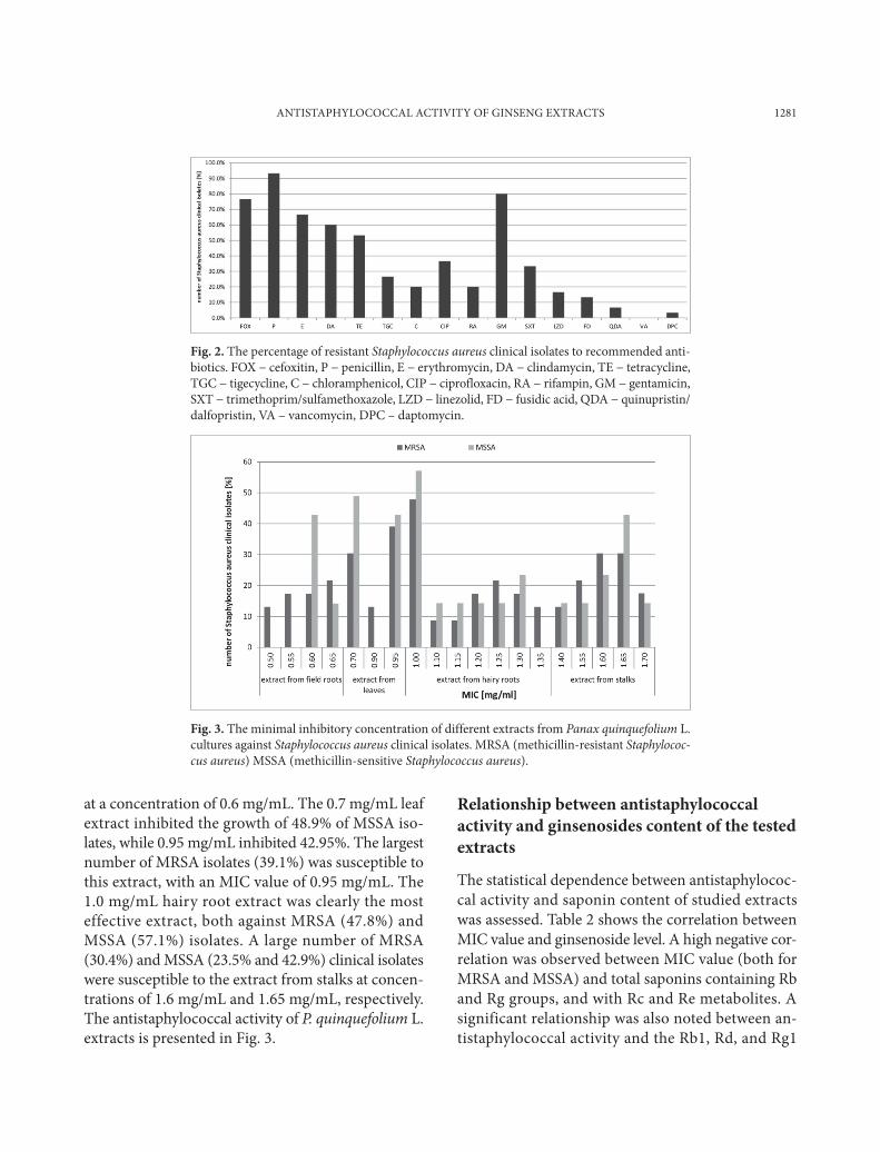
The results of susceptibility testing showed that the S. aureus isolates were highly resistant to β-lactam (penicillin for 93.3% of isolates), aminoglycoside (gentamicin for 80.0%), macrolide (erythromycin for 66.6%), lincosamide (clindamycin for 60.0%) and tetracycline (for 53.3% of clinical isolates). Among the 30 tested clinical strains, 23 (76.6%) were resis-tant to methicillin, based on tests with cefoxitin. The resistance of Staphylococcus aureus to recommended antibiotics is presented in Fig. 2.
Antimicrobial activity of P. quinquefolium L. extracts
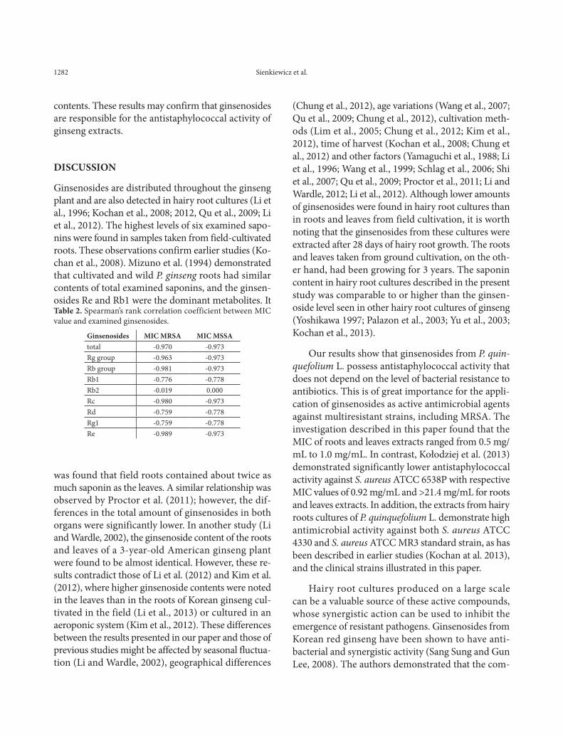
The tested extracts from leaves, stalks, hairy root cul-tures and field roots of P. quinquefolium L. demon-strated antistaphylococcal activity at concentrations from 0.5 mg/mL to 1.6 mg/mL. The highest activ-ity against all tested S. aureus clinical isolates was demonstrated by field root extracts, with MIC values ranging from 0.5 to 0.7 mg/mL. Higher MIC values (0.9-1.0 mg/mL) were obtained for leaf extracts, while the activity of hairy root extracts ranged from 1.1 mg/mL to 1.4 mg/mL. The lowest activity was demon-strated by the extract obtained from stalks, with MIC values ranging from 1.4 mg/mL to 1.7 mg. The 0.5 mg/mL and 0.55 mg/mL extracts from field roots in-hibited the growth of 30.5% MRSA cultures, while 42.9% of MSSA were susceptible to field root extract
Table 1. Ginsenoside contents in organs from field cultivation and in hairy roots cultures of P. quinquefolium. Each value is the mean of six replicates ± SE. Data analyzed using ANOVA Kruskal-Wallis test 0.05. The means with the same letter within the column do not differ significantly according to Kruskal Wallis test (p≤ 0.05)
Plant materialGinsenoside [mg/g d.w.]
Rb1 Rb2 Rc Rd Rg1 Re TotalHairy roots cultures
3.617±0.279ab
0.245±0.032b
1.057±0.034ab
0.081±0.007a
1.180±0.254ab
2.854±0.218a
9.035±0.079a
Stalks 0.721±0,099a
0.744±0.066ab
0.256±0,022a
0.989±0.044ab
0.417±0.103a 1.706 ±0.399a 4.832
±0.585a
Leaves 1.999±0.299ab
4.608±0.665a
2.689±0.818b
1.436±0.288ab
1.085±0.287ab
4.062±0.814a
15.870±1.94ab
Field roots 10.767±1.216b
0.356±0.050ab
3.724±0.615b
3.833±0.534b
3.179±0.343b
13.001±1.061b
34.859±1.646b
Sienkiewicz et al.
1281
at a concentration of 0.6 mg/mL. The 0.7 mg/mL leaf extract inhibited the growth of 48.9% of MSSA iso-lates, while 0.95 mg/mL inhibited 42.95%. The largest number of MRSA isolates (39.1%) was susceptible to this extract, with an MIC value of 0.95 mg/mL. The 1.0 mg/mL hairy root extract was clearly the most effective extract, both against MRSA (47.8%) and MSSA (57.1%) isolates. A large number of MRSA (30.4%) and MSSA (23.5% and 42.9%) clinical isolates were susceptible to the extract from stalks at concen-trations of 1.6 mg/mL and 1.65 mg/mL, respectively. The antistaphylococcal activity of P. quinquefolium L. extracts is presented in Fig. 3.
Relationship between antistaphylococcal activity and ginsenosides content of the tested extracts
The statistical dependence between antistaphylococ-cal activity and saponin content of studied extracts was assessed. Table 2 shows the correlation between MIC value and ginsenoside level. A high negative cor-relation was observed between MIC value (both for MRSA and MSSA) and total saponins containing Rb and Rg groups, and with Rc and Re metabolites. A significant relationship was also noted between an-tistaphylococcal activity and the Rb1, Rd, and Rg1
Fig. 2. The percentage of resistant Staphylococcus aureus clinical isolates to recommended anti-biotics. FOx − cefoxitin, P − penicillin, E − erythromycin, DA − clindamycin, TE − tetracycline, TGC − tigecycline, C − chloramphenicol, CIP − ciprofloxacin, RA − rifampin, GM − gentamicin, SxT − trimethoprim/sulfamethoxazole, LZD − linezolid, FD − fusidic acid, QDA − quinupristin/dalfopristin, VA – vancomycin, DPC – daptomycin.
Fig. 3. The minimal inhibitory concentration of different extracts from Panax quinquefolium L. cultures against Staphylococcus aureus clinical isolates. MRSA (methicillin-resistant Staphylococ-cus aureus) MSSA (methicillin-sensitive Staphylococcus aureus).
ANTISTAPHYLOCOCCAL ACTIVITY OF GINSENG ExTRACTS
1282
contents. These results may confirm that ginsenosides are responsible for the antistaphylococcal activity of ginseng extracts.
DISCUSSION
Ginsenosides are distributed throughout the ginseng plant and are also detected in hairy root cultures (Li et al., 1996; Kochan et al., 2008; 2012, Qu et al., 2009; Li et al., 2012). The highest levels of six examined sapo-nins were found in samples taken from field-cultivated roots. These observations confirm earlier studies (Ko-chan et al., 2008). Mizuno et al. (1994) demonstrated that cultivated and wild P. ginseng roots had similar contents of total examined saponins, and the ginsen-osides Re and Rb1 were the dominant metabolites. It
was found that field roots contained about twice as much saponin as the leaves. A similar relationship was observed by Proctor et al. (2011); however, the dif-ferences in the total amount of ginsenosides in both organs were significantly lower. In another study (Li and Wardle, 2002), the ginsenoside content of the roots and leaves of a 3-year-old American ginseng plant were found to be almost identical. However, these re-sults contradict those of Li et al. (2012) and Kim et al. (2012), where higher ginsenoside contents were noted in the leaves than in the roots of Korean ginseng cul-tivated in the field (Li et al., 2013) or cultured in an aeroponic system (Kim et al., 2012). These differences between the results presented in our paper and those of previous studies might be affected by seasonal fluctua-tion (Li and Wardle, 2002), geographical differences
(Chung et al., 2012), age variations (Wang et al., 2007; Qu et al., 2009; Chung et al., 2012), cultivation meth-ods (Lim et al., 2005; Chung et al., 2012; Kim et al., 2012), time of harvest (Kochan et al., 2008; Chung et al., 2012) and other factors (Yamaguchi et al., 1988; Li et al., 1996; Wang et al., 1999; Schlag et al., 2006; Shi et al., 2007; Qu et al., 2009; Proctor et al., 2011; Li and Wardle, 2012; Li et al., 2012). Although lower amounts of ginsenosides were found in hairy root cultures than in roots and leaves from field cultivation, it is worth noting that the ginsenosides from these cultures were extracted after 28 days of hairy root growth. The roots and leaves taken from ground cultivation, on the oth-er hand, had been growing for 3 years. The saponin content in hairy root cultures described in the present study was comparable to or higher than the ginsen-oside level seen in other hairy root cultures of ginseng (Yoshikawa 1997; Palazon et al., 2003; Yu et al., 2003; Kochan et al., 2013).
Our results show that ginsenosides from P. quin-quefolium L. possess antistaphylococcal activity that does not depend on the level of bacterial resistance to antibiotics. This is of great importance for the appli-cation of ginsenosides as active antimicrobial agents against multiresistant strains, including MRSA. The investigation described in this paper found that the MIC of roots and leaves extracts ranged from 0.5 mg/mL to 1.0 mg/mL. In contrast, Kołodziej et al. (2013) demonstrated significantly lower antistaphylococcal activity against S. aureus ATCC 6538P with respective MIC values of 0.92 mg/mL and >21.4 mg/mL for roots and leaves extracts. In addition, the extracts from hairy roots cultures of P. quinquefolium L. demonstrate high antimicrobial activity against both S. aureus ATCC 4330 and S. aureus ATCC MR3 standard strain, as has been described in earlier studies (Kochan at al. 2013), and the clinical strains illustrated in this paper.
Hairy root cultures produced on a large scale can be a valuable source of these active compounds, whose synergistic action can be used to inhibit the emergence of resistant pathogens. Ginsenosides from Korean red ginseng have been shown to have anti-bacterial and synergistic activity (Sang Sung and Gun Lee, 2008). The authors demonstrated that the com-
Table 2. Spearman’s rank correlation coefficient between MIC value and examined ginsenosides.
Ginsenosides MIC MRSA MIC MSSAtotal -0.970 -0.973Rg group -0.963 -0.973Rb group -0.981 -0.973Rb1 -0.776 -0.778Rb2 -0.019 0.000Rc -0.980 -0.973Rd -0.759 -0.778Rg1 -0.759 -0.778Re -0.989 -0.973
Sienkiewicz et al.
1283
bination of ginsenosides and kanamycin possesses a synergistic or additive effect against MRSA: the gin-senosides were shown to interact with the membrane, thus increasing the permeability of the plasma mem-brane to kanamycin.
CONCLUSIONS
The antistaphylococcal activity of extracts from leaves, stalks, hairy root cultures and field roots of P. quinquefolium L. depend on the ginsenoside content. The extracts containing ginsenosides have antistaphy-lococcal activity, irrespective of the level of bacterial antibiotic resistance. Excellent sources of active gin-senosides are not only extracts from leaves and field roots, but also those of hairy root cultures.
Acknowledgments: The research was supported by Grant No. 502 13 771 from the Medical University of Lodz and has not been submitted elsewhere.
Authors’ contributions: MS designed and performed the experiments, analyzed the data and prepared the manuscript. AG, EK assisted in data analysis. EK performed experiments, analyzed the data and assisted in manuscript preparation. All authors read and approved the final manuscript.
Conflict of interest disclosure: The authors report no con-flict of interest.
REFERENCES
Lowry, O.H., Rosebrough, N. J. Farr, A. L. and R. J. Randall (1951). Protein measurement with the folin phenol reagent. J. Biol. Chem. 193, 165-175.
Cho, I.H. (2012). Effects of Panax ginseng in neurodegenerative diseases. J. Ginseng Res. 36(4), 342-353.
Chung, I.M., Kim, J.W., Seguin, Ph., Jun, Y.M. and S.H. Kim (2012). Ginsenosides and phenolics in fresh and processed Korean Ginseng (Panax ginseng C.A. Meyer): effects of cul-tivation localisation, year, and storage period. Food Chem. 130, 73-83.
Ernst, E. (2010). Panax ginseng: An overview of the clinical evi-dence. J. Ginseng Res. 34, 259-263.
European Committee on Antimicrobial Susceptibility Testing (EUCAST). Breakpoint Tables for Interpretation of MICs and Zone Diameters, version 2.0; valid from 1 January
2012; Available online: www.eucast.org (accessed on 16 December 2012).
Gamborg, O.L., Miller, R.A. and K. Ojima (1968). Nutrient requirements of suspension cultures of soybean root cells. Exp. Cell Res. 50, 151-158.
Jorgensen, J.H. and J.D. Turnidge (2007). Antibacterial susceptibil-ity tests: dilution and disk diffusion methods. In: Manual of clinical microbiology (Eds. Murray, P.R., Baron, E.J., Jor-gensen, J.H., Landry, M.L. and M.A. Pfaller), 1152-1172. American Society for Microbiology, Washington, DC.
Kim, G.S., Lee, S.E., Noh, H.J., Kwon, H., Lee, S.W., Kim, S-Y. and Y-B. Kim (2012). Effects of natural products on the growth and ginsenoside contents of Panax ginseng cultured in an aeroponic system. J. Ginseng Res. 36(4), 430-441.
Kim, J-H. (2012). Cardiovascular diseases and Panax ginseng: a review on molecular mechanisms and medical applications. J. Ginseng Res. 36, 16-26.
Kochan, E., Wasiela, M. and M. Sienkiewicz (2013). The produc-tion of ginsenosides in hairy root cultures of American Ginseng, Panax quinquefolium L. and their antimicrobial activity. In Vitro Cell Dev. Biol. Pl. 49(1), 4-29.
Kochan, E., Królicka, A. and A. Chmiel (2012). Panax quinquefo-lium hairy roots cultivated in flasks and nutrient sprinkle bioreactor. Acta Physiol. Plant. 34, 1513-1518.
Kołodziej, B., Kowalski, R. and E. Hołderna-Kędzia (2013). Chem-ical composition and chosen bioactive properties of Panax quinquefolius extracts. Chemija. 24, 151-159.
Lee, K.A., Kim, W.J., Kim, H.J., Kim, K-T. and H-D. Paik (2013). Antibacterial activity of Ginseng (Panax ginseng C. A. Meyer) stems-leaves extract produced by subcritical water extraction Int. J. Food Sci & Technol. 48, 947-953
Li, T.S.C., Mazza, G., Cottrell, A.C. and L. Gao (1996). Ginsen-osides in roots and leaves of American Ginseng. J. Agric. Food Chem. 44, 717-720.
Li, X., Yan, Y.Z., Jin, X., Kim, Y.K., Uddin, M.R., Kim, Y.B., Bae, H., Kim, Y.C., Lee, S.W. and S.U. Park (2012). Ginsenoside content in the leaves and roots of Panax ginseng and dif-ferent ages. Life Sci. J. 9(4), 679-683.
Lim, W., Mudge, K.W. and F. Vermeylen (2005). Effect of popula-tion, age and cultivation methods on ginsenoside content of wild American Ginseng (Panax quinquefolium). J. Agrc. Food Chem. 53, 8498-8505.
Proctor, J.T.A., Sullivan, A.J., Rupasinghe, V.P.V. and Ch-JC. Jack-son (2011). Morphological and ginsenoside differences among North American ginseng leaves. J. Ginseng Res. 35(2), 155-161.
Sang Sung, W. and D. Gun Lee (2008). The combination effect of Korean red ginseng saponins with kanamycin and cefo-taxime against methicillin-resistant Staphylococcus aureus. Biol. Pharm. Bull. 31(8), 1614-1617.
Starrat, A.N., Hendel, J.G. and R.D. Reeleder (2001). Leaves of North American ginseng, Panax quinquefolium L. A renewable source of certain ginsenosides. Can. J. Plant Sc. 81(1), 65-67.
ANTISTAPHYLOCOCCAL ACTIVITY OF GINSENG ExTRACTS
1284
Wang, H., Peng, D. and J. Xie (2009). Ginseng life-stem: bioac-tive constituents and pharmacological functions. Chinese Med. 4, 20.
Wang, J., Gao, W-Y., Zhang, J., Zuo, B-M., Zhang, L-M. and L-Q. Huang (2012). Advances in study of ginsenoside biosyn-thesis pathway in Panax ginseng C.A. Meyer. Acta Physiol. Plant. 34, 397-403.
Wang, W, Li, Y.T., Zhang, H.Q. and L. Ding (2007). Investigation of ginsenosides in different parts and ages of Panax ginseng. Food Chem. 102, 664-668.
Wang, X., Sakuma, T., Asafu-Adjaye, E. and G.K. Shiu (1999). Determination of ginsenosides in plant extracts from Panax ginseng and Panax quinquefolium L. by LC/MS/MS. Anal. Chem. 71, 1579-1584.
Yamaguchi, H., Matsuura, H., Kasai, R., Tanaka, O., Satake, M., Kohda, H., Izumi, H., Nuno, M., Katsuki, S., Isoda, S., Shoji,
J. and K. Goto (1988). Analysis of saponins of wild Panax ginseng. Chem. Pharm. Bull. 36, 4177-4181.
Yan-Qing, J.I. (2013). Antioxidant and antibacterial activities of ginseng (Panax ginseng C.A. Meyer) extracts Asian Int. J. Life Sci. 22, 359-369.
Yoshikawa, T. (1997). Production of ginsenosides in ginseng hairy root cultures. In: Hairy roots. culture and applications (Ed. P.M. Doran), 73-79. Varwood Academic Publishers, Amsterdam.
Yu, K.W., Hahn, E.J. and K.Y. Paek (2003). Ginsenoside produc-tion by hairy root cultures of Panax ginseng C.A. Meyer in bioreactors. Acta. Hort. 597, 237-243.
Yuan, Ch-S., Wang, Ch-Z., Wicks, S.M. and L-W. Qi (2010). Chemical and pharmacological studies of saponins with focus on American ginseng. J. Ginseng Res. 34, 160-167.
Sienkiewicz et al.