Embed Size (px)
Citation preview

Original article
The activity of the 20S proteasome is maintained in detached wheatleaves during senescence in darkness
Irma Roberts, Pedro Fernández Murray, Susana Passeron, Atilio J. Barneix *
IBYF-CONICET, Facultad de Agronomía, Universidad de Buenos Aires, Avda, San Martín 4453, 1417 Buenos Aires, Argentina
Received 31 May 2001; accepted 20 September 2001
Abstract
In the present paper, we studied the participation of the 20S proteasome, the proteolytic component of the ubiquitin–proteasome pathway,in the remobilization of bulk proteins in senescing wheat leaves. The detached leaves of 15-d-old plants were incubated in darkness forseveral days, and various proteolytic activities were analysed in soluble extracts prepared at 0, 48 and 96 h after detachment. Theendoproteolytic activity, measured at pH 7.5 and 5.4, increased more than 10-fold and the total peptidasic activity increased up to 5-foldafter 96 h of incubation in the dark, when expressed as specific activity. In the same period, the leaf-protein content decreased to less than50% of that present at the initial time. The 20S proteasome chymotrypsin-like activity remained constant when it was expressed as activityper leaf fresh weight and resulted 2-fold higher in terms of specific activity. The western blot analysis showed that the amount of 20Sproteasome protein and ubiquitin–protein conjugates also remained constant until 4 d of incubation in darkness. These results indicate thatthe ubiquitin–proteasome pathway remains functional until the late phases of senescence suggesting that it may participate in the regulatoryaspects of the process rather than in the massive protein breakdown. © 2002 Éditions scientifiques et médicales Elsevier SAS. All rightsreserved.
Keywords: 20S proteasome; Proteolysis; Senescence; Ubiquitin; Wheat
1. Introduction
Senescence is the final stage of leaf development leadingto the death of the organ[11]. It is a genetically programmedprocess[26], involving leaf protein and chlorophyll degra-dation. Membrane integrity and the subcellular compart-mentation are maintained until the latest phase, allowing fora proper cooperation between various organelles involved inthe catabolism of leaf constituents[11].
Protein degradation during senescence involves the in-duction of several proteolytic activities[32] that contributeto the degradation of Rubisco (ribulose-1,5-bisphosphatecarboxylase/oxygenase; EC 4.1.1.39), the most abundant
protein in leaves, which is massively hydrolysed during thefirst stages of leaf senescence together with other proteins[11].
The ubiquitin–proteasome proteolytic system is respon-sible for the specific degradation of abnormal, short-livedand regulatory proteins in the cytoplasm and nucleus of theeukaryotic cells[20,22]. In addition, this pathway has beenshown to be involved in the degradation of the bulk ofproteins in the insect and mammalian cells under certainmetabolic conditions[19,28].
In higher plants, the ubiquitin–proteasome componentshave been isolated and characterised[3,32–34]; this pro-teolytic system has also been shown to participate incell-cycle progression[16], degradation of short-lived regu-latory proteins[8], elicitation of defence responses[2],auxin response, photomorphogenesis and pollen germina-tion [29] (for a review see[7,10,32,33]).
The involvement of the ubiquitin–proteasome pathway insenescence is suggested by biochemical analysis and evi-dence of the expression of genes coding for components ofthe ubiquitin pathway[9,15,17,27,31]. On the other hand,
Abbreviations: Cbz, benzyloxycarbonyl ; DEAE, diethylaminoethyl ;DTT, dithiothreitol ; NEM, N-ethyl-maleimide ; pNA, p-nitroanilide ;PVDF, polyvinylidene difluoride ; Rubisco, ribulose-1,5-bisphosphatecarboxylase ; oxygenase ; SDS-PAGE, sodium dodecyl sulphate–polya-crylamide gel electrophoresis ; TCA, trichloroacetic acid
* Corresponding author. Fax: +54-11-45-14-87-41.E-mail address: [email protected] (A.J. Barneix).
Plant Physiol. Biochem. 40 (2002) 161–166
www.elsevier.com/locate/plaphy
© 2002 Éditions scientifiques et médicales Elsevier SAS. All rights reserved.PII: S 0 9 8 1 - 9 4 2 8 ( 0 1 ) 0 1 3 4 9 - 3

studies on the expression of an α-type 20S proteasomesubunit gene provided no support for a role of the protea-some proteolytic pathway during the senescence process, atleast in massive protein degradation [1].
In order to clarify this point, we investigated severalproteolytic activities in senescing wheat leaves as well aspossible changes in the 20S proteasome proteolytic activity.Our results suggest that the induction of proteolytic activi-ties different from that of the 20S proteasome can accountfor the massive protein degradation observed. The fact thatthe 20S proteasome, as well as the ubiquitin conjugates, iskept at a constant level throughout the senescence processindicates that the ubiquitin–proteasome pathway remainsfunctional until the late phases of senescence, suggesting itsparticipation in the regulatory aspects of the process ratherthan in the bulk degradation of proteins.
2. Results
When excised leaves of 15-d-old wheat plants wereincubated in the dark for several days, there was a fastinduction of various proteolytic activities. After 96 h in thedark, the endoproteolytic activity increased nearly 400%when expressed as activity per leaf fresh weight (Fig. 1A),and as much as 1000% when expressed as specific activity(Fig. 1B). The total peptidasic activity increased more than200% on a fresh-weight basis, and up to 500% whenexpressed as specific activity (Fig. 1A, B). During thisperiod, the leaf-protein concentration decreased to less thanhalf of the initial concentration (Fig. 1A). The 20S protea-some activity remained constant on a fresh-weight basisduring the whole dark period, and only doubled its specificactivity in the same period (Fig. 1A, B). In order to furthersupport these results, the extracts obtained from fresh leavesand from 96 h dark-induced leaves, containing the sameamount of soluble protein, were fractionated by ion ex-change chromatography and analysed for Cbz–GGL–pNAand azocasein-hydrolysing activities. As can be seen in Fig.2A and C, one symmetrical peak of chymotrypsin-likeactivity, eluting at the same sodium chloride concentration,was detected in the eluates from extracts obtained fromfresh or dark-induced leaves. Remarkably, the total activityrecovered from the dark-induced leaf extract doubled thatobtained from the leaf extracts prepared from the non-induced leaves (Fig. 2A, C). Fractions were also analysedby western blotting using a polyclonal antiserum raisedagainst maize 20S proteasome. The fractions containingimmunoreactive proteins were coincident with those exhib-iting enzymatic activity in both senescing (Fig. 2D) andnon-senescing leaf samples (Fig. 2B).
Fig. 2A and C also shows the profile of azocasein-hydrolysing activity under both conditions (fresh and dark-induced leaf extracts). As can be seen, a broad peak, elutingahead of the 20S proteasome activity, was observed in bothcases. However, a second peak of endoprotease activity,
eluting with the void volume was present only in extractsderived from the dark-induced leaves. These activities arenow under study.
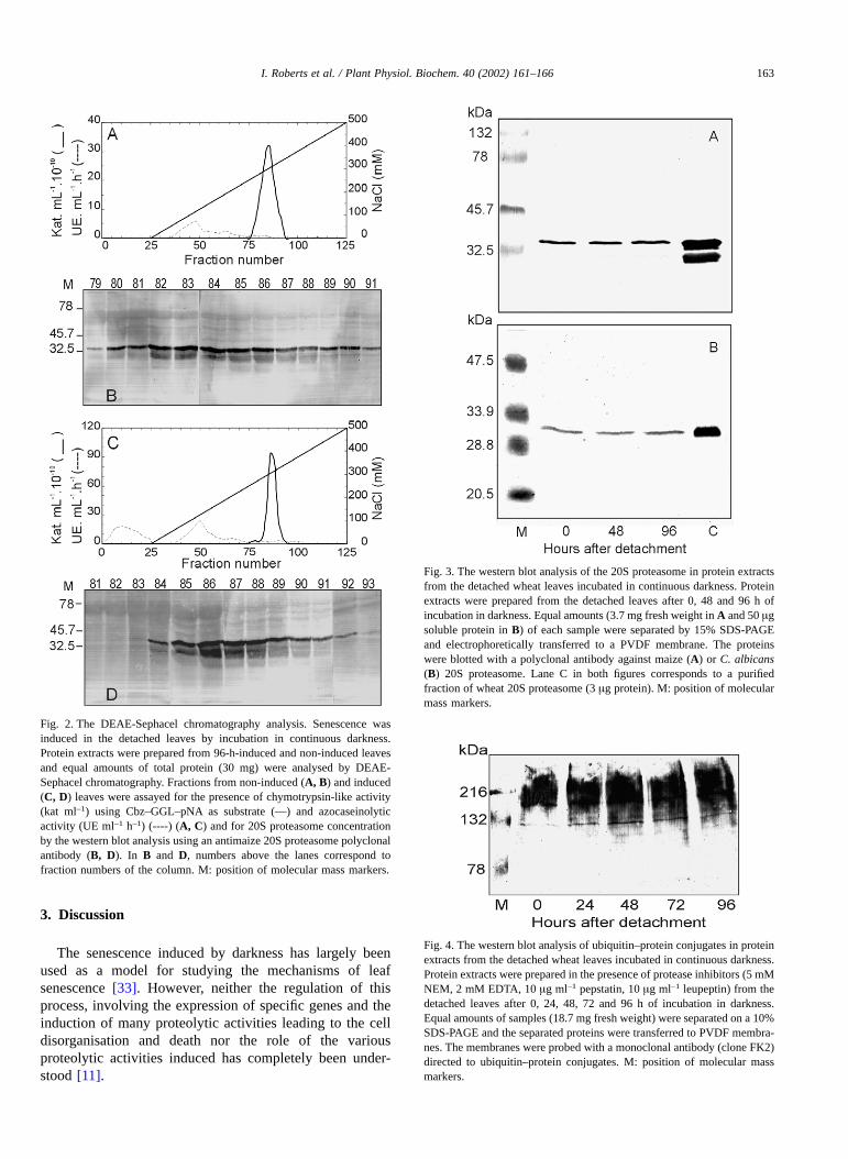
Western blot analysis of leaf extracts during the incuba-tion period in the dark showed that the amount of 20Sproteasome protein did not change as judged by the inten-sity of the main proteasomal component recognised byantibodies directed against maize (Fig. 3A) or Candidaalbicans 20S proteasome (Fig. 3B). The amount of ubiquitincomplexes, detected with a specific antibody, appearing asproteins of molecular masses above 100 kDa did not varysignificantly during the 96 h darkness incubation (Fig. 4).
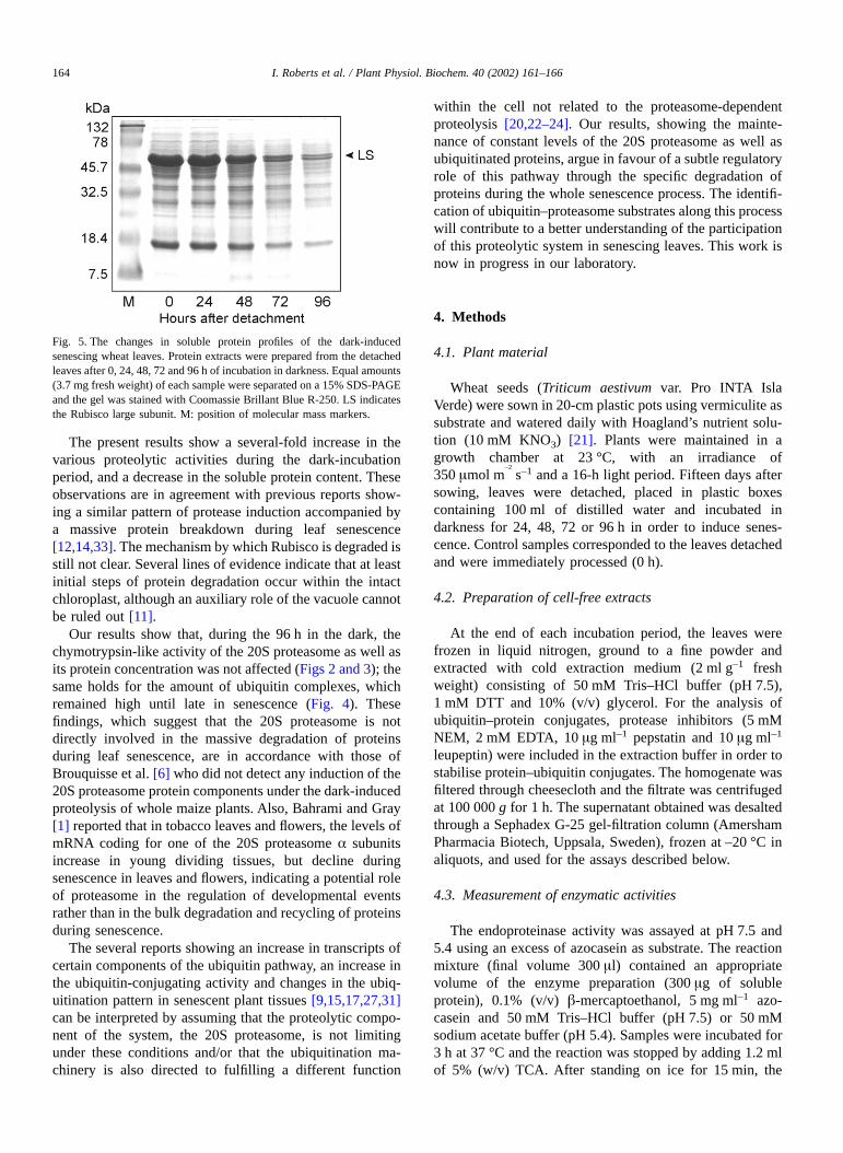
In young wheat leaves, more than 50% of soluble proteincan be accounted for by Rubisco. The SDS-PAGE pattern ofthe total leaf proteins during the dark incubation periodshowed a decrease in the intensity of all polypeptide bands.The degradation of a band near the 47.5 kDa marker,probably corresponding to the large subunit of the Rubisco(Fig. 5), was the most significant.
Fig. 1. Changes in proteolytic activities, during the dark-induced senes-cence in the detached wheat leaves. After 0, 48 and 96 h of incubation indarkness, leaf extracts were prepared and enzymatic activities weremeasured as described in the Methods. Activities are expressed per gram ofleaf fresh weight (A) or per milligram of total protein (B). Endoproteinaseactivity using azocasein as substrate was assayed at pH 7.5 ([) and pH 5.4(■ ), release of α-amino groups by autodigestion of extracts at pH 5.4 wasdetected with the ninhydrin reagent (▲) and 20S proteasome activity wasdetermined by hydrolysis of the synthetic peptide Cbz–GGL–pNA (● ).Protein content of extracts is also shown (·). Data are presented as relativevalues ± SEM of three replicates, with the data for 0 h after detachment setat 100. A, initial values are 55.5 and 55.8 UE h–1 g–1 for azocaseinolyticactivity at pH 7.5 and 5.4, respectively; 7.18 × 10⁻¹² kat g–1 for 20Sproteasome activity; 3.66 × 10⁻¹⁰ kat g–1 and 6.2 µg µl–1 for soluble proteinconcentration. B, initial values are 3.2 UE h–1 mg–1 for azocaseinolyticactivity at both pHs; 4.22 × 10⁻¹³ kat mg–1 for 20S proteasome activity and2.15 × 10–11 kat mg–1.
162 I. Roberts et al. / Plant Physiol. Biochem. 40 (2002) 161–166
3. Discussion
The senescence induced by darkness has largely beenused as a model for studying the mechanisms of leafsenescence [33]. However, neither the regulation of thisprocess, involving the expression of specific genes and theinduction of many proteolytic activities leading to the celldisorganisation and death nor the role of the variousproteolytic activities induced has completely been under-stood [11].
Fig. 2. The DEAE-Sephacel chromatography analysis. Senescence wasinduced in the detached leaves by incubation in continuous darkness.Protein extracts were prepared from 96-h-induced and non-induced leavesand equal amounts of total protein (30 mg) were analysed by DEAE-Sephacel chromatography. Fractions from non-induced (A, B) and induced(C, D) leaves were assayed for the presence of chymotrypsin-like activity(kat ml–1) using Cbz–GGL–pNA as substrate (—) and azocaseinolyticactivity (UE ml–1 h–1) (----) (A, C) and for 20S proteasome concentrationby the western blot analysis using an antimaize 20S proteasome polyclonalantibody (B, D). In B and D, numbers above the lanes correspond tofraction numbers of the column. M: position of molecular mass markers.
Fig. 3. The western blot analysis of the 20S proteasome in protein extractsfrom the detached wheat leaves incubated in continuous darkness. Proteinextracts were prepared from the detached leaves after 0, 48 and 96 h ofincubation in darkness. Equal amounts (3.7 mg fresh weight in A and 50 µgsoluble protein in B) of each sample were separated by 15% SDS-PAGEand electrophoretically transferred to a PVDF membrane. The proteinswere blotted with a polyclonal antibody against maize (A) or C. albicans(B) 20S proteasome. Lane C in both figures corresponds to a purifiedfraction of wheat 20S proteasome (3 µg protein). M: position of molecularmass markers.
Fig. 4. The western blot analysis of ubiquitin–protein conjugates in proteinextracts from the detached wheat leaves incubated in continuous darkness.Protein extracts were prepared in the presence of protease inhibitors (5 mMNEM, 2 mM EDTA, 10 µg ml–1 pepstatin, 10 µg ml–1 leupeptin) from thedetached leaves after 0, 24, 48, 72 and 96 h of incubation in darkness.Equal amounts of samples (18.7 mg fresh weight) were separated on a 10%SDS-PAGE and the separated proteins were transferred to PVDF membra-nes. The membranes were probed with a monoclonal antibody (clone FK2)directed to ubiquitin–protein conjugates. M: position of molecular massmarkers.
I. Roberts et al. / Plant Physiol. Biochem. 40 (2002) 161–166 163
The present results show a several-fold increase in thevarious proteolytic activities during the dark-incubationperiod, and a decrease in the soluble protein content. Theseobservations are in agreement with previous reports show-ing a similar pattern of protease induction accompanied bya massive protein breakdown during leaf senescence[12,14,33]. The mechanism by which Rubisco is degraded isstill not clear. Several lines of evidence indicate that at leastinitial steps of protein degradation occur within the intactchloroplast, although an auxiliary role of the vacuole cannotbe ruled out [11].
Our results show that, during the 96 h in the dark, thechymotrypsin-like activity of the 20S proteasome as well asits protein concentration was not affected (Figs 2 and 3); thesame holds for the amount of ubiquitin complexes, whichremained high until late in senescence (Fig. 4). Thesefindings, which suggest that the 20S proteasome is notdirectly involved in the massive degradation of proteinsduring leaf senescence, are in accordance with those ofBrouquisse et al. [6] who did not detect any induction of the20S proteasome protein components under the dark-inducedproteolysis of whole maize plants. Also, Bahrami and Gray[1] reported that in tobacco leaves and flowers, the levels ofmRNA coding for one of the 20S proteasome α subunitsincrease in young dividing tissues, but decline duringsenescence in leaves and flowers, indicating a potential roleof proteasome in the regulation of developmental eventsrather than in the bulk degradation and recycling of proteinsduring senescence.
The several reports showing an increase in transcripts ofcertain components of the ubiquitin pathway, an increase inthe ubiquitin-conjugating activity and changes in the ubiq-uitination pattern in senescent plant tissues [9,15,17,27,31]can be interpreted by assuming that the proteolytic compo-nent of the system, the 20S proteasome, is not limitingunder these conditions and/or that the ubiquitination ma-chinery is also directed to fulfilling a different function
within the cell not related to the proteasome-dependentproteolysis [20,22–24]. Our results, showing the mainte-nance of constant levels of the 20S proteasome as well asubiquitinated proteins, argue in favour of a subtle regulatoryrole of this pathway through the specific degradation ofproteins during the whole senescence process. The identifi-cation of ubiquitin–proteasome substrates along this processwill contribute to a better understanding of the participationof this proteolytic system in senescing leaves. This work isnow in progress in our laboratory.
4. Methods
4.1. Plant material
Wheat seeds (Triticum aestivum var. Pro INTA IslaVerde) were sown in 20-cm plastic pots using vermiculite assubstrate and watered daily with Hoagland’s nutrient solu-tion (10 mM KNO3) [21]. Plants were maintained in agrowth chamber at 23 °C, with an irradiance of350 µmol m
–2
s–1 and a 16-h light period. Fifteen days aftersowing, leaves were detached, placed in plastic boxescontaining 100 ml of distilled water and incubated indarkness for 24, 48, 72 or 96 h in order to induce senes-cence. Control samples corresponded to the leaves detachedand were immediately processed (0 h).
4.2. Preparation of cell-free extracts
At the end of each incubation period, the leaves werefrozen in liquid nitrogen, ground to a fine powder andextracted with cold extraction medium (2 ml g–1 freshweight) consisting of 50 mM Tris–HCl buffer (pH 7.5),1 mM DTT and 10% (v/v) glycerol. For the analysis ofubiquitin–protein conjugates, protease inhibitors (5 mMNEM, 2 mM EDTA, 10 µg ml–1 pepstatin and 10 µg ml–1
leupeptin) were included in the extraction buffer in order tostabilise protein–ubiquitin conjugates. The homogenate wasfiltered through cheesecloth and the filtrate was centrifugedat 100 000 g for 1 h. The supernatant obtained was desaltedthrough a Sephadex G-25 gel-filtration column (AmershamPharmacia Biotech, Uppsala, Sweden), frozen at –20 °C inaliquots, and used for the assays described below.
4.3. Measurement of enzymatic activities
The endoproteinase activity was assayed at pH 7.5 and5.4 using an excess of azocasein as substrate. The reactionmixture (final volume 300 µl) contained an appropriatevolume of the enzyme preparation (300 µg of solubleprotein), 0.1% (v/v) �-mercaptoethanol, 5 mg ml–1 azo-casein and 50 mM Tris–HCl buffer (pH 7.5) or 50 mMsodium acetate buffer (pH 5.4). Samples were incubated for3 h at 37 °C and the reaction was stopped by adding 1.2 mlof 5% (w/v) TCA. After standing on ice for 15 min, the
Fig. 5. The changes in soluble protein profiles of the dark-inducedsenescing wheat leaves. Protein extracts were prepared from the detachedleaves after 0, 24, 48, 72 and 96 h of incubation in darkness. Equal amounts(3.7 mg fresh weight) of each sample were separated on a 15% SDS-PAGEand the gel was stained with Coomassie Brillant Blue R-250. LS indicatesthe Rubisco large subunit. M: position of molecular mass markers.
164 I. Roberts et al. / Plant Physiol. Biochem. 40 (2002) 161–166

precipitate was removed by centrifugation at 16 000 g for10 min and acid-soluble products were determined spectro-photometrically at 340 nm. One unit of azocaseinolyticactivity was defined as the amount of protein causing a 0.01increase in A340 over the 0 time values (the reaction stoppedimmediately after starting). Chymotrypsinlike activity ofthe 20S proteasome was tested by hydrolysis of the syn-thetic peptide Cbz–GGL–pNA according to Bratton andMarshall [5] as modified by Goldbarg and Rutemburg [18],which is a reliable measure of the 20S proteasome activity.The release of α-amino groups by autodigestion of theextracts was determined with the ninhydrin reagent; 200 µlof an appropriate dilution of the samples (400 µg of solubleprotein) were incubated at 37 °C for 1 h with 200 µl of200 mM Tris–HCl buffer (pH 7.5). The reaction wasstopped with 400 µl of 5% TCA. After standing in ice for10 min, samples were centrifuged at 16 000 g for 10 minand 250 µl aliquots of the supernatant was taken for thedetermination of α-amino nitrogen by the ninhydrin method[36].
4.4. SDS-PAGE and immunoblotting
Protein extracts were analysed by 10 or 15% SDS-PAGEaccording to Laemmli [25]. For the visualisation of proteinbands, the gels were stained with Coomassie Brillant BlueR-250. For the western blot analysis, proteins were trans-ferred to PVDF membranes (Immobilon, Millipore), essen-tially as described by Towbin et al. [30]. Immunodetectionof wheat 20S proteasome was achieved by incubating themembrane with a polyclonal antiserum raised against C. al-bicans 20S proteasome [13] or an antimaize 20S protea-some antibody kindly supplied by Dr. R. Brouquisse [6].The ubiquitinated proteins were detected using a mono-clonal antibody directed against the ubiquitin–protein con-jugates, clone FK2 (Affinity Research Products, Ltd). Thebound antibodies were detected with the correspondingbiotinylated antirabbit (20S proteasome) or antimouse(ubiquitinated proteins) antibody and avidin-biotinylatedhorseradish peroxidase, using 3,3'-diaminobenzidine andH2O2 as substrates.
4.5. DEAE-Sephacel chromatography
Equal amounts of total protein (30 mg) of extracts frominduced (96 h) and non-induced (0 h) senescing leaves wereloaded on a DEAE–Sephacel column (Sigma) equilibratedwith the extraction buffer. The column (1 × 4.5 cm) waswashed with three volumes of extraction buffer and theneluted with 40 ml of a linear gradient of NaCl (0–500 mM)made in the same buffer. Fractions of 400 µl were collectedand aliquots of 200 µl of each fraction were assayed for thepresence of chymotrypsin-like activity using Cbz–G-GL–pNA as substrate. Proteins contained in 100 µl samplesof eluted fractions were precipitated by the methanol–chlo-roform method [35] and separated on a 15% SDS-PAGE.
The solved proteins were electrophoretically transferred toPVDF membranes and revealed with the anti-maize 20Sproteasome polyclonal antibody as described above.
4.6. Protein determination
The protein content of the leaf extracts was determinedaccording to Bradford [4] using bovine serum albumin asstandard.
Acknowledgements
This work was supported by the Consejo Nacional deInvestigaciones Científicas y Técnicas (CONICET), Argen-tina. We are very grateful to Dr. R. Brouquisse for hisgenerous donation of anti-maize 20S proteasome antiserumand to Dr. Eduardo J. Passeron for critical reading of themanuscript.
References
[1] A.R. Bahrami, J.E. Gray, Expression of a proteasome α-type subunitgene during tobacco development and senescence, Plant Mol. Biol. 39(1999) 325–333.
[2] J. Becker, R. Kempf, W. Jeblick, H. Kauss, Induction of competencefor elicitation of defence responses in cucumber hypocotyls requiresproteasome activity, Plant J. 21 (2000) 311–316.
[3] W.R. Belknap, J.E. Garbarino, The role of ubiquitin in plant senes-cence and stress responses, Trends Plant Sci. 1 (1996) 331–335.
[4] M.M. Bradford, A rapid and sensitive method for the quantitation ofmicrograms quantities of proteins utilizing the principle of protein-dye binding, Anal. Biochem. 72 (1976) 248–254.
[5] A.C. Bratton, E.K. Marshall, A new coupling component for sulfa-nilamide determination, J. Biol. Chem. 128 (1939) 537–550.
[6] R. Brouquisse, J.P. Gaudillere, P. Raymond, Induction of a carbon-starvation-related proteolysis in whole maize plants submitted tolight/dark cycles and to extended darkness, Plant Physiol. 117 (1988)1281–1291.
[7] J. Callis, R.D. Vierstra, Protein degradation in signaling, Curr. Opin.Plant Biol. 44 (2001) 85–410.
[8] R.C. Clough, E.T. Jordan-Beebe, K.N. Lohman, J.M. Marita,J.M. Walker, C. Gatz, R.D. Vierstra, Sequences within both the N- andC-terminal domains of phytochrome A are required for PFR ubiquiti-nation and degradation, Plant J. 17 (1999) 155–167.
[9] S.E. Courtney, C.C. Rider, A.D. Stead, Changes in protein ubiquiti-nation and the expression of ubiquitin encoding transcripts in day lilypetals during floral development and senescence, Physiol. Plant. 91(1994) 548–556.
[10] M. Estelle, Protease and cellular regulation in plants, Curr. Opin.Plant Biol. 4 (2001) 254–260.
[11] U. Feller, A. Fisher, Nitrogen metabolism in senescing leaves, Crit.Rev. Plant Sci. 13 (1994) 241–273.
[12] U. Feller, T. Soon, R. Haeman, Leaf proteolytic activities andsenescence during grain development of field-grown corn (Zea maysL.), Plant Physiol. 59 (1977) 290–294.
[13] P. Fernández Murray, M.J. Biscoglio, S. Passeron, Purification andcharacterization of Candida albicans 20S proteasome: identificationof four proteasomal subunits, Arch. Biochem. Biophys. 375 (2000)211–219.
I. Roberts et al. / Plant Physiol. Biochem. 40 (2002) 161–166 165

[14] A. Fischer, U. Feller, Senescence and protein degradation in leafsegments of young winter wheat: influence of leaf age, J. Exp. Bot. 45(1994) 103–109.
[15] J.E. Garbarino, T. Oosumi, W.R. Belknap, Isolation of a polyubiquitinpromoter and its expression in transgenic potato plants, Plant Physiol.109 (1995) 1371–1378.
[16] P. Genschik, M.C. Criqui, Y. Parmentier, A. Derevier, J. Fleck, Cellcycle-dependent proteolysis in plants: identification of the destructionbox pathway and metaphase arrest produced by the proteasomeinhibitor MG 132, Plant Cell 10 (1998) 2063–2076.
[17] P. Genschik, A. Durr, J. Fleck, Differential expression of severalE2-type ubiquitin-carrier protein genes at different developmentalstages of Arabidopsis thaliana and Nicotiana silvestris, Mol. Gen.Genet. 244 (1994) 548–556.
[18] J.A. Goldbarg, A.M. Rutemburg, The colorimetric determination ofleucine aminopeptidase in urine and serum of normal subjects andpatients with cancer and other diseases, Cancer 11 (1958) 283–291.
[19] A.L. Haas, O. Baboshina, B. Williams, L.M. Schwartz, Coordinatedinduction of the ubiquitin conjugation pathway accompanies thedevelopmentally programmed death of insect skeletal muscle, J. Biol.Chem. 270 (1995) 9407–9412.
[20] A. Hershko, A. Ciechanover, The ubiquitin system, Annu. Rev.Biochem. 67 (1998) 425–479.
[21] D.R. Hoagland, D.I. Arnon, The water culture method for growingplants without soil, Calif. Agric. Exp. Stn. Circ. 347 (1950).
[22] M. Hochstrasser, Ubiquitin-dependent protein degradation, Annu.Rev. Genet. 30 (1996) 405–439.
[23] H.P. Jennissen, Ubiquitin and the enigma of intracellular proteindegradation, Eur. J. Biochem. 231 (1996) 1–30.
[24] S. Jentsch, The ubiquitin-conjugation system, Annu. Rev. Genet. 26(1992) 179–207.
[25] U.K. Laemmli, Cleavage of structural proteins during the assembly ofthe head of bacteriophage T4, Nature 227 (1970) 680–685.
[26] L.D. Nooden, J.J. Guiamet, Genetic control of senescence and agingin plants, in: E.L. Schneider (Ed.), Handbook of the Biology of Aging,Academic Press Inc., San Diego, 1996, pp. 94–118.
[27] M.L. Pinedo, S.M. Goicoechea, L. Lamattina, R.D. Conde, Estima-tion of ubiquitin and ubiquitin mRNA content in dark senescing wheatleaves, Biol. Plant. 38 (1996) 321–328.
[28] K.L. Rock, C. Gramm, L. Rothstein, K. Clark, R. Stein, L. Dick,D. Hwang, A.L. Goldberg, Inhibitors of the proteasome block thedegradation of most cell proteins and the generation of peptidespresented on MHC class I molecules, Cell 78 (1994) 761–771.
[29] A. Speranza, R. Scoccianti, G.L. Calzoni, M. Magnani, Inhibition ofproteasome activity strongly affects kiwifruit pollen germination.Involvement of the ubiquitin/proteasome pathway as a major regula-tor, Plant Physiol. 126 (2001) 1150–1161.
[30] H. Towbin, T. Staehelin, J. Gordon, Electrophoretic transfer ofproteins from acrylamide gels to nitrocellulose sheets: procedure andapplications, Proc. Natl. Acad. Sci. USA 76 (1979) 4350–4354.
[31] B. Veierskov, I.B. Ferguson, Ubiquitin conjugating activity in leavesand isolates chloroplasts from Avena sativa L. during senescence,J. Plant Physiol. 138 (1991) 608–613.
[32] R.D. Vierstra, Protein degradation in plants, Annu. Rev. Plant Physiol.Plant Mol. Biol. 44 (1993) 385–410.
[33] R.D. Vierstra, Proteolysis in plants: mechanisms and functions, PlantMol. Biol. 32 (1996) 275–302.
[34] J. von Kampen, M. Wettern, M. Schultz, The ubiquitin system inplants, Physiol. Plant. 97 (1996) 618–624.
[35] D. Wessel, U.I. Flugge, A method for the quantitative recovery ofprotein in dilute solution in the presence of detergents and lipids,Anal. Biochem. 138 (1984) 141–143.
[36] E.W. Yem, E.C. Cocking, The determination of amino acids withninhydrin, Analyst 80 (1955) 209–213.
166 I. Roberts et al. / Plant Physiol. Biochem. 40 (2002) 161–166

![The age and sex specific decline of the 20s proteasome and ......age [30], and deletion of the 19S caps is not lethal [31]. Taken together, these findings indicate the need to reassess](https://img.pdfslide.net/doc/110x75/5fa2f9c797ae625bfb1e77f8/the-age-and-sex-specific-decline-of-the-20s-proteasome-and-age-30-and.jpg)