Embed Size (px)
Citation preview
TIBS -November I979
Future pruspects Although ten years have passed after
Avers and her co-worker [61 demonstrated the presence of peroxisomes in yeast cells, their detailed investigation started only three to four years ago. The nature of the enzymes localized in the peroxisomes of yeasts grown on alkanes and methanol indicates some of the metabolic roles of the organelles. However, the number of yeast peroxisomal enzymes studied to date is too limited, especially in the case of the methanol-utilizing yeasts, to establish all the functions that these organelles may play in the assimilation of alkanes and methanol. Furthermore, little information is available concerning the origin and development of peroxisomes. Where are the peroxisomal enzymes synthesized and how are they transferred to the organelles? What is the role of the peroxisomal DNA reported by Osumi et al. ? From where and how are the coenzymes essential for perox- isomal enzymes incorporated into the peroxisomes? There are many problems still to be solved and yeasts provide an important and convenient system to inves- tigate them, because by simply changing the growth substrate it is possible to observe the appearance and disappearance of peroxisomes.
References
8
9
10
11
1?
13
14
15
de Duve, C. and Baudhuin, P. (1966) Physiol. Rev. 46,323-257 Baudhuin, P., Beaufay, H. and de Duve, C. (1965) J. Cell Biol. 26,2 19-243 Lazarow, P. B. and de Duve, C. (1976)Proc. Nat. Acad. Sci. U.S.A. 73,2043-2046 Tolbert, N. E., Oeser, A., Kisaki, I., Hageman, R. H. and Yamazaki, R. K. (1968) J. Biol. Chem. 243,5179-5184 Breidenback, R. W. and Beevers, H. (1967) Biochem. Biophys. Res. Commun. 27,462-469 Avers, C. J. and Feederman, M. (1968) J. Cell Biol. 37,555-559 Osumi, M., Miwa, N., Teranishi, Y., Tanaka, A. and Fukui, S. (1974) Arch. Microbial. 99, 181-201 Osumi, M., Fukuzumi, F., Teranishi, Y., Tanaka, A. and Fukui, S. (1975) Arch. Microbial. 103, l-l 1 Fukui, S., Tanaka, A., Kawamoto, S., Yasuhara, S., Teranishi, Y. and Osumi, M. (1975) J. Bac- teriol. 123,317328 Tanaka, A., Yasuhara, S., Kawamoto, S., Fukui, S. and Osumi. M. (1976) J. Bucteriol. 126, 919-927 Kawamoto, S., Nozaki, C., Tanaka, A. and Fukui, S. (1978) Eur. J. B&hem. 83,609413 R’oggenkamp, R., Sahm, H.,HinkeImann, W. and Wagner, F..(1976) Eur. 1. B&hem. 59,231-236 Fukui, S., Kawamoto, S., Yasuhara, S., Tanaka, A., Osumi, M. and Imaizumi, F. (1975) Eur. J. B&hem. 59,561-566 Sahm, H., Hinkelmann, W., Roggenkamp, R. and Wagner, F. (1975)J. Gen. Microbial. 88,218-222 van Dijken, J. P., Veenhuis, M., Kreger-van Rij,
249
N. J. W. and Harder, W. (1975)Arch. Microbial. 24 Yasuhara, S., Kawamoto, S., Tanaka, A., Osumi, 102,4144 M. and Fukui, S (1976) Agric. Biol. Chem. 40,
16 Mishina, M., Kamiryo, T., Tashiro, S., Hagihara, 1771-1780 T., Tanaka, A., Fukui, S., Osumi, M. and Numa, S. 35 de Duve, C. (1973) J. Histochem. Cytochem. 21, MI (1978) Eur. J. Biochem. 89,321328
17 Kawamoto, S., Tanaka, A., Yamamura, M., Teranishi, Y., Fukui, S. and Osumi, M. (1977) 26 Arch. Microbial. 112. 1-8 27
941-948
Osumi, M. (1976)J. Electron Microsc. 25,4347
Osumi, M., Kazama, H. and Sato, S. (1978)FEBS Lett. 90,309-3 12 18
19
20
21
22
23
Tanaka, A., Yamamura, M., Kawamoto, S. and Fukui, S. (1977) Appl. Environ. Microbial. 34, 28 342-346 Kawamoto, S., Ueda, M., Nozaki, C., Yamamura, 29 M., Tanaka, A. and Fukui, S. (1978) FEBS Lett. 96,37-l0 Kawamoto, S., Yamada, T., Tanaka, A. and 3. Fukui, S. (1979) FEBS Lett. 97,253-256 Tanaka, A., Yasuhara, S., Osumi, M. and Fukui, S. (1977) Eur. J. Biochem. 80,193-l 97 31
Teranishi, Y., Tanaka, A., Osumi, M. and Fukui, S. (1974)Agric. Biol. Chem. 38,1213-1220 Teranishi, Y., Kawamoto, S., Tanaka, A., Osumi, 32 M. and Fukui, S. (1974) Agric. Biol. Chem. 38, 1221-1225
Tanaka, A., Takahashi, R., Kawamoto, S. and Fukui, S. (1976)J. Ferment. Technol. 54,850-855
Tanaka, A., Yasuhara, S., Gellf, G., Osumi, M. and Fukui, S. (1978) Eur. J. Appl. Microbial. Biotechnol. 5, 17-27 Mishina, M., Kamiryo, T., Tashiro, S. and Numa, S. (1978) Eur. J. Biochem. 82,347-354
Kamiryo, T., Mishina, M., Tashiro, S. and Numa, S. (1977) Proc. Natl. Acad. Sci. U.S.A. 74, 49474950
Tanaka, A., Hagihara, T., Kamiryo, T., Mishina, M., Tashiro, S., Numa, S. and Fukui, S. (1978) Eur. J. Appl. Microbial. Biotechnol. 5,79-86
The ADP,ATP shuttle of the mitochondrion
M. Klingenberg
The most active membrane transport system in eukaryotic cells -the ADP,ATP counter- exchange unique to mitochondria - in characterized by its transport activity, its coupling to energy transfer and its molecular properties. The study of tht3 system has advanced our knowledge of the mechanism of a membrane carrier, and has geuerated the
‘gated pore’ model.
In the aerobic eukaryotic cell most of the ATP utilized in the cytosol is generated in the inner mitochondrial matrix compart- ment which is enclosed by the inner mitochondrial membrane. Therefore, in the process of consumption and synthesis, ATP and ADP have to shuttle across this membrane.
The simple picture of an ATP-ADP shuttle was only slowly accepted. Up to
Metabolic
1965 transphosphorylation reactions or
considerations
direct access of ADP to the ATPase were
demand that this shuttle, together with
proposed. These problems were mainly due to the technical difficulties in measur-
transport of Pi, is the most active system for
ing transport in mitochondria, which were overcome in our laboratory in the early
moving metabolites across the mitochon-
1960s [4,5].
drial membranes (for more detailed reviews see [l-3]).
Most important was the recognition of an intramitochondrial pool of adenine nuc- leotides which is in direct contact with the
M. Klingenberg is at the Insritute for Physical Biochembtry, University of Munich, Munich, F. R.G.
system that synthesizes ATP. Extramito- chondrial nucleotides enter or leave this pool by a 1: 1 counterexchange across the inner mitochondrial membrane. This exchange was shown to be separate from the synthesis of ATP. It was only after the exchange was clearly defined and the tech-
To illustrate the importance of the ADP,ATP carrier, a profile of the ADP,ATP translocator system is pre- sented, featuring several ‘mosts’: The subs-
niques for measuring it were mastered, that
trates ADP and ATP are among the bul- kiest and most highly charged species trans-
the inhibitors of oxidative phosphorylation
located through
already known were recognized to be
membranes. The
inhibitors of transport, thus inhibiting the
ADP,ATP transport is the most active transport system in many eukaryotic cells,
phosphorylation of external but not of
and it forms the most vital link in the com- partmentation of metabolism in the
internal nucleotides [61.
eukaryotic cell. It is the most prominent example of anion transport driven by a membrane potential. It has two types of
@ Ekvier/North-Holland Biomedical Press 1979
TIBS -November 1979 250
CY tosol htermembrane outer space 1 K&h 1 Matrix space
ml tech membrane
h 3Synthose 1
Energy 7 consuming react ions 1
lntrami tech $@ ANP-pool
GDP-,
1
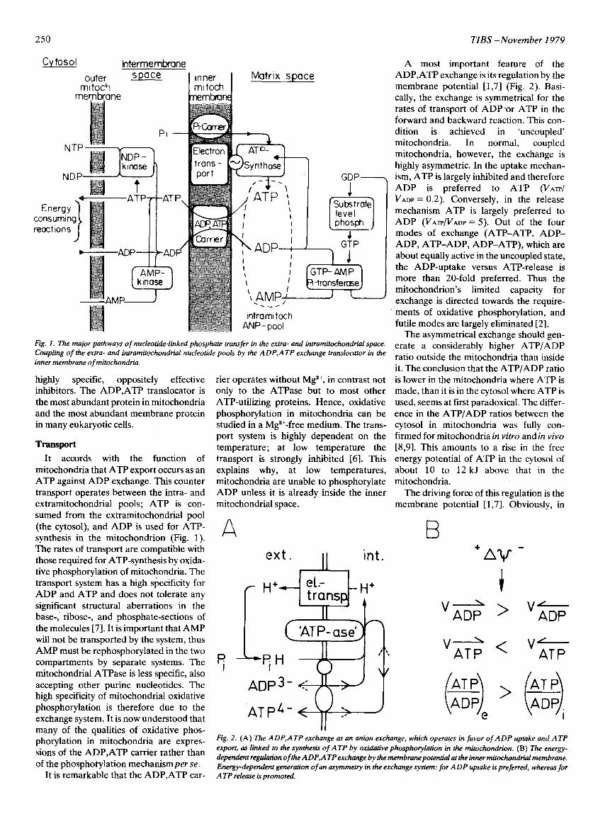
Fig. I. The major pathways of nucleotide-linked phosphate transfer in the extra- and intramitochondrial space. Coupling of the extra- and intramitochondrial nucleotide pools by the ADP,ATP exchange translocator in the inner membrane of mitochondria.
highly specific, oppositely effective inhibitors. The ADP,ATP translocator is the most abundant protein in mitochondria and the most abundant membrane protein in many eukaryotic cells.
TCUlSpOrt It accords with the function of
mitochondria that ATP export occurs as an ATP against ADP exchange. This counter transport operates between the intra- and extramitochondrial pools; ATP is con- sumed from the extramitochondrial pool (the cytosol), and ADP is used for ATP- synthesis in the mitochondrion (Fig. 1). The rates of transport are compatible with those required for ATP-synthesis by oxida- tive phosphorylation of mitochondria. The transport system has a high specificity for ADP and ATP and does not tolerate any significant structural aberrations in the base-,. ribose-, and phosphate-sections of the molecules [7]. It is important that AMP will not be transported by the system, thus AMP must be rephosphorylated in the two compartments by separate systems. The mitochondrial ATPase is less specific, also accepting other purine nucleotides. The high specificity of mitochondrial oxidative phosphorylation is therefore due to the exchange system. It is now understood that many of the qualities of oxidative phos- phorylation in mitochondria are expres- sions of the ADP,ATP carrier rather than of the phosphotylation mechanism per se.
It is remarkable that the ADP,ATP car-
rier operates without Mg2+, in contrast not only to the ATPase but to most other ATP-utilizing proteins. Hence, oxidative phosphorylation in mitochondria can be studied in a Mg*+-free medium. The trans- port system is highly dependent on the temperature; at low temperature the transport is strongly inhibited [6]. This explains why, at low temperatures, mitochondria are unable to phosphorylate ADP unless it is already inside the inner mitochondrial space.
( ‘ATP-ase’r 1
ATP4- <d/f+-)
A most important feature of the ADP,ATP exchange is its regulation by the membrane potential [1,71 (Fig. 2). Basi- cally, the exchange is symmetrical for the rates of transport of ADP -or ATP in the forward and backward reaction. This con- dition is achieved in ‘uncoupled’ mitochondria. In normal, coupled mitochondria, however, the exchange is highly asymmetric. In the uptake mechan- ism, ATP is largely inhibited and therefore ADP is preferred to ATP (VATP/ VADP = 0.2). Conversely, in the release mechanism ATP is largely preferred to ADP (VATP/VADP = 5). Out of the four modes of exchange (ATP-ATP, ADP- ADP, ATP-ADP, ADP-ATP), which are about equally active in the uncoupled state, the ADP-uptake versus ATP-release is more than 20-fold preferred. Thus the mitochondrion’s limited capacity for exchange is directed towards the require- ments of oxidative phosphorylation, and futile modes are largely eliminated [2].
The asymmetrical exchange should gen- erate a considerably higher ATP/ADP ratio outside the mitochondria than inside it. The conclusion that the ATP/ADP ratio is lower in the mitochondria where ATP is made, than it is in the cytosol where ATP is used, seems at first paradoxical. The differ- ence in the ATP/ADP ratios between the cytosol in mitochondria was fully con- firmed for mitochondria in vitro and in vivo [8,93. This amounts to a rise in the free energy potential of ATP in the cytosol of about 10 to 12 kJ above that in the mitochondria.
The driving force of this regulation is the membrane potential [1,7]. Obviously, in
B
+Av -
Fig. 2. (A) The ADP,ATP exchange as an anion exchange, which operates in favor of ADP uptake and ATP export, as linked to the synthesis of ATP by oxidative phosphorylation in the mitochondrion. (B) The energy- dependent regulation ofthe ADP,ATP exchange by the membranepotential at the inner mitochondr+al membrane. Energy-dependent generation of an asymmetry in the exchange system: for ADP uptake is preferred, whereas for ATP release is promoted.
TIBS -November 1979 251
the exchange one negative charge is trans- ferred as a result of ATPQ minus ADPS- charge difference. The energy required for this transfer comes from the same source tapped for the synthesis of ATP. Its impli- cations for stoichiometry of H+-transfer and respiration in ATP-synthesis were pointed out in 1968 1101, but are only now being widely recognized.
The ADP,ATP exchange system permits the ‘symbiosis’ of the intra- and extramitochondrial ATP systems which have to operate at different phosphoryl- ation potentials. The glycolytic system requires ATP to work at a higher phos- phorylation potential, about 66 kJ, than the intramitochondrial system, about 55 kJ. Only a membrane-controlled ADP,ATP exchange system can permit rapid interaction between both spaces.
structures (diterpenoide- and polyiso- prenoic-derivates) are so different from ADP, we believe that both of them bind to the substrate site of the carrier. In the membrane these sites were defined by measuring the binding of substrates and inhibitors to the mitochondria. By these studies the affinity for substrates and the inhibitors as well as the number of carrier sites were determined [2]. A high density of carrier sites was found, amounting to two sites per respiratory chain or ATPase com- plex.
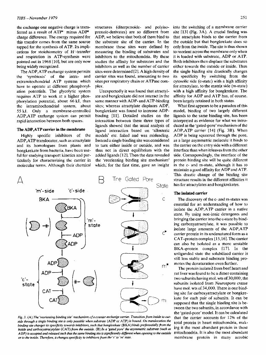
Unexpectedly it was found that atractyl- ate and bongkrekate did not interact in the same manner with ADP- and ATP-binding sites; whereas atractylate displaces ADP, bongkrekate was found to increase ADP- binding [ll]. Detailed studies on the interaction between these three types of ligands showed that the usual analysis of ligand interaction based on ‘allosteric models’ etc. failed and was misleading. Instead a single binding site was considered to turn either inside or outside, and was thus not in direct equilibrium with the added ligands [ 121. Then the data revealed the ‘reorienting binding site mechanism’ which,’ for the first time, gave an insight
into the switching of a membrane carrier site [13] (Fig. 3A). A crucial finding was that atractylate binds to the carrier from the outside but that bongkrekate does so only from the inside. The site is thus shown to reorient across the membrane only when it is loaded with substrate, ADP or ATP. Both inhibitors then displace the substrates either towards the outside or inside. Thus the single binding site drastically changes its specificity by switching from the cytosolic side (c-state) with a high affinity for atractylate, to the matrix side (m-state) with a high affinity for bongkrekate. The affinity for ADP and ATP has, of course, been largely retained in both states.
The ADP,ATP carrier in the membrane
Highly specific inhibitors of the ADP,ATP translocator, such as atractylate and its homologues from plants and bongkrekate from bacteria, have been use- ful for studying transport kinetics and par- ticularly for characterizing the carrier in molecular terms. Although their chemical
What first appears to be a paradox of this model, binding of two largely different ligands to the same binding site, has been interpreted as evidence for what we intro- duced as the ‘gated-pore’ mechanism of the ADP,ATP carrier 1141 (Fig. 3B). When ADP is being squeezed through the pore, as a large asymmetric molecule it binds to the carrier on the entry side with a different interface than when it leaves from the other side. Correspondingly, the interface of the protein binding site will be quite different in the c- and m-state, although it has to maintain a good affinity for ADP and ATP. This drastic change of the binding site structure results in the different affinities it has for atractylates and bongkrekates. A
‘ml-side ‘c’-side
BKA -v
B The Gated Pore
m’- state
.:.~:.>,..‘.,
F
.>:,:.>:.:.:.:I:.>. :.:,:.>>:.>:.:’ ,.,.,.(,.,.... .. C*
J
m*
, I C- state
Fig. 3. (A) The ‘reorienting binding site’ mechanism of a counter exchange carrier. Transition from inside to out. side through a single binding site is only possible when substrate (ADP or ATP) is bound. On transocation the binding site changes its specificity towards inhibitors, such that bongkrekate (BKA) binds preferentially from the inside and carboxyatracytylate (CAT) from the outsiak (B) In a ‘gated pore’ the asymmetric substrate (such as ADP) is accepted and released such that the same binding site is significantly different when opening to the outside or to the inside. Therefore, it changes specificiry to inhibitors from the ‘c’ to ‘m’ state.
The isolated carrier The discovery of the c- and m-states was
essential for an understanding of how to isolate the ADP,ATP carrier in a native state. By using non-ionic detergents and bringing the carrier into the c-state by bind- ing carboxyatractylate, it was possible to isolate large amounts of the ADP,ATP carrier protein in its undenatured form as a CAT-protein complex [ 15,161. The carrier can also be isolated as a more unstable BKA-protein complex 1171. In the unliganded state the solubilized carrier is still less stable and substrate binding pro- motes the denaturation even further.
The protein isolated from beef heart and rat liver was found to be a dimer containing two subunits having mol. wts of 30,000; the subunits isolated, from Neurosporu craw have mol. wts of 34,000. There is one bind- ing site for carboxyatractylate or bongkre- kate for each pair of subunits. It can be supposed that the single binding site is be- tween the two subunits, in accordance with the ‘gated-pore’ model. It can be calculated that the carrier accounts for 12% of the total protein in heart mitochondria, mak- ing it the most abundant protein in these mitochondria. It is also the most abundant membrane protein in many aerobic
252 TIBS -November 1979
eukaryotic cells. In view of the carrier’s relatively low specific activity, it is obvious that a large number of them are necessary to meet the high demand for ADP,ATP transport.
The hydrophobic nature of the protein can be deduced from the fact that each pro- tein molecule is able to bind 130 molecules of Triton. A helical content of 40% was determined by circular dichroism meas- urements. This figure is similar to that found for other hydrophilic nucleotide- binding proteins, but contrasts with the postulate that internal membrane proteins have a high helix content.
the cytosol and appears to be important for 8 the operation of the mitochondrial mem- brane -even to the extent that it may be the 9 last resort for maintaining the inner mitochondrial membrane potential in 10 these mutants, which we believe to be vital for the organization of this membrane.
References 11
The existence of two conformations, the c- and m-state, and the substrate-activated transition, could be fully substantiated with the isolated protein. Thus the isolated pro- tein exchanges bongkrekate against atrac- tylate, or vice versa, only in the presence of ADP [17]. The different conformations of the c- and m-states give rise to the activity of an SH-group in the m-state only; a dras- tic difference in the extent to which trypsin attacks the c- and m-states; and different antigenic properties.
Khngenberg, M. (1970) in Essays in Biochemistry (Campbell, P. N. and Dickens, F., eds), Vol. 6, pp. 119-159. Academic Press, New York Klingenberg, M. (1976) in The Enzymes of Biological Membranes: Membrane Transport (Martonosi, A. N., ed.), Vol. 3, pp. 383-438, Plenum Publishing Corporation, New York/ London
Heldt, H. W., Klingenberg, M. and Milovancev, M. (1972) Eur. J. Biochem. 30,434440 Elbers, R., Heldt, H. W., Schmucker, P., Soboll, S. and Wiese, H. (1974) Hoppe-Seyler’s Z. Physiol. Chem. 355,378-393 Klingenberg, M., Heldt, H. W. and Pfaff, E. (1969) in The Energy Level and Metabolic Control in Mitochondria (Papa, S., et al., eds), pp. 237-253, Adriatica Editrice, Bari Erdelt, H., Weidemann, M. J., Buchholz, M. and Klingenberg, M. (1972) Eur. J. Biochem. 30, 107-122
12
Vignais, P. V. (1976) Biochim. Biophys. Acra 456 (BR4), l-38 Klingenberg, M., Pfaff, E. and Kroger, A. (1964) Rapid Mixing and Sampling Techniques in Biochemistry, pp. 333-337, Academic Press, New York
13
14
Klingenberg, M. and Pfaff, E. (1966) in Regula- tion of Metabolic Processes in Mitochondtia (Tager, J. M., et al., eds), pp. 180-201, Elsevier, Amsterdam Heldt, H. W., Jacobs, H. and Klingenberg, M. (1965) B&hem. Biophys. Res. Commun. 18, 174-179
Reconstitution Pfaff, E. and Klingenberg, M. (1968) Eur. J. Biochem. 6,66-79
15
16
17
18
19
Klingenberg, M., Scherer, B., Stengel-Rutkowski, L., Buchholz, M. and Grebe, K. (1973) in Mechanisms in Bioenergetics (Azzone, G. F., et al., eds), pp. 257-284, Academic Press, New York/London Klingenberg, M. and Buchholz, M. (1973) Eur. J. Biochem. 38,346-358 Klingenberg, M., Riccio, P., Aquila, H., Bucha- nan, B. B. and Grebe, K. (1976) in The Structural Basis of Membrane Function (Hatefi, Y. and Djavadi-Ohaniance, L., eds), pp. 293-311, Academic Press, New York/London Riccio, P., Aquila, H. and Klingenberg, M. (1975) FEBS Len 56,133-138 Klingenberg, M., Riccio, P. and Aquila, H. (1978) Biochim. Biophys. Acta 503,193-210 Aquila, H., Eiermann, W., Babel, W. and Kling- enberg, M. (1978) Eur. J. B&hem. 85,549-560 Kramer, R. and Klingenberg, M. (1977) Biochemistry 16,4954-4961 Kramer, R. and Klingenberg, M. (1977) FEBS Lett. 82,363-367
The unloaded carrier protein could be stabilized by incorporating it into lipo- somes 1181. The binding affinities of the ligands for the reconstituted carrier are the same as those obtained for the carriers in vivo. The reconstituted carrier is also able to transport ADP and ATP, and thus ADP,ATP transport may be studied in a simple system [19j. The reconstituted sys- tem has the same inhibitory properties, fol- lows a 1 : 1 exchange and can also be regu- lated by the membrane potential in the membrane. It allows us to determine the separate rate constants of forward- and back-flux of ADP and ATP and their con- trol by the membrane potential. The recon- stitution also proves that the isolated 60,000 mol. wt protein forms the complete transport system and that there are no co- factors involved.
The chloroplast envelope - barrier or bridge?
U. Heber and D. A. Walker
The chloroplast envelope is a highly effective barrier against the unidirectional movement of most cations and anions. Still, in illuminated leaf cells there is a rapid exchange of anionic metabolites between chloroplasts and cytosol which profoundly infiuences extra-
chloroplast metabolism.
Green plants are capable of utilizing light energy for the photosynthetic reduction of COZ to [CHZO]. This is a complex process which in higher plants is performed in the chloroplasts. These green organelles con- tain pigment-bearing vesicular membranes called thylakoids and a matrix or stromal phase whieh is surrounded by an envelope consisting of two, more or less closely oppressed, biomembranes.
generated in the light and is reflected by increased stromal NADPH/NADP and ATP/ADP ratios.
Genetic considerations
The ADP,ATP carrier is unique to mitochondria and is the only well-defined molecular species which identifies the mitochondrion. Other typical components, such as cytochromes, quinones, cardolipin etc. are either found also in bacteria or in the cytosol of eukaryotic cells. We may state that no mitochondria are known without the ADP,ATP carrier, although some mutant mitochondria may be defi- cient in respiratory components, ATPase etc. The ADP,ATP carrier is synthesized in
The stroma contains a high concentra- tion of proteins. All the soluble enzymes required for the cyclic regeneration of the COz-acceptor molecule reside there. Some of them require small ions such as Mg2+ for activation. The stroma is also the solute phase for anionic photosynthetic inter- mediates. A driving force in photosynthesis is the so-called assimilatory power which is
In view of the complex dependence of photosynthesis on the presence of cations and organic anions such as phosphorylated sugars, adenylates and nicotinamide nuc- leotides it is most remarkable that isolated spinach chloroplasts suspended in an isotonic medium containing only a buffer, an osmoticum, bicarbonate and phosphate are photosynthetically competent and can photoreduce CO2 at rates which are as high as, or even higher than, the rate of photo- synthesis in a leaf. This ability is quickly destroyed by the addition of a small amount of a detergent which interacts with biomembranes.
U. Heber is Professor of Botany at the University of Wiirzburg, F.R.G., and D. A. Walker is Professor of Biology at the University of Sheffield, U.K.
It seems inescapable that photosynthesis is compartmented and that the chloroplast envelope is a barrier designed to keep the various components of the photosynthetic
0 Elsevier/North-Holland Biomedical Press 1979