Embed Size (px)
Citation preview
1466 The Journal of Clinical Investigation | November 2003 | Volume 112 | Number 10
IntroductionClinicians who deal with device-related and otherchronic bacterial infections have gradually defined anew category of infectious disease that differs radical-ly from the acute epidemic bacterial diseases that pre-dominated until the middle of the last century (1). Thehappy fact that acute epidemic diseases are largely inthe past is a consequence of progress in microbiology,and can be directly attributed to the vaccines andantibiotics that were developed as we came to under-stand planktonic bacteria. The free-floating bacteriathat caused diphtheria and cholera were accuratelymodeled in test tube cultures, their pathogenic mech-anisms (e.g., toxins) became epitopes for vaccines, andantibiotics that killed them were derived from natureor from directed synthesis. These specialized pathogenscaused disease in perfectly healthy individuals, they rantheir courses quickly, and they retreated to differentanimal populations or to natural reservoirs when thepopulation under attack became immune.
As we began to gain control over epidemic diseases,another type of disease came to the fore. These dis-eases are much less aggressive than acute infections,
they often persist for months or years, and theyprogress through periods of quiescence that alternatewith periods of acute exacerbation. When the sophis-ticated tools of microbiology were brought to bear onthese chronic diseases, many anomalies emerged. Thepathogens were common environmental organisms,with which individuals had daily contact and againstwhich they often had adequate immunity, and theirpathogenic mechanisms were often diffuse and poor-ly defined. When they were grown in conventional labcultures, these environmental organisms appeared tobe sensitive to conventional antibiotics, but thesesame antibiotics failed to resolve the bacterial infec-tions, although they gave some relief during acuteexacerbations. Even more puzzling was the observa-tion that, in many cases, it was not possible at all torecover any bacteria by traditional culture mecha-nisms. This led many investigators to posit that thesechronic disease states were sterile inflammatory con-ditions that persisted after the eradication of allmicroorganisms. However, the application of molec-ular diagnostics demonstrated unequivocally thatbacteria were both present and metabolically active,even when no bacteria were recovered by plating. Insome cases (e.g., otitis media, cholesteatoma, tonsili-tis), affected individuals were cured by surgical treat-ment or by growth-related anatomical changes, whilemany other victims were relegated to intermittentantibiotic therapy for the remainder of their lives(cystic fibrosis [CF], prostatitis). When we looked atthe infecting bacteria as they grew in affected tissues,we noted that they actually lived in matrix-enclosedcommunities that closely resembled the biofilms that
The application of biofilm science to the study and control of chronic bacterial infections
William Costerton,1 Richard Veeh,1 Mark Shirtliff,2 Mark Pasmore,1 Christopher Post,3
and Garth Ehrlich3,4
1Center for Biofilm Engineering, Montana State University, Bozeman, Montana, USA2Department of Biomedical Sciences, Dental School, University of Maryland–Baltimore, Baltimore, Maryland, USA3Center for Genomic Sciences, Allegheny Singer Research Institute, Pittsburgh, Pennsylvania, USA4Department of Microbiology and Immunology, Drexel University College of Medicine, Pittsburgh, Pennsylvania, USA
Unequivocal direct observations have established that the bacteria that cause device-related and otherchronic infections grow in matrix-enclosed biofilms. The diagnostic and therapeutic strategies that haveserved us so well in the partial eradication of acute epidemic bacterial diseases have not yielded accuratedata or favorable outcomes when applied to these biofilm diseases. We discuss the potential benefits ofthe application of the new methods and concepts developed by biofilm science and engineering to theclinical management of infectious diseases.
J. Clin. Invest. 112:1466–1477 (2003). doi:10.1172/JCI200320365.
PERSPECTIVE SERIESQuorum sensing | E. Peter Greenberg, Series Editor
Address correspondence to: William Costerton, Center forBiofilm Engineering, 366 EPS Building, P.O. Box 173980,Montana State University, Bozeman, Montana 59717-3980, USA.Phone: (406) 994-4770; Fax: (406) 994-6098; E-mail: [email protected] of interest: The authors have declared that no conflict ofinterest exists.Nonstandard abbreviations used: cystic fibrosis (CF); chronicotitis media with effusion (COME); expressed prostatic secretion(EPS); acyl homoserine lactone (AHL); polymorphonuclearneutrophil (PMN); intrauterine contraceptive device (IUD).
The Journal of Clinical Investigation | November 2003 | Volume 112 | Number 10 1467
are the predominant form of growth of bacteria inindustrial and environmental ecosystems. The sim-ple fact that the organisms that cause device-relatedand other chronic infections grow in biofilms (1)goes some distance toward explaining the perceivedanomalies of these diseases, and offers a measure ofhope that they can eventually be controlled.
Chronic bacterial disease: a clinical entity. When themodel used to analyze a natural process is incorrect,our attempts to understand and manipulate theprocess will fail, many honest and conscientious peo-ple will be frustrated, and the reputations of wholeresearch groups will be damaged. In the case ofchronic bacterial diseases, diagnostic microbiologylabs reported that cultures of Pseudomonas aeruginosafrom CF patients were sensitive to antibiotics (e.g.,cloxacillin), but pulmonary clinicians saw littleimprovement when these antibiotics were used. Thesera of CF patients contained very large amounts ofspecific antibodies against Pseudomonas, but the dis-ease persisted, and the use of anti-Pseudomonas vac-cines resulted in the deaths of some patients. Middleear specimens from children with chronic otitismedia with effusion (COME) yielded negative bacte-rial cultures, so that a host-sustained inflammatoryetiology was suspected, but the factors driving theinflammation could not be identified and serologydid not confirm the persistent involvement of virus-es. Patients with raging febrile prostate infectionsyielded expressed prostatic secretion (EPS) samplesthat produced cultures negative for bacteria, andmaterial recovered from osteomyelitis debridationswith frank pus yielded only a few colonies of skin and
environmental organisms. All was shadows and fog,and the reputations of the microbiology units ofmany hospitals plummeted from the high levels theyhad attained earlier.
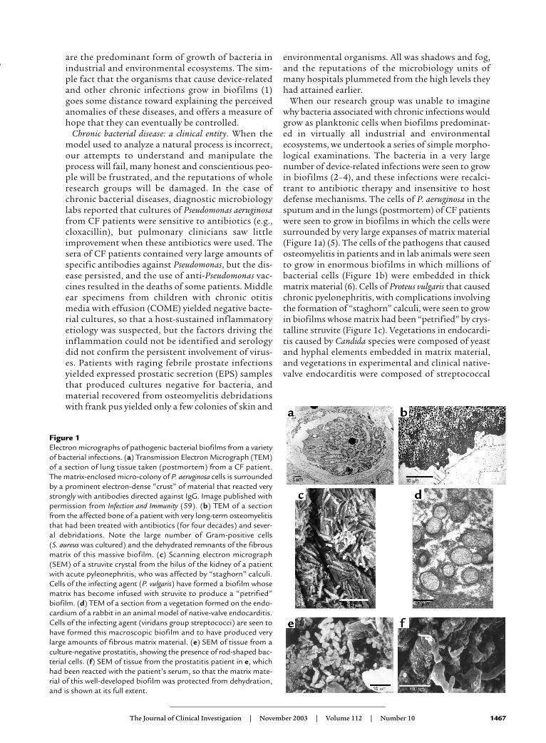
When our research group was unable to imaginewhy bacteria associated with chronic infections wouldgrow as planktonic cells when biofilms predominat-ed in virtually all industrial and environmentalecosystems, we undertook a series of simple morpho-logical examinations. The bacteria in a very largenumber of device-related infections were seen to growin biofilms (2–4), and these infections were recalci-trant to antibiotic therapy and insensitive to hostdefense mechanisms. The cells of P. aeruginosa in thesputum and in the lungs (postmortem) of CF patientswere seen to grow in biofilms in which the cells weresurrounded by very large expanses of matrix material(Figure 1a) (5). The cells of the pathogens that causedosteomyelitis in patients and in lab animals were seento grow in enormous biofilms in which millions ofbacterial cells (Figure 1b) were embedded in thickmatrix material (6). Cells of Proteus vulgaris that causedchronic pyelonephritis, with complications involvingthe formation of “staghorn” calculi, were seen to growin biofilms whose matrix had been “petrified” by crys-talline struvite (Figure 1c). Vegetations in endocardi-tis caused by Candida species were composed of yeastand hyphal elements embedded in matrix material,and vegetations in experimental and clinical native-valve endocarditis were composed of streptococcal
Figure 1Electron micrographs of pathogenic bacterial biofilms from a varietyof bacterial infections. (a) Transmission Electron Micrograph (TEM)of a section of lung tissue taken (postmortem) from a CF patient.The matrix-enclosed micro-colony of P. aeruginosa cells is surroundedby a prominent electron-dense “crust” of material that reacted verystrongly with antibodies directed against IgG. Image published withpermission from Infection and Immunity (59). (b) TEM of a sectionfrom the affected bone of a patient with very long-term osteomyelitisthat had been treated with antibiotics (for four decades) and sever-al debridations. Note the large number of Gram-positive cells (S. aureus was cultured) and the dehydrated remnants of the fibrousmatrix of this massive biofilm. (c) Scanning electron micrograph(SEM) of a struvite crystal from the hilus of the kidney of a patientwith acute pyleonephritis, who was affected by “staghorn” calculi.Cells of the infecting agent (P. vulgaris) have formed a biofilm whosematrix has become infused with struvite to produce a “petrified”biofilm. (d) TEM of a section from a vegetation formed on the endo-cardium of a rabbit in an animal model of native-valve endocarditis.Cells of the infecting agent (viridans group streptococci) are seen tohave formed this macroscopic biofilm and to have produced verylarge amounts of fibrous matrix material. (e) SEM of tissue from aculture-negative prostatitis, showing the presence of rod-shaped bac-terial cells. (f) SEM of tissue from the prostatitis patient in e, whichhad been reacted with the patient’s serum, so that the matrix mate-rial of this well-developed biofilm was protected from dehydration,and is shown at its full extent.
1468 The Journal of Clinical Investigation | November 2003 | Volume 112 | Number 10
cells in equally extensive fibrous matrices (Figure 1d).The prostate of a patient whose EPS samples hadyielded no bacteria was seen to be colonized by bacte-rial cells (Figure 1e) whose matrix material could bevisualized especially well when it was reacted with thepatient’s serum to yield the classic quellung reaction(Figure 1f). In COME luxuriant biofilms were demon-strated to be growing directly on the mucosal surface,and vital dye imaging showed that the embedded bac-teria were all viable in spite of antibiotic treatmentand loss of culturability. In some infections thebiofilms were composed almost exclusively of bacter-ial cells and matrix material, while the microbial com-munities in other infections also contained frag-mented platelets and host molecules (e.g., fibrin), sothat the bacteria were widely dispersed. However,biofilms were found in all of the chronic infectionsexamined in the 12 years during which this morpho-logical series was pursued.
Morphological data are unequivocal in that it ishighly unlikely that bacteria do not exist in a tissue inwhich we have found the distinctive features ofprokaryotic cells, and these cells are integrated intotheir matrices and into the structure of the tissueitself. However, morphological data tell us little aboutthe species of bacteria that are present in these chron-ic infections, or about their viability and their pheno-typic pattern of gene expression at the time that thesample was obtained. These data are provided bymodern techniques for the analysis of nucleic acids,and the Center for Genomic Sciences (Pittsburgh,Pennsylvania, USA) has established that living bacte-ria are present in otitis media and in otorrhea witheffusion (7). Curtis Nickel’s group has used nucleicacid analysis to establish that bacteria are present inEPS samples from prostatitis patients and from someindividuals without overt symptoms (8). Similaranalysis of sputum from CF patients has shown thatcells of P. aeruginosa are present and that they express
a phenotype different from that seen in planktoniccells in culture. Finally, Pradeep Singh and colleagueshave analyzed the ratio between two types of acylhomoserine lactone (AHL) signals to show that thecells of P. aeruginosa in the CF lung do indeed grow inthe biofilm phenotype (9). At some time in recent his-tory the conceptual balance tipped in favor of abiofilm etiology, at a different time in the case of eachchronic disease, and the general notion that device-related and other chronic bacterial infections arecaused by biofilms is now widely accepted (1).
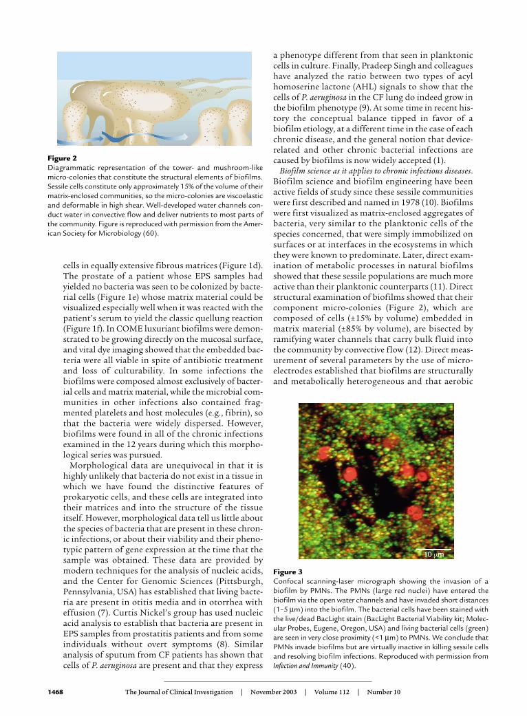
Biofilm science as it applies to chronic infectious diseases.Biofilm science and biofilm engineering have beenactive fields of study since these sessile communitieswere first described and named in 1978 (10). Biofilmswere first visualized as matrix-enclosed aggregates ofbacteria, very similar to the planktonic cells of thespecies concerned, that were simply immobilized onsurfaces or at interfaces in the ecosystems in whichthey were known to predominate. Later, direct exam-ination of metabolic processes in natural biofilmsshowed that these sessile populations are much moreactive than their planktonic counterparts (11). Directstructural examination of biofilms showed that theircomponent micro-colonies (Figure 2), which arecomposed of cells (±15% by volume) embedded inmatrix material (±85% by volume), are bisected byramifying water channels that carry bulk fluid intothe community by convective flow (12). Direct meas-urement of several parameters by the use of micro-electrodes established that biofilms are structurallyand metabolically heterogeneous and that aerobic
Figure 2Diagrammatic representation of the tower- and mushroom-likemicro-colonies that constitute the structural elements of biofilms.Sessile cells constitute only approximately 15% of the volume of theirmatrix-enclosed communities, so the micro-colonies are viscoelasticand deformable in high shear. Well-developed water channels con-duct water in convective flow and deliver nutrients to most parts ofthe community. Figure is reproduced with permission from the Amer-ican Society for Microbiology (60).
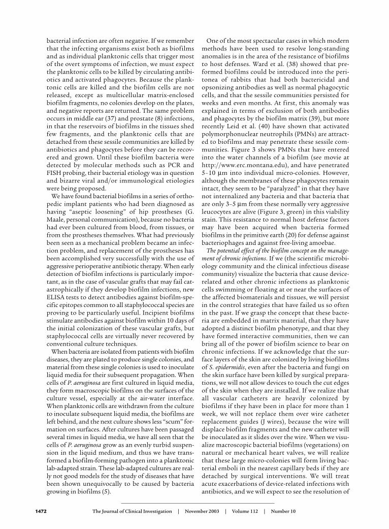
Figure 3Confocal scanning-laser micrograph showing the invasion of abiofilm by PMNs. The PMNs (large red nuclei) have entered thebiofilm via the open water channels and have invaded short distances(1–5 µm) into the biofilm. The bacterial cells have been stained withthe live/dead BacLight stain (BacLight Bacterial Viability kit; Molec-ular Probes, Eugene, Oregon, USA) and living bacterial cells (green)are seen in very close proximity (<1 µm) to PMNs. We conclude thatPMNs invade biofilms but are virtually inactive in killing sessile cellsand resolving biofilm infections. Reproduced with permission fromInfection and Immunity (40).
The Journal of Clinical Investigation | November 2003 | Volume 112 | Number 10 1469
and anaerobic processes occur simultaneously in dif-ferent parts of the multicellular community. Thestructural sophistication of biofilms suggested thatthese communities must be regulated by signals anal-ogous to the hormones and pheromones that regu-late multicellular eukaryotic communities, and thefirst of these regulatory signals were identified in1998 (13). As we began to study the patterns of geneexpression in biofilms, we found that these patternsproduce a distinct biofilm phenotype, which differsvery profoundly from that of the biofilms’ plankton-ic counterparts (14).
Biofilms as self-assembling multicellular communities.When planktonic bacteria encounter a surface or aninterface, they adhere to that interface in a reversiblefashion while they “explore” the locale to ascertainwhether it offers nutrient or other advantages. If the“decision” favors permanent settlement, the adherentcells upregulate the genes (15, 16) involved in matrixproduction (within as few as 12 minutes), and theprocess of biofilm formation begins. Kolter et al. (17)and others have described biofilm formation as adevelopmental sequence that varies to some extentbetween species but generally results in the formationof a mature community of tower- and mushroom-shaped micro-colonies (Figure 2). Both biofilm for-mation and biofilm detachment are under the controlof chemical signals of the same type that regulatesquorum sensing (18, 19), and these regulatory mole-cules guide the formation of slime-enclosed micro-colonies and water channels. The cells are remarkablyevenly distributed in the biofilm matrix, suggestingthat some matrix component may dictate their preciselocation (20); Stoodley et al. have shown that the com-munity as a whole has material properties similar tothose of a viscous fluid. The micro-colonies aredeformable, they oscillate in high-shear systems, andthey break and detach as biofilm fragments if theirtensile strength is exceeded by the shear forces (21).Biofilms also show “creeping” activity, in high shear,and whole biofilms can be seen moving across sur-faces, with the development of transitory waves and
areas of enhanced detachment. These mechanicalproperties of biofilms are documented in videosequences that are available on the Center for BiofilmEngineering website (http://www.erc.montana.edu).Biofilms form in many high-shear environments, likethe vegetations that form on native heart valves, sothese material properties are germane and can be usedto predict when and where fragments will detach andwhere they will end up in a flowing system.
Biofilms as protected sessile communities. Stoodley et al.(20) have made the point that microbial biofilms con-stitute the most “defensive” life strategy that can beadopted by prokaryotic cells. In very hostile environ-ments, in which many locations are too hot or too acidor too dry, the stationary mode of growth is inherently
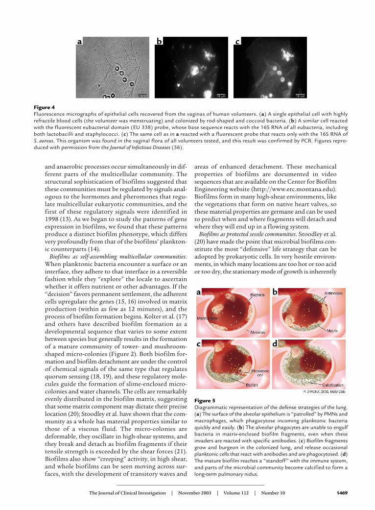
Figure 4Fluorescence micrographs of epithelial cells recovered from the vaginas of human volunteers. (a) A single epithelial cell with highlyrefractile blood cells (the volunteer was menstruating) and colonized by rod-shaped and coccoid bacteria. (b) A similar cell reactedwith the fluorescent eubacterial domain (EU 338) probe, whose base sequence reacts with the 16S RNA of all eubacteria, includingboth lactobacilli and staphylococci. (c) The same cell as in a reacted with a fluorescent probe that reacts only with the 16S RNA of S. aureus. This organism was found in the vaginal flora of all volunteers tested, and this result was confirmed by PCR. Figures repro-duced with permission from the Journal of Infectious Diseases (36).
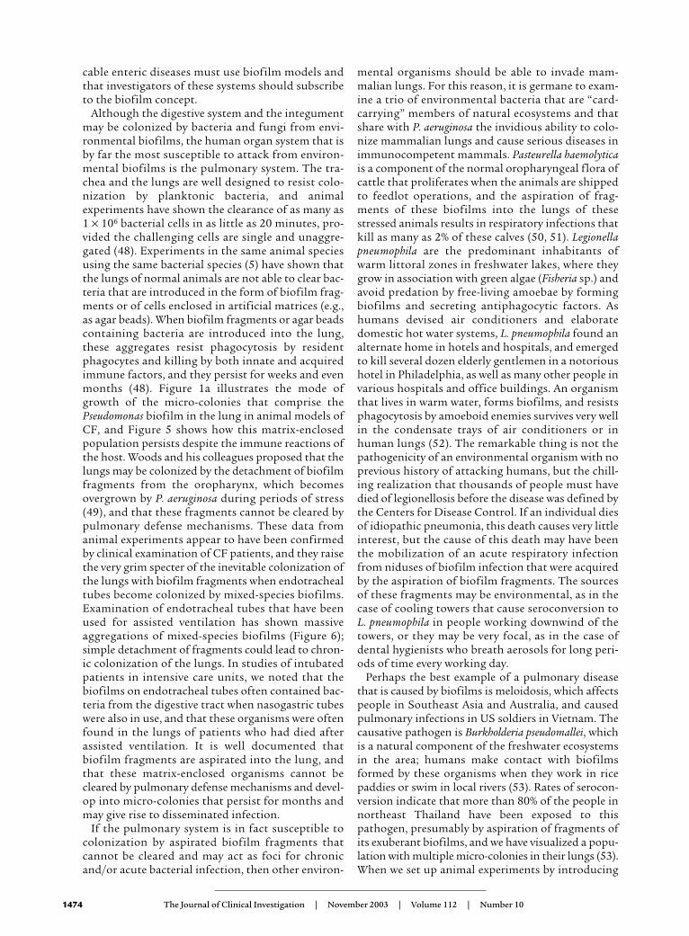
Figure 5Diagrammatic representation of the defense strategies of the lung.(a) The surface of the alveolar epithelium is “patrolled” by PMNs andmacrophages, which phagocytose incoming planktonic bacteriaquickly and easily. (b) The alveolar phagocytes are unable to engulfbacteria in matrix-enclosed biofilm fragments, even when theseinvaders are reacted with specific antibodies. (c) Biofilm fragmentsgrow and burgeon in the colonized lung, and release occasionalplanktonic cells that react with antibodies and are phagocytosed. (d)The mature biofilm reaches a “standoff” with the immune system,and parts of the microbial community become calcified to form along-term pulmonary nidus.
1470 The Journal of Clinical Investigation | November 2003 | Volume 112 | Number 10
defensive, because bacterial cells are not swept intoareas where they will be killed. These stationary sessilecommunities, which seem to have predominated inprimitive earth, were attacked by bacteriophages andby amoebae, and their collective growth in matrix-enclosed micro-colonies gave complete protectionfrom these predators. One of the most important pro-tections afforded by the biofilm mode of growth isprotection from drying and from ultraviolet light.These sessile communities are especially favored, andvery visible, in the intermittently wet splash zones offreshwater and marine ecosystems, where these com-munities allow prokaryotic cells to survive daylongexposures to drying and intense sunlight and to revivewhen they are rehydrated. Experiments conducted inmilitary defense establishments have shown thatplanktonic cells of lab-adapted strains of bacteria sur-vive for very short lengths of time when they arereleased from high-flying airplanes, whereas cells inbiofilm fragments survive long enough to reach theground. In their 2002 review, Stoodley et al. (20) madethe intriguing suggestion that bacteria may have devel-oped their biofilm phenotype early in the evolutionaryprocess, when survival in a hostile environment was asine qua non. They suggest that the planktonic pheno-type, with its genetically “expensive” and very sophis-ticated chemotactic and motility mechanisms, mayhave developed later, for the purposes of dissemina-tion and the colonization of new habitats.
The biofilm as a distinct, signal-controlled phenotype. Theinherent resistance of biofilm bacteria to antibiotics(22) and to virtually all antibacterial agents was an
early indication that sessile cells differ radically fromtheir planktonic counterparts. When it was estab-lished that this resistance was not the result of diffu-sion limitation (23) but was predicated on somechange in cellular characteristics (24), the researchcommunity missed the central point and wanderedclueless for more than a decade. Then various groupsbegan to use the tools of modern molecular biology,like mRNA analysis by gene arrays (25–27) and pro-teomic analysis of gene products (28–30), and theyshowed that cells in biofilms express a radically dif-ferent set of genes. This virtual “bombshell” of theconcept of a distinct biofilm phenotype has changedmost of the intellectual constructs in the biofilmfield. The genes expressed in a biofilm differ fromthose expressed in the corresponding planktonic cellmore than they do from the genes expressed in aspore or in the biofilm cells of another species (28).There is no single biofilm phenotype, but geneexpression in sessile communities goes through awhole spectrum of changes as the communitymatures (20), and the planktonic phenotype beginsto emerge as the biofilm begins to shed mobile cells.Now that we know that bacteria adopt a radically dif-ferent phenotype when they adhere to a surface orinterface and initiate biofilm formation, we under-stand such mysteries as the resistance of biofilms toantibiotics in terms of the expression of different setsof genes. Many other characteristics of sessile bacte-ria can now be explained in the same way, and matrix-assisted laser desorption-ionization time-of-flight(MALDITOF) and gene chip analyses of the genesexpressed in biofilms are already painting a pictureof considerable variety and specialization in thesemulticellular communities. In the microbiologicalcommunity, the notions are taking shape thatbiofilms predominate in most ecosystems and that
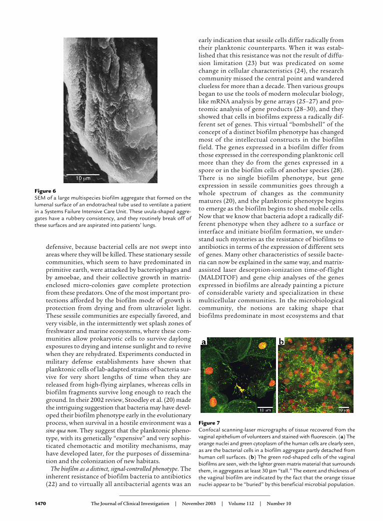
Figure 6SEM of a large multispecies biofilm aggregate that formed on thelumenal surface of an endotracheal tube used to ventilate a patientin a Systems Failure Intensive Care Unit. These uvula-shaped aggre-gates have a rubbery consistency, and they routinely break off ofthese surfaces and are aspirated into patients’ lungs.
Figure 7Confocal scanning-laser micrographs of tissue recovered from thevaginal epithelium of volunteers and stained with fluorescein. (a) Theorange nuclei and green cytoplasm of the human cells are clearly seen,as are the bacterial cells in a biofilm aggregate partly detached fromhuman cell surfaces. (b) The green rod-shaped cells of the vaginalbiofilms are seen, with the lighter green matrix material that surroundsthem, in aggregates at least 30 µm “tall.” The extent and thickness ofthe vaginal biofilm are indicated by the fact that the orange tissuenuclei appear to be “buried” by this beneficial microbial population.
The Journal of Clinical Investigation | November 2003 | Volume 112 | Number 10 1471
these sessile cells express genes we have never seenexpressed and reach levels of interaction and com-munity behavior that we have never imagined. Nowthat the phenotypes of planktonic and biofilm cellshave been shown to be so profoundly different, purecultures of planktonic bacteria seem to be a very poormodel for the study of these organisms, which actu-ally grow in mixed-species sessile communities innatural and medical systems (31).
The simple observations that microbial biofilms arecomposed of structured micro-colonies and thatwater channels are created and maintained suggestedthat some type of cell-cell signal system must be oper-ative in these multicellular communities. We specu-lated that these signals would resemble the hormonesthat dictate the structure of more complex eukaryot-ic organisms and communities, and we visualized atype of “embryology” of biofilms. We demonstratedthat the quorum-sensing molecules that had been dis-covered in planktonic bacteria (18) also controlbiofilm development (13) and many other bacterialbehaviors, and we now speculate that these signal sys-tems may have evolved to control biofilms, especiallyin the light organ of the squid. The AHL signals ofGram-negative bacteria and the two-component pep-tide signals of Gram-positive bacteria (32) have beenshown to affect biofilm formation, and the newly dis-covered autoinducer II signal (33) appears to have asimilar effect. Analysis of signal-negative mutantssuggests that all aspects of bacterial behavior are reg-ulated by an interactive network of signals, with largeinputs from environmental factors, so that biofilmscan now be seen as integrated communities that arevery reactive to their environments. This demonstra-tion of cross-talk among bacteria of the same and ofdifferent species presents us with the Rosetta stone of
the bacterial language that controls biofilm forma-tion, species interactions, growth rates, toxin produc-tion, invasive properties, and many other behaviors ofgreat interest to humankind. We estimate that dozensof new signal systems will be discovered in the nextdecade, and that control measures based on signalmanipulation will largely replace bactericidal agentsin human attempts to control bacteria (34). This newstrategy has gained some impetus from the observa-tion that many plants use natural analogues of bacte-rial signals to inhibit (35) or to encourage bacterialcolonization and biofilm formation on their surfaces.The pharmaceutical industry, which has been bedev-iled by the emergence of resistance to all of their mostcarefully crafted antibiotics, is intrigued by the factthat resistance to these natural antibiofilm signal ana-logues has not yet been recorded.
A re-examination of chronic bacterial diseases using biofilmconcepts. In view of the unequivocal demonstration thatthe bacteria that cause device-related and other chron-ic bacterial infections grow in biofilms, it seems usefulto survey this large number of infections for newinsights in terms of this new microbiological concept.
What new biofilm-specific methods tell us about biofilmbacteria. One of the most time-honored of microbio-logical conventions is the detection of bacteria byrecovery methods, which usually involve mechanicalremoval of the organisms (often by swabbing) andtheir propagation in liquid or on solid media. Werecently conducted a survey (36) of the bacterial col-onization of the vaginas of 3,000 human subjectsusing standard swabbing and culture (plating) tech-niques, and concluded (as most similar studies have)that 10.8% of individuals carried Staphylococcus aureus.We then examined subsets of these individuals, usingPCR to identify S. aureus by its DNA and using FISHprobes to identify cells of this species by their 16SRNA content, and we found that 100% of these indi-viduals carried this organism. We then examinedwhether individuals who yielded positive data inswab and plate tests carried more S. aureus as detect-ed by PCR and FISH, and found there was no corre-lation (36). If certain bacteria are present on a tissueor an inert surface, the swab may or may not pickthem up, they may be present in huge aggregates ofhundreds of cells that will yield only one colony onplating, and they may be expressing the set of genesthat constitute the biofilm phenotype and be unableto grow in the culture conditions provided. Swabbingand plating are techniques that date from the middle1800s, but they are still used to screen key medicalpersonnel for nasal carriage of S. aureus as well as todetect bacteria in suspected infections and to identi-fy the pathogens concerned. It may be time for us toadopt more modern and accurate methods.
The biofilm concept of chronic infections offers atleast a partial explanation for a phenomenon thathas troubled clinicians for some time, that blood cul-tures from patients who show many signs of overt
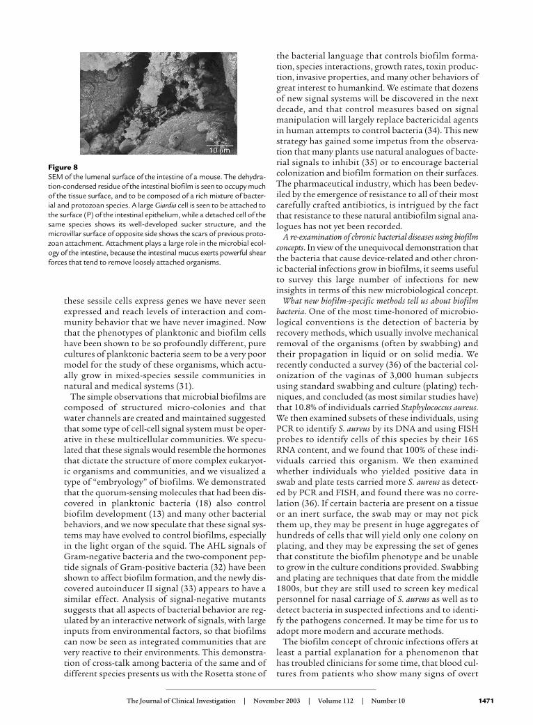
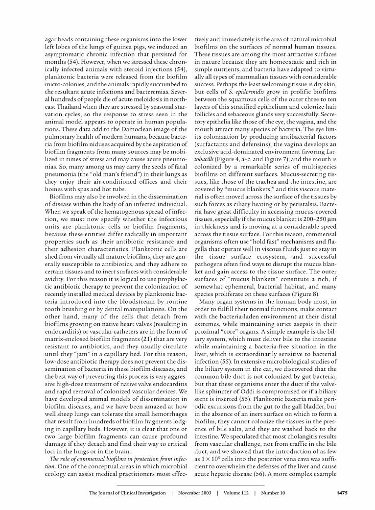
Figure 8SEM of the lumenal surface of the intestine of a mouse. The dehydra-tion-condensed residue of the intestinal biofilm is seen to occupy muchof the tissue surface, and to be composed of a rich mixture of bacter-ial and protozoan species. A large Giardia cell is seen to be attached tothe surface (P) of the intestinal epithelium, while a detached cell of thesame species shows its well-developed sucker structure, and themicrovillar surface of opposite side shows the scars of previous proto-zoan attachment. Attachment plays a large role in the microbial ecol-ogy of the intestine, because the intestinal mucus exerts powerful shearforces that tend to remove loosely attached organisms.
1472 The Journal of Clinical Investigation | November 2003 | Volume 112 | Number 10
bacterial infection are often negative. If we rememberthat the infecting organisms exist both as biofilmsand as individual planktonic cells that trigger mostof the overt symptoms of infection, we must expectthe planktonic cells to be killed by circulating antibi-otics and activated phagocytes. Because the plank-tonic cells are killed and the biofilm cells are notreleased, except as multicellular matrix-enclosedbiofilm fragments, no colonies develop on the plates,and negative reports are returned. The same problemoccurs in middle ear (37) and prostate (8) infections,in that the reservoirs of biofilms in the tissues shedfew fragments, and the planktonic cells that aredetached from these sessile communities are killed byantibiotics and phagocytes before they can be recov-ered and grown. Until these biofilm bacteria weredetected by molecular methods such as PCR andFISH probing, their bacterial etiology was in questionand bizarre viral and/or immunological etiologieswere being proposed.
We have found bacterial biofilms in a series of ortho-pedic implant patients who had been diagnosed ashaving “aseptic loosening” of hip prostheses (G.Maale, personal communication), because no bacteriahad ever been cultured from blood, from tissues, orfrom the prostheses themselves. What had previouslybeen seen as a mechanical problem became an infec-tion problem, and replacement of the prostheses hasbeen accomplished very successfully with the use ofaggressive perioperative antibiotic therapy. When earlydetection of biofilm infections is particularly impor-tant, as in the case of vascular grafts that may fail cat-astrophically if they develop biofilm infections, newELISA tests to detect antibodies against biofilm-spe-cific epitopes common to all staphylococcal species areproving to be particularly useful. Incipient biofilmsstimulate antibodies against biofilm within 10 days ofthe initial colonization of these vascular grafts, butstaphylococcal cells are virtually never recovered byconventional culture techniques.
When bacteria are isolated from patients with biofilmdiseases, they are plated to produce single colonies, andmaterial from these single colonies is used to inoculateliquid media for their subsequent propagation. Whencells of P. aeruginosa are first cultured in liquid media,they form macroscopic biofilms on the surfaces of theculture vessel, especially at the air-water interface.When planktonic cells are withdrawn from the cultureto inoculate subsequent liquid media, the biofilms areleft behind, and the next culture shows less “scum” for-mation on surfaces. After cultures have been passagedseveral times in liquid media, we have all seen that thecells of P. aeruginosa grow as an evenly turbid suspen-sion in the liquid medium, and thus we have trans-formed a biofilm-forming pathogen into a planktoniclab-adapted strain. These lab-adapted cultures are real-ly not good models for the study of diseases that havebeen shown unequivocally to be caused by bacteriagrowing in biofilms (5).
One of the most spectacular cases in which modernmethods have been used to resolve long-standinganomalies is in the area of the resistance of biofilmsto host defenses. Ward et al. (38) showed that pre-formed biofilms could be introduced into the peri-tonea of rabbits that had both bactericidal andopsonizing antibodies as well as normal phagocyticcells, and that the sessile communities persisted forweeks and even months. At first, this anomaly wasexplained in terms of exclusion of both antibodiesand phagocytes by the biofilm matrix (39), but morerecently Leid et al. (40) have shown that activatedpolymorphonuclear neutrophils (PMNs) are attract-ed to biofilms and may penetrate these sessile com-munities. Figure 3 shows PMNs that have enteredinto the water channels of a biofilm (see movie athttp://www.erc.montana.edu), and have penetrated5–10 µm into individual micro-colonies. However,although the membranes of these phagocytes remainintact, they seem to be “paralyzed” in that they havenot internalized any bacteria and that bacteria thatare only 3–5 µm from these normally very aggressiveleucocytes are alive (Figure 3, green) in this viabilitystain. This resistance to normal host defense factorsmay have been acquired when bacteria formedbiofilms in the primitive earth (20) for defense againstbacteriophages and against free-living amoebae.
The potential effect of the biofilm concept on the manage-ment of chronic infections. If we (the scientific microbi-ology community and the clinical infectious diseasecommunity) visualize the bacteria that cause device-related and other chronic infections as planktoniccells swimming or floating at or near the surfaces ofthe affected biomaterials and tissues, we will persistin the control strategies that have failed us so oftenin the past. If we grasp the concept that these bacte-ria are embedded in matrix material, that they haveadopted a distinct biofilm phenotype, and that theyhave formed interactive communities, then we canbring all of the power of biofilm science to bear onchronic infections. If we acknowledge that the sur-face layers of the skin are colonized by living biofilmsof S. epidermidis, even after the bacteria and fungi onthe skin surface have been killed by surgical prepara-tions, we will not allow devices to touch the cut edgesof the skin when they are installed. If we realize thatall vascular catheters are heavily colonized bybiofilms if they have been in place for more than 1week, we will not replace them over wire catheterreplacement guides (J wires), because the wire willdisplace biofilm fragments and the new catheter willbe inoculated as it slides over the wire. When we visu-alize macroscopic bacterial biofilms (vegetations) onnatural or mechanical heart valves, we will realizethat these large micro-colonies will form living bac-terial emboli in the nearest capillary beds if they aredetached by surgical interventions. We will treatacute exacerbations of device-related infections withantibiotics, and we will expect to see the resolution of
The Journal of Clinical Investigation | November 2003 | Volume 112 | Number 10 1473
many overt symptoms, but we will not expect to killall the sessile cells in the biofilm on the device. If werealize that we are confronted by classic biofilm infec-tions, we will probably remove devices (with theiradherent biofilms) sooner rather than later, and wewill use aggressive antibiotic therapy to prevent therecolonization of the replacement devices.
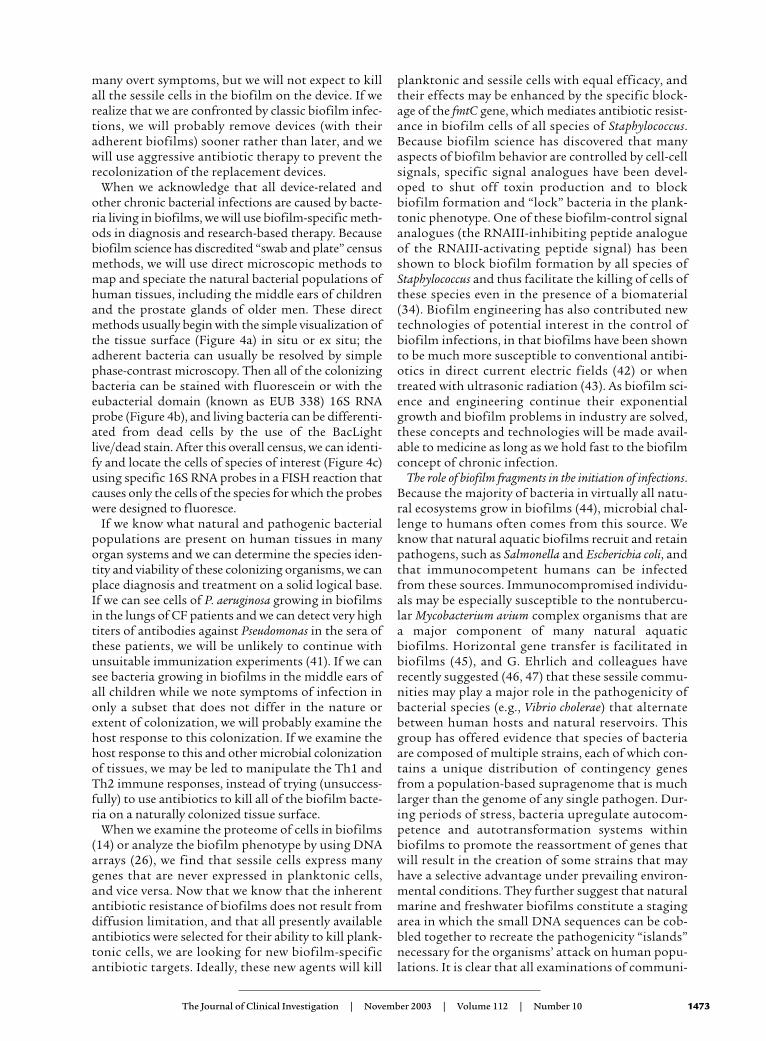
When we acknowledge that all device-related andother chronic bacterial infections are caused by bacte-ria living in biofilms, we will use biofilm-specific meth-ods in diagnosis and research-based therapy. Becausebiofilm science has discredited “swab and plate” censusmethods, we will use direct microscopic methods tomap and speciate the natural bacterial populations ofhuman tissues, including the middle ears of childrenand the prostate glands of older men. These directmethods usually begin with the simple visualization ofthe tissue surface (Figure 4a) in situ or ex situ; theadherent bacteria can usually be resolved by simplephase-contrast microscopy. Then all of the colonizingbacteria can be stained with fluorescein or with theeubacterial domain (known as EUB 338) 16S RNAprobe (Figure 4b), and living bacteria can be differenti-ated from dead cells by the use of the BacLightlive/dead stain. After this overall census, we can identi-fy and locate the cells of species of interest (Figure 4c)using specific 16S RNA probes in a FISH reaction thatcauses only the cells of the species for which the probeswere designed to fluoresce.
If we know what natural and pathogenic bacterialpopulations are present on human tissues in manyorgan systems and we can determine the species iden-tity and viability of these colonizing organisms, we canplace diagnosis and treatment on a solid logical base.If we can see cells of P. aeruginosa growing in biofilmsin the lungs of CF patients and we can detect very hightiters of antibodies against Pseudomonas in the sera ofthese patients, we will be unlikely to continue withunsuitable immunization experiments (41). If we cansee bacteria growing in biofilms in the middle ears ofall children while we note symptoms of infection inonly a subset that does not differ in the nature orextent of colonization, we will probably examine thehost response to this colonization. If we examine thehost response to this and other microbial colonizationof tissues, we may be led to manipulate the Th1 andTh2 immune responses, instead of trying (unsuccess-fully) to use antibiotics to kill all of the biofilm bacte-ria on a naturally colonized tissue surface.
When we examine the proteome of cells in biofilms(14) or analyze the biofilm phenotype by using DNAarrays (26), we find that sessile cells express manygenes that are never expressed in planktonic cells,and vice versa. Now that we know that the inherentantibiotic resistance of biofilms does not result fromdiffusion limitation, and that all presently availableantibiotics were selected for their ability to kill plank-tonic cells, we are looking for new biofilm-specificantibiotic targets. Ideally, these new agents will kill
planktonic and sessile cells with equal efficacy, andtheir effects may be enhanced by the specific block-age of the fmtC gene, which mediates antibiotic resist-ance in biofilm cells of all species of Staphylococcus.Because biofilm science has discovered that manyaspects of biofilm behavior are controlled by cell-cellsignals, specific signal analogues have been devel-oped to shut off toxin production and to blockbiofilm formation and “lock” bacteria in the plank-tonic phenotype. One of these biofilm-control signalanalogues (the RNAIII-inhibiting peptide analogueof the RNAIII-activating peptide signal) has beenshown to block biofilm formation by all species ofStaphylococcus and thus facilitate the killing of cells ofthese species even in the presence of a biomaterial(34). Biofilm engineering has also contributed newtechnologies of potential interest in the control ofbiofilm infections, in that biofilms have been shownto be much more susceptible to conventional antibi-otics in direct current electric fields (42) or whentreated with ultrasonic radiation (43). As biofilm sci-ence and engineering continue their exponentialgrowth and biofilm problems in industry are solved,these concepts and technologies will be made avail-able to medicine as long as we hold fast to the biofilmconcept of chronic infection.
The role of biofilm fragments in the initiation of infections.Because the majority of bacteria in virtually all natu-ral ecosystems grow in biofilms (44), microbial chal-lenge to humans often comes from this source. Weknow that natural aquatic biofilms recruit and retainpathogens, such as Salmonella and Escherichia coli, andthat immunocompetent humans can be infectedfrom these sources. Immunocompromised individu-als may be especially susceptible to the nontubercu-lar Mycobacterium avium complex organisms that area major component of many natural aquaticbiofilms. Horizontal gene transfer is facilitated inbiofilms (45), and G. Ehrlich and colleagues haverecently suggested (46, 47) that these sessile commu-nities may play a major role in the pathogenicity ofbacterial species (e.g., Vibrio cholerae) that alternatebetween human hosts and natural reservoirs. Thisgroup has offered evidence that species of bacteriaare composed of multiple strains, each of which con-tains a unique distribution of contingency genesfrom a population-based supragenome that is muchlarger than the genome of any single pathogen. Dur-ing periods of stress, bacteria upregulate autocom-petence and autotransformation systems withinbiofilms to promote the reassortment of genes thatwill result in the creation of some strains that mayhave a selective advantage under prevailing environ-mental conditions. They further suggest that naturalmarine and freshwater biofilms constitute a stagingarea in which the small DNA sequences can be cob-bled together to recreate the pathogenicity “islands”necessary for the organisms’ attack on human popu-lations. It is clear that all examinations of communi-
1474 The Journal of Clinical Investigation | November 2003 | Volume 112 | Number 10
cable enteric diseases must use biofilm models andthat investigators of these systems should subscribeto the biofilm concept.
Although the digestive system and the integumentmay be colonized by bacteria and fungi from envi-ronmental biofilms, the human organ system that isby far the most susceptible to attack from environ-mental biofilms is the pulmonary system. The tra-chea and the lungs are well designed to resist colo-nization by planktonic bacteria, and animalexperiments have shown the clearance of as many as1 × 106 bacterial cells in as little as 20 minutes, pro-vided the challenging cells are single and unaggre-gated (48). Experiments in the same animal speciesusing the same bacterial species (5) have shown thatthe lungs of normal animals are not able to clear bac-teria that are introduced in the form of biofilm frag-ments or of cells enclosed in artificial matrices (e.g.,as agar beads). When biofilm fragments or agar beadscontaining bacteria are introduced into the lung,these aggregates resist phagocytosis by residentphagocytes and killing by both innate and acquiredimmune factors, and they persist for weeks and evenmonths (48). Figure 1a illustrates the mode ofgrowth of the micro-colonies that comprise thePseudomonas biofilm in the lung in animal models ofCF, and Figure 5 shows how this matrix-enclosedpopulation persists despite the immune reactions ofthe host. Woods and his colleagues proposed that thelungs may be colonized by the detachment of biofilmfragments from the oropharynx, which becomesovergrown by P. aeruginosa during periods of stress(49), and that these fragments cannot be cleared bypulmonary defense mechanisms. These data fromanimal experiments appear to have been confirmedby clinical examination of CF patients, and they raisethe very grim specter of the inevitable colonization ofthe lungs with biofilm fragments when endotrachealtubes become colonized by mixed-species biofilms.Examination of endotracheal tubes that have beenused for assisted ventilation has shown massiveaggregations of mixed-species biofilms (Figure 6);simple detachment of fragments could lead to chron-ic colonization of the lungs. In studies of intubatedpatients in intensive care units, we noted that thebiofilms on endotracheal tubes often contained bac-teria from the digestive tract when nasogastric tubeswere also in use, and that these organisms were oftenfound in the lungs of patients who had died afterassisted ventilation. It is well documented thatbiofilm fragments are aspirated into the lung, andthat these matrix-enclosed organisms cannot becleared by pulmonary defense mechanisms and devel-op into micro-colonies that persist for months andmay give rise to disseminated infection.
If the pulmonary system is in fact susceptible tocolonization by aspirated biofilm fragments thatcannot be cleared and may act as foci for chronicand/or acute bacterial infection, then other environ-
mental organisms should be able to invade mam-malian lungs. For this reason, it is germane to exam-ine a trio of environmental bacteria that are “card-carrying” members of natural ecosystems and thatshare with P. aeruginosa the invidious ability to colo-nize mammalian lungs and cause serious diseases inimmunocompetent mammals. Pasteurella haemolyticais a component of the normal oropharyngeal flora ofcattle that proliferates when the animals are shippedto feedlot operations, and the aspiration of frag-ments of these biofilms into the lungs of thesestressed animals results in respiratory infections thatkill as many as 2% of these calves (50, 51). Legionellapneumophila are the predominant inhabitants ofwarm littoral zones in freshwater lakes, where theygrow in association with green algae (Fisheria sp.) andavoid predation by free-living amoebae by formingbiofilms and secreting antiphagocytic factors. Ashumans devised air conditioners and elaboratedomestic hot water systems, L. pneumophila found analternate home in hotels and hospitals, and emergedto kill several dozen elderly gentlemen in a notorioushotel in Philadelphia, as well as many other people invarious hospitals and office buildings. An organismthat lives in warm water, forms biofilms, and resistsphagocytosis by amoeboid enemies survives very wellin the condensate trays of air conditioners or inhuman lungs (52). The remarkable thing is not thepathogenicity of an environmental organism with noprevious history of attacking humans, but the chill-ing realization that thousands of people must havedied of legionellosis before the disease was defined bythe Centers for Disease Control. If an individual diesof idiopathic pneumonia, this death causes very littleinterest, but the cause of this death may have beenthe mobilization of an acute respiratory infectionfrom niduses of biofilm infection that were acquiredby the aspiration of biofilm fragments. The sourcesof these fragments may be environmental, as in thecase of cooling towers that cause seroconversion to L. pneumophila in people working downwind of thetowers, or they may be very focal, as in the case ofdental hygienists who breath aerosols for long peri-ods of time every working day.
Perhaps the best example of a pulmonary diseasethat is caused by biofilms is meloidosis, which affectspeople in Southeast Asia and Australia, and causedpulmonary infections in US soldiers in Vietnam. Thecausative pathogen is Burkholderia pseudomallei, whichis a natural component of the freshwater ecosystemsin the area; humans make contact with biofilmsformed by these organisms when they work in ricepaddies or swim in local rivers (53). Rates of serocon-version indicate that more than 80% of the people innortheast Thailand have been exposed to thispathogen, presumably by aspiration of fragments ofits exuberant biofilms, and we have visualized a popu-lation with multiple micro-colonies in their lungs (53).When we set up animal experiments by introducing
The Journal of Clinical Investigation | November 2003 | Volume 112 | Number 10 1475
agar beads containing these organisms into the lowerleft lobes of the lungs of guinea pigs, we induced anasymptomatic chronic infection that persisted formonths (54). However, when we stressed these chron-ically infected animals with steroid injections (54),planktonic bacteria were released from the biofilmmicro-colonies, and the animals rapidly succumbed tothe resultant acute infections and bacteremias. Sever-al hundreds of people die of acute meloidosis in north-east Thailand when they are stressed by seasonal star-vation cycles, so the response to stress seen in theanimal model appears to operate in human popula-tions. These data add to the Damoclean image of thepulmonary health of modern humans, because bacte-ria from biofilm niduses acquired by the aspiration ofbiofilm fragments from many sources may be mobi-lized in times of stress and may cause acute pneumo-nias. So, many among us may carry the seeds of fatalpneumonia (the “old man’s friend”) in their lungs asthey enjoy their air-conditioned offices and theirhomes with spas and hot tubs.
Biofilms may also be involved in the disseminationof disease within the body of an infected individual.When we speak of the hematogenous spread of infec-tion, we must now specify whether the infectiousunits are planktonic cells or biofilm fragments,because these entities differ radically in importantproperties such as their antibiotic resistance andtheir adhesion characteristics. Planktonic cells areshed from virtually all mature biofilms, they are gen-erally susceptible to antibiotics, and they adhere tocertain tissues and to inert surfaces with considerableavidity. For this reason it is logical to use prophylac-tic antibiotic therapy to prevent the colonization ofrecently installed medical devices by planktonic bac-teria introduced into the bloodstream by routinetooth brushing or by dental manipulations. On theother hand, many of the cells that detach frombiofilms growing on native heart valves (resulting inendocarditis) or vascular catheters are in the form ofmatrix-enclosed biofilm fragments (21) that are veryresistant to antibiotics, and they usually circulateuntil they “jam” in a capillary bed. For this reason,low-dose antibiotic therapy does not prevent the dis-semination of bacteria in these biofilm diseases, andthe best way of preventing this process is very aggres-sive high-dose treatment of native valve endocarditisand rapid removal of colonized vascular devices. Wehave developed animal models of dissemination inbiofilm diseases, and we have been amazed at howwell sheep lungs can tolerate the small hemorrhagesthat result from hundreds of biofilm fragments lodg-ing in capillary beds. However, it is clear that one ortwo large biofilm fragments can cause profounddamage if they detach and find their way to criticalloci in the lungs or in the brain.
The role of commensal biofilms in protection from infec-tion. One of the conceptual areas in which microbialecology can assist medical practitioners most effec-
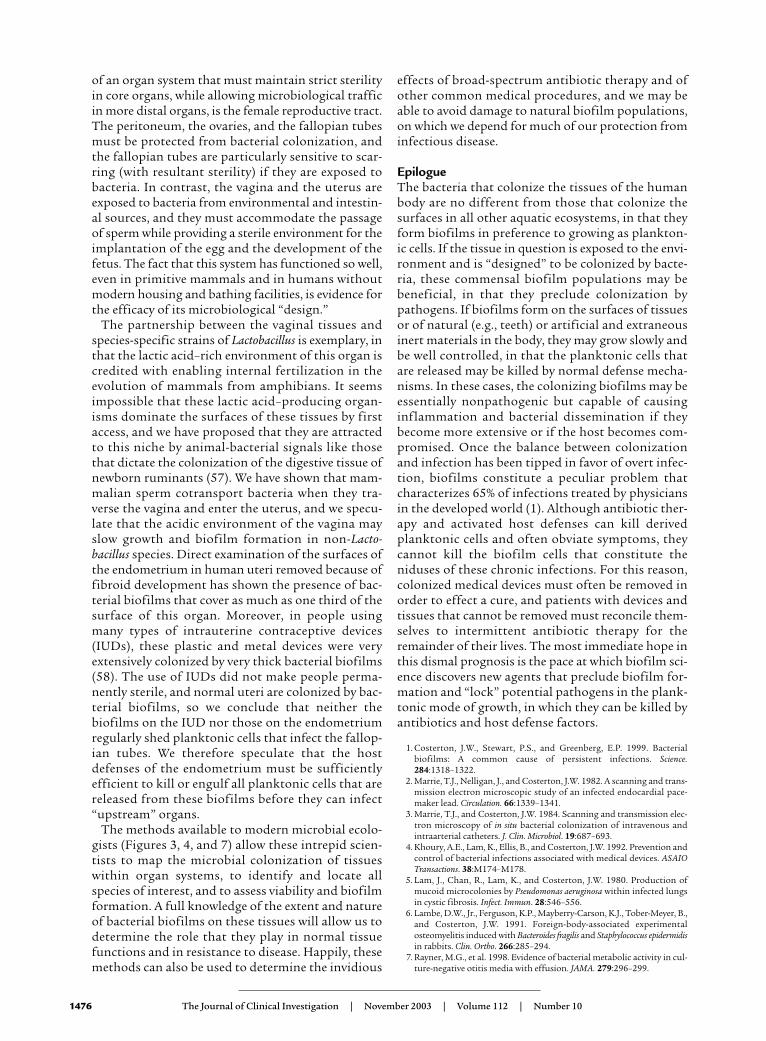
tively and immediately is the area of natural microbialbiofilms on the surfaces of normal human tissues.These tissues are among the most attractive surfacesin nature because they are homeostatic and rich insimple nutrients, and bacteria have adapted to virtu-ally all types of mammalian tissues with considerablesuccess. Perhaps the least welcoming tissue is dry skin,but cells of S. epidermidis grow in prolific biofilmsbetween the squamous cells of the outer three to tenlayers of this stratified epithelium and colonize hairfollicles and sebaceous glands very successfully. Secre-tory epithelia like those of the eye, the vagina, and themouth attract many species of bacteria. The eye lim-its colonization by producing antibacterial factors(surfactants and defensins); the vagina develops anexclusive acid-dominated environment favoring Lac-tobacilli (Figure 4, a–c, and Figure 7); and the mouth iscolonized by a remarkable series of multispeciesbiofilms on different surfaces. Mucus-secreting tis-sues, like those of the trachea and the intestine, arecovered by “mucus blankets,” and this viscous mate-rial is often moved across the surface of the tissues bysuch forces as ciliary beating or by peristalsis. Bacte-ria have great difficulty in accessing mucus-coveredtissues, especially if the mucus blanket is 200–250 µmin thickness and is moving at a considerable speedacross the tissue surface. For this reason, commensalorganisms often use “hold fast” mechanisms and fla-gella that operate well in viscous fluids just to stay inthe tissue surface ecosystem, and successfulpathogens often find ways to disrupt the mucus blan-ket and gain access to the tissue surface. The outersurfaces of “mucus blankets” constitute a rich, ifsomewhat ephemeral, bacterial habitat, and manyspecies proliferate on these surfaces (Figure 8).
Many organ systems in the human body must, inorder to fulfill their normal functions, make contactwith the bacteria-laden environment at their distalextremes, while maintaining strict asepsis in theirproximal “core” organs. A simple example is the bil-iary system, which must deliver bile to the intestinewhile maintaining a bacteria-free situation in theliver, which is extraordinarily sensitive to bacterialinfection (55). In extensive microbiological studies ofthe biliary system in the cat, we discovered that thecommon bile duct is not colonized by gut bacteria,but that these organisms enter the duct if the valve-like sphincter of Oddi is compromised or if a biliarystent is inserted (55). Planktonic bacteria make peri-odic excursions from the gut to the gall bladder, butin the absence of an inert surface on which to form abiofilm, they cannot colonize the tissues in the pres-ence of bile salts, and they are washed back to theintestine. We speculated that most cholangitis resultsfrom vascular challenge, not from traffic in the bileduct, and we showed that the introduction of as fewas 1 × 10⁵ cells into the posterior vena cava was suffi-cient to overwhelm the defenses of the liver and causeacute hepatic disease (56). A more complex example
1476 The Journal of Clinical Investigation | November 2003 | Volume 112 | Number 10
of an organ system that must maintain strict sterilityin core organs, while allowing microbiological trafficin more distal organs, is the female reproductive tract.The peritoneum, the ovaries, and the fallopian tubesmust be protected from bacterial colonization, andthe fallopian tubes are particularly sensitive to scar-ring (with resultant sterility) if they are exposed tobacteria. In contrast, the vagina and the uterus areexposed to bacteria from environmental and intestin-al sources, and they must accommodate the passageof sperm while providing a sterile environment for theimplantation of the egg and the development of thefetus. The fact that this system has functioned so well,even in primitive mammals and in humans withoutmodern housing and bathing facilities, is evidence forthe efficacy of its microbiological “design.”
The partnership between the vaginal tissues andspecies-specific strains of Lactobacillus is exemplary, inthat the lactic acid–rich environment of this organ iscredited with enabling internal fertilization in theevolution of mammals from amphibians. It seemsimpossible that these lactic acid–producing organ-isms dominate the surfaces of these tissues by firstaccess, and we have proposed that they are attractedto this niche by animal-bacterial signals like thosethat dictate the colonization of the digestive tissue ofnewborn ruminants (57). We have shown that mam-malian sperm cotransport bacteria when they tra-verse the vagina and enter the uterus, and we specu-late that the acidic environment of the vagina mayslow growth and biofilm formation in non-Lacto-bacillus species. Direct examination of the surfaces ofthe endometrium in human uteri removed because offibroid development has shown the presence of bac-terial biofilms that cover as much as one third of thesurface of this organ. Moreover, in people usingmany types of intrauterine contraceptive devices(IUDs), these plastic and metal devices were veryextensively colonized by very thick bacterial biofilms(58). The use of IUDs did not make people perma-nently sterile, and normal uteri are colonized by bac-terial biofilms, so we conclude that neither thebiofilms on the IUD nor those on the endometriumregularly shed planktonic cells that infect the fallop-ian tubes. We therefore speculate that the hostdefenses of the endometrium must be sufficientlyefficient to kill or engulf all planktonic cells that arereleased from these biofilms before they can infect“upstream” organs.
The methods available to modern microbial ecolo-gists (Figures 3, 4, and 7) allow these intrepid scien-tists to map the microbial colonization of tissueswithin organ systems, to identify and locate allspecies of interest, and to assess viability and biofilmformation. A full knowledge of the extent and natureof bacterial biofilms on these tissues will allow us todetermine the role that they play in normal tissuefunctions and in resistance to disease. Happily, thesemethods can also be used to determine the invidious
effects of broad-spectrum antibiotic therapy and ofother common medical procedures, and we may beable to avoid damage to natural biofilm populations,on which we depend for much of our protection frominfectious disease.
EpilogueThe bacteria that colonize the tissues of the humanbody are no different from those that colonize thesurfaces in all other aquatic ecosystems, in that theyform biofilms in preference to growing as plankton-ic cells. If the tissue in question is exposed to the envi-ronment and is “designed” to be colonized by bacte-ria, these commensal biofilm populations may bebeneficial, in that they preclude colonization bypathogens. If biofilms form on the surfaces of tissuesor of natural (e.g., teeth) or artificial and extraneousinert materials in the body, they may grow slowly andbe well controlled, in that the planktonic cells thatare released may be killed by normal defense mecha-nisms. In these cases, the colonizing biofilms may beessentially nonpathogenic but capable of causinginflammation and bacterial dissemination if theybecome more extensive or if the host becomes com-promised. Once the balance between colonizationand infection has been tipped in favor of overt infec-tion, biofilms constitute a peculiar problem thatcharacterizes 65% of infections treated by physiciansin the developed world (1). Although antibiotic ther-apy and activated host defenses can kill derivedplanktonic cells and often obviate symptoms, theycannot kill the biofilm cells that constitute theniduses of these chronic infections. For this reason,colonized medical devices must often be removed inorder to effect a cure, and patients with devices andtissues that cannot be removed must reconcile them-selves to intermittent antibiotic therapy for theremainder of their lives. The most immediate hope inthis dismal prognosis is the pace at which biofilm sci-ence discovers new agents that preclude biofilm for-mation and “lock” potential pathogens in the plank-tonic mode of growth, in which they can be killed byantibiotics and host defense factors.
1. Costerton, J.W., Stewart, P.S., and Greenberg, E.P. 1999. Bacterialbiofilms: A common cause of persistent infections. Science.284:1318–1322.
2. Marrie, T.J., Nelligan, J., and Costerton, J.W. 1982. A scanning and trans-mission electron microscopic study of an infected endocardial pace-maker lead. Circulation. 66:1339–1341.
3. Marrie, T.J., and Costerton, J.W. 1984. Scanning and transmission elec-tron microscopy of in situ bacterial colonization of intravenous andintraarterial catheters. J. Clin. Microbiol. 19:687–693.
4. Khoury, A.E., Lam, K., Ellis, B., and Costerton, J.W. 1992. Prevention andcontrol of bacterial infections associated with medical devices. ASAIOTransactions. 38:M174–M178.
5. Lam, J., Chan, R., Lam, K., and Costerton, J.W. 1980. Production ofmucoid microcolonies by Pseudomonas aeruginosa within infected lungsin cystic fibrosis. Infect. Immun. 28:546–556.
6. Lambe, D.W., Jr., Ferguson, K.P., Mayberry-Carson, K.J., Tober-Meyer, B.,and Costerton, J.W. 1991. Foreign-body-associated experimentalosteomyelitis induced with Bacteroides fragilis and Staphylococcus epidermidisin rabbits. Clin. Ortho. 266:285–294.
7. Rayner, M.G., et al. 1998. Evidence of bacterial metabolic activity in cul-ture-negative otitis media with effusion. JAMA. 279:296–299.
The Journal of Clinical Investigation | November 2003 | Volume 112 | Number 10 1477
8. Nickel, J.C., Costerton, J.W., McLean, R.J.C., and Olson, M. 1994. Bacte-rial biofilms: Influence on the pathogenesis, diagnosis and treatment ofurinary-tract infections. J. Antimicrob. Chemother. 33:31–41.
9. Singh, P.K., et al. 2000. Quorum-sensing signals indicate that cysticfibrosis lungs are infected with bacterial biofilms. Nature. 407:762–764.
10. Costerton, J.W., Geesey, G.G., and Cheng, G.K. 1978. How bacteria stick.Sci. Am. 238:86–95.
11. Wyndham, R.C., and Costerton, J.W. 1981. Heterotrophic potentials andhydrocarbon degradation potentials of sediment microorganisms with-in the Athabasca oil sands deposit. Appl. Environ. Microbiol. 41:783–790.
12. de Beer, D., Stoodley, P., and Lewandowski, Z. 1994. Liquid flow in het-erogeneous biofilms. Biotechnol. Bioeng. 44:636–641.
13. Davies, D.G., et al. 1998. The involvement of cell-to-cell signals in thedevelopment of a bacterial biofilm. Science. 280:295–298.
14. Sauer, K., Camper, A.K., Ehrlich, G.D., Costerton, J.W., and Davies, D.G.2002. Pseudomonas aeruginosa displays multiple phenotypes during devel-opment as a biofilm. J. Bacteriol. 184:1140–1154.
15. Davies, D.G., Chakrabarty, A.M., and Geesey, G.G. 1993. Exopolysaccha-ride production in biofilms: Substratum activation of alginate geneexpression by Pseudomonas aeruginosa. Appl. Envir. Microbiol. 59:1181–1186.
16. Davies, D.G., and Geesey, G.G. 1995. Regulation of the alginate biosyn-thesis gene algC in Pseudomonas aeruginosa during biofilm developmentin continuous culture. Appl. Environ. Microbiol. 61:860–867.
17. O’Toole, G.A., Kaplan, H.B., and Kolter, R. 2000. Biofilm formation asmicrobial development. Annu. Rev. Microbiol. 54:49–79.
18. Fuqua, W.C., Winans, E.P., and Greenberg, E.P. 1994. Quorum sensingin bacteria: The Lux R-Lux I family of cell density-responsive transcrip-tional regulators. J. Bacteriol. 176:269–275.
19. Fuqua, W.C., and Greenberg, E.P. 2002. Listening in on bacteria: acyl-homoserine lactone signaling. Nat. Rev. Mol. Cell Biol. 3:685–695.
20. Stoodley, P., Sauer, K., Davies, D.G., and Costerton, J.W. 2002. Biofilmsas complex differentiated communities. Annu. Rev. Microbiol. 56:187–209.
21. Stoodley, P., et al. 2001. Growth and detachment of cell clusters frommature mixed species biofilms. Appl. Environ. Microbiol. 67:5608–5613.
22. Nickel, J.C., Ruseska, I., Wright, J.B., and Costerton, J.W. 1985. Tobramycinresistance of cells of Pseudomonas aeruginosa growing as a biofilm on uri-nary catheter material. Antimicrob. Agents Chemother. 27:619–624.
23. Suci, P.A., Mittelman, M.W., Yu, F.P., and Geesey, G.G. 1994. Investiga-tion of ciprofloxacin penetration into Pseudomonas aeruginosa biofilms.Antimicrob. Agents Chemother. 38:2125–2133.
24. Stewart, P.S. 1996. Theoretical aspects of antibiotic diffusion into micro-bial biofilms. Antimicrob. Agents Chemother. 40:2517–2522.
25. Schoolnik, G.K., et al. 2001. Whole genome DNA microarray expressionanalysis of biofilm development by Vibrio cholerae O1 E1 Tor. MethodsEnzymol. 336:3–18.
26. Wagner, V.E., Bushnell, D., Passador, L., Brooks, A.I., and Iglewski, B.H.2003. Microarray analysis of Pseudomonas aeruginosa quorum-sensingreulons: effects of growth phase and environment. J. Bacteriol.185:2080–2095.
27. Shirtliff, M.E., Leid, J.G., and Costerton, J.W. 2003. Basic science in mus-culoskeletal infections. In Musculoskeletal Infections. J.T. Mader and J.H.Calhoun, editors. Marcel Dekker Inc. New York, New York, USA. 1–61.
28. Sauer, K., Camper, A.K., Ehrlich, G.D., Costerton, J.W., and Davies, D.G.2002. Pseudomonas aeruginosa displays multiple phenotypes during devel-opment as a biofilm. J. Bacteriol. 184:1140–1154.
29. Tremoulet, F., Duche, O., Namane, A., Martinie, B., and Labadie, J.C.2002. A proteomic study of Escherichia coli O157:H7 NCTC 12900 culti-vated in biofilm or in planktonic growth mode. FEMS Microbiol. Lett.215:7–14.
30. Miller, B.S., and Diaz-Torres, M.R. 1999. Proteome analysis of biofilms:growth of Bacillus subtilis on solid medium as model. Methods Enzymol.310:433–441.
31. Costerton, J.W., Lewandowski, Z., Caldwell, D.E., Korber, D.R., and Lap-pin-Scott, H.M. 1995. Microbial biofilms. Ann. Rev. Micro. 49:711–745.
32. Novick, R.P., et al. 1993. Synthesis of staphylococcal virulence factors iscontrolled by a regulatory RNA molecule. EMBO J. 12:3967–3975.
33. Xavier, K.B., and Bassler, B.L. 2003. LuxS quorum sensing: more thanjust a numbers game. Curr. Opin. Microbiol. 6:191–197.
34. Balaban, N., et al. 2003. Use of the quorum-sensing inhibitor RNAIII-inhibiting peptide to prevent biofilm formation in vivo by drug-resist-ant Staphylococcus epidermidis. J. Infect. Dis. 187:625–630.
35. de Nys, R., et al. 1995. Broad spectrum effects of secondary metabolitesfrom the red alga Delisea pulchra in antifouling assays. Biofouling. 8:259–271.
36. Veeh, R.H., et al. 2003. Detection of Staphylococcus aureus biofilm on tam-pons and menses components. J. Infect. Dis. 188:519–530.
37. Post, J.C. 2001. Direct evidence of bacterial biofilms in otitis media.Laryngoscope. 111:2083–2094.
38. Ward, K.H., Olson, M.E., Lam, K., and Costerton, J.W. 1992. Mechanismof persistent infection associated with peritoneal implants. J. Med. Micro.36:406–413.
39. Jensen, E.T., Kharazmi, A., Lam, K., Costerton, J.W., and Hoiby, N. 1990.Human polymorphonuclear leukocyte response to Pseudomonas aerugi-nosa grown in biofilms. Infect. Immun. 58:2383–2385.
40. Leid, J.G., Shirtliff, M.E., Costerton, J.W., and Stoodley, P. 2002. Humanleukocytes adhere to, penetrate, and respond to Staphylococcus aureusbiofilms. Infect. Immun. 70:6339–6345.
41. Costerton, J.W., and Anwar, H. 1994. Pseudomonas aeruginosa: Themicrobe and pathogen. In Pseudomonas aeruginosa infections and treatment.A.L. Baltch and R.P. Smith, editors. Marcel Dekker Inc. New York, NewYork, USA. 1–20.
42. Costerton, J.W., Ellis, B., Lam, K., Johnson, F., and Khoury, A.E. 1994.Mechanism of electrical enhancement of efficacy of antibiotics in killingbiofilm bacteria. Antimicrob. Agents Chemother. 38:2803–2809.
43. Rediske, A.M., Hymas, W.C., Wilkinson, R., and Pitt, W.G. 1998. Ultra-sonic enhancement of antibiotic action on several species of bacteria. J. Gen. Appl. Microbiol. 44:283–288.
44. Costerton, J.W., et al. 1987. Bacterial biofilms in nature and disease.Annu. Rev. Microbiol. 41:435–464.
45. Ghigo, J.-M. 2001. Natural conjugative plasmids induce biofilm devel-opment. Nature. 412:442–445.
46. Shen, K., Wang, X., Post, J.C., and Ehrlich, G.D. 2003. Molecular andtranslational research approaches for the study of bacterial pathogene-sis in otitis media. In Evidence-based otitis media. 2nd edition. R. Rosenfeldand C.D. Bluestone, editors. B.C. Decker Inc. Hamilton, Ontario, Cana-da. 91–119.
47. Ehrlich, G.D., Hu, Z.E., and Post, J.C. Role of biofilms in infectious dis-eases. ASM Press. Washington, DC, USA. In press.
48. Woods, D.E., Bass, J.A., and Johanson, W.G., Jr. 1980. Role of adherencein the pathogenesis of Pseudomonas aeruginosa lung infection in cysticfibrosis patients. Infect. Immun. 30:694–701.
49. Woods, D.E., Straus, D.C., Johanson, W.G., and Bass, J.A. 1981. Role offibronectin in the prevention of the adherence of Pseudomonas aeruginosato buccal cells. J. Infect. Dis. 143:784–790.
50. Morck, D.W., et al. 1987. Electron microscopic description of glycocalyxand fimbriae on the surface of Pasturella haemolytica. Can. J. Vet. Res.51:83–88.
51. Morck, D.W., et al. 1990. A guinea pig model of bovine pneumonic Pas-teurellosis. Can. J. Vet. Res. 54:139–145.
52. Wright, J.B., Athar, M.A., van Olm, T.M., Wootliff, J.S., and Costerton, J.W.1989. Atypical legionellosis: Isolation of Legionella pneumophila serogroup1 from a patient with aspiration pneumonia. J. Hosp. Inf. 13:187–190.
53. Vorachit, M., Lam, K., Jayanetra, P., and Costerton, J.W. 1995. Electronmicroscopy study of the mode of growth of Pseudomonas pseudomallei invitro and in vivo. J. Trop. Med. Hyg. 98:379–391.
54. Vorachit, M., Lam, K., Jayanetra, P., and Costerton, J.W. 1995. The studyof the pathogenicity of Burkholderia pseudomallei–a guinea pig model. J. Infect. Dis. Antimicrob. Agents. 12:115–121.
55. Sung, J.Y., et al. 1992. Ascending infection of the biliary tract after sur-gical sphincterotomy and biliary stenting. J. Gastroenterol. Hepatol.7:240–245.
56. Sung, J.Y., et al. 1991. Bacterial invasion of the biliary system by way ofthe portal-venous system. Hepatology. 14:313–317.
57. Cheng, K.-J., and Costerton, J.W. 1981. Adherent rumen bacteria: Theirrole in the digestion of plant material, urea, and epithelial cells. In Diges-tive physiology and metabolism in ruminants. Y. Ruchebusch and P. Thivend,editors. MTP Press. Lancaster, United Kingdom. 227–250.
58. Marrie, T.J., and Costerton, J.W. 1983. A scanning and transmission elec-tron microscopy study of the surfaces of intrauterine contraceptivedevices. Am. J. Obstet. Gynecol. 146:384–394.
59. Lam, J.S., et al. 1983. Immunogenicity of Pseudomonas aeruginosa outermembrane antigens examined by crossed immunoelectrophoresis. Infect.Immun. 42:88–89.
60. Costerton, J.W., and Stewart, P.S. 2000. Biofilms and device-related infec-tions. In Persistent bacterial infections. J.P. Nataro, M.J. Blaser, and S. Cun-ningham-Rundels, editors. American Society for Microbiology. Wash-ington, DC, USA. 423–437.