Embed Size (px)
Citation preview

The Arabidopsis Na+/H+ Antiporters NHX1 and NHX2 ControlVacuolar pH and K+ Homeostasis to Regulate Growth, FlowerDevelopment, and Reproduction C W
Elias Bassil,a Hiromi Tajima,a Yin-Chih Liang,a Masa-aki Ohto,a Koichiro Ushijima,b Ryohei Nakano,b
Tomoya Esumi,a Ardian Coku,a Mark Belmonte,c and Eduardo Blumwalda,1
a Department of Plant Sciences, University of California, Davis, California 95616b Department of Agriculture, Okayama University, Okayama 700-8530, Japanc Department of Biological Sciences, University of Manitoba, Winnipeg, Manitoba R3T 2N2, Canada
Intracellular Na+/H+ (NHX) antiporters have important roles in cellular pH and Na+, K+ homeostasis. The six Arabidopsis
thaliana intracellular NHX members are divided into two groups, endosomal (NHX5 and NHX6) and vacuolar (NHX1 to NHX4).
Of the vacuolar members, NHX1 has been characterized functionally, but the remaining members have largely unknown
roles. Using reverse genetics, we show that, unlike the single knockouts nhx1 or nhx2, the double knockout nhx1 nhx2 had
significantly reduced growth, smaller cells, shorter hypocotyls in etiolated seedlings and abnormal stamens in mature
flowers. Filaments of nhx1 nhx2 did not elongate and lacked the ability to dehisce and release pollen, resulting in a near lack
of silique formation. Pollen viability and germination was not affected. Quantification of vacuolar pH and intravacuolar K+
concentrations indicated that nhx1 nhx2 vacuoles were more acidic and accumulated only 30% of the wild-type K+
concentration, highlighting the roles of NHX1 and NHX2 in mediating vacuolar K+/H+ exchange. Growth under added Na+,
but not K+, partly rescued the flower and growth phenotypes. Our results demonstrate the roles of NHX1 and NHX2 in
regulating intravacuolar K+ and pH, which are essential to cell expansion and flower development.
INTRODUCTION
Ion homeostasis and the regulation of cellular pH are associated
with every aspect of the biology of the cell. The concerted action
of H+-translocating enzymes (H+-pumps) and cation/H+ ex-
changers are vital to establish and maintain optimal ion and pH
gradients, which are essential for cell function and plant devel-
opment (Martinoia et al., 2007; Amtmann and Leigh, 2010).
Intracellular Na+/H+ antiporters (NHXs) are integral membrane
proteins residing in the plasma membrane (Shi et al., 2000)
endosomal compartments (Rodriguez-Rosales et al., 2008;
Bassil et al., 2011), and vacuoles (Apse et al., 1999; Yokoi
et al., 2002; Apse and Blumwald, 2007; Hamaji et al., 2009; Leidi
et al., 2010). They belong to the monovalent cation/proton
antiporter (CPA1) family of transporters (Maser et al., 2001). In
plants, NHX antiporters catalyze the electroneutral exchange of
Na+ and/or K+ for H+ by using the electrochemical H+ gradients
generated by the (H+)–ATPases (in the plasma membrane)
and the (H+)–ATPase and (H+)–PPase (in the vacuole) to direct
either the movement of Na+ or K+ out of the cell or the luminal
movement of Na+ or K+ into the vacuoles and intracellular
organelles. TheNHX gene family ofArabidopsis thaliana includes
a class of six intracellular NHX-type antiporters, which are
classified into two groups (Brett et al., 2005a; Pardo et al.,
2006; Rodriguez-Rosales et al., 2009). Group I contains NHX1 to
NHX4 and localizes to vacuoles, whereas group II contains NHX5
and NHX6 and localizes to endosomal compartments (Bassil
et al., 2011). Several functions have been associated with plant
NHX transporters, including the regulation of flower colora-
tion (Yamaguchi et al., 2001; Ohnishi et al., 2005; Yoshida et al.,
2009), the regulation of cellular K+ homeostasis (Venema et al.,
2003; Pardo et al., 2006; Apse and Blumwald, 2007; Leidi
et al., 2010), cell expansion (Apse et al., 2003; Bassil et al., 2011),
vesicular trafficking and protein targeting (Bowers et al., 2000;
Brett et al., 2005b; Bassil et al., 2011), and salt tolerance (Apse
et al., 1999; Hernandez et al., 2009; Bassil et al., 2011). NHX1was
shown to be required for seedling establishment and leaf devel-
opment, especially in the formation of large epidermal cells (Apse
et al., 2003). Recently, NHX5 and NHX6 were shown to be
localized in the Golgi and trans-Golgi network and to function in
the trafficking of endosomal cargo to the vacuole, in salt stress,
and in cell expansion (Bassil et al., 2011). However, the roles of
the remainingmembers of the vacuolar group, NHX2, NHX3, and
NHX4, in growth and development remain largely unknown. To
comprehend the concerted role of all intracellular NHXs, an effort
to generate and characterize multiple knockout lines of Arabi-
dopsis NHX1 to NHX6 was initiated.
Although we have considerable information about the molec-
ular and biochemical networks acting during plant growth and
1Address correspondence to [email protected] author responsible for distribution of materials integral to thefindings presented in this article in accordance with the policy describedin the Instructions for Authors (www.plantcell.org) is: Eduardo Blumwald([email protected]).CSome figures in this article are displayed in color online but in blackand white in the print edition.WOnline version contains Web-only data.www.plantcell.org/cgi/doi/10.1105/tpc.111.089581
The Plant Cell, Vol. 23: 3482–3497, September 2011, www.plantcell.org ã 2011 American Society of Plant Biologists. All rights reserved.

development, we have little knowledge of how cells regulate
the transport of solutes and their intracellular pH. Ions (K+ in
particular) and pH gradients have a great effect on the regulation
of cell volume and the trafficking of membrane vesicles and
their protein cargo between the different cellular compartments
(Blumwald, 1987; Pardo et al., 2006; Bassil et al., 2011). These
processes are essential for plant growth, development, and the
response to environmental changes.
NHX-type cation/H+ antiporters are critical players in cell ex-
pansion. To understand the concerted role of intracellular Arabi-
dopsisNHXson cell expansion,we initiated the characterization of
the growth and development of NHX1 and NHX2 knockout lines.
We show that NHX1 and NHX2 function together to control cell
expansion in vegetative tissues andmale reproductive organs and
are required for normal flower development and set.We link flower
developmental processes to altered vacuolar pH and K+ homeo-
stasis and argue that NHX1 andNHX2mediate K+/H+ exchange to
accumulate K+ in the vacuole, a critical step for cell expansion and
growth, especially in rapidly expanding cells.
RESULTS
At the organ level, the expression of NHX1 and NHX2 indicated
that both genes are expressed ubiquitously, including roots,
rosette leaves, stems, flowers, and siliques (see Supplemental
Figure 1 online). The level of expression ofNHX1was significantly
higher than that of NHX2 in all organs examined, and NHX2
expression levels were similar to that of other NHX members
(Bassil et al., 2011). The nearly ubiquitous expression of NHX1
was also confirmed in publicly available expression data (http://
bbc.botany.utoronto.ca/efp/cgi-bin/efpWeb.cgi) as wells as
previously published work (Yokoi et al., 2002; Aharon et al.,
2003; Apse et al., 2003).
NHX1 and NHX2 Colocalize to the Vacuole
To assess the localization of NHX1 and NHX2 and their possible
vacuolar colocalization, we generated plants expressing C-terminal
translational fusion reporter constructs with NHX1 fused with
green fluorescent protein (GFP), or NHX2 fused with yellow
fluorescent protein (YFP) or red fluorescent protein (RFP), driven
by either the Ubiquitin-10 (Ub10) or 35S promoters. The growth
of the transgenic reporter lines was indistinguishable from that of
the wild-type plants, indicating that the fusion proteins did not
adversely affect the growth of the transgenic plants. In 35S-
NHX2-YFP plants, the fluorescence signals in roots and root tips
localized to vacuolar membranes (Figure 1; see Supplemental
Figure 2 online). The vacuolar localization was clearly indicated
by the nuclei seen outside the fluorescent boundary delineated
by the NHX2-associated YFP signal (arrows in Figure 1A). Trans-
vacuolar cytoplasmic strands were also clearly evident in root
cells, which further supported the NHX2-YFP vacuolar associa-
tion. Observation of 35S-NHX1-GFP reporter plants resulted in
a poor and inconsistent GFP signal that was often observed
within the vacuolar lumen and endosomal bodies in all lines
examined (see Supplemental Figure 2 online). To circumvent
possible problems associated with overexpression or nonstable
expression caused by the 35S promoter, NHX1-GFP was ex-
pressed under the control of theUb10promoter, which enabled a
milder, more stable expression of downstream genes (Grefen
et al., 2010). Transient transformation of Ub10-NHX1-GFP in
Arabidopsis seedlings resulted in a GFP signal associated with
vacuolar membranes and transvacuolar strands (Figure 1B; see
Supplemental Movie 1 online). Furthermore, plasmolysis of root
cells, induced by the addition of 1 M mannitol, caused a clear
recession of the tonoplast and the YFP and GFP signals, sup-
porting the vacuolar localization of NHX1 and NHX2 (see Sup-
plemental Figure 2 online). To determine whether NHX1 and
NHX2 colocalize, Ub10-NHX1-GFP and 35S-NHX2-RFP were
transiently coexpressed in Arabidopsis seedlings. As shown in
Figures 1C to 1E, NHX1-GFP and NHX2-RFP were highly colo-
calized to vacuolar membranes in cells expressing both trans-
genes. We also tested whether NHX1 colocalized with another
vacuolar membrane protein by transiently expressing Ub10-
NHX1-GFP in stable reporter lines of VAMP711-RFP, a R-SNARE
of the tonoplast (Uemura et al., 2004). As expected, NHX1-GFP
colocalized extensively with VAMP711 (Figures 1F to 1H).
nhx1 nhx2 Double Knockout Phenotypes
To investigate NHX1 and NHX2 functions, we used a reverse
genetic approach to generate knockouts from available T-DNA
insertion lines. Two independent knockout mutants using differ-
ent T-DNA insertion lines for each gene were identified. Single
knockouts, designated as nhx1-1, nhx1-2, nhx2-1, and nhx2-3
(see Methods), were genotyped and backcrossed twice before
their subsequent use in crosses to generate the independent
double knockout lines nhx1-1 nhx2-1, nhx1-2 nhx2-1, and nhx1-2
nhx2-3 (see Supplemental Figure 3 online). Lack of expression of
full-length NHX1 and NHX2 in all knockouts was confirmed with
RT-PCR (see Supplemental Figure 3 online). Experiments using
the double knockout lines produced identical results; therefore,
unless otherwise noted, results reported from here on are those
obtained with the line nhx1-1 nhx2-1.
The single knockout nhx1 displayed reduced growth com-
pared with the wild type (Columbia ecotype [Col-0]), similar to
that reported previously in nhx1 in the Wassilewskija ecotype
background (Apse et al., 2003). However, nhx2 did not show
any obvious growth phenotypes or developmental defects
when grown either in soil (Figure 2A) or on plates (see below).
By contrast, the double knockout nhx1 nhx2 displayed a signif-
icant reduction in growth (Figure 2). Rosette leaf size and
inflorescence height were markedly smaller than either the wild
type or the single knockouts. Cross sections through leaf
margins indicated that nhx1 nhx2 cells were significantly smaller
than comparable wild-type leaf cells (Figure 2B). nhx1 nhx2
leaf tissue remained highly organized with no apparent cell-
type disruptions. To confirm the nhx1 nhx2 phenotypes, we com-
plemented nhx1 nhx2 with functional copies of both genes.
Transformation of nhx1 nhx2 with either 35S-NHX1-GFP or 35S-
NHX2-YFP reverted the growth phenotype to the wild type
(Figure 2C), thereby demonstrating that the nhx1 nhx2 pheno-
types were caused by disruption of NHX1 and NHX2. In addi-
tion, C-terminal tagging of NHX1 and NHX2 did not affect the
function of either protein.
Vacuolar NHX1 and NHX2 3483

Stamens of nhx1 nhx2 Lack Filament Elongation and
Anther Dehiscence
nhx1 nhx2 plants displayed significant phenotypes in their
reproductive organs. nhx1 nhx2 flowers developed a low num-
ber of siliques, which contained few or no seeds (Figure 2D; see
arrows). At the same developmental stage, nhx1 nhx2 flowers
were significantly shorter and narrower than the wild-type
flowers (Figures 3A to 3C). A closer examination of flower
anatomy revealed at least two distinct developmental changes
associated with stamens. First, mature nhx1 nhx2 stamens had
markedly shorter filaments that did not extend far enough to
position the anthers at the height of the stigma (Figures 3B
[arrows] and 3D), as is typically seen in the wild-type flowers.
Quantitative measurements of filament lengths indicated that,
in nhx1 nhx2 flowers, both short and long filaments were
reduced in length (Figure 3D). A comparison of longitudinal
sections of filaments revealed that nhx1 nhx2 filament cells
Figure 1. Subcellular Localization of NHX1 and NHX2.
(A) NHX2-associated YFP fluorescence in mature root cells of stably transformed plants expressing 35S-NHX2-YFP. Arrows point to nuclei.
(B) Transient expression of Ub10-NHX1-GFP in Arabidopsis cotyledons.
(C) to (E) Transient coexpression of Ub10-NHX1-GFP and 35S-NHX2-RFP in Arabidopsis cotyledons.
(F) to (H) Transient expression of Ub10-NHX1-GFP in the Ub10-VAMP711-RFP background.
(C) and (F) are GFP images; (D) and (G) are RFP images; (E) and (H) are merged images. Arrowheads in (B), (E), and (H) point to the tonoplast and
transvacuolar cytoplasmic strands.
Bar in (A) = 50 mm; bars in (B) to (H) = 20 mm.
3484 The Plant Cell

were only 55% the length and 60% the width of the wild-type
filament cells (Figures 3E to 3G). The second stamen phenotype
was that nhx1 nhx2 anthers did not dehisce at the same
frequency seen in the wild-type anthers (Figures 3B and
3C [insets]). Scoring of anthers from 40 nhx1 nhx2 plants
(;80 anthers) indicated that only 3% showed normal anther
dehiscence. Cross sections of anthers showed that, in a high
proportion of nhx1 nhx2 anthers, the stomium (St in Figures 3H
and 3I) did not rupture sufficiently to release pollen from
individual anther locules (Figures 3H and 3I). When anther
dehiscence did occur it was not sufficient to release and
expose the majority of pollen. Therefore, nhx1 nhx2 anthers
lacked the characteristic yellowish color typical of anthers after
dehiscence and remained the distinctive color of immature
anthers (i.e., off-white) (Figures 3A and 3B [insets]). Eventually
nhx1 nhx2 flowers senesced without setting, and inflores-
cences of nhx1 nhx2 did not form siliques (Figure 2B). A
comparison of flower anatomy, the lack of anther dehiscence,
and filament elongation in nhx1 nhx2 are shown in the scanning
electron micrographs of Figure 4.
To test specific expression of NHX1 and NHX2 in floral organs,
we used NHX1 and NHX2 promoters to drive b-glucuronidase
(GUS) expression in transgenic plants (see Supplemental Fig-
ure 4 online). GUS analysis indicated that the expression of
NHX1 was abundant in most flower organs (see Supplemental
Figure 4A online) but was low in nonelongated filaments and
immature anthers (see Supplemental Figure 4B online) and was
highly expressed in elongated filaments (see Supplemental
Figure 4C online). On the other hand, NHX2 was highly ex-
pressed in anthers and pollen. These results might suggest
Figure 2. Growth and Development of the Single and Double Knockouts of NHX1 and NHX2.
(A) Four-week-old plants grown under 16-h days (Top), and 5-week-old plants grown under 8-h days (Bottom).
(B) Cross section through the margin of a 4-week-old wild-type leaf (Top) and comparable nhx1 nhx2 knockout leaf (Bottom). Sections were stained
with toluidine blue O.
(C) The nhx1 nhx2 growth phenotype was rescued by transformation with either 35S-NHX1-GFP or 35S-NHX2-YFP. The wild type and nhx1 nhx2 are
included for comparison. Representative images of 4-week-old T1 transformants are shown.
(D) Inflorescence stalks of wild-type, nhx1, nhx2, and nhx1 nhx2 plants. Note the lack of siliques in nhx1 nhx2 (arrows).
Bar in (B) = 30 mm.
Vacuolar NHX1 and NHX2 3485

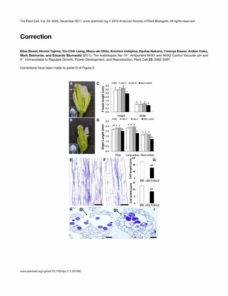
Figure 3. Stamens of the Double Knockout nhx1 nhx2 Have Reduced Filament Elongation and Anther Dehiscence.
(A) Dissected wild-type flower showing normal filaments and anthers.
(B) Dissected nhx1 nhx2 flower showing the short filament (arrows) and nondehiscent anther (inset) phenotypes.
(C) Quantification of whole flower size and (D) floral organ size in single and double knockouts of NHX1 and NHX2. Wt, wild type.
(E) Longitudinal section of mature wild-type filament.
(F) Longitudinal section of mature nhx1 nhx2 filament.
(G) Comparison of filament cell length and width.
(H) Cross section of a mature wild-type dehiscent anther with a ruptured stomium (St).
3486 The Plant Cell
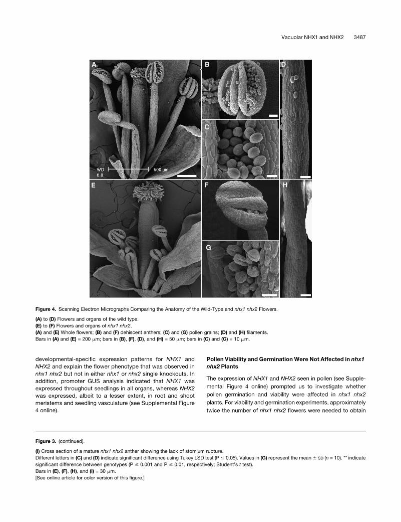
developmental-specific expression patterns for NHX1 and
NHX2 and explain the flower phenotype that was observed in
nhx1 nhx2 but not in either nhx1 or nhx2 single knockouts. In
addition, promoter GUS analysis indicated that NHX1 was
expressed throughout seedlings in all organs, whereas NHX2
was expressed, albeit to a lesser extent, in root and shoot
meristems and seedling vasculature (see Supplemental Figure
4 online).
Pollen Viability and Germination Were Not Affected in nhx1
nhx2 Plants
The expression of NHX1 and NHX2 seen in pollen (see Supple-
mental Figure 4 online) prompted us to investigate whether
pollen germination and viability were affected in nhx1 nhx2
plants. For viability and germination experiments, approximately
twice the number of nhx1 nhx2 flowers were needed to obtain
Figure 3. (continued).
(I) Cross section of a mature nhx1 nhx2 anther showing the lack of stomium rupture.
Different letters in (C) and (D) indicate significant difference using Tukey LSD test (P # 0.05). Values in (G) represent the mean 6 SD (n = 10). ** indicate
significant difference between genotypes (P # 0.001 and P # 0.01, respectively; Student’s t test).
Bars in (E), (F), (H), and (I) = 30 mm.
[See online article for color version of this figure.]
Figure 4. Scanning Electron Micrographs Comparing the Anatomy of the Wild-Type and nhx1 nhx2 Flowers.
(A) to (D) Flowers and organs of the wild type.
(E) to (F) Flowers and organs of nhx1 nhx2.
(A) and (E) Whole flowers; (B) and (F) dehiscent anthers; (C) and (G) pollen grains; (D) and (H) filaments.
Bars in (A) and (E) = 200 mm; bars in (B), (F), (D), and (H) = 50 mm; bars in (C) and (G) = 10 mm.
Vacuolar NHX1 and NHX2 3487
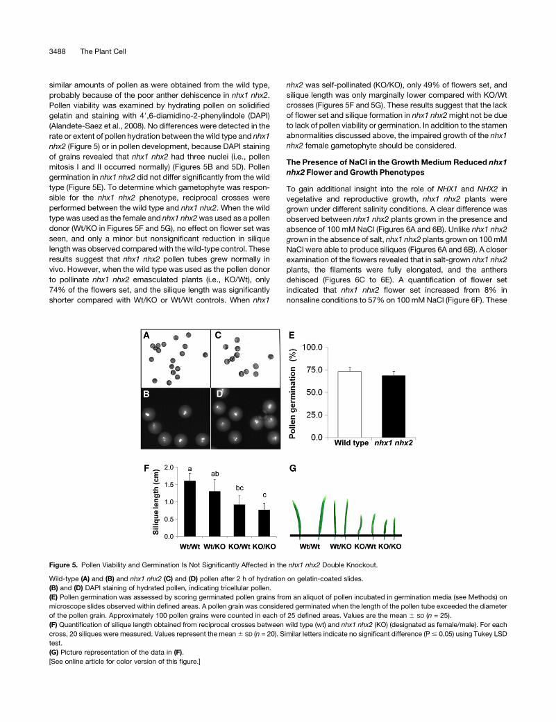
similar amounts of pollen as were obtained from the wild type,
probably because of the poor anther dehiscence in nhx1 nhx2.
Pollen viability was examined by hydrating pollen on solidified
gelatin and staining with 49,6-diamidino-2-phenylindole (DAPI)
(Alandete-Saez et al., 2008). No differences were detected in the
rate or extent of pollen hydration between the wild type and nhx1
nhx2 (Figure 5) or in pollen development, because DAPI staining
of grains revealed that nhx1 nhx2 had three nuclei (i.e., pollen
mitosis I and II occurred normally) (Figures 5B and 5D). Pollen
germination in nhx1 nhx2 did not differ significantly from the wild
type (Figure 5E). To determine which gametophyte was respon-
sible for the nhx1 nhx2 phenotype, reciprocal crosses were
performed between the wild type and nhx1 nhx2. When the wild
type was used as the female and nhx1 nhx2was used as a pollen
donor (Wt/KO in Figures 5F and 5G), no effect on flower set was
seen, and only a minor but nonsignificant reduction in silique
lengthwas observed comparedwith thewild-type control. These
results suggest that nhx1 nhx2 pollen tubes grew normally in
vivo. However, when the wild type was used as the pollen donor
to pollinate nhx1 nhx2 emasculated plants (i.e., KO/Wt), only
74% of the flowers set, and the silique length was significantly
shorter compared with Wt/KO or Wt/Wt controls. When nhx1
nhx2 was self-pollinated (KO/KO), only 49% of flowers set, and
silique length was only marginally lower compared with KO/Wt
crosses (Figures 5F and 5G). These results suggest that the lack
of flower set and silique formation in nhx1 nhx2might not be due
to lack of pollen viability or germination. In addition to the stamen
abnormalities discussed above, the impaired growth of the nhx1
nhx2 female gametophyte should be considered.
The Presence of NaCl in the Growth Medium Reduced nhx1
nhx2 Flower and Growth Phenotypes
To gain additional insight into the role of NHX1 and NHX2 in
vegetative and reproductive growth, nhx1 nhx2 plants were
grown under different salinity conditions. A clear difference was
observed between nhx1 nhx2 plants grown in the presence and
absence of 100 mM NaCl (Figures 6A and 6B). Unlike nhx1 nhx2
grown in the absence of salt, nhx1 nhx2 plants grown on 100mM
NaCl were able to produce siliques (Figures 6A and 6B). A closer
examination of the flowers revealed that in salt-grown nhx1 nhx2
plants, the filaments were fully elongated, and the anthers
dehisced (Figures 6C to 6E). A quantification of flower set
indicated that nhx1 nhx2 flower set increased from 8% in
nonsaline conditions to 57% on 100 mMNaCl (Figure 6F). These
Figure 5. Pollen Viability and Germination Is Not Significantly Affected in the nhx1 nhx2 Double Knockout.
Wild-type (A) and (B) and nhx1 nhx2 (C) and (D) pollen after 2 h of hydration on gelatin-coated slides.
(B) and (D) DAPI staining of hydrated pollen, indicating tricellular pollen.
(E) Pollen germination was assessed by scoring germinated pollen grains from an aliquot of pollen incubated in germination media (see Methods) on
microscope slides observed within defined areas. A pollen grain was considered germinated when the length of the pollen tube exceeded the diameter
of the pollen grain. Approximately 100 pollen grains were counted in each of 25 defined areas. Values are the mean 6 SD (n = 25).
(F) Quantification of silique length obtained from reciprocal crosses between wild type (wt) and nhx1 nhx2 (KO) (designated as female/male). For each
cross, 20 siliques were measured. Values represent the mean6 SD (n = 20). Similar letters indicate no significant difference (P# 0.05) using Tukey LSD
test.
(G) Picture representation of the data in (F).
[See online article for color version of this figure.]
3488 The Plant Cell
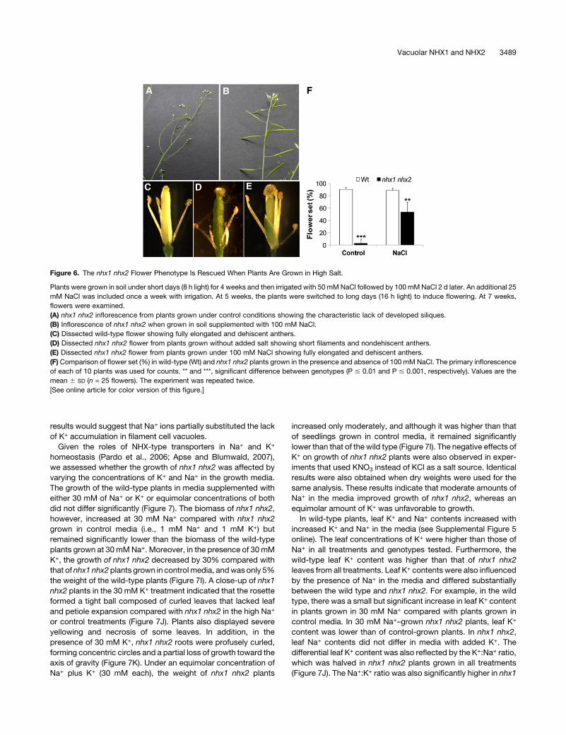
results would suggest that Na+ ions partially substituted the lack
of K+ accumulation in filament cell vacuoles.
Given the roles of NHX-type transporters in Na+ and K+
homeostasis (Pardo et al., 2006; Apse and Blumwald, 2007),
we assessed whether the growth of nhx1 nhx2 was affected by
varying the concentrations of K+ and Na+ in the growth media.
The growth of the wild-type plants in media supplemented with
either 30 mM of Na+ or K+ or equimolar concentrations of both
did not differ significantly (Figure 7). The biomass of nhx1 nhx2,
however, increased at 30 mM Na+ compared with nhx1 nhx2
grown in control media (i.e., 1 mM Na+ and 1 mM K+) but
remained significantly lower than the biomass of the wild-type
plants grown at 30 mMNa+. Moreover, in the presence of 30 mM
K+, the growth of nhx1 nhx2 decreased by 30% compared with
that of nhx1 nhx2plants grown in controlmedia, andwas only 5%
the weight of the wild-type plants (Figure 7I). A close-up of nhx1
nhx2 plants in the 30 mM K+ treatment indicated that the rosette
formed a tight ball composed of curled leaves that lacked leaf
and petiole expansion compared with nhx1 nhx2 in the high Na+
or control treatments (Figure 7J). Plants also displayed severe
yellowing and necrosis of some leaves. In addition, in the
presence of 30 mM K+, nhx1 nhx2 roots were profusely curled,
forming concentric circles and a partial loss of growth toward the
axis of gravity (Figure 7K). Under an equimolar concentration of
Na+ plus K+ (30 mM each), the weight of nhx1 nhx2 plants
increased only moderately, and although it was higher than that
of seedlings grown in control media, it remained significantly
lower than that of the wild type (Figure 7I). The negative effects of
K+ on growth of nhx1 nhx2 plants were also observed in exper-
iments that used KNO3 instead of KCl as a salt source. Identical
results were also obtained when dry weights were used for the
same analysis. These results indicate that moderate amounts of
Na+ in the media improved growth of nhx1 nhx2, whereas an
equimolar amount of K+ was unfavorable to growth.
In wild-type plants, leaf K+ and Na+ contents increased with
increased K+ and Na+ in the media (see Supplemental Figure 5
online). The leaf concentrations of K+ were higher than those of
Na+ in all treatments and genotypes tested. Furthermore, the
wild-type leaf K+ content was higher than that of nhx1 nhx2
leaves from all treatments. Leaf K+ contents were also influenced
by the presence of Na+ in the media and differed substantially
between the wild type and nhx1 nhx2. For example, in the wild
type, there was a small but significant increase in leaf K+ content
in plants grown in 30 mM Na+ compared with plants grown in
control media. In 30 mM Na+–grown nhx1 nhx2 plants, leaf K+
content was lower than of control-grown plants. In nhx1 nhx2,
leaf Na+ contents did not differ in media with added K+. The
differential leaf K+ content was also reflected by the K+:Na+ ratio,
which was halved in nhx1 nhx2 plants grown in all treatments
(Figure 7J). The Na+:K+ ratio was also significantly higher in nhx1
Figure 6. The nhx1 nhx2 Flower Phenotype Is Rescued When Plants Are Grown in High Salt.
Plants were grown in soil under short days (8 h light) for 4 weeks and then irrigated with 50 mMNaCl followed by 100mMNaCl 2 d later. An additional 25
mM NaCl was included once a week with irrigation. At 5 weeks, the plants were switched to long days (16 h light) to induce flowering. At 7 weeks,
flowers were examined.
(A) nhx1 nhx2 inflorescence from plants grown under control conditions showing the characteristic lack of developed siliques.
(B) Inflorescence of nhx1 nhx2 when grown in soil supplemented with 100 mM NaCl.
(C) Dissected wild-type flower showing fully elongated and dehiscent anthers.
(D) Dissected nhx1 nhx2 flower from plants grown without added salt showing short filaments and nondehiscent anthers.
(E) Dissected nhx1 nhx2 flower from plants grown under 100 mM NaCl showing fully elongated and dehiscent anthers.
(F) Comparison of flower set (%) in wild-type (Wt) and nhx1 nhx2 plants grown in the presence and absence of 100 mM NaCl. The primary inflorescence
of each of 10 plants was used for counts. ** and ***, significant difference between genotypes (P # 0.01 and P # 0.001, respectively). Values are the
mean 6 SD (n = 25 flowers). The experiment was repeated twice.
[See online article for color version of this figure.]
Vacuolar NHX1 and NHX2 3489
nhx2 than in thewild type at both 30mMNa+ and 30mMNa+ plus
30 mM K+ treatments. Collectively, these data indicate that the
ability of nhx1 nhx2 plants to accumulate K+ in favor of Na+ was
compromised and that these plants were not able to adjust their
K+ content when challenged with Na+, as seen in the wild-type
plants.
Etiolation of nhx1 nhx2 Seedlings Is Cation Dependent
We reasoned that a good experimental system to test the role(s)
of NHX1 andNHX2 in rapid cell expansion would be the etiolation
response of dark-grown seedlings, because etiolated seedlings
are amenable to physiological and cellular measurements of pH
and K+ homeostasis. Furthermore, the rapid elongation of hypo-
cotyls in etiolated seedlings closely parallels the elongation of
anther filaments during flower development. When grown on
solidified media in the dark, nhx1 nhx2 etiolated seedlings
displayed significantly shorter hypocotyls and longer roots than
the wild-type seedlings (see Supplemental Figure 6A online).
Etiolated seedlings grown on increasing concentrations of K+
exhibited a progressively stronger phenotype (i.e., significantly
shorter hypocotyls and longer roots than control-grown nhx1
nhx2). Interestingly, when K+ concentrations in the media were
10mMormore, roots of nhx1 nhx2 grew by curling away from the
direction of gravity, forming concentric circles of winding roots,
which was probably caused by uneven expansion of epidermal
cells (see Supplemental Figure 6 online). Similar to the response
of long-term plant growth experiments (Figure 7), hypocotyls of
nhx1 nhx2 seedlings grown in 30 mM Na+ elongated more than
those of nhx1 nhx2 in control media and were not significantly
different in length from the wild-type hypocotyls. The root re-
sponse, however, did not recover under increasedNa+ as it did in
hypocotyls. The consistent phenotype and response of etiolated
seedlings allowed for a reliable experimental system to perform
additional cellular measurements (as described below).
NHX1 and NHX2 Regulate Vacuolar pH and K+ Homeostasis
To assess the roles of NHX1 and NHX2 in pH regulation, we
used the ratiometric fluorescein-based pH sensitive dye, 29,79-bis-(2-carboxyethyl)-5-(and-6)-carboxyfluorescein (BCECF) in
an imaging-based approach to measure vacuolar pH in vivo.
Ratiometric dyes have the distinct advantage of not being
significantly affected by dye loading, cell size, or tissue mor-
phology, unlike other nonratiometric dyes. The membrane-
permeant BCECF-acetoxymethyl (AM) derivative readily loads
into vacuoles of intact root cells (Swanson et al., 1998; Krebs
et al., 2010). pH values were calculated from fluorescence ratios
of confocal images using an in situ calibration curve (see Sup-
plemental Figure 7 online). Because of the imaging nature of the
technique, it was possible to measure vacuolar pH along the
entire root zone of seedlings from root tip to hypocotyl. Vacuolar
Figure 7. Growth and Ion Content of the nhx1 nhx2 Double Knockout Depends on K+ and Na+ in Media.
Four-week-old wild-type plants (A), (C), (E), and (G) and nhx1 nhx2 (B), (D), (F), and (H) were grown in modified Spalding media containing:
(A) and (B) 1 mM K+ and 1 mM Na+ (control), (C) and (D) 1 mM K+ and 30 mM Na+ (30Na), (E) and (F) 30 mM K+ and 1 mM Na+ (30K), and (G) and (H) 30
mM K+ and 30 mM Na+ (30K30Na).
(I) Relative shoot fresh weight (FW) (compared with control media of each genotype) of plants shown in (A) to (H). Values are the mean 6 SD (n = 25
plants). Similar letters indicate no significant difference (P < 0.05) using Tukey LSD test. ***, significant difference between genotypes (P # 0.01;
Student’s t test). Wt, wild type.
(J) Close-up image of nhx1 nhx2 shoots grown in 30 mM K+.
(K) Close-up image of nhx1 nhx2 showing severe root curling in 30 mM K+ but not 30 mM Na+. Arrows point to regions of profuse root curling.
(L)Ratio of leaf K+ and Na+ content. Values are themean6 SD (n = 5). Similar letters indicate no significant difference (P < 0.05) using Tukey LSD test. ***,
significant difference between genotypes (P # 0.001, respectively).
3490 The Plant Cell

pH in root tip cells (i.e., cells that were not fully expanded, as seen
in Supplemental Figure 7B online), was lower (pH 5.5) than in the
mature zone (pH 6.3 to 5.8) but was similar to that in hypocotyl
cells (pH 5.5 to 5.2) (Figure 8A). In root tip cells, no significant
difference between thewild-type and nhx1 nhx2 vacuolar pHwas
observed. However, in cells of the mature root zone and the
hypocotyl, vacuolar pH was nearly 0.5 and 0.35 pH units lower,
respectively, in nhx1 nhx2 (Figure 8A). Figures 8B and 8C show
the typical ratio images showing the BCECF-associated vacuo-
lar fluorescence in mature root zone cells from the wild type and
nhx1 nhx2.
We next measured vacuolar K+ with a similar imaging ap-
proach (to that of pH discussed above), using the ratiometric
K+-sensitive dye potassium-binding benzofuran isophthalate
acetoxymethyl ester (PBFI-AM) (Halperin and Lynch, 2003). We
found that a longer incubation time was needed to load the AM
form of the dye into the vacuole of cortical and hypocotyl cells
than was reported for loading into the cytosol of root hair cells
(Halperin and Lynch, 2003). The vacuolar localization of the dye
was confirmed using confocal images of root tip cells, in which it
is easy to discern vacuole from cytosol (see Supplemental Figure
7B online). Under our experimental conditions, the vacuolar K+
concentrations of root cells of the mature zone as well as
hypocotyl cells were;75mM (612mM). In nhx1 nhx2, however,
K+ concentrations were significantly lower and were near 20 mM
(64mM). Both a lower vacuolar pH and lower K+ concentration in
nhx1 nhx2 are consistent with the notion of tonoplast-localized
NHX1 and NHX2 mediating vacuolar K+/H+ exchange.
DISCUSSION
The in planta localization of NHX1 and NHX2 provides key
information on their functions. In Arabidopsis cell cultures,
Hamaji et al. (2009) immunolocalized NHX1 to vacuoles, and
Yokoi et al. (2002) transiently expressedNHX2 in onion epidermal
cells to show vacuolar localization, whereas Apse et al. (1999)
used membrane fractionation to localize NHX1 to the tonoplast.
Here we show that NHX1 and NHX2 colocalize to the vacuole.
We also observed that in some reporter lines, the NHX1-GFP
signal was associated with punctate and motile endosomal
bodies in addition to labeling of the vacuole. Although this might
be an artifact of the 35S promoter-driver expression, it is inter-
esting to note that Hamaji et al. (2009) also observed similarly
sized vesicles that were immunolabeled with NHX1 in salt-
treated plants. They suggested that NHX1 may have roles in
salt stress, either through tonoplast augmentation via vesicular
trafficking, or that NHX1 in an endosomal or prevacuolar com-
partment may be critical for salt stress, as suggested by
Hernandez et al. (2009). A role of NHX1 in vesicular trafficking
was also inferred frommicroarray analysis of nhx1 transcripts, in
which many vesicular trafficking genes were differentially ex-
pressed (Sottosanto et al., 2004).
Using a reverse genetic approach, we characterized the
growth and development phenotypes of several T-DNA insertion
lines of NHX1 and NHX2. In the Col-0 background, nhx1 knock-
out plants displayed a similar reduction in growth as described
previously for nhx1 in the WS background (Apse et al., 2003). In
Figure 8. Vacuoles of nhx1 nhx2 in Root and Hypocotyl Cells Are More
Acidic and Contain Less K+ than Comparable Wild-Type Cells.
(A) Vacuolar pH in roots and hypocotyl of 4-d etiolated seedlings. Wt,
wild type.
(B) Ratio images of wild-type or (C) nhx1 nhx2 cells of the mature root
zone, indicating lower pH in nhx1 nhx2. The Intensity Modulated Display
mode of MetaMorph (Molecular Devices) was used to generate the ratio
images and accompanying scale bar. pH was calculated from the fluo-
rescence ratio of confocal images collected in roots and hypocotyls cells
of seedlings loaded with the pH-sensitive dye BCECF-AM. After back-
ground correction, an integrated pixel intensity value (ImageJ 1.43,
National Institutes of Health) was calculated in emission (535 to 550 nm)
images after excitation with 488 nm and divided by those acquired when
excited by 458 nm to obtain ratio images. Ratio images were used to
calculate pH from a calibration curve (see Supplemental Figure 7 online)
generated as described in Methods. Error bars are the SD of 35 measure-
ments (i.e., ratio images) representing approximately 15 to 20 cells in at least
10 different seedlings. The experiment was repeated six times. Particular
care was taken to use images from similar regions of the root, because a
comparison of pH along the root indicated that vacuolar pH is not uniform
across all cells along the seedling root (mature root and root tip pH values).
(D) The vacuolar K+ concentration is lower in nhx1 nhx2 seedlings than
the wild type. Vacuolar K+ was measured as described for BCECF-AM
except the dye PBFI-AM was used (see Methods). Values are the mean6
SD (n = 30). ***, significant difference (P # 0.001; Student’s t test).
Vacuolar NHX1 and NHX2 3491

the former study, we showed that epidermal cell expansion,
especially that of large highly vacuolated cells, was significantly
reduced in the single knockout nhx1, which caused a mild but
significant decrease in overall plant growth (Apse et al., 2003).
Here we show that the single knockout nhx2 did not exhibit any
obvious growth or developmental phenotypes. However, the
double knockout nhx1 nhx2 had a more severe reduction in
vegetative growth than that of nhx1, and this reduction was
associated with a substantial decrease in cell size rather than
defects in tissue organization. Our data support the notion that
NHX1 and NHX2 might function in a partially redundant manner
to control cell expansion.
Roles of NHX1 and NHX2 in Stamen Development
and Function
The double knockout nhx1 nhx2 also displayed two distinct floral
organ developmental phenotypes, which were not seen in either
of the single knockouts, nhx1 or nhx2. The first phenotype was
that filaments did not elongate far enough to place anthers at the
height of the stigma. Longitudinal sections of filaments indicated
that individual filament cells in nhx1 nhx2were 70% the length of
the wild-type cells. The second phenotype was that nhx1 nhx2
anther dehiscence occurred in only 7% of flowers. Together,
both phenotypes likely lead to the observed lack of flower set
and silique formation in nhx1 nhx2. Given the role of NHX1 in
mediating vacuolar K+/H+ exchange (Blumwald and Poole, 1985;
Zhang and Blumwald, 2001; Venema et al., 2002; Yamaguchi
et al., 2003; Leidi et al., 2010) and the proposed specific role of
K+ in filament extension (Heslop-Harrison and Heslop-Harrison,
1996), it is plausible that the reduced cell expansion observed
in nhx1 nhx2 filaments might result from a lack of sufficient
K+ accumulation in vacuoles of filament cells, which is necessary
for osmotic driven cell expansion.
Anther dehiscence is a coordinated process that depends on
the active hydration and subsequent dehydration of select anther
tissues, including the endothecium and epidermal cells. Hydra-
tion directs a turgor-induced force that ultimately ruptures a
weakened stomium and causes the opening of the anther locules
to expose and release pollen (Scott et al., 2004). Water move-
ment within anthers has been suggested to occur as a conse-
quence of localized accumulation of ions and K+ in particular
(Matsui et al., 2000; Rehman and Yun, 2006). A transfer of K+
from the anther locule to pollen grains has also been postulated
to cause swelling of pollen grains, which contributes to stomium
rupture (Wilson et al., 2011). It is worth noting that we also
observed that a significant number of nhx1 nhx2 pollen grains
shrunk (Figures 3I and 4G), perhaps because of insufficient
swelling.
Here, the stamen phenotypes correlated with filament- and
anther-specific expression of NHX1 and NHX2. These observa-
tions are supported by available microarray data (Zimmermann
et al., 2004) as well as by previous results (Apse et al., 2003).
These data also suggest that NHX1 and NHX2 might have
specific roles in pollen development and/or pollen tube growth.
Recently, it was also shown that two cation/proton exchangers
(CHXs) are needed to guide pollen tubes toward the ovule (Lu
et al., 2011). However, pollen viability, germination, and recipro-
cal crossing experiments between the wild-type and nhx1 nhx2
plants suggested that pollen development and its ability to
germinate in vitro and in vivo might not be significantly affected
in nhx1 nhx2 plants. Thus, it is possible that the roles of CHXs and
NHX1 and NHX2 are complementary and that CHXs have both
broad and specialized roles in development. Collectively, our
data point to a developmentally coordinated role in which NHX1
and NHX2 regulate K+ homeostasis in stamens, and this homeo-
stasis enables filament elongation and anther dehiscence to
occur.
Interestingly, when soil-grown nhx1 nhx2 plants were watered
with solutions containing 100 mM NaCl, filament elongation and
flower set was restored, along with a concomitant and significant
increase in silique formation (Figure 6). These results suggest
that other vacuolar transporters, possibly NHX3, NHX4, and/
or CHXs, are mediating H+-driven Na+ uptake into vacuoles,
allowing for osmotic-driven vacuolar expansion to elongate the
filament and bypass the specific requirement of K+ accumulation
in filament cells.
Regulation of Vacuolar pH and K+ Homeostasis to Control
Cell Expansion
The lack of cell expansion and tissue elongation in nhx1 nhx2
plants was not limited to leaves and filaments but was also
observed in hypocotyls of etiolated seedlings, in which nhx1
nhx2 hypocotyls were shorter than the wild type. Using seed-
lings, wemeasured vacuolar pH and K+ content in hypocotyl and
root cell vacuoles and found that in nhx1 nhx2, vacuolar pH was
more acidic, andK+ content was only 30%of that in thewild-type
cells. These differences were only observed in highly vacuolated
cells of the mature root zone and the hypocotyl and not in the
nonvacuolated cells of the root tip, suggesting a close relation-
ship between NHX1, NHX2, vacuolar K+ and pH homeostasis,
and cell size. Our values for vacuolar pH (5.5) and K+ (75 mM in
the wild type) were similar to other reported values obtained
using 31P NMR (Martinez and Lauchli, 1993) ion-selective mi-
croelectrodes (Walker et al., 1996; Carden et al., 2003; Leidi
et al., 2010) and fluorescent dyes (Swanson et al., 1998; Halperin
and Lynch, 2003; Krebs et al., 2010). Assuming a cytosolic
concentration of 100 mM K+, a tonoplast membrane potential of
30 mV, and steady state with the cytosol, the vacuolar K+
concentration would be ;20 mM and similar to the vacuolar
K+ content we measured in nhx1 nhx2 cells. A more acidic pH
and significantly lower K+ content of nhx1 nhx2 are consistent
with the localization and biochemical function of NHX trans-
porters at the tonoplast.
In the single knockout nhx1, Apse et al. (2003) measured a
reduction in K+/H+ and Na+/H+ exchange and, because of this
and the reduced cell expansion phenotype of nhx1, proposed
that under normal physiological conditions, NHX1 regulates
vacuolar K+ and/or pH to control cell expansion. Using NHX1-
overexpressing tomatoes, Leidi et al. (2010) concluded that
NHX1 overexpression led to increased vacuolar K+ accumulation
at the expense of cytosolic K+ depletion, which was correlated
with early K+ deficiency symptoms, upregulation of HAK5
expression, and greater K+ uptake compared with the wild
type, despite the measurement of higher tissue K+ content in
3492 The Plant Cell
transgenic plants. Our results closely mirror the findings of Leidi
et al. (2010) and essentially point to a similar function of NHX1
and NHX2. Under normal growth conditions (i.e., low Na+ con-
centrations), these two exchangers primarily mediate K+/H+
exchange, as supported by earlier findings (Zhang and Blumwald,
2001; Apse et al., 2003).
Most cellular K+ is located within the vacuole, where it serves
as an osmoticum to drive turgor, but significant amounts (80 to
100 mM) also exist in the cytosol, where it is critical for protein
function and stability and as an enzyme cofactor (Leigh and
Jones, 1984; Amtmann and Leigh, 2010). The vacuolar K+ pool is
a main supplier of cytosolic K+, because under varying external
K+ concentrations, cytosolic K+ is tightly maintained, whereas
the vacuolar K+ pool fluctuates with K+ supply and tissue content
(Walker et al., 1996). The data of Walker et al. strongly implied
that cytosolic K+ might be maintained in part by exchange with
the vacuole, a role that can be satisfied in part by NHX-type
antiporters. At the typical tonoplastmembranepotential (;30mV,
positive inside), transport of K+ would occur against its potential,
which suggests that it must be energized (Martinoia et al., 2000).
Our data support this notion. When grown on K+-supplemented
media (30 mM), nhx1 nhx2 growth was adversely affected
compared with that in normal K+ (1 mM). Given that the electro-
chemical potential of K+ favors uptake into the cell as well as the
lack in K+ vacuolar accumulation, we reason that a high supply of
K+ was deleterious to nhx1 nhx2 growth, probably because K+
was accumulating to toxic levels in the cytosol. Although there is
significant information on the consequences of limiting K+ uptake
at the plasma membrane, little is known about the possible
consequences of increasing cytosolic K+ by limiting vacuolar
K+ sequestration. In other words, little is known about cellular
responses to elevated cytosolic K+. This is probably because
when plants are exposed to high external K+ concentrations,
changes in cytosolic K+ are efficiently modulated by the com-
partmentation of K+ into vacuoles (Walker et al., 1996; Leigh,
2001). In this respect, given the low exchange of K+ at the
tonoplast in nhx1 nhx2, these plants could serve as a useful tool
to study the cellular effects of shifts in cytosolic K+ homeostasis.
The adverse response of nhx1 nhx2 plants to high external K+
raises interesting questions about cellular K+ homeostasis and
feedback control between vacuole, cytosol, and the extracellular
medium. The toxic effects of such relatively low (30 mM) K+
concentration on the nhx1 nhx2 seedlings might also suggest
indirect effects on other cellular functions. For example, cytosolic
K+ could induce the dephosphorylation of the plasmamembrane
(H+)–ATPase, downregulating its activity with the concomitant
decrease in the transmembrane electrochemical H+ gradient
(Buch-Pedersen et al., 2006). These aspects are beyond the
scope of this study and require further investigation.
The growth of nhx1 nhx2 plants increased significantly in the
presence of moderate amounts of Na+ (30 mM), supporting the
notion that Na+ uptake into the vacuole does not depend solely
onNHX1 andNHX2 activities, and that Na+ is transported into the
vacuole through other transporters. Na+ likely substituted in part
for the lack of K+ accumulation in vacuoles, where it induced cell
expansion by increasing the osmotic potential and turgor that
was probably insufficient in nonsalinized nhx1 nhx2. It is well
known that at low K+ conditions, Na+ can partly substitute for K+
in the vacuole and can therefore positively affect growth (Bartels
and Sunkar, 2005). At low concentrations (<30 mM NaCl in the
case of Arabidosis), Na+ was beneficial to growth, because it
probably served as a metabolically cheap osmoticum (Bartels
and Sunkar, 2005). Nevertheless, Na+ did not replace the plant’s
K+ requirements, because the vegetative and reproductive phe-
notype of nhx1 nhx2 when grown under added Na+ were not
completely rescued.
Increased vacuolar acidification in nhx1 nhx2would also result in
changes in the electrical potential differences across the tonoplast
(i.e., depolarization), which could affect the vacuolar H+-pump
activity and the activities of other transporters, in particular gated
ion channels (Pantoja et al., 1992). A critical role of NHX antiporters
in pH regulation is best exemplifiedby their function in alkalinization
of the vacuole during petal development and color transitions in
Japanese morning glory (Ipomoea nil) (Yamaguchi et al., 2001;
Ohnishi et al., 2005). In addition, NHX1 activity is regulated by a
calmodulin-like protein at the C terminus in a Ca2+- and pH-
dependent manner. Binding of the calmodulin-like protein to NHX1
decreased the Vmax for Na+/H+ exchange but not for K+/H+ ex-
change, thereby modulating H+-coupled Na+ or K+ exchange
(Yamaguchi et al., 2005). This regulation has important implications
under salt stress, in which a lowNa+:K+ ratiomust bemaintained in
the cytosol. Given the lower vacuolar pH of nhx1 nhx2, it is possible
that the regulation of NHX1 selectivity may be altered, influencing
the differential response of nhx1 nhx2 plants to Na+ versus K+.
Other roles of vacuolar pH, especially those associated with cell
expansion, remain largely unknown but may be linked to vesicular
trafficking and membrane fusion (Honsbein et al., 2011).
In summary, NHX1 and NHX2 are vacuolar proteins that
control vacuolar pH and K+ homeostasis. We provide evidence
to support the role of NHX1 and NHX2 in mediating K+/H+
exchange. This exchange regulates cell expansion in rapidly
elongating tissues, such as filaments and hypocotyls, and plays
unique and specific roles in flower development.
METHODS
Plant Materials and Growth Conditions
Arabidopsis thaliana (Col-0) were grown in soil at 228C under diurnal light
conditions as specified below. For plate-grown plants, modified Mura-
shige and Skoog media (Spalding et al., 1999), with 1% phytagel (Sigma-
Aldrich), no Suc, and pH 5.7. 1 mM Na+ was considered the control Na+
concentration. For Na+ and K+ experiments, media were supplemented
with KCl or NaCl as indicated in the figure legends. Plate-grown plants
were incubated at 228C for 12 h light and 12 h dark. For flower analysis,
the wild-type and nhx1 nhx2 plants were grown in soil under short days
(8 h light), and at 4weeks, were irrigatedwith 50mMNaCl followed by 100
mM NaCl 2 d later. An additional 25 mM NaCl was included once a week
with irrigation. At 5 weeks, the plants were switched to long days (16 h
light) to induce flowering.
T-DNA insertion mutants (Col-0) (http://signal.salk.edu/cgi-bin/
tdnaexpress) forNHX1werenhx1-1 (SALK_34001)andnhx1-2 (SALK_065623)
and for NHX2 were nhx2-1 (SALK_036114) and nhx2-3 (SALK_084844).
Positions of T-DNA insertion sites are shown in Supplemental Figure 3
online. RT-PCRconfirmednoDNAamplificationwith allele-specific primers
from knockout-derived cDNA. All single knockouts were backcrossed with
Col-0 wild-type plants twice, and corresponding lines with single T-DNA
insertions were selected by DNA gel blot hybridization using the left border
Vacuolar NHX1 and NHX2 3493

sequence as the probe. Primer sequences for the confirmation of homo-
zygous T-DNA insertions are listed in Supplemental Table 1 online.
Plasmid Construction and Plant Transformation
All of the constructs in this study were generated using the Gateway
system (Invitrogen). cDNAs of NHX1 and NHX2 (without stop codons)
were cloned into pDONR207 (Invitrogen) to generate entry vectors
(pDONR207-NHX1, pDONR207-NHX2) and recombined into pEarley-
Gate103 for GFP fusion and pEarleyGate101 for YFP fusion (Earley et al.,
2006). The constructs were introduced into Arabidopsis Col-0 plants
expressing translational fusion proteins, and into nhx1-1 nhx2-1 for
complementation by Agrobacterium tumefaciens (GV3101) using the
floral dipping method (Clough and Bent, 1998). The Ub10-NHX1-GFP
construct was generated using Multisite Gateway method (Invitrogen).
634 bp of Ubiquitin-10 promoter was cloned from pNIGEL07 (Geldner
et al., 2009) into pDONRP4P1r (Invitrogen) to generate pDONRL4R1-
UBQ10pro as a first fragment entry vector. The pDONRL4R1-UBQ10pro
was recombined with the entry vectors pDONR207-NHX1 and pEN-
R2-F-L3 into pB7m34GW destination vector (Karimi et al., 2007). For
the 35S-NHX2-RFP construct, pDONR207-NHX2 was recombined
into pH7RWG2 (Karimi et al., 2007). The Ub10-NHX1-GFP and the
35S-NHX2-RFP were introduced into A. tumefaciens strain EHA105.
Those constructs were transiently expressed in cotyledons of the wild-
type and VAMP711-RFP Arabidopsis seedlings (Marion et al., 2008). A list
of primers is included in Supplemental Table 1 online.
Pollen Viability and Germination
Flowers of the wild type and nhx1 nhx2 were harvested in the morning
and used to dust pollen on 2.5% gelatin (from porcine skin; Sigma-Aldrich)
solidified on glass plates. To assay pollen viability, several drops of water
containing 1 mM DAPI were added to the gelatin pad. Pollen germination
was modified from the protocol of (Boavida and McCormick, 2007). Pollen
from 50 wild-type flowers and 100 nhx1 nhx2 flowers were collected in a
1.5-mL tube with liquid germination medium (0.01% H3BO3, 5 mM CaCl2,
5 mMKCl, 1 mMMgSO4, 5 mMTris-MES [pH 7.5], 10%Suc) and vortexed
briefly. Pollen was pelleted for 1 min at 13,000 rpm, resuspended in 250 mL
fresh pollen germination medium, and transferred to small glass vials with
their screwcaps loosely attached. Pollenwas germinatedat 228C in light for
6 h, without agitation. 50-mL aliquots were used to assess percent germi-
nation fromdefined areas of slides. Pollen germinationmediumwas always
made fresh from stock solutions. For reciprocal crosses, flowers were
emasculated at night 18 to 20 h before cross-pollination. Given the slower
development ofnhx1nhx2 flowers, and to ensure that all flowerswere at the
same developmental stage, nhx1 nhx2 flowers used for crosses were
;20 h older than the wild-type flowers.
Histological Analysis
Preparation of plant tissues was performed as previously reported (Bassil
et al., 2011). Briefly, tissue was fixed in formalin–acetic acid–alcohol,
under vacuum, rinsed, and dehydrated in a graded series of ethanol
before being mixed with xylenes and paraffin and sectioned. Serial
sections were stained with periodic acid–Schiff for total carbohydrates
and were counterstained with amido black 10B for protein or toluidine
blue O for general histological organization.
Fluorescence and Light Microscopy
Fluorescence microscopy was performed using a Leica confocal laser-
scanning microscope (DM RXE 6 TCS-SP2 AOBS) equipped with a 633
water immersion objective. The excitation wavelength was 488 nm for
GFP and 594 nm for RFP, and emission was 500 to 535 nm for GFP and
600 to 660 for RFP. To avoid crosstalk between fluorescence channels,
sequential scanning was used when necessary. Images were processed
with ImageJ (http://rsbweb.nih.gov/ij/).
Electron Microscopy
Mature flowers were partly dissected to expose floral organs before
fixation (2.5% paraformaldehyde, 2.0% glutaraldehyde in 0.08 M sodium
phosphate buffer, pH 7.2) under vacuum (1 h) and were rinsed with 0.1 M
sodium phosphate buffer. Tissue was then dehydrated in ascending
concentrations of ethyl alcohol (30, 50, 70, 95, and 100%), then dried in a
critical point dryer, mounted onto aluminum stubs, and coated with gold.
Flowers were viewed with a scanning electron microscope using 20 kV
(Philips XL30 TMP; FEI) at the University of California, Davis (Electron
Microscopy Laboratory, Department of Pathology and Laboratory Med-
icine, School of Medicine).
Measurement of Vacuolar K+ and pH
Vacuolar pH was measured using the pH-sensitive dye BCECF-AM in
root and hypocotyls cells of 5-d-old seedlings grown on vertical plates.
Dye loading was performed as described by Krebs et al. (2010) withminor
modifications. Briefly, seedlings were incubated in 1/10 Murashige and
Skoog liquid medium (0.5% Suc, 10 mM MES, pH 5.7) containing 10 mM
BCECF-AM 0.02% pluronic F-127 (Molecular Probes) for 1 h in darkness
at 228C and were washed twice before microscopy. Dye fluorescence
images were collected using a Leica confocal laser-scanningmicroscope
(DM RXE 6 TCS-SP2 AOBS) equipped with a 203 objective after
excitation with 458 and 488 nm. Single emission between 525 and 550
nm was collected for each excitation wavelength. Images were collected
from root tip cells, mature root cells of the elongation zone, and hypo-
cotyls cells. After background correction, the integrated pixel intensity
was measured for both the 458 nm-excited images and the 488 nm-
excited images, and ratios were calculated (ImageJ v1.43; National
Institutes of Health). Fluorescence ratio values were used to calculate the
pH from a calibration curve. For pH calibration, seedlings were equil-
ibrated 15 min before observation in equilibration buffer containing 50
mM BTP-HEPES or MES (pH 5.0 to 7.4) and 50 mM ammonium acetate.
For each ratio value, 10 images from each of 25 seedlings were mea-
sured, and the experiment was repeated at least five times.
For determination of vacuolar K+, the ratiometric dye PBFI-AM (Mo-
lecular Probes) procedure (Halperin and Lynch, 2003) was used with
slight modifications. PBFI-AM was loaded into 5-d-old seedlings as
described for BCECF-AM above, except that 20 mM and 18 h of loading
were used. Images above 500 nm emission were collected when roots
and hypocotyls were excited with 360 nmand 380 nm, respectively, using
an Hg lamp source fitted to a Leica DMRE running MetaMorph v7.1
(Molecular Devices). In situ calibration of vacuolar K+ was performed by
incubating dye-loaded tissue with 2 mM gramicidin in dye-loading buffer
and concentrations of KCl (0 to 100 mM) to generate a standard curve of
fluorescence ratio (Halperin and Lynch, 2003). There is an inherent
difficulty in buffering K+ near zero when performing in vivo calibrations,
but this was not a significant limitation, because all measured values fell
within the calibration range (i.e., ratios corresponding to 15 mM and
above). Images were corrected for background fluorescence as de-
scribed for BCECF-AM.
Leaf Ion Contents
The samples were weighed and digested in;3 mL of 3:1 HNO3:H2O2 for
24 h. An aliquot of the digest was diluted 30-fold with 3% HNO3 and was
3494 The Plant Cell
measured with inductively coupled plasma atomic emission spectros-
copy.
RNA Preparation and Expression Analysis
Total RNA was extracted from rosette leaves using RNeasy Mini kit
(Qiagen) with six biological replicates, treated with DNaseI, and subse-
quently purified with RNeasy RNA purification column (Qiagen). First-
strand cDNA was synthesized from 1 mg of total RNA with the QuantiTect
Reverse Transcription Kit (Qiagen). Primer Express (Applied Biosystem,
Life Technologies) was used to design primers. Quantitative PCR
(qPCR) was performed on the StepOnePlus (Applied Biosystems) using
SYBR GREEN (Bio–Rad). The reaction volume included 2 mL template,
0.3 mL of reverse primer, 0.3 mL of forward primer, 7.5 mL SYBR Green
Master Mix, and 4.9 mL RNA-free water (total 15 mL). qPCR was
performed as follows: 958C for 10 min followed by 40 cycles of 958C
for 30 s and 608C for 30 s. The cycle threshold (CT) 22DDCT method
(Livak and Schmittgen, 2001) was used to determine the relative mRNA
using PP2A as an internal reference, because PP2A was previously
found to express similarly in all genotypes and organs examined here
(Bassil et al., 2011). Primer sequences for RT-PCR and qPCR are listed in
Supplemental Table 1 online.
Accession Numbers
Sequence data from this article can be found in the Arabidopsis Genome
Initiative database under the following accession numbers: NHX1
(At5g27150) and NHX2 (At3g05030).
Supplemental Data
The following materials are available in the online version of this article.
Supplemental Figure 1. Tissue-Specific Expression of NHX1 and
NHX2.
Supplemental Figure 2. Subcellular Localization of NHX1 and NHX2.
Supplemental Figure 3. T-DNA Insertion Mutants of NHX1 and
NHX2.
Supplemental Figure 4. Organ-Specific Expression of NHX1 and
NHX2 in Inflorescence Clusters and Flowers.
Supplemental Figure 5. Concentrations of Na+ and K+ in Rosette
Leaves of nhx1 nhx2.
Supplemental Figure 6. Etiolation Response of Germinating nhx1
nhx2 Seedlings Depends on Ion Content of Media.
Supplemental Figure 7. pH Calibration Curve of BCECF-AM Dye-
Loaded Roots.
Supplemental Table 1. List of Primers Used in This Study.
Supplemental Movie 1. Movie of Figure 1.
Supplemental Movie Legends. Vacuolar Colocalization of NHX1-
GFP and NHX2-RFP.
ACKNOWLEDGMENTS
We thank Martin Kottackal, Zvi Peleg, Carla A. Delatorre, Yuval Cohen,
and Monica Aladente-Saez for helpful discussions and Pat Kysar
for assistance with electron microscopy. We thank the Salk Institute
Genomic Analysis Laboratory for generating the sequence-indexed
Arabidopsis T-DNA insertion mutants and the ABRC for proving them.
This work was supported in part by grants from the National Science
Foundation (MCB-0343279; IOS-0820112) and the Will W. Lester En-
dowment, University of California, to E. Blumwald.
AUTHOR CONTRIBUTIONS
E. Bassil and E. Blumwald designed the research. E. Bassil, H.T., Y.-C.L.,
M.O., T.E., K.U., R.N., A.C., and M.B. performed the research. E. Bassil
and E. Blumwald wrote the article.
Received July 25, 2011; revised August 17, 2011; accepted September 7,
2011; published September 27, 2011.
REFERENCES
Aharon, G.S., Apse, M.P., Duan, S., Hua, X., and Blumwald, E. (2003).
Characterization of a family of vacuolar Na+/H+ antiporters in Arabi-
dopsis thaliana. Plant Soil 253: 245–256.
Alandete-Saez, M., Ron, M., and McCormick, S. (2008). GEX3,
expressed in the male gametophyte and in the egg cell of Arabidopsis
thaliana, is essential for micropylar pollen tube guidance and plays a
role during early embryogenesis. Molecular Plant 1: 586–598.
Amtmann, A., and Leigh, R. (2010). Ion homeostasis. In Abiotic Stress
Adaptation in Plants, A. Pareek, S.K. Sopory, and H.J. Bohnert, eds
(Dordrecht, The Netherlands: Springer), pp. 245–262.
Apse, M.P., and Blumwald, E. (2007). Na+ transport in plants. FEBS
Lett. 581: 2247–2254.
Apse, M.P., Sottosanto, J.B., and Blumwald, E. (2003). Vacuolar
cation/H+ exchange, ion homeostasis, and leaf development are
altered in a T-DNA insertional mutant of AtNHX1, the Arabidopsis
vacuolar Na+/H+ antiporter. Plant J. 36: 229–239.
Apse, M.P., Aharon, G.S., Snedden, W.A., and Blumwald, E. (1999).
Salt tolerance conferred by overexpression of a vacuolar Na+/H+
antiport in Arabidopsis. Science 285: 1256–1258.
Bartels, D., and Sunkar, R. (2005). Drought and salt tolerance in plants.
Crit. Rev. Plant Sci. 24: 23–58.
Bassil, E., Ohto, M.A., Esumi, T., Tajima, H., Zhu, Z., Cagnac, O.,
Belmonte, M., Peleg, Z., Yamaguchi, T., and Blumwald, E. (2011).
The Arabidopsis intracellular Na+/H+ antiporters NHX5 and NHX6 are
endosome associated and necessary for plant growth and develop-
ment. Plant Cell 23: 224–239.
Blumwald, E. (1987). Tonoplast vescicles as a tool in the study of ion-
transport at the plant vacuole. Physiol. Plant. 69: 731–734.
Blumwald, E., and Poole, R.J. (1985). Na+/H+ antiport in isolated
tonoplast vesicles from storage tissue of Beta vulgaris. Plant Physiol.
78: 163–167.
Boavida, L.C., and McCormick, S. (2007). Temperature as a determi-
nant factor for increased and reproducible in vitro pollen germination
in Arabidopsis thaliana. Plant J. 52: 570–582.
Bowers, K., Levi, B.P., Patel, F.I., and Stevens, T.H. (2000). The
sodium/proton exchanger Nhx1p is required for endosomal protein
trafficking in the yeast Saccharomyces cerevisiae. Mol. Biol. Cell 11:
4277–4294.
Brett, C.L., Donowitz, M., and Rao, R. (2005a). Evolutionary origins of
eukaryotic sodium/proton exchangers. Am. J. Physiol. Cell Physiol.
288: C223–C239.
Brett, C.L., Tukaye, D.N., Mukherjee, S., and Rao, R.J. (2005b). The
yeast endosomal Na+(K+)/H+ exchanger Nhx1 regulates cellular pH
to control vesicle trafficking. Mol. Biol. Cell 16: 1396–1405.
Buch-Pedersen, M.J., Rudashevskaya, E.L., Berner, T.S., Venema,
K., and Palmgren, M.G. (2006). Potassium as an intrinsic uncoupler
of the plasma membrane H+-ATPase. J. Biol. Chem. 281: 38285–
38292.
Carden, D.E., Walker, D.J., Flowers, T.J., and Miller, A.J. (2003).
Single-cell measurements of the contributions of cytosolic Na+ and
K+ to salt tolerance. Plant Physiol. 131: 676–683.
Vacuolar NHX1 and NHX2 3495
Clough, S.J., and Bent, A.F. (1998). Floral dip: A simplified method for
Agrobacterium-mediated transformation of Arabidopsis thaliana. Plant
J. 16: 735–743.
Earley, K.W., Haag, J.R., Pontes, O., Opper, K., Juehne, T., Song, K.,
and Pikaard, C.S. (2006). Gateway-compatible vectors for plant
functional genomics and proteomics. Plant J. 45: 616–629.
Geldner, N., Denervaud-Tendon, V., Hyman, D.L., Mayer, U., Stierhof,
Y.D., and Chory, J. (2009). Rapid, combinatorial analysis of membrane
compartments in intact plants with a multicolor marker set. Plant J. 59:
169–178.
Grefen, C., Donald, N., Hashimoto, K., Kudla, J., Schumacher, K.,
and Blatt, M.R. (2010). A ubiquitin-10 promoter-based vector set for
fluorescent protein tagging facilitates temporal stability and native
protein distribution in transient and stable expression studies. Plant J.
64: 355–365.
Halperin, S.J., and Lynch, J.P. (2003). Effects of salinity on cytosolic
Na+ and K+ in root hairs of Arabidopsis thaliana: In vivo measure-
ments using the fluorescent dyes SBFI and PBFI. J. Exp. Bot. 54:
2035–2043.
Hamaji, K., et al. (2009). Dynamic aspects of ion accumulation by
vesicle traffic under salt stress in Arabidopsis. Plant Cell Physiol. 50:
2023–2033.
Hernandez, A., Jiang, X.Y., Cubero, B., Nieto, P.M., Bressan, R.A.,
Hasegawa, P.M., and Pardo, J.M. (2009). Mutants of the Arabidopsis
thaliana cation/H+ antiporter AtNHX1 conferring increased salt toler-
ance in yeast. The endosome/prevacuolar compartment is a target for
salt toxicity. J. Biol. Chem. 284: 14276–14285.
Heslop-Harrison, Y., and Heslop-Harrison, J.S. (1996). Lodicule
function and filament extension in the grasses: Potassium ion move-
ment and tissue specialization. Ann. Bot. (Lond.) 77: 573–582.
Honsbein, A., Blatt, M.R., and Grefen, C. (2011). A molecular frame-
work for coupling cellular volume and osmotic solute transport
control. J. Exp. Bot. 62: 2363–2370.
Karimi, M., Bleys, A., Vanderhaeghen, R., and Hilson, P. (2007).
Building blocks for plant gene assembly. Plant Physiol. 145: 1183–1191.
Krebs, M., Beyhl, D., Gorlich, E., Al-Rasheid, K.A.S., Marten, I.,
Stierhof, Y.D., Hedrich, R., and Schumacher, K. (2010). Arabidopsis
V-ATPase activity at the tonoplast is required for efficient nutrient
storage but not for sodium accumulation. Proc. Natl. Acad. Sci. USA
107: 3251–3256.
Leidi, E.O., Barragan, V., Rubio, L., El-Hamdaoui, A., Ruiz, M.T.,
Cubero, B., Fernandez, J.A., Bressan, R.A., Hasegawa, P.M.,
Quintero, F.J., and Pardo, J.M. (2010). The AtNHX1 exchanger
mediates potassium compartmentation in vacuoles of transgenic
tomato. Plant J. 61: 495–506.
Leigh, R.A. (2001). Potassium homeostasis and membrane transport.
J. Plant Nutr. Soil Sci. 164: 193–198.
Leigh, R.A., and Jones, R.G.W. (1984). A hypothesis relating critical
potassium concentrations for growth to the distribution and functions
of this ion in the plant-cell. New Phytol. 97: 1–13.
Livak, K.J., and Schmittgen, T.D. (2001). Analysis of relative gene
expression data using real-time quantitative PCR and the 2(-Delta
Delta C(T)) method. Methods 25: 402–408.
Lu, Y., Chanroj, S., Zulkifli, L., Johnson, M.A., Uozumi, N., Cheung,
A., and Sze, H. (2011). Pollen tubes lacking a pair of K+ transporters
fail to target ovules in Arabidopsis. Plant Cell 23: 81–93.
Marion, J., Bach, L., Bellec, Y., Meyer, C., Gissot, L., and Faure, J.D.
(2008). Systematic analysis of protein subcellular localization and
interaction using high-throughput transient transformation of Arabi-
dopsis seedlings. Plant J. 56: 169–179.
Martinez, V., and Lauchli, A. (1993). Effects of Ca2+ on the salt-stress
response of barley roots as observed by in-vivo P-31-nuclear magnetic-
resonance and in-vitro analysis. Planta 190: 519–524.
Martinoia, E., Massonneau, A., and Frangne, N. (2000). Transport
processes of solutes across the vacuolar membrane of higher plants.
Plant Cell Physiol. 41: 1175–1186.
Martinoia, E., Maeshima, M., and Neuhaus, H.E. (2007). Vacuolar
transporters and their essential role in plant metabolism. J. Exp. Bot.
58: 83–102.
Maser, P., et al. (2001). Phylogenetic relationships within cation trans-
porter families of Arabidopsis. Plant Physiol. 126: 1646–1667.
Matsui, T., Omasa, K., and Horie, T. (2000). High temperature at
flowering inhibits swelling of pollen grains, a driving force for thecae
dehiscence in rice (Oryza sativa L.). Plant Prod. Sci. 3: 430–434.
Ohnishi, M., Fukada-Tanaka, S., Hoshino, A., Takada, J., Inagaki, Y.,
and Iida, S. (2005). Characterization of a novel Na+/H+ antiporter
gene InNHX2 and comparison of InNHX2 with InNHX1, which is
responsible for blue flower coloration by increasing the vacuolar pH in
the Japanese morning glory. Plant Cell Physiol. 46: 259–267.
Pantoja, O., Gelli, A., and Blumwald, E. (1992). Characterization of
vacuolar malate and K+ channels under physiological conditions.
Plant Physiol. 100: 1137–1141.
Pardo, J.M., Cubero, B., Leidi, E.O., and Quintero, F.J. (2006). Alkali
cation exchangers: Roles in cellular homeostasis and stress toler-
ance. J. Exp. Bot. 57: 1181–1199.
Rehman, S., and Yun, S.J. (2006). Developmental regulation of K
accumulation in pollen, anthers, and papillae: Are anther dehiscence,
papillae hydration, and pollen swelling leading to pollination and
fertilization in barley (Hordeum vulgare L.) regulated by changes in K
concentration? J. Exp. Bot. 57: 1315–1321.
Rodriguez-Rosales, M.P., Jiang, X.Y., Galvez, F.J., Aranda, M.N.,
Cubero, B., and Venema, K. (2008). Overexpression of the tomato
K+/H+ antiporter LeNHX2 confers salt tolerance by improving potas-
sium compartmentalization. New Phytol. 179: 366–377.
Rodriguez-Rosales, M.P., Galvez, F.J., Huertas, R., Aranda, M.N.,
Baghour, M., Cagnac, O., and Venema, K. (2009). Plant NHX cation/
proton antiporters. Plant Signal. Behav. 4: 265–276.
Scott, R.J., Spielman, M., and Dickinson, H.G. (2004). Stamen struc-
ture and function. Plant Cell 16: S46–S60.
Shi, H., Ishitani, M., Kim, C., and Zhu, J.-K. (2000). The Arabidopsis
thaliana salt tolerance gene SOS1 encodes a putative Na+/H+ anti-
porter. Proc. Natl. Acad. Sci. USA 97: 6896–6901.
Sottosanto, J.B., Gelli, A., and Blumwald, E. (2004). DNA array
analyses of Arabidopsis thaliana lacking a vacuolar Na+/H+ antiporter:
Impact of AtNHX1 on gene expression. Plant J. 40: 752–771.
Spalding, E.P., Hirsch, R.E., Lewis, D.R., Qi, Z., Sussman, M.R., and
Lewis, B.D. (1999). Potassium uptake supporting plant growth in the
absence of AKT1 channel activity: Inhibition by ammonium and
stimulation by sodium. J. Gen. Physiol. 113: 909–918.
Swanson, S.J., Bethke, P.C., and Jones, R.L. (1998). Barley aleurone
cells contain two types of vacuoles: Characterization of lytic organ-
elles by use of fluorescent probes. Plant Cell 10: 685–698.
Uemura, T., Ueda, T., Ohniwa, R.L., Nakano, A., Takeyasu, K., and
Sato, M.H. (2004). Systematic analysis of SNARE molecules in
Arabidopsis: Dissection of the post-Golgi network in plant cells. Cell
Struct. Funct. 29: 49–65.
Venema, K., Quintero, F.J., Pardo, J.M., and Donaire, J.P. (2002). The
Arabidopsis Na+/H+ exchanger AtNHX1 catalyzes low affinity Na+
and K+ transport in reconstituted liposomes. J. Biol. Chem. 277:
2413–2418.
Venema, K., Belver, A., Marin-Manzano, M.C., Rodriguez-Rosales,
M.P., and Donaire, J.P. (2003). A novel intracellular K+/H+ antiporter
related to Na+/H+ antiporters is important for K+ ion homeostasis in
plants. J. Biol. Chem. 278: 22453–22459.
Walker, D.J., Leigh, R.A., andMiller, A.J. (1996). Potassium homeostasis
in vacuolate plant cells. Proc. Natl. Acad. Sci. USA 93: 10510–10514.
3496 The Plant Cell

Wilson, Z.A., Song, J., Taylor, B., and Yang, C. (2011). The final split:
The regulation of anther dehiscence. J. Exp. Bot. 62: 1633–1649.
Yamaguchi, T., Apse, M.P., Shi, H.Z., and Blumwald, E. (2003).
Topological analysis of a plant vacuolar Na+/H+ antiporter reveals a
luminal C terminus that regulates antiporter cation selectivity. Proc.
Natl. Acad. Sci. USA 100: 12510–12515.
Yamaguchi, T., Aharon, G.S., Sottosanto, J.B., and Blumwald, E.
(2005). Vacuolar Na+/H+ antiporter cation selectivity is regulated by
calmodulin from within the vacuole in a Ca2+- and pH-dependent
manner. Proc. Natl. Acad. Sci. USA 102: 16107–16112.
Yamaguchi, T., Fukada-Tanaka, S., Inagaki, Y., Saito, N., Yonekura-
Sakakibara, K., Tanaka, Y., Kusumi, T., and Iida, S. (2001). Genes
encoding the vacuolar Na+/H+ exchanger and flower coloration. Plant
Cell Physiol. 42: 451–461.
Yokoi, S., Quintero, F.J., Cubero, B., Ruiz, M.T., Bressan, R.A.,
Hasegawa, P.M., and Pardo, J.M. (2002). Differential expression and
function of Arabidopsis thaliana NHX Na+/H+ antiporters in the salt
stress response. Plant J. 30: 529–539.
Yoshida, K., Miki, N., Momonoi, K., Kawachi, M., Katou, K., Okazaki,
Y., Uozumi, N., Maeshima, M., and Kondo, T. (2009). Synchrony
between flower opening and petal-color change from red to blue in
morning glory, Ipomoea tricolor cv. Heavenly Blue. Proc. Jpn. Acad.
Ser. B Phys. Biol. Sci. 85: 187–197.
Zhang, H.X., and Blumwald, E. (2001). Transgenic salt-tolerant tomato plants
accumulate salt in foliage but not in fruit. Nat. Biotechnol. 19: 765–768.
Zimmermann, P., Hirsch-Hoffmann, M., Hennig, L., and Gruissem,
W. (2004). GENEVESTIGATOR. Arabidopsis Microarray Database and
Analysis Toolbox. Plant Physiol. 136: 2621–2632.
Vacuolar NHX1 and NHX2 3497
Correction
Elias Bassil, Hiromi Tajima, Yin-Chih Liang, Masa-aki Ohto, Koichiro Ushijima, Ryohei Nakano, Tomoya Esumi, Ardian Coku,
Mark Belmonte, and Eduardo Blumwald (2011). The Arabidopsis Na1/H1 Antiporters NHX1 and NHX2 Control Vacuolar pH and
K1 Homeostasis to Regulate Growth, Flower Development, and Reproduction. Plant Cell 23: 3482–3497.
Corrections have been made to panel D of Figure 3.
www.plantcell.org/cgi/doi/10.1105/tpc.111.231260
The Plant Cell, Vol. 23: 4526, December 2011, www.plantcell.org ã 2012 American Society of Plant Biologists. All rights reserved.

DOI 10.1105/tpc.111.089581; originally published online September 27, 2011; 2011;23;3482-3497Plant Cell
Tomoya Esumi, Ardian Coku, Mark Belmonte and Eduardo BlumwaldElias Bassil, Hiromi Tajima, Yin-Chih Liang, Masa-aki Ohto, Koichiro Ushijima, Ryohei Nakano,
to Regulate Growth, Flower Development, and Reproduction Homeostasis+ Antiporters NHX1 and NHX2 Control Vacuolar pH and K+/H+ NaArabidopsisThe
This information is current as of October 7, 2018
Supplemental Data /content/suppl/2011/10/26/tpc.111.089581.DC2.html /content/suppl/2011/09/15/tpc.111.089581.DC1.html
References /content/23/9/3482.full.html#ref-list-1
This article cites 60 articles, 23 of which can be accessed free at:
Permissions https://www.copyright.com/ccc/openurl.do?sid=pd_hw1532298X&issn=1532298X&WT.mc_id=pd_hw1532298X
eTOCs http://www.plantcell.org/cgi/alerts/ctmain
Sign up for eTOCs at:
CiteTrack Alerts http://www.plantcell.org/cgi/alerts/ctmain
Sign up for CiteTrack Alerts at:
Subscription Information http://www.aspb.org/publications/subscriptions.cfm
is available at:Plant Physiology and The Plant CellSubscription Information for
Errata
/content/23/12/4526.full.pdfbe accessed at:An erratum has been published regarding this article. It is appended to this PDF and can also
ADVANCING THE SCIENCE OF PLANT BIOLOGY © American Society of Plant Biologists