Embed Size (px)
Citation preview
The Autonomic Nervous System and
Visceral Sensory Neurons
The Autonomic Nervous System and Visceral Sensory Neurons
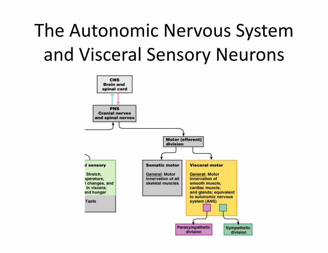
• The ANS – a system of motor neurons– The general visceral motor division of the PNS– Innervates smooth muscle, cardiac muscle, and glands
– Regulates visceral functions• Heart rate, blood pressure, digestion, urination . . .
The Autonomic Nervous System and Visceral Sensory Neurons
Comparison of Autonomic and Somatic Motor Systems
• Somatic motor system– One motor neuron extends from the CNS to skeletal muscle
– Axons are well myelinated, conduct impulses rapidly
• Visceral Motor (Autonomic nervous) system– Chain of two motor neurons
• Preganglionic neuron• Ganglionic neuron
– Conduction is slower due to thinly or unmyelinated axons
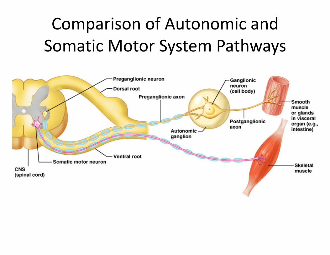
Comparison of Autonomic and Somatic Motor System Pathways
Divisions of the Autonomic Nervous System
• Sympathetic and parasympathetic divisions– Chains of two motor neurons
• Exhibits dual innervation– Nerves of both divisions innervate mostly the same structures
• Cause opposite effects
• Sympathetic – “fight, flight, or fright”– Activated during exercise, excitement, and emergencies– Concerned with liberating energy resources
• Parasympathetic – “rest and digest”– Concerned with conserving and storage of energy
Differences in ANS Divisions
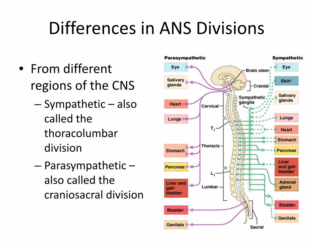
• From different regions of the CNS– Sympathetic – also called the thoracolumbar division
– Parasympathetic –also called the craniosacral division
Differences in ANS Divisions
• Length of postganglionic fibers– Sympathetic – long postganglionic fibers– Parasympathetic – short postganglionic fibers
• Branching of axons– Sympathetic axons – highly branched
• Influences many organs– Parasympathetic axons – few branches
• Localized effect
• Neurotransmitter released by postganglionic axons– Sympathetic – most release norepinephrine (adrenergic)– Parasympathetic – release acetylcholine
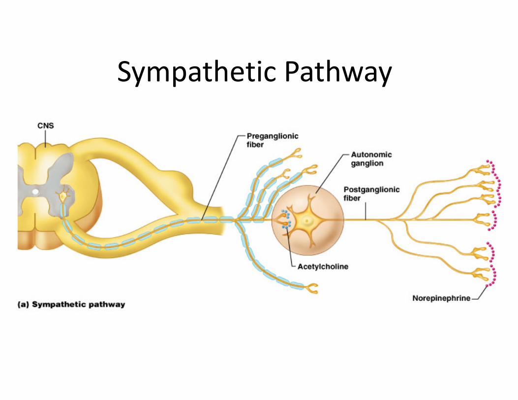
Sympathetic Pathway
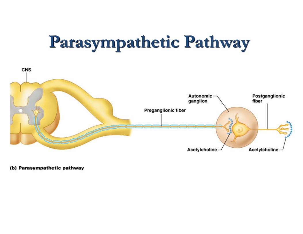
Parasympathetic Pathway
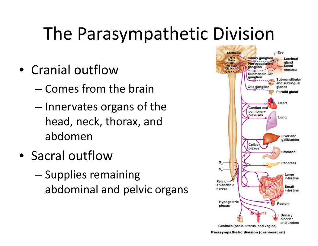
The Parasympathetic Division
• Cranial outflow – Comes from the brain– Innervates organs of the head, neck, thorax, and abdomen
• Sacral outflow – Supplies remaining abdominal and pelvic organs
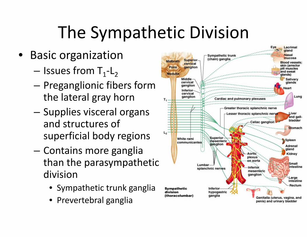
The Sympathetic Division• Basic organization
– Issues from T1‐L2– Preganglionic fibers form the lateral gray horn
– Supplies visceral organs and structures of superficial body regions
– Contains more ganglia than the parasympathetic division
• Sympathetic trunk ganglia• Prevertebral ganglia
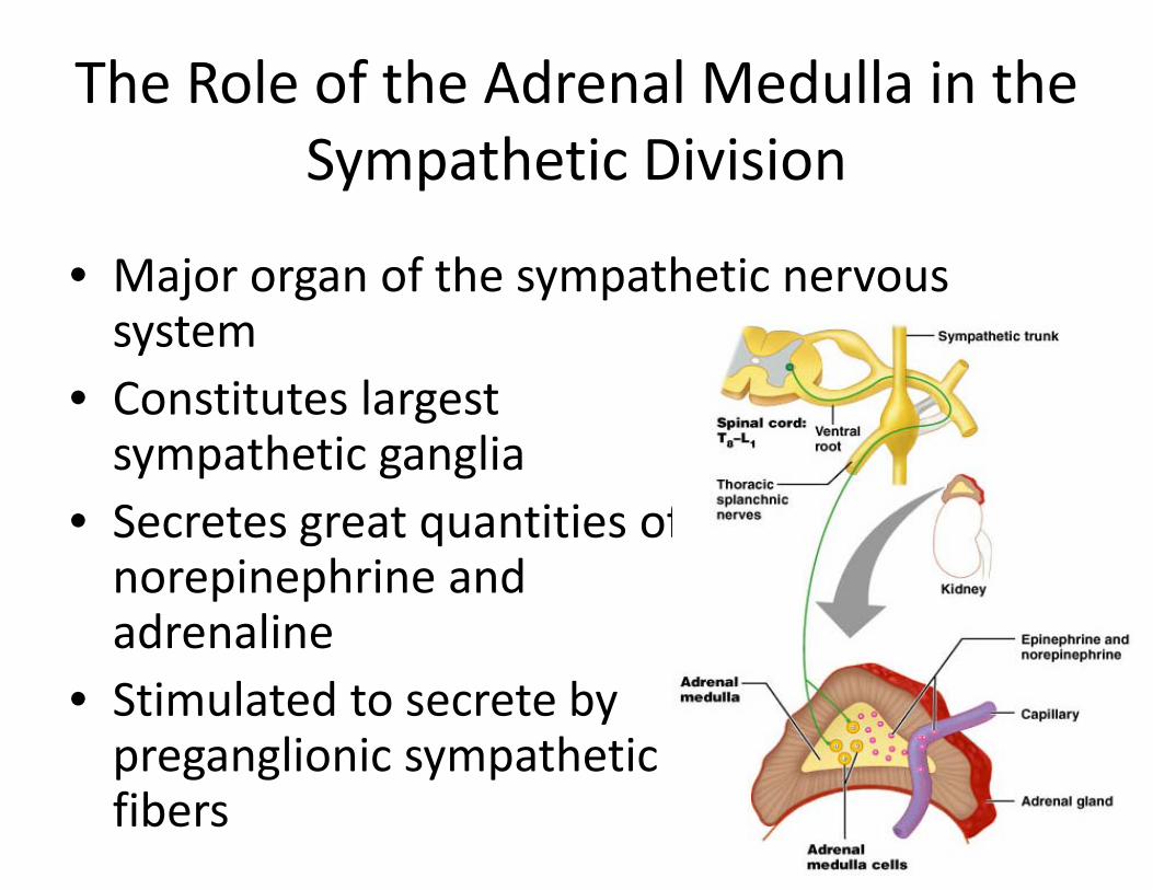
The Role of the Adrenal Medulla in the Sympathetic Division
• Major organ of the sympathetic nervous system
• Constitutes largest sympathetic ganglia
• Secretes great quantities of norepinephrine and adrenaline
• Stimulated to secrete by preganglionic sympathetic fibers
Visceral Sensory Neurons
• General visceral sensory neurons monitor:– Stretch, temperature, chemical changes, and irritation
• Cell bodies are located in the dorsal root ganglia
• Visceral pain – perceived to be somatic in origin– Referred pain
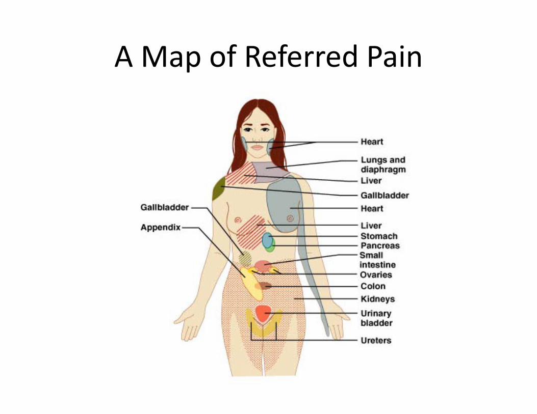
A Map of Referred Pain
Visceral Reflexes
• Visceral sensory and autonomic neurons– Participate in visceral reflex arcs
• Defecation reflex• Micturition reflex
• Some are simple spinal reflexes• Others do not involve the CNS
– Strictly peripheral reflexes
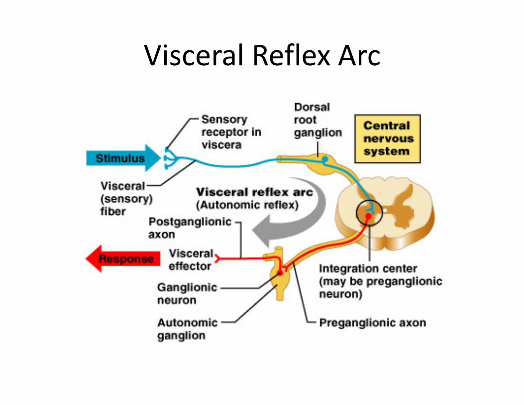
Visceral Reflex Arc
Special Senses
• Senses that have specific concentration of receptors– Vision– Hearing/Equilibrium– Smell– Taste
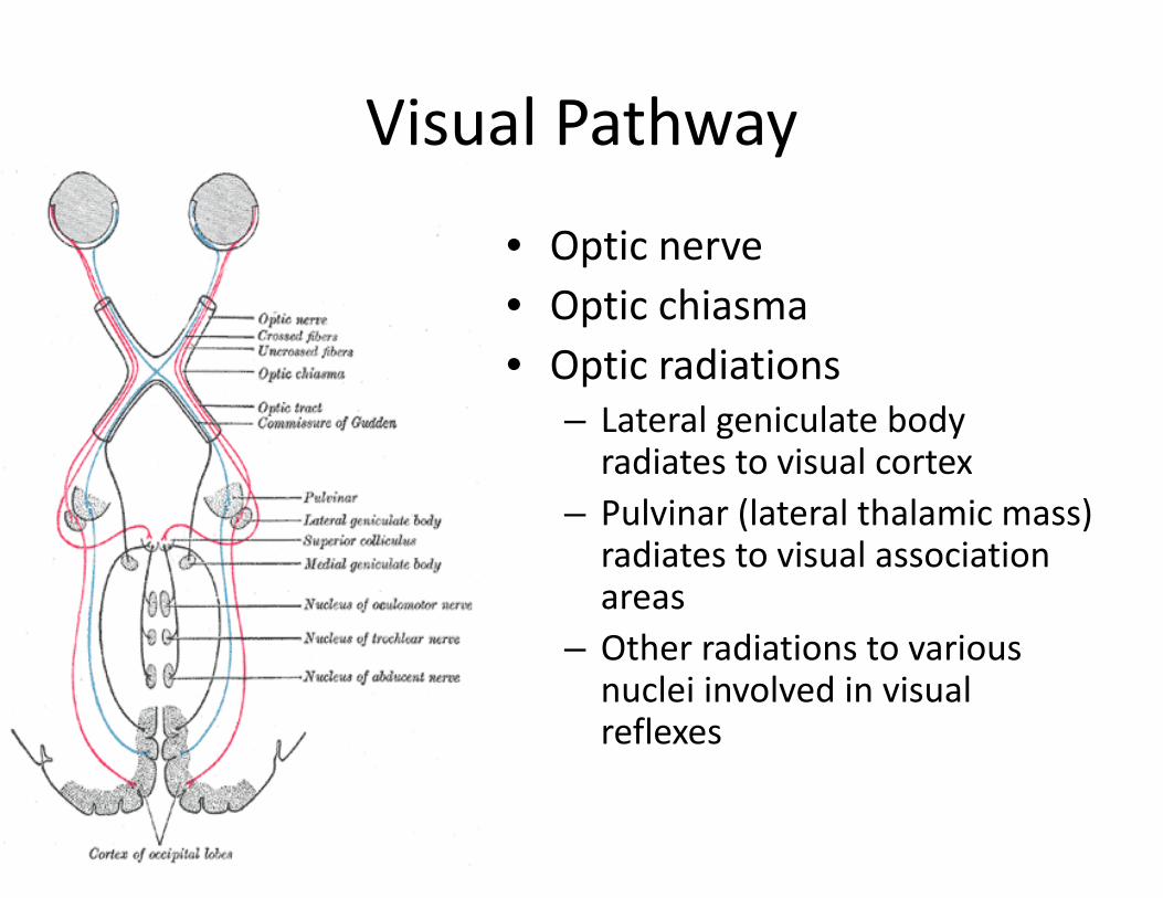
Visual Pathway
• Optic nerve• Optic chiasma• Optic radiations
– Lateral geniculate body radiates to visual cortex
– Pulvinar (lateral thalamic mass) radiates to visual association areas
– Other radiations to various nuclei involved in visual reflexes
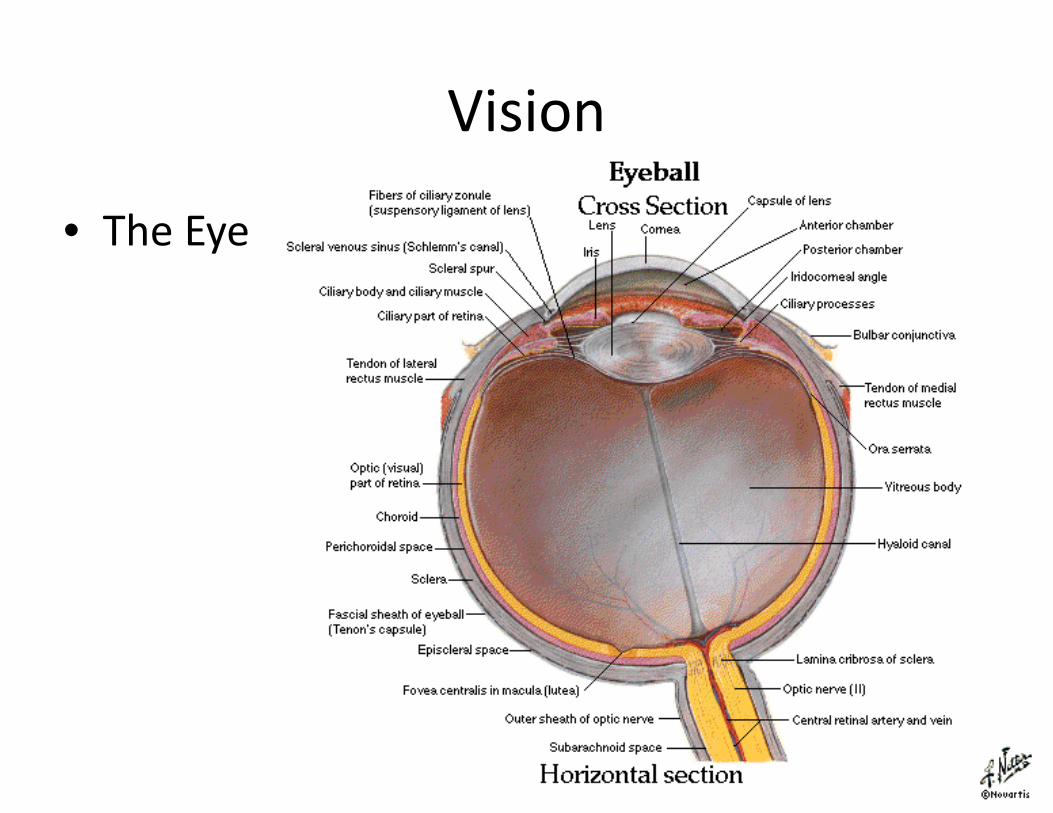
Vision
• The Eye
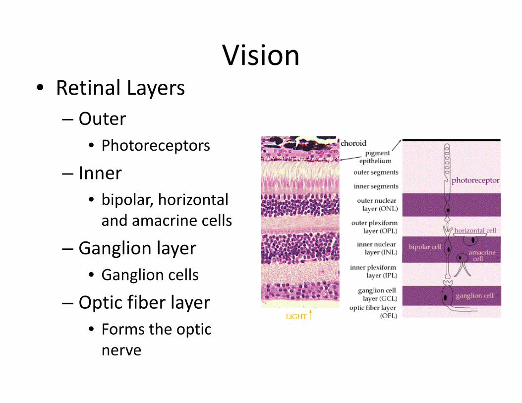
Vision• Retinal Layers
– Outer• Photoreceptors
– Inner• bipolar, horizontal and amacrine cells
– Ganglion layer• Ganglion cells
– Optic fiber layer• Forms the optic nerve
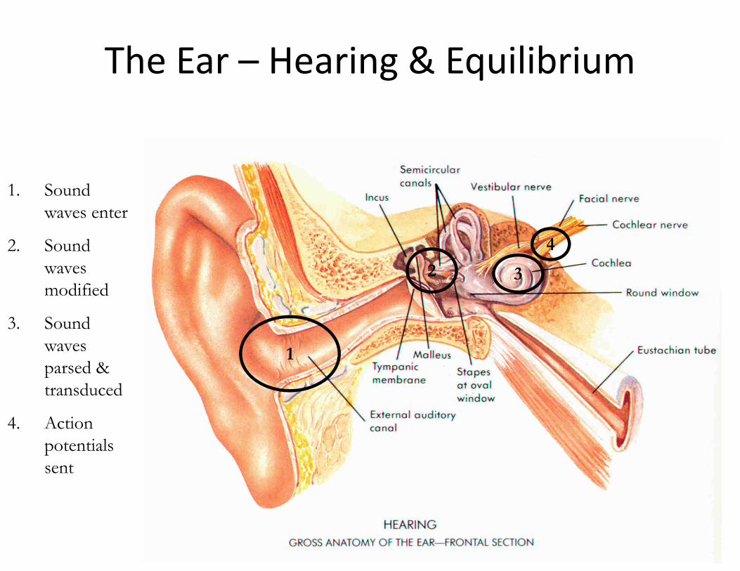
The Ear – Hearing & Equilibrium
1
2 3
1. Sound waves enter
2. Sound waves modified
3. Sound waves parsed & transduced
4. Action potentials sent
4
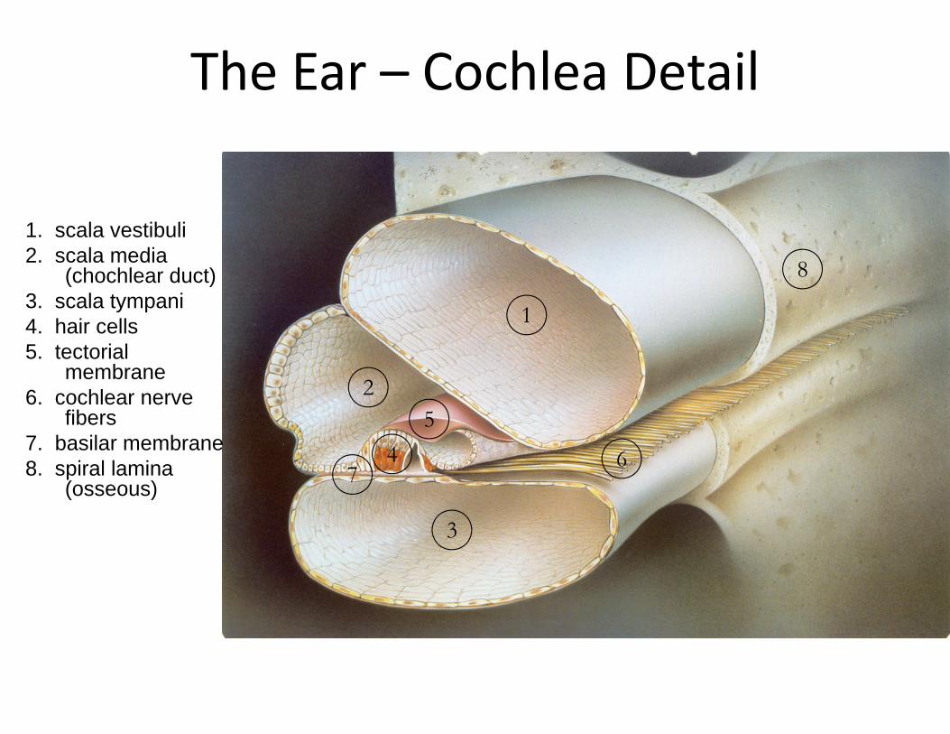
The Ear – Cochlea Detail
1. scala vestibuli2. scala media
(chochlear duct)3. scala tympani4. hair cells5. tectorial
membrane6. cochlear nerve
fibers7. basilar membrane8. spiral lamina
(osseous)
1
45
6
3
2
7
8
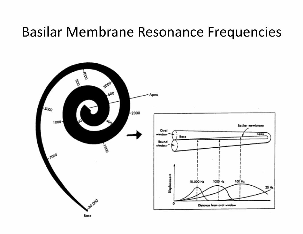
Basilar Membrane Resonance Frequencies
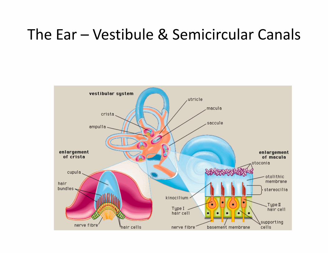
The Ear – Vestibule & Semicircular Canals
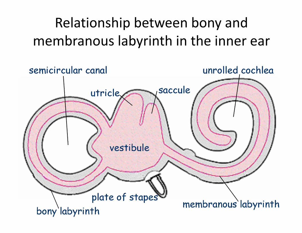
Relationship between bony and membranous labyrinth in the inner ear
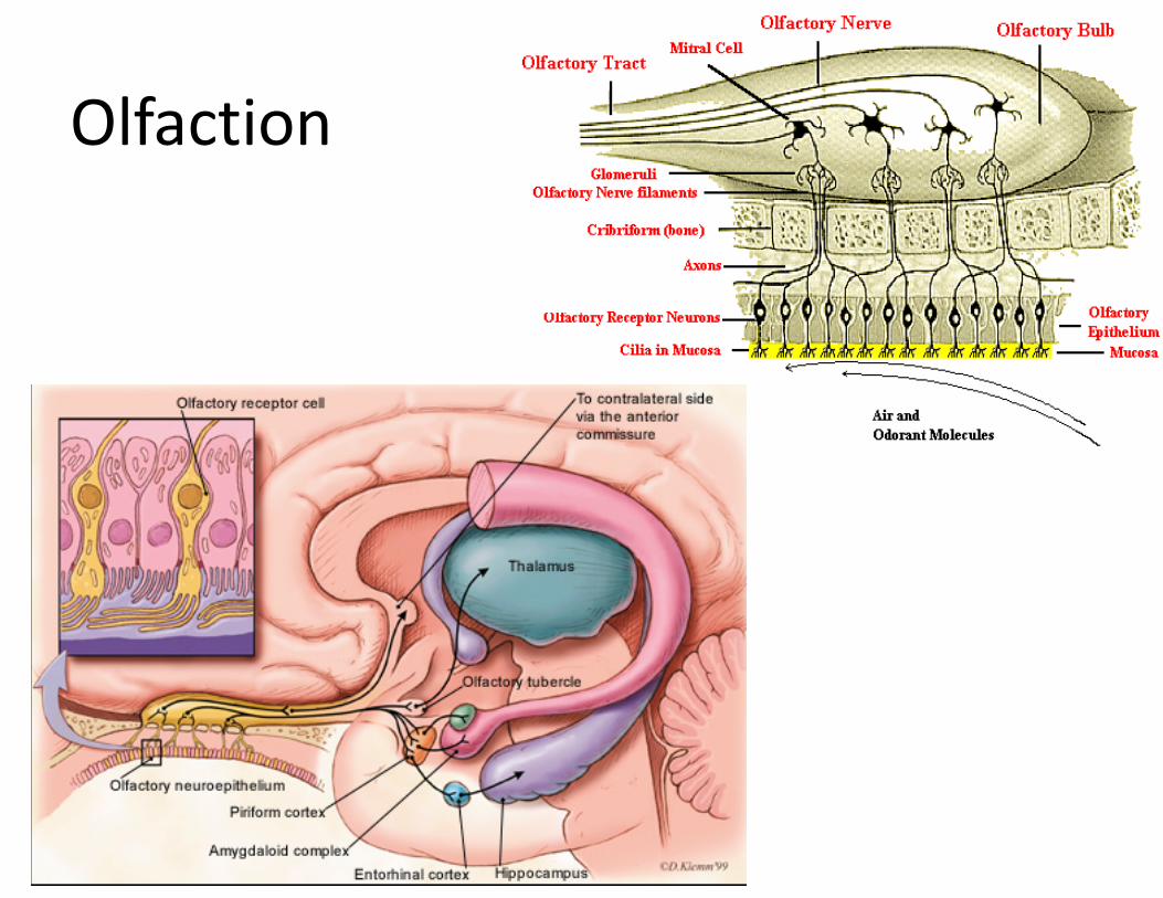
Olfaction
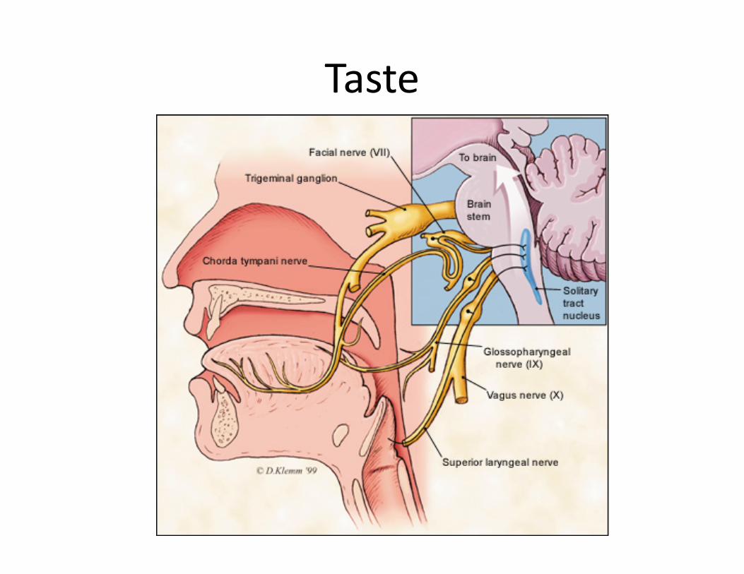
Taste
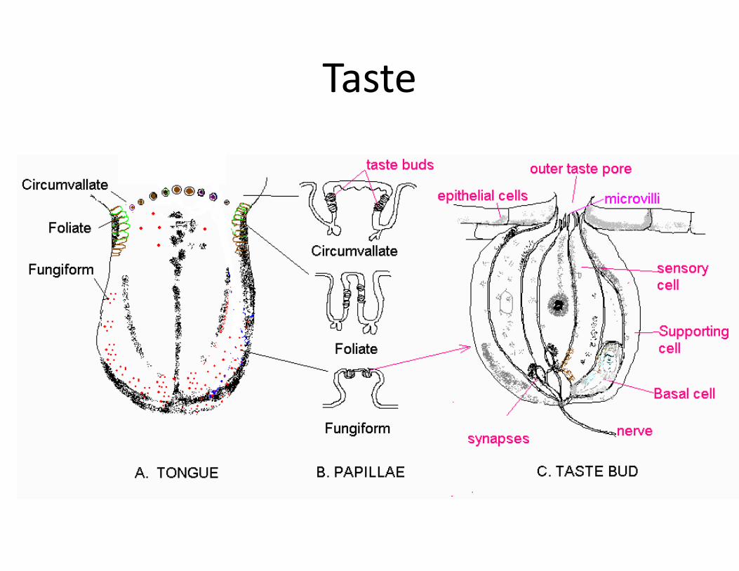
Taste