Embed Size (px)
Citation preview
1
The biofilm matrix scaffold of Pseudomonas species consists of non-1
canonically base paired extracellular DNA and RNA 2
Thomas Seviour1
Fernaldo Richtia Winnerdy2 Lan Li Wong
1 Xiangyan Shi
2 Sudarsan 3
Mugunthan1 Gurjeet Singh Kohli
1 Heather M Shewan
3 Jason R Stokes
3 Scott A Rice
145 Anh 4
Tuacircn Phan2 Staffan Kjelleberg
1 5 6 5
1 Singapore Centre for Environmental Life Sciences Engineering Nanyang Technological 6
University 637551 Singapore 7
2 School of Physical and Mathematical Sciences Nanyang Technological University 637371 8
Singapore 9
3 School of Chemical Engineering The University of Queensland 4072 Brisbane Australia 10
4 The iThree Institute The University of Technology Sydney Sydney 2007 Australia 11
5 School of Biological Sciences Nanyang Technological University 637551 Singapore 12
6 Centre for Marine Bio-Innovation School of Biological Earth and Environmental Sciences 13
University of New South Wales Sydney 2052 Australia 14
Correspondence to twseviourntuedusg 15
16
CC-BY 40 International licensecertified by peer review) is the authorfunder It is made available under aThe copyright holder for this preprint (which was notthis version posted January 22 2019 httpsdoiorg101101527267doi bioRxiv preprint
2
Abstract 17
While the array of emergent properties assigned to biofilms is extensive (eg antimicrobial 18
tolerance) the mechanisms that underpin these are largely unknown In particular the 19
extracellular matrix a defining feature of biofilms remains poorly understood in terms of its 20
composition and contribution to biofilm structure and function Here we demonstrate that 21
extracellular DNA exists in a complex with RNA that forms the main cross-linking exopolymer 22
of Pseudomonas biofilms and explains biofilm elasticity The RNA has a high purine content 23
and our solid-state NMR data indicate the formation of Hoogsteen guanine base pairs This may 24
suggest the presence of G-quadruplexes which is also corroborated by the enhancement of 25
biofilm formation in the presence of potassium The finding that non-canonical interactions 26
mediate networking of matrix-forming extracellular nucleic acids addresses how eDNA is 27
organized and contributes to matrix biophysical properties This understanding will allow for the 28
development of more effective biofilm control strategies 29
30
CC-BY 40 International licensecertified by peer review) is the authorfunder It is made available under aThe copyright holder for this preprint (which was notthis version posted January 22 2019 httpsdoiorg101101527267doi bioRxiv preprint
3
Introduction 31
Biofilms are key microbial ecosystems that contribute to bacterial pathogenicity (Phillips and 32
Schultz 2012) disrupt flow in water filtration systems (Drescher et al 2013) and facilitate 33
wastewater treatment bioprocesses (Seviour et al 2011) They represent bacterial adaptation 34
strategies allowing for increased antibiotic tolerance (Hoslashiby et al 2010) enhanced resource 35
capture (Kurniawan et al 2012) and the establishment of ecological microniches (de Kreuk et 36
al 2007) Such properties are unique to biofilms in contrast to planktonic bacteria and are not 37
mediated directly by the cells but instead by an extracellular polymeric matrix the cells secrete 38
(Flemming and Wingender 2010) 39
Exopolymer functions in biofilms have been studied extensively (Seviour et al 2012) 40
particularly for Pseudomonas aeruginosa which contributes to one in five clinical infections 41
(Bodey et al 1983) No fewer than eight exopolymers have been identified as supporting key 42
traits in P aeruginosa biofilms including three exopolysaccharides (Colvin et al 2012) four 43
proteins (Allesen-Holm et al 2006 Borlee et al 2010 Seviour et al 2015a) and extracellular 44
DNA (eDNA) (Okshevsky and Meyer 2015) Each putative exopolysaccharide (Colvin et al 45
2012) has been identified as a primary structural agent suggesting the existence of functional 46
redundancy Other exopolymers have multiple roles (Irie et al 2012) and a wide range of 47
secondary regulatory responses associated with P aeruginosa biofilm exopolymer expression 48
has been elucidated (Herbst et al 2015) 49
Despite the ambiguity of the contributions of individual exopolymers biofilm formation is the 50
result of these exopolymers changing the matrixrsquos viscous and elastic properties (ie 51
viscoelasticity) where viscosity refers to its fluid properties and elasticity its networked 52
properties Polymeric networking is a fundamental requirement for any biofilm (Chew et al 53
CC-BY 40 International licensecertified by peer review) is the authorfunder It is made available under aThe copyright holder for this preprint (which was notthis version posted January 22 2019 httpsdoiorg101101527267doi bioRxiv preprint
4
2014) We undertook to identify the foundation polymers in P aeruginosa biofilms which are 54
defined here as those that either dominate biofilm elasticity or constitute the primary structural 55
agents 56
Results 57
eDNA dominates the elastic response of Pseudomonas aeruginosa 58
To characterize the foundation polymer of P aeruginosa biofilms we exploited the reported 59
ability of the ionic liquid 1-ethyl-3-methyl-imidazolium acetate (EMIM-Ac) to dissolve a range 60
of recalcitrant biopolymers including DNA(Zhao 2015) and cellulose (Vitz et al 2009) which 61
led us to demonstrate this also for P aeruginosa biofilm exopolymers (Seviour et al 2015b) 62
Here when P aeruginosa biofilms were dissolved in EMIM-Ac the subsequent fluid was highly 63
viscoelastic We measured non-linear elasticity as a shear-rate dependent high normal stress 64
difference (N1-N2) where N1 and N2 are primary and secondary normal stress differences 65
respectively Elasticity dominated the viscous flow properties for the wild type biofilm in 66
EMIM-Ac with (N1-N2) an order of magnitude greater than shear stress (Figure 1A Wild type) 67
The solvent (EMIM-Ac) alone exhibited no elasticity indicating that the elastic properties are 68
transferred to the EMIM-Ac from the biofilm matrix Viscosity was slightly shear-thinning 69
(Supplementary Figure 1A and Supplementary Table 1) which would be expected from dilute 70
polymer solutions in viscous fluids (ie Boger fluids) (Scirocco et al 2005) (N1-N2) has a 71
power law dependence on the shear rate (n) of 14 (Supplementary Figure 1B Wild type) and 72
viscoelasticity was accurately modeled by the modified finitely-extensible nonlinear elastic 73
(FENE-P) polymer model (Figure 1A-B Supplementary Tables 1 and 2) The rheological 74
CC-BY 40 International licensecertified by peer review) is the authorfunder It is made available under aThe copyright holder for this preprint (which was notthis version posted January 22 2019 httpsdoiorg101101527267doi bioRxiv preprint
5
behavior is thus characteristic of solutions containing semi-flexible polymers such as 75
DNA(Stokes et al 2001) 76
The polysaccharides alginate Pel and Psl have been suggested as the structural scaffolds of P 77
aeruginosa biofilms (Colvin et al 2012) Their contribution to elasticity was determined using 78
the mucoid alginate over-expressing strain PDO300 and isogenic Pel and Psl genetic knockout 79
mutants (Ghafoor et al 2011) When Psl is absent the elasticity (ie N1-N2) and viscosity 80
increased relative to the wild type and the mucoid strain PDO300 which was less elastic than the 81
wild type despite the over-expression of alginate (Figure 1 ΔPsl) In contrast when Pel is 82
absent there was a slight decrease in both elasticity and viscosity relative to both the wild type 83
and PDO300 (Figure 1 ΔPel) 84
The contributions of proteins RNA and DNA were also explored using pronase RNase A and 85
DNase I respectively (Figure 1 Pronase RNase DNase) Removing each exopolymer 86
component individually decreased biofilm elasticity With the exception of the DNase I-treated 87
biofilm all treatments displayed n values of 09 ndash 17 (Supplementary Table 1) consistent with 88
semi-flexible polymers like DNA(Mansfield et al 2015) While viscosity was unchanged 89
following pronase treatment there was a slight decrease in elasticity This decrease in elasticity 90
was even greater following RNase treatment and elasticity was completely lost upon DNase 91
treatment with the normal stress difference reduced to zero Thus the elastic response of the 92
biofilm in EMIM-Ac can be primarily attributed to DNA 93
The fundamental rheology described here for the P aeruginosa biofilm therefore arises 94
principally from DNA This was elucidated for biofilms dissolved in EMIM-Ac as well as for 95
hydrated native biofilms displaying the same structural dependence on DNA (Supplementary 96
Figure 2) This approach of dissolving biofilm in EMIM-Ac to assess the contribution of 97
CC-BY 40 International licensecertified by peer review) is the authorfunder It is made available under aThe copyright holder for this preprint (which was notthis version posted January 22 2019 httpsdoiorg101101527267doi bioRxiv preprint
6
individual exopolymers to its properties is thus validated The data suggest that multiple 98
exopolymers contribute to the rheology with the matrix from the ΔPsl mutant being the most 99
elastic followed by wild type and RNase A-treated biofilms which were the least elastic It has 100
been noted previously that a range of P aeruginosa exopolymers influence biofilm extracellular 101
matrix crosslinking (Colvin et al 2012) and the data presented further clarifies that these 102
exopolymers can only modulate biofilm rheology if eDNA is present An eDNA scaffold is 103
therefore a prerequisite for this rheological differentiation of the matrix Further support for the 104
presence of an eDNA scaffold is provided in the micrograph of a biofilm stained with TOTO-1 105
for DNA visualization (Figure 1C) which shows DNA fibers in the extracellular matrix 106
107
Figure 1 (A) N1-N2 and (B) viscosity against shear rate for Pseudomonas aeruginosa biofilm wild type PDO300 108
ΔPsl ΔPel and pronase RNase A and DNase I digested wild type biofilm immediately following dissolution in 1-109
ethyl-3-methylimidazolium acetate (40 mgmL) at 25 degC 100 microm gap This is measured as a function of shear stress 110
CC-BY 40 International licensecertified by peer review) is the authorfunder It is made available under aThe copyright holder for this preprint (which was notthis version posted January 22 2019 httpsdoiorg101101527267doi bioRxiv preprint
7
from 10 to 1000 Pa (N1 ndashN2) is not described for DNase I digested biofilm in Figure 1A and Supplementary Figure 111
1B as their normal force (FN) is less than the resolution of the rheometer (ie 01 N) and set to zero for calculating 112
(N1 ndashN2) Both the N1-N2 and viscosity data are fitted with the FENE-P model a rigid dumbell model for polymer 113
solutions Fitting parameters are shown in Supplementary Table 2 (C) Micrograph of P aeruginosa biofilm DNA 114
stained green with TOTO-1 (scale bar 10 microm) (D) Phase separation of extracellular nucleic acids extracted from P 115
aeruginosa biofilms into a gel occurs upon transfer from 1-ethyl-3-methylimidazolium acetate into water 116
Furthermore EMIM-Ac did not lyse either biofilm or planktonic cells as indicated by the intact 117
cell morphology and the absence of phospholipids and lipopolysaccharides in EMIM-Ac 118
following biofilm dissolution (Supplementary Figure 3) Therefore it was concluded that the 119
DNA dissolved following treatment of P aeruginosa planktonic cells and biofilms with EMIM-120
Ac is extracellular and due to extraction of intracellular DNA 121
This finding that eDNA is a prerequisite for matrix building is consistent with recent studies 122
reporting the observation of eDNA in biofilm extracellular matrices (Jennings et al 2015) That 123
DNA dominated the elastic response would indicate eDNA is not an incidental matrix 124
component but instead a primary or foundation structural component of P aeruginosa biofilms 125
Isolated nucleic acids mimic gel-forming property of P aeruginosa biofilms 126
eDNA is known to complex and co-localize with exopolysaccharides Furthermore eDNA 127
sequencing shows that there is no bias towards any specific region of the chromosome (Turnbull 128
et al 2016) In contrast to intracellular chromosomal DNA which is supercoiled and folded for 129
example by HU proteins and topoisomerases and then relaxed by gyrases to allow replication 130
and transcription to occur the organization of eDNA remains elusive We contend that a 131
molecular understanding of how the eDNA is assembled and organized is key to answering how 132
and why DNA transforms from the chromosomal form to that found in the biofilm matrix 133
CC-BY 40 International licensecertified by peer review) is the authorfunder It is made available under aThe copyright holder for this preprint (which was notthis version posted January 22 2019 httpsdoiorg101101527267doi bioRxiv preprint
8
We recovered the eDNA from EMIM-Ac following biofilm dissolution by exploiting the ability 134
of perchloric acid to selectively precipitate DNA over protein (Supplementary Figure 4) 135
Following further purification by gel permeation chromatography the polymer phase-separated 136
into a gel upon transfer from EMIM-Ac into water (ie the gel isolate) mimicking the formation 137
of networks in native biofilms (Figure 1D) A higher Grsquo was recorded for the gel isolate than for 138
the P aeruginosa biofilm (Supplementary Figure 5) which is consistent with it having a higher 139
DNA concentration Furthermore DNase degraded the isolated gel into shorter DNA fragments 140
(Supplementary Figure 6) Other fractions including those not precipitated by perchloric acid 141
did not self-assemble into gels (Supplementary Figure 7A) Similarly calf thymus DNA did not 142
form gels when processed the same way either with or without added cations (Supplementary 143
Figure 7B) suggesting that this behavior is not a universal property of DNA 144
Nucleic acid conformation is preserved during isolation 145
Circular dichroism (CD) is an extremely sensitive spectroscopic technique for determining the 146
secondary structure of biomolecules particularly proteins and nucleic acids It was used here to 147
understand whether the nucleic acid (NA) conformation was modified during extraction and 148
isolation Unprocessed P aeruginosa biofilms displayed a major circular dichroism (CD) peak at 149
250-285 nm (Figure 2A) which is consistent with the presence of NA (Kypr et al 2009) and 150
this peak was also observed to dominate the CD spectrum of the gel isolate (Figure 2B) The 151
spectral trough at 2255-2265 nm is typical for proteins (Greenfield 2006) NA can also display 152
a trough in this region although the relative depth of the trough and its absence after proteolysis 153
and fractional precipitation against proteins (Supplementary Figure 4) suggest that it denotes 154
proteinaceous material Hence even though the CD spectrum shows that proteins are significant 155
CC-BY 40 International licensecertified by peer review) is the authorfunder It is made available under aThe copyright holder for this preprint (which was notthis version posted January 22 2019 httpsdoiorg101101527267doi bioRxiv preprint
9
components of the biofilm matrix they are not major contributors to biofilm elasticity (Figure 156
1A) 157
158
Figure 2 Circular dichroism (CD) spectra of (A) Pseudomonas aeruginosa biofilm and (B) extracellular nucleic 159
acids (NA) gel isolate at temperatures between 30 and 95degC (C) Amplitude of dominant NA peak CDmax (260-285 160
nm) from CD spectra of P aeruginosa biofilm (seen in (A)) and its extracted extracellular nucleic acid gel (seen in 161
(B)) from T = 30degC to T = 95degC 162
The CD spectrum of the isolated NA gel (Figure 2B) is dominated by the same NA peak that 163
appears in the biofilm CD spectrum To confirm this similarity CD melting experiments were 164
performed on both samples The resulting melting curves (Figure 2C) show that their maximum 165
absorbance values follow the same trend across the temperature profiles with a minimum 166
absorbance at 40degC a maximum at 60degC and only partial absorbance attenuation at 95degC NA 167
conformation was thus preserved during its isolation and the NA are more stable than typical 168
canonical DNA duplexes (Khandelwal and Bhyravabhotla 2010) The extracted NA gel isolate 169
is therefore an accurate proxy for understanding the intermolecular interactions that stabilize the 170
extracellular matrices of P aeruginosa biofilms Moreover the CD spectrum of the NA gel 171
isolate informs on the nucleic acid conformation In addition to the peak at 272 nm there is a 172
trough that shifts from 245 to 230 nm with increasing temperature another maximum at 215 nm 173
210 240 270 300
-40
-30
-20
-10
0
210 240 270 300-20
-10
0
10
20
30
30 oC
40 oC
60 oC
95 oCC
ircu
lar
dic
hro
ism
(m
de
g)
Circu
lar
dic
hro
ism
(m
de
g)
Wavelength (nm)
30 oC
40 oC
60 oC
95 oC
CB
Wavelength (nm)
A
30 40 50 60 70 80 90
26
28
30
32
34
36
38
NA gel
CD
max (
md
eg
) -
Iso
late
Temperature (oC)
1
2
3
4
5
6
Biofilm
CD
max (
md
eg
) -
Bio
film
CC-BY 40 International licensecertified by peer review) is the authorfunder It is made available under aThe copyright holder for this preprint (which was notthis version posted January 22 2019 httpsdoiorg101101527267doi bioRxiv preprint
10
that remains constant with temperature and other sub-maxima at 260 and 253 nm that change 174
with temperature The absence of a trough at 200-215 nm precludes the possibility of NA in A- 175
or Z- conformations (Kypr et al 2009) The dominant maximum and minimum could indicate 176
either B-DNA or G-quadruplex conformations (del Villar-Guerra et al 2018) although the peak 177
at 215 nm is not a feature of B-DNA CD spectra and is slightly higher than the characteristic G-178
quadruplex low wavelength peak of 210 nm Nonetheless the appearance of several peaks in the 179
NA region of 250-285 nm indicates that while the dominant NA conformation is unclear several 180
conformations likely contribute to phase separation of the NA 181
Purine-rich ribonucleotides and eDNA are present in isolated gel 182
Total correlation spectroscopy (TOCSY) and 1H-
13C heteronuclear single quantum coherence 183
(HSQC) are nuclear magnetic resonance (NMR) techniques that can identify proton NMR 184
correlations within individual ribose sugars and their proton-carbon single bond correlations 185
respectively Raising the pH can solubilize P aeruginosa biofilms (Friedman and Kolter 2004) 186
and here we show that after alkalinization the NA peaks dominated the solution 1H-
13C HSQC 187
spectra for the isolated material with no indication that proteins or polysaccharides were present 188
(Supplementary Figure 8 (Wuumlthrich 2008)) The 1H-
13C HSQC-TOCSY spectrum of the 189
isolated gel when dissolved by alkalinization shows two clusters of sugar proton peaks (C1rsquo-H1rsquo) 190
with correlations to neighboring carbons (C2rsquo-H1rsquo) (Figure 3A) 191
The first cluster (rectangles) has C2rsquo chemical shift values of ~40 ppm and the second cluster 192
(ovals) has C2rsquo values of ~70 ppm The 1H-
13C correlations denoted by the rectangles therefore 193
arise from deoxyribose and those denoted by the ovals from ribose sugar conformations The 194
broadened form of the deoxyribonucleotide peaks is consistent with high molecular weight 195
(MW) molecules while the sharpness of the ribonucleotide peaks suggests that they could arise 196
CC-BY 40 International licensecertified by peer review) is the authorfunder It is made available under aThe copyright holder for this preprint (which was notthis version posted January 22 2019 httpsdoiorg101101527267doi bioRxiv preprint
11
from small possibly even single nucleotides Hence eDNA and small ribonucleotides are both 197
likely to be present in the gel dissolved by alkalinization 198
Spin-systems for the eight sharp ribonucleotide peaks (Figure 3B) were assigned by HSQC-199
TOCSY and correlation spectroscopy (COSY) (Supplementary Figure 9) with absolute 200
identification achieved by comparing the heteronuclear multiple bond correlation HMBC spectra 201
of samples and monoribonucleotide standards (Supplementary Figure 10) The 31
P NMR 202
spectrum of the NA gel isolate at elevated pH revealed the presence of a mixture of 203
monoesterified (34 to 41 ppm) and diesterified phosphates (-08 to -12 ppm) indicating the 204
coexistence of monoribonucleotides and DNA respectively (Figure 3C (i)) Long-range 31
P-1H 205
correlations (ie from NA phosphorous to adjoined ribose proton) were observed in the 31
P-1H 206
heteronuclear correlation (HETCOR) spectrum (Figure 3C (ii)) for both monoribonucleotides 207
and DNA The broad 31
P-1H cross peaks at ~-10 ppm (Figure 3C (ii)) correspond to long-range 208
correlations between DNA phosphorous to H3rsquo and H5rsquoH5rsquorsquo protons The eight 209
monoribonucleotides were assigned to 2rsquo- and 3rsquo-(A U G and C)-monophosphates We 210
therefore have both 2rsquo and 3rsquo monoribonucleotides present with the eDNA in the gel solubilized 211
under alkaline conditions 212
However the monoribonucleotide peaks only became resolved upon alkaline dissolution of the 213
biofilm in contrast to the broad peaks evident at pH 7 (Supplementary Figure 11) This indicates 214
that the monoribonucleotides are derived from chain structures that exist at biological pH and 215
assemble with DNA chains into higher-order networked structures which is consistent with the 216
observation in Figure 1A that RNA is a major contributor to P aeruginosa biofilm elasticity 217
218
219
CC-BY 40 International licensecertified by peer review) is the authorfunder It is made available under aThe copyright holder for this preprint (which was notthis version posted January 22 2019 httpsdoiorg101101527267doi bioRxiv preprint
12
220
221
222
223
224
225
226
227
Figure 3 (A) Nuclear magnetic resonance (NMR) 1H-13C 228
heteronuclear single quantum coherence (HSQC)-total 229
correlation spectroscopy (TOCSY) spectrum at 25degC of 230
extracellular nucleic acids (NA) gel isolate after 231
alkalinization showing the C1rsquo-H1rsquo cross peaks of RNA 232
(blue ovals) and DNA (red rectangles) and their correlations 233
to the neighboring carbon C2rsquo-H1rsquo Schematics (right) 234
illustrate these correlations (B) 1H-13C HSQC spectrum of 235
extracellular NA gel isolate at 25degC after alkalinization 236
identifying eight monoribonucleic acid ribose spin systems 237
(C) 1-D 31P NMR of NA isolate gel and Pseudomomonas 238
aeruginosa biofilm with proton decoupling showing the 239
presence of monoesterified (ie monoribonucleotides) and 240
diesterified (ie DNA) phosphate peaks (i) and 2-D 1H-31P heteronuclear correlation (HETCOR) spectrum of 241
extracellular NA showing the 31P-1H cross-peaks of monoribonucleotides and DNA (ii) Couplings of 242
monoesterified phosphates to H2rsquo or H3rsquo of eight monoesterified monoribonucleotides (from left to right 3rsquo 243
AMP3rsquo GMP 3rsquo CMP3rsquo UMP 2rsquo CMP2rsquo UMP 2rsquo AMP 2rsquo GMP) and diesterified phosphate to DNA H3rsquo and 244
H5rsquoH5rsquorsquo protons are denoted by the dashed lines There is a discontinuity (wavy line) in the 31P axis due to the 245
different thresholds required to illustrate the 31P-1H correlations in the mono-esterified and di-esterified regions All 246
samples were prepared in 01 M NaOD (10 mgmL) and preheated to 55degC for 2 h 247
CC-BY 40 International licensecertified by peer review) is the authorfunder It is made available under aThe copyright holder for this preprint (which was notthis version posted January 22 2019 httpsdoiorg101101527267doi bioRxiv preprint
13
The molar ratio of the individual ribonucleotides could be determined from the 31
P spectrum of 248
the gel dissolved at high pH (Table 1) While several peaks could not be separated it was 249
possible to deduce that the RNA is purine rich (ie 57 mol A+G) and that the G+C mol 250
content of 46-50 differs from that of the P aeruginosa genome (ie 67 mol) (Shen et al 251
2006) The same peaks were also observed in the biofilm 31
P spectrum after alkalinization 252
(Figure 3C(i)) 253
Table 1 Relative abundances of monoribonucleotides in extracellular NA gel isolate from Pseudomonas 254 aeruginosa biofilm as determined by integrating 31P NMR spectrum following alkalinization 255
31P shift
(ppm)
Ribonucleotide Relative
abundance
403 3rsquoAMP 180
401 3rsquo GMP 172
390 3rsquo CMP3rsquo UMP 89
387 3rsquo CMP3rsquo UMP 113
368 2rsquo CMP2rsquo UMP 95
366 2rsquo CMP2rsquo UMP 117
350 2rsquo AMP 104
342 2rsquo GMP 130
256
Extracellular DNA and RNA interact to form a network 257
We applied Magic-angle spinning (MAS) solid-state NMR (SSNMR) to confirm that the 258
networks in the gel isolate and biofilm are comprised of DNA and RNA in chain form This 259
technique is ideal for intractable systems such as biofilms to eliminate solubility and extraction 260
biases (Reichhardt and Cegelski 2014) and to analyze inter-molecular H-bond interactions to 261
describe for example inter-nucleotide base pairing in RNA (Marchanka et al 2015) MAS 262
SSNMR averages anisotropic interactions to provide high-resolution spectral characterization of 263
insoluble and large biomolecular systems By analyzing dipolar interactions through-space 264
heteronuclear correlations (eg N∙∙∙H) can be detected In contrast to the liquid state 31
P NMR 265
spectrum of the alkali-dissolved isolate (Figure 3C (i)) the 31
P SSNMR spectrum of the gel 266
CC-BY 40 International licensecertified by peer review) is the authorfunder It is made available under aThe copyright holder for this preprint (which was notthis version posted January 22 2019 httpsdoiorg101101527267doi bioRxiv preprint
14
isolate (ie no alkali treatment Figure 4A (i)) showed a single peak in the diesterified phosphate 267
region consistent with the presence of both DNA and RNA chains There are no sharp peaks 268
present in the monoesterified phosphate region further demonstrating that the 269
monoribonucleotides are a consequence of alkali transesterification (Radak et al 2013) 270
Only the diesterified phosphate peak was observed in the 31
P SSNMR spectrum of the biofilm 271
(Figure 4A (iii)) while both RNA-derived monoesterified and DNA-derived diesterified 272
phosphate peaks were present in the 31
P SSNMR spectrum of alkali digested NA gel-isolate and 273
biofilm (lyophilized) (Figure 4A (ii) and Figure 4A (iv) respectively) Complete alkali RNA 274
transesterification was confirmed by the full conversion of diesterified to monoesterified 275
phosphates (from liquid state 31
P NMR spectra) for the RNA standard when dissolved in 01 M 276
NaOH (Supplementary Figure 12A) Conversely RNA diesterified phosphate peaks were 277
preserved in the RNA standard spectrum following dissolution in EMIM-Ac and recovery by 278
perchloric acid (Supplementary Figure 12B) Hence the alkaline conditions break down the 279
RNA chains into individual monoribonucleotides while the ionic liquid-based extraction does 280
not This suggests that the conventional method for P aeruginosa biofilm dissolution in alkali 281
(Friedman and Kolter 2004) may in fact work by transesterifying RNA as a primary structural 282
component and illustrates the importance of the ionic liquid-based extraction protocol described 283
here as a non-destructive method for interrogating biofilm structural polymers 284
CC-BY 40 International licensecertified by peer review) is the authorfunder It is made available under aThe copyright holder for this preprint (which was notthis version posted January 22 2019 httpsdoiorg101101527267doi bioRxiv preprint
15
285
Figure 4 (A) Solid-state 31P NMR spectra at T = 25degC of extracellular NA gel isolate (i) alkalinized and 286
lyophilized NA gel isolate (ii) Pseudomonas aeruginosa biofilm (iii) and alkalinized and lyophilized P aeruginosa 287
biofilm (iv) showing the presence of diesterified phosphate peaks and the absence of monoesterified phosphate 288
peaks for both NA gel isolate and biofilm in double distilled water and the coexistence of diesterified and 289
monoesterified phosphate peaks for both samples after alkalinization This indicates that alkalinization of the matrix 290
results in RNA transesterification Micrographs of P aeruginosa NA gel isolate stained green with (B) SYTO 291
RNASelect showing RNA fibers and (C) TOTO-1 showing DNA fibers (scale bar 10 microm) 292
293
To visualize the DNA-RNA network organization we used nucleic acid-specific stains ie RNA 294
specific SYTO RNASelect dye (Figure 4B) and the eDNA-specific TOTO-1 dye (Figure 4C) 295
(Okshevsky and Meyer 2014) Fibrous structures which are typical of networked polymer gels 296
(Cornwell and Smith 2015) were observed in the NA gel isolate stained with both nucleic acid 297
dyes as well as in the biofilm stained with the eDNA specific dye (Figure 1C) This is consistent 298
with our observation that the biofilm is readily degraded by DNase I (Figure 1A) (Chen et al 299
2004) Furthermore the NA gel isolate was stained positively with the RNA-specific dye even 300
after DNase I digestion providing further evidence that RNA contributes to networking 301
BeRNA fibers
C
eDNA fibers
A
CC-BY 40 International licensecertified by peer review) is the authorfunder It is made available under aThe copyright holder for this preprint (which was notthis version posted January 22 2019 httpsdoiorg101101527267doi bioRxiv preprint
16
(Supplementary Figure 13) However digestion with RNases III A and H did not degrade the 302
biofilm (Figure 1A Supplementary Figure 14) This may suggest that the RNase binding was 303
shielded by hairpins in the network or the presence of non-canonical DNA-RNA interactions 304
(Geerts-Dimitriadou et al 2012 Nakamura et al 1991) 305
Non-canonical and Watson-Crick base pairs and tetrads support extracellular network 306
Sequence analysis of the extracted material indicated that the gene coverage was even for both 307
chromosomal and extracellular DNA with the exception of bacteriophage Pf1 genes 308
(Supplementary Figure 15) However the Pf1 knockout mutant of P aeruginosa also displayed 309
an elastic response when dissolved in EMIM-Ac (Supplementary Figure 16 Supplementary 310
Tables 1 and 2) indicating that Pf1 DNA is unlikely to be responsible for the phase-separating 311
behavior of P aeruginosa biofilms DNA and RNA interaction therefore cannot be explained by 312
uneven gene coverage To elucidate the mechanism of DNA-RNA gelation we generated a 2D 313
through-space 15
N-1H HETCOR spectrum of
15N
labeled DNA-RNA gel isolated from a P 314
aeruginosa biofilm matrix (Figure 5A) This spectrum was run with longer cross polarization 315
contact times so that correlations between distant 1H and
15N could be observed This confirmed 316
the complete absence of proteins and supported our conclusion that proteins do not contribute to 317
the DNA-RNA interaction The HETCOR spectrum showed four signal clusters at δH 10-14 318
ppm and δN 140-160 ppm (Figure 5A) which arose from direct N-H couplings of TU and G 319
nucleobase imino groups Two of these clusters (δH 12-14 ppm) resulted from imino protons 320
hydrogen-bonded to a nucleobase nitrogen (ie N-H∙∙∙N) and the other two (δH 10 -12 ppm) 321
from imino protons hydrogen-bonded to a nucleobase carbonyl oxygen (ie N-H∙∙∙O) Due to the 322
longer cross polarization times we were able to observe a strong long-range and indirect (ie 323
intermolecular) correlation at δN 196 ppm arising from G-C Watson-Crick base pairing ie 324
CC-BY 40 International licensecertified by peer review) is the authorfunder It is made available under aThe copyright holder for this preprint (which was notthis version posted January 22 2019 httpsdoiorg101101527267doi bioRxiv preprint
17
C(N3)-G(H1) There was also a weak and indirect correlation at δN 220 ppm resulting from A-325
TU Watson-Crick base pairing ie A(N1) to UT(H1) 326
327
328
Figure 5 (A) Solid-state 2D 1H-15N through-space heteronuclear correlation (HETCOR) spectrum of extracellular 329
nucleic acid (NA) gel isolate in double distilled water (2 mg) T = 25degC showing direct N-H couplings for G imino 330
N (N3) and TU imino N (N3) bonded to nucleobase nitrogen (NHN) This indicates Watson-Crick (WC) G-C and 331
UT-A base pairings respectively and for G imino N (N1) and TU imino N (N3) bonded to nucleobase carbonyl 332
oxygen (ie NHO) indicating non-canonical G-G G-TU and UT-UT base pairings or tetrads The spectrum also 333
displayed indirect N-H correlations for the WC base pairs (ie C(N3) to G(H1) and A(N1) to UT(H1)) and no 334
indirect correlations for the non-canonical bonded nucleobases The threshold for the A(N1)-TU(H3) correlation 335
was increased due to lower signal intensity for that particular coupling (B) Pseudomonas aeruginosa biofilm 336
growth yield at 37degC (5 d) as a function of [K+] [Na+] or [Li+] showing the growth dependence on monovalent 337
cation concentration (K+gtNa+gtLi+) Error bars indicate standard deviation (C) Illustration of how structural 338
variability of RNA may allow for the formation of extracellular RNA and DNA networks through hairpins and a 339
mixture of canonical and non-canonical base pairs or tetrads (ie Hoogsteen and G-quadruplexes) 340
341
The absence of long-range correlations between the clusters at δH 10 -12 ppm is consistent with 342
non-Watson-Crick pairings for the G and TU nucleobases The observation of NH to O 343
interactions suggests the formation of G-G TU-TU G-TU Hoogsteen base pairs or tetrads 344
CA B C
CC-BY 40 International licensecertified by peer review) is the authorfunder It is made available under aThe copyright holder for this preprint (which was notthis version posted January 22 2019 httpsdoiorg101101527267doi bioRxiv preprint
18
Hoogsteen H-bonded G-bases can assemble planarly into tetrads and self-stack to form G-345
quadruplexes (Haumlnsel-Hertsch et al 2017) G-quadruplexes are stabilized by the presence of a 346
monovalent cation with potassium providing the greatest stability We experimentally confirmed 347
that potassium had a similar effect in a static biofilm growth assay We found that potassium 348
promoted P aeruginosa biofilm growth to a greater extent than either sodium or lithium (Figure 349
5B) Additionally this was a biofilm-specific effect as there was no effect of monovalent cation 350
on planktonic growth (Supplementary Figure 17) Based on the SSNMR data coupled with the 351
potassium effect and the earlier observation of several maxima in the CD spectrum of the NA gel 352
isolate across the characteristic NA region of 250-285 nm which suggested the coincidence of 353
several NA conformations contributing to phase separation it is proposed that G-quadruplexes 354
contribute to extracellular DNA-RNA networking 355
Discussion 356
We extracted a gel-forming complex composed of DNA and RNA from the extracellular matrix 357
of P aeruginosa biofilms without disrupting its fundamental structural or chemical organization 358
We report the novel observation that extracellular RNA contributes structurally to biofilms 359
While gel-forming exopolymers have been detected in other biofilms (Seviour et al 2009) no 360
functional DNA gel has been described for any biological system much less one also including 361
extracellular RNA 362
While it is not possible to identify the precise nature of the DNA-RNA interaction based on the 363
information presented here purine-rich RNA as described here for the extracellular NA gel 364
isolate (57 mol A-G) is a characteristic of DNA-RNA duplexes because RNA purines bind 365
more strongly to their DNA pyrimidine complement than vice versa (Nadel et al 2015) Both 366
Watson-Crick and non-canonical interactions were present in the NA gel isolate which is also 367
CC-BY 40 International licensecertified by peer review) is the authorfunder It is made available under aThe copyright holder for this preprint (which was notthis version posted January 22 2019 httpsdoiorg101101527267doi bioRxiv preprint
19
consistent with the discrepancy observed between RNA and DNA G+C mol contents (Table 368
1) It is possible therefore that interactions between DNA and RNA as described in this study 369
account for the formation of a highly stable nucleic acid gel 370
Non-canonical base pairing and hairpinning are commonly seen with RNA and cause multivalent 371
intermolecular interactions between RNA strands that lead to a sol-gel transition (Jain and Vale 372
2017) The greater structural versatility of RNA could be a key to the ability of the eDNA to 373
form networks with hairpins as well as canonical and non-canonical base pair interactions all 374
contributing to the assembly of higher order NA structures (Figure 5C) RNA has recently been 375
found to promote intracellular phase separations although this has not been extended to the 376
extracellular domain of prokaryotic cells (Shin and Brangwynne 2017) Additionally G-rich 377
RNA sequences are more predisposed to forming G-quadruplexes because they do not have a 378
complementary strand and in fact form more stable G-quadruplexes than DNA (Guo and Bartel 379
2016) It is therefore probable that the RNA but not DNA enables DNA-RNA networking and 380
gelation 381
The results presented here provide unprecedented resolution of the biofilm exopolymeric matrix 382
and its key foundation structural components We have developed a methodology that preserved 383
the molecular organization of the foundational polymer in its native state upon extraction and 384
isolation This enabled us to use SSNMR to describe intermolecular associations at the atomic 385
level 386
Our findings that a DNA-RNA gel network can provide the foundation for biofilm matrices in P 387
aeruginosa mark a departure from the prevailing paradigm that biofilm gelation is only due to 388
polysaccharides The elastic and film-forming properties were also observed for Pseudomonas 389
putida and Pseudomonas protegens (Figs S16 and S18 Supplementary Tables 1 and 2) 390
CC-BY 40 International licensecertified by peer review) is the authorfunder It is made available under aThe copyright holder for this preprint (which was notthis version posted January 22 2019 httpsdoiorg101101527267doi bioRxiv preprint
20
suggesting that eDNA is broadly important for biofilm formation in members of this genus In 391
addition to Pseudomonas spp other organisms are known to have eDNA including clinical 392
organisms Staphylococcus aureus (Mann et al 2009) Staphylococcus epidermidis (Adam et al 393
2002) and Mycobacterium abscessus (Rose et al 2015) as well as environmental isolates such 394
as strain F8 from the South Saskatchewan River (Boumlckelmann et al 2006) 395
While eDNA is commonly thought to be the product of cell lysis and has been shown to be 396
released by a sub-population of lytic P aeruginosa cells (Turnbull et al 2016) there is an 397
increasing awareness that it serves an important structural function such as in activated sludge 398
granules (Cheng et al 2011) Additionally Bockelmann et al (2006) observed that stable 399
filamentous networks produced by the aquatic strain F8 were comprised of DNA This is the first 400
study that provides an explanation for how eDNA is organized structurally However the 401
environmental factors contributing to eDNA release and how this is regulated (ie whether it is 402
an active or passive process) are still unclear It is possible that DNA-RNA foundational gel 403
networks as described here for the Pseudomonads are broad-scoping phenomena in biofilms 404
and that interactions with RNA enable an extracellular structural function for DNA Thus 405
elucidating how nucleic acids including RNA are integral to the biophysical and other emergent 406
properties imparted on the biofilm via the extracellular matrix will inform the regulation and 407
control of extracellular nucleic acid release across environmental and clinical biofilms 408
Materials and Methods 409
Bacterial strains 410
Unless otherwise stated all experiments were undertaken directly on P aeruginosa PAO1 411
biofilms grown in lysogeny broth at 37degC The P aeruginosa PAO1 Δpf4 knockout mutant is a 412
CC-BY 40 International licensecertified by peer review) is the authorfunder It is made available under aThe copyright holder for this preprint (which was notthis version posted January 22 2019 httpsdoiorg101101527267doi bioRxiv preprint
21
defined Pf4 chromosomal deletion mutant of the entire Pf4 prophage genome(Rice et al 413
2009) 414
P aeruginosa PDO300 PDO300(Δpel) and PDO300(Δpsl) mutant strains were gratefully 415
received from Professor Bernd H A Rehm Institute of Molecular Biosciences Massey 416
University New Zealand (Ghafoor et al 2011) PDO300 PDO300(Δpel) PDO300(Δpsl) and 417
PAO1 (Δpf4) were also grown in LB at 37degC P protogens Pf-5 and P putida ATCC BAA-477 418
and S12 strain were were grown in LB at 30degC 419
Biofilm growth assay 420
Ten milliliter aliquots of P aeruginosa planktonic pre-cultures (LB 200 rpm 37degC OD600 240 421
16 h) were diluted 50 times with LB in 2 L Erlenmeyer flasks and incubated for 5 d under static 422
conditions Supplementary Figure 15A displays an image of 5-d old biofilm in LB The cultures 423
were centrifuged at 10000 x g for 15 min the supernatant removed by decanting and the 424
biofilm then collected and lyophilized (LabConco) Supplementary Figure 15B displays an 425
image of the culture after centrifugation showing clear separation of biofilm and supernatant 426
Enzymatic digestions 427
Twenty milligrams of lyophilized biofilm was resuspended in 1 mL of either i) RNase buffer (50 428
mM Tris HCl 10 mM EDTA pH8) with 02 mg RNaseA from bovine pancreas (Sigma Aldrich) 429
ii) storage buffer (10 mM NaCl 10 mM Tris-HCl) with 01 mg Pronase E from Streptomyces 430
grisens (Sigma Aldrich) with 05 (vv) iii) 1X RNase H reaction buffer (20 mM Tris-HCl pH 431
78 40 mM KCl 8 mM MgCl2 1 mM DTT) with 04 mg RNase H (Thermo Fisher Scientific) 432
iv) 1X RNase III reaction buffer (500 mM NaCl 100 mM Tris pH 79 100 mM MgCl2 10 mM 433
DTT) with 06 mg RNase II (Thermo Fisher Scientific) or v) DNase I buffer (100mM Tris (pH 434
75) 25 mM MgCl2 and CaCl2) with 02 mg DNAse I from bovine pancreas (Sigma Aldrich) All 435
CC-BY 40 International licensecertified by peer review) is the authorfunder It is made available under aThe copyright holder for this preprint (which was notthis version posted January 22 2019 httpsdoiorg101101527267doi bioRxiv preprint
22
digestions were performed with shaking at 200 rpm at 37degC for 16 h The suspensions were then 436
centrifuged (10000 x g 15 min) the supernatant was discarded and the pellets of the biofilm 437
materials were lyophilized 438
Normal force measurement 439
Forty milligrams per milliliter solutions of lyophilized biofilms were added to 1 mL 1-ethyl-3-440
methylimidazolium acetate and incubated in 55degC for either 2 h A Haake Mars 3 (Thermo 441
Fisher Scientific) stress-controlled rotational rheometer with Peltier controlled element at 25degC 442
was used for rheological measurements Thirty five-illimeter diameter parallel plate geometry 443
was used with smooth titanium plates to measure viscosity and normal stress difference (N1 ndash 444
N2) Prior to measurement the gap error was zeroed at 4 N and gap error calculated as previously 445
described (Bird et al 1987 Davies and Stokes 2008 Kravchuk and Stokes 2013) One 446
hundred microliters of sample was deposited on the plates The plates were closed to 100 microm the 447
sample trimmed and the sample allowed to sit for 5 min prior to measurement All measurements 448
with Normal force (FN) less than the resolution of the rheometer (ie lt 01 N) were set to 0 449
before calculation of N1-N2 using equations 9-11 and viscosity from equation 3-5 in Davies and 450
Stokes(Davies and Stokes 2008) for the parallel plate geometry 451
Only the linearly increasing portion of the normal stress difference curves are presented Above 452
this range normal stress difference begins to decrease again which may be due to elastic 453
instabilities or associating polymers (Annable et al 1993) Corrections were made to N1 and N1 454
ndash N2 to account for inertia using equation 17 in Davies and Stokes (Davies and Stokes 2008) and 455
to correct for the baseline residual force in the samples Except for the DNase I-treated biofilm 456
the shear rheology for all treatments could be modelled using the finitely extensible non-linear 457
elastic with Gaussian closure proposed by Peterlin (FENE-P) constitutive model by varying four 458
CC-BY 40 International licensecertified by peer review) is the authorfunder It is made available under aThe copyright holder for this preprint (which was notthis version posted January 22 2019 httpsdoiorg101101527267doi bioRxiv preprint
23
parameters to fit shear viscosity and normal stress difference as a function of shear rate 459
(Supplementary Table 2) Fitting parameters for the FENE-P model include 1 = relaxation time 460
b = a measure of the relative extensibility of the model spring s = solvent viscosity p = 461
polymer contribution to the viscosity The FENE-P equations can be written in the following 462
format as shown by Bird et al(1987) 463
120578 = 120578119904 +120578119901
1205821((1198622 + 1198621)
1
3 minus (1198622 minus 1198621)1
3) (Equation 1) 464
465
1198731 = 2120578119901
1205821((1198622 + 1198621)
1
3 minus (1198622 minus 1198621)1
3)2
(Equation 2) 466
Where 467
1198621 = 119887
41205821 (Equation 3) 468
1198622 = (11986212 + (
119887+3
6)
3
)12
(Equation 4) 469
470
All measurements were performed in triplicate For clarity one representative data set is 471
presented in Figure 1A-B and Supplementary Figure 1A-B with the respective Power Law and 472
FENE-P model fit to that data set Averaged values for the FENE-P and Power law fits with the 473
standard deviation across three replicates are shown in Supplementary Tables 1 and 2 474
Oscillatory measurements were carried out with controlled frequency 01 rads across an 475
amplitude range of 001 to 10 476
Extracellular polymeric substances (EPS) extraction 477
Lyophilized biofilms were dissolved in ionic liquid mixture (40 (vv) 1-ethyl-3-478
methylimidazolium acetate (EMIM Ac) 60 (vv) NN-dimethyl acetamide (DMAc)) at 55degC 479
CC-BY 40 International licensecertified by peer review) is the authorfunder It is made available under aThe copyright holder for this preprint (which was notthis version posted January 22 2019 httpsdoiorg101101527267doi bioRxiv preprint
24
for 16 h The solution was centrifuged (10000 x g) to remove any undissolved material 480
Perchloric acid (70) was added (005 vv) to the viscosified centrate (on ice) After 15 min 481
incubation the solution was centrifuged at 10000 x g at 4degC for 15 min and the pellet recovered 482
This was repeated on the centrate two to four times until the solution was not viscous The 483
precipitate was dialysed against double distilled water for 2 d at 4degC (SnakeSkintrade Dialysis 484
Tubing 35K MWCO 22 mm) and the retentate lyophilized (FreeZone Plus 45 Liter Cascade 485
Benchtop Freeze Dry System) The same procedure was performed on calf thymus DNA lipase 486
cytochrome C for the purposes of determining recovery yield of representative exoproteins and 487
on RNA from torula yeast for assessing by 31
P NMR whether the extraction procedure 488
contributed to RNA transesterification (all from Sigma Aldrich) 489
Extracellular nucleic acid isolation 490
Twenty milligrams of lyophilized retentate (ie post perchloric acid precipitation) were dissolved 491
in 1 mL of 40 (vv) EMIM-Ac 60 DMAc (vv) (55degC 16 h) Chromatographic separation 492
was achieved in a Shimadzu system comprising DGU-20A 3r Prominence Degasser and LC-493
20AD Solvent Delivery Unit fitted with two Agilent PLgel 10 μm column of 105Aring pore size for 494
separation across the MW range 200 kDa to 2000 kDa The eluent flow rate was 30 mLmin-1
495
and the injection volume 1 mL The fractions with molecular weight range of 2000-800kDa and 496
800-200kDa were pooled and dialyzed for 2 d at 4degC (SnakeSkintrade Dialysis Tubing 35K 497
MWCO 22 mm) against double distilled water to induce gelation The gel was then collected 498
from the dialysis tubing 499
Solution-state nuclear magnetic resonance (NMR) 500
Solution-state NMR experiments were performed on an 800 MHz Bruker Avance III 501
spectrometer at 25oC Sample concentration was 10 mg (dry weight)mL
-1 unless otherwise 502
CC-BY 40 International licensecertified by peer review) is the authorfunder It is made available under aThe copyright holder for this preprint (which was notthis version posted January 22 2019 httpsdoiorg101101527267doi bioRxiv preprint
25
specified Spectra were recorded either under conditions of neutral pH in 100 D2O (Cambridge 503
Isotope Laboratories) or following alkalinization (ie transesterification 01 M NaOD 55oC 2 504
h) 1-D NMR experiments include 1H and
31P direct detection while 2-D NMR analysis include 505
13C-HSQC
13C-HSQC-TOCSY
1H-
31P HETCOR HMBC and COSY All spectral analyses 506
were performed using Topspin and SPARKY software 507
Asolectin (Sigma Aldrich) standard (10 mgmL) and lyophilized Pseudomonas aeruginosa 508
PAO1 biofilm (10mgmL) were dissolved in 40 (vv) EMIM Ac 60 (vv) DMAc at (55oC 2 509
h) P aeruginosa PAO1 pre-culture cell lysate was prepared by lysing pre-culture cells with 510
lysozyme in PBS 10 (vv) of D2O was added to all samples for locking purposes 511
Solid-state NMR 512
For solid-state NMR experiments performed on the NA gel isolate 15
N labeled NH4Cl-513
supplemented M9 minimal media was used for biofilm growth M9 consisted of 9552 gL-1
514
Na2HPO42H2O 441g litre-1
KH2PO4 171 gL-1
NaCl 1 gL-1
15
NH4Cl 024 gL-1
MgSO4 515
0011 gL-1
CaCl2 2 gL-1
casamino acids and 04 gL-1
glucose The NA gel was prepared from 516
the M9-grown P aeruginosa PAO1 biofilm as described above 517
Solid-state NMR experiments were performed on 141 T Bruker Advance III instruments 518
equipped with a 19 mm MAS probe operated in double mode The typical 1H
15N and
31P π2 519
pulse lengths were 23 37 and 45 μs respectively 2D dipolar-based 15
N-1H heteronulcear-520
correlation (HETCOR) experiments were conducted on the 15
N-labelled NA gel isolate at 37 kHz 521
MAS spinning frequency Variable temperature was regulated at -20ordmC and the sample 522
temperature was 12ordmC (calibrated using ethylene glycol) In the 15
N-1H HETCOR experiments 523
the initially excited 1H magnetization was transferred to
15N through a cross polarization step 524
followed by t1 evolution Then the 15
N magnetization was flipped to longitudinal axis and 400 525
CC-BY 40 International licensecertified by peer review) is the authorfunder It is made available under aThe copyright holder for this preprint (which was notthis version posted January 22 2019 httpsdoiorg101101527267doi bioRxiv preprint
26
ms proton saturation pulses were applied for water suppression Subsequently the 15
N 526
magnetization was flipped to the transverse plane and transferred to 1H via a second CP step for 527
signal acquisition Two 15
N-1H HETCOR experiments were collected one with 400 s and the 528
other with 2 ms contact times applied for both of the CP steps Low power XiX 1H decoupling 529
(~10 kHz) was employed during 15
N evolution and WALTZ-16 decoupling (10 kHz) was 530
implemented on 15
N channel during 1H acquisition 531
1-D 31
P experiments were performed on 15
N-labelled NA gel isolate and 15
N-labelled P 532
aeruginosa PAO biofilm both directly after dialysis against double distilled water at 4degC for 2 d 533
(SnakeSkintrade Dialysis Tubing 35K MWCO 22 mm) and following alkalinization (01 M 534
NaOD 55ordmC 15 min) and lyophilization (FreeZone Plus 45 Liter Cascade Benchtop Freeze Dry 535
System) 15 kHz MAS spinning frequency and a sample temperature of 27ordmC 75 kHz 536
SPINAL64 1H decoupling was applied during
31P acquisition time All chemical shifts were 537
indirectly referenced using adamantane as a secondary standard (downfield peak is at 4048 ppm 538
DSS scale) 539
Monovalent cation-dependent biofilm growth 540
Ten milliliters aliquots of P aeruginosa pre-cultures (supplemented M9 200 rpm OD600 240 541
16 h) were transferred into 500 mL M9 minimal media (85 gL-1
Na2HPO42H2O 20 gL-1
of 542
NaH2PO4H2O 10 gL-1
KH2PO4 10 gL-1
NH4Cl 048 gL-1
MgSO4 0011 gL-1
CaCl2 2 gL-1
543
casamino acids and 04 gL-1
glucose) supplemented with either KCl LiCl or NaCl at either of 544
three different concentrations (0 002 mM and 008 mM) Biofilms were collected by 545
centrifugation (10000 x g 15 min) as described above lyophilized (LabConco) and weighed To 546
describe the growth curve P aeruginosa wild type PAO1 pre-culture was incubated in M9 547
media at 37degC 200 rpm for 16 h PAO1 WT pre-culture was then diluted with supplemented M9 548
CC-BY 40 International licensecertified by peer review) is the authorfunder It is made available under aThe copyright holder for this preprint (which was notthis version posted January 22 2019 httpsdoiorg101101527267doi bioRxiv preprint
27
media to a volume of 75 mL and a starting OD600 of 001 in a 250 mL Erlenmeyer flask The 549
mixture was incubated at 37degC with shaking (200rpm) The OD600 of the bacteria was measured 550
hourly for 9 h followed by final time point at 24 h 551
DNA Sequencing 552
Genomic DNA was extracted from the biofilm using FastDNA SPIN Kit for soil (MP 553
Biomedicals USA) as per the standard protocol Briefly biofilm was resuspended in Sodium 554
Phosphate Buffer in was lysed (Lysing Matrix) homogenized (FastPrepreg
40 seconds speed 555
setting 60) and the cell debris removed by centrifugation (14000 x g 5 min) Proteins were 556
removed by precipitation (250 μl Protein Precipitation Solution) the supernatant mixed with 557
DNA Binding Matrix which was then homogenized and transferred to a SPINtrade Filter Excess 558
supernatant was removed by centrifugation (14000 x g 5 min) DNA was then eluted from air 559
dried DNA Binding Matrix with DNase Pyrogen-Free Water 560
The NA gel isolate was resuspended in 500 uL of 1x Protease K solution (10times Protease K 561
solution 10 mM Tris HCl 1 SDS and 10 mM EDTA pH 8 buffer (10times protease K buffer 562
containing 500 mM Tris-HCl 10 SDS 10 mM CaCl2) and 10 L of Protease K (20 mgmL-1
563
Thermo Fisher Scientific) was added and the mixture incubated at 56degC for 2-16 h after which 564
DNA was extracted as previously described in the phenol-chloroform method(Ausubel 2002) 565
Samples before sequencing were further purified to remove any remaining protein and RNA by 566
RNase and Proteinase K treatment The DNA was then isolated using phenol-chloroform 567
precipitation as described above The DNA precipitate was dissolved in TE buffer the purity 568
confirmed by 260280 value in Nanodrop (acceptable range value 18-20) and Qubitreg
20 569
fluorometer 570
CC-BY 40 International licensecertified by peer review) is the authorfunder It is made available under aThe copyright holder for this preprint (which was notthis version posted January 22 2019 httpsdoiorg101101527267doi bioRxiv preprint
28
The molecular weight distributions of extracellular and genomic DNA were measured on a 1 571
agarose gel which was prepared from Viviantis LE grade agarose using 1x TAE buffer (40 mM 572
Tris 20 mM Acetate and 1 mM EDTA pH 86) Gels were run horizontally After 573
electrophoresis the gel was stained for 05 h with ethidium bromide and visualized under 574
UV(Lee et al 2012) 575
Three replicates were used for each DNA sequence analysis Library was produced using 576
Illumina DNA sample preparation kit The libraries were sequenced using Illumina MiSeq 577
platform (Illumina San Diego Ca) with paired-end protocol to read lengths of 600 nt generating 578
a total of 1614106 and 1848846 paired end reads Raw reads were quality filtered (reads 579
remaining after trimming PPG1-1549104 PBLC1-1666280) and aligned to the P aeruginosa 580
PAO1 (AE004091) genome using CLC Genomics Workbench 90 (CLC bio Cambridge MA) 581
Circular Dichroism CD 582
Five-day old biofilm and NA gel isolate were resuspended in double distilled water to achieve 583
UV absorbance reading 1 and ddH2O served as a blank The heat-treated samples were analyzed 584
by JASCO-815 spectropolarimeter in a 1 cm path length quartz cuvette containing a solution 585
volume 500 μL Spectra (200-320 nm) were measured at 1degC increments from 30 - 90degC For 586
each measurement an average of three scans was taken and the buffer spectra subtracted Each 587
spectrum presented is the rolling average across 5 temperatures 588
Staining and microscopy 589
Microscopic imaging was conducted on a confocal microscope Zeiss LSM 780 with a 63times 590
objective Extracellular RNA in the gel isolate were stained using SYTO RNASelect (Thermo 591
Fisher Scientific) green fluorescent cell stain (5 mM solution in DMSO) Five microM stain solution 592
was prepared from 1 microL of stock in 1X PBS solution (137 mM NaCl 27 mM KCl 43 mM 593
CC-BY 40 International licensecertified by peer review) is the authorfunder It is made available under aThe copyright holder for this preprint (which was notthis version posted January 22 2019 httpsdoiorg101101527267doi bioRxiv preprint
29
Na2HPO4 147 mM KH2PO4) The gel isolate was labelled with 5 microM stain solution kept at 594
37degC for 20 min and then transferred to glass side for imaging 595
eDNA staining was achieved by depositing biofilm or NA gel isolate on a glass slide air-drying 596
overnight and incubating with 2 microM TOTO-1 iodide (1 mM solution in DMSO Thermo Fisher 597
Scientific) for 15 min 598
Acknowledgments 599
We acknowledge Prof Bernd Rehm for supplying polysaccharide deletion mutants of P 600
aeruginosa Dan Roizman for providing P putida Long Yu for assistance with rheology Dr 601
Gleb Yakubov for coordinating sample preparation for rheological measurements Ravi 602
Jagadeeshan for discussion regarding DNA normal force analysis and Florentin Constancias for 603
analyzing the sequencing data SCELSE is funded by Singaporersquos Ministry of Education 604
National Research Federation Nanyang Technological University (NTU) and National 605
University of Singapore (NUS) and hosted by NTU in partnership with NUS 606
Author Contributions 607
TS FRW WLL SM HMS and XS performed experiments TS and ATP designed 608
the experiments TS FRW ATP HMS GSK and JRS analysed the data TS 609
FRW WLL HMS SAR ATP and SK wrote the manuscript 610
Competing Interests 611
All authors have no competing interests 612
References 613
Mixed species biofilms of Candida albicans and Staphylococcus epidermidis 614 B Adam GS Baillie LJ Douglas (2002) 615
CC-BY 40 International licensecertified by peer review) is the authorfunder It is made available under aThe copyright holder for this preprint (which was notthis version posted January 22 2019 httpsdoiorg101101527267doi bioRxiv preprint
30
Journal of Medical Microbiology 51344-349 616
doi1010990022-1317-51-4-344 617
A characterization of DNA release in Pseudomonas aeruginosa cultures and biofilms 618
M Allesen-Holm KB Barken L Yang M Klausen JS Webb S Kjelleberg S Molin M Givskov T Tolker-Nielsen 619
(2006) 620
Molecular Microbiology 591114-1128 621
doi101111j1365-2958200505008x 622
The rheology of solutions of associating polymers Comparison of experimental behavior with transient 623
network theory 624
T Annable R Buscall R Ettelaie D Whittlestone (1993) 625
Journal of Rheology 37695-726 626
doi1011221550391 627
Short protocols in molecular biology a compendium of methods from Current protocols in molecular 628
biology 629
FM Ausubel (2002) 630
5th ed New York Wiley 631
Dynamics of Polymeric Liquids 632
RB Bird RC Armstrong O Hassager (1987) 633
2nd ed 634
Bacterial extracellular DNA forming a defined network-like structure 635
U Boumlckelmann A Janke R Kuhn TR Neu J Wecke JR Lawrence U Szewzyk (2006) 636
FEMS Microbiology Letters 26231-38 637
doi101111j1574-6968200600361x 638
Infections caused by Pseudomonas aeruginosa 639
GP Bodey R Bolivar V Fainstein L Jadeja (1983) 640
Review of Infectious Diseases 5279-313 641
doi101093clinids52279 642
Pseudomonas aeruginosa uses a cyclic-di-GMP-regulated adhesin to reinforce the biofilm extracellular matrix 643 BR Borlee AD Goldman K Murakami R Samudrala DJ Wozniak MR Parsek (2010) 644
Molecular Microbiology 75827-842 645
doi101111j1365-2958200906991x 646
DNARNA helicase gene mutations in a form of juvenile amyotrophic lateral sclerosis (ALS4) 647
YZ Chen CL Bennett HM Huynh IP Blair I Puls J Irobi I Dierick A Abel ML Kennerson BA Rabin GA 648
Nicholson M Auer-Grumbach K Wagner P De Jonghe JW Griffin KH Fischbeck V Timmerman DR Cornblath 649
PF Chance (2004) 650
Am J Hum Genet 741128-1135 651
doi101086421054 652
Evidence of compositional differences between the extracellular and intracellular DNA of a granular sludge 653
biofilm 654 M Cheng AE Cook T Fukushima PL Bond (2011) 655
Letters in Applied Microbiology 531-7 656
doidoi101111j1472-765X201103074x 657
Dynamic Remodeling of Microbial Biofilms by Functionally Distinct Exopolysaccharides 658
SC Chew B Kundukad T Seviour JRC Van Der Maarel L Yang SA Rice P Doyle S Kjelleberg (2014) 659
mBio 5e01536-01514 660
doi101128mBio01536-14 661
The Pel and Psl polysaccharides provide Pseudomonas aeruginosa structural redundancy within the biofilm 662
matrix 663 KM Colvin Y Irie CS Tart R Urbano JC Whitney C Ryder PL Howell DJ Wozniak MR Parsek (2012) 664
Environmental Microbiology 141913-1928 665
doi101111j1462-2920201102657x 666
Expanding the scope of gels - Combining polymers with low-molecular-weight gelators to yield modified self-667
assembling smart materials with high-tech applications 668 DJ Cornwell DK Smith (2015) 669
Materials Horizons 2279-293 670
doi101039c4mh00245h 671
CC-BY 40 International licensecertified by peer review) is the authorfunder It is made available under aThe copyright holder for this preprint (which was notthis version posted January 22 2019 httpsdoiorg101101527267doi bioRxiv preprint
31
Thin film and high shear rheology of multiphase complex fluids 672
GA Davies JR Stokes (2008) 673
Journal of Non-Newtonian Fluid Mechanics 14873-87 674
doi101016jjnnfm200704013 675
Kinetic model of a granular sludge SBR Influences on nutrient removal 676 MK De Kreuk C Picioreanu M Hosseini JB Xavier MCM Van Loosdrecht (2007) 677
Biotechnology and Bioengineering 97801-815 678
doi101002bit21196 679
G-Quadruplex secondary structure obtained from circular dichroism spectroscopy 680
R Del Villar-Guerra JO Trent JB Chaires (2018) 681
Angewandte Chemie International Edition 577171-7175 682
doidoi101002anie201709184 683
Biofilm streamers cause catastrophic disruption of flow with consequences for environmental and medical 684
systems 685
K Drescher Y Shen BL Bassler HA Stone (2013) 686
Proceedings of the National Academy of Sciences of the United States of America 1104345-4350 687
doi101073pnas1300321110 688
The biofilm matrix 689
H-C Flemming J Wingender (2010) 690
Nature Reviews Microbiology 8623-633 691
doi101038nrmicro2415 692
Two genetic loci produce distinct carbohydrate-rich structural components of the Pseudomonas aeruginosa 693
biofilm matrix 694
L Friedman R Kolter (2004) 695
Journal of Bacteriology 1864457-4465 696
doi101128jb186144457-44652004 697
Analysis of the Tomato spotted wilt virus Ambisense S RNA-Encoded Hairpin Structure in Translation 698
C Geerts-Dimitriadou Y-Y Lu C Geertsema R Goldbach R Kormelink (2012) 699
PLOS ONE 7e31013 700
doi101371journalpone0031013 701
Role of exopolysaccharides in Pseudomonas aeruginosa biofilm formation and architecture 702 A Ghafoor ID Hay BHA Rehm (2011) 703
Applied and Environmental Microbiology 775238-5246 704
doi101128aem00637-11 705
Using circular dichroism spectra to estimate protein secondary structure 706
NJ Greenfield (2006) 707
Nature protocols 12876-2890 708
doi101038nprot2006202 709
RNA G-quadruplexes are globally unfolded in eukaryotic cells and depleted in bacteria 710 JU Guo DP Bartel (2016) 711
Science 353aaf5371 712
doi101126scienceaaf5371 713
DNA G-quadruplexes in the human genome detection functions and therapeutic potential 714
R Haumlnsel-Hertsch M Di Antonio S Balasubramanian (2017) 715
Nature Reviews Molecular Cell Biology 18279 716
doi101038nrm20173 717
Major proteomic changes associated with amyloid-induced biofilm formation in Pseudomonas aeruginosa 718
PAO1 719 F-A Herbst MT Soslashndergaard H Kjeldal A Stensballe PH Nielsen MS Dueholm (2015) 720
Journal of Proteome Research 1472-81 721
doi101021pr500938x 722
Antibiotic resistance of bacterial biofilms 723
N Hoslashiby T Bjarnsholt M Givskov SR Molin O Ciofu (2010) 724
International Journal of Antimicrobial Agents 35322-332 725
doi101016jijantimicag200912011 726
Self-produced exopolysaccharide is a signal that stimulates biofilm formation in Pseudomonas aeruginosa 727
CC-BY 40 International licensecertified by peer review) is the authorfunder It is made available under aThe copyright holder for this preprint (which was notthis version posted January 22 2019 httpsdoiorg101101527267doi bioRxiv preprint
32
Y Irie BR Borlee JR Oconnor PJ Hill CS Harwood DJ Wozniak MR Parsek (2012) 728
Proceedings of the National Academy of Sciences of the United States of America 10920632-20636 729
doi101073pnas1217993109 730
RNA phase transitions in repeat expansion disorders 731
A Jain RD Vale (2017) 732
Nature 546243-247 733
doi101038nature22386 734
Pel is a cationic exopolysaccharide that cross-links extracellular DNA in the Pseudomonas aeruginosa biofilm 735
matrix 736
LK Jennings KM Storek HE Ledvina C Coulon LS Marmont I Sadovskaya PR Secor BS Tseng M Scian A 737
Filloux DJ Wozniak PL Howell MR Parsek (2015) 738
Proceedings of the National Academy of Sciences of the United States of America 11211353-11358 739
doi101073pnas1503058112 740
A phenomenological model for predicting melting temperatures of DNA sequences 741
G Khandelwal J Bhyravabhotla (2010) 742
PLOS ONE 5e12433 743
doi101371journalpone0012433 744
Review of algorithms for estimating the gap error correction in narrow gap parallel plate rheology 745
O Kravchuk JR Stokes (2013) 746
Journal of Rheology 57365-375 747
doi10112214774323 748
Analysis of the ion adsorptionndashdesorption characteristics of biofilm matrices 749 A Kurniawan T Yamamoto Y Tsuchiya H Morisaki (2012) 750
Microbes and Environments 27399-406 751
doi101264jsme2ME11339 752
Circular dichroism and conformational polymorphism of DNA 753
J Kypr I Kejnovskaacute D Renčiuk M Vorliacutečkovaacute (2009) 754
Nucleic Acids Research 371713-1725 755
doi101093nargkp026 756
Agarose gel electrophoresis for the separation of DNA fragments 757
PY Lee J Costumbrado C-Y Hsu YH Kim (2012) 758
Journal of Visualized Experiments JoVE 3923 759
doi1037913923 760
Modulation of eDNA release and degradation affects Staphylococcus aureus biofilm maturation 761
EE Mann KC Rice BR Boles JL Endres D Ranjit L Chandramohan LH Tsang MS Smeltzer AR Horswill KW 762
Bayles (2009) 763
PLoS One 4e5822 764
doi101371journalpone0005822 765
Persistent draining crossover in DNA and other semi-flexible polymers Evidence from hydrodynamic models 766
and extensive measurements on DNA solutions 767
ML Mansfield A Tsortos JF Douglas (2015) 768
The Journal of Chemical Physics 143124903 769
doi10106314930918 770
RNA structure determination by solid-state NMR spectroscopy 771
A Marchanka B Simon G Althoff-Ospelt T Carlomagno (2015) 772
Nature Communications 67024 773
doi101038ncomms8024 774
RNADNA hybrids in the human genome have distinctive nucleotide characteristics chromatin composition 775
and transcriptional relationships 776
J Nadel R Athanasiadou C Lemetre NA Wijetunga P Oacute Broin H Sato Z Zhang J Jeddeloh C Montagna A 777
Golden C Seoighe JM Greally (2015) 778
Epigenetics amp Chromatin 846 779
doi101186s13072-015-0040-6 780
How does RNase H recognize a DNARNA hybrid 781
H Nakamura Y Oda S Iwai H Inoue E Ohtsuka S Kanaya S Kimura C Katsuda K Katayanagi K Morikawa 782
(1991) 783
CC-BY 40 International licensecertified by peer review) is the authorfunder It is made available under aThe copyright holder for this preprint (which was notthis version posted January 22 2019 httpsdoiorg101101527267doi bioRxiv preprint
33
Proceedings of the National Academy of Sciences 8811535-11539 784
Evaluation of fluorescent stains for visualizing extracellular DNA in biofilms 785 M Okshevsky RL Meyer (2014) 786
Journal of Microbiological Methods 105102-104 787
doi101016jmimet201407010 788
The role of extracellular DNA in the establishment maintenance and perpetuation of bacterial biofilms 789
M Okshevsky RL Meyer (2015) 790
Critical Reviews in Microbiology 41341-352 791
doi1031091040841x2013841639 792
Molecular mechanisms of biofilm infection biofilm virulence factors 793
PL Phillips GS Schultz (2012) 794
Advances in Wound Care 1109-114 795
doi101089wound20110301 796
Molecular simulations of RNA 2prime-o-transesterification reaction models in solution 797
BK Radak ME Harris DM York (2013) 798
The Journal of Physical Chemistry B 11794-103 799
doi101021jp3084277 800
Solid-state NMR for bacterial biofilms 801 C Reichhardt L Cegelski (2014) 802
Molecular physics 112887-894 803
doi101080002689762013837983 804
The biofilm life cycle and virulence of Pseudomonas aeruginosa are dependent on a filamentous prophage 805
SA Rice CH Tan PJ Mikkelsen V Kung J Woo M Tay A Hauser D Mcdougald JS Webb S Kjelleberg (2009) 806
The ISME journal 3271-282 807
doi101038ismej2008109 808
Mycobacterium avium possesses extracellular DNA that contributes to biofilm formation structural integrity 809
and tolerance to antibiotics 810
SJ Rose LM Babrak LE Bermudez (2015) 811
PLOS ONE 10e0128772 812
doi101371journalpone0128772 813
Shear thickening in filled Boger fluids 814
R Scirocco J Vermant J Mewis (2005) 815
Journal of Rheology 49551-567 816
doi10112211849185 817
Functional amyloids keep quorum-sensing molecules in check 818 T Seviour SH Hansen L Yang YH Yau VB Wang MR Stenvang G Christiansen E Marsili M Givskov Y Chen 819
DE Otzen PH Nielsen S Geifman-Shochat S Kjelleberg MS Dueholm (2015a) 820
Journal of Biological Chemistry 2906457-6469 821
doi101074jbcM114613810 822
Selectively inducing the synthesis of a key structural exopolysaccharide in aerobic granules by enriching for 823
Candidatus Competibacter phosphatis 824 T Seviour LK Lambert M Pijuan Z Yuan (2011) 825
Applied Microbiology and Biotechnology 921297-1305 826
doi101007s00253-011-3385-1 827
Molecular dynamics unlocks atomic level self-assembly of the exopolysaccharide matrix of water-treatment 828
granular biofilms 829 T Seviour AK Malde S Kjelleberg Z Yuan AE Mark (2012) 830
Biomacromolecules 131965-1972 831
doi101021bm3005808 832
Gel-forming exopolysaccharides explain basic differences between structures of aerobic sludge granules and 833
floccular sludges 834
T Seviour M Pijuan T Nicholson J Keller Z Yuan (2009) 835
Water Research 434469-4478 836
doi101016jwatres200907018 837
Solvent optimization for bacterial extracellular matrices a solution for the insoluble 838 T Seviour P Weerachanchai J Hinks D Roizman SA Rice L Bai J-M Lee S Kjelleberg (2015b) 839
CC-BY 40 International licensecertified by peer review) is the authorfunder It is made available under aThe copyright holder for this preprint (which was notthis version posted January 22 2019 httpsdoiorg101101527267doi bioRxiv preprint
34
RSC Advances 57469-7478 840
doi101039c4ra10930a 841
Extensive genomic plasticity in Pseudomonas aeruginosa revealed by identification and distribution studies of 842
novel genes among clinical isolates 843
K Shen S Sayeed P Antalis J Gladitz A Ahmed B Dice B Janto R Dopico R Keefe J Hayes S Johnson S Yu 844
N Ehrlich J Jocz L Kropp R Wong RM Wadowsky M Slifkin RA Preston G Erdos JC Post GD Ehrlich FZ Hu 845
(2006) 846
Infection and Immunity 745272-5283 847
doi101128IAI00546-06 848
Liquid phase condensation in cell physiology and disease 849
Y Shin CP Brangwynne (2017) 850
Science 357doi101126scienceaaf4382 851
Swirling flow of viscoelastic fluids Part 2 Elastic effects 852 JR Stokes LJW Graham NJ Lawson DV Boger (2001) 853
Journal of Fluid Mechanics 429117-153 854
doi101017S0022112000002901 855
Explosive cell lysis as a mechanism for the biogenesis of bacterial membrane vesicles and biofilms 856
L Turnbull M Toyofuku AL Hynen M Kurosawa G Pessi NK Petty SR Osvath G Carcamo-Oyarce ES Gloag R 857
Shimoni U Omasits S Ito X Yap LG Monahan R Cavaliere CH Ahrens IG Charles N Nomura L Eberl CB 858
Whitchurch (2016) 859
Nature Communications 711220 860
doi101038ncomms11220 861
Extended dissolution studies of cellulose in imidazolium based ionic liquids 862
J Vitz T Erdmenger C Haensch US Schubert (2009) 863
Green Chemistry 11417-424 864
doi101039b818061j 865
NMR of proteins and nucleic acids 866
K Wuumlthrich (2008) 867
2nd ed New York John Wiley and Sons 868
DNA stability in ionic liquids and deep eutectic solvents 869
H Zhao (2015) 870
Journal of Chemical Technology amp Biotechnology 9019-25 871
doidoi101002jctb4511 872
873
CC-BY 40 International licensecertified by peer review) is the authorfunder It is made available under aThe copyright holder for this preprint (which was notthis version posted January 22 2019 httpsdoiorg101101527267doi bioRxiv preprint
Figure 1 (A) N1-N2 and (B) viscosity against shear rate for Pseudomonas aeruginosa biofilm wild type PDO300
ΔPsl ΔPel and pronase RNase A and DNase I digested wild type biofilm immediately following dissolution in 1-
ethyl-3-methylimidazolium acetate (40 mgmL) at 25 degC 100 microm gap This is measured as a function of shear stress
from 10 to 1000 Pa (N1 ndashN2) is not described for DNase I digested biofilm in Figure 1A and Supplementary Figure
1B as their normal force (FN) is less than the resolution of the rheometer (ie 01 N) and set to zero for calculating
(N1 ndashN2) Both the N1-N2 and viscosity data are fitted with the FENE-P model a rigid dumbell model for polymer
solutions Fitting parameters are shown in Supplementary Table 2 (C) Micrograph of P aeruginosa biofilm DNA
stained green with TOTO-1 (scale bar 10 microm) (D) Phase separation of extracellular nucleic acids extracted from P
aeruginosa biofilms into a gel occurs upon transfer from 1-ethyl-3-methylimidazolium acetate into water
CC-BY 40 International licensecertified by peer review) is the authorfunder It is made available under aThe copyright holder for this preprint (which was notthis version posted January 22 2019 httpsdoiorg101101527267doi bioRxiv preprint
Figure 2 Circular dichroism (CD) spectra of (A) Pseudomonas aeruginosa biofilm and (B) extracellular nucleic
acids (NA) gel isolate at temperatures between 30 and 95degC (C) Amplitude of dominant NA peak CDmax (260-285
nm) from CD spectra of P aeruginosa biofilm (seen in (A)) and its extracted extracellular nucleic acid gel (seen in
(B)) from T = 30degC to T = 95degC
210 240 270 300
-40
-30
-20
-10
0
210 240 270 300-20
-10
0
10
20
30
30 oC
40 oC
60 oC
95 oCC
ircu
lar
dic
hro
ism
(m
de
g)
Circu
lar
dic
hro
ism
(m
de
g)
Wavelength (nm)
30 oC
40 oC
60 oC
95 oC
CB
Wavelength (nm)
A
30 40 50 60 70 80 90
26
28
30
32
34
36
38
NA gel
CD
max (
md
eg
) -
Iso
late
Temperature (oC)
1
2
3
4
5
6
Biofilm
CD
max (
md
eg
) -
Bio
film
CC-BY 40 International licensecertified by peer review) is the authorfunder It is made available under aThe copyright holder for this preprint (which was notthis version posted January 22 2019 httpsdoiorg101101527267doi bioRxiv preprint
Figure 3 (A) Nuclear magnetic resonance (NMR) 1H-13C
heteronuclear single quantum coherence (HSQC)-total
correlation spectroscopy (TOCSY) spectrum at 25degC of
extracellular nucleic acids (NA) gel isolate after
alkalinization showing the C1rsquo-H1rsquo cross peaks of RNA
(blue ovals) and DNA (red rectangles) and their correlations
to the neighboring carbon C2rsquo-H1rsquo Schematics (right)
illustrate these correlations (B) 1H-13C HSQC spectrum of
extracellular NA gel isolate at 25degC after alkalinization
identifying eight monoribonucleic acid ribose spin systems
(C) 1-D 31P NMR of NA isolate gel and Pseudomomonas
aeruginosa biofilm with proton decoupling showing the
presence of monoesterified (ie monoribonucleotides) and
diesterified (ie DNA) phosphate peaks (i) and 2-D 1H-31P
heteronuclear correlation (HETCOR) spectrum of
extracellular NA showing the 31P-1H cross-peaks of
monoribonucleotides and DNA (ii) Couplings of
monoesterified phosphates to H2rsquo or H3rsquo of eight
monoesterified monoribonucleotides (from left to right 3rsquo
AMP3rsquo GMP 3rsquo CMP3rsquo UMP 2rsquo CMP2rsquo UMP 2rsquo AMP 2rsquo GMP) and diesterified phosphate to DNA H3rsquo and
H5rsquoH5rsquorsquo protons are denoted by the dashed lines There is a discontinuity (wavy line) in the 31P axis due to the
different thresholds required to illustrate the 31P-1H correlations in the mono-esterified and di-esterified regions All
samples were prepared in 01 M NaOD (10 mgmL) and preheated to 55degC for 2 h
CC-BY 40 International licensecertified by peer review) is the authorfunder It is made available under aThe copyright holder for this preprint (which was notthis version posted January 22 2019 httpsdoiorg101101527267doi bioRxiv preprint
Figure 4 (A) Solid-state 31P NMR spectra at T = 25degC of extracellular NA gel isolate (i) alkalinized and
lyophilized NA gel isolate (ii) Pseudomonas aeruginosa biofilm (iii) and alkalinized and lyophilized P aeruginosa
biofilm (iv) showing the presence of diesterified phosphate peaks and the absence of monoesterified phosphate
peaks for both NA gel isolate and biofilm in double distilled water and the coexistence of diesterified and
monoesterified phosphate peaks for both samples after alkalinization This indicates that alkalinization of the matrix
results in RNA transesterification Micrographs of P aeruginosa NA gel isolate stained green with (B) SYTO
RNASelect showing RNA fibers and (C) TOTO-1 showing DNA fibers (scale bar 10 microm)
BeRNA fibers
C
eDNA fibers
A
CC-BY 40 International licensecertified by peer review) is the authorfunder It is made available under aThe copyright holder for this preprint (which was notthis version posted January 22 2019 httpsdoiorg101101527267doi bioRxiv preprint
Figure 5 (A) Solid-state 2D 1H-15N through-space heteronuclear correlation (HETCOR) spectrum of extracellular
nucleic acid (NA) gel isolate in double distilled water (2 mg) T = 25degC showing direct N-H couplings for G imino
N (N3) and TU imino N (N3) bonded to nucleobase nitrogen (NHN) This indicates Watson-Crick (WC) G-C and
UT-A base pairings respectively and for G imino N (N1) and TU imino N (N3) bonded to nucleobase carbonyl
oxygen (ie NHO) indicating non-canonical G-G G-TU and UT-UT base pairings or tetrads The spectrum also
displayed indirect N-H correlations for the WC base pairs (ie C(N3) to G(H1) and A(N1) to UT(H1)) and no
indirect correlations for the non-canonical bonded nucleobases The threshold for the A(N1)-TU(H3) correlation
was increased due to lower signal intensity for that particular coupling (B) Pseudomonas aeruginosa biofilm
growth yield at 37degC (5 d) as a function of [K+] [Na+] or [Li+] showing the growth dependence on monovalent
cation concentration (K+gtNa+gtLi+) Error bars indicate standard deviation (C) Illustration of how structural
variability of RNA may allow for the formation of extracellular RNA and DNA networks through hairpins and a
mixture of canonical and non-canonical base pairs or tetrads (ie Hoogsteen and G-quadruplexes)
CA B C
CC-BY 40 International licensecertified by peer review) is the authorfunder It is made available under aThe copyright holder for this preprint (which was notthis version posted January 22 2019 httpsdoiorg101101527267doi bioRxiv preprint
1
Supplementary Information for 5
The biofilm matrix scaffold of Pseudomonas species consists of non-
canonically base paired extracellular DNA and RNA
Thomas Seviour1
Fernaldo Richtia Winnerdy2 Wong Lan Li
1 Xiangyan Shi
2 Sudarsan
Mugunthan1 Gurjeet Singh Kohli
1 Heather M Shewan
3 Jason R Stokes
3 Scott A Rice
14 Anh
Tuacircn Phan2 Staffan Kjelleberg
1 5 6 10
1Singapore Centre for Environmental Life Sciences Engineering Nanyang Technological
University 637551 Singapore 2School of Physical and Mathematical Sciences Nanyang
Technological University 637371 Singapore 3School of Chemical Engineering The University
of Queensland 4072 Brisbane Australia 4
The iThree Institute The University of Technology 15
Sydney Sydney 2007 Australia 5 School of Biological Sciences Nanyang Technological
University 637551 Singapore 6
Centre for Marine Bio-Innovation School of Biological Earth
and Environmental Sciences University of New South Wales Sydney 2052 Australia
20
Correspondence to twseviourntuedusg
This PDF file includes
25
Supplementary Figs 1 to 19
Supplementary Tables 1-2
30
35
CC-BY 40 International licensecertified by peer review) is the authorfunder It is made available under aThe copyright holder for this preprint (which was notthis version posted January 22 2019 httpsdoiorg101101527267doi bioRxiv preprint
2
Supplementary Fig 1 (A) Shear stress and (B) normal stress differences (N1 ndashN2) as a function of shear rate for P
aeruginosa biofilm wild type PDO300 ΔPsl ΔPel pronase digested RNase A digested and DNase I digested
wild type biofilm dissolved in 1-ethyl-3-methylimidazolium acetate (40 mgmL) at 25 degC 100 microm rheometer
measurement gap shear stress sweep from 10 to 1000 Pa (N1 ndashN2) is not described for DNase I digested biofilm as 5
its normal force (FN) is less than the resolution of the rheometer (ie 01 N) and is set to zero for calculating (N1 ndash
N2) Lines indicate power-law fits to the data The power law dependences of shear stress on shear rate (m) (see
Table S1) indicate Newtonian-like rheological properties
10
A B
CC-BY 40 International licensecertified by peer review) is the authorfunder It is made available under aThe copyright holder for this preprint (which was notthis version posted January 22 2019 httpsdoiorg101101527267doi bioRxiv preprint
3
Supplementary Fig 2 Storage modulus (Grsquo) loss modulus (Grsquorsquo) and complex viscosity (η) of P aeruginosa wild
type biofilm in amplitude sweep at 25degC 50 microm gap 01 s-1 frequency showing that the biofilm behaves like a gel
at low amplitude (ie Grsquo gt Grsquorsquo) Note Grsquo is not measurable in the DNase I treated biofilm 5
10
01 1 10 100
001
01
1 G
G
G and G
P
a
1
10
100
P
as
CC-BY 40 International licensecertified by peer review) is the authorfunder It is made available under aThe copyright holder for this preprint (which was notthis version posted January 22 2019 httpsdoiorg101101527267doi bioRxiv preprint
4
Supplementary Fig 3
31P NMR spectrum of H3PO4 solution in EMIM-Ac ( upper) lyophilized P aeruginosa
wild type biofilm in EMIM-Ac (10 mgmL-1) ( second upper) asolectin standard in EMIM-Ac ( second lower) 5
and SDS and lysozyme-treated P aeruginosa wild type planktonic cells ( lower) at 25degC showing phospholipid
peaks for asolectin in EMIM-Ac for lysed P aeruginosa cells (SDS lysozyme) in water and the absence of
phospholipid peaks in the spectrum of P aeruginosa treated with EMIM-Ac
CC-BY 40 International licensecertified by peer review) is the authorfunder It is made available under aThe copyright holder for this preprint (which was notthis version posted January 22 2019 httpsdoiorg101101527267doi bioRxiv preprint
5
Supplementary Fig 4 Recovery yield of calf thymus DNA lipase and cytochrome c standards following EMIM-
Ac solubilization and recovery with perchloric acid (upper) followed by ethanol (lower) Error bars indicate standard
deviation
5
DNA Lipase Cyt C0
10
20
30
40
50
60
Ethanol Precipitate
Re
co
ve
ry y
ield
(
ww
)Perchloric Precipitate
DNA Lipase Cyt C0
10
20
30
40
50
60
70
Sample
CC-BY 40 International licensecertified by peer review) is the authorfunder It is made available under aThe copyright holder for this preprint (which was notthis version posted January 22 2019 httpsdoiorg101101527267doi bioRxiv preprint
6
Supplementary Fig 5 Storage modulus (Grsquo) loss modulus (Grsquorsquo) and complex viscosity (η) of P aeruginosa wild
type biofilm extracellular nucleic acid gel isolate in amplitude sweep at 25degC 50 microm gap 01 s-1 frequency showing
that the biofilm behaves like a gel at low amplitude (ie Grsquo gt Grsquorsquo) 5
01 10
1
10
100
1000 G
G
GG
(P
a)
1000
10000
100000
1000000
(
Pas
)
CC-BY 40 International licensecertified by peer review) is the authorfunder It is made available under aThe copyright holder for this preprint (which was notthis version posted January 22 2019 httpsdoiorg101101527267doi bioRxiv preprint
7
Supplementary Fig 6 Agarose electrophoretic gel showing P aeruginosa eDNA (Lanes 2 and 3) and digested
with DNase (Lane 4) Lane 1 is the GeneRuler 1 kbp ladder 5
Recovered
fractions
DNase I
treated
bp
10000
1000
250
1 2 3 4
CC-BY 40 International licensecertified by peer review) is the authorfunder It is made available under aThe copyright holder for this preprint (which was notthis version posted January 22 2019 httpsdoiorg101101527267doi bioRxiv preprint
8
5
10
15
20
Supplementary Fig 7 (A) Photograph of P aeruginosa biofilm extracellular polymeric substances recovered by
ethanol precipitation (70 vv) from 1-ethyl-3-methylimidazolium dialyzed against double distilled water and then 25
pelleted by centrifugation (10000 g 4degC) after it was dissolved (20 mgmL-1) and eDNA first removed by
perchloric acid precipitation (5 vv) showing that gelation was not observed once the nucleic acids were
removed (B) Photograph of calf thymus DNA recovered from 1-ethyl-3-methylimidazolium after dissolution (6
mgmL-1) perchloric acid precipitation and dialysis against double distilled water showing that gelation is not a
universal feature of all DNA following dissolution in 1-ethyl-3-methylimidazolium acetate and perchloric acid 30
precipitation
A B
CC-BY 40 International licensecertified by peer review) is the authorfunder It is made available under aThe copyright holder for this preprint (which was notthis version posted January 22 2019 httpsdoiorg101101527267doi bioRxiv preprint
9
5
10
15
20
Supplementary Fig 8 1H-13C HSQC-TOCSY and 1-D 1H NMR spectra of P aeruginosa biofilm extracellular NA
gel isolate following alkalinization (01 M NaOD 10 mgmL-1 55degC 2 h) at 25degC showing a distribution of 1H-13C
HSQC-TOCSY cross peaks that is consistent with the presence of nucleic acids and the absence of proteins and
hexose-based sugars
25
13
C
1
H
CC-BY 40 International licensecertified by peer review) is the authorfunder It is made available under aThe copyright holder for this preprint (which was notthis version posted January 22 2019 httpsdoiorg101101527267doi bioRxiv preprint
10
Supplementary Fig 9
1H-1H COSY NMR spectrum of P aeruginosa biofilm extracellular NA gel isolate
following alkalinization (01 M NaOD 10 mgmL-1 55degC 2 h)
5
CC-BY 40 International licensecertified by peer review) is the authorfunder It is made available under aThe copyright holder for this preprint (which was notthis version posted January 22 2019 httpsdoiorg101101527267doi bioRxiv preprint
11
Supplementary Fig 10 1H-13C HMBC NMR spectrum of P aeruginosa biofilm extracellular NA gel isolate at
25degC following alkalinization (01 M NaOD 10 mgmL-1 55degC 2 h)
5
CC-BY 40 International licensecertified by peer review) is the authorfunder It is made available under aThe copyright holder for this preprint (which was notthis version posted January 22 2019 httpsdoiorg101101527267doi bioRxiv preprint
12
Supplementary Fig 11 1H NMR spectra of P aeruginosa biofilm NA gel isolate in D2O at 25degC
10 8 6 4 2 0
1H (ppm)
CC-BY 40 International licensecertified by peer review) is the authorfunder It is made available under aThe copyright holder for this preprint (which was notthis version posted January 22 2019 httpsdoiorg101101527267doi bioRxiv preprint
13
5
Supplementary Fig 12 31P NMR spectrum of RNA standard (torula yeast) at 25degC following alkalinization (01 M
NaOD 10 mgmL-1 55degC 2 h) (A) and in D2O following EMIM-Ac solubilisation and perchloric acid recovery (10
mgmL-1) (B) demonstrating that alkalinization only leads to RNA transesterification
000E+000
100E+008
200E+008
300E+008
400E+008
500E+008
Inte
nsity (
au
)
5 4 3 2 1 0 -1 -2 -3-600E+008
-400E+008
-200E+008
000E+000
200E+008 B
Inte
nsity (
au
)
31P (ppm)
A
CC-BY 40 International licensecertified by peer review) is the authorfunder It is made available under aThe copyright holder for this preprint (which was notthis version posted January 22 2019 httpsdoiorg101101527267doi bioRxiv preprint
14
5
10
15
20
25
Supplementary Fig 13 Micrograph of P aeruginosa NA gel isolate digested with DNase I and stained green with
SYTO RNASelect showing that SYTO RNASelect is binding to extracellular RNA (scale bar 10 microm) 30
CC-BY 40 International licensecertified by peer review) is the authorfunder It is made available under aThe copyright holder for this preprint (which was notthis version posted January 22 2019 httpsdoiorg101101527267doi bioRxiv preprint
15
Supplementary Fig 14 Storage modulus (Grsquo) loss modulus (Grsquorsquo) and complex viscosity (η) of P aeruginosa
wild type biofilm extracellular nucleic acid gel isolate in amplitude sweep at 25degC 50 microm gap 01 s-1 frequency
showing that the biofilm behaves like a gel at low amplitude (ie Grsquo gt Grsquorsquo) even after digestion with RNase III (A)
and RNase H (B) 5
001 01 1 10 100
001
01
1
G
G
GG
P
a
10
100
(m
Pas
)
001 01 1 10 100
001
01
1
GG
P
a
10
100
B
(m
Pas
)
A
CC-BY 40 International licensecertified by peer review) is the authorfunder It is made available under aThe copyright holder for this preprint (which was notthis version posted January 22 2019 httpsdoiorg101101527267doi bioRxiv preprint
16
Supplementary Fig 15 Agarose gel loaded with chromosomal DNA extracted from P aeruginosa pre-culture
planktonic cells (upper) and extracellular NA gel isolate (lower) (A) Gene coverage of P aeruginosa biofilm
chromosomal (upper) and extracellular (lower) DNA normalized against rpoB numbers The red oval denotes the 5
peak resulting from bacteriophage Pf1 genes (B)
115866E6 239371E6 364123E6 486767E6 608308E6
0
2
4
6
8
10
Extracellular DNA
Mea
n g
en
e c
overa
ge
Chromosomal DNA
115866E6 239371E6 364123E6 486767E6 608308E6
0
2
4
6
8
10
Region
Chromosomal DNA
Extracellular NA
A B
CC-BY 40 International licensecertified by peer review) is the authorfunder It is made available under aThe copyright holder for this preprint (which was notthis version posted January 22 2019 httpsdoiorg101101527267doi bioRxiv preprint
17
Supplementary Fig 16 (A) Normal stress differences (N1 ndashN2) and (B) shear stress as a function of shear rate for
Pseudomonas biofilms P putida P protogens and P aeruginosa ΔPf4 dissolved in 1-ethyl-3-methylimidazolium
acetate (40 mgmL-1) at 25 degC Lines indicate FENE-P fits to the data 5
A B
CC-BY 40 International licensecertified by peer review) is the authorfunder It is made available under aThe copyright holder for this preprint (which was notthis version posted January 22 2019 httpsdoiorg101101527267doi bioRxiv preprint
18
Supplementary Fig 17 Overnight growth curve for P aeruginosa cells grown in lithium and potassium enhanced
modified M9 media showing that potassium does not enhance planktonic growth over lithium
5
CC-BY 40 International licensecertified by peer review) is the authorfunder It is made available under aThe copyright holder for this preprint (which was notthis version posted January 22 2019 httpsdoiorg101101527267doi bioRxiv preprint
19
5
10
15
Supplementary Fig 18 Photograph of nucleic acid gel extracted from (A) P protegens and (B) P putida
A B
CC-BY 40 International licensecertified by peer review) is the authorfunder It is made available under aThe copyright holder for this preprint (which was notthis version posted January 22 2019 httpsdoiorg101101527267doi bioRxiv preprint
20
5
10
15
20
Supplementary Fig 19 Photographs of P aeruginosa biofilm after 5-d static growth in lysogeny broth medium at
37degC in 2 L Erlenmeyer flask (A) before centrifugation and (B) in 50 mL centrifuge tube after centrifugation
showing the separation of biofilm from supernatant 25
Biofilm
Supernatant
A B
CC-BY 40 International licensecertified by peer review) is the authorfunder It is made available under aThe copyright holder for this preprint (which was notthis version posted January 22 2019 httpsdoiorg101101527267doi bioRxiv preprint
21
Supplementary Table 1 Power law exponent (ie n in 1198731 = 119870119899 and m in 120590 = 119870120590119898) of P aeruginosa biofilms
following dissolution in EMIM-Ac m values approaching unity indicate a Newtonian-like fluid property Viscosity 5
is slightly shear thinning (m = 08 to 09) for all samples except RNase DNase and EMIM-Ac which are Newtonian
like (m ge 093) which would be expected from dilute polymer solutions in viscous fluids (ie Boger fluids)
120590 = 119870120590119898 1198731 = 1198701198731
119899
Sample K m KN1 n
Ave Std Dev Ave Std Dev Ave Std Dev Ave Std Dev
P
aeruginosa
wild type 063
008
084 001 144 045 136 0
Psl 126 035 076 004 781 518 121 015
Pel 035 0 089 0 058 015 134 002
Pronase 055 003 089 0 018 011 164 008
RNase 034 001 093 0 211 009 091 001
P putida 132 001 091 001 056 016 148 008
PDO300 073 035 089 001 233 059 110 003
P protegens 114 001 090 001 275 130 110 011
Pf4 057 002 087 001 159 125 133 024
DNase 020 001 099 001 - -
EMIM-Ac 013 0 098 0 - -
10
Supplementary Table 2 Fitting parameters for the FENE-P model including 1 = relaxation time b = a measure of
the relative extensibility of the model spring s = solvent viscosity p = polymer contribution to the
viscosity Molecular extensibility and relaxation times as predicted by FENE-P decrease in accordance with
elasticity
Average Std Deviation
Sample b 1 p s b 1 p s
P
aeruginosa
wild type 22485 0322 0260 0145
6431 0066 0040 00026
Psl 35115 0676 0381 0161 10009 0219 0113 00114
Pel 20008 0227 0110 0133 6378 0056 0000 00025
Pronase 18718 0103 0197 0167 6931 0047 0015 00339
RNase 3992 0102 0095 0183 176 0008 0003 00077
PDO300 9094 0453 0138 0147 1342 0023 0026 00007
DNase 1357 0005 0020 0129 1068 0001 0011 00112
Pf4 8654 0230 0243 0166 1299 0034 0022 00072
P protegens 1372 0175 0501 0414 192 0028 0025 00013
P putida 3369 0071 0544 0440 535 0002 0018 00199 15
CC-BY 40 International licensecertified by peer review) is the authorfunder It is made available under aThe copyright holder for this preprint (which was notthis version posted January 22 2019 httpsdoiorg101101527267doi bioRxiv preprint
2
Abstract 17
While the array of emergent properties assigned to biofilms is extensive (eg antimicrobial 18
tolerance) the mechanisms that underpin these are largely unknown In particular the 19
extracellular matrix a defining feature of biofilms remains poorly understood in terms of its 20
composition and contribution to biofilm structure and function Here we demonstrate that 21
extracellular DNA exists in a complex with RNA that forms the main cross-linking exopolymer 22
of Pseudomonas biofilms and explains biofilm elasticity The RNA has a high purine content 23
and our solid-state NMR data indicate the formation of Hoogsteen guanine base pairs This may 24
suggest the presence of G-quadruplexes which is also corroborated by the enhancement of 25
biofilm formation in the presence of potassium The finding that non-canonical interactions 26
mediate networking of matrix-forming extracellular nucleic acids addresses how eDNA is 27
organized and contributes to matrix biophysical properties This understanding will allow for the 28
development of more effective biofilm control strategies 29
30
CC-BY 40 International licensecertified by peer review) is the authorfunder It is made available under aThe copyright holder for this preprint (which was notthis version posted January 22 2019 httpsdoiorg101101527267doi bioRxiv preprint
3
Introduction 31
Biofilms are key microbial ecosystems that contribute to bacterial pathogenicity (Phillips and 32
Schultz 2012) disrupt flow in water filtration systems (Drescher et al 2013) and facilitate 33
wastewater treatment bioprocesses (Seviour et al 2011) They represent bacterial adaptation 34
strategies allowing for increased antibiotic tolerance (Hoslashiby et al 2010) enhanced resource 35
capture (Kurniawan et al 2012) and the establishment of ecological microniches (de Kreuk et 36
al 2007) Such properties are unique to biofilms in contrast to planktonic bacteria and are not 37
mediated directly by the cells but instead by an extracellular polymeric matrix the cells secrete 38
(Flemming and Wingender 2010) 39
Exopolymer functions in biofilms have been studied extensively (Seviour et al 2012) 40
particularly for Pseudomonas aeruginosa which contributes to one in five clinical infections 41
(Bodey et al 1983) No fewer than eight exopolymers have been identified as supporting key 42
traits in P aeruginosa biofilms including three exopolysaccharides (Colvin et al 2012) four 43
proteins (Allesen-Holm et al 2006 Borlee et al 2010 Seviour et al 2015a) and extracellular 44
DNA (eDNA) (Okshevsky and Meyer 2015) Each putative exopolysaccharide (Colvin et al 45
2012) has been identified as a primary structural agent suggesting the existence of functional 46
redundancy Other exopolymers have multiple roles (Irie et al 2012) and a wide range of 47
secondary regulatory responses associated with P aeruginosa biofilm exopolymer expression 48
has been elucidated (Herbst et al 2015) 49
Despite the ambiguity of the contributions of individual exopolymers biofilm formation is the 50
result of these exopolymers changing the matrixrsquos viscous and elastic properties (ie 51
viscoelasticity) where viscosity refers to its fluid properties and elasticity its networked 52
properties Polymeric networking is a fundamental requirement for any biofilm (Chew et al 53
CC-BY 40 International licensecertified by peer review) is the authorfunder It is made available under aThe copyright holder for this preprint (which was notthis version posted January 22 2019 httpsdoiorg101101527267doi bioRxiv preprint
4
2014) We undertook to identify the foundation polymers in P aeruginosa biofilms which are 54
defined here as those that either dominate biofilm elasticity or constitute the primary structural 55
agents 56
Results 57
eDNA dominates the elastic response of Pseudomonas aeruginosa 58
To characterize the foundation polymer of P aeruginosa biofilms we exploited the reported 59
ability of the ionic liquid 1-ethyl-3-methyl-imidazolium acetate (EMIM-Ac) to dissolve a range 60
of recalcitrant biopolymers including DNA(Zhao 2015) and cellulose (Vitz et al 2009) which 61
led us to demonstrate this also for P aeruginosa biofilm exopolymers (Seviour et al 2015b) 62
Here when P aeruginosa biofilms were dissolved in EMIM-Ac the subsequent fluid was highly 63
viscoelastic We measured non-linear elasticity as a shear-rate dependent high normal stress 64
difference (N1-N2) where N1 and N2 are primary and secondary normal stress differences 65
respectively Elasticity dominated the viscous flow properties for the wild type biofilm in 66
EMIM-Ac with (N1-N2) an order of magnitude greater than shear stress (Figure 1A Wild type) 67
The solvent (EMIM-Ac) alone exhibited no elasticity indicating that the elastic properties are 68
transferred to the EMIM-Ac from the biofilm matrix Viscosity was slightly shear-thinning 69
(Supplementary Figure 1A and Supplementary Table 1) which would be expected from dilute 70
polymer solutions in viscous fluids (ie Boger fluids) (Scirocco et al 2005) (N1-N2) has a 71
power law dependence on the shear rate (n) of 14 (Supplementary Figure 1B Wild type) and 72
viscoelasticity was accurately modeled by the modified finitely-extensible nonlinear elastic 73
(FENE-P) polymer model (Figure 1A-B Supplementary Tables 1 and 2) The rheological 74
CC-BY 40 International licensecertified by peer review) is the authorfunder It is made available under aThe copyright holder for this preprint (which was notthis version posted January 22 2019 httpsdoiorg101101527267doi bioRxiv preprint
5
behavior is thus characteristic of solutions containing semi-flexible polymers such as 75
DNA(Stokes et al 2001) 76
The polysaccharides alginate Pel and Psl have been suggested as the structural scaffolds of P 77
aeruginosa biofilms (Colvin et al 2012) Their contribution to elasticity was determined using 78
the mucoid alginate over-expressing strain PDO300 and isogenic Pel and Psl genetic knockout 79
mutants (Ghafoor et al 2011) When Psl is absent the elasticity (ie N1-N2) and viscosity 80
increased relative to the wild type and the mucoid strain PDO300 which was less elastic than the 81
wild type despite the over-expression of alginate (Figure 1 ΔPsl) In contrast when Pel is 82
absent there was a slight decrease in both elasticity and viscosity relative to both the wild type 83
and PDO300 (Figure 1 ΔPel) 84
The contributions of proteins RNA and DNA were also explored using pronase RNase A and 85
DNase I respectively (Figure 1 Pronase RNase DNase) Removing each exopolymer 86
component individually decreased biofilm elasticity With the exception of the DNase I-treated 87
biofilm all treatments displayed n values of 09 ndash 17 (Supplementary Table 1) consistent with 88
semi-flexible polymers like DNA(Mansfield et al 2015) While viscosity was unchanged 89
following pronase treatment there was a slight decrease in elasticity This decrease in elasticity 90
was even greater following RNase treatment and elasticity was completely lost upon DNase 91
treatment with the normal stress difference reduced to zero Thus the elastic response of the 92
biofilm in EMIM-Ac can be primarily attributed to DNA 93
The fundamental rheology described here for the P aeruginosa biofilm therefore arises 94
principally from DNA This was elucidated for biofilms dissolved in EMIM-Ac as well as for 95
hydrated native biofilms displaying the same structural dependence on DNA (Supplementary 96
Figure 2) This approach of dissolving biofilm in EMIM-Ac to assess the contribution of 97
CC-BY 40 International licensecertified by peer review) is the authorfunder It is made available under aThe copyright holder for this preprint (which was notthis version posted January 22 2019 httpsdoiorg101101527267doi bioRxiv preprint
6
individual exopolymers to its properties is thus validated The data suggest that multiple 98
exopolymers contribute to the rheology with the matrix from the ΔPsl mutant being the most 99
elastic followed by wild type and RNase A-treated biofilms which were the least elastic It has 100
been noted previously that a range of P aeruginosa exopolymers influence biofilm extracellular 101
matrix crosslinking (Colvin et al 2012) and the data presented further clarifies that these 102
exopolymers can only modulate biofilm rheology if eDNA is present An eDNA scaffold is 103
therefore a prerequisite for this rheological differentiation of the matrix Further support for the 104
presence of an eDNA scaffold is provided in the micrograph of a biofilm stained with TOTO-1 105
for DNA visualization (Figure 1C) which shows DNA fibers in the extracellular matrix 106
107
Figure 1 (A) N1-N2 and (B) viscosity against shear rate for Pseudomonas aeruginosa biofilm wild type PDO300 108
ΔPsl ΔPel and pronase RNase A and DNase I digested wild type biofilm immediately following dissolution in 1-109
ethyl-3-methylimidazolium acetate (40 mgmL) at 25 degC 100 microm gap This is measured as a function of shear stress 110
CC-BY 40 International licensecertified by peer review) is the authorfunder It is made available under aThe copyright holder for this preprint (which was notthis version posted January 22 2019 httpsdoiorg101101527267doi bioRxiv preprint
7
from 10 to 1000 Pa (N1 ndashN2) is not described for DNase I digested biofilm in Figure 1A and Supplementary Figure 111
1B as their normal force (FN) is less than the resolution of the rheometer (ie 01 N) and set to zero for calculating 112
(N1 ndashN2) Both the N1-N2 and viscosity data are fitted with the FENE-P model a rigid dumbell model for polymer 113
solutions Fitting parameters are shown in Supplementary Table 2 (C) Micrograph of P aeruginosa biofilm DNA 114
stained green with TOTO-1 (scale bar 10 microm) (D) Phase separation of extracellular nucleic acids extracted from P 115
aeruginosa biofilms into a gel occurs upon transfer from 1-ethyl-3-methylimidazolium acetate into water 116
Furthermore EMIM-Ac did not lyse either biofilm or planktonic cells as indicated by the intact 117
cell morphology and the absence of phospholipids and lipopolysaccharides in EMIM-Ac 118
following biofilm dissolution (Supplementary Figure 3) Therefore it was concluded that the 119
DNA dissolved following treatment of P aeruginosa planktonic cells and biofilms with EMIM-120
Ac is extracellular and due to extraction of intracellular DNA 121
This finding that eDNA is a prerequisite for matrix building is consistent with recent studies 122
reporting the observation of eDNA in biofilm extracellular matrices (Jennings et al 2015) That 123
DNA dominated the elastic response would indicate eDNA is not an incidental matrix 124
component but instead a primary or foundation structural component of P aeruginosa biofilms 125
Isolated nucleic acids mimic gel-forming property of P aeruginosa biofilms 126
eDNA is known to complex and co-localize with exopolysaccharides Furthermore eDNA 127
sequencing shows that there is no bias towards any specific region of the chromosome (Turnbull 128
et al 2016) In contrast to intracellular chromosomal DNA which is supercoiled and folded for 129
example by HU proteins and topoisomerases and then relaxed by gyrases to allow replication 130
and transcription to occur the organization of eDNA remains elusive We contend that a 131
molecular understanding of how the eDNA is assembled and organized is key to answering how 132
and why DNA transforms from the chromosomal form to that found in the biofilm matrix 133
CC-BY 40 International licensecertified by peer review) is the authorfunder It is made available under aThe copyright holder for this preprint (which was notthis version posted January 22 2019 httpsdoiorg101101527267doi bioRxiv preprint
8
We recovered the eDNA from EMIM-Ac following biofilm dissolution by exploiting the ability 134
of perchloric acid to selectively precipitate DNA over protein (Supplementary Figure 4) 135
Following further purification by gel permeation chromatography the polymer phase-separated 136
into a gel upon transfer from EMIM-Ac into water (ie the gel isolate) mimicking the formation 137
of networks in native biofilms (Figure 1D) A higher Grsquo was recorded for the gel isolate than for 138
the P aeruginosa biofilm (Supplementary Figure 5) which is consistent with it having a higher 139
DNA concentration Furthermore DNase degraded the isolated gel into shorter DNA fragments 140
(Supplementary Figure 6) Other fractions including those not precipitated by perchloric acid 141
did not self-assemble into gels (Supplementary Figure 7A) Similarly calf thymus DNA did not 142
form gels when processed the same way either with or without added cations (Supplementary 143
Figure 7B) suggesting that this behavior is not a universal property of DNA 144
Nucleic acid conformation is preserved during isolation 145
Circular dichroism (CD) is an extremely sensitive spectroscopic technique for determining the 146
secondary structure of biomolecules particularly proteins and nucleic acids It was used here to 147
understand whether the nucleic acid (NA) conformation was modified during extraction and 148
isolation Unprocessed P aeruginosa biofilms displayed a major circular dichroism (CD) peak at 149
250-285 nm (Figure 2A) which is consistent with the presence of NA (Kypr et al 2009) and 150
this peak was also observed to dominate the CD spectrum of the gel isolate (Figure 2B) The 151
spectral trough at 2255-2265 nm is typical for proteins (Greenfield 2006) NA can also display 152
a trough in this region although the relative depth of the trough and its absence after proteolysis 153
and fractional precipitation against proteins (Supplementary Figure 4) suggest that it denotes 154
proteinaceous material Hence even though the CD spectrum shows that proteins are significant 155
CC-BY 40 International licensecertified by peer review) is the authorfunder It is made available under aThe copyright holder for this preprint (which was notthis version posted January 22 2019 httpsdoiorg101101527267doi bioRxiv preprint
9
components of the biofilm matrix they are not major contributors to biofilm elasticity (Figure 156
1A) 157
158
Figure 2 Circular dichroism (CD) spectra of (A) Pseudomonas aeruginosa biofilm and (B) extracellular nucleic 159
acids (NA) gel isolate at temperatures between 30 and 95degC (C) Amplitude of dominant NA peak CDmax (260-285 160
nm) from CD spectra of P aeruginosa biofilm (seen in (A)) and its extracted extracellular nucleic acid gel (seen in 161
(B)) from T = 30degC to T = 95degC 162
The CD spectrum of the isolated NA gel (Figure 2B) is dominated by the same NA peak that 163
appears in the biofilm CD spectrum To confirm this similarity CD melting experiments were 164
performed on both samples The resulting melting curves (Figure 2C) show that their maximum 165
absorbance values follow the same trend across the temperature profiles with a minimum 166
absorbance at 40degC a maximum at 60degC and only partial absorbance attenuation at 95degC NA 167
conformation was thus preserved during its isolation and the NA are more stable than typical 168
canonical DNA duplexes (Khandelwal and Bhyravabhotla 2010) The extracted NA gel isolate 169
is therefore an accurate proxy for understanding the intermolecular interactions that stabilize the 170
extracellular matrices of P aeruginosa biofilms Moreover the CD spectrum of the NA gel 171
isolate informs on the nucleic acid conformation In addition to the peak at 272 nm there is a 172
trough that shifts from 245 to 230 nm with increasing temperature another maximum at 215 nm 173
210 240 270 300
-40
-30
-20
-10
0
210 240 270 300-20
-10
0
10
20
30
30 oC
40 oC
60 oC
95 oCC
ircu
lar
dic
hro
ism
(m
de
g)
Circu
lar
dic
hro
ism
(m
de
g)
Wavelength (nm)
30 oC
40 oC
60 oC
95 oC
CB
Wavelength (nm)
A
30 40 50 60 70 80 90
26
28
30
32
34
36
38
NA gel
CD
max (
md
eg
) -
Iso
late
Temperature (oC)
1
2
3
4
5
6
Biofilm
CD
max (
md
eg
) -
Bio
film
CC-BY 40 International licensecertified by peer review) is the authorfunder It is made available under aThe copyright holder for this preprint (which was notthis version posted January 22 2019 httpsdoiorg101101527267doi bioRxiv preprint
10
that remains constant with temperature and other sub-maxima at 260 and 253 nm that change 174
with temperature The absence of a trough at 200-215 nm precludes the possibility of NA in A- 175
or Z- conformations (Kypr et al 2009) The dominant maximum and minimum could indicate 176
either B-DNA or G-quadruplex conformations (del Villar-Guerra et al 2018) although the peak 177
at 215 nm is not a feature of B-DNA CD spectra and is slightly higher than the characteristic G-178
quadruplex low wavelength peak of 210 nm Nonetheless the appearance of several peaks in the 179
NA region of 250-285 nm indicates that while the dominant NA conformation is unclear several 180
conformations likely contribute to phase separation of the NA 181
Purine-rich ribonucleotides and eDNA are present in isolated gel 182
Total correlation spectroscopy (TOCSY) and 1H-
13C heteronuclear single quantum coherence 183
(HSQC) are nuclear magnetic resonance (NMR) techniques that can identify proton NMR 184
correlations within individual ribose sugars and their proton-carbon single bond correlations 185
respectively Raising the pH can solubilize P aeruginosa biofilms (Friedman and Kolter 2004) 186
and here we show that after alkalinization the NA peaks dominated the solution 1H-
13C HSQC 187
spectra for the isolated material with no indication that proteins or polysaccharides were present 188
(Supplementary Figure 8 (Wuumlthrich 2008)) The 1H-
13C HSQC-TOCSY spectrum of the 189
isolated gel when dissolved by alkalinization shows two clusters of sugar proton peaks (C1rsquo-H1rsquo) 190
with correlations to neighboring carbons (C2rsquo-H1rsquo) (Figure 3A) 191
The first cluster (rectangles) has C2rsquo chemical shift values of ~40 ppm and the second cluster 192
(ovals) has C2rsquo values of ~70 ppm The 1H-
13C correlations denoted by the rectangles therefore 193
arise from deoxyribose and those denoted by the ovals from ribose sugar conformations The 194
broadened form of the deoxyribonucleotide peaks is consistent with high molecular weight 195
(MW) molecules while the sharpness of the ribonucleotide peaks suggests that they could arise 196
CC-BY 40 International licensecertified by peer review) is the authorfunder It is made available under aThe copyright holder for this preprint (which was notthis version posted January 22 2019 httpsdoiorg101101527267doi bioRxiv preprint
11
from small possibly even single nucleotides Hence eDNA and small ribonucleotides are both 197
likely to be present in the gel dissolved by alkalinization 198
Spin-systems for the eight sharp ribonucleotide peaks (Figure 3B) were assigned by HSQC-199
TOCSY and correlation spectroscopy (COSY) (Supplementary Figure 9) with absolute 200
identification achieved by comparing the heteronuclear multiple bond correlation HMBC spectra 201
of samples and monoribonucleotide standards (Supplementary Figure 10) The 31
P NMR 202
spectrum of the NA gel isolate at elevated pH revealed the presence of a mixture of 203
monoesterified (34 to 41 ppm) and diesterified phosphates (-08 to -12 ppm) indicating the 204
coexistence of monoribonucleotides and DNA respectively (Figure 3C (i)) Long-range 31
P-1H 205
correlations (ie from NA phosphorous to adjoined ribose proton) were observed in the 31
P-1H 206
heteronuclear correlation (HETCOR) spectrum (Figure 3C (ii)) for both monoribonucleotides 207
and DNA The broad 31
P-1H cross peaks at ~-10 ppm (Figure 3C (ii)) correspond to long-range 208
correlations between DNA phosphorous to H3rsquo and H5rsquoH5rsquorsquo protons The eight 209
monoribonucleotides were assigned to 2rsquo- and 3rsquo-(A U G and C)-monophosphates We 210
therefore have both 2rsquo and 3rsquo monoribonucleotides present with the eDNA in the gel solubilized 211
under alkaline conditions 212
However the monoribonucleotide peaks only became resolved upon alkaline dissolution of the 213
biofilm in contrast to the broad peaks evident at pH 7 (Supplementary Figure 11) This indicates 214
that the monoribonucleotides are derived from chain structures that exist at biological pH and 215
assemble with DNA chains into higher-order networked structures which is consistent with the 216
observation in Figure 1A that RNA is a major contributor to P aeruginosa biofilm elasticity 217
218
219
CC-BY 40 International licensecertified by peer review) is the authorfunder It is made available under aThe copyright holder for this preprint (which was notthis version posted January 22 2019 httpsdoiorg101101527267doi bioRxiv preprint
12
220
221
222
223
224
225
226
227
Figure 3 (A) Nuclear magnetic resonance (NMR) 1H-13C 228
heteronuclear single quantum coherence (HSQC)-total 229
correlation spectroscopy (TOCSY) spectrum at 25degC of 230
extracellular nucleic acids (NA) gel isolate after 231
alkalinization showing the C1rsquo-H1rsquo cross peaks of RNA 232
(blue ovals) and DNA (red rectangles) and their correlations 233
to the neighboring carbon C2rsquo-H1rsquo Schematics (right) 234
illustrate these correlations (B) 1H-13C HSQC spectrum of 235
extracellular NA gel isolate at 25degC after alkalinization 236
identifying eight monoribonucleic acid ribose spin systems 237
(C) 1-D 31P NMR of NA isolate gel and Pseudomomonas 238
aeruginosa biofilm with proton decoupling showing the 239
presence of monoesterified (ie monoribonucleotides) and 240
diesterified (ie DNA) phosphate peaks (i) and 2-D 1H-31P heteronuclear correlation (HETCOR) spectrum of 241
extracellular NA showing the 31P-1H cross-peaks of monoribonucleotides and DNA (ii) Couplings of 242
monoesterified phosphates to H2rsquo or H3rsquo of eight monoesterified monoribonucleotides (from left to right 3rsquo 243
AMP3rsquo GMP 3rsquo CMP3rsquo UMP 2rsquo CMP2rsquo UMP 2rsquo AMP 2rsquo GMP) and diesterified phosphate to DNA H3rsquo and 244
H5rsquoH5rsquorsquo protons are denoted by the dashed lines There is a discontinuity (wavy line) in the 31P axis due to the 245
different thresholds required to illustrate the 31P-1H correlations in the mono-esterified and di-esterified regions All 246
samples were prepared in 01 M NaOD (10 mgmL) and preheated to 55degC for 2 h 247
CC-BY 40 International licensecertified by peer review) is the authorfunder It is made available under aThe copyright holder for this preprint (which was notthis version posted January 22 2019 httpsdoiorg101101527267doi bioRxiv preprint
13
The molar ratio of the individual ribonucleotides could be determined from the 31
P spectrum of 248
the gel dissolved at high pH (Table 1) While several peaks could not be separated it was 249
possible to deduce that the RNA is purine rich (ie 57 mol A+G) and that the G+C mol 250
content of 46-50 differs from that of the P aeruginosa genome (ie 67 mol) (Shen et al 251
2006) The same peaks were also observed in the biofilm 31
P spectrum after alkalinization 252
(Figure 3C(i)) 253
Table 1 Relative abundances of monoribonucleotides in extracellular NA gel isolate from Pseudomonas 254 aeruginosa biofilm as determined by integrating 31P NMR spectrum following alkalinization 255
31P shift
(ppm)
Ribonucleotide Relative
abundance
403 3rsquoAMP 180
401 3rsquo GMP 172
390 3rsquo CMP3rsquo UMP 89
387 3rsquo CMP3rsquo UMP 113
368 2rsquo CMP2rsquo UMP 95
366 2rsquo CMP2rsquo UMP 117
350 2rsquo AMP 104
342 2rsquo GMP 130
256
Extracellular DNA and RNA interact to form a network 257
We applied Magic-angle spinning (MAS) solid-state NMR (SSNMR) to confirm that the 258
networks in the gel isolate and biofilm are comprised of DNA and RNA in chain form This 259
technique is ideal for intractable systems such as biofilms to eliminate solubility and extraction 260
biases (Reichhardt and Cegelski 2014) and to analyze inter-molecular H-bond interactions to 261
describe for example inter-nucleotide base pairing in RNA (Marchanka et al 2015) MAS 262
SSNMR averages anisotropic interactions to provide high-resolution spectral characterization of 263
insoluble and large biomolecular systems By analyzing dipolar interactions through-space 264
heteronuclear correlations (eg N∙∙∙H) can be detected In contrast to the liquid state 31
P NMR 265
spectrum of the alkali-dissolved isolate (Figure 3C (i)) the 31
P SSNMR spectrum of the gel 266
CC-BY 40 International licensecertified by peer review) is the authorfunder It is made available under aThe copyright holder for this preprint (which was notthis version posted January 22 2019 httpsdoiorg101101527267doi bioRxiv preprint
14
isolate (ie no alkali treatment Figure 4A (i)) showed a single peak in the diesterified phosphate 267
region consistent with the presence of both DNA and RNA chains There are no sharp peaks 268
present in the monoesterified phosphate region further demonstrating that the 269
monoribonucleotides are a consequence of alkali transesterification (Radak et al 2013) 270
Only the diesterified phosphate peak was observed in the 31
P SSNMR spectrum of the biofilm 271
(Figure 4A (iii)) while both RNA-derived monoesterified and DNA-derived diesterified 272
phosphate peaks were present in the 31
P SSNMR spectrum of alkali digested NA gel-isolate and 273
biofilm (lyophilized) (Figure 4A (ii) and Figure 4A (iv) respectively) Complete alkali RNA 274
transesterification was confirmed by the full conversion of diesterified to monoesterified 275
phosphates (from liquid state 31
P NMR spectra) for the RNA standard when dissolved in 01 M 276
NaOH (Supplementary Figure 12A) Conversely RNA diesterified phosphate peaks were 277
preserved in the RNA standard spectrum following dissolution in EMIM-Ac and recovery by 278
perchloric acid (Supplementary Figure 12B) Hence the alkaline conditions break down the 279
RNA chains into individual monoribonucleotides while the ionic liquid-based extraction does 280
not This suggests that the conventional method for P aeruginosa biofilm dissolution in alkali 281
(Friedman and Kolter 2004) may in fact work by transesterifying RNA as a primary structural 282
component and illustrates the importance of the ionic liquid-based extraction protocol described 283
here as a non-destructive method for interrogating biofilm structural polymers 284
CC-BY 40 International licensecertified by peer review) is the authorfunder It is made available under aThe copyright holder for this preprint (which was notthis version posted January 22 2019 httpsdoiorg101101527267doi bioRxiv preprint
15
285
Figure 4 (A) Solid-state 31P NMR spectra at T = 25degC of extracellular NA gel isolate (i) alkalinized and 286
lyophilized NA gel isolate (ii) Pseudomonas aeruginosa biofilm (iii) and alkalinized and lyophilized P aeruginosa 287
biofilm (iv) showing the presence of diesterified phosphate peaks and the absence of monoesterified phosphate 288
peaks for both NA gel isolate and biofilm in double distilled water and the coexistence of diesterified and 289
monoesterified phosphate peaks for both samples after alkalinization This indicates that alkalinization of the matrix 290
results in RNA transesterification Micrographs of P aeruginosa NA gel isolate stained green with (B) SYTO 291
RNASelect showing RNA fibers and (C) TOTO-1 showing DNA fibers (scale bar 10 microm) 292
293
To visualize the DNA-RNA network organization we used nucleic acid-specific stains ie RNA 294
specific SYTO RNASelect dye (Figure 4B) and the eDNA-specific TOTO-1 dye (Figure 4C) 295
(Okshevsky and Meyer 2014) Fibrous structures which are typical of networked polymer gels 296
(Cornwell and Smith 2015) were observed in the NA gel isolate stained with both nucleic acid 297
dyes as well as in the biofilm stained with the eDNA specific dye (Figure 1C) This is consistent 298
with our observation that the biofilm is readily degraded by DNase I (Figure 1A) (Chen et al 299
2004) Furthermore the NA gel isolate was stained positively with the RNA-specific dye even 300
after DNase I digestion providing further evidence that RNA contributes to networking 301
BeRNA fibers
C
eDNA fibers
A
CC-BY 40 International licensecertified by peer review) is the authorfunder It is made available under aThe copyright holder for this preprint (which was notthis version posted January 22 2019 httpsdoiorg101101527267doi bioRxiv preprint
16
(Supplementary Figure 13) However digestion with RNases III A and H did not degrade the 302
biofilm (Figure 1A Supplementary Figure 14) This may suggest that the RNase binding was 303
shielded by hairpins in the network or the presence of non-canonical DNA-RNA interactions 304
(Geerts-Dimitriadou et al 2012 Nakamura et al 1991) 305
Non-canonical and Watson-Crick base pairs and tetrads support extracellular network 306
Sequence analysis of the extracted material indicated that the gene coverage was even for both 307
chromosomal and extracellular DNA with the exception of bacteriophage Pf1 genes 308
(Supplementary Figure 15) However the Pf1 knockout mutant of P aeruginosa also displayed 309
an elastic response when dissolved in EMIM-Ac (Supplementary Figure 16 Supplementary 310
Tables 1 and 2) indicating that Pf1 DNA is unlikely to be responsible for the phase-separating 311
behavior of P aeruginosa biofilms DNA and RNA interaction therefore cannot be explained by 312
uneven gene coverage To elucidate the mechanism of DNA-RNA gelation we generated a 2D 313
through-space 15
N-1H HETCOR spectrum of
15N
labeled DNA-RNA gel isolated from a P 314
aeruginosa biofilm matrix (Figure 5A) This spectrum was run with longer cross polarization 315
contact times so that correlations between distant 1H and
15N could be observed This confirmed 316
the complete absence of proteins and supported our conclusion that proteins do not contribute to 317
the DNA-RNA interaction The HETCOR spectrum showed four signal clusters at δH 10-14 318
ppm and δN 140-160 ppm (Figure 5A) which arose from direct N-H couplings of TU and G 319
nucleobase imino groups Two of these clusters (δH 12-14 ppm) resulted from imino protons 320
hydrogen-bonded to a nucleobase nitrogen (ie N-H∙∙∙N) and the other two (δH 10 -12 ppm) 321
from imino protons hydrogen-bonded to a nucleobase carbonyl oxygen (ie N-H∙∙∙O) Due to the 322
longer cross polarization times we were able to observe a strong long-range and indirect (ie 323
intermolecular) correlation at δN 196 ppm arising from G-C Watson-Crick base pairing ie 324
CC-BY 40 International licensecertified by peer review) is the authorfunder It is made available under aThe copyright holder for this preprint (which was notthis version posted January 22 2019 httpsdoiorg101101527267doi bioRxiv preprint
17
C(N3)-G(H1) There was also a weak and indirect correlation at δN 220 ppm resulting from A-325
TU Watson-Crick base pairing ie A(N1) to UT(H1) 326
327
328
Figure 5 (A) Solid-state 2D 1H-15N through-space heteronuclear correlation (HETCOR) spectrum of extracellular 329
nucleic acid (NA) gel isolate in double distilled water (2 mg) T = 25degC showing direct N-H couplings for G imino 330
N (N3) and TU imino N (N3) bonded to nucleobase nitrogen (NHN) This indicates Watson-Crick (WC) G-C and 331
UT-A base pairings respectively and for G imino N (N1) and TU imino N (N3) bonded to nucleobase carbonyl 332
oxygen (ie NHO) indicating non-canonical G-G G-TU and UT-UT base pairings or tetrads The spectrum also 333
displayed indirect N-H correlations for the WC base pairs (ie C(N3) to G(H1) and A(N1) to UT(H1)) and no 334
indirect correlations for the non-canonical bonded nucleobases The threshold for the A(N1)-TU(H3) correlation 335
was increased due to lower signal intensity for that particular coupling (B) Pseudomonas aeruginosa biofilm 336
growth yield at 37degC (5 d) as a function of [K+] [Na+] or [Li+] showing the growth dependence on monovalent 337
cation concentration (K+gtNa+gtLi+) Error bars indicate standard deviation (C) Illustration of how structural 338
variability of RNA may allow for the formation of extracellular RNA and DNA networks through hairpins and a 339
mixture of canonical and non-canonical base pairs or tetrads (ie Hoogsteen and G-quadruplexes) 340
341
The absence of long-range correlations between the clusters at δH 10 -12 ppm is consistent with 342
non-Watson-Crick pairings for the G and TU nucleobases The observation of NH to O 343
interactions suggests the formation of G-G TU-TU G-TU Hoogsteen base pairs or tetrads 344
CA B C
CC-BY 40 International licensecertified by peer review) is the authorfunder It is made available under aThe copyright holder for this preprint (which was notthis version posted January 22 2019 httpsdoiorg101101527267doi bioRxiv preprint
18
Hoogsteen H-bonded G-bases can assemble planarly into tetrads and self-stack to form G-345
quadruplexes (Haumlnsel-Hertsch et al 2017) G-quadruplexes are stabilized by the presence of a 346
monovalent cation with potassium providing the greatest stability We experimentally confirmed 347
that potassium had a similar effect in a static biofilm growth assay We found that potassium 348
promoted P aeruginosa biofilm growth to a greater extent than either sodium or lithium (Figure 349
5B) Additionally this was a biofilm-specific effect as there was no effect of monovalent cation 350
on planktonic growth (Supplementary Figure 17) Based on the SSNMR data coupled with the 351
potassium effect and the earlier observation of several maxima in the CD spectrum of the NA gel 352
isolate across the characteristic NA region of 250-285 nm which suggested the coincidence of 353
several NA conformations contributing to phase separation it is proposed that G-quadruplexes 354
contribute to extracellular DNA-RNA networking 355
Discussion 356
We extracted a gel-forming complex composed of DNA and RNA from the extracellular matrix 357
of P aeruginosa biofilms without disrupting its fundamental structural or chemical organization 358
We report the novel observation that extracellular RNA contributes structurally to biofilms 359
While gel-forming exopolymers have been detected in other biofilms (Seviour et al 2009) no 360
functional DNA gel has been described for any biological system much less one also including 361
extracellular RNA 362
While it is not possible to identify the precise nature of the DNA-RNA interaction based on the 363
information presented here purine-rich RNA as described here for the extracellular NA gel 364
isolate (57 mol A-G) is a characteristic of DNA-RNA duplexes because RNA purines bind 365
more strongly to their DNA pyrimidine complement than vice versa (Nadel et al 2015) Both 366
Watson-Crick and non-canonical interactions were present in the NA gel isolate which is also 367
CC-BY 40 International licensecertified by peer review) is the authorfunder It is made available under aThe copyright holder for this preprint (which was notthis version posted January 22 2019 httpsdoiorg101101527267doi bioRxiv preprint
19
consistent with the discrepancy observed between RNA and DNA G+C mol contents (Table 368
1) It is possible therefore that interactions between DNA and RNA as described in this study 369
account for the formation of a highly stable nucleic acid gel 370
Non-canonical base pairing and hairpinning are commonly seen with RNA and cause multivalent 371
intermolecular interactions between RNA strands that lead to a sol-gel transition (Jain and Vale 372
2017) The greater structural versatility of RNA could be a key to the ability of the eDNA to 373
form networks with hairpins as well as canonical and non-canonical base pair interactions all 374
contributing to the assembly of higher order NA structures (Figure 5C) RNA has recently been 375
found to promote intracellular phase separations although this has not been extended to the 376
extracellular domain of prokaryotic cells (Shin and Brangwynne 2017) Additionally G-rich 377
RNA sequences are more predisposed to forming G-quadruplexes because they do not have a 378
complementary strand and in fact form more stable G-quadruplexes than DNA (Guo and Bartel 379
2016) It is therefore probable that the RNA but not DNA enables DNA-RNA networking and 380
gelation 381
The results presented here provide unprecedented resolution of the biofilm exopolymeric matrix 382
and its key foundation structural components We have developed a methodology that preserved 383
the molecular organization of the foundational polymer in its native state upon extraction and 384
isolation This enabled us to use SSNMR to describe intermolecular associations at the atomic 385
level 386
Our findings that a DNA-RNA gel network can provide the foundation for biofilm matrices in P 387
aeruginosa mark a departure from the prevailing paradigm that biofilm gelation is only due to 388
polysaccharides The elastic and film-forming properties were also observed for Pseudomonas 389
putida and Pseudomonas protegens (Figs S16 and S18 Supplementary Tables 1 and 2) 390
CC-BY 40 International licensecertified by peer review) is the authorfunder It is made available under aThe copyright holder for this preprint (which was notthis version posted January 22 2019 httpsdoiorg101101527267doi bioRxiv preprint
20
suggesting that eDNA is broadly important for biofilm formation in members of this genus In 391
addition to Pseudomonas spp other organisms are known to have eDNA including clinical 392
organisms Staphylococcus aureus (Mann et al 2009) Staphylococcus epidermidis (Adam et al 393
2002) and Mycobacterium abscessus (Rose et al 2015) as well as environmental isolates such 394
as strain F8 from the South Saskatchewan River (Boumlckelmann et al 2006) 395
While eDNA is commonly thought to be the product of cell lysis and has been shown to be 396
released by a sub-population of lytic P aeruginosa cells (Turnbull et al 2016) there is an 397
increasing awareness that it serves an important structural function such as in activated sludge 398
granules (Cheng et al 2011) Additionally Bockelmann et al (2006) observed that stable 399
filamentous networks produced by the aquatic strain F8 were comprised of DNA This is the first 400
study that provides an explanation for how eDNA is organized structurally However the 401
environmental factors contributing to eDNA release and how this is regulated (ie whether it is 402
an active or passive process) are still unclear It is possible that DNA-RNA foundational gel 403
networks as described here for the Pseudomonads are broad-scoping phenomena in biofilms 404
and that interactions with RNA enable an extracellular structural function for DNA Thus 405
elucidating how nucleic acids including RNA are integral to the biophysical and other emergent 406
properties imparted on the biofilm via the extracellular matrix will inform the regulation and 407
control of extracellular nucleic acid release across environmental and clinical biofilms 408
Materials and Methods 409
Bacterial strains 410
Unless otherwise stated all experiments were undertaken directly on P aeruginosa PAO1 411
biofilms grown in lysogeny broth at 37degC The P aeruginosa PAO1 Δpf4 knockout mutant is a 412
CC-BY 40 International licensecertified by peer review) is the authorfunder It is made available under aThe copyright holder for this preprint (which was notthis version posted January 22 2019 httpsdoiorg101101527267doi bioRxiv preprint
21
defined Pf4 chromosomal deletion mutant of the entire Pf4 prophage genome(Rice et al 413
2009) 414
P aeruginosa PDO300 PDO300(Δpel) and PDO300(Δpsl) mutant strains were gratefully 415
received from Professor Bernd H A Rehm Institute of Molecular Biosciences Massey 416
University New Zealand (Ghafoor et al 2011) PDO300 PDO300(Δpel) PDO300(Δpsl) and 417
PAO1 (Δpf4) were also grown in LB at 37degC P protogens Pf-5 and P putida ATCC BAA-477 418
and S12 strain were were grown in LB at 30degC 419
Biofilm growth assay 420
Ten milliliter aliquots of P aeruginosa planktonic pre-cultures (LB 200 rpm 37degC OD600 240 421
16 h) were diluted 50 times with LB in 2 L Erlenmeyer flasks and incubated for 5 d under static 422
conditions Supplementary Figure 15A displays an image of 5-d old biofilm in LB The cultures 423
were centrifuged at 10000 x g for 15 min the supernatant removed by decanting and the 424
biofilm then collected and lyophilized (LabConco) Supplementary Figure 15B displays an 425
image of the culture after centrifugation showing clear separation of biofilm and supernatant 426
Enzymatic digestions 427
Twenty milligrams of lyophilized biofilm was resuspended in 1 mL of either i) RNase buffer (50 428
mM Tris HCl 10 mM EDTA pH8) with 02 mg RNaseA from bovine pancreas (Sigma Aldrich) 429
ii) storage buffer (10 mM NaCl 10 mM Tris-HCl) with 01 mg Pronase E from Streptomyces 430
grisens (Sigma Aldrich) with 05 (vv) iii) 1X RNase H reaction buffer (20 mM Tris-HCl pH 431
78 40 mM KCl 8 mM MgCl2 1 mM DTT) with 04 mg RNase H (Thermo Fisher Scientific) 432
iv) 1X RNase III reaction buffer (500 mM NaCl 100 mM Tris pH 79 100 mM MgCl2 10 mM 433
DTT) with 06 mg RNase II (Thermo Fisher Scientific) or v) DNase I buffer (100mM Tris (pH 434
75) 25 mM MgCl2 and CaCl2) with 02 mg DNAse I from bovine pancreas (Sigma Aldrich) All 435
CC-BY 40 International licensecertified by peer review) is the authorfunder It is made available under aThe copyright holder for this preprint (which was notthis version posted January 22 2019 httpsdoiorg101101527267doi bioRxiv preprint
22
digestions were performed with shaking at 200 rpm at 37degC for 16 h The suspensions were then 436
centrifuged (10000 x g 15 min) the supernatant was discarded and the pellets of the biofilm 437
materials were lyophilized 438
Normal force measurement 439
Forty milligrams per milliliter solutions of lyophilized biofilms were added to 1 mL 1-ethyl-3-440
methylimidazolium acetate and incubated in 55degC for either 2 h A Haake Mars 3 (Thermo 441
Fisher Scientific) stress-controlled rotational rheometer with Peltier controlled element at 25degC 442
was used for rheological measurements Thirty five-illimeter diameter parallel plate geometry 443
was used with smooth titanium plates to measure viscosity and normal stress difference (N1 ndash 444
N2) Prior to measurement the gap error was zeroed at 4 N and gap error calculated as previously 445
described (Bird et al 1987 Davies and Stokes 2008 Kravchuk and Stokes 2013) One 446
hundred microliters of sample was deposited on the plates The plates were closed to 100 microm the 447
sample trimmed and the sample allowed to sit for 5 min prior to measurement All measurements 448
with Normal force (FN) less than the resolution of the rheometer (ie lt 01 N) were set to 0 449
before calculation of N1-N2 using equations 9-11 and viscosity from equation 3-5 in Davies and 450
Stokes(Davies and Stokes 2008) for the parallel plate geometry 451
Only the linearly increasing portion of the normal stress difference curves are presented Above 452
this range normal stress difference begins to decrease again which may be due to elastic 453
instabilities or associating polymers (Annable et al 1993) Corrections were made to N1 and N1 454
ndash N2 to account for inertia using equation 17 in Davies and Stokes (Davies and Stokes 2008) and 455
to correct for the baseline residual force in the samples Except for the DNase I-treated biofilm 456
the shear rheology for all treatments could be modelled using the finitely extensible non-linear 457
elastic with Gaussian closure proposed by Peterlin (FENE-P) constitutive model by varying four 458
CC-BY 40 International licensecertified by peer review) is the authorfunder It is made available under aThe copyright holder for this preprint (which was notthis version posted January 22 2019 httpsdoiorg101101527267doi bioRxiv preprint
23
parameters to fit shear viscosity and normal stress difference as a function of shear rate 459
(Supplementary Table 2) Fitting parameters for the FENE-P model include 1 = relaxation time 460
b = a measure of the relative extensibility of the model spring s = solvent viscosity p = 461
polymer contribution to the viscosity The FENE-P equations can be written in the following 462
format as shown by Bird et al(1987) 463
120578 = 120578119904 +120578119901
1205821((1198622 + 1198621)
1
3 minus (1198622 minus 1198621)1
3) (Equation 1) 464
465
1198731 = 2120578119901
1205821((1198622 + 1198621)
1
3 minus (1198622 minus 1198621)1
3)2
(Equation 2) 466
Where 467
1198621 = 119887
41205821 (Equation 3) 468
1198622 = (11986212 + (
119887+3
6)
3
)12
(Equation 4) 469
470
All measurements were performed in triplicate For clarity one representative data set is 471
presented in Figure 1A-B and Supplementary Figure 1A-B with the respective Power Law and 472
FENE-P model fit to that data set Averaged values for the FENE-P and Power law fits with the 473
standard deviation across three replicates are shown in Supplementary Tables 1 and 2 474
Oscillatory measurements were carried out with controlled frequency 01 rads across an 475
amplitude range of 001 to 10 476
Extracellular polymeric substances (EPS) extraction 477
Lyophilized biofilms were dissolved in ionic liquid mixture (40 (vv) 1-ethyl-3-478
methylimidazolium acetate (EMIM Ac) 60 (vv) NN-dimethyl acetamide (DMAc)) at 55degC 479
CC-BY 40 International licensecertified by peer review) is the authorfunder It is made available under aThe copyright holder for this preprint (which was notthis version posted January 22 2019 httpsdoiorg101101527267doi bioRxiv preprint
24
for 16 h The solution was centrifuged (10000 x g) to remove any undissolved material 480
Perchloric acid (70) was added (005 vv) to the viscosified centrate (on ice) After 15 min 481
incubation the solution was centrifuged at 10000 x g at 4degC for 15 min and the pellet recovered 482
This was repeated on the centrate two to four times until the solution was not viscous The 483
precipitate was dialysed against double distilled water for 2 d at 4degC (SnakeSkintrade Dialysis 484
Tubing 35K MWCO 22 mm) and the retentate lyophilized (FreeZone Plus 45 Liter Cascade 485
Benchtop Freeze Dry System) The same procedure was performed on calf thymus DNA lipase 486
cytochrome C for the purposes of determining recovery yield of representative exoproteins and 487
on RNA from torula yeast for assessing by 31
P NMR whether the extraction procedure 488
contributed to RNA transesterification (all from Sigma Aldrich) 489
Extracellular nucleic acid isolation 490
Twenty milligrams of lyophilized retentate (ie post perchloric acid precipitation) were dissolved 491
in 1 mL of 40 (vv) EMIM-Ac 60 DMAc (vv) (55degC 16 h) Chromatographic separation 492
was achieved in a Shimadzu system comprising DGU-20A 3r Prominence Degasser and LC-493
20AD Solvent Delivery Unit fitted with two Agilent PLgel 10 μm column of 105Aring pore size for 494
separation across the MW range 200 kDa to 2000 kDa The eluent flow rate was 30 mLmin-1
495
and the injection volume 1 mL The fractions with molecular weight range of 2000-800kDa and 496
800-200kDa were pooled and dialyzed for 2 d at 4degC (SnakeSkintrade Dialysis Tubing 35K 497
MWCO 22 mm) against double distilled water to induce gelation The gel was then collected 498
from the dialysis tubing 499
Solution-state nuclear magnetic resonance (NMR) 500
Solution-state NMR experiments were performed on an 800 MHz Bruker Avance III 501
spectrometer at 25oC Sample concentration was 10 mg (dry weight)mL
-1 unless otherwise 502
CC-BY 40 International licensecertified by peer review) is the authorfunder It is made available under aThe copyright holder for this preprint (which was notthis version posted January 22 2019 httpsdoiorg101101527267doi bioRxiv preprint
25
specified Spectra were recorded either under conditions of neutral pH in 100 D2O (Cambridge 503
Isotope Laboratories) or following alkalinization (ie transesterification 01 M NaOD 55oC 2 504
h) 1-D NMR experiments include 1H and
31P direct detection while 2-D NMR analysis include 505
13C-HSQC
13C-HSQC-TOCSY
1H-
31P HETCOR HMBC and COSY All spectral analyses 506
were performed using Topspin and SPARKY software 507
Asolectin (Sigma Aldrich) standard (10 mgmL) and lyophilized Pseudomonas aeruginosa 508
PAO1 biofilm (10mgmL) were dissolved in 40 (vv) EMIM Ac 60 (vv) DMAc at (55oC 2 509
h) P aeruginosa PAO1 pre-culture cell lysate was prepared by lysing pre-culture cells with 510
lysozyme in PBS 10 (vv) of D2O was added to all samples for locking purposes 511
Solid-state NMR 512
For solid-state NMR experiments performed on the NA gel isolate 15
N labeled NH4Cl-513
supplemented M9 minimal media was used for biofilm growth M9 consisted of 9552 gL-1
514
Na2HPO42H2O 441g litre-1
KH2PO4 171 gL-1
NaCl 1 gL-1
15
NH4Cl 024 gL-1
MgSO4 515
0011 gL-1
CaCl2 2 gL-1
casamino acids and 04 gL-1
glucose The NA gel was prepared from 516
the M9-grown P aeruginosa PAO1 biofilm as described above 517
Solid-state NMR experiments were performed on 141 T Bruker Advance III instruments 518
equipped with a 19 mm MAS probe operated in double mode The typical 1H
15N and
31P π2 519
pulse lengths were 23 37 and 45 μs respectively 2D dipolar-based 15
N-1H heteronulcear-520
correlation (HETCOR) experiments were conducted on the 15
N-labelled NA gel isolate at 37 kHz 521
MAS spinning frequency Variable temperature was regulated at -20ordmC and the sample 522
temperature was 12ordmC (calibrated using ethylene glycol) In the 15
N-1H HETCOR experiments 523
the initially excited 1H magnetization was transferred to
15N through a cross polarization step 524
followed by t1 evolution Then the 15
N magnetization was flipped to longitudinal axis and 400 525
CC-BY 40 International licensecertified by peer review) is the authorfunder It is made available under aThe copyright holder for this preprint (which was notthis version posted January 22 2019 httpsdoiorg101101527267doi bioRxiv preprint
26
ms proton saturation pulses were applied for water suppression Subsequently the 15
N 526
magnetization was flipped to the transverse plane and transferred to 1H via a second CP step for 527
signal acquisition Two 15
N-1H HETCOR experiments were collected one with 400 s and the 528
other with 2 ms contact times applied for both of the CP steps Low power XiX 1H decoupling 529
(~10 kHz) was employed during 15
N evolution and WALTZ-16 decoupling (10 kHz) was 530
implemented on 15
N channel during 1H acquisition 531
1-D 31
P experiments were performed on 15
N-labelled NA gel isolate and 15
N-labelled P 532
aeruginosa PAO biofilm both directly after dialysis against double distilled water at 4degC for 2 d 533
(SnakeSkintrade Dialysis Tubing 35K MWCO 22 mm) and following alkalinization (01 M 534
NaOD 55ordmC 15 min) and lyophilization (FreeZone Plus 45 Liter Cascade Benchtop Freeze Dry 535
System) 15 kHz MAS spinning frequency and a sample temperature of 27ordmC 75 kHz 536
SPINAL64 1H decoupling was applied during
31P acquisition time All chemical shifts were 537
indirectly referenced using adamantane as a secondary standard (downfield peak is at 4048 ppm 538
DSS scale) 539
Monovalent cation-dependent biofilm growth 540
Ten milliliters aliquots of P aeruginosa pre-cultures (supplemented M9 200 rpm OD600 240 541
16 h) were transferred into 500 mL M9 minimal media (85 gL-1
Na2HPO42H2O 20 gL-1
of 542
NaH2PO4H2O 10 gL-1
KH2PO4 10 gL-1
NH4Cl 048 gL-1
MgSO4 0011 gL-1
CaCl2 2 gL-1
543
casamino acids and 04 gL-1
glucose) supplemented with either KCl LiCl or NaCl at either of 544
three different concentrations (0 002 mM and 008 mM) Biofilms were collected by 545
centrifugation (10000 x g 15 min) as described above lyophilized (LabConco) and weighed To 546
describe the growth curve P aeruginosa wild type PAO1 pre-culture was incubated in M9 547
media at 37degC 200 rpm for 16 h PAO1 WT pre-culture was then diluted with supplemented M9 548
CC-BY 40 International licensecertified by peer review) is the authorfunder It is made available under aThe copyright holder for this preprint (which was notthis version posted January 22 2019 httpsdoiorg101101527267doi bioRxiv preprint
27
media to a volume of 75 mL and a starting OD600 of 001 in a 250 mL Erlenmeyer flask The 549
mixture was incubated at 37degC with shaking (200rpm) The OD600 of the bacteria was measured 550
hourly for 9 h followed by final time point at 24 h 551
DNA Sequencing 552
Genomic DNA was extracted from the biofilm using FastDNA SPIN Kit for soil (MP 553
Biomedicals USA) as per the standard protocol Briefly biofilm was resuspended in Sodium 554
Phosphate Buffer in was lysed (Lysing Matrix) homogenized (FastPrepreg
40 seconds speed 555
setting 60) and the cell debris removed by centrifugation (14000 x g 5 min) Proteins were 556
removed by precipitation (250 μl Protein Precipitation Solution) the supernatant mixed with 557
DNA Binding Matrix which was then homogenized and transferred to a SPINtrade Filter Excess 558
supernatant was removed by centrifugation (14000 x g 5 min) DNA was then eluted from air 559
dried DNA Binding Matrix with DNase Pyrogen-Free Water 560
The NA gel isolate was resuspended in 500 uL of 1x Protease K solution (10times Protease K 561
solution 10 mM Tris HCl 1 SDS and 10 mM EDTA pH 8 buffer (10times protease K buffer 562
containing 500 mM Tris-HCl 10 SDS 10 mM CaCl2) and 10 L of Protease K (20 mgmL-1
563
Thermo Fisher Scientific) was added and the mixture incubated at 56degC for 2-16 h after which 564
DNA was extracted as previously described in the phenol-chloroform method(Ausubel 2002) 565
Samples before sequencing were further purified to remove any remaining protein and RNA by 566
RNase and Proteinase K treatment The DNA was then isolated using phenol-chloroform 567
precipitation as described above The DNA precipitate was dissolved in TE buffer the purity 568
confirmed by 260280 value in Nanodrop (acceptable range value 18-20) and Qubitreg
20 569
fluorometer 570
CC-BY 40 International licensecertified by peer review) is the authorfunder It is made available under aThe copyright holder for this preprint (which was notthis version posted January 22 2019 httpsdoiorg101101527267doi bioRxiv preprint
28
The molecular weight distributions of extracellular and genomic DNA were measured on a 1 571
agarose gel which was prepared from Viviantis LE grade agarose using 1x TAE buffer (40 mM 572
Tris 20 mM Acetate and 1 mM EDTA pH 86) Gels were run horizontally After 573
electrophoresis the gel was stained for 05 h with ethidium bromide and visualized under 574
UV(Lee et al 2012) 575
Three replicates were used for each DNA sequence analysis Library was produced using 576
Illumina DNA sample preparation kit The libraries were sequenced using Illumina MiSeq 577
platform (Illumina San Diego Ca) with paired-end protocol to read lengths of 600 nt generating 578
a total of 1614106 and 1848846 paired end reads Raw reads were quality filtered (reads 579
remaining after trimming PPG1-1549104 PBLC1-1666280) and aligned to the P aeruginosa 580
PAO1 (AE004091) genome using CLC Genomics Workbench 90 (CLC bio Cambridge MA) 581
Circular Dichroism CD 582
Five-day old biofilm and NA gel isolate were resuspended in double distilled water to achieve 583
UV absorbance reading 1 and ddH2O served as a blank The heat-treated samples were analyzed 584
by JASCO-815 spectropolarimeter in a 1 cm path length quartz cuvette containing a solution 585
volume 500 μL Spectra (200-320 nm) were measured at 1degC increments from 30 - 90degC For 586
each measurement an average of three scans was taken and the buffer spectra subtracted Each 587
spectrum presented is the rolling average across 5 temperatures 588
Staining and microscopy 589
Microscopic imaging was conducted on a confocal microscope Zeiss LSM 780 with a 63times 590
objective Extracellular RNA in the gel isolate were stained using SYTO RNASelect (Thermo 591
Fisher Scientific) green fluorescent cell stain (5 mM solution in DMSO) Five microM stain solution 592
was prepared from 1 microL of stock in 1X PBS solution (137 mM NaCl 27 mM KCl 43 mM 593
CC-BY 40 International licensecertified by peer review) is the authorfunder It is made available under aThe copyright holder for this preprint (which was notthis version posted January 22 2019 httpsdoiorg101101527267doi bioRxiv preprint
29
Na2HPO4 147 mM KH2PO4) The gel isolate was labelled with 5 microM stain solution kept at 594
37degC for 20 min and then transferred to glass side for imaging 595
eDNA staining was achieved by depositing biofilm or NA gel isolate on a glass slide air-drying 596
overnight and incubating with 2 microM TOTO-1 iodide (1 mM solution in DMSO Thermo Fisher 597
Scientific) for 15 min 598
Acknowledgments 599
We acknowledge Prof Bernd Rehm for supplying polysaccharide deletion mutants of P 600
aeruginosa Dan Roizman for providing P putida Long Yu for assistance with rheology Dr 601
Gleb Yakubov for coordinating sample preparation for rheological measurements Ravi 602
Jagadeeshan for discussion regarding DNA normal force analysis and Florentin Constancias for 603
analyzing the sequencing data SCELSE is funded by Singaporersquos Ministry of Education 604
National Research Federation Nanyang Technological University (NTU) and National 605
University of Singapore (NUS) and hosted by NTU in partnership with NUS 606
Author Contributions 607
TS FRW WLL SM HMS and XS performed experiments TS and ATP designed 608
the experiments TS FRW ATP HMS GSK and JRS analysed the data TS 609
FRW WLL HMS SAR ATP and SK wrote the manuscript 610
Competing Interests 611
All authors have no competing interests 612
References 613
Mixed species biofilms of Candida albicans and Staphylococcus epidermidis 614 B Adam GS Baillie LJ Douglas (2002) 615
CC-BY 40 International licensecertified by peer review) is the authorfunder It is made available under aThe copyright holder for this preprint (which was notthis version posted January 22 2019 httpsdoiorg101101527267doi bioRxiv preprint
30
Journal of Medical Microbiology 51344-349 616
doi1010990022-1317-51-4-344 617
A characterization of DNA release in Pseudomonas aeruginosa cultures and biofilms 618
M Allesen-Holm KB Barken L Yang M Klausen JS Webb S Kjelleberg S Molin M Givskov T Tolker-Nielsen 619
(2006) 620
Molecular Microbiology 591114-1128 621
doi101111j1365-2958200505008x 622
The rheology of solutions of associating polymers Comparison of experimental behavior with transient 623
network theory 624
T Annable R Buscall R Ettelaie D Whittlestone (1993) 625
Journal of Rheology 37695-726 626
doi1011221550391 627
Short protocols in molecular biology a compendium of methods from Current protocols in molecular 628
biology 629
FM Ausubel (2002) 630
5th ed New York Wiley 631
Dynamics of Polymeric Liquids 632
RB Bird RC Armstrong O Hassager (1987) 633
2nd ed 634
Bacterial extracellular DNA forming a defined network-like structure 635
U Boumlckelmann A Janke R Kuhn TR Neu J Wecke JR Lawrence U Szewzyk (2006) 636
FEMS Microbiology Letters 26231-38 637
doi101111j1574-6968200600361x 638
Infections caused by Pseudomonas aeruginosa 639
GP Bodey R Bolivar V Fainstein L Jadeja (1983) 640
Review of Infectious Diseases 5279-313 641
doi101093clinids52279 642
Pseudomonas aeruginosa uses a cyclic-di-GMP-regulated adhesin to reinforce the biofilm extracellular matrix 643 BR Borlee AD Goldman K Murakami R Samudrala DJ Wozniak MR Parsek (2010) 644
Molecular Microbiology 75827-842 645
doi101111j1365-2958200906991x 646
DNARNA helicase gene mutations in a form of juvenile amyotrophic lateral sclerosis (ALS4) 647
YZ Chen CL Bennett HM Huynh IP Blair I Puls J Irobi I Dierick A Abel ML Kennerson BA Rabin GA 648
Nicholson M Auer-Grumbach K Wagner P De Jonghe JW Griffin KH Fischbeck V Timmerman DR Cornblath 649
PF Chance (2004) 650
Am J Hum Genet 741128-1135 651
doi101086421054 652
Evidence of compositional differences between the extracellular and intracellular DNA of a granular sludge 653
biofilm 654 M Cheng AE Cook T Fukushima PL Bond (2011) 655
Letters in Applied Microbiology 531-7 656
doidoi101111j1472-765X201103074x 657
Dynamic Remodeling of Microbial Biofilms by Functionally Distinct Exopolysaccharides 658
SC Chew B Kundukad T Seviour JRC Van Der Maarel L Yang SA Rice P Doyle S Kjelleberg (2014) 659
mBio 5e01536-01514 660
doi101128mBio01536-14 661
The Pel and Psl polysaccharides provide Pseudomonas aeruginosa structural redundancy within the biofilm 662
matrix 663 KM Colvin Y Irie CS Tart R Urbano JC Whitney C Ryder PL Howell DJ Wozniak MR Parsek (2012) 664
Environmental Microbiology 141913-1928 665
doi101111j1462-2920201102657x 666
Expanding the scope of gels - Combining polymers with low-molecular-weight gelators to yield modified self-667
assembling smart materials with high-tech applications 668 DJ Cornwell DK Smith (2015) 669
Materials Horizons 2279-293 670
doi101039c4mh00245h 671
CC-BY 40 International licensecertified by peer review) is the authorfunder It is made available under aThe copyright holder for this preprint (which was notthis version posted January 22 2019 httpsdoiorg101101527267doi bioRxiv preprint
31
Thin film and high shear rheology of multiphase complex fluids 672
GA Davies JR Stokes (2008) 673
Journal of Non-Newtonian Fluid Mechanics 14873-87 674
doi101016jjnnfm200704013 675
Kinetic model of a granular sludge SBR Influences on nutrient removal 676 MK De Kreuk C Picioreanu M Hosseini JB Xavier MCM Van Loosdrecht (2007) 677
Biotechnology and Bioengineering 97801-815 678
doi101002bit21196 679
G-Quadruplex secondary structure obtained from circular dichroism spectroscopy 680
R Del Villar-Guerra JO Trent JB Chaires (2018) 681
Angewandte Chemie International Edition 577171-7175 682
doidoi101002anie201709184 683
Biofilm streamers cause catastrophic disruption of flow with consequences for environmental and medical 684
systems 685
K Drescher Y Shen BL Bassler HA Stone (2013) 686
Proceedings of the National Academy of Sciences of the United States of America 1104345-4350 687
doi101073pnas1300321110 688
The biofilm matrix 689
H-C Flemming J Wingender (2010) 690
Nature Reviews Microbiology 8623-633 691
doi101038nrmicro2415 692
Two genetic loci produce distinct carbohydrate-rich structural components of the Pseudomonas aeruginosa 693
biofilm matrix 694
L Friedman R Kolter (2004) 695
Journal of Bacteriology 1864457-4465 696
doi101128jb186144457-44652004 697
Analysis of the Tomato spotted wilt virus Ambisense S RNA-Encoded Hairpin Structure in Translation 698
C Geerts-Dimitriadou Y-Y Lu C Geertsema R Goldbach R Kormelink (2012) 699
PLOS ONE 7e31013 700
doi101371journalpone0031013 701
Role of exopolysaccharides in Pseudomonas aeruginosa biofilm formation and architecture 702 A Ghafoor ID Hay BHA Rehm (2011) 703
Applied and Environmental Microbiology 775238-5246 704
doi101128aem00637-11 705
Using circular dichroism spectra to estimate protein secondary structure 706
NJ Greenfield (2006) 707
Nature protocols 12876-2890 708
doi101038nprot2006202 709
RNA G-quadruplexes are globally unfolded in eukaryotic cells and depleted in bacteria 710 JU Guo DP Bartel (2016) 711
Science 353aaf5371 712
doi101126scienceaaf5371 713
DNA G-quadruplexes in the human genome detection functions and therapeutic potential 714
R Haumlnsel-Hertsch M Di Antonio S Balasubramanian (2017) 715
Nature Reviews Molecular Cell Biology 18279 716
doi101038nrm20173 717
Major proteomic changes associated with amyloid-induced biofilm formation in Pseudomonas aeruginosa 718
PAO1 719 F-A Herbst MT Soslashndergaard H Kjeldal A Stensballe PH Nielsen MS Dueholm (2015) 720
Journal of Proteome Research 1472-81 721
doi101021pr500938x 722
Antibiotic resistance of bacterial biofilms 723
N Hoslashiby T Bjarnsholt M Givskov SR Molin O Ciofu (2010) 724
International Journal of Antimicrobial Agents 35322-332 725
doi101016jijantimicag200912011 726
Self-produced exopolysaccharide is a signal that stimulates biofilm formation in Pseudomonas aeruginosa 727
CC-BY 40 International licensecertified by peer review) is the authorfunder It is made available under aThe copyright holder for this preprint (which was notthis version posted January 22 2019 httpsdoiorg101101527267doi bioRxiv preprint
32
Y Irie BR Borlee JR Oconnor PJ Hill CS Harwood DJ Wozniak MR Parsek (2012) 728
Proceedings of the National Academy of Sciences of the United States of America 10920632-20636 729
doi101073pnas1217993109 730
RNA phase transitions in repeat expansion disorders 731
A Jain RD Vale (2017) 732
Nature 546243-247 733
doi101038nature22386 734
Pel is a cationic exopolysaccharide that cross-links extracellular DNA in the Pseudomonas aeruginosa biofilm 735
matrix 736
LK Jennings KM Storek HE Ledvina C Coulon LS Marmont I Sadovskaya PR Secor BS Tseng M Scian A 737
Filloux DJ Wozniak PL Howell MR Parsek (2015) 738
Proceedings of the National Academy of Sciences of the United States of America 11211353-11358 739
doi101073pnas1503058112 740
A phenomenological model for predicting melting temperatures of DNA sequences 741
G Khandelwal J Bhyravabhotla (2010) 742
PLOS ONE 5e12433 743
doi101371journalpone0012433 744
Review of algorithms for estimating the gap error correction in narrow gap parallel plate rheology 745
O Kravchuk JR Stokes (2013) 746
Journal of Rheology 57365-375 747
doi10112214774323 748
Analysis of the ion adsorptionndashdesorption characteristics of biofilm matrices 749 A Kurniawan T Yamamoto Y Tsuchiya H Morisaki (2012) 750
Microbes and Environments 27399-406 751
doi101264jsme2ME11339 752
Circular dichroism and conformational polymorphism of DNA 753
J Kypr I Kejnovskaacute D Renčiuk M Vorliacutečkovaacute (2009) 754
Nucleic Acids Research 371713-1725 755
doi101093nargkp026 756
Agarose gel electrophoresis for the separation of DNA fragments 757
PY Lee J Costumbrado C-Y Hsu YH Kim (2012) 758
Journal of Visualized Experiments JoVE 3923 759
doi1037913923 760
Modulation of eDNA release and degradation affects Staphylococcus aureus biofilm maturation 761
EE Mann KC Rice BR Boles JL Endres D Ranjit L Chandramohan LH Tsang MS Smeltzer AR Horswill KW 762
Bayles (2009) 763
PLoS One 4e5822 764
doi101371journalpone0005822 765
Persistent draining crossover in DNA and other semi-flexible polymers Evidence from hydrodynamic models 766
and extensive measurements on DNA solutions 767
ML Mansfield A Tsortos JF Douglas (2015) 768
The Journal of Chemical Physics 143124903 769
doi10106314930918 770
RNA structure determination by solid-state NMR spectroscopy 771
A Marchanka B Simon G Althoff-Ospelt T Carlomagno (2015) 772
Nature Communications 67024 773
doi101038ncomms8024 774
RNADNA hybrids in the human genome have distinctive nucleotide characteristics chromatin composition 775
and transcriptional relationships 776
J Nadel R Athanasiadou C Lemetre NA Wijetunga P Oacute Broin H Sato Z Zhang J Jeddeloh C Montagna A 777
Golden C Seoighe JM Greally (2015) 778
Epigenetics amp Chromatin 846 779
doi101186s13072-015-0040-6 780
How does RNase H recognize a DNARNA hybrid 781
H Nakamura Y Oda S Iwai H Inoue E Ohtsuka S Kanaya S Kimura C Katsuda K Katayanagi K Morikawa 782
(1991) 783
CC-BY 40 International licensecertified by peer review) is the authorfunder It is made available under aThe copyright holder for this preprint (which was notthis version posted January 22 2019 httpsdoiorg101101527267doi bioRxiv preprint
33
Proceedings of the National Academy of Sciences 8811535-11539 784
Evaluation of fluorescent stains for visualizing extracellular DNA in biofilms 785 M Okshevsky RL Meyer (2014) 786
Journal of Microbiological Methods 105102-104 787
doi101016jmimet201407010 788
The role of extracellular DNA in the establishment maintenance and perpetuation of bacterial biofilms 789
M Okshevsky RL Meyer (2015) 790
Critical Reviews in Microbiology 41341-352 791
doi1031091040841x2013841639 792
Molecular mechanisms of biofilm infection biofilm virulence factors 793
PL Phillips GS Schultz (2012) 794
Advances in Wound Care 1109-114 795
doi101089wound20110301 796
Molecular simulations of RNA 2prime-o-transesterification reaction models in solution 797
BK Radak ME Harris DM York (2013) 798
The Journal of Physical Chemistry B 11794-103 799
doi101021jp3084277 800
Solid-state NMR for bacterial biofilms 801 C Reichhardt L Cegelski (2014) 802
Molecular physics 112887-894 803
doi101080002689762013837983 804
The biofilm life cycle and virulence of Pseudomonas aeruginosa are dependent on a filamentous prophage 805
SA Rice CH Tan PJ Mikkelsen V Kung J Woo M Tay A Hauser D Mcdougald JS Webb S Kjelleberg (2009) 806
The ISME journal 3271-282 807
doi101038ismej2008109 808
Mycobacterium avium possesses extracellular DNA that contributes to biofilm formation structural integrity 809
and tolerance to antibiotics 810
SJ Rose LM Babrak LE Bermudez (2015) 811
PLOS ONE 10e0128772 812
doi101371journalpone0128772 813
Shear thickening in filled Boger fluids 814
R Scirocco J Vermant J Mewis (2005) 815
Journal of Rheology 49551-567 816
doi10112211849185 817
Functional amyloids keep quorum-sensing molecules in check 818 T Seviour SH Hansen L Yang YH Yau VB Wang MR Stenvang G Christiansen E Marsili M Givskov Y Chen 819
DE Otzen PH Nielsen S Geifman-Shochat S Kjelleberg MS Dueholm (2015a) 820
Journal of Biological Chemistry 2906457-6469 821
doi101074jbcM114613810 822
Selectively inducing the synthesis of a key structural exopolysaccharide in aerobic granules by enriching for 823
Candidatus Competibacter phosphatis 824 T Seviour LK Lambert M Pijuan Z Yuan (2011) 825
Applied Microbiology and Biotechnology 921297-1305 826
doi101007s00253-011-3385-1 827
Molecular dynamics unlocks atomic level self-assembly of the exopolysaccharide matrix of water-treatment 828
granular biofilms 829 T Seviour AK Malde S Kjelleberg Z Yuan AE Mark (2012) 830
Biomacromolecules 131965-1972 831
doi101021bm3005808 832
Gel-forming exopolysaccharides explain basic differences between structures of aerobic sludge granules and 833
floccular sludges 834
T Seviour M Pijuan T Nicholson J Keller Z Yuan (2009) 835
Water Research 434469-4478 836
doi101016jwatres200907018 837
Solvent optimization for bacterial extracellular matrices a solution for the insoluble 838 T Seviour P Weerachanchai J Hinks D Roizman SA Rice L Bai J-M Lee S Kjelleberg (2015b) 839
CC-BY 40 International licensecertified by peer review) is the authorfunder It is made available under aThe copyright holder for this preprint (which was notthis version posted January 22 2019 httpsdoiorg101101527267doi bioRxiv preprint
34
RSC Advances 57469-7478 840
doi101039c4ra10930a 841
Extensive genomic plasticity in Pseudomonas aeruginosa revealed by identification and distribution studies of 842
novel genes among clinical isolates 843
K Shen S Sayeed P Antalis J Gladitz A Ahmed B Dice B Janto R Dopico R Keefe J Hayes S Johnson S Yu 844
N Ehrlich J Jocz L Kropp R Wong RM Wadowsky M Slifkin RA Preston G Erdos JC Post GD Ehrlich FZ Hu 845
(2006) 846
Infection and Immunity 745272-5283 847
doi101128IAI00546-06 848
Liquid phase condensation in cell physiology and disease 849
Y Shin CP Brangwynne (2017) 850
Science 357doi101126scienceaaf4382 851
Swirling flow of viscoelastic fluids Part 2 Elastic effects 852 JR Stokes LJW Graham NJ Lawson DV Boger (2001) 853
Journal of Fluid Mechanics 429117-153 854
doi101017S0022112000002901 855
Explosive cell lysis as a mechanism for the biogenesis of bacterial membrane vesicles and biofilms 856
L Turnbull M Toyofuku AL Hynen M Kurosawa G Pessi NK Petty SR Osvath G Carcamo-Oyarce ES Gloag R 857
Shimoni U Omasits S Ito X Yap LG Monahan R Cavaliere CH Ahrens IG Charles N Nomura L Eberl CB 858
Whitchurch (2016) 859
Nature Communications 711220 860
doi101038ncomms11220 861
Extended dissolution studies of cellulose in imidazolium based ionic liquids 862
J Vitz T Erdmenger C Haensch US Schubert (2009) 863
Green Chemistry 11417-424 864
doi101039b818061j 865
NMR of proteins and nucleic acids 866
K Wuumlthrich (2008) 867
2nd ed New York John Wiley and Sons 868
DNA stability in ionic liquids and deep eutectic solvents 869
H Zhao (2015) 870
Journal of Chemical Technology amp Biotechnology 9019-25 871
doidoi101002jctb4511 872
873
CC-BY 40 International licensecertified by peer review) is the authorfunder It is made available under aThe copyright holder for this preprint (which was notthis version posted January 22 2019 httpsdoiorg101101527267doi bioRxiv preprint
Figure 1 (A) N1-N2 and (B) viscosity against shear rate for Pseudomonas aeruginosa biofilm wild type PDO300
ΔPsl ΔPel and pronase RNase A and DNase I digested wild type biofilm immediately following dissolution in 1-
ethyl-3-methylimidazolium acetate (40 mgmL) at 25 degC 100 microm gap This is measured as a function of shear stress
from 10 to 1000 Pa (N1 ndashN2) is not described for DNase I digested biofilm in Figure 1A and Supplementary Figure
1B as their normal force (FN) is less than the resolution of the rheometer (ie 01 N) and set to zero for calculating
(N1 ndashN2) Both the N1-N2 and viscosity data are fitted with the FENE-P model a rigid dumbell model for polymer
solutions Fitting parameters are shown in Supplementary Table 2 (C) Micrograph of P aeruginosa biofilm DNA
stained green with TOTO-1 (scale bar 10 microm) (D) Phase separation of extracellular nucleic acids extracted from P
aeruginosa biofilms into a gel occurs upon transfer from 1-ethyl-3-methylimidazolium acetate into water
CC-BY 40 International licensecertified by peer review) is the authorfunder It is made available under aThe copyright holder for this preprint (which was notthis version posted January 22 2019 httpsdoiorg101101527267doi bioRxiv preprint
Figure 2 Circular dichroism (CD) spectra of (A) Pseudomonas aeruginosa biofilm and (B) extracellular nucleic
acids (NA) gel isolate at temperatures between 30 and 95degC (C) Amplitude of dominant NA peak CDmax (260-285
nm) from CD spectra of P aeruginosa biofilm (seen in (A)) and its extracted extracellular nucleic acid gel (seen in
(B)) from T = 30degC to T = 95degC
210 240 270 300
-40
-30
-20
-10
0
210 240 270 300-20
-10
0
10
20
30
30 oC
40 oC
60 oC
95 oCC
ircu
lar
dic
hro
ism
(m
de
g)
Circu
lar
dic
hro
ism
(m
de
g)
Wavelength (nm)
30 oC
40 oC
60 oC
95 oC
CB
Wavelength (nm)
A
30 40 50 60 70 80 90
26
28
30
32
34
36
38
NA gel
CD
max (
md
eg
) -
Iso
late
Temperature (oC)
1
2
3
4
5
6
Biofilm
CD
max (
md
eg
) -
Bio
film
CC-BY 40 International licensecertified by peer review) is the authorfunder It is made available under aThe copyright holder for this preprint (which was notthis version posted January 22 2019 httpsdoiorg101101527267doi bioRxiv preprint
Figure 3 (A) Nuclear magnetic resonance (NMR) 1H-13C
heteronuclear single quantum coherence (HSQC)-total
correlation spectroscopy (TOCSY) spectrum at 25degC of
extracellular nucleic acids (NA) gel isolate after
alkalinization showing the C1rsquo-H1rsquo cross peaks of RNA
(blue ovals) and DNA (red rectangles) and their correlations
to the neighboring carbon C2rsquo-H1rsquo Schematics (right)
illustrate these correlations (B) 1H-13C HSQC spectrum of
extracellular NA gel isolate at 25degC after alkalinization
identifying eight monoribonucleic acid ribose spin systems
(C) 1-D 31P NMR of NA isolate gel and Pseudomomonas
aeruginosa biofilm with proton decoupling showing the
presence of monoesterified (ie monoribonucleotides) and
diesterified (ie DNA) phosphate peaks (i) and 2-D 1H-31P
heteronuclear correlation (HETCOR) spectrum of
extracellular NA showing the 31P-1H cross-peaks of
monoribonucleotides and DNA (ii) Couplings of
monoesterified phosphates to H2rsquo or H3rsquo of eight
monoesterified monoribonucleotides (from left to right 3rsquo
AMP3rsquo GMP 3rsquo CMP3rsquo UMP 2rsquo CMP2rsquo UMP 2rsquo AMP 2rsquo GMP) and diesterified phosphate to DNA H3rsquo and
H5rsquoH5rsquorsquo protons are denoted by the dashed lines There is a discontinuity (wavy line) in the 31P axis due to the
different thresholds required to illustrate the 31P-1H correlations in the mono-esterified and di-esterified regions All
samples were prepared in 01 M NaOD (10 mgmL) and preheated to 55degC for 2 h
CC-BY 40 International licensecertified by peer review) is the authorfunder It is made available under aThe copyright holder for this preprint (which was notthis version posted January 22 2019 httpsdoiorg101101527267doi bioRxiv preprint
Figure 4 (A) Solid-state 31P NMR spectra at T = 25degC of extracellular NA gel isolate (i) alkalinized and
lyophilized NA gel isolate (ii) Pseudomonas aeruginosa biofilm (iii) and alkalinized and lyophilized P aeruginosa
biofilm (iv) showing the presence of diesterified phosphate peaks and the absence of monoesterified phosphate
peaks for both NA gel isolate and biofilm in double distilled water and the coexistence of diesterified and
monoesterified phosphate peaks for both samples after alkalinization This indicates that alkalinization of the matrix
results in RNA transesterification Micrographs of P aeruginosa NA gel isolate stained green with (B) SYTO
RNASelect showing RNA fibers and (C) TOTO-1 showing DNA fibers (scale bar 10 microm)
BeRNA fibers
C
eDNA fibers
A
CC-BY 40 International licensecertified by peer review) is the authorfunder It is made available under aThe copyright holder for this preprint (which was notthis version posted January 22 2019 httpsdoiorg101101527267doi bioRxiv preprint
Figure 5 (A) Solid-state 2D 1H-15N through-space heteronuclear correlation (HETCOR) spectrum of extracellular
nucleic acid (NA) gel isolate in double distilled water (2 mg) T = 25degC showing direct N-H couplings for G imino
N (N3) and TU imino N (N3) bonded to nucleobase nitrogen (NHN) This indicates Watson-Crick (WC) G-C and
UT-A base pairings respectively and for G imino N (N1) and TU imino N (N3) bonded to nucleobase carbonyl
oxygen (ie NHO) indicating non-canonical G-G G-TU and UT-UT base pairings or tetrads The spectrum also
displayed indirect N-H correlations for the WC base pairs (ie C(N3) to G(H1) and A(N1) to UT(H1)) and no
indirect correlations for the non-canonical bonded nucleobases The threshold for the A(N1)-TU(H3) correlation
was increased due to lower signal intensity for that particular coupling (B) Pseudomonas aeruginosa biofilm
growth yield at 37degC (5 d) as a function of [K+] [Na+] or [Li+] showing the growth dependence on monovalent
cation concentration (K+gtNa+gtLi+) Error bars indicate standard deviation (C) Illustration of how structural
variability of RNA may allow for the formation of extracellular RNA and DNA networks through hairpins and a
mixture of canonical and non-canonical base pairs or tetrads (ie Hoogsteen and G-quadruplexes)
CA B C
CC-BY 40 International licensecertified by peer review) is the authorfunder It is made available under aThe copyright holder for this preprint (which was notthis version posted January 22 2019 httpsdoiorg101101527267doi bioRxiv preprint
1
Supplementary Information for 5
The biofilm matrix scaffold of Pseudomonas species consists of non-
canonically base paired extracellular DNA and RNA
Thomas Seviour1
Fernaldo Richtia Winnerdy2 Wong Lan Li
1 Xiangyan Shi
2 Sudarsan
Mugunthan1 Gurjeet Singh Kohli
1 Heather M Shewan
3 Jason R Stokes
3 Scott A Rice
14 Anh
Tuacircn Phan2 Staffan Kjelleberg
1 5 6 10
1Singapore Centre for Environmental Life Sciences Engineering Nanyang Technological
University 637551 Singapore 2School of Physical and Mathematical Sciences Nanyang
Technological University 637371 Singapore 3School of Chemical Engineering The University
of Queensland 4072 Brisbane Australia 4
The iThree Institute The University of Technology 15
Sydney Sydney 2007 Australia 5 School of Biological Sciences Nanyang Technological
University 637551 Singapore 6
Centre for Marine Bio-Innovation School of Biological Earth
and Environmental Sciences University of New South Wales Sydney 2052 Australia
20
Correspondence to twseviourntuedusg
This PDF file includes
25
Supplementary Figs 1 to 19
Supplementary Tables 1-2
30
35
CC-BY 40 International licensecertified by peer review) is the authorfunder It is made available under aThe copyright holder for this preprint (which was notthis version posted January 22 2019 httpsdoiorg101101527267doi bioRxiv preprint
2
Supplementary Fig 1 (A) Shear stress and (B) normal stress differences (N1 ndashN2) as a function of shear rate for P
aeruginosa biofilm wild type PDO300 ΔPsl ΔPel pronase digested RNase A digested and DNase I digested
wild type biofilm dissolved in 1-ethyl-3-methylimidazolium acetate (40 mgmL) at 25 degC 100 microm rheometer
measurement gap shear stress sweep from 10 to 1000 Pa (N1 ndashN2) is not described for DNase I digested biofilm as 5
its normal force (FN) is less than the resolution of the rheometer (ie 01 N) and is set to zero for calculating (N1 ndash
N2) Lines indicate power-law fits to the data The power law dependences of shear stress on shear rate (m) (see
Table S1) indicate Newtonian-like rheological properties
10
A B
CC-BY 40 International licensecertified by peer review) is the authorfunder It is made available under aThe copyright holder for this preprint (which was notthis version posted January 22 2019 httpsdoiorg101101527267doi bioRxiv preprint
3
Supplementary Fig 2 Storage modulus (Grsquo) loss modulus (Grsquorsquo) and complex viscosity (η) of P aeruginosa wild
type biofilm in amplitude sweep at 25degC 50 microm gap 01 s-1 frequency showing that the biofilm behaves like a gel
at low amplitude (ie Grsquo gt Grsquorsquo) Note Grsquo is not measurable in the DNase I treated biofilm 5
10
01 1 10 100
001
01
1 G
G
G and G
P
a
1
10
100
P
as
CC-BY 40 International licensecertified by peer review) is the authorfunder It is made available under aThe copyright holder for this preprint (which was notthis version posted January 22 2019 httpsdoiorg101101527267doi bioRxiv preprint
4
Supplementary Fig 3
31P NMR spectrum of H3PO4 solution in EMIM-Ac ( upper) lyophilized P aeruginosa
wild type biofilm in EMIM-Ac (10 mgmL-1) ( second upper) asolectin standard in EMIM-Ac ( second lower) 5
and SDS and lysozyme-treated P aeruginosa wild type planktonic cells ( lower) at 25degC showing phospholipid
peaks for asolectin in EMIM-Ac for lysed P aeruginosa cells (SDS lysozyme) in water and the absence of
phospholipid peaks in the spectrum of P aeruginosa treated with EMIM-Ac
CC-BY 40 International licensecertified by peer review) is the authorfunder It is made available under aThe copyright holder for this preprint (which was notthis version posted January 22 2019 httpsdoiorg101101527267doi bioRxiv preprint
5
Supplementary Fig 4 Recovery yield of calf thymus DNA lipase and cytochrome c standards following EMIM-
Ac solubilization and recovery with perchloric acid (upper) followed by ethanol (lower) Error bars indicate standard
deviation
5
DNA Lipase Cyt C0
10
20
30
40
50
60
Ethanol Precipitate
Re
co
ve
ry y
ield
(
ww
)Perchloric Precipitate
DNA Lipase Cyt C0
10
20
30
40
50
60
70
Sample
CC-BY 40 International licensecertified by peer review) is the authorfunder It is made available under aThe copyright holder for this preprint (which was notthis version posted January 22 2019 httpsdoiorg101101527267doi bioRxiv preprint
6
Supplementary Fig 5 Storage modulus (Grsquo) loss modulus (Grsquorsquo) and complex viscosity (η) of P aeruginosa wild
type biofilm extracellular nucleic acid gel isolate in amplitude sweep at 25degC 50 microm gap 01 s-1 frequency showing
that the biofilm behaves like a gel at low amplitude (ie Grsquo gt Grsquorsquo) 5
01 10
1
10
100
1000 G
G
GG
(P
a)
1000
10000
100000
1000000
(
Pas
)
CC-BY 40 International licensecertified by peer review) is the authorfunder It is made available under aThe copyright holder for this preprint (which was notthis version posted January 22 2019 httpsdoiorg101101527267doi bioRxiv preprint
7
Supplementary Fig 6 Agarose electrophoretic gel showing P aeruginosa eDNA (Lanes 2 and 3) and digested
with DNase (Lane 4) Lane 1 is the GeneRuler 1 kbp ladder 5
Recovered
fractions
DNase I
treated
bp
10000
1000
250
1 2 3 4
CC-BY 40 International licensecertified by peer review) is the authorfunder It is made available under aThe copyright holder for this preprint (which was notthis version posted January 22 2019 httpsdoiorg101101527267doi bioRxiv preprint
8
5
10
15
20
Supplementary Fig 7 (A) Photograph of P aeruginosa biofilm extracellular polymeric substances recovered by
ethanol precipitation (70 vv) from 1-ethyl-3-methylimidazolium dialyzed against double distilled water and then 25
pelleted by centrifugation (10000 g 4degC) after it was dissolved (20 mgmL-1) and eDNA first removed by
perchloric acid precipitation (5 vv) showing that gelation was not observed once the nucleic acids were
removed (B) Photograph of calf thymus DNA recovered from 1-ethyl-3-methylimidazolium after dissolution (6
mgmL-1) perchloric acid precipitation and dialysis against double distilled water showing that gelation is not a
universal feature of all DNA following dissolution in 1-ethyl-3-methylimidazolium acetate and perchloric acid 30
precipitation
A B
CC-BY 40 International licensecertified by peer review) is the authorfunder It is made available under aThe copyright holder for this preprint (which was notthis version posted January 22 2019 httpsdoiorg101101527267doi bioRxiv preprint
9
5
10
15
20
Supplementary Fig 8 1H-13C HSQC-TOCSY and 1-D 1H NMR spectra of P aeruginosa biofilm extracellular NA
gel isolate following alkalinization (01 M NaOD 10 mgmL-1 55degC 2 h) at 25degC showing a distribution of 1H-13C
HSQC-TOCSY cross peaks that is consistent with the presence of nucleic acids and the absence of proteins and
hexose-based sugars
25
13
C
1
H
CC-BY 40 International licensecertified by peer review) is the authorfunder It is made available under aThe copyright holder for this preprint (which was notthis version posted January 22 2019 httpsdoiorg101101527267doi bioRxiv preprint
10
Supplementary Fig 9
1H-1H COSY NMR spectrum of P aeruginosa biofilm extracellular NA gel isolate
following alkalinization (01 M NaOD 10 mgmL-1 55degC 2 h)
5
CC-BY 40 International licensecertified by peer review) is the authorfunder It is made available under aThe copyright holder for this preprint (which was notthis version posted January 22 2019 httpsdoiorg101101527267doi bioRxiv preprint
11
Supplementary Fig 10 1H-13C HMBC NMR spectrum of P aeruginosa biofilm extracellular NA gel isolate at
25degC following alkalinization (01 M NaOD 10 mgmL-1 55degC 2 h)
5
CC-BY 40 International licensecertified by peer review) is the authorfunder It is made available under aThe copyright holder for this preprint (which was notthis version posted January 22 2019 httpsdoiorg101101527267doi bioRxiv preprint
12
Supplementary Fig 11 1H NMR spectra of P aeruginosa biofilm NA gel isolate in D2O at 25degC
10 8 6 4 2 0
1H (ppm)
CC-BY 40 International licensecertified by peer review) is the authorfunder It is made available under aThe copyright holder for this preprint (which was notthis version posted January 22 2019 httpsdoiorg101101527267doi bioRxiv preprint
13
5
Supplementary Fig 12 31P NMR spectrum of RNA standard (torula yeast) at 25degC following alkalinization (01 M
NaOD 10 mgmL-1 55degC 2 h) (A) and in D2O following EMIM-Ac solubilisation and perchloric acid recovery (10
mgmL-1) (B) demonstrating that alkalinization only leads to RNA transesterification
000E+000
100E+008
200E+008
300E+008
400E+008
500E+008
Inte
nsity (
au
)
5 4 3 2 1 0 -1 -2 -3-600E+008
-400E+008
-200E+008
000E+000
200E+008 B
Inte
nsity (
au
)
31P (ppm)
A
CC-BY 40 International licensecertified by peer review) is the authorfunder It is made available under aThe copyright holder for this preprint (which was notthis version posted January 22 2019 httpsdoiorg101101527267doi bioRxiv preprint
14
5
10
15
20
25
Supplementary Fig 13 Micrograph of P aeruginosa NA gel isolate digested with DNase I and stained green with
SYTO RNASelect showing that SYTO RNASelect is binding to extracellular RNA (scale bar 10 microm) 30
CC-BY 40 International licensecertified by peer review) is the authorfunder It is made available under aThe copyright holder for this preprint (which was notthis version posted January 22 2019 httpsdoiorg101101527267doi bioRxiv preprint
15
Supplementary Fig 14 Storage modulus (Grsquo) loss modulus (Grsquorsquo) and complex viscosity (η) of P aeruginosa
wild type biofilm extracellular nucleic acid gel isolate in amplitude sweep at 25degC 50 microm gap 01 s-1 frequency
showing that the biofilm behaves like a gel at low amplitude (ie Grsquo gt Grsquorsquo) even after digestion with RNase III (A)
and RNase H (B) 5
001 01 1 10 100
001
01
1
G
G
GG
P
a
10
100
(m
Pas
)
001 01 1 10 100
001
01
1
GG
P
a
10
100
B
(m
Pas
)
A
CC-BY 40 International licensecertified by peer review) is the authorfunder It is made available under aThe copyright holder for this preprint (which was notthis version posted January 22 2019 httpsdoiorg101101527267doi bioRxiv preprint
16
Supplementary Fig 15 Agarose gel loaded with chromosomal DNA extracted from P aeruginosa pre-culture
planktonic cells (upper) and extracellular NA gel isolate (lower) (A) Gene coverage of P aeruginosa biofilm
chromosomal (upper) and extracellular (lower) DNA normalized against rpoB numbers The red oval denotes the 5
peak resulting from bacteriophage Pf1 genes (B)
115866E6 239371E6 364123E6 486767E6 608308E6
0
2
4
6
8
10
Extracellular DNA
Mea
n g
en
e c
overa
ge
Chromosomal DNA
115866E6 239371E6 364123E6 486767E6 608308E6
0
2
4
6
8
10
Region
Chromosomal DNA
Extracellular NA
A B
CC-BY 40 International licensecertified by peer review) is the authorfunder It is made available under aThe copyright holder for this preprint (which was notthis version posted January 22 2019 httpsdoiorg101101527267doi bioRxiv preprint
17
Supplementary Fig 16 (A) Normal stress differences (N1 ndashN2) and (B) shear stress as a function of shear rate for
Pseudomonas biofilms P putida P protogens and P aeruginosa ΔPf4 dissolved in 1-ethyl-3-methylimidazolium
acetate (40 mgmL-1) at 25 degC Lines indicate FENE-P fits to the data 5
A B
CC-BY 40 International licensecertified by peer review) is the authorfunder It is made available under aThe copyright holder for this preprint (which was notthis version posted January 22 2019 httpsdoiorg101101527267doi bioRxiv preprint
18
Supplementary Fig 17 Overnight growth curve for P aeruginosa cells grown in lithium and potassium enhanced
modified M9 media showing that potassium does not enhance planktonic growth over lithium
5
CC-BY 40 International licensecertified by peer review) is the authorfunder It is made available under aThe copyright holder for this preprint (which was notthis version posted January 22 2019 httpsdoiorg101101527267doi bioRxiv preprint
19
5
10
15
Supplementary Fig 18 Photograph of nucleic acid gel extracted from (A) P protegens and (B) P putida
A B
CC-BY 40 International licensecertified by peer review) is the authorfunder It is made available under aThe copyright holder for this preprint (which was notthis version posted January 22 2019 httpsdoiorg101101527267doi bioRxiv preprint
20
5
10
15
20
Supplementary Fig 19 Photographs of P aeruginosa biofilm after 5-d static growth in lysogeny broth medium at
37degC in 2 L Erlenmeyer flask (A) before centrifugation and (B) in 50 mL centrifuge tube after centrifugation
showing the separation of biofilm from supernatant 25
Biofilm
Supernatant
A B
CC-BY 40 International licensecertified by peer review) is the authorfunder It is made available under aThe copyright holder for this preprint (which was notthis version posted January 22 2019 httpsdoiorg101101527267doi bioRxiv preprint
21
Supplementary Table 1 Power law exponent (ie n in 1198731 = 119870119899 and m in 120590 = 119870120590119898) of P aeruginosa biofilms
following dissolution in EMIM-Ac m values approaching unity indicate a Newtonian-like fluid property Viscosity 5
is slightly shear thinning (m = 08 to 09) for all samples except RNase DNase and EMIM-Ac which are Newtonian
like (m ge 093) which would be expected from dilute polymer solutions in viscous fluids (ie Boger fluids)
120590 = 119870120590119898 1198731 = 1198701198731
119899
Sample K m KN1 n
Ave Std Dev Ave Std Dev Ave Std Dev Ave Std Dev
P
aeruginosa
wild type 063
008
084 001 144 045 136 0
Psl 126 035 076 004 781 518 121 015
Pel 035 0 089 0 058 015 134 002
Pronase 055 003 089 0 018 011 164 008
RNase 034 001 093 0 211 009 091 001
P putida 132 001 091 001 056 016 148 008
PDO300 073 035 089 001 233 059 110 003
P protegens 114 001 090 001 275 130 110 011
Pf4 057 002 087 001 159 125 133 024
DNase 020 001 099 001 - -
EMIM-Ac 013 0 098 0 - -
10
Supplementary Table 2 Fitting parameters for the FENE-P model including 1 = relaxation time b = a measure of
the relative extensibility of the model spring s = solvent viscosity p = polymer contribution to the
viscosity Molecular extensibility and relaxation times as predicted by FENE-P decrease in accordance with
elasticity
Average Std Deviation
Sample b 1 p s b 1 p s
P
aeruginosa
wild type 22485 0322 0260 0145
6431 0066 0040 00026
Psl 35115 0676 0381 0161 10009 0219 0113 00114
Pel 20008 0227 0110 0133 6378 0056 0000 00025
Pronase 18718 0103 0197 0167 6931 0047 0015 00339
RNase 3992 0102 0095 0183 176 0008 0003 00077
PDO300 9094 0453 0138 0147 1342 0023 0026 00007
DNase 1357 0005 0020 0129 1068 0001 0011 00112
Pf4 8654 0230 0243 0166 1299 0034 0022 00072
P protegens 1372 0175 0501 0414 192 0028 0025 00013
P putida 3369 0071 0544 0440 535 0002 0018 00199 15
CC-BY 40 International licensecertified by peer review) is the authorfunder It is made available under aThe copyright holder for this preprint (which was notthis version posted January 22 2019 httpsdoiorg101101527267doi bioRxiv preprint
3
Introduction 31
Biofilms are key microbial ecosystems that contribute to bacterial pathogenicity (Phillips and 32
Schultz 2012) disrupt flow in water filtration systems (Drescher et al 2013) and facilitate 33
wastewater treatment bioprocesses (Seviour et al 2011) They represent bacterial adaptation 34
strategies allowing for increased antibiotic tolerance (Hoslashiby et al 2010) enhanced resource 35
capture (Kurniawan et al 2012) and the establishment of ecological microniches (de Kreuk et 36
al 2007) Such properties are unique to biofilms in contrast to planktonic bacteria and are not 37
mediated directly by the cells but instead by an extracellular polymeric matrix the cells secrete 38
(Flemming and Wingender 2010) 39
Exopolymer functions in biofilms have been studied extensively (Seviour et al 2012) 40
particularly for Pseudomonas aeruginosa which contributes to one in five clinical infections 41
(Bodey et al 1983) No fewer than eight exopolymers have been identified as supporting key 42
traits in P aeruginosa biofilms including three exopolysaccharides (Colvin et al 2012) four 43
proteins (Allesen-Holm et al 2006 Borlee et al 2010 Seviour et al 2015a) and extracellular 44
DNA (eDNA) (Okshevsky and Meyer 2015) Each putative exopolysaccharide (Colvin et al 45
2012) has been identified as a primary structural agent suggesting the existence of functional 46
redundancy Other exopolymers have multiple roles (Irie et al 2012) and a wide range of 47
secondary regulatory responses associated with P aeruginosa biofilm exopolymer expression 48
has been elucidated (Herbst et al 2015) 49
Despite the ambiguity of the contributions of individual exopolymers biofilm formation is the 50
result of these exopolymers changing the matrixrsquos viscous and elastic properties (ie 51
viscoelasticity) where viscosity refers to its fluid properties and elasticity its networked 52
properties Polymeric networking is a fundamental requirement for any biofilm (Chew et al 53
CC-BY 40 International licensecertified by peer review) is the authorfunder It is made available under aThe copyright holder for this preprint (which was notthis version posted January 22 2019 httpsdoiorg101101527267doi bioRxiv preprint
4
2014) We undertook to identify the foundation polymers in P aeruginosa biofilms which are 54
defined here as those that either dominate biofilm elasticity or constitute the primary structural 55
agents 56
Results 57
eDNA dominates the elastic response of Pseudomonas aeruginosa 58
To characterize the foundation polymer of P aeruginosa biofilms we exploited the reported 59
ability of the ionic liquid 1-ethyl-3-methyl-imidazolium acetate (EMIM-Ac) to dissolve a range 60
of recalcitrant biopolymers including DNA(Zhao 2015) and cellulose (Vitz et al 2009) which 61
led us to demonstrate this also for P aeruginosa biofilm exopolymers (Seviour et al 2015b) 62
Here when P aeruginosa biofilms were dissolved in EMIM-Ac the subsequent fluid was highly 63
viscoelastic We measured non-linear elasticity as a shear-rate dependent high normal stress 64
difference (N1-N2) where N1 and N2 are primary and secondary normal stress differences 65
respectively Elasticity dominated the viscous flow properties for the wild type biofilm in 66
EMIM-Ac with (N1-N2) an order of magnitude greater than shear stress (Figure 1A Wild type) 67
The solvent (EMIM-Ac) alone exhibited no elasticity indicating that the elastic properties are 68
transferred to the EMIM-Ac from the biofilm matrix Viscosity was slightly shear-thinning 69
(Supplementary Figure 1A and Supplementary Table 1) which would be expected from dilute 70
polymer solutions in viscous fluids (ie Boger fluids) (Scirocco et al 2005) (N1-N2) has a 71
power law dependence on the shear rate (n) of 14 (Supplementary Figure 1B Wild type) and 72
viscoelasticity was accurately modeled by the modified finitely-extensible nonlinear elastic 73
(FENE-P) polymer model (Figure 1A-B Supplementary Tables 1 and 2) The rheological 74
CC-BY 40 International licensecertified by peer review) is the authorfunder It is made available under aThe copyright holder for this preprint (which was notthis version posted January 22 2019 httpsdoiorg101101527267doi bioRxiv preprint
5
behavior is thus characteristic of solutions containing semi-flexible polymers such as 75
DNA(Stokes et al 2001) 76
The polysaccharides alginate Pel and Psl have been suggested as the structural scaffolds of P 77
aeruginosa biofilms (Colvin et al 2012) Their contribution to elasticity was determined using 78
the mucoid alginate over-expressing strain PDO300 and isogenic Pel and Psl genetic knockout 79
mutants (Ghafoor et al 2011) When Psl is absent the elasticity (ie N1-N2) and viscosity 80
increased relative to the wild type and the mucoid strain PDO300 which was less elastic than the 81
wild type despite the over-expression of alginate (Figure 1 ΔPsl) In contrast when Pel is 82
absent there was a slight decrease in both elasticity and viscosity relative to both the wild type 83
and PDO300 (Figure 1 ΔPel) 84
The contributions of proteins RNA and DNA were also explored using pronase RNase A and 85
DNase I respectively (Figure 1 Pronase RNase DNase) Removing each exopolymer 86
component individually decreased biofilm elasticity With the exception of the DNase I-treated 87
biofilm all treatments displayed n values of 09 ndash 17 (Supplementary Table 1) consistent with 88
semi-flexible polymers like DNA(Mansfield et al 2015) While viscosity was unchanged 89
following pronase treatment there was a slight decrease in elasticity This decrease in elasticity 90
was even greater following RNase treatment and elasticity was completely lost upon DNase 91
treatment with the normal stress difference reduced to zero Thus the elastic response of the 92
biofilm in EMIM-Ac can be primarily attributed to DNA 93
The fundamental rheology described here for the P aeruginosa biofilm therefore arises 94
principally from DNA This was elucidated for biofilms dissolved in EMIM-Ac as well as for 95
hydrated native biofilms displaying the same structural dependence on DNA (Supplementary 96
Figure 2) This approach of dissolving biofilm in EMIM-Ac to assess the contribution of 97
CC-BY 40 International licensecertified by peer review) is the authorfunder It is made available under aThe copyright holder for this preprint (which was notthis version posted January 22 2019 httpsdoiorg101101527267doi bioRxiv preprint
6
individual exopolymers to its properties is thus validated The data suggest that multiple 98
exopolymers contribute to the rheology with the matrix from the ΔPsl mutant being the most 99
elastic followed by wild type and RNase A-treated biofilms which were the least elastic It has 100
been noted previously that a range of P aeruginosa exopolymers influence biofilm extracellular 101
matrix crosslinking (Colvin et al 2012) and the data presented further clarifies that these 102
exopolymers can only modulate biofilm rheology if eDNA is present An eDNA scaffold is 103
therefore a prerequisite for this rheological differentiation of the matrix Further support for the 104
presence of an eDNA scaffold is provided in the micrograph of a biofilm stained with TOTO-1 105
for DNA visualization (Figure 1C) which shows DNA fibers in the extracellular matrix 106
107
Figure 1 (A) N1-N2 and (B) viscosity against shear rate for Pseudomonas aeruginosa biofilm wild type PDO300 108
ΔPsl ΔPel and pronase RNase A and DNase I digested wild type biofilm immediately following dissolution in 1-109
ethyl-3-methylimidazolium acetate (40 mgmL) at 25 degC 100 microm gap This is measured as a function of shear stress 110
CC-BY 40 International licensecertified by peer review) is the authorfunder It is made available under aThe copyright holder for this preprint (which was notthis version posted January 22 2019 httpsdoiorg101101527267doi bioRxiv preprint
7
from 10 to 1000 Pa (N1 ndashN2) is not described for DNase I digested biofilm in Figure 1A and Supplementary Figure 111
1B as their normal force (FN) is less than the resolution of the rheometer (ie 01 N) and set to zero for calculating 112
(N1 ndashN2) Both the N1-N2 and viscosity data are fitted with the FENE-P model a rigid dumbell model for polymer 113
solutions Fitting parameters are shown in Supplementary Table 2 (C) Micrograph of P aeruginosa biofilm DNA 114
stained green with TOTO-1 (scale bar 10 microm) (D) Phase separation of extracellular nucleic acids extracted from P 115
aeruginosa biofilms into a gel occurs upon transfer from 1-ethyl-3-methylimidazolium acetate into water 116
Furthermore EMIM-Ac did not lyse either biofilm or planktonic cells as indicated by the intact 117
cell morphology and the absence of phospholipids and lipopolysaccharides in EMIM-Ac 118
following biofilm dissolution (Supplementary Figure 3) Therefore it was concluded that the 119
DNA dissolved following treatment of P aeruginosa planktonic cells and biofilms with EMIM-120
Ac is extracellular and due to extraction of intracellular DNA 121
This finding that eDNA is a prerequisite for matrix building is consistent with recent studies 122
reporting the observation of eDNA in biofilm extracellular matrices (Jennings et al 2015) That 123
DNA dominated the elastic response would indicate eDNA is not an incidental matrix 124
component but instead a primary or foundation structural component of P aeruginosa biofilms 125
Isolated nucleic acids mimic gel-forming property of P aeruginosa biofilms 126
eDNA is known to complex and co-localize with exopolysaccharides Furthermore eDNA 127
sequencing shows that there is no bias towards any specific region of the chromosome (Turnbull 128
et al 2016) In contrast to intracellular chromosomal DNA which is supercoiled and folded for 129
example by HU proteins and topoisomerases and then relaxed by gyrases to allow replication 130
and transcription to occur the organization of eDNA remains elusive We contend that a 131
molecular understanding of how the eDNA is assembled and organized is key to answering how 132
and why DNA transforms from the chromosomal form to that found in the biofilm matrix 133
CC-BY 40 International licensecertified by peer review) is the authorfunder It is made available under aThe copyright holder for this preprint (which was notthis version posted January 22 2019 httpsdoiorg101101527267doi bioRxiv preprint
8
We recovered the eDNA from EMIM-Ac following biofilm dissolution by exploiting the ability 134
of perchloric acid to selectively precipitate DNA over protein (Supplementary Figure 4) 135
Following further purification by gel permeation chromatography the polymer phase-separated 136
into a gel upon transfer from EMIM-Ac into water (ie the gel isolate) mimicking the formation 137
of networks in native biofilms (Figure 1D) A higher Grsquo was recorded for the gel isolate than for 138
the P aeruginosa biofilm (Supplementary Figure 5) which is consistent with it having a higher 139
DNA concentration Furthermore DNase degraded the isolated gel into shorter DNA fragments 140
(Supplementary Figure 6) Other fractions including those not precipitated by perchloric acid 141
did not self-assemble into gels (Supplementary Figure 7A) Similarly calf thymus DNA did not 142
form gels when processed the same way either with or without added cations (Supplementary 143
Figure 7B) suggesting that this behavior is not a universal property of DNA 144
Nucleic acid conformation is preserved during isolation 145
Circular dichroism (CD) is an extremely sensitive spectroscopic technique for determining the 146
secondary structure of biomolecules particularly proteins and nucleic acids It was used here to 147
understand whether the nucleic acid (NA) conformation was modified during extraction and 148
isolation Unprocessed P aeruginosa biofilms displayed a major circular dichroism (CD) peak at 149
250-285 nm (Figure 2A) which is consistent with the presence of NA (Kypr et al 2009) and 150
this peak was also observed to dominate the CD spectrum of the gel isolate (Figure 2B) The 151
spectral trough at 2255-2265 nm is typical for proteins (Greenfield 2006) NA can also display 152
a trough in this region although the relative depth of the trough and its absence after proteolysis 153
and fractional precipitation against proteins (Supplementary Figure 4) suggest that it denotes 154
proteinaceous material Hence even though the CD spectrum shows that proteins are significant 155
CC-BY 40 International licensecertified by peer review) is the authorfunder It is made available under aThe copyright holder for this preprint (which was notthis version posted January 22 2019 httpsdoiorg101101527267doi bioRxiv preprint
9
components of the biofilm matrix they are not major contributors to biofilm elasticity (Figure 156
1A) 157
158
Figure 2 Circular dichroism (CD) spectra of (A) Pseudomonas aeruginosa biofilm and (B) extracellular nucleic 159
acids (NA) gel isolate at temperatures between 30 and 95degC (C) Amplitude of dominant NA peak CDmax (260-285 160
nm) from CD spectra of P aeruginosa biofilm (seen in (A)) and its extracted extracellular nucleic acid gel (seen in 161
(B)) from T = 30degC to T = 95degC 162
The CD spectrum of the isolated NA gel (Figure 2B) is dominated by the same NA peak that 163
appears in the biofilm CD spectrum To confirm this similarity CD melting experiments were 164
performed on both samples The resulting melting curves (Figure 2C) show that their maximum 165
absorbance values follow the same trend across the temperature profiles with a minimum 166
absorbance at 40degC a maximum at 60degC and only partial absorbance attenuation at 95degC NA 167
conformation was thus preserved during its isolation and the NA are more stable than typical 168
canonical DNA duplexes (Khandelwal and Bhyravabhotla 2010) The extracted NA gel isolate 169
is therefore an accurate proxy for understanding the intermolecular interactions that stabilize the 170
extracellular matrices of P aeruginosa biofilms Moreover the CD spectrum of the NA gel 171
isolate informs on the nucleic acid conformation In addition to the peak at 272 nm there is a 172
trough that shifts from 245 to 230 nm with increasing temperature another maximum at 215 nm 173
210 240 270 300
-40
-30
-20
-10
0
210 240 270 300-20
-10
0
10
20
30
30 oC
40 oC
60 oC
95 oCC
ircu
lar
dic
hro
ism
(m
de
g)
Circu
lar
dic
hro
ism
(m
de
g)
Wavelength (nm)
30 oC
40 oC
60 oC
95 oC
CB
Wavelength (nm)
A
30 40 50 60 70 80 90
26
28
30
32
34
36
38
NA gel
CD
max (
md
eg
) -
Iso
late
Temperature (oC)
1
2
3
4
5
6
Biofilm
CD
max (
md
eg
) -
Bio
film
CC-BY 40 International licensecertified by peer review) is the authorfunder It is made available under aThe copyright holder for this preprint (which was notthis version posted January 22 2019 httpsdoiorg101101527267doi bioRxiv preprint
10
that remains constant with temperature and other sub-maxima at 260 and 253 nm that change 174
with temperature The absence of a trough at 200-215 nm precludes the possibility of NA in A- 175
or Z- conformations (Kypr et al 2009) The dominant maximum and minimum could indicate 176
either B-DNA or G-quadruplex conformations (del Villar-Guerra et al 2018) although the peak 177
at 215 nm is not a feature of B-DNA CD spectra and is slightly higher than the characteristic G-178
quadruplex low wavelength peak of 210 nm Nonetheless the appearance of several peaks in the 179
NA region of 250-285 nm indicates that while the dominant NA conformation is unclear several 180
conformations likely contribute to phase separation of the NA 181
Purine-rich ribonucleotides and eDNA are present in isolated gel 182
Total correlation spectroscopy (TOCSY) and 1H-
13C heteronuclear single quantum coherence 183
(HSQC) are nuclear magnetic resonance (NMR) techniques that can identify proton NMR 184
correlations within individual ribose sugars and their proton-carbon single bond correlations 185
respectively Raising the pH can solubilize P aeruginosa biofilms (Friedman and Kolter 2004) 186
and here we show that after alkalinization the NA peaks dominated the solution 1H-
13C HSQC 187
spectra for the isolated material with no indication that proteins or polysaccharides were present 188
(Supplementary Figure 8 (Wuumlthrich 2008)) The 1H-
13C HSQC-TOCSY spectrum of the 189
isolated gel when dissolved by alkalinization shows two clusters of sugar proton peaks (C1rsquo-H1rsquo) 190
with correlations to neighboring carbons (C2rsquo-H1rsquo) (Figure 3A) 191
The first cluster (rectangles) has C2rsquo chemical shift values of ~40 ppm and the second cluster 192
(ovals) has C2rsquo values of ~70 ppm The 1H-
13C correlations denoted by the rectangles therefore 193
arise from deoxyribose and those denoted by the ovals from ribose sugar conformations The 194
broadened form of the deoxyribonucleotide peaks is consistent with high molecular weight 195
(MW) molecules while the sharpness of the ribonucleotide peaks suggests that they could arise 196
CC-BY 40 International licensecertified by peer review) is the authorfunder It is made available under aThe copyright holder for this preprint (which was notthis version posted January 22 2019 httpsdoiorg101101527267doi bioRxiv preprint
11
from small possibly even single nucleotides Hence eDNA and small ribonucleotides are both 197
likely to be present in the gel dissolved by alkalinization 198
Spin-systems for the eight sharp ribonucleotide peaks (Figure 3B) were assigned by HSQC-199
TOCSY and correlation spectroscopy (COSY) (Supplementary Figure 9) with absolute 200
identification achieved by comparing the heteronuclear multiple bond correlation HMBC spectra 201
of samples and monoribonucleotide standards (Supplementary Figure 10) The 31
P NMR 202
spectrum of the NA gel isolate at elevated pH revealed the presence of a mixture of 203
monoesterified (34 to 41 ppm) and diesterified phosphates (-08 to -12 ppm) indicating the 204
coexistence of monoribonucleotides and DNA respectively (Figure 3C (i)) Long-range 31
P-1H 205
correlations (ie from NA phosphorous to adjoined ribose proton) were observed in the 31
P-1H 206
heteronuclear correlation (HETCOR) spectrum (Figure 3C (ii)) for both monoribonucleotides 207
and DNA The broad 31
P-1H cross peaks at ~-10 ppm (Figure 3C (ii)) correspond to long-range 208
correlations between DNA phosphorous to H3rsquo and H5rsquoH5rsquorsquo protons The eight 209
monoribonucleotides were assigned to 2rsquo- and 3rsquo-(A U G and C)-monophosphates We 210
therefore have both 2rsquo and 3rsquo monoribonucleotides present with the eDNA in the gel solubilized 211
under alkaline conditions 212
However the monoribonucleotide peaks only became resolved upon alkaline dissolution of the 213
biofilm in contrast to the broad peaks evident at pH 7 (Supplementary Figure 11) This indicates 214
that the monoribonucleotides are derived from chain structures that exist at biological pH and 215
assemble with DNA chains into higher-order networked structures which is consistent with the 216
observation in Figure 1A that RNA is a major contributor to P aeruginosa biofilm elasticity 217
218
219
CC-BY 40 International licensecertified by peer review) is the authorfunder It is made available under aThe copyright holder for this preprint (which was notthis version posted January 22 2019 httpsdoiorg101101527267doi bioRxiv preprint
12
220
221
222
223
224
225
226
227
Figure 3 (A) Nuclear magnetic resonance (NMR) 1H-13C 228
heteronuclear single quantum coherence (HSQC)-total 229
correlation spectroscopy (TOCSY) spectrum at 25degC of 230
extracellular nucleic acids (NA) gel isolate after 231
alkalinization showing the C1rsquo-H1rsquo cross peaks of RNA 232
(blue ovals) and DNA (red rectangles) and their correlations 233
to the neighboring carbon C2rsquo-H1rsquo Schematics (right) 234
illustrate these correlations (B) 1H-13C HSQC spectrum of 235
extracellular NA gel isolate at 25degC after alkalinization 236
identifying eight monoribonucleic acid ribose spin systems 237
(C) 1-D 31P NMR of NA isolate gel and Pseudomomonas 238
aeruginosa biofilm with proton decoupling showing the 239
presence of monoesterified (ie monoribonucleotides) and 240
diesterified (ie DNA) phosphate peaks (i) and 2-D 1H-31P heteronuclear correlation (HETCOR) spectrum of 241
extracellular NA showing the 31P-1H cross-peaks of monoribonucleotides and DNA (ii) Couplings of 242
monoesterified phosphates to H2rsquo or H3rsquo of eight monoesterified monoribonucleotides (from left to right 3rsquo 243
AMP3rsquo GMP 3rsquo CMP3rsquo UMP 2rsquo CMP2rsquo UMP 2rsquo AMP 2rsquo GMP) and diesterified phosphate to DNA H3rsquo and 244
H5rsquoH5rsquorsquo protons are denoted by the dashed lines There is a discontinuity (wavy line) in the 31P axis due to the 245
different thresholds required to illustrate the 31P-1H correlations in the mono-esterified and di-esterified regions All 246
samples were prepared in 01 M NaOD (10 mgmL) and preheated to 55degC for 2 h 247
CC-BY 40 International licensecertified by peer review) is the authorfunder It is made available under aThe copyright holder for this preprint (which was notthis version posted January 22 2019 httpsdoiorg101101527267doi bioRxiv preprint
13
The molar ratio of the individual ribonucleotides could be determined from the 31
P spectrum of 248
the gel dissolved at high pH (Table 1) While several peaks could not be separated it was 249
possible to deduce that the RNA is purine rich (ie 57 mol A+G) and that the G+C mol 250
content of 46-50 differs from that of the P aeruginosa genome (ie 67 mol) (Shen et al 251
2006) The same peaks were also observed in the biofilm 31
P spectrum after alkalinization 252
(Figure 3C(i)) 253
Table 1 Relative abundances of monoribonucleotides in extracellular NA gel isolate from Pseudomonas 254 aeruginosa biofilm as determined by integrating 31P NMR spectrum following alkalinization 255
31P shift
(ppm)
Ribonucleotide Relative
abundance
403 3rsquoAMP 180
401 3rsquo GMP 172
390 3rsquo CMP3rsquo UMP 89
387 3rsquo CMP3rsquo UMP 113
368 2rsquo CMP2rsquo UMP 95
366 2rsquo CMP2rsquo UMP 117
350 2rsquo AMP 104
342 2rsquo GMP 130
256
Extracellular DNA and RNA interact to form a network 257
We applied Magic-angle spinning (MAS) solid-state NMR (SSNMR) to confirm that the 258
networks in the gel isolate and biofilm are comprised of DNA and RNA in chain form This 259
technique is ideal for intractable systems such as biofilms to eliminate solubility and extraction 260
biases (Reichhardt and Cegelski 2014) and to analyze inter-molecular H-bond interactions to 261
describe for example inter-nucleotide base pairing in RNA (Marchanka et al 2015) MAS 262
SSNMR averages anisotropic interactions to provide high-resolution spectral characterization of 263
insoluble and large biomolecular systems By analyzing dipolar interactions through-space 264
heteronuclear correlations (eg N∙∙∙H) can be detected In contrast to the liquid state 31
P NMR 265
spectrum of the alkali-dissolved isolate (Figure 3C (i)) the 31
P SSNMR spectrum of the gel 266
CC-BY 40 International licensecertified by peer review) is the authorfunder It is made available under aThe copyright holder for this preprint (which was notthis version posted January 22 2019 httpsdoiorg101101527267doi bioRxiv preprint
14
isolate (ie no alkali treatment Figure 4A (i)) showed a single peak in the diesterified phosphate 267
region consistent with the presence of both DNA and RNA chains There are no sharp peaks 268
present in the monoesterified phosphate region further demonstrating that the 269
monoribonucleotides are a consequence of alkali transesterification (Radak et al 2013) 270
Only the diesterified phosphate peak was observed in the 31
P SSNMR spectrum of the biofilm 271
(Figure 4A (iii)) while both RNA-derived monoesterified and DNA-derived diesterified 272
phosphate peaks were present in the 31
P SSNMR spectrum of alkali digested NA gel-isolate and 273
biofilm (lyophilized) (Figure 4A (ii) and Figure 4A (iv) respectively) Complete alkali RNA 274
transesterification was confirmed by the full conversion of diesterified to monoesterified 275
phosphates (from liquid state 31
P NMR spectra) for the RNA standard when dissolved in 01 M 276
NaOH (Supplementary Figure 12A) Conversely RNA diesterified phosphate peaks were 277
preserved in the RNA standard spectrum following dissolution in EMIM-Ac and recovery by 278
perchloric acid (Supplementary Figure 12B) Hence the alkaline conditions break down the 279
RNA chains into individual monoribonucleotides while the ionic liquid-based extraction does 280
not This suggests that the conventional method for P aeruginosa biofilm dissolution in alkali 281
(Friedman and Kolter 2004) may in fact work by transesterifying RNA as a primary structural 282
component and illustrates the importance of the ionic liquid-based extraction protocol described 283
here as a non-destructive method for interrogating biofilm structural polymers 284
CC-BY 40 International licensecertified by peer review) is the authorfunder It is made available under aThe copyright holder for this preprint (which was notthis version posted January 22 2019 httpsdoiorg101101527267doi bioRxiv preprint
15
285
Figure 4 (A) Solid-state 31P NMR spectra at T = 25degC of extracellular NA gel isolate (i) alkalinized and 286
lyophilized NA gel isolate (ii) Pseudomonas aeruginosa biofilm (iii) and alkalinized and lyophilized P aeruginosa 287
biofilm (iv) showing the presence of diesterified phosphate peaks and the absence of monoesterified phosphate 288
peaks for both NA gel isolate and biofilm in double distilled water and the coexistence of diesterified and 289
monoesterified phosphate peaks for both samples after alkalinization This indicates that alkalinization of the matrix 290
results in RNA transesterification Micrographs of P aeruginosa NA gel isolate stained green with (B) SYTO 291
RNASelect showing RNA fibers and (C) TOTO-1 showing DNA fibers (scale bar 10 microm) 292
293
To visualize the DNA-RNA network organization we used nucleic acid-specific stains ie RNA 294
specific SYTO RNASelect dye (Figure 4B) and the eDNA-specific TOTO-1 dye (Figure 4C) 295
(Okshevsky and Meyer 2014) Fibrous structures which are typical of networked polymer gels 296
(Cornwell and Smith 2015) were observed in the NA gel isolate stained with both nucleic acid 297
dyes as well as in the biofilm stained with the eDNA specific dye (Figure 1C) This is consistent 298
with our observation that the biofilm is readily degraded by DNase I (Figure 1A) (Chen et al 299
2004) Furthermore the NA gel isolate was stained positively with the RNA-specific dye even 300
after DNase I digestion providing further evidence that RNA contributes to networking 301
BeRNA fibers
C
eDNA fibers
A
CC-BY 40 International licensecertified by peer review) is the authorfunder It is made available under aThe copyright holder for this preprint (which was notthis version posted January 22 2019 httpsdoiorg101101527267doi bioRxiv preprint
16
(Supplementary Figure 13) However digestion with RNases III A and H did not degrade the 302
biofilm (Figure 1A Supplementary Figure 14) This may suggest that the RNase binding was 303
shielded by hairpins in the network or the presence of non-canonical DNA-RNA interactions 304
(Geerts-Dimitriadou et al 2012 Nakamura et al 1991) 305
Non-canonical and Watson-Crick base pairs and tetrads support extracellular network 306
Sequence analysis of the extracted material indicated that the gene coverage was even for both 307
chromosomal and extracellular DNA with the exception of bacteriophage Pf1 genes 308
(Supplementary Figure 15) However the Pf1 knockout mutant of P aeruginosa also displayed 309
an elastic response when dissolved in EMIM-Ac (Supplementary Figure 16 Supplementary 310
Tables 1 and 2) indicating that Pf1 DNA is unlikely to be responsible for the phase-separating 311
behavior of P aeruginosa biofilms DNA and RNA interaction therefore cannot be explained by 312
uneven gene coverage To elucidate the mechanism of DNA-RNA gelation we generated a 2D 313
through-space 15
N-1H HETCOR spectrum of
15N
labeled DNA-RNA gel isolated from a P 314
aeruginosa biofilm matrix (Figure 5A) This spectrum was run with longer cross polarization 315
contact times so that correlations between distant 1H and
15N could be observed This confirmed 316
the complete absence of proteins and supported our conclusion that proteins do not contribute to 317
the DNA-RNA interaction The HETCOR spectrum showed four signal clusters at δH 10-14 318
ppm and δN 140-160 ppm (Figure 5A) which arose from direct N-H couplings of TU and G 319
nucleobase imino groups Two of these clusters (δH 12-14 ppm) resulted from imino protons 320
hydrogen-bonded to a nucleobase nitrogen (ie N-H∙∙∙N) and the other two (δH 10 -12 ppm) 321
from imino protons hydrogen-bonded to a nucleobase carbonyl oxygen (ie N-H∙∙∙O) Due to the 322
longer cross polarization times we were able to observe a strong long-range and indirect (ie 323
intermolecular) correlation at δN 196 ppm arising from G-C Watson-Crick base pairing ie 324
CC-BY 40 International licensecertified by peer review) is the authorfunder It is made available under aThe copyright holder for this preprint (which was notthis version posted January 22 2019 httpsdoiorg101101527267doi bioRxiv preprint
17
C(N3)-G(H1) There was also a weak and indirect correlation at δN 220 ppm resulting from A-325
TU Watson-Crick base pairing ie A(N1) to UT(H1) 326
327
328
Figure 5 (A) Solid-state 2D 1H-15N through-space heteronuclear correlation (HETCOR) spectrum of extracellular 329
nucleic acid (NA) gel isolate in double distilled water (2 mg) T = 25degC showing direct N-H couplings for G imino 330
N (N3) and TU imino N (N3) bonded to nucleobase nitrogen (NHN) This indicates Watson-Crick (WC) G-C and 331
UT-A base pairings respectively and for G imino N (N1) and TU imino N (N3) bonded to nucleobase carbonyl 332
oxygen (ie NHO) indicating non-canonical G-G G-TU and UT-UT base pairings or tetrads The spectrum also 333
displayed indirect N-H correlations for the WC base pairs (ie C(N3) to G(H1) and A(N1) to UT(H1)) and no 334
indirect correlations for the non-canonical bonded nucleobases The threshold for the A(N1)-TU(H3) correlation 335
was increased due to lower signal intensity for that particular coupling (B) Pseudomonas aeruginosa biofilm 336
growth yield at 37degC (5 d) as a function of [K+] [Na+] or [Li+] showing the growth dependence on monovalent 337
cation concentration (K+gtNa+gtLi+) Error bars indicate standard deviation (C) Illustration of how structural 338
variability of RNA may allow for the formation of extracellular RNA and DNA networks through hairpins and a 339
mixture of canonical and non-canonical base pairs or tetrads (ie Hoogsteen and G-quadruplexes) 340
341
The absence of long-range correlations between the clusters at δH 10 -12 ppm is consistent with 342
non-Watson-Crick pairings for the G and TU nucleobases The observation of NH to O 343
interactions suggests the formation of G-G TU-TU G-TU Hoogsteen base pairs or tetrads 344
CA B C
CC-BY 40 International licensecertified by peer review) is the authorfunder It is made available under aThe copyright holder for this preprint (which was notthis version posted January 22 2019 httpsdoiorg101101527267doi bioRxiv preprint
18
Hoogsteen H-bonded G-bases can assemble planarly into tetrads and self-stack to form G-345
quadruplexes (Haumlnsel-Hertsch et al 2017) G-quadruplexes are stabilized by the presence of a 346
monovalent cation with potassium providing the greatest stability We experimentally confirmed 347
that potassium had a similar effect in a static biofilm growth assay We found that potassium 348
promoted P aeruginosa biofilm growth to a greater extent than either sodium or lithium (Figure 349
5B) Additionally this was a biofilm-specific effect as there was no effect of monovalent cation 350
on planktonic growth (Supplementary Figure 17) Based on the SSNMR data coupled with the 351
potassium effect and the earlier observation of several maxima in the CD spectrum of the NA gel 352
isolate across the characteristic NA region of 250-285 nm which suggested the coincidence of 353
several NA conformations contributing to phase separation it is proposed that G-quadruplexes 354
contribute to extracellular DNA-RNA networking 355
Discussion 356
We extracted a gel-forming complex composed of DNA and RNA from the extracellular matrix 357
of P aeruginosa biofilms without disrupting its fundamental structural or chemical organization 358
We report the novel observation that extracellular RNA contributes structurally to biofilms 359
While gel-forming exopolymers have been detected in other biofilms (Seviour et al 2009) no 360
functional DNA gel has been described for any biological system much less one also including 361
extracellular RNA 362
While it is not possible to identify the precise nature of the DNA-RNA interaction based on the 363
information presented here purine-rich RNA as described here for the extracellular NA gel 364
isolate (57 mol A-G) is a characteristic of DNA-RNA duplexes because RNA purines bind 365
more strongly to their DNA pyrimidine complement than vice versa (Nadel et al 2015) Both 366
Watson-Crick and non-canonical interactions were present in the NA gel isolate which is also 367
CC-BY 40 International licensecertified by peer review) is the authorfunder It is made available under aThe copyright holder for this preprint (which was notthis version posted January 22 2019 httpsdoiorg101101527267doi bioRxiv preprint
19
consistent with the discrepancy observed between RNA and DNA G+C mol contents (Table 368
1) It is possible therefore that interactions between DNA and RNA as described in this study 369
account for the formation of a highly stable nucleic acid gel 370
Non-canonical base pairing and hairpinning are commonly seen with RNA and cause multivalent 371
intermolecular interactions between RNA strands that lead to a sol-gel transition (Jain and Vale 372
2017) The greater structural versatility of RNA could be a key to the ability of the eDNA to 373
form networks with hairpins as well as canonical and non-canonical base pair interactions all 374
contributing to the assembly of higher order NA structures (Figure 5C) RNA has recently been 375
found to promote intracellular phase separations although this has not been extended to the 376
extracellular domain of prokaryotic cells (Shin and Brangwynne 2017) Additionally G-rich 377
RNA sequences are more predisposed to forming G-quadruplexes because they do not have a 378
complementary strand and in fact form more stable G-quadruplexes than DNA (Guo and Bartel 379
2016) It is therefore probable that the RNA but not DNA enables DNA-RNA networking and 380
gelation 381
The results presented here provide unprecedented resolution of the biofilm exopolymeric matrix 382
and its key foundation structural components We have developed a methodology that preserved 383
the molecular organization of the foundational polymer in its native state upon extraction and 384
isolation This enabled us to use SSNMR to describe intermolecular associations at the atomic 385
level 386
Our findings that a DNA-RNA gel network can provide the foundation for biofilm matrices in P 387
aeruginosa mark a departure from the prevailing paradigm that biofilm gelation is only due to 388
polysaccharides The elastic and film-forming properties were also observed for Pseudomonas 389
putida and Pseudomonas protegens (Figs S16 and S18 Supplementary Tables 1 and 2) 390
CC-BY 40 International licensecertified by peer review) is the authorfunder It is made available under aThe copyright holder for this preprint (which was notthis version posted January 22 2019 httpsdoiorg101101527267doi bioRxiv preprint
20
suggesting that eDNA is broadly important for biofilm formation in members of this genus In 391
addition to Pseudomonas spp other organisms are known to have eDNA including clinical 392
organisms Staphylococcus aureus (Mann et al 2009) Staphylococcus epidermidis (Adam et al 393
2002) and Mycobacterium abscessus (Rose et al 2015) as well as environmental isolates such 394
as strain F8 from the South Saskatchewan River (Boumlckelmann et al 2006) 395
While eDNA is commonly thought to be the product of cell lysis and has been shown to be 396
released by a sub-population of lytic P aeruginosa cells (Turnbull et al 2016) there is an 397
increasing awareness that it serves an important structural function such as in activated sludge 398
granules (Cheng et al 2011) Additionally Bockelmann et al (2006) observed that stable 399
filamentous networks produced by the aquatic strain F8 were comprised of DNA This is the first 400
study that provides an explanation for how eDNA is organized structurally However the 401
environmental factors contributing to eDNA release and how this is regulated (ie whether it is 402
an active or passive process) are still unclear It is possible that DNA-RNA foundational gel 403
networks as described here for the Pseudomonads are broad-scoping phenomena in biofilms 404
and that interactions with RNA enable an extracellular structural function for DNA Thus 405
elucidating how nucleic acids including RNA are integral to the biophysical and other emergent 406
properties imparted on the biofilm via the extracellular matrix will inform the regulation and 407
control of extracellular nucleic acid release across environmental and clinical biofilms 408
Materials and Methods 409
Bacterial strains 410
Unless otherwise stated all experiments were undertaken directly on P aeruginosa PAO1 411
biofilms grown in lysogeny broth at 37degC The P aeruginosa PAO1 Δpf4 knockout mutant is a 412
CC-BY 40 International licensecertified by peer review) is the authorfunder It is made available under aThe copyright holder for this preprint (which was notthis version posted January 22 2019 httpsdoiorg101101527267doi bioRxiv preprint
21
defined Pf4 chromosomal deletion mutant of the entire Pf4 prophage genome(Rice et al 413
2009) 414
P aeruginosa PDO300 PDO300(Δpel) and PDO300(Δpsl) mutant strains were gratefully 415
received from Professor Bernd H A Rehm Institute of Molecular Biosciences Massey 416
University New Zealand (Ghafoor et al 2011) PDO300 PDO300(Δpel) PDO300(Δpsl) and 417
PAO1 (Δpf4) were also grown in LB at 37degC P protogens Pf-5 and P putida ATCC BAA-477 418
and S12 strain were were grown in LB at 30degC 419
Biofilm growth assay 420
Ten milliliter aliquots of P aeruginosa planktonic pre-cultures (LB 200 rpm 37degC OD600 240 421
16 h) were diluted 50 times with LB in 2 L Erlenmeyer flasks and incubated for 5 d under static 422
conditions Supplementary Figure 15A displays an image of 5-d old biofilm in LB The cultures 423
were centrifuged at 10000 x g for 15 min the supernatant removed by decanting and the 424
biofilm then collected and lyophilized (LabConco) Supplementary Figure 15B displays an 425
image of the culture after centrifugation showing clear separation of biofilm and supernatant 426
Enzymatic digestions 427
Twenty milligrams of lyophilized biofilm was resuspended in 1 mL of either i) RNase buffer (50 428
mM Tris HCl 10 mM EDTA pH8) with 02 mg RNaseA from bovine pancreas (Sigma Aldrich) 429
ii) storage buffer (10 mM NaCl 10 mM Tris-HCl) with 01 mg Pronase E from Streptomyces 430
grisens (Sigma Aldrich) with 05 (vv) iii) 1X RNase H reaction buffer (20 mM Tris-HCl pH 431
78 40 mM KCl 8 mM MgCl2 1 mM DTT) with 04 mg RNase H (Thermo Fisher Scientific) 432
iv) 1X RNase III reaction buffer (500 mM NaCl 100 mM Tris pH 79 100 mM MgCl2 10 mM 433
DTT) with 06 mg RNase II (Thermo Fisher Scientific) or v) DNase I buffer (100mM Tris (pH 434
75) 25 mM MgCl2 and CaCl2) with 02 mg DNAse I from bovine pancreas (Sigma Aldrich) All 435
CC-BY 40 International licensecertified by peer review) is the authorfunder It is made available under aThe copyright holder for this preprint (which was notthis version posted January 22 2019 httpsdoiorg101101527267doi bioRxiv preprint
22
digestions were performed with shaking at 200 rpm at 37degC for 16 h The suspensions were then 436
centrifuged (10000 x g 15 min) the supernatant was discarded and the pellets of the biofilm 437
materials were lyophilized 438
Normal force measurement 439
Forty milligrams per milliliter solutions of lyophilized biofilms were added to 1 mL 1-ethyl-3-440
methylimidazolium acetate and incubated in 55degC for either 2 h A Haake Mars 3 (Thermo 441
Fisher Scientific) stress-controlled rotational rheometer with Peltier controlled element at 25degC 442
was used for rheological measurements Thirty five-illimeter diameter parallel plate geometry 443
was used with smooth titanium plates to measure viscosity and normal stress difference (N1 ndash 444
N2) Prior to measurement the gap error was zeroed at 4 N and gap error calculated as previously 445
described (Bird et al 1987 Davies and Stokes 2008 Kravchuk and Stokes 2013) One 446
hundred microliters of sample was deposited on the plates The plates were closed to 100 microm the 447
sample trimmed and the sample allowed to sit for 5 min prior to measurement All measurements 448
with Normal force (FN) less than the resolution of the rheometer (ie lt 01 N) were set to 0 449
before calculation of N1-N2 using equations 9-11 and viscosity from equation 3-5 in Davies and 450
Stokes(Davies and Stokes 2008) for the parallel plate geometry 451
Only the linearly increasing portion of the normal stress difference curves are presented Above 452
this range normal stress difference begins to decrease again which may be due to elastic 453
instabilities or associating polymers (Annable et al 1993) Corrections were made to N1 and N1 454
ndash N2 to account for inertia using equation 17 in Davies and Stokes (Davies and Stokes 2008) and 455
to correct for the baseline residual force in the samples Except for the DNase I-treated biofilm 456
the shear rheology for all treatments could be modelled using the finitely extensible non-linear 457
elastic with Gaussian closure proposed by Peterlin (FENE-P) constitutive model by varying four 458
CC-BY 40 International licensecertified by peer review) is the authorfunder It is made available under aThe copyright holder for this preprint (which was notthis version posted January 22 2019 httpsdoiorg101101527267doi bioRxiv preprint
23
parameters to fit shear viscosity and normal stress difference as a function of shear rate 459
(Supplementary Table 2) Fitting parameters for the FENE-P model include 1 = relaxation time 460
b = a measure of the relative extensibility of the model spring s = solvent viscosity p = 461
polymer contribution to the viscosity The FENE-P equations can be written in the following 462
format as shown by Bird et al(1987) 463
120578 = 120578119904 +120578119901
1205821((1198622 + 1198621)
1
3 minus (1198622 minus 1198621)1
3) (Equation 1) 464
465
1198731 = 2120578119901
1205821((1198622 + 1198621)
1
3 minus (1198622 minus 1198621)1
3)2
(Equation 2) 466
Where 467
1198621 = 119887
41205821 (Equation 3) 468
1198622 = (11986212 + (
119887+3
6)
3
)12
(Equation 4) 469
470
All measurements were performed in triplicate For clarity one representative data set is 471
presented in Figure 1A-B and Supplementary Figure 1A-B with the respective Power Law and 472
FENE-P model fit to that data set Averaged values for the FENE-P and Power law fits with the 473
standard deviation across three replicates are shown in Supplementary Tables 1 and 2 474
Oscillatory measurements were carried out with controlled frequency 01 rads across an 475
amplitude range of 001 to 10 476
Extracellular polymeric substances (EPS) extraction 477
Lyophilized biofilms were dissolved in ionic liquid mixture (40 (vv) 1-ethyl-3-478
methylimidazolium acetate (EMIM Ac) 60 (vv) NN-dimethyl acetamide (DMAc)) at 55degC 479
CC-BY 40 International licensecertified by peer review) is the authorfunder It is made available under aThe copyright holder for this preprint (which was notthis version posted January 22 2019 httpsdoiorg101101527267doi bioRxiv preprint
24
for 16 h The solution was centrifuged (10000 x g) to remove any undissolved material 480
Perchloric acid (70) was added (005 vv) to the viscosified centrate (on ice) After 15 min 481
incubation the solution was centrifuged at 10000 x g at 4degC for 15 min and the pellet recovered 482
This was repeated on the centrate two to four times until the solution was not viscous The 483
precipitate was dialysed against double distilled water for 2 d at 4degC (SnakeSkintrade Dialysis 484
Tubing 35K MWCO 22 mm) and the retentate lyophilized (FreeZone Plus 45 Liter Cascade 485
Benchtop Freeze Dry System) The same procedure was performed on calf thymus DNA lipase 486
cytochrome C for the purposes of determining recovery yield of representative exoproteins and 487
on RNA from torula yeast for assessing by 31
P NMR whether the extraction procedure 488
contributed to RNA transesterification (all from Sigma Aldrich) 489
Extracellular nucleic acid isolation 490
Twenty milligrams of lyophilized retentate (ie post perchloric acid precipitation) were dissolved 491
in 1 mL of 40 (vv) EMIM-Ac 60 DMAc (vv) (55degC 16 h) Chromatographic separation 492
was achieved in a Shimadzu system comprising DGU-20A 3r Prominence Degasser and LC-493
20AD Solvent Delivery Unit fitted with two Agilent PLgel 10 μm column of 105Aring pore size for 494
separation across the MW range 200 kDa to 2000 kDa The eluent flow rate was 30 mLmin-1
495
and the injection volume 1 mL The fractions with molecular weight range of 2000-800kDa and 496
800-200kDa were pooled and dialyzed for 2 d at 4degC (SnakeSkintrade Dialysis Tubing 35K 497
MWCO 22 mm) against double distilled water to induce gelation The gel was then collected 498
from the dialysis tubing 499
Solution-state nuclear magnetic resonance (NMR) 500
Solution-state NMR experiments were performed on an 800 MHz Bruker Avance III 501
spectrometer at 25oC Sample concentration was 10 mg (dry weight)mL
-1 unless otherwise 502
CC-BY 40 International licensecertified by peer review) is the authorfunder It is made available under aThe copyright holder for this preprint (which was notthis version posted January 22 2019 httpsdoiorg101101527267doi bioRxiv preprint
25
specified Spectra were recorded either under conditions of neutral pH in 100 D2O (Cambridge 503
Isotope Laboratories) or following alkalinization (ie transesterification 01 M NaOD 55oC 2 504
h) 1-D NMR experiments include 1H and
31P direct detection while 2-D NMR analysis include 505
13C-HSQC
13C-HSQC-TOCSY
1H-
31P HETCOR HMBC and COSY All spectral analyses 506
were performed using Topspin and SPARKY software 507
Asolectin (Sigma Aldrich) standard (10 mgmL) and lyophilized Pseudomonas aeruginosa 508
PAO1 biofilm (10mgmL) were dissolved in 40 (vv) EMIM Ac 60 (vv) DMAc at (55oC 2 509
h) P aeruginosa PAO1 pre-culture cell lysate was prepared by lysing pre-culture cells with 510
lysozyme in PBS 10 (vv) of D2O was added to all samples for locking purposes 511
Solid-state NMR 512
For solid-state NMR experiments performed on the NA gel isolate 15
N labeled NH4Cl-513
supplemented M9 minimal media was used for biofilm growth M9 consisted of 9552 gL-1
514
Na2HPO42H2O 441g litre-1
KH2PO4 171 gL-1
NaCl 1 gL-1
15
NH4Cl 024 gL-1
MgSO4 515
0011 gL-1
CaCl2 2 gL-1
casamino acids and 04 gL-1
glucose The NA gel was prepared from 516
the M9-grown P aeruginosa PAO1 biofilm as described above 517
Solid-state NMR experiments were performed on 141 T Bruker Advance III instruments 518
equipped with a 19 mm MAS probe operated in double mode The typical 1H
15N and
31P π2 519
pulse lengths were 23 37 and 45 μs respectively 2D dipolar-based 15
N-1H heteronulcear-520
correlation (HETCOR) experiments were conducted on the 15
N-labelled NA gel isolate at 37 kHz 521
MAS spinning frequency Variable temperature was regulated at -20ordmC and the sample 522
temperature was 12ordmC (calibrated using ethylene glycol) In the 15
N-1H HETCOR experiments 523
the initially excited 1H magnetization was transferred to
15N through a cross polarization step 524
followed by t1 evolution Then the 15
N magnetization was flipped to longitudinal axis and 400 525
CC-BY 40 International licensecertified by peer review) is the authorfunder It is made available under aThe copyright holder for this preprint (which was notthis version posted January 22 2019 httpsdoiorg101101527267doi bioRxiv preprint
26
ms proton saturation pulses were applied for water suppression Subsequently the 15
N 526
magnetization was flipped to the transverse plane and transferred to 1H via a second CP step for 527
signal acquisition Two 15
N-1H HETCOR experiments were collected one with 400 s and the 528
other with 2 ms contact times applied for both of the CP steps Low power XiX 1H decoupling 529
(~10 kHz) was employed during 15
N evolution and WALTZ-16 decoupling (10 kHz) was 530
implemented on 15
N channel during 1H acquisition 531
1-D 31
P experiments were performed on 15
N-labelled NA gel isolate and 15
N-labelled P 532
aeruginosa PAO biofilm both directly after dialysis against double distilled water at 4degC for 2 d 533
(SnakeSkintrade Dialysis Tubing 35K MWCO 22 mm) and following alkalinization (01 M 534
NaOD 55ordmC 15 min) and lyophilization (FreeZone Plus 45 Liter Cascade Benchtop Freeze Dry 535
System) 15 kHz MAS spinning frequency and a sample temperature of 27ordmC 75 kHz 536
SPINAL64 1H decoupling was applied during
31P acquisition time All chemical shifts were 537
indirectly referenced using adamantane as a secondary standard (downfield peak is at 4048 ppm 538
DSS scale) 539
Monovalent cation-dependent biofilm growth 540
Ten milliliters aliquots of P aeruginosa pre-cultures (supplemented M9 200 rpm OD600 240 541
16 h) were transferred into 500 mL M9 minimal media (85 gL-1
Na2HPO42H2O 20 gL-1
of 542
NaH2PO4H2O 10 gL-1
KH2PO4 10 gL-1
NH4Cl 048 gL-1
MgSO4 0011 gL-1
CaCl2 2 gL-1
543
casamino acids and 04 gL-1
glucose) supplemented with either KCl LiCl or NaCl at either of 544
three different concentrations (0 002 mM and 008 mM) Biofilms were collected by 545
centrifugation (10000 x g 15 min) as described above lyophilized (LabConco) and weighed To 546
describe the growth curve P aeruginosa wild type PAO1 pre-culture was incubated in M9 547
media at 37degC 200 rpm for 16 h PAO1 WT pre-culture was then diluted with supplemented M9 548
CC-BY 40 International licensecertified by peer review) is the authorfunder It is made available under aThe copyright holder for this preprint (which was notthis version posted January 22 2019 httpsdoiorg101101527267doi bioRxiv preprint
27
media to a volume of 75 mL and a starting OD600 of 001 in a 250 mL Erlenmeyer flask The 549
mixture was incubated at 37degC with shaking (200rpm) The OD600 of the bacteria was measured 550
hourly for 9 h followed by final time point at 24 h 551
DNA Sequencing 552
Genomic DNA was extracted from the biofilm using FastDNA SPIN Kit for soil (MP 553
Biomedicals USA) as per the standard protocol Briefly biofilm was resuspended in Sodium 554
Phosphate Buffer in was lysed (Lysing Matrix) homogenized (FastPrepreg
40 seconds speed 555
setting 60) and the cell debris removed by centrifugation (14000 x g 5 min) Proteins were 556
removed by precipitation (250 μl Protein Precipitation Solution) the supernatant mixed with 557
DNA Binding Matrix which was then homogenized and transferred to a SPINtrade Filter Excess 558
supernatant was removed by centrifugation (14000 x g 5 min) DNA was then eluted from air 559
dried DNA Binding Matrix with DNase Pyrogen-Free Water 560
The NA gel isolate was resuspended in 500 uL of 1x Protease K solution (10times Protease K 561
solution 10 mM Tris HCl 1 SDS and 10 mM EDTA pH 8 buffer (10times protease K buffer 562
containing 500 mM Tris-HCl 10 SDS 10 mM CaCl2) and 10 L of Protease K (20 mgmL-1
563
Thermo Fisher Scientific) was added and the mixture incubated at 56degC for 2-16 h after which 564
DNA was extracted as previously described in the phenol-chloroform method(Ausubel 2002) 565
Samples before sequencing were further purified to remove any remaining protein and RNA by 566
RNase and Proteinase K treatment The DNA was then isolated using phenol-chloroform 567
precipitation as described above The DNA precipitate was dissolved in TE buffer the purity 568
confirmed by 260280 value in Nanodrop (acceptable range value 18-20) and Qubitreg
20 569
fluorometer 570
CC-BY 40 International licensecertified by peer review) is the authorfunder It is made available under aThe copyright holder for this preprint (which was notthis version posted January 22 2019 httpsdoiorg101101527267doi bioRxiv preprint
28
The molecular weight distributions of extracellular and genomic DNA were measured on a 1 571
agarose gel which was prepared from Viviantis LE grade agarose using 1x TAE buffer (40 mM 572
Tris 20 mM Acetate and 1 mM EDTA pH 86) Gels were run horizontally After 573
electrophoresis the gel was stained for 05 h with ethidium bromide and visualized under 574
UV(Lee et al 2012) 575
Three replicates were used for each DNA sequence analysis Library was produced using 576
Illumina DNA sample preparation kit The libraries were sequenced using Illumina MiSeq 577
platform (Illumina San Diego Ca) with paired-end protocol to read lengths of 600 nt generating 578
a total of 1614106 and 1848846 paired end reads Raw reads were quality filtered (reads 579
remaining after trimming PPG1-1549104 PBLC1-1666280) and aligned to the P aeruginosa 580
PAO1 (AE004091) genome using CLC Genomics Workbench 90 (CLC bio Cambridge MA) 581
Circular Dichroism CD 582
Five-day old biofilm and NA gel isolate were resuspended in double distilled water to achieve 583
UV absorbance reading 1 and ddH2O served as a blank The heat-treated samples were analyzed 584
by JASCO-815 spectropolarimeter in a 1 cm path length quartz cuvette containing a solution 585
volume 500 μL Spectra (200-320 nm) were measured at 1degC increments from 30 - 90degC For 586
each measurement an average of three scans was taken and the buffer spectra subtracted Each 587
spectrum presented is the rolling average across 5 temperatures 588
Staining and microscopy 589
Microscopic imaging was conducted on a confocal microscope Zeiss LSM 780 with a 63times 590
objective Extracellular RNA in the gel isolate were stained using SYTO RNASelect (Thermo 591
Fisher Scientific) green fluorescent cell stain (5 mM solution in DMSO) Five microM stain solution 592
was prepared from 1 microL of stock in 1X PBS solution (137 mM NaCl 27 mM KCl 43 mM 593
CC-BY 40 International licensecertified by peer review) is the authorfunder It is made available under aThe copyright holder for this preprint (which was notthis version posted January 22 2019 httpsdoiorg101101527267doi bioRxiv preprint
29
Na2HPO4 147 mM KH2PO4) The gel isolate was labelled with 5 microM stain solution kept at 594
37degC for 20 min and then transferred to glass side for imaging 595
eDNA staining was achieved by depositing biofilm or NA gel isolate on a glass slide air-drying 596
overnight and incubating with 2 microM TOTO-1 iodide (1 mM solution in DMSO Thermo Fisher 597
Scientific) for 15 min 598
Acknowledgments 599
We acknowledge Prof Bernd Rehm for supplying polysaccharide deletion mutants of P 600
aeruginosa Dan Roizman for providing P putida Long Yu for assistance with rheology Dr 601
Gleb Yakubov for coordinating sample preparation for rheological measurements Ravi 602
Jagadeeshan for discussion regarding DNA normal force analysis and Florentin Constancias for 603
analyzing the sequencing data SCELSE is funded by Singaporersquos Ministry of Education 604
National Research Federation Nanyang Technological University (NTU) and National 605
University of Singapore (NUS) and hosted by NTU in partnership with NUS 606
Author Contributions 607
TS FRW WLL SM HMS and XS performed experiments TS and ATP designed 608
the experiments TS FRW ATP HMS GSK and JRS analysed the data TS 609
FRW WLL HMS SAR ATP and SK wrote the manuscript 610
Competing Interests 611
All authors have no competing interests 612
References 613
Mixed species biofilms of Candida albicans and Staphylococcus epidermidis 614 B Adam GS Baillie LJ Douglas (2002) 615
CC-BY 40 International licensecertified by peer review) is the authorfunder It is made available under aThe copyright holder for this preprint (which was notthis version posted January 22 2019 httpsdoiorg101101527267doi bioRxiv preprint
30
Journal of Medical Microbiology 51344-349 616
doi1010990022-1317-51-4-344 617
A characterization of DNA release in Pseudomonas aeruginosa cultures and biofilms 618
M Allesen-Holm KB Barken L Yang M Klausen JS Webb S Kjelleberg S Molin M Givskov T Tolker-Nielsen 619
(2006) 620
Molecular Microbiology 591114-1128 621
doi101111j1365-2958200505008x 622
The rheology of solutions of associating polymers Comparison of experimental behavior with transient 623
network theory 624
T Annable R Buscall R Ettelaie D Whittlestone (1993) 625
Journal of Rheology 37695-726 626
doi1011221550391 627
Short protocols in molecular biology a compendium of methods from Current protocols in molecular 628
biology 629
FM Ausubel (2002) 630
5th ed New York Wiley 631
Dynamics of Polymeric Liquids 632
RB Bird RC Armstrong O Hassager (1987) 633
2nd ed 634
Bacterial extracellular DNA forming a defined network-like structure 635
U Boumlckelmann A Janke R Kuhn TR Neu J Wecke JR Lawrence U Szewzyk (2006) 636
FEMS Microbiology Letters 26231-38 637
doi101111j1574-6968200600361x 638
Infections caused by Pseudomonas aeruginosa 639
GP Bodey R Bolivar V Fainstein L Jadeja (1983) 640
Review of Infectious Diseases 5279-313 641
doi101093clinids52279 642
Pseudomonas aeruginosa uses a cyclic-di-GMP-regulated adhesin to reinforce the biofilm extracellular matrix 643 BR Borlee AD Goldman K Murakami R Samudrala DJ Wozniak MR Parsek (2010) 644
Molecular Microbiology 75827-842 645
doi101111j1365-2958200906991x 646
DNARNA helicase gene mutations in a form of juvenile amyotrophic lateral sclerosis (ALS4) 647
YZ Chen CL Bennett HM Huynh IP Blair I Puls J Irobi I Dierick A Abel ML Kennerson BA Rabin GA 648
Nicholson M Auer-Grumbach K Wagner P De Jonghe JW Griffin KH Fischbeck V Timmerman DR Cornblath 649
PF Chance (2004) 650
Am J Hum Genet 741128-1135 651
doi101086421054 652
Evidence of compositional differences between the extracellular and intracellular DNA of a granular sludge 653
biofilm 654 M Cheng AE Cook T Fukushima PL Bond (2011) 655
Letters in Applied Microbiology 531-7 656
doidoi101111j1472-765X201103074x 657
Dynamic Remodeling of Microbial Biofilms by Functionally Distinct Exopolysaccharides 658
SC Chew B Kundukad T Seviour JRC Van Der Maarel L Yang SA Rice P Doyle S Kjelleberg (2014) 659
mBio 5e01536-01514 660
doi101128mBio01536-14 661
The Pel and Psl polysaccharides provide Pseudomonas aeruginosa structural redundancy within the biofilm 662
matrix 663 KM Colvin Y Irie CS Tart R Urbano JC Whitney C Ryder PL Howell DJ Wozniak MR Parsek (2012) 664
Environmental Microbiology 141913-1928 665
doi101111j1462-2920201102657x 666
Expanding the scope of gels - Combining polymers with low-molecular-weight gelators to yield modified self-667
assembling smart materials with high-tech applications 668 DJ Cornwell DK Smith (2015) 669
Materials Horizons 2279-293 670
doi101039c4mh00245h 671
CC-BY 40 International licensecertified by peer review) is the authorfunder It is made available under aThe copyright holder for this preprint (which was notthis version posted January 22 2019 httpsdoiorg101101527267doi bioRxiv preprint
31
Thin film and high shear rheology of multiphase complex fluids 672
GA Davies JR Stokes (2008) 673
Journal of Non-Newtonian Fluid Mechanics 14873-87 674
doi101016jjnnfm200704013 675
Kinetic model of a granular sludge SBR Influences on nutrient removal 676 MK De Kreuk C Picioreanu M Hosseini JB Xavier MCM Van Loosdrecht (2007) 677
Biotechnology and Bioengineering 97801-815 678
doi101002bit21196 679
G-Quadruplex secondary structure obtained from circular dichroism spectroscopy 680
R Del Villar-Guerra JO Trent JB Chaires (2018) 681
Angewandte Chemie International Edition 577171-7175 682
doidoi101002anie201709184 683
Biofilm streamers cause catastrophic disruption of flow with consequences for environmental and medical 684
systems 685
K Drescher Y Shen BL Bassler HA Stone (2013) 686
Proceedings of the National Academy of Sciences of the United States of America 1104345-4350 687
doi101073pnas1300321110 688
The biofilm matrix 689
H-C Flemming J Wingender (2010) 690
Nature Reviews Microbiology 8623-633 691
doi101038nrmicro2415 692
Two genetic loci produce distinct carbohydrate-rich structural components of the Pseudomonas aeruginosa 693
biofilm matrix 694
L Friedman R Kolter (2004) 695
Journal of Bacteriology 1864457-4465 696
doi101128jb186144457-44652004 697
Analysis of the Tomato spotted wilt virus Ambisense S RNA-Encoded Hairpin Structure in Translation 698
C Geerts-Dimitriadou Y-Y Lu C Geertsema R Goldbach R Kormelink (2012) 699
PLOS ONE 7e31013 700
doi101371journalpone0031013 701
Role of exopolysaccharides in Pseudomonas aeruginosa biofilm formation and architecture 702 A Ghafoor ID Hay BHA Rehm (2011) 703
Applied and Environmental Microbiology 775238-5246 704
doi101128aem00637-11 705
Using circular dichroism spectra to estimate protein secondary structure 706
NJ Greenfield (2006) 707
Nature protocols 12876-2890 708
doi101038nprot2006202 709
RNA G-quadruplexes are globally unfolded in eukaryotic cells and depleted in bacteria 710 JU Guo DP Bartel (2016) 711
Science 353aaf5371 712
doi101126scienceaaf5371 713
DNA G-quadruplexes in the human genome detection functions and therapeutic potential 714
R Haumlnsel-Hertsch M Di Antonio S Balasubramanian (2017) 715
Nature Reviews Molecular Cell Biology 18279 716
doi101038nrm20173 717
Major proteomic changes associated with amyloid-induced biofilm formation in Pseudomonas aeruginosa 718
PAO1 719 F-A Herbst MT Soslashndergaard H Kjeldal A Stensballe PH Nielsen MS Dueholm (2015) 720
Journal of Proteome Research 1472-81 721
doi101021pr500938x 722
Antibiotic resistance of bacterial biofilms 723
N Hoslashiby T Bjarnsholt M Givskov SR Molin O Ciofu (2010) 724
International Journal of Antimicrobial Agents 35322-332 725
doi101016jijantimicag200912011 726
Self-produced exopolysaccharide is a signal that stimulates biofilm formation in Pseudomonas aeruginosa 727
CC-BY 40 International licensecertified by peer review) is the authorfunder It is made available under aThe copyright holder for this preprint (which was notthis version posted January 22 2019 httpsdoiorg101101527267doi bioRxiv preprint
32
Y Irie BR Borlee JR Oconnor PJ Hill CS Harwood DJ Wozniak MR Parsek (2012) 728
Proceedings of the National Academy of Sciences of the United States of America 10920632-20636 729
doi101073pnas1217993109 730
RNA phase transitions in repeat expansion disorders 731
A Jain RD Vale (2017) 732
Nature 546243-247 733
doi101038nature22386 734
Pel is a cationic exopolysaccharide that cross-links extracellular DNA in the Pseudomonas aeruginosa biofilm 735
matrix 736
LK Jennings KM Storek HE Ledvina C Coulon LS Marmont I Sadovskaya PR Secor BS Tseng M Scian A 737
Filloux DJ Wozniak PL Howell MR Parsek (2015) 738
Proceedings of the National Academy of Sciences of the United States of America 11211353-11358 739
doi101073pnas1503058112 740
A phenomenological model for predicting melting temperatures of DNA sequences 741
G Khandelwal J Bhyravabhotla (2010) 742
PLOS ONE 5e12433 743
doi101371journalpone0012433 744
Review of algorithms for estimating the gap error correction in narrow gap parallel plate rheology 745
O Kravchuk JR Stokes (2013) 746
Journal of Rheology 57365-375 747
doi10112214774323 748
Analysis of the ion adsorptionndashdesorption characteristics of biofilm matrices 749 A Kurniawan T Yamamoto Y Tsuchiya H Morisaki (2012) 750
Microbes and Environments 27399-406 751
doi101264jsme2ME11339 752
Circular dichroism and conformational polymorphism of DNA 753
J Kypr I Kejnovskaacute D Renčiuk M Vorliacutečkovaacute (2009) 754
Nucleic Acids Research 371713-1725 755
doi101093nargkp026 756
Agarose gel electrophoresis for the separation of DNA fragments 757
PY Lee J Costumbrado C-Y Hsu YH Kim (2012) 758
Journal of Visualized Experiments JoVE 3923 759
doi1037913923 760
Modulation of eDNA release and degradation affects Staphylococcus aureus biofilm maturation 761
EE Mann KC Rice BR Boles JL Endres D Ranjit L Chandramohan LH Tsang MS Smeltzer AR Horswill KW 762
Bayles (2009) 763
PLoS One 4e5822 764
doi101371journalpone0005822 765
Persistent draining crossover in DNA and other semi-flexible polymers Evidence from hydrodynamic models 766
and extensive measurements on DNA solutions 767
ML Mansfield A Tsortos JF Douglas (2015) 768
The Journal of Chemical Physics 143124903 769
doi10106314930918 770
RNA structure determination by solid-state NMR spectroscopy 771
A Marchanka B Simon G Althoff-Ospelt T Carlomagno (2015) 772
Nature Communications 67024 773
doi101038ncomms8024 774
RNADNA hybrids in the human genome have distinctive nucleotide characteristics chromatin composition 775
and transcriptional relationships 776
J Nadel R Athanasiadou C Lemetre NA Wijetunga P Oacute Broin H Sato Z Zhang J Jeddeloh C Montagna A 777
Golden C Seoighe JM Greally (2015) 778
Epigenetics amp Chromatin 846 779
doi101186s13072-015-0040-6 780
How does RNase H recognize a DNARNA hybrid 781
H Nakamura Y Oda S Iwai H Inoue E Ohtsuka S Kanaya S Kimura C Katsuda K Katayanagi K Morikawa 782
(1991) 783
CC-BY 40 International licensecertified by peer review) is the authorfunder It is made available under aThe copyright holder for this preprint (which was notthis version posted January 22 2019 httpsdoiorg101101527267doi bioRxiv preprint
33
Proceedings of the National Academy of Sciences 8811535-11539 784
Evaluation of fluorescent stains for visualizing extracellular DNA in biofilms 785 M Okshevsky RL Meyer (2014) 786
Journal of Microbiological Methods 105102-104 787
doi101016jmimet201407010 788
The role of extracellular DNA in the establishment maintenance and perpetuation of bacterial biofilms 789
M Okshevsky RL Meyer (2015) 790
Critical Reviews in Microbiology 41341-352 791
doi1031091040841x2013841639 792
Molecular mechanisms of biofilm infection biofilm virulence factors 793
PL Phillips GS Schultz (2012) 794
Advances in Wound Care 1109-114 795
doi101089wound20110301 796
Molecular simulations of RNA 2prime-o-transesterification reaction models in solution 797
BK Radak ME Harris DM York (2013) 798
The Journal of Physical Chemistry B 11794-103 799
doi101021jp3084277 800
Solid-state NMR for bacterial biofilms 801 C Reichhardt L Cegelski (2014) 802
Molecular physics 112887-894 803
doi101080002689762013837983 804
The biofilm life cycle and virulence of Pseudomonas aeruginosa are dependent on a filamentous prophage 805
SA Rice CH Tan PJ Mikkelsen V Kung J Woo M Tay A Hauser D Mcdougald JS Webb S Kjelleberg (2009) 806
The ISME journal 3271-282 807
doi101038ismej2008109 808
Mycobacterium avium possesses extracellular DNA that contributes to biofilm formation structural integrity 809
and tolerance to antibiotics 810
SJ Rose LM Babrak LE Bermudez (2015) 811
PLOS ONE 10e0128772 812
doi101371journalpone0128772 813
Shear thickening in filled Boger fluids 814
R Scirocco J Vermant J Mewis (2005) 815
Journal of Rheology 49551-567 816
doi10112211849185 817
Functional amyloids keep quorum-sensing molecules in check 818 T Seviour SH Hansen L Yang YH Yau VB Wang MR Stenvang G Christiansen E Marsili M Givskov Y Chen 819
DE Otzen PH Nielsen S Geifman-Shochat S Kjelleberg MS Dueholm (2015a) 820
Journal of Biological Chemistry 2906457-6469 821
doi101074jbcM114613810 822
Selectively inducing the synthesis of a key structural exopolysaccharide in aerobic granules by enriching for 823
Candidatus Competibacter phosphatis 824 T Seviour LK Lambert M Pijuan Z Yuan (2011) 825
Applied Microbiology and Biotechnology 921297-1305 826
doi101007s00253-011-3385-1 827
Molecular dynamics unlocks atomic level self-assembly of the exopolysaccharide matrix of water-treatment 828
granular biofilms 829 T Seviour AK Malde S Kjelleberg Z Yuan AE Mark (2012) 830
Biomacromolecules 131965-1972 831
doi101021bm3005808 832
Gel-forming exopolysaccharides explain basic differences between structures of aerobic sludge granules and 833
floccular sludges 834
T Seviour M Pijuan T Nicholson J Keller Z Yuan (2009) 835
Water Research 434469-4478 836
doi101016jwatres200907018 837
Solvent optimization for bacterial extracellular matrices a solution for the insoluble 838 T Seviour P Weerachanchai J Hinks D Roizman SA Rice L Bai J-M Lee S Kjelleberg (2015b) 839
CC-BY 40 International licensecertified by peer review) is the authorfunder It is made available under aThe copyright holder for this preprint (which was notthis version posted January 22 2019 httpsdoiorg101101527267doi bioRxiv preprint
34
RSC Advances 57469-7478 840
doi101039c4ra10930a 841
Extensive genomic plasticity in Pseudomonas aeruginosa revealed by identification and distribution studies of 842
novel genes among clinical isolates 843
K Shen S Sayeed P Antalis J Gladitz A Ahmed B Dice B Janto R Dopico R Keefe J Hayes S Johnson S Yu 844
N Ehrlich J Jocz L Kropp R Wong RM Wadowsky M Slifkin RA Preston G Erdos JC Post GD Ehrlich FZ Hu 845
(2006) 846
Infection and Immunity 745272-5283 847
doi101128IAI00546-06 848
Liquid phase condensation in cell physiology and disease 849
Y Shin CP Brangwynne (2017) 850
Science 357doi101126scienceaaf4382 851
Swirling flow of viscoelastic fluids Part 2 Elastic effects 852 JR Stokes LJW Graham NJ Lawson DV Boger (2001) 853
Journal of Fluid Mechanics 429117-153 854
doi101017S0022112000002901 855
Explosive cell lysis as a mechanism for the biogenesis of bacterial membrane vesicles and biofilms 856
L Turnbull M Toyofuku AL Hynen M Kurosawa G Pessi NK Petty SR Osvath G Carcamo-Oyarce ES Gloag R 857
Shimoni U Omasits S Ito X Yap LG Monahan R Cavaliere CH Ahrens IG Charles N Nomura L Eberl CB 858
Whitchurch (2016) 859
Nature Communications 711220 860
doi101038ncomms11220 861
Extended dissolution studies of cellulose in imidazolium based ionic liquids 862
J Vitz T Erdmenger C Haensch US Schubert (2009) 863
Green Chemistry 11417-424 864
doi101039b818061j 865
NMR of proteins and nucleic acids 866
K Wuumlthrich (2008) 867
2nd ed New York John Wiley and Sons 868
DNA stability in ionic liquids and deep eutectic solvents 869
H Zhao (2015) 870
Journal of Chemical Technology amp Biotechnology 9019-25 871
doidoi101002jctb4511 872
873
CC-BY 40 International licensecertified by peer review) is the authorfunder It is made available under aThe copyright holder for this preprint (which was notthis version posted January 22 2019 httpsdoiorg101101527267doi bioRxiv preprint
Figure 1 (A) N1-N2 and (B) viscosity against shear rate for Pseudomonas aeruginosa biofilm wild type PDO300
ΔPsl ΔPel and pronase RNase A and DNase I digested wild type biofilm immediately following dissolution in 1-
ethyl-3-methylimidazolium acetate (40 mgmL) at 25 degC 100 microm gap This is measured as a function of shear stress
from 10 to 1000 Pa (N1 ndashN2) is not described for DNase I digested biofilm in Figure 1A and Supplementary Figure
1B as their normal force (FN) is less than the resolution of the rheometer (ie 01 N) and set to zero for calculating
(N1 ndashN2) Both the N1-N2 and viscosity data are fitted with the FENE-P model a rigid dumbell model for polymer
solutions Fitting parameters are shown in Supplementary Table 2 (C) Micrograph of P aeruginosa biofilm DNA
stained green with TOTO-1 (scale bar 10 microm) (D) Phase separation of extracellular nucleic acids extracted from P
aeruginosa biofilms into a gel occurs upon transfer from 1-ethyl-3-methylimidazolium acetate into water
CC-BY 40 International licensecertified by peer review) is the authorfunder It is made available under aThe copyright holder for this preprint (which was notthis version posted January 22 2019 httpsdoiorg101101527267doi bioRxiv preprint
Figure 2 Circular dichroism (CD) spectra of (A) Pseudomonas aeruginosa biofilm and (B) extracellular nucleic
acids (NA) gel isolate at temperatures between 30 and 95degC (C) Amplitude of dominant NA peak CDmax (260-285
nm) from CD spectra of P aeruginosa biofilm (seen in (A)) and its extracted extracellular nucleic acid gel (seen in
(B)) from T = 30degC to T = 95degC
210 240 270 300
-40
-30
-20
-10
0
210 240 270 300-20
-10
0
10
20
30
30 oC
40 oC
60 oC
95 oCC
ircu
lar
dic
hro
ism
(m
de
g)
Circu
lar
dic
hro
ism
(m
de
g)
Wavelength (nm)
30 oC
40 oC
60 oC
95 oC
CB
Wavelength (nm)
A
30 40 50 60 70 80 90
26
28
30
32
34
36
38
NA gel
CD
max (
md
eg
) -
Iso
late
Temperature (oC)
1
2
3
4
5
6
Biofilm
CD
max (
md
eg
) -
Bio
film
CC-BY 40 International licensecertified by peer review) is the authorfunder It is made available under aThe copyright holder for this preprint (which was notthis version posted January 22 2019 httpsdoiorg101101527267doi bioRxiv preprint
Figure 3 (A) Nuclear magnetic resonance (NMR) 1H-13C
heteronuclear single quantum coherence (HSQC)-total
correlation spectroscopy (TOCSY) spectrum at 25degC of
extracellular nucleic acids (NA) gel isolate after
alkalinization showing the C1rsquo-H1rsquo cross peaks of RNA
(blue ovals) and DNA (red rectangles) and their correlations
to the neighboring carbon C2rsquo-H1rsquo Schematics (right)
illustrate these correlations (B) 1H-13C HSQC spectrum of
extracellular NA gel isolate at 25degC after alkalinization
identifying eight monoribonucleic acid ribose spin systems
(C) 1-D 31P NMR of NA isolate gel and Pseudomomonas
aeruginosa biofilm with proton decoupling showing the
presence of monoesterified (ie monoribonucleotides) and
diesterified (ie DNA) phosphate peaks (i) and 2-D 1H-31P
heteronuclear correlation (HETCOR) spectrum of
extracellular NA showing the 31P-1H cross-peaks of
monoribonucleotides and DNA (ii) Couplings of
monoesterified phosphates to H2rsquo or H3rsquo of eight
monoesterified monoribonucleotides (from left to right 3rsquo
AMP3rsquo GMP 3rsquo CMP3rsquo UMP 2rsquo CMP2rsquo UMP 2rsquo AMP 2rsquo GMP) and diesterified phosphate to DNA H3rsquo and
H5rsquoH5rsquorsquo protons are denoted by the dashed lines There is a discontinuity (wavy line) in the 31P axis due to the
different thresholds required to illustrate the 31P-1H correlations in the mono-esterified and di-esterified regions All
samples were prepared in 01 M NaOD (10 mgmL) and preheated to 55degC for 2 h
CC-BY 40 International licensecertified by peer review) is the authorfunder It is made available under aThe copyright holder for this preprint (which was notthis version posted January 22 2019 httpsdoiorg101101527267doi bioRxiv preprint
Figure 4 (A) Solid-state 31P NMR spectra at T = 25degC of extracellular NA gel isolate (i) alkalinized and
lyophilized NA gel isolate (ii) Pseudomonas aeruginosa biofilm (iii) and alkalinized and lyophilized P aeruginosa
biofilm (iv) showing the presence of diesterified phosphate peaks and the absence of monoesterified phosphate
peaks for both NA gel isolate and biofilm in double distilled water and the coexistence of diesterified and
monoesterified phosphate peaks for both samples after alkalinization This indicates that alkalinization of the matrix
results in RNA transesterification Micrographs of P aeruginosa NA gel isolate stained green with (B) SYTO
RNASelect showing RNA fibers and (C) TOTO-1 showing DNA fibers (scale bar 10 microm)
BeRNA fibers
C
eDNA fibers
A
CC-BY 40 International licensecertified by peer review) is the authorfunder It is made available under aThe copyright holder for this preprint (which was notthis version posted January 22 2019 httpsdoiorg101101527267doi bioRxiv preprint
Figure 5 (A) Solid-state 2D 1H-15N through-space heteronuclear correlation (HETCOR) spectrum of extracellular
nucleic acid (NA) gel isolate in double distilled water (2 mg) T = 25degC showing direct N-H couplings for G imino
N (N3) and TU imino N (N3) bonded to nucleobase nitrogen (NHN) This indicates Watson-Crick (WC) G-C and
UT-A base pairings respectively and for G imino N (N1) and TU imino N (N3) bonded to nucleobase carbonyl
oxygen (ie NHO) indicating non-canonical G-G G-TU and UT-UT base pairings or tetrads The spectrum also
displayed indirect N-H correlations for the WC base pairs (ie C(N3) to G(H1) and A(N1) to UT(H1)) and no
indirect correlations for the non-canonical bonded nucleobases The threshold for the A(N1)-TU(H3) correlation
was increased due to lower signal intensity for that particular coupling (B) Pseudomonas aeruginosa biofilm
growth yield at 37degC (5 d) as a function of [K+] [Na+] or [Li+] showing the growth dependence on monovalent
cation concentration (K+gtNa+gtLi+) Error bars indicate standard deviation (C) Illustration of how structural
variability of RNA may allow for the formation of extracellular RNA and DNA networks through hairpins and a
mixture of canonical and non-canonical base pairs or tetrads (ie Hoogsteen and G-quadruplexes)
CA B C
CC-BY 40 International licensecertified by peer review) is the authorfunder It is made available under aThe copyright holder for this preprint (which was notthis version posted January 22 2019 httpsdoiorg101101527267doi bioRxiv preprint
1
Supplementary Information for 5
The biofilm matrix scaffold of Pseudomonas species consists of non-
canonically base paired extracellular DNA and RNA
Thomas Seviour1
Fernaldo Richtia Winnerdy2 Wong Lan Li
1 Xiangyan Shi
2 Sudarsan
Mugunthan1 Gurjeet Singh Kohli
1 Heather M Shewan
3 Jason R Stokes
3 Scott A Rice
14 Anh
Tuacircn Phan2 Staffan Kjelleberg
1 5 6 10
1Singapore Centre for Environmental Life Sciences Engineering Nanyang Technological
University 637551 Singapore 2School of Physical and Mathematical Sciences Nanyang
Technological University 637371 Singapore 3School of Chemical Engineering The University
of Queensland 4072 Brisbane Australia 4
The iThree Institute The University of Technology 15
Sydney Sydney 2007 Australia 5 School of Biological Sciences Nanyang Technological
University 637551 Singapore 6
Centre for Marine Bio-Innovation School of Biological Earth
and Environmental Sciences University of New South Wales Sydney 2052 Australia
20
Correspondence to twseviourntuedusg
This PDF file includes
25
Supplementary Figs 1 to 19
Supplementary Tables 1-2
30
35
CC-BY 40 International licensecertified by peer review) is the authorfunder It is made available under aThe copyright holder for this preprint (which was notthis version posted January 22 2019 httpsdoiorg101101527267doi bioRxiv preprint
2
Supplementary Fig 1 (A) Shear stress and (B) normal stress differences (N1 ndashN2) as a function of shear rate for P
aeruginosa biofilm wild type PDO300 ΔPsl ΔPel pronase digested RNase A digested and DNase I digested
wild type biofilm dissolved in 1-ethyl-3-methylimidazolium acetate (40 mgmL) at 25 degC 100 microm rheometer
measurement gap shear stress sweep from 10 to 1000 Pa (N1 ndashN2) is not described for DNase I digested biofilm as 5
its normal force (FN) is less than the resolution of the rheometer (ie 01 N) and is set to zero for calculating (N1 ndash
N2) Lines indicate power-law fits to the data The power law dependences of shear stress on shear rate (m) (see
Table S1) indicate Newtonian-like rheological properties
10
A B
CC-BY 40 International licensecertified by peer review) is the authorfunder It is made available under aThe copyright holder for this preprint (which was notthis version posted January 22 2019 httpsdoiorg101101527267doi bioRxiv preprint
3
Supplementary Fig 2 Storage modulus (Grsquo) loss modulus (Grsquorsquo) and complex viscosity (η) of P aeruginosa wild
type biofilm in amplitude sweep at 25degC 50 microm gap 01 s-1 frequency showing that the biofilm behaves like a gel
at low amplitude (ie Grsquo gt Grsquorsquo) Note Grsquo is not measurable in the DNase I treated biofilm 5
10
01 1 10 100
001
01
1 G
G
G and G
P
a
1
10
100
P
as
CC-BY 40 International licensecertified by peer review) is the authorfunder It is made available under aThe copyright holder for this preprint (which was notthis version posted January 22 2019 httpsdoiorg101101527267doi bioRxiv preprint
4
Supplementary Fig 3
31P NMR spectrum of H3PO4 solution in EMIM-Ac ( upper) lyophilized P aeruginosa
wild type biofilm in EMIM-Ac (10 mgmL-1) ( second upper) asolectin standard in EMIM-Ac ( second lower) 5
and SDS and lysozyme-treated P aeruginosa wild type planktonic cells ( lower) at 25degC showing phospholipid
peaks for asolectin in EMIM-Ac for lysed P aeruginosa cells (SDS lysozyme) in water and the absence of
phospholipid peaks in the spectrum of P aeruginosa treated with EMIM-Ac
CC-BY 40 International licensecertified by peer review) is the authorfunder It is made available under aThe copyright holder for this preprint (which was notthis version posted January 22 2019 httpsdoiorg101101527267doi bioRxiv preprint
5
Supplementary Fig 4 Recovery yield of calf thymus DNA lipase and cytochrome c standards following EMIM-
Ac solubilization and recovery with perchloric acid (upper) followed by ethanol (lower) Error bars indicate standard
deviation
5
DNA Lipase Cyt C0
10
20
30
40
50
60
Ethanol Precipitate
Re
co
ve
ry y
ield
(
ww
)Perchloric Precipitate
DNA Lipase Cyt C0
10
20
30
40
50
60
70
Sample
CC-BY 40 International licensecertified by peer review) is the authorfunder It is made available under aThe copyright holder for this preprint (which was notthis version posted January 22 2019 httpsdoiorg101101527267doi bioRxiv preprint
6
Supplementary Fig 5 Storage modulus (Grsquo) loss modulus (Grsquorsquo) and complex viscosity (η) of P aeruginosa wild
type biofilm extracellular nucleic acid gel isolate in amplitude sweep at 25degC 50 microm gap 01 s-1 frequency showing
that the biofilm behaves like a gel at low amplitude (ie Grsquo gt Grsquorsquo) 5
01 10
1
10
100
1000 G
G
GG
(P
a)
1000
10000
100000
1000000
(
Pas
)
CC-BY 40 International licensecertified by peer review) is the authorfunder It is made available under aThe copyright holder for this preprint (which was notthis version posted January 22 2019 httpsdoiorg101101527267doi bioRxiv preprint
7
Supplementary Fig 6 Agarose electrophoretic gel showing P aeruginosa eDNA (Lanes 2 and 3) and digested
with DNase (Lane 4) Lane 1 is the GeneRuler 1 kbp ladder 5
Recovered
fractions
DNase I
treated
bp
10000
1000
250
1 2 3 4
CC-BY 40 International licensecertified by peer review) is the authorfunder It is made available under aThe copyright holder for this preprint (which was notthis version posted January 22 2019 httpsdoiorg101101527267doi bioRxiv preprint
8
5
10
15
20
Supplementary Fig 7 (A) Photograph of P aeruginosa biofilm extracellular polymeric substances recovered by
ethanol precipitation (70 vv) from 1-ethyl-3-methylimidazolium dialyzed against double distilled water and then 25
pelleted by centrifugation (10000 g 4degC) after it was dissolved (20 mgmL-1) and eDNA first removed by
perchloric acid precipitation (5 vv) showing that gelation was not observed once the nucleic acids were
removed (B) Photograph of calf thymus DNA recovered from 1-ethyl-3-methylimidazolium after dissolution (6
mgmL-1) perchloric acid precipitation and dialysis against double distilled water showing that gelation is not a
universal feature of all DNA following dissolution in 1-ethyl-3-methylimidazolium acetate and perchloric acid 30
precipitation
A B
CC-BY 40 International licensecertified by peer review) is the authorfunder It is made available under aThe copyright holder for this preprint (which was notthis version posted January 22 2019 httpsdoiorg101101527267doi bioRxiv preprint
9
5
10
15
20
Supplementary Fig 8 1H-13C HSQC-TOCSY and 1-D 1H NMR spectra of P aeruginosa biofilm extracellular NA
gel isolate following alkalinization (01 M NaOD 10 mgmL-1 55degC 2 h) at 25degC showing a distribution of 1H-13C
HSQC-TOCSY cross peaks that is consistent with the presence of nucleic acids and the absence of proteins and
hexose-based sugars
25
13
C
1
H
CC-BY 40 International licensecertified by peer review) is the authorfunder It is made available under aThe copyright holder for this preprint (which was notthis version posted January 22 2019 httpsdoiorg101101527267doi bioRxiv preprint
10
Supplementary Fig 9
1H-1H COSY NMR spectrum of P aeruginosa biofilm extracellular NA gel isolate
following alkalinization (01 M NaOD 10 mgmL-1 55degC 2 h)
5
CC-BY 40 International licensecertified by peer review) is the authorfunder It is made available under aThe copyright holder for this preprint (which was notthis version posted January 22 2019 httpsdoiorg101101527267doi bioRxiv preprint
11
Supplementary Fig 10 1H-13C HMBC NMR spectrum of P aeruginosa biofilm extracellular NA gel isolate at
25degC following alkalinization (01 M NaOD 10 mgmL-1 55degC 2 h)
5
CC-BY 40 International licensecertified by peer review) is the authorfunder It is made available under aThe copyright holder for this preprint (which was notthis version posted January 22 2019 httpsdoiorg101101527267doi bioRxiv preprint
12
Supplementary Fig 11 1H NMR spectra of P aeruginosa biofilm NA gel isolate in D2O at 25degC
10 8 6 4 2 0
1H (ppm)
CC-BY 40 International licensecertified by peer review) is the authorfunder It is made available under aThe copyright holder for this preprint (which was notthis version posted January 22 2019 httpsdoiorg101101527267doi bioRxiv preprint
13
5
Supplementary Fig 12 31P NMR spectrum of RNA standard (torula yeast) at 25degC following alkalinization (01 M
NaOD 10 mgmL-1 55degC 2 h) (A) and in D2O following EMIM-Ac solubilisation and perchloric acid recovery (10
mgmL-1) (B) demonstrating that alkalinization only leads to RNA transesterification
000E+000
100E+008
200E+008
300E+008
400E+008
500E+008
Inte
nsity (
au
)
5 4 3 2 1 0 -1 -2 -3-600E+008
-400E+008
-200E+008
000E+000
200E+008 B
Inte
nsity (
au
)
31P (ppm)
A
CC-BY 40 International licensecertified by peer review) is the authorfunder It is made available under aThe copyright holder for this preprint (which was notthis version posted January 22 2019 httpsdoiorg101101527267doi bioRxiv preprint
14
5
10
15
20
25
Supplementary Fig 13 Micrograph of P aeruginosa NA gel isolate digested with DNase I and stained green with
SYTO RNASelect showing that SYTO RNASelect is binding to extracellular RNA (scale bar 10 microm) 30
CC-BY 40 International licensecertified by peer review) is the authorfunder It is made available under aThe copyright holder for this preprint (which was notthis version posted January 22 2019 httpsdoiorg101101527267doi bioRxiv preprint
15
Supplementary Fig 14 Storage modulus (Grsquo) loss modulus (Grsquorsquo) and complex viscosity (η) of P aeruginosa
wild type biofilm extracellular nucleic acid gel isolate in amplitude sweep at 25degC 50 microm gap 01 s-1 frequency
showing that the biofilm behaves like a gel at low amplitude (ie Grsquo gt Grsquorsquo) even after digestion with RNase III (A)
and RNase H (B) 5
001 01 1 10 100
001
01
1
G
G
GG
P
a
10
100
(m
Pas
)
001 01 1 10 100
001
01
1
GG
P
a
10
100
B
(m
Pas
)
A
CC-BY 40 International licensecertified by peer review) is the authorfunder It is made available under aThe copyright holder for this preprint (which was notthis version posted January 22 2019 httpsdoiorg101101527267doi bioRxiv preprint
16
Supplementary Fig 15 Agarose gel loaded with chromosomal DNA extracted from P aeruginosa pre-culture
planktonic cells (upper) and extracellular NA gel isolate (lower) (A) Gene coverage of P aeruginosa biofilm
chromosomal (upper) and extracellular (lower) DNA normalized against rpoB numbers The red oval denotes the 5
peak resulting from bacteriophage Pf1 genes (B)
115866E6 239371E6 364123E6 486767E6 608308E6
0
2
4
6
8
10
Extracellular DNA
Mea
n g
en
e c
overa
ge
Chromosomal DNA
115866E6 239371E6 364123E6 486767E6 608308E6
0
2
4
6
8
10
Region
Chromosomal DNA
Extracellular NA
A B
CC-BY 40 International licensecertified by peer review) is the authorfunder It is made available under aThe copyright holder for this preprint (which was notthis version posted January 22 2019 httpsdoiorg101101527267doi bioRxiv preprint
17
Supplementary Fig 16 (A) Normal stress differences (N1 ndashN2) and (B) shear stress as a function of shear rate for
Pseudomonas biofilms P putida P protogens and P aeruginosa ΔPf4 dissolved in 1-ethyl-3-methylimidazolium
acetate (40 mgmL-1) at 25 degC Lines indicate FENE-P fits to the data 5
A B
CC-BY 40 International licensecertified by peer review) is the authorfunder It is made available under aThe copyright holder for this preprint (which was notthis version posted January 22 2019 httpsdoiorg101101527267doi bioRxiv preprint
18
Supplementary Fig 17 Overnight growth curve for P aeruginosa cells grown in lithium and potassium enhanced
modified M9 media showing that potassium does not enhance planktonic growth over lithium
5
CC-BY 40 International licensecertified by peer review) is the authorfunder It is made available under aThe copyright holder for this preprint (which was notthis version posted January 22 2019 httpsdoiorg101101527267doi bioRxiv preprint
19
5
10
15
Supplementary Fig 18 Photograph of nucleic acid gel extracted from (A) P protegens and (B) P putida
A B
CC-BY 40 International licensecertified by peer review) is the authorfunder It is made available under aThe copyright holder for this preprint (which was notthis version posted January 22 2019 httpsdoiorg101101527267doi bioRxiv preprint
20
5
10
15
20
Supplementary Fig 19 Photographs of P aeruginosa biofilm after 5-d static growth in lysogeny broth medium at
37degC in 2 L Erlenmeyer flask (A) before centrifugation and (B) in 50 mL centrifuge tube after centrifugation
showing the separation of biofilm from supernatant 25
Biofilm
Supernatant
A B
CC-BY 40 International licensecertified by peer review) is the authorfunder It is made available under aThe copyright holder for this preprint (which was notthis version posted January 22 2019 httpsdoiorg101101527267doi bioRxiv preprint
21
Supplementary Table 1 Power law exponent (ie n in 1198731 = 119870119899 and m in 120590 = 119870120590119898) of P aeruginosa biofilms
following dissolution in EMIM-Ac m values approaching unity indicate a Newtonian-like fluid property Viscosity 5
is slightly shear thinning (m = 08 to 09) for all samples except RNase DNase and EMIM-Ac which are Newtonian
like (m ge 093) which would be expected from dilute polymer solutions in viscous fluids (ie Boger fluids)
120590 = 119870120590119898 1198731 = 1198701198731
119899
Sample K m KN1 n
Ave Std Dev Ave Std Dev Ave Std Dev Ave Std Dev
P
aeruginosa
wild type 063
008
084 001 144 045 136 0
Psl 126 035 076 004 781 518 121 015
Pel 035 0 089 0 058 015 134 002
Pronase 055 003 089 0 018 011 164 008
RNase 034 001 093 0 211 009 091 001
P putida 132 001 091 001 056 016 148 008
PDO300 073 035 089 001 233 059 110 003
P protegens 114 001 090 001 275 130 110 011
Pf4 057 002 087 001 159 125 133 024
DNase 020 001 099 001 - -
EMIM-Ac 013 0 098 0 - -
10
Supplementary Table 2 Fitting parameters for the FENE-P model including 1 = relaxation time b = a measure of
the relative extensibility of the model spring s = solvent viscosity p = polymer contribution to the
viscosity Molecular extensibility and relaxation times as predicted by FENE-P decrease in accordance with
elasticity
Average Std Deviation
Sample b 1 p s b 1 p s
P
aeruginosa
wild type 22485 0322 0260 0145
6431 0066 0040 00026
Psl 35115 0676 0381 0161 10009 0219 0113 00114
Pel 20008 0227 0110 0133 6378 0056 0000 00025
Pronase 18718 0103 0197 0167 6931 0047 0015 00339
RNase 3992 0102 0095 0183 176 0008 0003 00077
PDO300 9094 0453 0138 0147 1342 0023 0026 00007
DNase 1357 0005 0020 0129 1068 0001 0011 00112
Pf4 8654 0230 0243 0166 1299 0034 0022 00072
P protegens 1372 0175 0501 0414 192 0028 0025 00013
P putida 3369 0071 0544 0440 535 0002 0018 00199 15
CC-BY 40 International licensecertified by peer review) is the authorfunder It is made available under aThe copyright holder for this preprint (which was notthis version posted January 22 2019 httpsdoiorg101101527267doi bioRxiv preprint
4
2014) We undertook to identify the foundation polymers in P aeruginosa biofilms which are 54
defined here as those that either dominate biofilm elasticity or constitute the primary structural 55
agents 56
Results 57
eDNA dominates the elastic response of Pseudomonas aeruginosa 58
To characterize the foundation polymer of P aeruginosa biofilms we exploited the reported 59
ability of the ionic liquid 1-ethyl-3-methyl-imidazolium acetate (EMIM-Ac) to dissolve a range 60
of recalcitrant biopolymers including DNA(Zhao 2015) and cellulose (Vitz et al 2009) which 61
led us to demonstrate this also for P aeruginosa biofilm exopolymers (Seviour et al 2015b) 62
Here when P aeruginosa biofilms were dissolved in EMIM-Ac the subsequent fluid was highly 63
viscoelastic We measured non-linear elasticity as a shear-rate dependent high normal stress 64
difference (N1-N2) where N1 and N2 are primary and secondary normal stress differences 65
respectively Elasticity dominated the viscous flow properties for the wild type biofilm in 66
EMIM-Ac with (N1-N2) an order of magnitude greater than shear stress (Figure 1A Wild type) 67
The solvent (EMIM-Ac) alone exhibited no elasticity indicating that the elastic properties are 68
transferred to the EMIM-Ac from the biofilm matrix Viscosity was slightly shear-thinning 69
(Supplementary Figure 1A and Supplementary Table 1) which would be expected from dilute 70
polymer solutions in viscous fluids (ie Boger fluids) (Scirocco et al 2005) (N1-N2) has a 71
power law dependence on the shear rate (n) of 14 (Supplementary Figure 1B Wild type) and 72
viscoelasticity was accurately modeled by the modified finitely-extensible nonlinear elastic 73
(FENE-P) polymer model (Figure 1A-B Supplementary Tables 1 and 2) The rheological 74
CC-BY 40 International licensecertified by peer review) is the authorfunder It is made available under aThe copyright holder for this preprint (which was notthis version posted January 22 2019 httpsdoiorg101101527267doi bioRxiv preprint
5
behavior is thus characteristic of solutions containing semi-flexible polymers such as 75
DNA(Stokes et al 2001) 76
The polysaccharides alginate Pel and Psl have been suggested as the structural scaffolds of P 77
aeruginosa biofilms (Colvin et al 2012) Their contribution to elasticity was determined using 78
the mucoid alginate over-expressing strain PDO300 and isogenic Pel and Psl genetic knockout 79
mutants (Ghafoor et al 2011) When Psl is absent the elasticity (ie N1-N2) and viscosity 80
increased relative to the wild type and the mucoid strain PDO300 which was less elastic than the 81
wild type despite the over-expression of alginate (Figure 1 ΔPsl) In contrast when Pel is 82
absent there was a slight decrease in both elasticity and viscosity relative to both the wild type 83
and PDO300 (Figure 1 ΔPel) 84
The contributions of proteins RNA and DNA were also explored using pronase RNase A and 85
DNase I respectively (Figure 1 Pronase RNase DNase) Removing each exopolymer 86
component individually decreased biofilm elasticity With the exception of the DNase I-treated 87
biofilm all treatments displayed n values of 09 ndash 17 (Supplementary Table 1) consistent with 88
semi-flexible polymers like DNA(Mansfield et al 2015) While viscosity was unchanged 89
following pronase treatment there was a slight decrease in elasticity This decrease in elasticity 90
was even greater following RNase treatment and elasticity was completely lost upon DNase 91
treatment with the normal stress difference reduced to zero Thus the elastic response of the 92
biofilm in EMIM-Ac can be primarily attributed to DNA 93
The fundamental rheology described here for the P aeruginosa biofilm therefore arises 94
principally from DNA This was elucidated for biofilms dissolved in EMIM-Ac as well as for 95
hydrated native biofilms displaying the same structural dependence on DNA (Supplementary 96
Figure 2) This approach of dissolving biofilm in EMIM-Ac to assess the contribution of 97
CC-BY 40 International licensecertified by peer review) is the authorfunder It is made available under aThe copyright holder for this preprint (which was notthis version posted January 22 2019 httpsdoiorg101101527267doi bioRxiv preprint
6
individual exopolymers to its properties is thus validated The data suggest that multiple 98
exopolymers contribute to the rheology with the matrix from the ΔPsl mutant being the most 99
elastic followed by wild type and RNase A-treated biofilms which were the least elastic It has 100
been noted previously that a range of P aeruginosa exopolymers influence biofilm extracellular 101
matrix crosslinking (Colvin et al 2012) and the data presented further clarifies that these 102
exopolymers can only modulate biofilm rheology if eDNA is present An eDNA scaffold is 103
therefore a prerequisite for this rheological differentiation of the matrix Further support for the 104
presence of an eDNA scaffold is provided in the micrograph of a biofilm stained with TOTO-1 105
for DNA visualization (Figure 1C) which shows DNA fibers in the extracellular matrix 106
107
Figure 1 (A) N1-N2 and (B) viscosity against shear rate for Pseudomonas aeruginosa biofilm wild type PDO300 108
ΔPsl ΔPel and pronase RNase A and DNase I digested wild type biofilm immediately following dissolution in 1-109
ethyl-3-methylimidazolium acetate (40 mgmL) at 25 degC 100 microm gap This is measured as a function of shear stress 110
CC-BY 40 International licensecertified by peer review) is the authorfunder It is made available under aThe copyright holder for this preprint (which was notthis version posted January 22 2019 httpsdoiorg101101527267doi bioRxiv preprint
7
from 10 to 1000 Pa (N1 ndashN2) is not described for DNase I digested biofilm in Figure 1A and Supplementary Figure 111
1B as their normal force (FN) is less than the resolution of the rheometer (ie 01 N) and set to zero for calculating 112
(N1 ndashN2) Both the N1-N2 and viscosity data are fitted with the FENE-P model a rigid dumbell model for polymer 113
solutions Fitting parameters are shown in Supplementary Table 2 (C) Micrograph of P aeruginosa biofilm DNA 114
stained green with TOTO-1 (scale bar 10 microm) (D) Phase separation of extracellular nucleic acids extracted from P 115
aeruginosa biofilms into a gel occurs upon transfer from 1-ethyl-3-methylimidazolium acetate into water 116
Furthermore EMIM-Ac did not lyse either biofilm or planktonic cells as indicated by the intact 117
cell morphology and the absence of phospholipids and lipopolysaccharides in EMIM-Ac 118
following biofilm dissolution (Supplementary Figure 3) Therefore it was concluded that the 119
DNA dissolved following treatment of P aeruginosa planktonic cells and biofilms with EMIM-120
Ac is extracellular and due to extraction of intracellular DNA 121
This finding that eDNA is a prerequisite for matrix building is consistent with recent studies 122
reporting the observation of eDNA in biofilm extracellular matrices (Jennings et al 2015) That 123
DNA dominated the elastic response would indicate eDNA is not an incidental matrix 124
component but instead a primary or foundation structural component of P aeruginosa biofilms 125
Isolated nucleic acids mimic gel-forming property of P aeruginosa biofilms 126
eDNA is known to complex and co-localize with exopolysaccharides Furthermore eDNA 127
sequencing shows that there is no bias towards any specific region of the chromosome (Turnbull 128
et al 2016) In contrast to intracellular chromosomal DNA which is supercoiled and folded for 129
example by HU proteins and topoisomerases and then relaxed by gyrases to allow replication 130
and transcription to occur the organization of eDNA remains elusive We contend that a 131
molecular understanding of how the eDNA is assembled and organized is key to answering how 132
and why DNA transforms from the chromosomal form to that found in the biofilm matrix 133
CC-BY 40 International licensecertified by peer review) is the authorfunder It is made available under aThe copyright holder for this preprint (which was notthis version posted January 22 2019 httpsdoiorg101101527267doi bioRxiv preprint
8
We recovered the eDNA from EMIM-Ac following biofilm dissolution by exploiting the ability 134
of perchloric acid to selectively precipitate DNA over protein (Supplementary Figure 4) 135
Following further purification by gel permeation chromatography the polymer phase-separated 136
into a gel upon transfer from EMIM-Ac into water (ie the gel isolate) mimicking the formation 137
of networks in native biofilms (Figure 1D) A higher Grsquo was recorded for the gel isolate than for 138
the P aeruginosa biofilm (Supplementary Figure 5) which is consistent with it having a higher 139
DNA concentration Furthermore DNase degraded the isolated gel into shorter DNA fragments 140
(Supplementary Figure 6) Other fractions including those not precipitated by perchloric acid 141
did not self-assemble into gels (Supplementary Figure 7A) Similarly calf thymus DNA did not 142
form gels when processed the same way either with or without added cations (Supplementary 143
Figure 7B) suggesting that this behavior is not a universal property of DNA 144
Nucleic acid conformation is preserved during isolation 145
Circular dichroism (CD) is an extremely sensitive spectroscopic technique for determining the 146
secondary structure of biomolecules particularly proteins and nucleic acids It was used here to 147
understand whether the nucleic acid (NA) conformation was modified during extraction and 148
isolation Unprocessed P aeruginosa biofilms displayed a major circular dichroism (CD) peak at 149
250-285 nm (Figure 2A) which is consistent with the presence of NA (Kypr et al 2009) and 150
this peak was also observed to dominate the CD spectrum of the gel isolate (Figure 2B) The 151
spectral trough at 2255-2265 nm is typical for proteins (Greenfield 2006) NA can also display 152
a trough in this region although the relative depth of the trough and its absence after proteolysis 153
and fractional precipitation against proteins (Supplementary Figure 4) suggest that it denotes 154
proteinaceous material Hence even though the CD spectrum shows that proteins are significant 155
CC-BY 40 International licensecertified by peer review) is the authorfunder It is made available under aThe copyright holder for this preprint (which was notthis version posted January 22 2019 httpsdoiorg101101527267doi bioRxiv preprint
9
components of the biofilm matrix they are not major contributors to biofilm elasticity (Figure 156
1A) 157
158
Figure 2 Circular dichroism (CD) spectra of (A) Pseudomonas aeruginosa biofilm and (B) extracellular nucleic 159
acids (NA) gel isolate at temperatures between 30 and 95degC (C) Amplitude of dominant NA peak CDmax (260-285 160
nm) from CD spectra of P aeruginosa biofilm (seen in (A)) and its extracted extracellular nucleic acid gel (seen in 161
(B)) from T = 30degC to T = 95degC 162
The CD spectrum of the isolated NA gel (Figure 2B) is dominated by the same NA peak that 163
appears in the biofilm CD spectrum To confirm this similarity CD melting experiments were 164
performed on both samples The resulting melting curves (Figure 2C) show that their maximum 165
absorbance values follow the same trend across the temperature profiles with a minimum 166
absorbance at 40degC a maximum at 60degC and only partial absorbance attenuation at 95degC NA 167
conformation was thus preserved during its isolation and the NA are more stable than typical 168
canonical DNA duplexes (Khandelwal and Bhyravabhotla 2010) The extracted NA gel isolate 169
is therefore an accurate proxy for understanding the intermolecular interactions that stabilize the 170
extracellular matrices of P aeruginosa biofilms Moreover the CD spectrum of the NA gel 171
isolate informs on the nucleic acid conformation In addition to the peak at 272 nm there is a 172
trough that shifts from 245 to 230 nm with increasing temperature another maximum at 215 nm 173
210 240 270 300
-40
-30
-20
-10
0
210 240 270 300-20
-10
0
10
20
30
30 oC
40 oC
60 oC
95 oCC
ircu
lar
dic
hro
ism
(m
de
g)
Circu
lar
dic
hro
ism
(m
de
g)
Wavelength (nm)
30 oC
40 oC
60 oC
95 oC
CB
Wavelength (nm)
A
30 40 50 60 70 80 90
26
28
30
32
34
36
38
NA gel
CD
max (
md
eg
) -
Iso
late
Temperature (oC)
1
2
3
4
5
6
Biofilm
CD
max (
md
eg
) -
Bio
film
CC-BY 40 International licensecertified by peer review) is the authorfunder It is made available under aThe copyright holder for this preprint (which was notthis version posted January 22 2019 httpsdoiorg101101527267doi bioRxiv preprint
10
that remains constant with temperature and other sub-maxima at 260 and 253 nm that change 174
with temperature The absence of a trough at 200-215 nm precludes the possibility of NA in A- 175
or Z- conformations (Kypr et al 2009) The dominant maximum and minimum could indicate 176
either B-DNA or G-quadruplex conformations (del Villar-Guerra et al 2018) although the peak 177
at 215 nm is not a feature of B-DNA CD spectra and is slightly higher than the characteristic G-178
quadruplex low wavelength peak of 210 nm Nonetheless the appearance of several peaks in the 179
NA region of 250-285 nm indicates that while the dominant NA conformation is unclear several 180
conformations likely contribute to phase separation of the NA 181
Purine-rich ribonucleotides and eDNA are present in isolated gel 182
Total correlation spectroscopy (TOCSY) and 1H-
13C heteronuclear single quantum coherence 183
(HSQC) are nuclear magnetic resonance (NMR) techniques that can identify proton NMR 184
correlations within individual ribose sugars and their proton-carbon single bond correlations 185
respectively Raising the pH can solubilize P aeruginosa biofilms (Friedman and Kolter 2004) 186
and here we show that after alkalinization the NA peaks dominated the solution 1H-
13C HSQC 187
spectra for the isolated material with no indication that proteins or polysaccharides were present 188
(Supplementary Figure 8 (Wuumlthrich 2008)) The 1H-
13C HSQC-TOCSY spectrum of the 189
isolated gel when dissolved by alkalinization shows two clusters of sugar proton peaks (C1rsquo-H1rsquo) 190
with correlations to neighboring carbons (C2rsquo-H1rsquo) (Figure 3A) 191
The first cluster (rectangles) has C2rsquo chemical shift values of ~40 ppm and the second cluster 192
(ovals) has C2rsquo values of ~70 ppm The 1H-
13C correlations denoted by the rectangles therefore 193
arise from deoxyribose and those denoted by the ovals from ribose sugar conformations The 194
broadened form of the deoxyribonucleotide peaks is consistent with high molecular weight 195
(MW) molecules while the sharpness of the ribonucleotide peaks suggests that they could arise 196
CC-BY 40 International licensecertified by peer review) is the authorfunder It is made available under aThe copyright holder for this preprint (which was notthis version posted January 22 2019 httpsdoiorg101101527267doi bioRxiv preprint
11
from small possibly even single nucleotides Hence eDNA and small ribonucleotides are both 197
likely to be present in the gel dissolved by alkalinization 198
Spin-systems for the eight sharp ribonucleotide peaks (Figure 3B) were assigned by HSQC-199
TOCSY and correlation spectroscopy (COSY) (Supplementary Figure 9) with absolute 200
identification achieved by comparing the heteronuclear multiple bond correlation HMBC spectra 201
of samples and monoribonucleotide standards (Supplementary Figure 10) The 31
P NMR 202
spectrum of the NA gel isolate at elevated pH revealed the presence of a mixture of 203
monoesterified (34 to 41 ppm) and diesterified phosphates (-08 to -12 ppm) indicating the 204
coexistence of monoribonucleotides and DNA respectively (Figure 3C (i)) Long-range 31
P-1H 205
correlations (ie from NA phosphorous to adjoined ribose proton) were observed in the 31
P-1H 206
heteronuclear correlation (HETCOR) spectrum (Figure 3C (ii)) for both monoribonucleotides 207
and DNA The broad 31
P-1H cross peaks at ~-10 ppm (Figure 3C (ii)) correspond to long-range 208
correlations between DNA phosphorous to H3rsquo and H5rsquoH5rsquorsquo protons The eight 209
monoribonucleotides were assigned to 2rsquo- and 3rsquo-(A U G and C)-monophosphates We 210
therefore have both 2rsquo and 3rsquo monoribonucleotides present with the eDNA in the gel solubilized 211
under alkaline conditions 212
However the monoribonucleotide peaks only became resolved upon alkaline dissolution of the 213
biofilm in contrast to the broad peaks evident at pH 7 (Supplementary Figure 11) This indicates 214
that the monoribonucleotides are derived from chain structures that exist at biological pH and 215
assemble with DNA chains into higher-order networked structures which is consistent with the 216
observation in Figure 1A that RNA is a major contributor to P aeruginosa biofilm elasticity 217
218
219
CC-BY 40 International licensecertified by peer review) is the authorfunder It is made available under aThe copyright holder for this preprint (which was notthis version posted January 22 2019 httpsdoiorg101101527267doi bioRxiv preprint
12
220
221
222
223
224
225
226
227
Figure 3 (A) Nuclear magnetic resonance (NMR) 1H-13C 228
heteronuclear single quantum coherence (HSQC)-total 229
correlation spectroscopy (TOCSY) spectrum at 25degC of 230
extracellular nucleic acids (NA) gel isolate after 231
alkalinization showing the C1rsquo-H1rsquo cross peaks of RNA 232
(blue ovals) and DNA (red rectangles) and their correlations 233
to the neighboring carbon C2rsquo-H1rsquo Schematics (right) 234
illustrate these correlations (B) 1H-13C HSQC spectrum of 235
extracellular NA gel isolate at 25degC after alkalinization 236
identifying eight monoribonucleic acid ribose spin systems 237
(C) 1-D 31P NMR of NA isolate gel and Pseudomomonas 238
aeruginosa biofilm with proton decoupling showing the 239
presence of monoesterified (ie monoribonucleotides) and 240
diesterified (ie DNA) phosphate peaks (i) and 2-D 1H-31P heteronuclear correlation (HETCOR) spectrum of 241
extracellular NA showing the 31P-1H cross-peaks of monoribonucleotides and DNA (ii) Couplings of 242
monoesterified phosphates to H2rsquo or H3rsquo of eight monoesterified monoribonucleotides (from left to right 3rsquo 243
AMP3rsquo GMP 3rsquo CMP3rsquo UMP 2rsquo CMP2rsquo UMP 2rsquo AMP 2rsquo GMP) and diesterified phosphate to DNA H3rsquo and 244
H5rsquoH5rsquorsquo protons are denoted by the dashed lines There is a discontinuity (wavy line) in the 31P axis due to the 245
different thresholds required to illustrate the 31P-1H correlations in the mono-esterified and di-esterified regions All 246
samples were prepared in 01 M NaOD (10 mgmL) and preheated to 55degC for 2 h 247
CC-BY 40 International licensecertified by peer review) is the authorfunder It is made available under aThe copyright holder for this preprint (which was notthis version posted January 22 2019 httpsdoiorg101101527267doi bioRxiv preprint
13
The molar ratio of the individual ribonucleotides could be determined from the 31
P spectrum of 248
the gel dissolved at high pH (Table 1) While several peaks could not be separated it was 249
possible to deduce that the RNA is purine rich (ie 57 mol A+G) and that the G+C mol 250
content of 46-50 differs from that of the P aeruginosa genome (ie 67 mol) (Shen et al 251
2006) The same peaks were also observed in the biofilm 31
P spectrum after alkalinization 252
(Figure 3C(i)) 253
Table 1 Relative abundances of monoribonucleotides in extracellular NA gel isolate from Pseudomonas 254 aeruginosa biofilm as determined by integrating 31P NMR spectrum following alkalinization 255
31P shift
(ppm)
Ribonucleotide Relative
abundance
403 3rsquoAMP 180
401 3rsquo GMP 172
390 3rsquo CMP3rsquo UMP 89
387 3rsquo CMP3rsquo UMP 113
368 2rsquo CMP2rsquo UMP 95
366 2rsquo CMP2rsquo UMP 117
350 2rsquo AMP 104
342 2rsquo GMP 130
256
Extracellular DNA and RNA interact to form a network 257
We applied Magic-angle spinning (MAS) solid-state NMR (SSNMR) to confirm that the 258
networks in the gel isolate and biofilm are comprised of DNA and RNA in chain form This 259
technique is ideal for intractable systems such as biofilms to eliminate solubility and extraction 260
biases (Reichhardt and Cegelski 2014) and to analyze inter-molecular H-bond interactions to 261
describe for example inter-nucleotide base pairing in RNA (Marchanka et al 2015) MAS 262
SSNMR averages anisotropic interactions to provide high-resolution spectral characterization of 263
insoluble and large biomolecular systems By analyzing dipolar interactions through-space 264
heteronuclear correlations (eg N∙∙∙H) can be detected In contrast to the liquid state 31
P NMR 265
spectrum of the alkali-dissolved isolate (Figure 3C (i)) the 31
P SSNMR spectrum of the gel 266
CC-BY 40 International licensecertified by peer review) is the authorfunder It is made available under aThe copyright holder for this preprint (which was notthis version posted January 22 2019 httpsdoiorg101101527267doi bioRxiv preprint
14
isolate (ie no alkali treatment Figure 4A (i)) showed a single peak in the diesterified phosphate 267
region consistent with the presence of both DNA and RNA chains There are no sharp peaks 268
present in the monoesterified phosphate region further demonstrating that the 269
monoribonucleotides are a consequence of alkali transesterification (Radak et al 2013) 270
Only the diesterified phosphate peak was observed in the 31
P SSNMR spectrum of the biofilm 271
(Figure 4A (iii)) while both RNA-derived monoesterified and DNA-derived diesterified 272
phosphate peaks were present in the 31
P SSNMR spectrum of alkali digested NA gel-isolate and 273
biofilm (lyophilized) (Figure 4A (ii) and Figure 4A (iv) respectively) Complete alkali RNA 274
transesterification was confirmed by the full conversion of diesterified to monoesterified 275
phosphates (from liquid state 31
P NMR spectra) for the RNA standard when dissolved in 01 M 276
NaOH (Supplementary Figure 12A) Conversely RNA diesterified phosphate peaks were 277
preserved in the RNA standard spectrum following dissolution in EMIM-Ac and recovery by 278
perchloric acid (Supplementary Figure 12B) Hence the alkaline conditions break down the 279
RNA chains into individual monoribonucleotides while the ionic liquid-based extraction does 280
not This suggests that the conventional method for P aeruginosa biofilm dissolution in alkali 281
(Friedman and Kolter 2004) may in fact work by transesterifying RNA as a primary structural 282
component and illustrates the importance of the ionic liquid-based extraction protocol described 283
here as a non-destructive method for interrogating biofilm structural polymers 284
CC-BY 40 International licensecertified by peer review) is the authorfunder It is made available under aThe copyright holder for this preprint (which was notthis version posted January 22 2019 httpsdoiorg101101527267doi bioRxiv preprint
15
285
Figure 4 (A) Solid-state 31P NMR spectra at T = 25degC of extracellular NA gel isolate (i) alkalinized and 286
lyophilized NA gel isolate (ii) Pseudomonas aeruginosa biofilm (iii) and alkalinized and lyophilized P aeruginosa 287
biofilm (iv) showing the presence of diesterified phosphate peaks and the absence of monoesterified phosphate 288
peaks for both NA gel isolate and biofilm in double distilled water and the coexistence of diesterified and 289
monoesterified phosphate peaks for both samples after alkalinization This indicates that alkalinization of the matrix 290
results in RNA transesterification Micrographs of P aeruginosa NA gel isolate stained green with (B) SYTO 291
RNASelect showing RNA fibers and (C) TOTO-1 showing DNA fibers (scale bar 10 microm) 292
293
To visualize the DNA-RNA network organization we used nucleic acid-specific stains ie RNA 294
specific SYTO RNASelect dye (Figure 4B) and the eDNA-specific TOTO-1 dye (Figure 4C) 295
(Okshevsky and Meyer 2014) Fibrous structures which are typical of networked polymer gels 296
(Cornwell and Smith 2015) were observed in the NA gel isolate stained with both nucleic acid 297
dyes as well as in the biofilm stained with the eDNA specific dye (Figure 1C) This is consistent 298
with our observation that the biofilm is readily degraded by DNase I (Figure 1A) (Chen et al 299
2004) Furthermore the NA gel isolate was stained positively with the RNA-specific dye even 300
after DNase I digestion providing further evidence that RNA contributes to networking 301
BeRNA fibers
C
eDNA fibers
A
CC-BY 40 International licensecertified by peer review) is the authorfunder It is made available under aThe copyright holder for this preprint (which was notthis version posted January 22 2019 httpsdoiorg101101527267doi bioRxiv preprint
16
(Supplementary Figure 13) However digestion with RNases III A and H did not degrade the 302
biofilm (Figure 1A Supplementary Figure 14) This may suggest that the RNase binding was 303
shielded by hairpins in the network or the presence of non-canonical DNA-RNA interactions 304
(Geerts-Dimitriadou et al 2012 Nakamura et al 1991) 305
Non-canonical and Watson-Crick base pairs and tetrads support extracellular network 306
Sequence analysis of the extracted material indicated that the gene coverage was even for both 307
chromosomal and extracellular DNA with the exception of bacteriophage Pf1 genes 308
(Supplementary Figure 15) However the Pf1 knockout mutant of P aeruginosa also displayed 309
an elastic response when dissolved in EMIM-Ac (Supplementary Figure 16 Supplementary 310
Tables 1 and 2) indicating that Pf1 DNA is unlikely to be responsible for the phase-separating 311
behavior of P aeruginosa biofilms DNA and RNA interaction therefore cannot be explained by 312
uneven gene coverage To elucidate the mechanism of DNA-RNA gelation we generated a 2D 313
through-space 15
N-1H HETCOR spectrum of
15N
labeled DNA-RNA gel isolated from a P 314
aeruginosa biofilm matrix (Figure 5A) This spectrum was run with longer cross polarization 315
contact times so that correlations between distant 1H and
15N could be observed This confirmed 316
the complete absence of proteins and supported our conclusion that proteins do not contribute to 317
the DNA-RNA interaction The HETCOR spectrum showed four signal clusters at δH 10-14 318
ppm and δN 140-160 ppm (Figure 5A) which arose from direct N-H couplings of TU and G 319
nucleobase imino groups Two of these clusters (δH 12-14 ppm) resulted from imino protons 320
hydrogen-bonded to a nucleobase nitrogen (ie N-H∙∙∙N) and the other two (δH 10 -12 ppm) 321
from imino protons hydrogen-bonded to a nucleobase carbonyl oxygen (ie N-H∙∙∙O) Due to the 322
longer cross polarization times we were able to observe a strong long-range and indirect (ie 323
intermolecular) correlation at δN 196 ppm arising from G-C Watson-Crick base pairing ie 324
CC-BY 40 International licensecertified by peer review) is the authorfunder It is made available under aThe copyright holder for this preprint (which was notthis version posted January 22 2019 httpsdoiorg101101527267doi bioRxiv preprint
17
C(N3)-G(H1) There was also a weak and indirect correlation at δN 220 ppm resulting from A-325
TU Watson-Crick base pairing ie A(N1) to UT(H1) 326
327
328
Figure 5 (A) Solid-state 2D 1H-15N through-space heteronuclear correlation (HETCOR) spectrum of extracellular 329
nucleic acid (NA) gel isolate in double distilled water (2 mg) T = 25degC showing direct N-H couplings for G imino 330
N (N3) and TU imino N (N3) bonded to nucleobase nitrogen (NHN) This indicates Watson-Crick (WC) G-C and 331
UT-A base pairings respectively and for G imino N (N1) and TU imino N (N3) bonded to nucleobase carbonyl 332
oxygen (ie NHO) indicating non-canonical G-G G-TU and UT-UT base pairings or tetrads The spectrum also 333
displayed indirect N-H correlations for the WC base pairs (ie C(N3) to G(H1) and A(N1) to UT(H1)) and no 334
indirect correlations for the non-canonical bonded nucleobases The threshold for the A(N1)-TU(H3) correlation 335
was increased due to lower signal intensity for that particular coupling (B) Pseudomonas aeruginosa biofilm 336
growth yield at 37degC (5 d) as a function of [K+] [Na+] or [Li+] showing the growth dependence on monovalent 337
cation concentration (K+gtNa+gtLi+) Error bars indicate standard deviation (C) Illustration of how structural 338
variability of RNA may allow for the formation of extracellular RNA and DNA networks through hairpins and a 339
mixture of canonical and non-canonical base pairs or tetrads (ie Hoogsteen and G-quadruplexes) 340
341
The absence of long-range correlations between the clusters at δH 10 -12 ppm is consistent with 342
non-Watson-Crick pairings for the G and TU nucleobases The observation of NH to O 343
interactions suggests the formation of G-G TU-TU G-TU Hoogsteen base pairs or tetrads 344
CA B C
CC-BY 40 International licensecertified by peer review) is the authorfunder It is made available under aThe copyright holder for this preprint (which was notthis version posted January 22 2019 httpsdoiorg101101527267doi bioRxiv preprint
18
Hoogsteen H-bonded G-bases can assemble planarly into tetrads and self-stack to form G-345
quadruplexes (Haumlnsel-Hertsch et al 2017) G-quadruplexes are stabilized by the presence of a 346
monovalent cation with potassium providing the greatest stability We experimentally confirmed 347
that potassium had a similar effect in a static biofilm growth assay We found that potassium 348
promoted P aeruginosa biofilm growth to a greater extent than either sodium or lithium (Figure 349
5B) Additionally this was a biofilm-specific effect as there was no effect of monovalent cation 350
on planktonic growth (Supplementary Figure 17) Based on the SSNMR data coupled with the 351
potassium effect and the earlier observation of several maxima in the CD spectrum of the NA gel 352
isolate across the characteristic NA region of 250-285 nm which suggested the coincidence of 353
several NA conformations contributing to phase separation it is proposed that G-quadruplexes 354
contribute to extracellular DNA-RNA networking 355
Discussion 356
We extracted a gel-forming complex composed of DNA and RNA from the extracellular matrix 357
of P aeruginosa biofilms without disrupting its fundamental structural or chemical organization 358
We report the novel observation that extracellular RNA contributes structurally to biofilms 359
While gel-forming exopolymers have been detected in other biofilms (Seviour et al 2009) no 360
functional DNA gel has been described for any biological system much less one also including 361
extracellular RNA 362
While it is not possible to identify the precise nature of the DNA-RNA interaction based on the 363
information presented here purine-rich RNA as described here for the extracellular NA gel 364
isolate (57 mol A-G) is a characteristic of DNA-RNA duplexes because RNA purines bind 365
more strongly to their DNA pyrimidine complement than vice versa (Nadel et al 2015) Both 366
Watson-Crick and non-canonical interactions were present in the NA gel isolate which is also 367
CC-BY 40 International licensecertified by peer review) is the authorfunder It is made available under aThe copyright holder for this preprint (which was notthis version posted January 22 2019 httpsdoiorg101101527267doi bioRxiv preprint
19
consistent with the discrepancy observed between RNA and DNA G+C mol contents (Table 368
1) It is possible therefore that interactions between DNA and RNA as described in this study 369
account for the formation of a highly stable nucleic acid gel 370
Non-canonical base pairing and hairpinning are commonly seen with RNA and cause multivalent 371
intermolecular interactions between RNA strands that lead to a sol-gel transition (Jain and Vale 372
2017) The greater structural versatility of RNA could be a key to the ability of the eDNA to 373
form networks with hairpins as well as canonical and non-canonical base pair interactions all 374
contributing to the assembly of higher order NA structures (Figure 5C) RNA has recently been 375
found to promote intracellular phase separations although this has not been extended to the 376
extracellular domain of prokaryotic cells (Shin and Brangwynne 2017) Additionally G-rich 377
RNA sequences are more predisposed to forming G-quadruplexes because they do not have a 378
complementary strand and in fact form more stable G-quadruplexes than DNA (Guo and Bartel 379
2016) It is therefore probable that the RNA but not DNA enables DNA-RNA networking and 380
gelation 381
The results presented here provide unprecedented resolution of the biofilm exopolymeric matrix 382
and its key foundation structural components We have developed a methodology that preserved 383
the molecular organization of the foundational polymer in its native state upon extraction and 384
isolation This enabled us to use SSNMR to describe intermolecular associations at the atomic 385
level 386
Our findings that a DNA-RNA gel network can provide the foundation for biofilm matrices in P 387
aeruginosa mark a departure from the prevailing paradigm that biofilm gelation is only due to 388
polysaccharides The elastic and film-forming properties were also observed for Pseudomonas 389
putida and Pseudomonas protegens (Figs S16 and S18 Supplementary Tables 1 and 2) 390
CC-BY 40 International licensecertified by peer review) is the authorfunder It is made available under aThe copyright holder for this preprint (which was notthis version posted January 22 2019 httpsdoiorg101101527267doi bioRxiv preprint
20
suggesting that eDNA is broadly important for biofilm formation in members of this genus In 391
addition to Pseudomonas spp other organisms are known to have eDNA including clinical 392
organisms Staphylococcus aureus (Mann et al 2009) Staphylococcus epidermidis (Adam et al 393
2002) and Mycobacterium abscessus (Rose et al 2015) as well as environmental isolates such 394
as strain F8 from the South Saskatchewan River (Boumlckelmann et al 2006) 395
While eDNA is commonly thought to be the product of cell lysis and has been shown to be 396
released by a sub-population of lytic P aeruginosa cells (Turnbull et al 2016) there is an 397
increasing awareness that it serves an important structural function such as in activated sludge 398
granules (Cheng et al 2011) Additionally Bockelmann et al (2006) observed that stable 399
filamentous networks produced by the aquatic strain F8 were comprised of DNA This is the first 400
study that provides an explanation for how eDNA is organized structurally However the 401
environmental factors contributing to eDNA release and how this is regulated (ie whether it is 402
an active or passive process) are still unclear It is possible that DNA-RNA foundational gel 403
networks as described here for the Pseudomonads are broad-scoping phenomena in biofilms 404
and that interactions with RNA enable an extracellular structural function for DNA Thus 405
elucidating how nucleic acids including RNA are integral to the biophysical and other emergent 406
properties imparted on the biofilm via the extracellular matrix will inform the regulation and 407
control of extracellular nucleic acid release across environmental and clinical biofilms 408
Materials and Methods 409
Bacterial strains 410
Unless otherwise stated all experiments were undertaken directly on P aeruginosa PAO1 411
biofilms grown in lysogeny broth at 37degC The P aeruginosa PAO1 Δpf4 knockout mutant is a 412
CC-BY 40 International licensecertified by peer review) is the authorfunder It is made available under aThe copyright holder for this preprint (which was notthis version posted January 22 2019 httpsdoiorg101101527267doi bioRxiv preprint
21
defined Pf4 chromosomal deletion mutant of the entire Pf4 prophage genome(Rice et al 413
2009) 414
P aeruginosa PDO300 PDO300(Δpel) and PDO300(Δpsl) mutant strains were gratefully 415
received from Professor Bernd H A Rehm Institute of Molecular Biosciences Massey 416
University New Zealand (Ghafoor et al 2011) PDO300 PDO300(Δpel) PDO300(Δpsl) and 417
PAO1 (Δpf4) were also grown in LB at 37degC P protogens Pf-5 and P putida ATCC BAA-477 418
and S12 strain were were grown in LB at 30degC 419
Biofilm growth assay 420
Ten milliliter aliquots of P aeruginosa planktonic pre-cultures (LB 200 rpm 37degC OD600 240 421
16 h) were diluted 50 times with LB in 2 L Erlenmeyer flasks and incubated for 5 d under static 422
conditions Supplementary Figure 15A displays an image of 5-d old biofilm in LB The cultures 423
were centrifuged at 10000 x g for 15 min the supernatant removed by decanting and the 424
biofilm then collected and lyophilized (LabConco) Supplementary Figure 15B displays an 425
image of the culture after centrifugation showing clear separation of biofilm and supernatant 426
Enzymatic digestions 427
Twenty milligrams of lyophilized biofilm was resuspended in 1 mL of either i) RNase buffer (50 428
mM Tris HCl 10 mM EDTA pH8) with 02 mg RNaseA from bovine pancreas (Sigma Aldrich) 429
ii) storage buffer (10 mM NaCl 10 mM Tris-HCl) with 01 mg Pronase E from Streptomyces 430
grisens (Sigma Aldrich) with 05 (vv) iii) 1X RNase H reaction buffer (20 mM Tris-HCl pH 431
78 40 mM KCl 8 mM MgCl2 1 mM DTT) with 04 mg RNase H (Thermo Fisher Scientific) 432
iv) 1X RNase III reaction buffer (500 mM NaCl 100 mM Tris pH 79 100 mM MgCl2 10 mM 433
DTT) with 06 mg RNase II (Thermo Fisher Scientific) or v) DNase I buffer (100mM Tris (pH 434
75) 25 mM MgCl2 and CaCl2) with 02 mg DNAse I from bovine pancreas (Sigma Aldrich) All 435
CC-BY 40 International licensecertified by peer review) is the authorfunder It is made available under aThe copyright holder for this preprint (which was notthis version posted January 22 2019 httpsdoiorg101101527267doi bioRxiv preprint
22
digestions were performed with shaking at 200 rpm at 37degC for 16 h The suspensions were then 436
centrifuged (10000 x g 15 min) the supernatant was discarded and the pellets of the biofilm 437
materials were lyophilized 438
Normal force measurement 439
Forty milligrams per milliliter solutions of lyophilized biofilms were added to 1 mL 1-ethyl-3-440
methylimidazolium acetate and incubated in 55degC for either 2 h A Haake Mars 3 (Thermo 441
Fisher Scientific) stress-controlled rotational rheometer with Peltier controlled element at 25degC 442
was used for rheological measurements Thirty five-illimeter diameter parallel plate geometry 443
was used with smooth titanium plates to measure viscosity and normal stress difference (N1 ndash 444
N2) Prior to measurement the gap error was zeroed at 4 N and gap error calculated as previously 445
described (Bird et al 1987 Davies and Stokes 2008 Kravchuk and Stokes 2013) One 446
hundred microliters of sample was deposited on the plates The plates were closed to 100 microm the 447
sample trimmed and the sample allowed to sit for 5 min prior to measurement All measurements 448
with Normal force (FN) less than the resolution of the rheometer (ie lt 01 N) were set to 0 449
before calculation of N1-N2 using equations 9-11 and viscosity from equation 3-5 in Davies and 450
Stokes(Davies and Stokes 2008) for the parallel plate geometry 451
Only the linearly increasing portion of the normal stress difference curves are presented Above 452
this range normal stress difference begins to decrease again which may be due to elastic 453
instabilities or associating polymers (Annable et al 1993) Corrections were made to N1 and N1 454
ndash N2 to account for inertia using equation 17 in Davies and Stokes (Davies and Stokes 2008) and 455
to correct for the baseline residual force in the samples Except for the DNase I-treated biofilm 456
the shear rheology for all treatments could be modelled using the finitely extensible non-linear 457
elastic with Gaussian closure proposed by Peterlin (FENE-P) constitutive model by varying four 458
CC-BY 40 International licensecertified by peer review) is the authorfunder It is made available under aThe copyright holder for this preprint (which was notthis version posted January 22 2019 httpsdoiorg101101527267doi bioRxiv preprint
23
parameters to fit shear viscosity and normal stress difference as a function of shear rate 459
(Supplementary Table 2) Fitting parameters for the FENE-P model include 1 = relaxation time 460
b = a measure of the relative extensibility of the model spring s = solvent viscosity p = 461
polymer contribution to the viscosity The FENE-P equations can be written in the following 462
format as shown by Bird et al(1987) 463
120578 = 120578119904 +120578119901
1205821((1198622 + 1198621)
1
3 minus (1198622 minus 1198621)1
3) (Equation 1) 464
465
1198731 = 2120578119901
1205821((1198622 + 1198621)
1
3 minus (1198622 minus 1198621)1
3)2
(Equation 2) 466
Where 467
1198621 = 119887
41205821 (Equation 3) 468
1198622 = (11986212 + (
119887+3
6)
3
)12
(Equation 4) 469
470
All measurements were performed in triplicate For clarity one representative data set is 471
presented in Figure 1A-B and Supplementary Figure 1A-B with the respective Power Law and 472
FENE-P model fit to that data set Averaged values for the FENE-P and Power law fits with the 473
standard deviation across three replicates are shown in Supplementary Tables 1 and 2 474
Oscillatory measurements were carried out with controlled frequency 01 rads across an 475
amplitude range of 001 to 10 476
Extracellular polymeric substances (EPS) extraction 477
Lyophilized biofilms were dissolved in ionic liquid mixture (40 (vv) 1-ethyl-3-478
methylimidazolium acetate (EMIM Ac) 60 (vv) NN-dimethyl acetamide (DMAc)) at 55degC 479
CC-BY 40 International licensecertified by peer review) is the authorfunder It is made available under aThe copyright holder for this preprint (which was notthis version posted January 22 2019 httpsdoiorg101101527267doi bioRxiv preprint
24
for 16 h The solution was centrifuged (10000 x g) to remove any undissolved material 480
Perchloric acid (70) was added (005 vv) to the viscosified centrate (on ice) After 15 min 481
incubation the solution was centrifuged at 10000 x g at 4degC for 15 min and the pellet recovered 482
This was repeated on the centrate two to four times until the solution was not viscous The 483
precipitate was dialysed against double distilled water for 2 d at 4degC (SnakeSkintrade Dialysis 484
Tubing 35K MWCO 22 mm) and the retentate lyophilized (FreeZone Plus 45 Liter Cascade 485
Benchtop Freeze Dry System) The same procedure was performed on calf thymus DNA lipase 486
cytochrome C for the purposes of determining recovery yield of representative exoproteins and 487
on RNA from torula yeast for assessing by 31
P NMR whether the extraction procedure 488
contributed to RNA transesterification (all from Sigma Aldrich) 489
Extracellular nucleic acid isolation 490
Twenty milligrams of lyophilized retentate (ie post perchloric acid precipitation) were dissolved 491
in 1 mL of 40 (vv) EMIM-Ac 60 DMAc (vv) (55degC 16 h) Chromatographic separation 492
was achieved in a Shimadzu system comprising DGU-20A 3r Prominence Degasser and LC-493
20AD Solvent Delivery Unit fitted with two Agilent PLgel 10 μm column of 105Aring pore size for 494
separation across the MW range 200 kDa to 2000 kDa The eluent flow rate was 30 mLmin-1
495
and the injection volume 1 mL The fractions with molecular weight range of 2000-800kDa and 496
800-200kDa were pooled and dialyzed for 2 d at 4degC (SnakeSkintrade Dialysis Tubing 35K 497
MWCO 22 mm) against double distilled water to induce gelation The gel was then collected 498
from the dialysis tubing 499
Solution-state nuclear magnetic resonance (NMR) 500
Solution-state NMR experiments were performed on an 800 MHz Bruker Avance III 501
spectrometer at 25oC Sample concentration was 10 mg (dry weight)mL
-1 unless otherwise 502
CC-BY 40 International licensecertified by peer review) is the authorfunder It is made available under aThe copyright holder for this preprint (which was notthis version posted January 22 2019 httpsdoiorg101101527267doi bioRxiv preprint
25
specified Spectra were recorded either under conditions of neutral pH in 100 D2O (Cambridge 503
Isotope Laboratories) or following alkalinization (ie transesterification 01 M NaOD 55oC 2 504
h) 1-D NMR experiments include 1H and
31P direct detection while 2-D NMR analysis include 505
13C-HSQC
13C-HSQC-TOCSY
1H-
31P HETCOR HMBC and COSY All spectral analyses 506
were performed using Topspin and SPARKY software 507
Asolectin (Sigma Aldrich) standard (10 mgmL) and lyophilized Pseudomonas aeruginosa 508
PAO1 biofilm (10mgmL) were dissolved in 40 (vv) EMIM Ac 60 (vv) DMAc at (55oC 2 509
h) P aeruginosa PAO1 pre-culture cell lysate was prepared by lysing pre-culture cells with 510
lysozyme in PBS 10 (vv) of D2O was added to all samples for locking purposes 511
Solid-state NMR 512
For solid-state NMR experiments performed on the NA gel isolate 15
N labeled NH4Cl-513
supplemented M9 minimal media was used for biofilm growth M9 consisted of 9552 gL-1
514
Na2HPO42H2O 441g litre-1
KH2PO4 171 gL-1
NaCl 1 gL-1
15
NH4Cl 024 gL-1
MgSO4 515
0011 gL-1
CaCl2 2 gL-1
casamino acids and 04 gL-1
glucose The NA gel was prepared from 516
the M9-grown P aeruginosa PAO1 biofilm as described above 517
Solid-state NMR experiments were performed on 141 T Bruker Advance III instruments 518
equipped with a 19 mm MAS probe operated in double mode The typical 1H
15N and
31P π2 519
pulse lengths were 23 37 and 45 μs respectively 2D dipolar-based 15
N-1H heteronulcear-520
correlation (HETCOR) experiments were conducted on the 15
N-labelled NA gel isolate at 37 kHz 521
MAS spinning frequency Variable temperature was regulated at -20ordmC and the sample 522
temperature was 12ordmC (calibrated using ethylene glycol) In the 15
N-1H HETCOR experiments 523
the initially excited 1H magnetization was transferred to
15N through a cross polarization step 524
followed by t1 evolution Then the 15
N magnetization was flipped to longitudinal axis and 400 525
CC-BY 40 International licensecertified by peer review) is the authorfunder It is made available under aThe copyright holder for this preprint (which was notthis version posted January 22 2019 httpsdoiorg101101527267doi bioRxiv preprint
26
ms proton saturation pulses were applied for water suppression Subsequently the 15
N 526
magnetization was flipped to the transverse plane and transferred to 1H via a second CP step for 527
signal acquisition Two 15
N-1H HETCOR experiments were collected one with 400 s and the 528
other with 2 ms contact times applied for both of the CP steps Low power XiX 1H decoupling 529
(~10 kHz) was employed during 15
N evolution and WALTZ-16 decoupling (10 kHz) was 530
implemented on 15
N channel during 1H acquisition 531
1-D 31
P experiments were performed on 15
N-labelled NA gel isolate and 15
N-labelled P 532
aeruginosa PAO biofilm both directly after dialysis against double distilled water at 4degC for 2 d 533
(SnakeSkintrade Dialysis Tubing 35K MWCO 22 mm) and following alkalinization (01 M 534
NaOD 55ordmC 15 min) and lyophilization (FreeZone Plus 45 Liter Cascade Benchtop Freeze Dry 535
System) 15 kHz MAS spinning frequency and a sample temperature of 27ordmC 75 kHz 536
SPINAL64 1H decoupling was applied during
31P acquisition time All chemical shifts were 537
indirectly referenced using adamantane as a secondary standard (downfield peak is at 4048 ppm 538
DSS scale) 539
Monovalent cation-dependent biofilm growth 540
Ten milliliters aliquots of P aeruginosa pre-cultures (supplemented M9 200 rpm OD600 240 541
16 h) were transferred into 500 mL M9 minimal media (85 gL-1
Na2HPO42H2O 20 gL-1
of 542
NaH2PO4H2O 10 gL-1
KH2PO4 10 gL-1
NH4Cl 048 gL-1
MgSO4 0011 gL-1
CaCl2 2 gL-1
543
casamino acids and 04 gL-1
glucose) supplemented with either KCl LiCl or NaCl at either of 544
three different concentrations (0 002 mM and 008 mM) Biofilms were collected by 545
centrifugation (10000 x g 15 min) as described above lyophilized (LabConco) and weighed To 546
describe the growth curve P aeruginosa wild type PAO1 pre-culture was incubated in M9 547
media at 37degC 200 rpm for 16 h PAO1 WT pre-culture was then diluted with supplemented M9 548
CC-BY 40 International licensecertified by peer review) is the authorfunder It is made available under aThe copyright holder for this preprint (which was notthis version posted January 22 2019 httpsdoiorg101101527267doi bioRxiv preprint
27
media to a volume of 75 mL and a starting OD600 of 001 in a 250 mL Erlenmeyer flask The 549
mixture was incubated at 37degC with shaking (200rpm) The OD600 of the bacteria was measured 550
hourly for 9 h followed by final time point at 24 h 551
DNA Sequencing 552
Genomic DNA was extracted from the biofilm using FastDNA SPIN Kit for soil (MP 553
Biomedicals USA) as per the standard protocol Briefly biofilm was resuspended in Sodium 554
Phosphate Buffer in was lysed (Lysing Matrix) homogenized (FastPrepreg
40 seconds speed 555
setting 60) and the cell debris removed by centrifugation (14000 x g 5 min) Proteins were 556
removed by precipitation (250 μl Protein Precipitation Solution) the supernatant mixed with 557
DNA Binding Matrix which was then homogenized and transferred to a SPINtrade Filter Excess 558
supernatant was removed by centrifugation (14000 x g 5 min) DNA was then eluted from air 559
dried DNA Binding Matrix with DNase Pyrogen-Free Water 560
The NA gel isolate was resuspended in 500 uL of 1x Protease K solution (10times Protease K 561
solution 10 mM Tris HCl 1 SDS and 10 mM EDTA pH 8 buffer (10times protease K buffer 562
containing 500 mM Tris-HCl 10 SDS 10 mM CaCl2) and 10 L of Protease K (20 mgmL-1
563
Thermo Fisher Scientific) was added and the mixture incubated at 56degC for 2-16 h after which 564
DNA was extracted as previously described in the phenol-chloroform method(Ausubel 2002) 565
Samples before sequencing were further purified to remove any remaining protein and RNA by 566
RNase and Proteinase K treatment The DNA was then isolated using phenol-chloroform 567
precipitation as described above The DNA precipitate was dissolved in TE buffer the purity 568
confirmed by 260280 value in Nanodrop (acceptable range value 18-20) and Qubitreg
20 569
fluorometer 570
CC-BY 40 International licensecertified by peer review) is the authorfunder It is made available under aThe copyright holder for this preprint (which was notthis version posted January 22 2019 httpsdoiorg101101527267doi bioRxiv preprint
28
The molecular weight distributions of extracellular and genomic DNA were measured on a 1 571
agarose gel which was prepared from Viviantis LE grade agarose using 1x TAE buffer (40 mM 572
Tris 20 mM Acetate and 1 mM EDTA pH 86) Gels were run horizontally After 573
electrophoresis the gel was stained for 05 h with ethidium bromide and visualized under 574
UV(Lee et al 2012) 575
Three replicates were used for each DNA sequence analysis Library was produced using 576
Illumina DNA sample preparation kit The libraries were sequenced using Illumina MiSeq 577
platform (Illumina San Diego Ca) with paired-end protocol to read lengths of 600 nt generating 578
a total of 1614106 and 1848846 paired end reads Raw reads were quality filtered (reads 579
remaining after trimming PPG1-1549104 PBLC1-1666280) and aligned to the P aeruginosa 580
PAO1 (AE004091) genome using CLC Genomics Workbench 90 (CLC bio Cambridge MA) 581
Circular Dichroism CD 582
Five-day old biofilm and NA gel isolate were resuspended in double distilled water to achieve 583
UV absorbance reading 1 and ddH2O served as a blank The heat-treated samples were analyzed 584
by JASCO-815 spectropolarimeter in a 1 cm path length quartz cuvette containing a solution 585
volume 500 μL Spectra (200-320 nm) were measured at 1degC increments from 30 - 90degC For 586
each measurement an average of three scans was taken and the buffer spectra subtracted Each 587
spectrum presented is the rolling average across 5 temperatures 588
Staining and microscopy 589
Microscopic imaging was conducted on a confocal microscope Zeiss LSM 780 with a 63times 590
objective Extracellular RNA in the gel isolate were stained using SYTO RNASelect (Thermo 591
Fisher Scientific) green fluorescent cell stain (5 mM solution in DMSO) Five microM stain solution 592
was prepared from 1 microL of stock in 1X PBS solution (137 mM NaCl 27 mM KCl 43 mM 593
CC-BY 40 International licensecertified by peer review) is the authorfunder It is made available under aThe copyright holder for this preprint (which was notthis version posted January 22 2019 httpsdoiorg101101527267doi bioRxiv preprint
29
Na2HPO4 147 mM KH2PO4) The gel isolate was labelled with 5 microM stain solution kept at 594
37degC for 20 min and then transferred to glass side for imaging 595
eDNA staining was achieved by depositing biofilm or NA gel isolate on a glass slide air-drying 596
overnight and incubating with 2 microM TOTO-1 iodide (1 mM solution in DMSO Thermo Fisher 597
Scientific) for 15 min 598
Acknowledgments 599
We acknowledge Prof Bernd Rehm for supplying polysaccharide deletion mutants of P 600
aeruginosa Dan Roizman for providing P putida Long Yu for assistance with rheology Dr 601
Gleb Yakubov for coordinating sample preparation for rheological measurements Ravi 602
Jagadeeshan for discussion regarding DNA normal force analysis and Florentin Constancias for 603
analyzing the sequencing data SCELSE is funded by Singaporersquos Ministry of Education 604
National Research Federation Nanyang Technological University (NTU) and National 605
University of Singapore (NUS) and hosted by NTU in partnership with NUS 606
Author Contributions 607
TS FRW WLL SM HMS and XS performed experiments TS and ATP designed 608
the experiments TS FRW ATP HMS GSK and JRS analysed the data TS 609
FRW WLL HMS SAR ATP and SK wrote the manuscript 610
Competing Interests 611
All authors have no competing interests 612
References 613
Mixed species biofilms of Candida albicans and Staphylococcus epidermidis 614 B Adam GS Baillie LJ Douglas (2002) 615
CC-BY 40 International licensecertified by peer review) is the authorfunder It is made available under aThe copyright holder for this preprint (which was notthis version posted January 22 2019 httpsdoiorg101101527267doi bioRxiv preprint
30
Journal of Medical Microbiology 51344-349 616
doi1010990022-1317-51-4-344 617
A characterization of DNA release in Pseudomonas aeruginosa cultures and biofilms 618
M Allesen-Holm KB Barken L Yang M Klausen JS Webb S Kjelleberg S Molin M Givskov T Tolker-Nielsen 619
(2006) 620
Molecular Microbiology 591114-1128 621
doi101111j1365-2958200505008x 622
The rheology of solutions of associating polymers Comparison of experimental behavior with transient 623
network theory 624
T Annable R Buscall R Ettelaie D Whittlestone (1993) 625
Journal of Rheology 37695-726 626
doi1011221550391 627
Short protocols in molecular biology a compendium of methods from Current protocols in molecular 628
biology 629
FM Ausubel (2002) 630
5th ed New York Wiley 631
Dynamics of Polymeric Liquids 632
RB Bird RC Armstrong O Hassager (1987) 633
2nd ed 634
Bacterial extracellular DNA forming a defined network-like structure 635
U Boumlckelmann A Janke R Kuhn TR Neu J Wecke JR Lawrence U Szewzyk (2006) 636
FEMS Microbiology Letters 26231-38 637
doi101111j1574-6968200600361x 638
Infections caused by Pseudomonas aeruginosa 639
GP Bodey R Bolivar V Fainstein L Jadeja (1983) 640
Review of Infectious Diseases 5279-313 641
doi101093clinids52279 642
Pseudomonas aeruginosa uses a cyclic-di-GMP-regulated adhesin to reinforce the biofilm extracellular matrix 643 BR Borlee AD Goldman K Murakami R Samudrala DJ Wozniak MR Parsek (2010) 644
Molecular Microbiology 75827-842 645
doi101111j1365-2958200906991x 646
DNARNA helicase gene mutations in a form of juvenile amyotrophic lateral sclerosis (ALS4) 647
YZ Chen CL Bennett HM Huynh IP Blair I Puls J Irobi I Dierick A Abel ML Kennerson BA Rabin GA 648
Nicholson M Auer-Grumbach K Wagner P De Jonghe JW Griffin KH Fischbeck V Timmerman DR Cornblath 649
PF Chance (2004) 650
Am J Hum Genet 741128-1135 651
doi101086421054 652
Evidence of compositional differences between the extracellular and intracellular DNA of a granular sludge 653
biofilm 654 M Cheng AE Cook T Fukushima PL Bond (2011) 655
Letters in Applied Microbiology 531-7 656
doidoi101111j1472-765X201103074x 657
Dynamic Remodeling of Microbial Biofilms by Functionally Distinct Exopolysaccharides 658
SC Chew B Kundukad T Seviour JRC Van Der Maarel L Yang SA Rice P Doyle S Kjelleberg (2014) 659
mBio 5e01536-01514 660
doi101128mBio01536-14 661
The Pel and Psl polysaccharides provide Pseudomonas aeruginosa structural redundancy within the biofilm 662
matrix 663 KM Colvin Y Irie CS Tart R Urbano JC Whitney C Ryder PL Howell DJ Wozniak MR Parsek (2012) 664
Environmental Microbiology 141913-1928 665
doi101111j1462-2920201102657x 666
Expanding the scope of gels - Combining polymers with low-molecular-weight gelators to yield modified self-667
assembling smart materials with high-tech applications 668 DJ Cornwell DK Smith (2015) 669
Materials Horizons 2279-293 670
doi101039c4mh00245h 671
CC-BY 40 International licensecertified by peer review) is the authorfunder It is made available under aThe copyright holder for this preprint (which was notthis version posted January 22 2019 httpsdoiorg101101527267doi bioRxiv preprint
31
Thin film and high shear rheology of multiphase complex fluids 672
GA Davies JR Stokes (2008) 673
Journal of Non-Newtonian Fluid Mechanics 14873-87 674
doi101016jjnnfm200704013 675
Kinetic model of a granular sludge SBR Influences on nutrient removal 676 MK De Kreuk C Picioreanu M Hosseini JB Xavier MCM Van Loosdrecht (2007) 677
Biotechnology and Bioengineering 97801-815 678
doi101002bit21196 679
G-Quadruplex secondary structure obtained from circular dichroism spectroscopy 680
R Del Villar-Guerra JO Trent JB Chaires (2018) 681
Angewandte Chemie International Edition 577171-7175 682
doidoi101002anie201709184 683
Biofilm streamers cause catastrophic disruption of flow with consequences for environmental and medical 684
systems 685
K Drescher Y Shen BL Bassler HA Stone (2013) 686
Proceedings of the National Academy of Sciences of the United States of America 1104345-4350 687
doi101073pnas1300321110 688
The biofilm matrix 689
H-C Flemming J Wingender (2010) 690
Nature Reviews Microbiology 8623-633 691
doi101038nrmicro2415 692
Two genetic loci produce distinct carbohydrate-rich structural components of the Pseudomonas aeruginosa 693
biofilm matrix 694
L Friedman R Kolter (2004) 695
Journal of Bacteriology 1864457-4465 696
doi101128jb186144457-44652004 697
Analysis of the Tomato spotted wilt virus Ambisense S RNA-Encoded Hairpin Structure in Translation 698
C Geerts-Dimitriadou Y-Y Lu C Geertsema R Goldbach R Kormelink (2012) 699
PLOS ONE 7e31013 700
doi101371journalpone0031013 701
Role of exopolysaccharides in Pseudomonas aeruginosa biofilm formation and architecture 702 A Ghafoor ID Hay BHA Rehm (2011) 703
Applied and Environmental Microbiology 775238-5246 704
doi101128aem00637-11 705
Using circular dichroism spectra to estimate protein secondary structure 706
NJ Greenfield (2006) 707
Nature protocols 12876-2890 708
doi101038nprot2006202 709
RNA G-quadruplexes are globally unfolded in eukaryotic cells and depleted in bacteria 710 JU Guo DP Bartel (2016) 711
Science 353aaf5371 712
doi101126scienceaaf5371 713
DNA G-quadruplexes in the human genome detection functions and therapeutic potential 714
R Haumlnsel-Hertsch M Di Antonio S Balasubramanian (2017) 715
Nature Reviews Molecular Cell Biology 18279 716
doi101038nrm20173 717
Major proteomic changes associated with amyloid-induced biofilm formation in Pseudomonas aeruginosa 718
PAO1 719 F-A Herbst MT Soslashndergaard H Kjeldal A Stensballe PH Nielsen MS Dueholm (2015) 720
Journal of Proteome Research 1472-81 721
doi101021pr500938x 722
Antibiotic resistance of bacterial biofilms 723
N Hoslashiby T Bjarnsholt M Givskov SR Molin O Ciofu (2010) 724
International Journal of Antimicrobial Agents 35322-332 725
doi101016jijantimicag200912011 726
Self-produced exopolysaccharide is a signal that stimulates biofilm formation in Pseudomonas aeruginosa 727
CC-BY 40 International licensecertified by peer review) is the authorfunder It is made available under aThe copyright holder for this preprint (which was notthis version posted January 22 2019 httpsdoiorg101101527267doi bioRxiv preprint
32
Y Irie BR Borlee JR Oconnor PJ Hill CS Harwood DJ Wozniak MR Parsek (2012) 728
Proceedings of the National Academy of Sciences of the United States of America 10920632-20636 729
doi101073pnas1217993109 730
RNA phase transitions in repeat expansion disorders 731
A Jain RD Vale (2017) 732
Nature 546243-247 733
doi101038nature22386 734
Pel is a cationic exopolysaccharide that cross-links extracellular DNA in the Pseudomonas aeruginosa biofilm 735
matrix 736
LK Jennings KM Storek HE Ledvina C Coulon LS Marmont I Sadovskaya PR Secor BS Tseng M Scian A 737
Filloux DJ Wozniak PL Howell MR Parsek (2015) 738
Proceedings of the National Academy of Sciences of the United States of America 11211353-11358 739
doi101073pnas1503058112 740
A phenomenological model for predicting melting temperatures of DNA sequences 741
G Khandelwal J Bhyravabhotla (2010) 742
PLOS ONE 5e12433 743
doi101371journalpone0012433 744
Review of algorithms for estimating the gap error correction in narrow gap parallel plate rheology 745
O Kravchuk JR Stokes (2013) 746
Journal of Rheology 57365-375 747
doi10112214774323 748
Analysis of the ion adsorptionndashdesorption characteristics of biofilm matrices 749 A Kurniawan T Yamamoto Y Tsuchiya H Morisaki (2012) 750
Microbes and Environments 27399-406 751
doi101264jsme2ME11339 752
Circular dichroism and conformational polymorphism of DNA 753
J Kypr I Kejnovskaacute D Renčiuk M Vorliacutečkovaacute (2009) 754
Nucleic Acids Research 371713-1725 755
doi101093nargkp026 756
Agarose gel electrophoresis for the separation of DNA fragments 757
PY Lee J Costumbrado C-Y Hsu YH Kim (2012) 758
Journal of Visualized Experiments JoVE 3923 759
doi1037913923 760
Modulation of eDNA release and degradation affects Staphylococcus aureus biofilm maturation 761
EE Mann KC Rice BR Boles JL Endres D Ranjit L Chandramohan LH Tsang MS Smeltzer AR Horswill KW 762
Bayles (2009) 763
PLoS One 4e5822 764
doi101371journalpone0005822 765
Persistent draining crossover in DNA and other semi-flexible polymers Evidence from hydrodynamic models 766
and extensive measurements on DNA solutions 767
ML Mansfield A Tsortos JF Douglas (2015) 768
The Journal of Chemical Physics 143124903 769
doi10106314930918 770
RNA structure determination by solid-state NMR spectroscopy 771
A Marchanka B Simon G Althoff-Ospelt T Carlomagno (2015) 772
Nature Communications 67024 773
doi101038ncomms8024 774
RNADNA hybrids in the human genome have distinctive nucleotide characteristics chromatin composition 775
and transcriptional relationships 776
J Nadel R Athanasiadou C Lemetre NA Wijetunga P Oacute Broin H Sato Z Zhang J Jeddeloh C Montagna A 777
Golden C Seoighe JM Greally (2015) 778
Epigenetics amp Chromatin 846 779
doi101186s13072-015-0040-6 780
How does RNase H recognize a DNARNA hybrid 781
H Nakamura Y Oda S Iwai H Inoue E Ohtsuka S Kanaya S Kimura C Katsuda K Katayanagi K Morikawa 782
(1991) 783
CC-BY 40 International licensecertified by peer review) is the authorfunder It is made available under aThe copyright holder for this preprint (which was notthis version posted January 22 2019 httpsdoiorg101101527267doi bioRxiv preprint
33
Proceedings of the National Academy of Sciences 8811535-11539 784
Evaluation of fluorescent stains for visualizing extracellular DNA in biofilms 785 M Okshevsky RL Meyer (2014) 786
Journal of Microbiological Methods 105102-104 787
doi101016jmimet201407010 788
The role of extracellular DNA in the establishment maintenance and perpetuation of bacterial biofilms 789
M Okshevsky RL Meyer (2015) 790
Critical Reviews in Microbiology 41341-352 791
doi1031091040841x2013841639 792
Molecular mechanisms of biofilm infection biofilm virulence factors 793
PL Phillips GS Schultz (2012) 794
Advances in Wound Care 1109-114 795
doi101089wound20110301 796
Molecular simulations of RNA 2prime-o-transesterification reaction models in solution 797
BK Radak ME Harris DM York (2013) 798
The Journal of Physical Chemistry B 11794-103 799
doi101021jp3084277 800
Solid-state NMR for bacterial biofilms 801 C Reichhardt L Cegelski (2014) 802
Molecular physics 112887-894 803
doi101080002689762013837983 804
The biofilm life cycle and virulence of Pseudomonas aeruginosa are dependent on a filamentous prophage 805
SA Rice CH Tan PJ Mikkelsen V Kung J Woo M Tay A Hauser D Mcdougald JS Webb S Kjelleberg (2009) 806
The ISME journal 3271-282 807
doi101038ismej2008109 808
Mycobacterium avium possesses extracellular DNA that contributes to biofilm formation structural integrity 809
and tolerance to antibiotics 810
SJ Rose LM Babrak LE Bermudez (2015) 811
PLOS ONE 10e0128772 812
doi101371journalpone0128772 813
Shear thickening in filled Boger fluids 814
R Scirocco J Vermant J Mewis (2005) 815
Journal of Rheology 49551-567 816
doi10112211849185 817
Functional amyloids keep quorum-sensing molecules in check 818 T Seviour SH Hansen L Yang YH Yau VB Wang MR Stenvang G Christiansen E Marsili M Givskov Y Chen 819
DE Otzen PH Nielsen S Geifman-Shochat S Kjelleberg MS Dueholm (2015a) 820
Journal of Biological Chemistry 2906457-6469 821
doi101074jbcM114613810 822
Selectively inducing the synthesis of a key structural exopolysaccharide in aerobic granules by enriching for 823
Candidatus Competibacter phosphatis 824 T Seviour LK Lambert M Pijuan Z Yuan (2011) 825
Applied Microbiology and Biotechnology 921297-1305 826
doi101007s00253-011-3385-1 827
Molecular dynamics unlocks atomic level self-assembly of the exopolysaccharide matrix of water-treatment 828
granular biofilms 829 T Seviour AK Malde S Kjelleberg Z Yuan AE Mark (2012) 830
Biomacromolecules 131965-1972 831
doi101021bm3005808 832
Gel-forming exopolysaccharides explain basic differences between structures of aerobic sludge granules and 833
floccular sludges 834
T Seviour M Pijuan T Nicholson J Keller Z Yuan (2009) 835
Water Research 434469-4478 836
doi101016jwatres200907018 837
Solvent optimization for bacterial extracellular matrices a solution for the insoluble 838 T Seviour P Weerachanchai J Hinks D Roizman SA Rice L Bai J-M Lee S Kjelleberg (2015b) 839
CC-BY 40 International licensecertified by peer review) is the authorfunder It is made available under aThe copyright holder for this preprint (which was notthis version posted January 22 2019 httpsdoiorg101101527267doi bioRxiv preprint
34
RSC Advances 57469-7478 840
doi101039c4ra10930a 841
Extensive genomic plasticity in Pseudomonas aeruginosa revealed by identification and distribution studies of 842
novel genes among clinical isolates 843
K Shen S Sayeed P Antalis J Gladitz A Ahmed B Dice B Janto R Dopico R Keefe J Hayes S Johnson S Yu 844
N Ehrlich J Jocz L Kropp R Wong RM Wadowsky M Slifkin RA Preston G Erdos JC Post GD Ehrlich FZ Hu 845
(2006) 846
Infection and Immunity 745272-5283 847
doi101128IAI00546-06 848
Liquid phase condensation in cell physiology and disease 849
Y Shin CP Brangwynne (2017) 850
Science 357doi101126scienceaaf4382 851
Swirling flow of viscoelastic fluids Part 2 Elastic effects 852 JR Stokes LJW Graham NJ Lawson DV Boger (2001) 853
Journal of Fluid Mechanics 429117-153 854
doi101017S0022112000002901 855
Explosive cell lysis as a mechanism for the biogenesis of bacterial membrane vesicles and biofilms 856
L Turnbull M Toyofuku AL Hynen M Kurosawa G Pessi NK Petty SR Osvath G Carcamo-Oyarce ES Gloag R 857
Shimoni U Omasits S Ito X Yap LG Monahan R Cavaliere CH Ahrens IG Charles N Nomura L Eberl CB 858
Whitchurch (2016) 859
Nature Communications 711220 860
doi101038ncomms11220 861
Extended dissolution studies of cellulose in imidazolium based ionic liquids 862
J Vitz T Erdmenger C Haensch US Schubert (2009) 863
Green Chemistry 11417-424 864
doi101039b818061j 865
NMR of proteins and nucleic acids 866
K Wuumlthrich (2008) 867
2nd ed New York John Wiley and Sons 868
DNA stability in ionic liquids and deep eutectic solvents 869
H Zhao (2015) 870
Journal of Chemical Technology amp Biotechnology 9019-25 871
doidoi101002jctb4511 872
873
CC-BY 40 International licensecertified by peer review) is the authorfunder It is made available under aThe copyright holder for this preprint (which was notthis version posted January 22 2019 httpsdoiorg101101527267doi bioRxiv preprint
Figure 1 (A) N1-N2 and (B) viscosity against shear rate for Pseudomonas aeruginosa biofilm wild type PDO300
ΔPsl ΔPel and pronase RNase A and DNase I digested wild type biofilm immediately following dissolution in 1-
ethyl-3-methylimidazolium acetate (40 mgmL) at 25 degC 100 microm gap This is measured as a function of shear stress
from 10 to 1000 Pa (N1 ndashN2) is not described for DNase I digested biofilm in Figure 1A and Supplementary Figure
1B as their normal force (FN) is less than the resolution of the rheometer (ie 01 N) and set to zero for calculating
(N1 ndashN2) Both the N1-N2 and viscosity data are fitted with the FENE-P model a rigid dumbell model for polymer
solutions Fitting parameters are shown in Supplementary Table 2 (C) Micrograph of P aeruginosa biofilm DNA
stained green with TOTO-1 (scale bar 10 microm) (D) Phase separation of extracellular nucleic acids extracted from P
aeruginosa biofilms into a gel occurs upon transfer from 1-ethyl-3-methylimidazolium acetate into water
CC-BY 40 International licensecertified by peer review) is the authorfunder It is made available under aThe copyright holder for this preprint (which was notthis version posted January 22 2019 httpsdoiorg101101527267doi bioRxiv preprint
Figure 2 Circular dichroism (CD) spectra of (A) Pseudomonas aeruginosa biofilm and (B) extracellular nucleic
acids (NA) gel isolate at temperatures between 30 and 95degC (C) Amplitude of dominant NA peak CDmax (260-285
nm) from CD spectra of P aeruginosa biofilm (seen in (A)) and its extracted extracellular nucleic acid gel (seen in
(B)) from T = 30degC to T = 95degC
210 240 270 300
-40
-30
-20
-10
0
210 240 270 300-20
-10
0
10
20
30
30 oC
40 oC
60 oC
95 oCC
ircu
lar
dic
hro
ism
(m
de
g)
Circu
lar
dic
hro
ism
(m
de
g)
Wavelength (nm)
30 oC
40 oC
60 oC
95 oC
CB
Wavelength (nm)
A
30 40 50 60 70 80 90
26
28
30
32
34
36
38
NA gel
CD
max (
md
eg
) -
Iso
late
Temperature (oC)
1
2
3
4
5
6
Biofilm
CD
max (
md
eg
) -
Bio
film
CC-BY 40 International licensecertified by peer review) is the authorfunder It is made available under aThe copyright holder for this preprint (which was notthis version posted January 22 2019 httpsdoiorg101101527267doi bioRxiv preprint
Figure 3 (A) Nuclear magnetic resonance (NMR) 1H-13C
heteronuclear single quantum coherence (HSQC)-total
correlation spectroscopy (TOCSY) spectrum at 25degC of
extracellular nucleic acids (NA) gel isolate after
alkalinization showing the C1rsquo-H1rsquo cross peaks of RNA
(blue ovals) and DNA (red rectangles) and their correlations
to the neighboring carbon C2rsquo-H1rsquo Schematics (right)
illustrate these correlations (B) 1H-13C HSQC spectrum of
extracellular NA gel isolate at 25degC after alkalinization
identifying eight monoribonucleic acid ribose spin systems
(C) 1-D 31P NMR of NA isolate gel and Pseudomomonas
aeruginosa biofilm with proton decoupling showing the
presence of monoesterified (ie monoribonucleotides) and
diesterified (ie DNA) phosphate peaks (i) and 2-D 1H-31P
heteronuclear correlation (HETCOR) spectrum of
extracellular NA showing the 31P-1H cross-peaks of
monoribonucleotides and DNA (ii) Couplings of
monoesterified phosphates to H2rsquo or H3rsquo of eight
monoesterified monoribonucleotides (from left to right 3rsquo
AMP3rsquo GMP 3rsquo CMP3rsquo UMP 2rsquo CMP2rsquo UMP 2rsquo AMP 2rsquo GMP) and diesterified phosphate to DNA H3rsquo and
H5rsquoH5rsquorsquo protons are denoted by the dashed lines There is a discontinuity (wavy line) in the 31P axis due to the
different thresholds required to illustrate the 31P-1H correlations in the mono-esterified and di-esterified regions All
samples were prepared in 01 M NaOD (10 mgmL) and preheated to 55degC for 2 h
CC-BY 40 International licensecertified by peer review) is the authorfunder It is made available under aThe copyright holder for this preprint (which was notthis version posted January 22 2019 httpsdoiorg101101527267doi bioRxiv preprint
Figure 4 (A) Solid-state 31P NMR spectra at T = 25degC of extracellular NA gel isolate (i) alkalinized and
lyophilized NA gel isolate (ii) Pseudomonas aeruginosa biofilm (iii) and alkalinized and lyophilized P aeruginosa
biofilm (iv) showing the presence of diesterified phosphate peaks and the absence of monoesterified phosphate
peaks for both NA gel isolate and biofilm in double distilled water and the coexistence of diesterified and
monoesterified phosphate peaks for both samples after alkalinization This indicates that alkalinization of the matrix
results in RNA transesterification Micrographs of P aeruginosa NA gel isolate stained green with (B) SYTO
RNASelect showing RNA fibers and (C) TOTO-1 showing DNA fibers (scale bar 10 microm)
BeRNA fibers
C
eDNA fibers
A
CC-BY 40 International licensecertified by peer review) is the authorfunder It is made available under aThe copyright holder for this preprint (which was notthis version posted January 22 2019 httpsdoiorg101101527267doi bioRxiv preprint
Figure 5 (A) Solid-state 2D 1H-15N through-space heteronuclear correlation (HETCOR) spectrum of extracellular
nucleic acid (NA) gel isolate in double distilled water (2 mg) T = 25degC showing direct N-H couplings for G imino
N (N3) and TU imino N (N3) bonded to nucleobase nitrogen (NHN) This indicates Watson-Crick (WC) G-C and
UT-A base pairings respectively and for G imino N (N1) and TU imino N (N3) bonded to nucleobase carbonyl
oxygen (ie NHO) indicating non-canonical G-G G-TU and UT-UT base pairings or tetrads The spectrum also
displayed indirect N-H correlations for the WC base pairs (ie C(N3) to G(H1) and A(N1) to UT(H1)) and no
indirect correlations for the non-canonical bonded nucleobases The threshold for the A(N1)-TU(H3) correlation
was increased due to lower signal intensity for that particular coupling (B) Pseudomonas aeruginosa biofilm
growth yield at 37degC (5 d) as a function of [K+] [Na+] or [Li+] showing the growth dependence on monovalent
cation concentration (K+gtNa+gtLi+) Error bars indicate standard deviation (C) Illustration of how structural
variability of RNA may allow for the formation of extracellular RNA and DNA networks through hairpins and a
mixture of canonical and non-canonical base pairs or tetrads (ie Hoogsteen and G-quadruplexes)
CA B C
CC-BY 40 International licensecertified by peer review) is the authorfunder It is made available under aThe copyright holder for this preprint (which was notthis version posted January 22 2019 httpsdoiorg101101527267doi bioRxiv preprint
1
Supplementary Information for 5
The biofilm matrix scaffold of Pseudomonas species consists of non-
canonically base paired extracellular DNA and RNA
Thomas Seviour1
Fernaldo Richtia Winnerdy2 Wong Lan Li
1 Xiangyan Shi
2 Sudarsan
Mugunthan1 Gurjeet Singh Kohli
1 Heather M Shewan
3 Jason R Stokes
3 Scott A Rice
14 Anh
Tuacircn Phan2 Staffan Kjelleberg
1 5 6 10
1Singapore Centre for Environmental Life Sciences Engineering Nanyang Technological
University 637551 Singapore 2School of Physical and Mathematical Sciences Nanyang
Technological University 637371 Singapore 3School of Chemical Engineering The University
of Queensland 4072 Brisbane Australia 4
The iThree Institute The University of Technology 15
Sydney Sydney 2007 Australia 5 School of Biological Sciences Nanyang Technological
University 637551 Singapore 6
Centre for Marine Bio-Innovation School of Biological Earth
and Environmental Sciences University of New South Wales Sydney 2052 Australia
20
Correspondence to twseviourntuedusg
This PDF file includes
25
Supplementary Figs 1 to 19
Supplementary Tables 1-2
30
35
CC-BY 40 International licensecertified by peer review) is the authorfunder It is made available under aThe copyright holder for this preprint (which was notthis version posted January 22 2019 httpsdoiorg101101527267doi bioRxiv preprint
2
Supplementary Fig 1 (A) Shear stress and (B) normal stress differences (N1 ndashN2) as a function of shear rate for P
aeruginosa biofilm wild type PDO300 ΔPsl ΔPel pronase digested RNase A digested and DNase I digested
wild type biofilm dissolved in 1-ethyl-3-methylimidazolium acetate (40 mgmL) at 25 degC 100 microm rheometer
measurement gap shear stress sweep from 10 to 1000 Pa (N1 ndashN2) is not described for DNase I digested biofilm as 5
its normal force (FN) is less than the resolution of the rheometer (ie 01 N) and is set to zero for calculating (N1 ndash
N2) Lines indicate power-law fits to the data The power law dependences of shear stress on shear rate (m) (see
Table S1) indicate Newtonian-like rheological properties
10
A B
CC-BY 40 International licensecertified by peer review) is the authorfunder It is made available under aThe copyright holder for this preprint (which was notthis version posted January 22 2019 httpsdoiorg101101527267doi bioRxiv preprint
3
Supplementary Fig 2 Storage modulus (Grsquo) loss modulus (Grsquorsquo) and complex viscosity (η) of P aeruginosa wild
type biofilm in amplitude sweep at 25degC 50 microm gap 01 s-1 frequency showing that the biofilm behaves like a gel
at low amplitude (ie Grsquo gt Grsquorsquo) Note Grsquo is not measurable in the DNase I treated biofilm 5
10
01 1 10 100
001
01
1 G
G
G and G
P
a
1
10
100
P
as
CC-BY 40 International licensecertified by peer review) is the authorfunder It is made available under aThe copyright holder for this preprint (which was notthis version posted January 22 2019 httpsdoiorg101101527267doi bioRxiv preprint
4
Supplementary Fig 3
31P NMR spectrum of H3PO4 solution in EMIM-Ac ( upper) lyophilized P aeruginosa
wild type biofilm in EMIM-Ac (10 mgmL-1) ( second upper) asolectin standard in EMIM-Ac ( second lower) 5
and SDS and lysozyme-treated P aeruginosa wild type planktonic cells ( lower) at 25degC showing phospholipid
peaks for asolectin in EMIM-Ac for lysed P aeruginosa cells (SDS lysozyme) in water and the absence of
phospholipid peaks in the spectrum of P aeruginosa treated with EMIM-Ac
CC-BY 40 International licensecertified by peer review) is the authorfunder It is made available under aThe copyright holder for this preprint (which was notthis version posted January 22 2019 httpsdoiorg101101527267doi bioRxiv preprint
5
Supplementary Fig 4 Recovery yield of calf thymus DNA lipase and cytochrome c standards following EMIM-
Ac solubilization and recovery with perchloric acid (upper) followed by ethanol (lower) Error bars indicate standard
deviation
5
DNA Lipase Cyt C0
10
20
30
40
50
60
Ethanol Precipitate
Re
co
ve
ry y
ield
(
ww
)Perchloric Precipitate
DNA Lipase Cyt C0
10
20
30
40
50
60
70
Sample
CC-BY 40 International licensecertified by peer review) is the authorfunder It is made available under aThe copyright holder for this preprint (which was notthis version posted January 22 2019 httpsdoiorg101101527267doi bioRxiv preprint
6
Supplementary Fig 5 Storage modulus (Grsquo) loss modulus (Grsquorsquo) and complex viscosity (η) of P aeruginosa wild
type biofilm extracellular nucleic acid gel isolate in amplitude sweep at 25degC 50 microm gap 01 s-1 frequency showing
that the biofilm behaves like a gel at low amplitude (ie Grsquo gt Grsquorsquo) 5
01 10
1
10
100
1000 G
G
GG
(P
a)
1000
10000
100000
1000000
(
Pas
)
CC-BY 40 International licensecertified by peer review) is the authorfunder It is made available under aThe copyright holder for this preprint (which was notthis version posted January 22 2019 httpsdoiorg101101527267doi bioRxiv preprint
7
Supplementary Fig 6 Agarose electrophoretic gel showing P aeruginosa eDNA (Lanes 2 and 3) and digested
with DNase (Lane 4) Lane 1 is the GeneRuler 1 kbp ladder 5
Recovered
fractions
DNase I
treated
bp
10000
1000
250
1 2 3 4
CC-BY 40 International licensecertified by peer review) is the authorfunder It is made available under aThe copyright holder for this preprint (which was notthis version posted January 22 2019 httpsdoiorg101101527267doi bioRxiv preprint
8
5
10
15
20
Supplementary Fig 7 (A) Photograph of P aeruginosa biofilm extracellular polymeric substances recovered by
ethanol precipitation (70 vv) from 1-ethyl-3-methylimidazolium dialyzed against double distilled water and then 25
pelleted by centrifugation (10000 g 4degC) after it was dissolved (20 mgmL-1) and eDNA first removed by
perchloric acid precipitation (5 vv) showing that gelation was not observed once the nucleic acids were
removed (B) Photograph of calf thymus DNA recovered from 1-ethyl-3-methylimidazolium after dissolution (6
mgmL-1) perchloric acid precipitation and dialysis against double distilled water showing that gelation is not a
universal feature of all DNA following dissolution in 1-ethyl-3-methylimidazolium acetate and perchloric acid 30
precipitation
A B
CC-BY 40 International licensecertified by peer review) is the authorfunder It is made available under aThe copyright holder for this preprint (which was notthis version posted January 22 2019 httpsdoiorg101101527267doi bioRxiv preprint
9
5
10
15
20
Supplementary Fig 8 1H-13C HSQC-TOCSY and 1-D 1H NMR spectra of P aeruginosa biofilm extracellular NA
gel isolate following alkalinization (01 M NaOD 10 mgmL-1 55degC 2 h) at 25degC showing a distribution of 1H-13C
HSQC-TOCSY cross peaks that is consistent with the presence of nucleic acids and the absence of proteins and
hexose-based sugars
25
13
C
1
H
CC-BY 40 International licensecertified by peer review) is the authorfunder It is made available under aThe copyright holder for this preprint (which was notthis version posted January 22 2019 httpsdoiorg101101527267doi bioRxiv preprint
10
Supplementary Fig 9
1H-1H COSY NMR spectrum of P aeruginosa biofilm extracellular NA gel isolate
following alkalinization (01 M NaOD 10 mgmL-1 55degC 2 h)
5
CC-BY 40 International licensecertified by peer review) is the authorfunder It is made available under aThe copyright holder for this preprint (which was notthis version posted January 22 2019 httpsdoiorg101101527267doi bioRxiv preprint
11
Supplementary Fig 10 1H-13C HMBC NMR spectrum of P aeruginosa biofilm extracellular NA gel isolate at
25degC following alkalinization (01 M NaOD 10 mgmL-1 55degC 2 h)
5
CC-BY 40 International licensecertified by peer review) is the authorfunder It is made available under aThe copyright holder for this preprint (which was notthis version posted January 22 2019 httpsdoiorg101101527267doi bioRxiv preprint
12
Supplementary Fig 11 1H NMR spectra of P aeruginosa biofilm NA gel isolate in D2O at 25degC
10 8 6 4 2 0
1H (ppm)
CC-BY 40 International licensecertified by peer review) is the authorfunder It is made available under aThe copyright holder for this preprint (which was notthis version posted January 22 2019 httpsdoiorg101101527267doi bioRxiv preprint
13
5
Supplementary Fig 12 31P NMR spectrum of RNA standard (torula yeast) at 25degC following alkalinization (01 M
NaOD 10 mgmL-1 55degC 2 h) (A) and in D2O following EMIM-Ac solubilisation and perchloric acid recovery (10
mgmL-1) (B) demonstrating that alkalinization only leads to RNA transesterification
000E+000
100E+008
200E+008
300E+008
400E+008
500E+008
Inte
nsity (
au
)
5 4 3 2 1 0 -1 -2 -3-600E+008
-400E+008
-200E+008
000E+000
200E+008 B
Inte
nsity (
au
)
31P (ppm)
A
CC-BY 40 International licensecertified by peer review) is the authorfunder It is made available under aThe copyright holder for this preprint (which was notthis version posted January 22 2019 httpsdoiorg101101527267doi bioRxiv preprint
14
5
10
15
20
25
Supplementary Fig 13 Micrograph of P aeruginosa NA gel isolate digested with DNase I and stained green with
SYTO RNASelect showing that SYTO RNASelect is binding to extracellular RNA (scale bar 10 microm) 30
CC-BY 40 International licensecertified by peer review) is the authorfunder It is made available under aThe copyright holder for this preprint (which was notthis version posted January 22 2019 httpsdoiorg101101527267doi bioRxiv preprint
15
Supplementary Fig 14 Storage modulus (Grsquo) loss modulus (Grsquorsquo) and complex viscosity (η) of P aeruginosa
wild type biofilm extracellular nucleic acid gel isolate in amplitude sweep at 25degC 50 microm gap 01 s-1 frequency
showing that the biofilm behaves like a gel at low amplitude (ie Grsquo gt Grsquorsquo) even after digestion with RNase III (A)
and RNase H (B) 5
001 01 1 10 100
001
01
1
G
G
GG
P
a
10
100
(m
Pas
)
001 01 1 10 100
001
01
1
GG
P
a
10
100
B
(m
Pas
)
A
CC-BY 40 International licensecertified by peer review) is the authorfunder It is made available under aThe copyright holder for this preprint (which was notthis version posted January 22 2019 httpsdoiorg101101527267doi bioRxiv preprint
16
Supplementary Fig 15 Agarose gel loaded with chromosomal DNA extracted from P aeruginosa pre-culture
planktonic cells (upper) and extracellular NA gel isolate (lower) (A) Gene coverage of P aeruginosa biofilm
chromosomal (upper) and extracellular (lower) DNA normalized against rpoB numbers The red oval denotes the 5
peak resulting from bacteriophage Pf1 genes (B)
115866E6 239371E6 364123E6 486767E6 608308E6
0
2
4
6
8
10
Extracellular DNA
Mea
n g
en
e c
overa
ge
Chromosomal DNA
115866E6 239371E6 364123E6 486767E6 608308E6
0
2
4
6
8
10
Region
Chromosomal DNA
Extracellular NA
A B
CC-BY 40 International licensecertified by peer review) is the authorfunder It is made available under aThe copyright holder for this preprint (which was notthis version posted January 22 2019 httpsdoiorg101101527267doi bioRxiv preprint
17
Supplementary Fig 16 (A) Normal stress differences (N1 ndashN2) and (B) shear stress as a function of shear rate for
Pseudomonas biofilms P putida P protogens and P aeruginosa ΔPf4 dissolved in 1-ethyl-3-methylimidazolium
acetate (40 mgmL-1) at 25 degC Lines indicate FENE-P fits to the data 5
A B
CC-BY 40 International licensecertified by peer review) is the authorfunder It is made available under aThe copyright holder for this preprint (which was notthis version posted January 22 2019 httpsdoiorg101101527267doi bioRxiv preprint
18
Supplementary Fig 17 Overnight growth curve for P aeruginosa cells grown in lithium and potassium enhanced
modified M9 media showing that potassium does not enhance planktonic growth over lithium
5
CC-BY 40 International licensecertified by peer review) is the authorfunder It is made available under aThe copyright holder for this preprint (which was notthis version posted January 22 2019 httpsdoiorg101101527267doi bioRxiv preprint
19
5
10
15
Supplementary Fig 18 Photograph of nucleic acid gel extracted from (A) P protegens and (B) P putida
A B
CC-BY 40 International licensecertified by peer review) is the authorfunder It is made available under aThe copyright holder for this preprint (which was notthis version posted January 22 2019 httpsdoiorg101101527267doi bioRxiv preprint
20
5
10
15
20
Supplementary Fig 19 Photographs of P aeruginosa biofilm after 5-d static growth in lysogeny broth medium at
37degC in 2 L Erlenmeyer flask (A) before centrifugation and (B) in 50 mL centrifuge tube after centrifugation
showing the separation of biofilm from supernatant 25
Biofilm
Supernatant
A B
CC-BY 40 International licensecertified by peer review) is the authorfunder It is made available under aThe copyright holder for this preprint (which was notthis version posted January 22 2019 httpsdoiorg101101527267doi bioRxiv preprint
21
Supplementary Table 1 Power law exponent (ie n in 1198731 = 119870119899 and m in 120590 = 119870120590119898) of P aeruginosa biofilms
following dissolution in EMIM-Ac m values approaching unity indicate a Newtonian-like fluid property Viscosity 5
is slightly shear thinning (m = 08 to 09) for all samples except RNase DNase and EMIM-Ac which are Newtonian
like (m ge 093) which would be expected from dilute polymer solutions in viscous fluids (ie Boger fluids)
120590 = 119870120590119898 1198731 = 1198701198731
119899
Sample K m KN1 n
Ave Std Dev Ave Std Dev Ave Std Dev Ave Std Dev
P
aeruginosa
wild type 063
008
084 001 144 045 136 0
Psl 126 035 076 004 781 518 121 015
Pel 035 0 089 0 058 015 134 002
Pronase 055 003 089 0 018 011 164 008
RNase 034 001 093 0 211 009 091 001
P putida 132 001 091 001 056 016 148 008
PDO300 073 035 089 001 233 059 110 003
P protegens 114 001 090 001 275 130 110 011
Pf4 057 002 087 001 159 125 133 024
DNase 020 001 099 001 - -
EMIM-Ac 013 0 098 0 - -
10
Supplementary Table 2 Fitting parameters for the FENE-P model including 1 = relaxation time b = a measure of
the relative extensibility of the model spring s = solvent viscosity p = polymer contribution to the
viscosity Molecular extensibility and relaxation times as predicted by FENE-P decrease in accordance with
elasticity
Average Std Deviation
Sample b 1 p s b 1 p s
P
aeruginosa
wild type 22485 0322 0260 0145
6431 0066 0040 00026
Psl 35115 0676 0381 0161 10009 0219 0113 00114
Pel 20008 0227 0110 0133 6378 0056 0000 00025
Pronase 18718 0103 0197 0167 6931 0047 0015 00339
RNase 3992 0102 0095 0183 176 0008 0003 00077
PDO300 9094 0453 0138 0147 1342 0023 0026 00007
DNase 1357 0005 0020 0129 1068 0001 0011 00112
Pf4 8654 0230 0243 0166 1299 0034 0022 00072
P protegens 1372 0175 0501 0414 192 0028 0025 00013
P putida 3369 0071 0544 0440 535 0002 0018 00199 15
CC-BY 40 International licensecertified by peer review) is the authorfunder It is made available under aThe copyright holder for this preprint (which was notthis version posted January 22 2019 httpsdoiorg101101527267doi bioRxiv preprint
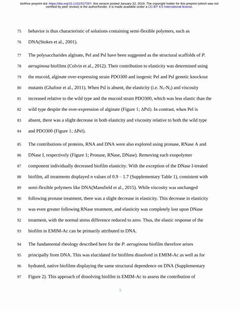
5
behavior is thus characteristic of solutions containing semi-flexible polymers such as 75
DNA(Stokes et al 2001) 76
The polysaccharides alginate Pel and Psl have been suggested as the structural scaffolds of P 77
aeruginosa biofilms (Colvin et al 2012) Their contribution to elasticity was determined using 78
the mucoid alginate over-expressing strain PDO300 and isogenic Pel and Psl genetic knockout 79
mutants (Ghafoor et al 2011) When Psl is absent the elasticity (ie N1-N2) and viscosity 80
increased relative to the wild type and the mucoid strain PDO300 which was less elastic than the 81
wild type despite the over-expression of alginate (Figure 1 ΔPsl) In contrast when Pel is 82
absent there was a slight decrease in both elasticity and viscosity relative to both the wild type 83
and PDO300 (Figure 1 ΔPel) 84
The contributions of proteins RNA and DNA were also explored using pronase RNase A and 85
DNase I respectively (Figure 1 Pronase RNase DNase) Removing each exopolymer 86
component individually decreased biofilm elasticity With the exception of the DNase I-treated 87
biofilm all treatments displayed n values of 09 ndash 17 (Supplementary Table 1) consistent with 88
semi-flexible polymers like DNA(Mansfield et al 2015) While viscosity was unchanged 89
following pronase treatment there was a slight decrease in elasticity This decrease in elasticity 90
was even greater following RNase treatment and elasticity was completely lost upon DNase 91
treatment with the normal stress difference reduced to zero Thus the elastic response of the 92
biofilm in EMIM-Ac can be primarily attributed to DNA 93
The fundamental rheology described here for the P aeruginosa biofilm therefore arises 94
principally from DNA This was elucidated for biofilms dissolved in EMIM-Ac as well as for 95
hydrated native biofilms displaying the same structural dependence on DNA (Supplementary 96
Figure 2) This approach of dissolving biofilm in EMIM-Ac to assess the contribution of 97
CC-BY 40 International licensecertified by peer review) is the authorfunder It is made available under aThe copyright holder for this preprint (which was notthis version posted January 22 2019 httpsdoiorg101101527267doi bioRxiv preprint
6
individual exopolymers to its properties is thus validated The data suggest that multiple 98
exopolymers contribute to the rheology with the matrix from the ΔPsl mutant being the most 99
elastic followed by wild type and RNase A-treated biofilms which were the least elastic It has 100
been noted previously that a range of P aeruginosa exopolymers influence biofilm extracellular 101
matrix crosslinking (Colvin et al 2012) and the data presented further clarifies that these 102
exopolymers can only modulate biofilm rheology if eDNA is present An eDNA scaffold is 103
therefore a prerequisite for this rheological differentiation of the matrix Further support for the 104
presence of an eDNA scaffold is provided in the micrograph of a biofilm stained with TOTO-1 105
for DNA visualization (Figure 1C) which shows DNA fibers in the extracellular matrix 106
107
Figure 1 (A) N1-N2 and (B) viscosity against shear rate for Pseudomonas aeruginosa biofilm wild type PDO300 108
ΔPsl ΔPel and pronase RNase A and DNase I digested wild type biofilm immediately following dissolution in 1-109
ethyl-3-methylimidazolium acetate (40 mgmL) at 25 degC 100 microm gap This is measured as a function of shear stress 110
CC-BY 40 International licensecertified by peer review) is the authorfunder It is made available under aThe copyright holder for this preprint (which was notthis version posted January 22 2019 httpsdoiorg101101527267doi bioRxiv preprint
7
from 10 to 1000 Pa (N1 ndashN2) is not described for DNase I digested biofilm in Figure 1A and Supplementary Figure 111
1B as their normal force (FN) is less than the resolution of the rheometer (ie 01 N) and set to zero for calculating 112
(N1 ndashN2) Both the N1-N2 and viscosity data are fitted with the FENE-P model a rigid dumbell model for polymer 113
solutions Fitting parameters are shown in Supplementary Table 2 (C) Micrograph of P aeruginosa biofilm DNA 114
stained green with TOTO-1 (scale bar 10 microm) (D) Phase separation of extracellular nucleic acids extracted from P 115
aeruginosa biofilms into a gel occurs upon transfer from 1-ethyl-3-methylimidazolium acetate into water 116
Furthermore EMIM-Ac did not lyse either biofilm or planktonic cells as indicated by the intact 117
cell morphology and the absence of phospholipids and lipopolysaccharides in EMIM-Ac 118
following biofilm dissolution (Supplementary Figure 3) Therefore it was concluded that the 119
DNA dissolved following treatment of P aeruginosa planktonic cells and biofilms with EMIM-120
Ac is extracellular and due to extraction of intracellular DNA 121
This finding that eDNA is a prerequisite for matrix building is consistent with recent studies 122
reporting the observation of eDNA in biofilm extracellular matrices (Jennings et al 2015) That 123
DNA dominated the elastic response would indicate eDNA is not an incidental matrix 124
component but instead a primary or foundation structural component of P aeruginosa biofilms 125
Isolated nucleic acids mimic gel-forming property of P aeruginosa biofilms 126
eDNA is known to complex and co-localize with exopolysaccharides Furthermore eDNA 127
sequencing shows that there is no bias towards any specific region of the chromosome (Turnbull 128
et al 2016) In contrast to intracellular chromosomal DNA which is supercoiled and folded for 129
example by HU proteins and topoisomerases and then relaxed by gyrases to allow replication 130
and transcription to occur the organization of eDNA remains elusive We contend that a 131
molecular understanding of how the eDNA is assembled and organized is key to answering how 132
and why DNA transforms from the chromosomal form to that found in the biofilm matrix 133
CC-BY 40 International licensecertified by peer review) is the authorfunder It is made available under aThe copyright holder for this preprint (which was notthis version posted January 22 2019 httpsdoiorg101101527267doi bioRxiv preprint
8
We recovered the eDNA from EMIM-Ac following biofilm dissolution by exploiting the ability 134
of perchloric acid to selectively precipitate DNA over protein (Supplementary Figure 4) 135
Following further purification by gel permeation chromatography the polymer phase-separated 136
into a gel upon transfer from EMIM-Ac into water (ie the gel isolate) mimicking the formation 137
of networks in native biofilms (Figure 1D) A higher Grsquo was recorded for the gel isolate than for 138
the P aeruginosa biofilm (Supplementary Figure 5) which is consistent with it having a higher 139
DNA concentration Furthermore DNase degraded the isolated gel into shorter DNA fragments 140
(Supplementary Figure 6) Other fractions including those not precipitated by perchloric acid 141
did not self-assemble into gels (Supplementary Figure 7A) Similarly calf thymus DNA did not 142
form gels when processed the same way either with or without added cations (Supplementary 143
Figure 7B) suggesting that this behavior is not a universal property of DNA 144
Nucleic acid conformation is preserved during isolation 145
Circular dichroism (CD) is an extremely sensitive spectroscopic technique for determining the 146
secondary structure of biomolecules particularly proteins and nucleic acids It was used here to 147
understand whether the nucleic acid (NA) conformation was modified during extraction and 148
isolation Unprocessed P aeruginosa biofilms displayed a major circular dichroism (CD) peak at 149
250-285 nm (Figure 2A) which is consistent with the presence of NA (Kypr et al 2009) and 150
this peak was also observed to dominate the CD spectrum of the gel isolate (Figure 2B) The 151
spectral trough at 2255-2265 nm is typical for proteins (Greenfield 2006) NA can also display 152
a trough in this region although the relative depth of the trough and its absence after proteolysis 153
and fractional precipitation against proteins (Supplementary Figure 4) suggest that it denotes 154
proteinaceous material Hence even though the CD spectrum shows that proteins are significant 155
CC-BY 40 International licensecertified by peer review) is the authorfunder It is made available under aThe copyright holder for this preprint (which was notthis version posted January 22 2019 httpsdoiorg101101527267doi bioRxiv preprint
9
components of the biofilm matrix they are not major contributors to biofilm elasticity (Figure 156
1A) 157
158
Figure 2 Circular dichroism (CD) spectra of (A) Pseudomonas aeruginosa biofilm and (B) extracellular nucleic 159
acids (NA) gel isolate at temperatures between 30 and 95degC (C) Amplitude of dominant NA peak CDmax (260-285 160
nm) from CD spectra of P aeruginosa biofilm (seen in (A)) and its extracted extracellular nucleic acid gel (seen in 161
(B)) from T = 30degC to T = 95degC 162
The CD spectrum of the isolated NA gel (Figure 2B) is dominated by the same NA peak that 163
appears in the biofilm CD spectrum To confirm this similarity CD melting experiments were 164
performed on both samples The resulting melting curves (Figure 2C) show that their maximum 165
absorbance values follow the same trend across the temperature profiles with a minimum 166
absorbance at 40degC a maximum at 60degC and only partial absorbance attenuation at 95degC NA 167
conformation was thus preserved during its isolation and the NA are more stable than typical 168
canonical DNA duplexes (Khandelwal and Bhyravabhotla 2010) The extracted NA gel isolate 169
is therefore an accurate proxy for understanding the intermolecular interactions that stabilize the 170
extracellular matrices of P aeruginosa biofilms Moreover the CD spectrum of the NA gel 171
isolate informs on the nucleic acid conformation In addition to the peak at 272 nm there is a 172
trough that shifts from 245 to 230 nm with increasing temperature another maximum at 215 nm 173
210 240 270 300
-40
-30
-20
-10
0
210 240 270 300-20
-10
0
10
20
30
30 oC
40 oC
60 oC
95 oCC
ircu
lar
dic
hro
ism
(m
de
g)
Circu
lar
dic
hro
ism
(m
de
g)
Wavelength (nm)
30 oC
40 oC
60 oC
95 oC
CB
Wavelength (nm)
A
30 40 50 60 70 80 90
26
28
30
32
34
36
38
NA gel
CD
max (
md
eg
) -
Iso
late
Temperature (oC)
1
2
3
4
5
6
Biofilm
CD
max (
md
eg
) -
Bio
film
CC-BY 40 International licensecertified by peer review) is the authorfunder It is made available under aThe copyright holder for this preprint (which was notthis version posted January 22 2019 httpsdoiorg101101527267doi bioRxiv preprint
10
that remains constant with temperature and other sub-maxima at 260 and 253 nm that change 174
with temperature The absence of a trough at 200-215 nm precludes the possibility of NA in A- 175
or Z- conformations (Kypr et al 2009) The dominant maximum and minimum could indicate 176
either B-DNA or G-quadruplex conformations (del Villar-Guerra et al 2018) although the peak 177
at 215 nm is not a feature of B-DNA CD spectra and is slightly higher than the characteristic G-178
quadruplex low wavelength peak of 210 nm Nonetheless the appearance of several peaks in the 179
NA region of 250-285 nm indicates that while the dominant NA conformation is unclear several 180
conformations likely contribute to phase separation of the NA 181
Purine-rich ribonucleotides and eDNA are present in isolated gel 182
Total correlation spectroscopy (TOCSY) and 1H-
13C heteronuclear single quantum coherence 183
(HSQC) are nuclear magnetic resonance (NMR) techniques that can identify proton NMR 184
correlations within individual ribose sugars and their proton-carbon single bond correlations 185
respectively Raising the pH can solubilize P aeruginosa biofilms (Friedman and Kolter 2004) 186
and here we show that after alkalinization the NA peaks dominated the solution 1H-
13C HSQC 187
spectra for the isolated material with no indication that proteins or polysaccharides were present 188
(Supplementary Figure 8 (Wuumlthrich 2008)) The 1H-
13C HSQC-TOCSY spectrum of the 189
isolated gel when dissolved by alkalinization shows two clusters of sugar proton peaks (C1rsquo-H1rsquo) 190
with correlations to neighboring carbons (C2rsquo-H1rsquo) (Figure 3A) 191
The first cluster (rectangles) has C2rsquo chemical shift values of ~40 ppm and the second cluster 192
(ovals) has C2rsquo values of ~70 ppm The 1H-
13C correlations denoted by the rectangles therefore 193
arise from deoxyribose and those denoted by the ovals from ribose sugar conformations The 194
broadened form of the deoxyribonucleotide peaks is consistent with high molecular weight 195
(MW) molecules while the sharpness of the ribonucleotide peaks suggests that they could arise 196
CC-BY 40 International licensecertified by peer review) is the authorfunder It is made available under aThe copyright holder for this preprint (which was notthis version posted January 22 2019 httpsdoiorg101101527267doi bioRxiv preprint
11
from small possibly even single nucleotides Hence eDNA and small ribonucleotides are both 197
likely to be present in the gel dissolved by alkalinization 198
Spin-systems for the eight sharp ribonucleotide peaks (Figure 3B) were assigned by HSQC-199
TOCSY and correlation spectroscopy (COSY) (Supplementary Figure 9) with absolute 200
identification achieved by comparing the heteronuclear multiple bond correlation HMBC spectra 201
of samples and monoribonucleotide standards (Supplementary Figure 10) The 31
P NMR 202
spectrum of the NA gel isolate at elevated pH revealed the presence of a mixture of 203
monoesterified (34 to 41 ppm) and diesterified phosphates (-08 to -12 ppm) indicating the 204
coexistence of monoribonucleotides and DNA respectively (Figure 3C (i)) Long-range 31
P-1H 205
correlations (ie from NA phosphorous to adjoined ribose proton) were observed in the 31
P-1H 206
heteronuclear correlation (HETCOR) spectrum (Figure 3C (ii)) for both monoribonucleotides 207
and DNA The broad 31
P-1H cross peaks at ~-10 ppm (Figure 3C (ii)) correspond to long-range 208
correlations between DNA phosphorous to H3rsquo and H5rsquoH5rsquorsquo protons The eight 209
monoribonucleotides were assigned to 2rsquo- and 3rsquo-(A U G and C)-monophosphates We 210
therefore have both 2rsquo and 3rsquo monoribonucleotides present with the eDNA in the gel solubilized 211
under alkaline conditions 212
However the monoribonucleotide peaks only became resolved upon alkaline dissolution of the 213
biofilm in contrast to the broad peaks evident at pH 7 (Supplementary Figure 11) This indicates 214
that the monoribonucleotides are derived from chain structures that exist at biological pH and 215
assemble with DNA chains into higher-order networked structures which is consistent with the 216
observation in Figure 1A that RNA is a major contributor to P aeruginosa biofilm elasticity 217
218
219
CC-BY 40 International licensecertified by peer review) is the authorfunder It is made available under aThe copyright holder for this preprint (which was notthis version posted January 22 2019 httpsdoiorg101101527267doi bioRxiv preprint
12
220
221
222
223
224
225
226
227
Figure 3 (A) Nuclear magnetic resonance (NMR) 1H-13C 228
heteronuclear single quantum coherence (HSQC)-total 229
correlation spectroscopy (TOCSY) spectrum at 25degC of 230
extracellular nucleic acids (NA) gel isolate after 231
alkalinization showing the C1rsquo-H1rsquo cross peaks of RNA 232
(blue ovals) and DNA (red rectangles) and their correlations 233
to the neighboring carbon C2rsquo-H1rsquo Schematics (right) 234
illustrate these correlations (B) 1H-13C HSQC spectrum of 235
extracellular NA gel isolate at 25degC after alkalinization 236
identifying eight monoribonucleic acid ribose spin systems 237
(C) 1-D 31P NMR of NA isolate gel and Pseudomomonas 238
aeruginosa biofilm with proton decoupling showing the 239
presence of monoesterified (ie monoribonucleotides) and 240
diesterified (ie DNA) phosphate peaks (i) and 2-D 1H-31P heteronuclear correlation (HETCOR) spectrum of 241
extracellular NA showing the 31P-1H cross-peaks of monoribonucleotides and DNA (ii) Couplings of 242
monoesterified phosphates to H2rsquo or H3rsquo of eight monoesterified monoribonucleotides (from left to right 3rsquo 243
AMP3rsquo GMP 3rsquo CMP3rsquo UMP 2rsquo CMP2rsquo UMP 2rsquo AMP 2rsquo GMP) and diesterified phosphate to DNA H3rsquo and 244
H5rsquoH5rsquorsquo protons are denoted by the dashed lines There is a discontinuity (wavy line) in the 31P axis due to the 245
different thresholds required to illustrate the 31P-1H correlations in the mono-esterified and di-esterified regions All 246
samples were prepared in 01 M NaOD (10 mgmL) and preheated to 55degC for 2 h 247
CC-BY 40 International licensecertified by peer review) is the authorfunder It is made available under aThe copyright holder for this preprint (which was notthis version posted January 22 2019 httpsdoiorg101101527267doi bioRxiv preprint
13
The molar ratio of the individual ribonucleotides could be determined from the 31
P spectrum of 248
the gel dissolved at high pH (Table 1) While several peaks could not be separated it was 249
possible to deduce that the RNA is purine rich (ie 57 mol A+G) and that the G+C mol 250
content of 46-50 differs from that of the P aeruginosa genome (ie 67 mol) (Shen et al 251
2006) The same peaks were also observed in the biofilm 31
P spectrum after alkalinization 252
(Figure 3C(i)) 253
Table 1 Relative abundances of monoribonucleotides in extracellular NA gel isolate from Pseudomonas 254 aeruginosa biofilm as determined by integrating 31P NMR spectrum following alkalinization 255
31P shift
(ppm)
Ribonucleotide Relative
abundance
403 3rsquoAMP 180
401 3rsquo GMP 172
390 3rsquo CMP3rsquo UMP 89
387 3rsquo CMP3rsquo UMP 113
368 2rsquo CMP2rsquo UMP 95
366 2rsquo CMP2rsquo UMP 117
350 2rsquo AMP 104
342 2rsquo GMP 130
256
Extracellular DNA and RNA interact to form a network 257
We applied Magic-angle spinning (MAS) solid-state NMR (SSNMR) to confirm that the 258
networks in the gel isolate and biofilm are comprised of DNA and RNA in chain form This 259
technique is ideal for intractable systems such as biofilms to eliminate solubility and extraction 260
biases (Reichhardt and Cegelski 2014) and to analyze inter-molecular H-bond interactions to 261
describe for example inter-nucleotide base pairing in RNA (Marchanka et al 2015) MAS 262
SSNMR averages anisotropic interactions to provide high-resolution spectral characterization of 263
insoluble and large biomolecular systems By analyzing dipolar interactions through-space 264
heteronuclear correlations (eg N∙∙∙H) can be detected In contrast to the liquid state 31
P NMR 265
spectrum of the alkali-dissolved isolate (Figure 3C (i)) the 31
P SSNMR spectrum of the gel 266
CC-BY 40 International licensecertified by peer review) is the authorfunder It is made available under aThe copyright holder for this preprint (which was notthis version posted January 22 2019 httpsdoiorg101101527267doi bioRxiv preprint
14
isolate (ie no alkali treatment Figure 4A (i)) showed a single peak in the diesterified phosphate 267
region consistent with the presence of both DNA and RNA chains There are no sharp peaks 268
present in the monoesterified phosphate region further demonstrating that the 269
monoribonucleotides are a consequence of alkali transesterification (Radak et al 2013) 270
Only the diesterified phosphate peak was observed in the 31
P SSNMR spectrum of the biofilm 271
(Figure 4A (iii)) while both RNA-derived monoesterified and DNA-derived diesterified 272
phosphate peaks were present in the 31
P SSNMR spectrum of alkali digested NA gel-isolate and 273
biofilm (lyophilized) (Figure 4A (ii) and Figure 4A (iv) respectively) Complete alkali RNA 274
transesterification was confirmed by the full conversion of diesterified to monoesterified 275
phosphates (from liquid state 31
P NMR spectra) for the RNA standard when dissolved in 01 M 276
NaOH (Supplementary Figure 12A) Conversely RNA diesterified phosphate peaks were 277
preserved in the RNA standard spectrum following dissolution in EMIM-Ac and recovery by 278
perchloric acid (Supplementary Figure 12B) Hence the alkaline conditions break down the 279
RNA chains into individual monoribonucleotides while the ionic liquid-based extraction does 280
not This suggests that the conventional method for P aeruginosa biofilm dissolution in alkali 281
(Friedman and Kolter 2004) may in fact work by transesterifying RNA as a primary structural 282
component and illustrates the importance of the ionic liquid-based extraction protocol described 283
here as a non-destructive method for interrogating biofilm structural polymers 284
CC-BY 40 International licensecertified by peer review) is the authorfunder It is made available under aThe copyright holder for this preprint (which was notthis version posted January 22 2019 httpsdoiorg101101527267doi bioRxiv preprint
15
285
Figure 4 (A) Solid-state 31P NMR spectra at T = 25degC of extracellular NA gel isolate (i) alkalinized and 286
lyophilized NA gel isolate (ii) Pseudomonas aeruginosa biofilm (iii) and alkalinized and lyophilized P aeruginosa 287
biofilm (iv) showing the presence of diesterified phosphate peaks and the absence of monoesterified phosphate 288
peaks for both NA gel isolate and biofilm in double distilled water and the coexistence of diesterified and 289
monoesterified phosphate peaks for both samples after alkalinization This indicates that alkalinization of the matrix 290
results in RNA transesterification Micrographs of P aeruginosa NA gel isolate stained green with (B) SYTO 291
RNASelect showing RNA fibers and (C) TOTO-1 showing DNA fibers (scale bar 10 microm) 292
293
To visualize the DNA-RNA network organization we used nucleic acid-specific stains ie RNA 294
specific SYTO RNASelect dye (Figure 4B) and the eDNA-specific TOTO-1 dye (Figure 4C) 295
(Okshevsky and Meyer 2014) Fibrous structures which are typical of networked polymer gels 296
(Cornwell and Smith 2015) were observed in the NA gel isolate stained with both nucleic acid 297
dyes as well as in the biofilm stained with the eDNA specific dye (Figure 1C) This is consistent 298
with our observation that the biofilm is readily degraded by DNase I (Figure 1A) (Chen et al 299
2004) Furthermore the NA gel isolate was stained positively with the RNA-specific dye even 300
after DNase I digestion providing further evidence that RNA contributes to networking 301
BeRNA fibers
C
eDNA fibers
A
CC-BY 40 International licensecertified by peer review) is the authorfunder It is made available under aThe copyright holder for this preprint (which was notthis version posted January 22 2019 httpsdoiorg101101527267doi bioRxiv preprint
16
(Supplementary Figure 13) However digestion with RNases III A and H did not degrade the 302
biofilm (Figure 1A Supplementary Figure 14) This may suggest that the RNase binding was 303
shielded by hairpins in the network or the presence of non-canonical DNA-RNA interactions 304
(Geerts-Dimitriadou et al 2012 Nakamura et al 1991) 305
Non-canonical and Watson-Crick base pairs and tetrads support extracellular network 306
Sequence analysis of the extracted material indicated that the gene coverage was even for both 307
chromosomal and extracellular DNA with the exception of bacteriophage Pf1 genes 308
(Supplementary Figure 15) However the Pf1 knockout mutant of P aeruginosa also displayed 309
an elastic response when dissolved in EMIM-Ac (Supplementary Figure 16 Supplementary 310
Tables 1 and 2) indicating that Pf1 DNA is unlikely to be responsible for the phase-separating 311
behavior of P aeruginosa biofilms DNA and RNA interaction therefore cannot be explained by 312
uneven gene coverage To elucidate the mechanism of DNA-RNA gelation we generated a 2D 313
through-space 15
N-1H HETCOR spectrum of
15N
labeled DNA-RNA gel isolated from a P 314
aeruginosa biofilm matrix (Figure 5A) This spectrum was run with longer cross polarization 315
contact times so that correlations between distant 1H and
15N could be observed This confirmed 316
the complete absence of proteins and supported our conclusion that proteins do not contribute to 317
the DNA-RNA interaction The HETCOR spectrum showed four signal clusters at δH 10-14 318
ppm and δN 140-160 ppm (Figure 5A) which arose from direct N-H couplings of TU and G 319
nucleobase imino groups Two of these clusters (δH 12-14 ppm) resulted from imino protons 320
hydrogen-bonded to a nucleobase nitrogen (ie N-H∙∙∙N) and the other two (δH 10 -12 ppm) 321
from imino protons hydrogen-bonded to a nucleobase carbonyl oxygen (ie N-H∙∙∙O) Due to the 322
longer cross polarization times we were able to observe a strong long-range and indirect (ie 323
intermolecular) correlation at δN 196 ppm arising from G-C Watson-Crick base pairing ie 324
CC-BY 40 International licensecertified by peer review) is the authorfunder It is made available under aThe copyright holder for this preprint (which was notthis version posted January 22 2019 httpsdoiorg101101527267doi bioRxiv preprint
17
C(N3)-G(H1) There was also a weak and indirect correlation at δN 220 ppm resulting from A-325
TU Watson-Crick base pairing ie A(N1) to UT(H1) 326
327
328
Figure 5 (A) Solid-state 2D 1H-15N through-space heteronuclear correlation (HETCOR) spectrum of extracellular 329
nucleic acid (NA) gel isolate in double distilled water (2 mg) T = 25degC showing direct N-H couplings for G imino 330
N (N3) and TU imino N (N3) bonded to nucleobase nitrogen (NHN) This indicates Watson-Crick (WC) G-C and 331
UT-A base pairings respectively and for G imino N (N1) and TU imino N (N3) bonded to nucleobase carbonyl 332
oxygen (ie NHO) indicating non-canonical G-G G-TU and UT-UT base pairings or tetrads The spectrum also 333
displayed indirect N-H correlations for the WC base pairs (ie C(N3) to G(H1) and A(N1) to UT(H1)) and no 334
indirect correlations for the non-canonical bonded nucleobases The threshold for the A(N1)-TU(H3) correlation 335
was increased due to lower signal intensity for that particular coupling (B) Pseudomonas aeruginosa biofilm 336
growth yield at 37degC (5 d) as a function of [K+] [Na+] or [Li+] showing the growth dependence on monovalent 337
cation concentration (K+gtNa+gtLi+) Error bars indicate standard deviation (C) Illustration of how structural 338
variability of RNA may allow for the formation of extracellular RNA and DNA networks through hairpins and a 339
mixture of canonical and non-canonical base pairs or tetrads (ie Hoogsteen and G-quadruplexes) 340
341
The absence of long-range correlations between the clusters at δH 10 -12 ppm is consistent with 342
non-Watson-Crick pairings for the G and TU nucleobases The observation of NH to O 343
interactions suggests the formation of G-G TU-TU G-TU Hoogsteen base pairs or tetrads 344
CA B C
CC-BY 40 International licensecertified by peer review) is the authorfunder It is made available under aThe copyright holder for this preprint (which was notthis version posted January 22 2019 httpsdoiorg101101527267doi bioRxiv preprint
18
Hoogsteen H-bonded G-bases can assemble planarly into tetrads and self-stack to form G-345
quadruplexes (Haumlnsel-Hertsch et al 2017) G-quadruplexes are stabilized by the presence of a 346
monovalent cation with potassium providing the greatest stability We experimentally confirmed 347
that potassium had a similar effect in a static biofilm growth assay We found that potassium 348
promoted P aeruginosa biofilm growth to a greater extent than either sodium or lithium (Figure 349
5B) Additionally this was a biofilm-specific effect as there was no effect of monovalent cation 350
on planktonic growth (Supplementary Figure 17) Based on the SSNMR data coupled with the 351
potassium effect and the earlier observation of several maxima in the CD spectrum of the NA gel 352
isolate across the characteristic NA region of 250-285 nm which suggested the coincidence of 353
several NA conformations contributing to phase separation it is proposed that G-quadruplexes 354
contribute to extracellular DNA-RNA networking 355
Discussion 356
We extracted a gel-forming complex composed of DNA and RNA from the extracellular matrix 357
of P aeruginosa biofilms without disrupting its fundamental structural or chemical organization 358
We report the novel observation that extracellular RNA contributes structurally to biofilms 359
While gel-forming exopolymers have been detected in other biofilms (Seviour et al 2009) no 360
functional DNA gel has been described for any biological system much less one also including 361
extracellular RNA 362
While it is not possible to identify the precise nature of the DNA-RNA interaction based on the 363
information presented here purine-rich RNA as described here for the extracellular NA gel 364
isolate (57 mol A-G) is a characteristic of DNA-RNA duplexes because RNA purines bind 365
more strongly to their DNA pyrimidine complement than vice versa (Nadel et al 2015) Both 366
Watson-Crick and non-canonical interactions were present in the NA gel isolate which is also 367
CC-BY 40 International licensecertified by peer review) is the authorfunder It is made available under aThe copyright holder for this preprint (which was notthis version posted January 22 2019 httpsdoiorg101101527267doi bioRxiv preprint
19
consistent with the discrepancy observed between RNA and DNA G+C mol contents (Table 368
1) It is possible therefore that interactions between DNA and RNA as described in this study 369
account for the formation of a highly stable nucleic acid gel 370
Non-canonical base pairing and hairpinning are commonly seen with RNA and cause multivalent 371
intermolecular interactions between RNA strands that lead to a sol-gel transition (Jain and Vale 372
2017) The greater structural versatility of RNA could be a key to the ability of the eDNA to 373
form networks with hairpins as well as canonical and non-canonical base pair interactions all 374
contributing to the assembly of higher order NA structures (Figure 5C) RNA has recently been 375
found to promote intracellular phase separations although this has not been extended to the 376
extracellular domain of prokaryotic cells (Shin and Brangwynne 2017) Additionally G-rich 377
RNA sequences are more predisposed to forming G-quadruplexes because they do not have a 378
complementary strand and in fact form more stable G-quadruplexes than DNA (Guo and Bartel 379
2016) It is therefore probable that the RNA but not DNA enables DNA-RNA networking and 380
gelation 381
The results presented here provide unprecedented resolution of the biofilm exopolymeric matrix 382
and its key foundation structural components We have developed a methodology that preserved 383
the molecular organization of the foundational polymer in its native state upon extraction and 384
isolation This enabled us to use SSNMR to describe intermolecular associations at the atomic 385
level 386
Our findings that a DNA-RNA gel network can provide the foundation for biofilm matrices in P 387
aeruginosa mark a departure from the prevailing paradigm that biofilm gelation is only due to 388
polysaccharides The elastic and film-forming properties were also observed for Pseudomonas 389
putida and Pseudomonas protegens (Figs S16 and S18 Supplementary Tables 1 and 2) 390
CC-BY 40 International licensecertified by peer review) is the authorfunder It is made available under aThe copyright holder for this preprint (which was notthis version posted January 22 2019 httpsdoiorg101101527267doi bioRxiv preprint
20
suggesting that eDNA is broadly important for biofilm formation in members of this genus In 391
addition to Pseudomonas spp other organisms are known to have eDNA including clinical 392
organisms Staphylococcus aureus (Mann et al 2009) Staphylococcus epidermidis (Adam et al 393
2002) and Mycobacterium abscessus (Rose et al 2015) as well as environmental isolates such 394
as strain F8 from the South Saskatchewan River (Boumlckelmann et al 2006) 395
While eDNA is commonly thought to be the product of cell lysis and has been shown to be 396
released by a sub-population of lytic P aeruginosa cells (Turnbull et al 2016) there is an 397
increasing awareness that it serves an important structural function such as in activated sludge 398
granules (Cheng et al 2011) Additionally Bockelmann et al (2006) observed that stable 399
filamentous networks produced by the aquatic strain F8 were comprised of DNA This is the first 400
study that provides an explanation for how eDNA is organized structurally However the 401
environmental factors contributing to eDNA release and how this is regulated (ie whether it is 402
an active or passive process) are still unclear It is possible that DNA-RNA foundational gel 403
networks as described here for the Pseudomonads are broad-scoping phenomena in biofilms 404
and that interactions with RNA enable an extracellular structural function for DNA Thus 405
elucidating how nucleic acids including RNA are integral to the biophysical and other emergent 406
properties imparted on the biofilm via the extracellular matrix will inform the regulation and 407
control of extracellular nucleic acid release across environmental and clinical biofilms 408
Materials and Methods 409
Bacterial strains 410
Unless otherwise stated all experiments were undertaken directly on P aeruginosa PAO1 411
biofilms grown in lysogeny broth at 37degC The P aeruginosa PAO1 Δpf4 knockout mutant is a 412
CC-BY 40 International licensecertified by peer review) is the authorfunder It is made available under aThe copyright holder for this preprint (which was notthis version posted January 22 2019 httpsdoiorg101101527267doi bioRxiv preprint
21
defined Pf4 chromosomal deletion mutant of the entire Pf4 prophage genome(Rice et al 413
2009) 414
P aeruginosa PDO300 PDO300(Δpel) and PDO300(Δpsl) mutant strains were gratefully 415
received from Professor Bernd H A Rehm Institute of Molecular Biosciences Massey 416
University New Zealand (Ghafoor et al 2011) PDO300 PDO300(Δpel) PDO300(Δpsl) and 417
PAO1 (Δpf4) were also grown in LB at 37degC P protogens Pf-5 and P putida ATCC BAA-477 418
and S12 strain were were grown in LB at 30degC 419
Biofilm growth assay 420
Ten milliliter aliquots of P aeruginosa planktonic pre-cultures (LB 200 rpm 37degC OD600 240 421
16 h) were diluted 50 times with LB in 2 L Erlenmeyer flasks and incubated for 5 d under static 422
conditions Supplementary Figure 15A displays an image of 5-d old biofilm in LB The cultures 423
were centrifuged at 10000 x g for 15 min the supernatant removed by decanting and the 424
biofilm then collected and lyophilized (LabConco) Supplementary Figure 15B displays an 425
image of the culture after centrifugation showing clear separation of biofilm and supernatant 426
Enzymatic digestions 427
Twenty milligrams of lyophilized biofilm was resuspended in 1 mL of either i) RNase buffer (50 428
mM Tris HCl 10 mM EDTA pH8) with 02 mg RNaseA from bovine pancreas (Sigma Aldrich) 429
ii) storage buffer (10 mM NaCl 10 mM Tris-HCl) with 01 mg Pronase E from Streptomyces 430
grisens (Sigma Aldrich) with 05 (vv) iii) 1X RNase H reaction buffer (20 mM Tris-HCl pH 431
78 40 mM KCl 8 mM MgCl2 1 mM DTT) with 04 mg RNase H (Thermo Fisher Scientific) 432
iv) 1X RNase III reaction buffer (500 mM NaCl 100 mM Tris pH 79 100 mM MgCl2 10 mM 433
DTT) with 06 mg RNase II (Thermo Fisher Scientific) or v) DNase I buffer (100mM Tris (pH 434
75) 25 mM MgCl2 and CaCl2) with 02 mg DNAse I from bovine pancreas (Sigma Aldrich) All 435
CC-BY 40 International licensecertified by peer review) is the authorfunder It is made available under aThe copyright holder for this preprint (which was notthis version posted January 22 2019 httpsdoiorg101101527267doi bioRxiv preprint
22
digestions were performed with shaking at 200 rpm at 37degC for 16 h The suspensions were then 436
centrifuged (10000 x g 15 min) the supernatant was discarded and the pellets of the biofilm 437
materials were lyophilized 438
Normal force measurement 439
Forty milligrams per milliliter solutions of lyophilized biofilms were added to 1 mL 1-ethyl-3-440
methylimidazolium acetate and incubated in 55degC for either 2 h A Haake Mars 3 (Thermo 441
Fisher Scientific) stress-controlled rotational rheometer with Peltier controlled element at 25degC 442
was used for rheological measurements Thirty five-illimeter diameter parallel plate geometry 443
was used with smooth titanium plates to measure viscosity and normal stress difference (N1 ndash 444
N2) Prior to measurement the gap error was zeroed at 4 N and gap error calculated as previously 445
described (Bird et al 1987 Davies and Stokes 2008 Kravchuk and Stokes 2013) One 446
hundred microliters of sample was deposited on the plates The plates were closed to 100 microm the 447
sample trimmed and the sample allowed to sit for 5 min prior to measurement All measurements 448
with Normal force (FN) less than the resolution of the rheometer (ie lt 01 N) were set to 0 449
before calculation of N1-N2 using equations 9-11 and viscosity from equation 3-5 in Davies and 450
Stokes(Davies and Stokes 2008) for the parallel plate geometry 451
Only the linearly increasing portion of the normal stress difference curves are presented Above 452
this range normal stress difference begins to decrease again which may be due to elastic 453
instabilities or associating polymers (Annable et al 1993) Corrections were made to N1 and N1 454
ndash N2 to account for inertia using equation 17 in Davies and Stokes (Davies and Stokes 2008) and 455
to correct for the baseline residual force in the samples Except for the DNase I-treated biofilm 456
the shear rheology for all treatments could be modelled using the finitely extensible non-linear 457
elastic with Gaussian closure proposed by Peterlin (FENE-P) constitutive model by varying four 458
CC-BY 40 International licensecertified by peer review) is the authorfunder It is made available under aThe copyright holder for this preprint (which was notthis version posted January 22 2019 httpsdoiorg101101527267doi bioRxiv preprint
23
parameters to fit shear viscosity and normal stress difference as a function of shear rate 459
(Supplementary Table 2) Fitting parameters for the FENE-P model include 1 = relaxation time 460
b = a measure of the relative extensibility of the model spring s = solvent viscosity p = 461
polymer contribution to the viscosity The FENE-P equations can be written in the following 462
format as shown by Bird et al(1987) 463
120578 = 120578119904 +120578119901
1205821((1198622 + 1198621)
1
3 minus (1198622 minus 1198621)1
3) (Equation 1) 464
465
1198731 = 2120578119901
1205821((1198622 + 1198621)
1
3 minus (1198622 minus 1198621)1
3)2
(Equation 2) 466
Where 467
1198621 = 119887
41205821 (Equation 3) 468
1198622 = (11986212 + (
119887+3
6)
3
)12
(Equation 4) 469
470
All measurements were performed in triplicate For clarity one representative data set is 471
presented in Figure 1A-B and Supplementary Figure 1A-B with the respective Power Law and 472
FENE-P model fit to that data set Averaged values for the FENE-P and Power law fits with the 473
standard deviation across three replicates are shown in Supplementary Tables 1 and 2 474
Oscillatory measurements were carried out with controlled frequency 01 rads across an 475
amplitude range of 001 to 10 476
Extracellular polymeric substances (EPS) extraction 477
Lyophilized biofilms were dissolved in ionic liquid mixture (40 (vv) 1-ethyl-3-478
methylimidazolium acetate (EMIM Ac) 60 (vv) NN-dimethyl acetamide (DMAc)) at 55degC 479
CC-BY 40 International licensecertified by peer review) is the authorfunder It is made available under aThe copyright holder for this preprint (which was notthis version posted January 22 2019 httpsdoiorg101101527267doi bioRxiv preprint
24
for 16 h The solution was centrifuged (10000 x g) to remove any undissolved material 480
Perchloric acid (70) was added (005 vv) to the viscosified centrate (on ice) After 15 min 481
incubation the solution was centrifuged at 10000 x g at 4degC for 15 min and the pellet recovered 482
This was repeated on the centrate two to four times until the solution was not viscous The 483
precipitate was dialysed against double distilled water for 2 d at 4degC (SnakeSkintrade Dialysis 484
Tubing 35K MWCO 22 mm) and the retentate lyophilized (FreeZone Plus 45 Liter Cascade 485
Benchtop Freeze Dry System) The same procedure was performed on calf thymus DNA lipase 486
cytochrome C for the purposes of determining recovery yield of representative exoproteins and 487
on RNA from torula yeast for assessing by 31
P NMR whether the extraction procedure 488
contributed to RNA transesterification (all from Sigma Aldrich) 489
Extracellular nucleic acid isolation 490
Twenty milligrams of lyophilized retentate (ie post perchloric acid precipitation) were dissolved 491
in 1 mL of 40 (vv) EMIM-Ac 60 DMAc (vv) (55degC 16 h) Chromatographic separation 492
was achieved in a Shimadzu system comprising DGU-20A 3r Prominence Degasser and LC-493
20AD Solvent Delivery Unit fitted with two Agilent PLgel 10 μm column of 105Aring pore size for 494
separation across the MW range 200 kDa to 2000 kDa The eluent flow rate was 30 mLmin-1
495
and the injection volume 1 mL The fractions with molecular weight range of 2000-800kDa and 496
800-200kDa were pooled and dialyzed for 2 d at 4degC (SnakeSkintrade Dialysis Tubing 35K 497
MWCO 22 mm) against double distilled water to induce gelation The gel was then collected 498
from the dialysis tubing 499
Solution-state nuclear magnetic resonance (NMR) 500
Solution-state NMR experiments were performed on an 800 MHz Bruker Avance III 501
spectrometer at 25oC Sample concentration was 10 mg (dry weight)mL
-1 unless otherwise 502
CC-BY 40 International licensecertified by peer review) is the authorfunder It is made available under aThe copyright holder for this preprint (which was notthis version posted January 22 2019 httpsdoiorg101101527267doi bioRxiv preprint
25
specified Spectra were recorded either under conditions of neutral pH in 100 D2O (Cambridge 503
Isotope Laboratories) or following alkalinization (ie transesterification 01 M NaOD 55oC 2 504
h) 1-D NMR experiments include 1H and
31P direct detection while 2-D NMR analysis include 505
13C-HSQC
13C-HSQC-TOCSY
1H-
31P HETCOR HMBC and COSY All spectral analyses 506
were performed using Topspin and SPARKY software 507
Asolectin (Sigma Aldrich) standard (10 mgmL) and lyophilized Pseudomonas aeruginosa 508
PAO1 biofilm (10mgmL) were dissolved in 40 (vv) EMIM Ac 60 (vv) DMAc at (55oC 2 509
h) P aeruginosa PAO1 pre-culture cell lysate was prepared by lysing pre-culture cells with 510
lysozyme in PBS 10 (vv) of D2O was added to all samples for locking purposes 511
Solid-state NMR 512
For solid-state NMR experiments performed on the NA gel isolate 15
N labeled NH4Cl-513
supplemented M9 minimal media was used for biofilm growth M9 consisted of 9552 gL-1
514
Na2HPO42H2O 441g litre-1
KH2PO4 171 gL-1
NaCl 1 gL-1
15
NH4Cl 024 gL-1
MgSO4 515
0011 gL-1
CaCl2 2 gL-1
casamino acids and 04 gL-1
glucose The NA gel was prepared from 516
the M9-grown P aeruginosa PAO1 biofilm as described above 517
Solid-state NMR experiments were performed on 141 T Bruker Advance III instruments 518
equipped with a 19 mm MAS probe operated in double mode The typical 1H
15N and
31P π2 519
pulse lengths were 23 37 and 45 μs respectively 2D dipolar-based 15
N-1H heteronulcear-520
correlation (HETCOR) experiments were conducted on the 15
N-labelled NA gel isolate at 37 kHz 521
MAS spinning frequency Variable temperature was regulated at -20ordmC and the sample 522
temperature was 12ordmC (calibrated using ethylene glycol) In the 15
N-1H HETCOR experiments 523
the initially excited 1H magnetization was transferred to
15N through a cross polarization step 524
followed by t1 evolution Then the 15
N magnetization was flipped to longitudinal axis and 400 525
CC-BY 40 International licensecertified by peer review) is the authorfunder It is made available under aThe copyright holder for this preprint (which was notthis version posted January 22 2019 httpsdoiorg101101527267doi bioRxiv preprint
26
ms proton saturation pulses were applied for water suppression Subsequently the 15
N 526
magnetization was flipped to the transverse plane and transferred to 1H via a second CP step for 527
signal acquisition Two 15
N-1H HETCOR experiments were collected one with 400 s and the 528
other with 2 ms contact times applied for both of the CP steps Low power XiX 1H decoupling 529
(~10 kHz) was employed during 15
N evolution and WALTZ-16 decoupling (10 kHz) was 530
implemented on 15
N channel during 1H acquisition 531
1-D 31
P experiments were performed on 15
N-labelled NA gel isolate and 15
N-labelled P 532
aeruginosa PAO biofilm both directly after dialysis against double distilled water at 4degC for 2 d 533
(SnakeSkintrade Dialysis Tubing 35K MWCO 22 mm) and following alkalinization (01 M 534
NaOD 55ordmC 15 min) and lyophilization (FreeZone Plus 45 Liter Cascade Benchtop Freeze Dry 535
System) 15 kHz MAS spinning frequency and a sample temperature of 27ordmC 75 kHz 536
SPINAL64 1H decoupling was applied during
31P acquisition time All chemical shifts were 537
indirectly referenced using adamantane as a secondary standard (downfield peak is at 4048 ppm 538
DSS scale) 539
Monovalent cation-dependent biofilm growth 540
Ten milliliters aliquots of P aeruginosa pre-cultures (supplemented M9 200 rpm OD600 240 541
16 h) were transferred into 500 mL M9 minimal media (85 gL-1
Na2HPO42H2O 20 gL-1
of 542
NaH2PO4H2O 10 gL-1
KH2PO4 10 gL-1
NH4Cl 048 gL-1
MgSO4 0011 gL-1
CaCl2 2 gL-1
543
casamino acids and 04 gL-1
glucose) supplemented with either KCl LiCl or NaCl at either of 544
three different concentrations (0 002 mM and 008 mM) Biofilms were collected by 545
centrifugation (10000 x g 15 min) as described above lyophilized (LabConco) and weighed To 546
describe the growth curve P aeruginosa wild type PAO1 pre-culture was incubated in M9 547
media at 37degC 200 rpm for 16 h PAO1 WT pre-culture was then diluted with supplemented M9 548
CC-BY 40 International licensecertified by peer review) is the authorfunder It is made available under aThe copyright holder for this preprint (which was notthis version posted January 22 2019 httpsdoiorg101101527267doi bioRxiv preprint
27
media to a volume of 75 mL and a starting OD600 of 001 in a 250 mL Erlenmeyer flask The 549
mixture was incubated at 37degC with shaking (200rpm) The OD600 of the bacteria was measured 550
hourly for 9 h followed by final time point at 24 h 551
DNA Sequencing 552
Genomic DNA was extracted from the biofilm using FastDNA SPIN Kit for soil (MP 553
Biomedicals USA) as per the standard protocol Briefly biofilm was resuspended in Sodium 554
Phosphate Buffer in was lysed (Lysing Matrix) homogenized (FastPrepreg
40 seconds speed 555
setting 60) and the cell debris removed by centrifugation (14000 x g 5 min) Proteins were 556
removed by precipitation (250 μl Protein Precipitation Solution) the supernatant mixed with 557
DNA Binding Matrix which was then homogenized and transferred to a SPINtrade Filter Excess 558
supernatant was removed by centrifugation (14000 x g 5 min) DNA was then eluted from air 559
dried DNA Binding Matrix with DNase Pyrogen-Free Water 560
The NA gel isolate was resuspended in 500 uL of 1x Protease K solution (10times Protease K 561
solution 10 mM Tris HCl 1 SDS and 10 mM EDTA pH 8 buffer (10times protease K buffer 562
containing 500 mM Tris-HCl 10 SDS 10 mM CaCl2) and 10 L of Protease K (20 mgmL-1
563
Thermo Fisher Scientific) was added and the mixture incubated at 56degC for 2-16 h after which 564
DNA was extracted as previously described in the phenol-chloroform method(Ausubel 2002) 565
Samples before sequencing were further purified to remove any remaining protein and RNA by 566
RNase and Proteinase K treatment The DNA was then isolated using phenol-chloroform 567
precipitation as described above The DNA precipitate was dissolved in TE buffer the purity 568
confirmed by 260280 value in Nanodrop (acceptable range value 18-20) and Qubitreg
20 569
fluorometer 570
CC-BY 40 International licensecertified by peer review) is the authorfunder It is made available under aThe copyright holder for this preprint (which was notthis version posted January 22 2019 httpsdoiorg101101527267doi bioRxiv preprint
28
The molecular weight distributions of extracellular and genomic DNA were measured on a 1 571
agarose gel which was prepared from Viviantis LE grade agarose using 1x TAE buffer (40 mM 572
Tris 20 mM Acetate and 1 mM EDTA pH 86) Gels were run horizontally After 573
electrophoresis the gel was stained for 05 h with ethidium bromide and visualized under 574
UV(Lee et al 2012) 575
Three replicates were used for each DNA sequence analysis Library was produced using 576
Illumina DNA sample preparation kit The libraries were sequenced using Illumina MiSeq 577
platform (Illumina San Diego Ca) with paired-end protocol to read lengths of 600 nt generating 578
a total of 1614106 and 1848846 paired end reads Raw reads were quality filtered (reads 579
remaining after trimming PPG1-1549104 PBLC1-1666280) and aligned to the P aeruginosa 580
PAO1 (AE004091) genome using CLC Genomics Workbench 90 (CLC bio Cambridge MA) 581
Circular Dichroism CD 582
Five-day old biofilm and NA gel isolate were resuspended in double distilled water to achieve 583
UV absorbance reading 1 and ddH2O served as a blank The heat-treated samples were analyzed 584
by JASCO-815 spectropolarimeter in a 1 cm path length quartz cuvette containing a solution 585
volume 500 μL Spectra (200-320 nm) were measured at 1degC increments from 30 - 90degC For 586
each measurement an average of three scans was taken and the buffer spectra subtracted Each 587
spectrum presented is the rolling average across 5 temperatures 588
Staining and microscopy 589
Microscopic imaging was conducted on a confocal microscope Zeiss LSM 780 with a 63times 590
objective Extracellular RNA in the gel isolate were stained using SYTO RNASelect (Thermo 591
Fisher Scientific) green fluorescent cell stain (5 mM solution in DMSO) Five microM stain solution 592
was prepared from 1 microL of stock in 1X PBS solution (137 mM NaCl 27 mM KCl 43 mM 593
CC-BY 40 International licensecertified by peer review) is the authorfunder It is made available under aThe copyright holder for this preprint (which was notthis version posted January 22 2019 httpsdoiorg101101527267doi bioRxiv preprint
29
Na2HPO4 147 mM KH2PO4) The gel isolate was labelled with 5 microM stain solution kept at 594
37degC for 20 min and then transferred to glass side for imaging 595
eDNA staining was achieved by depositing biofilm or NA gel isolate on a glass slide air-drying 596
overnight and incubating with 2 microM TOTO-1 iodide (1 mM solution in DMSO Thermo Fisher 597
Scientific) for 15 min 598
Acknowledgments 599
We acknowledge Prof Bernd Rehm for supplying polysaccharide deletion mutants of P 600
aeruginosa Dan Roizman for providing P putida Long Yu for assistance with rheology Dr 601
Gleb Yakubov for coordinating sample preparation for rheological measurements Ravi 602
Jagadeeshan for discussion regarding DNA normal force analysis and Florentin Constancias for 603
analyzing the sequencing data SCELSE is funded by Singaporersquos Ministry of Education 604
National Research Federation Nanyang Technological University (NTU) and National 605
University of Singapore (NUS) and hosted by NTU in partnership with NUS 606
Author Contributions 607
TS FRW WLL SM HMS and XS performed experiments TS and ATP designed 608
the experiments TS FRW ATP HMS GSK and JRS analysed the data TS 609
FRW WLL HMS SAR ATP and SK wrote the manuscript 610
Competing Interests 611
All authors have no competing interests 612
References 613
Mixed species biofilms of Candida albicans and Staphylococcus epidermidis 614 B Adam GS Baillie LJ Douglas (2002) 615
CC-BY 40 International licensecertified by peer review) is the authorfunder It is made available under aThe copyright holder for this preprint (which was notthis version posted January 22 2019 httpsdoiorg101101527267doi bioRxiv preprint
30
Journal of Medical Microbiology 51344-349 616
doi1010990022-1317-51-4-344 617
A characterization of DNA release in Pseudomonas aeruginosa cultures and biofilms 618
M Allesen-Holm KB Barken L Yang M Klausen JS Webb S Kjelleberg S Molin M Givskov T Tolker-Nielsen 619
(2006) 620
Molecular Microbiology 591114-1128 621
doi101111j1365-2958200505008x 622
The rheology of solutions of associating polymers Comparison of experimental behavior with transient 623
network theory 624
T Annable R Buscall R Ettelaie D Whittlestone (1993) 625
Journal of Rheology 37695-726 626
doi1011221550391 627
Short protocols in molecular biology a compendium of methods from Current protocols in molecular 628
biology 629
FM Ausubel (2002) 630
5th ed New York Wiley 631
Dynamics of Polymeric Liquids 632
RB Bird RC Armstrong O Hassager (1987) 633
2nd ed 634
Bacterial extracellular DNA forming a defined network-like structure 635
U Boumlckelmann A Janke R Kuhn TR Neu J Wecke JR Lawrence U Szewzyk (2006) 636
FEMS Microbiology Letters 26231-38 637
doi101111j1574-6968200600361x 638
Infections caused by Pseudomonas aeruginosa 639
GP Bodey R Bolivar V Fainstein L Jadeja (1983) 640
Review of Infectious Diseases 5279-313 641
doi101093clinids52279 642
Pseudomonas aeruginosa uses a cyclic-di-GMP-regulated adhesin to reinforce the biofilm extracellular matrix 643 BR Borlee AD Goldman K Murakami R Samudrala DJ Wozniak MR Parsek (2010) 644
Molecular Microbiology 75827-842 645
doi101111j1365-2958200906991x 646
DNARNA helicase gene mutations in a form of juvenile amyotrophic lateral sclerosis (ALS4) 647
YZ Chen CL Bennett HM Huynh IP Blair I Puls J Irobi I Dierick A Abel ML Kennerson BA Rabin GA 648
Nicholson M Auer-Grumbach K Wagner P De Jonghe JW Griffin KH Fischbeck V Timmerman DR Cornblath 649
PF Chance (2004) 650
Am J Hum Genet 741128-1135 651
doi101086421054 652
Evidence of compositional differences between the extracellular and intracellular DNA of a granular sludge 653
biofilm 654 M Cheng AE Cook T Fukushima PL Bond (2011) 655
Letters in Applied Microbiology 531-7 656
doidoi101111j1472-765X201103074x 657
Dynamic Remodeling of Microbial Biofilms by Functionally Distinct Exopolysaccharides 658
SC Chew B Kundukad T Seviour JRC Van Der Maarel L Yang SA Rice P Doyle S Kjelleberg (2014) 659
mBio 5e01536-01514 660
doi101128mBio01536-14 661
The Pel and Psl polysaccharides provide Pseudomonas aeruginosa structural redundancy within the biofilm 662
matrix 663 KM Colvin Y Irie CS Tart R Urbano JC Whitney C Ryder PL Howell DJ Wozniak MR Parsek (2012) 664
Environmental Microbiology 141913-1928 665
doi101111j1462-2920201102657x 666
Expanding the scope of gels - Combining polymers with low-molecular-weight gelators to yield modified self-667
assembling smart materials with high-tech applications 668 DJ Cornwell DK Smith (2015) 669
Materials Horizons 2279-293 670
doi101039c4mh00245h 671
CC-BY 40 International licensecertified by peer review) is the authorfunder It is made available under aThe copyright holder for this preprint (which was notthis version posted January 22 2019 httpsdoiorg101101527267doi bioRxiv preprint
31
Thin film and high shear rheology of multiphase complex fluids 672
GA Davies JR Stokes (2008) 673
Journal of Non-Newtonian Fluid Mechanics 14873-87 674
doi101016jjnnfm200704013 675
Kinetic model of a granular sludge SBR Influences on nutrient removal 676 MK De Kreuk C Picioreanu M Hosseini JB Xavier MCM Van Loosdrecht (2007) 677
Biotechnology and Bioengineering 97801-815 678
doi101002bit21196 679
G-Quadruplex secondary structure obtained from circular dichroism spectroscopy 680
R Del Villar-Guerra JO Trent JB Chaires (2018) 681
Angewandte Chemie International Edition 577171-7175 682
doidoi101002anie201709184 683
Biofilm streamers cause catastrophic disruption of flow with consequences for environmental and medical 684
systems 685
K Drescher Y Shen BL Bassler HA Stone (2013) 686
Proceedings of the National Academy of Sciences of the United States of America 1104345-4350 687
doi101073pnas1300321110 688
The biofilm matrix 689
H-C Flemming J Wingender (2010) 690
Nature Reviews Microbiology 8623-633 691
doi101038nrmicro2415 692
Two genetic loci produce distinct carbohydrate-rich structural components of the Pseudomonas aeruginosa 693
biofilm matrix 694
L Friedman R Kolter (2004) 695
Journal of Bacteriology 1864457-4465 696
doi101128jb186144457-44652004 697
Analysis of the Tomato spotted wilt virus Ambisense S RNA-Encoded Hairpin Structure in Translation 698
C Geerts-Dimitriadou Y-Y Lu C Geertsema R Goldbach R Kormelink (2012) 699
PLOS ONE 7e31013 700
doi101371journalpone0031013 701
Role of exopolysaccharides in Pseudomonas aeruginosa biofilm formation and architecture 702 A Ghafoor ID Hay BHA Rehm (2011) 703
Applied and Environmental Microbiology 775238-5246 704
doi101128aem00637-11 705
Using circular dichroism spectra to estimate protein secondary structure 706
NJ Greenfield (2006) 707
Nature protocols 12876-2890 708
doi101038nprot2006202 709
RNA G-quadruplexes are globally unfolded in eukaryotic cells and depleted in bacteria 710 JU Guo DP Bartel (2016) 711
Science 353aaf5371 712
doi101126scienceaaf5371 713
DNA G-quadruplexes in the human genome detection functions and therapeutic potential 714
R Haumlnsel-Hertsch M Di Antonio S Balasubramanian (2017) 715
Nature Reviews Molecular Cell Biology 18279 716
doi101038nrm20173 717
Major proteomic changes associated with amyloid-induced biofilm formation in Pseudomonas aeruginosa 718
PAO1 719 F-A Herbst MT Soslashndergaard H Kjeldal A Stensballe PH Nielsen MS Dueholm (2015) 720
Journal of Proteome Research 1472-81 721
doi101021pr500938x 722
Antibiotic resistance of bacterial biofilms 723
N Hoslashiby T Bjarnsholt M Givskov SR Molin O Ciofu (2010) 724
International Journal of Antimicrobial Agents 35322-332 725
doi101016jijantimicag200912011 726
Self-produced exopolysaccharide is a signal that stimulates biofilm formation in Pseudomonas aeruginosa 727
CC-BY 40 International licensecertified by peer review) is the authorfunder It is made available under aThe copyright holder for this preprint (which was notthis version posted January 22 2019 httpsdoiorg101101527267doi bioRxiv preprint
32
Y Irie BR Borlee JR Oconnor PJ Hill CS Harwood DJ Wozniak MR Parsek (2012) 728
Proceedings of the National Academy of Sciences of the United States of America 10920632-20636 729
doi101073pnas1217993109 730
RNA phase transitions in repeat expansion disorders 731
A Jain RD Vale (2017) 732
Nature 546243-247 733
doi101038nature22386 734
Pel is a cationic exopolysaccharide that cross-links extracellular DNA in the Pseudomonas aeruginosa biofilm 735
matrix 736
LK Jennings KM Storek HE Ledvina C Coulon LS Marmont I Sadovskaya PR Secor BS Tseng M Scian A 737
Filloux DJ Wozniak PL Howell MR Parsek (2015) 738
Proceedings of the National Academy of Sciences of the United States of America 11211353-11358 739
doi101073pnas1503058112 740
A phenomenological model for predicting melting temperatures of DNA sequences 741
G Khandelwal J Bhyravabhotla (2010) 742
PLOS ONE 5e12433 743
doi101371journalpone0012433 744
Review of algorithms for estimating the gap error correction in narrow gap parallel plate rheology 745
O Kravchuk JR Stokes (2013) 746
Journal of Rheology 57365-375 747
doi10112214774323 748
Analysis of the ion adsorptionndashdesorption characteristics of biofilm matrices 749 A Kurniawan T Yamamoto Y Tsuchiya H Morisaki (2012) 750
Microbes and Environments 27399-406 751
doi101264jsme2ME11339 752
Circular dichroism and conformational polymorphism of DNA 753
J Kypr I Kejnovskaacute D Renčiuk M Vorliacutečkovaacute (2009) 754
Nucleic Acids Research 371713-1725 755
doi101093nargkp026 756
Agarose gel electrophoresis for the separation of DNA fragments 757
PY Lee J Costumbrado C-Y Hsu YH Kim (2012) 758
Journal of Visualized Experiments JoVE 3923 759
doi1037913923 760
Modulation of eDNA release and degradation affects Staphylococcus aureus biofilm maturation 761
EE Mann KC Rice BR Boles JL Endres D Ranjit L Chandramohan LH Tsang MS Smeltzer AR Horswill KW 762
Bayles (2009) 763
PLoS One 4e5822 764
doi101371journalpone0005822 765
Persistent draining crossover in DNA and other semi-flexible polymers Evidence from hydrodynamic models 766
and extensive measurements on DNA solutions 767
ML Mansfield A Tsortos JF Douglas (2015) 768
The Journal of Chemical Physics 143124903 769
doi10106314930918 770
RNA structure determination by solid-state NMR spectroscopy 771
A Marchanka B Simon G Althoff-Ospelt T Carlomagno (2015) 772
Nature Communications 67024 773
doi101038ncomms8024 774
RNADNA hybrids in the human genome have distinctive nucleotide characteristics chromatin composition 775
and transcriptional relationships 776
J Nadel R Athanasiadou C Lemetre NA Wijetunga P Oacute Broin H Sato Z Zhang J Jeddeloh C Montagna A 777
Golden C Seoighe JM Greally (2015) 778
Epigenetics amp Chromatin 846 779
doi101186s13072-015-0040-6 780
How does RNase H recognize a DNARNA hybrid 781
H Nakamura Y Oda S Iwai H Inoue E Ohtsuka S Kanaya S Kimura C Katsuda K Katayanagi K Morikawa 782
(1991) 783
CC-BY 40 International licensecertified by peer review) is the authorfunder It is made available under aThe copyright holder for this preprint (which was notthis version posted January 22 2019 httpsdoiorg101101527267doi bioRxiv preprint
33
Proceedings of the National Academy of Sciences 8811535-11539 784
Evaluation of fluorescent stains for visualizing extracellular DNA in biofilms 785 M Okshevsky RL Meyer (2014) 786
Journal of Microbiological Methods 105102-104 787
doi101016jmimet201407010 788
The role of extracellular DNA in the establishment maintenance and perpetuation of bacterial biofilms 789
M Okshevsky RL Meyer (2015) 790
Critical Reviews in Microbiology 41341-352 791
doi1031091040841x2013841639 792
Molecular mechanisms of biofilm infection biofilm virulence factors 793
PL Phillips GS Schultz (2012) 794
Advances in Wound Care 1109-114 795
doi101089wound20110301 796
Molecular simulations of RNA 2prime-o-transesterification reaction models in solution 797
BK Radak ME Harris DM York (2013) 798
The Journal of Physical Chemistry B 11794-103 799
doi101021jp3084277 800
Solid-state NMR for bacterial biofilms 801 C Reichhardt L Cegelski (2014) 802
Molecular physics 112887-894 803
doi101080002689762013837983 804
The biofilm life cycle and virulence of Pseudomonas aeruginosa are dependent on a filamentous prophage 805
SA Rice CH Tan PJ Mikkelsen V Kung J Woo M Tay A Hauser D Mcdougald JS Webb S Kjelleberg (2009) 806
The ISME journal 3271-282 807
doi101038ismej2008109 808
Mycobacterium avium possesses extracellular DNA that contributes to biofilm formation structural integrity 809
and tolerance to antibiotics 810
SJ Rose LM Babrak LE Bermudez (2015) 811
PLOS ONE 10e0128772 812
doi101371journalpone0128772 813
Shear thickening in filled Boger fluids 814
R Scirocco J Vermant J Mewis (2005) 815
Journal of Rheology 49551-567 816
doi10112211849185 817
Functional amyloids keep quorum-sensing molecules in check 818 T Seviour SH Hansen L Yang YH Yau VB Wang MR Stenvang G Christiansen E Marsili M Givskov Y Chen 819
DE Otzen PH Nielsen S Geifman-Shochat S Kjelleberg MS Dueholm (2015a) 820
Journal of Biological Chemistry 2906457-6469 821
doi101074jbcM114613810 822
Selectively inducing the synthesis of a key structural exopolysaccharide in aerobic granules by enriching for 823
Candidatus Competibacter phosphatis 824 T Seviour LK Lambert M Pijuan Z Yuan (2011) 825
Applied Microbiology and Biotechnology 921297-1305 826
doi101007s00253-011-3385-1 827
Molecular dynamics unlocks atomic level self-assembly of the exopolysaccharide matrix of water-treatment 828
granular biofilms 829 T Seviour AK Malde S Kjelleberg Z Yuan AE Mark (2012) 830
Biomacromolecules 131965-1972 831
doi101021bm3005808 832
Gel-forming exopolysaccharides explain basic differences between structures of aerobic sludge granules and 833
floccular sludges 834
T Seviour M Pijuan T Nicholson J Keller Z Yuan (2009) 835
Water Research 434469-4478 836
doi101016jwatres200907018 837
Solvent optimization for bacterial extracellular matrices a solution for the insoluble 838 T Seviour P Weerachanchai J Hinks D Roizman SA Rice L Bai J-M Lee S Kjelleberg (2015b) 839
CC-BY 40 International licensecertified by peer review) is the authorfunder It is made available under aThe copyright holder for this preprint (which was notthis version posted January 22 2019 httpsdoiorg101101527267doi bioRxiv preprint
34
RSC Advances 57469-7478 840
doi101039c4ra10930a 841
Extensive genomic plasticity in Pseudomonas aeruginosa revealed by identification and distribution studies of 842
novel genes among clinical isolates 843
K Shen S Sayeed P Antalis J Gladitz A Ahmed B Dice B Janto R Dopico R Keefe J Hayes S Johnson S Yu 844
N Ehrlich J Jocz L Kropp R Wong RM Wadowsky M Slifkin RA Preston G Erdos JC Post GD Ehrlich FZ Hu 845
(2006) 846
Infection and Immunity 745272-5283 847
doi101128IAI00546-06 848
Liquid phase condensation in cell physiology and disease 849
Y Shin CP Brangwynne (2017) 850
Science 357doi101126scienceaaf4382 851
Swirling flow of viscoelastic fluids Part 2 Elastic effects 852 JR Stokes LJW Graham NJ Lawson DV Boger (2001) 853
Journal of Fluid Mechanics 429117-153 854
doi101017S0022112000002901 855
Explosive cell lysis as a mechanism for the biogenesis of bacterial membrane vesicles and biofilms 856
L Turnbull M Toyofuku AL Hynen M Kurosawa G Pessi NK Petty SR Osvath G Carcamo-Oyarce ES Gloag R 857
Shimoni U Omasits S Ito X Yap LG Monahan R Cavaliere CH Ahrens IG Charles N Nomura L Eberl CB 858
Whitchurch (2016) 859
Nature Communications 711220 860
doi101038ncomms11220 861
Extended dissolution studies of cellulose in imidazolium based ionic liquids 862
J Vitz T Erdmenger C Haensch US Schubert (2009) 863
Green Chemistry 11417-424 864
doi101039b818061j 865
NMR of proteins and nucleic acids 866
K Wuumlthrich (2008) 867
2nd ed New York John Wiley and Sons 868
DNA stability in ionic liquids and deep eutectic solvents 869
H Zhao (2015) 870
Journal of Chemical Technology amp Biotechnology 9019-25 871
doidoi101002jctb4511 872
873
CC-BY 40 International licensecertified by peer review) is the authorfunder It is made available under aThe copyright holder for this preprint (which was notthis version posted January 22 2019 httpsdoiorg101101527267doi bioRxiv preprint
Figure 1 (A) N1-N2 and (B) viscosity against shear rate for Pseudomonas aeruginosa biofilm wild type PDO300
ΔPsl ΔPel and pronase RNase A and DNase I digested wild type biofilm immediately following dissolution in 1-
ethyl-3-methylimidazolium acetate (40 mgmL) at 25 degC 100 microm gap This is measured as a function of shear stress
from 10 to 1000 Pa (N1 ndashN2) is not described for DNase I digested biofilm in Figure 1A and Supplementary Figure
1B as their normal force (FN) is less than the resolution of the rheometer (ie 01 N) and set to zero for calculating
(N1 ndashN2) Both the N1-N2 and viscosity data are fitted with the FENE-P model a rigid dumbell model for polymer
solutions Fitting parameters are shown in Supplementary Table 2 (C) Micrograph of P aeruginosa biofilm DNA
stained green with TOTO-1 (scale bar 10 microm) (D) Phase separation of extracellular nucleic acids extracted from P
aeruginosa biofilms into a gel occurs upon transfer from 1-ethyl-3-methylimidazolium acetate into water
CC-BY 40 International licensecertified by peer review) is the authorfunder It is made available under aThe copyright holder for this preprint (which was notthis version posted January 22 2019 httpsdoiorg101101527267doi bioRxiv preprint
Figure 2 Circular dichroism (CD) spectra of (A) Pseudomonas aeruginosa biofilm and (B) extracellular nucleic
acids (NA) gel isolate at temperatures between 30 and 95degC (C) Amplitude of dominant NA peak CDmax (260-285
nm) from CD spectra of P aeruginosa biofilm (seen in (A)) and its extracted extracellular nucleic acid gel (seen in
(B)) from T = 30degC to T = 95degC
210 240 270 300
-40
-30
-20
-10
0
210 240 270 300-20
-10
0
10
20
30
30 oC
40 oC
60 oC
95 oCC
ircu
lar
dic
hro
ism
(m
de
g)
Circu
lar
dic
hro
ism
(m
de
g)
Wavelength (nm)
30 oC
40 oC
60 oC
95 oC
CB
Wavelength (nm)
A
30 40 50 60 70 80 90
26
28
30
32
34
36
38
NA gel
CD
max (
md
eg
) -
Iso
late
Temperature (oC)
1
2
3
4
5
6
Biofilm
CD
max (
md
eg
) -
Bio
film
CC-BY 40 International licensecertified by peer review) is the authorfunder It is made available under aThe copyright holder for this preprint (which was notthis version posted January 22 2019 httpsdoiorg101101527267doi bioRxiv preprint
Figure 3 (A) Nuclear magnetic resonance (NMR) 1H-13C
heteronuclear single quantum coherence (HSQC)-total
correlation spectroscopy (TOCSY) spectrum at 25degC of
extracellular nucleic acids (NA) gel isolate after
alkalinization showing the C1rsquo-H1rsquo cross peaks of RNA
(blue ovals) and DNA (red rectangles) and their correlations
to the neighboring carbon C2rsquo-H1rsquo Schematics (right)
illustrate these correlations (B) 1H-13C HSQC spectrum of
extracellular NA gel isolate at 25degC after alkalinization
identifying eight monoribonucleic acid ribose spin systems
(C) 1-D 31P NMR of NA isolate gel and Pseudomomonas
aeruginosa biofilm with proton decoupling showing the
presence of monoesterified (ie monoribonucleotides) and
diesterified (ie DNA) phosphate peaks (i) and 2-D 1H-31P
heteronuclear correlation (HETCOR) spectrum of
extracellular NA showing the 31P-1H cross-peaks of
monoribonucleotides and DNA (ii) Couplings of
monoesterified phosphates to H2rsquo or H3rsquo of eight
monoesterified monoribonucleotides (from left to right 3rsquo
AMP3rsquo GMP 3rsquo CMP3rsquo UMP 2rsquo CMP2rsquo UMP 2rsquo AMP 2rsquo GMP) and diesterified phosphate to DNA H3rsquo and
H5rsquoH5rsquorsquo protons are denoted by the dashed lines There is a discontinuity (wavy line) in the 31P axis due to the
different thresholds required to illustrate the 31P-1H correlations in the mono-esterified and di-esterified regions All
samples were prepared in 01 M NaOD (10 mgmL) and preheated to 55degC for 2 h
CC-BY 40 International licensecertified by peer review) is the authorfunder It is made available under aThe copyright holder for this preprint (which was notthis version posted January 22 2019 httpsdoiorg101101527267doi bioRxiv preprint
Figure 4 (A) Solid-state 31P NMR spectra at T = 25degC of extracellular NA gel isolate (i) alkalinized and
lyophilized NA gel isolate (ii) Pseudomonas aeruginosa biofilm (iii) and alkalinized and lyophilized P aeruginosa
biofilm (iv) showing the presence of diesterified phosphate peaks and the absence of monoesterified phosphate
peaks for both NA gel isolate and biofilm in double distilled water and the coexistence of diesterified and
monoesterified phosphate peaks for both samples after alkalinization This indicates that alkalinization of the matrix
results in RNA transesterification Micrographs of P aeruginosa NA gel isolate stained green with (B) SYTO
RNASelect showing RNA fibers and (C) TOTO-1 showing DNA fibers (scale bar 10 microm)
BeRNA fibers
C
eDNA fibers
A
CC-BY 40 International licensecertified by peer review) is the authorfunder It is made available under aThe copyright holder for this preprint (which was notthis version posted January 22 2019 httpsdoiorg101101527267doi bioRxiv preprint
Figure 5 (A) Solid-state 2D 1H-15N through-space heteronuclear correlation (HETCOR) spectrum of extracellular
nucleic acid (NA) gel isolate in double distilled water (2 mg) T = 25degC showing direct N-H couplings for G imino
N (N3) and TU imino N (N3) bonded to nucleobase nitrogen (NHN) This indicates Watson-Crick (WC) G-C and
UT-A base pairings respectively and for G imino N (N1) and TU imino N (N3) bonded to nucleobase carbonyl
oxygen (ie NHO) indicating non-canonical G-G G-TU and UT-UT base pairings or tetrads The spectrum also
displayed indirect N-H correlations for the WC base pairs (ie C(N3) to G(H1) and A(N1) to UT(H1)) and no
indirect correlations for the non-canonical bonded nucleobases The threshold for the A(N1)-TU(H3) correlation
was increased due to lower signal intensity for that particular coupling (B) Pseudomonas aeruginosa biofilm
growth yield at 37degC (5 d) as a function of [K+] [Na+] or [Li+] showing the growth dependence on monovalent
cation concentration (K+gtNa+gtLi+) Error bars indicate standard deviation (C) Illustration of how structural
variability of RNA may allow for the formation of extracellular RNA and DNA networks through hairpins and a
mixture of canonical and non-canonical base pairs or tetrads (ie Hoogsteen and G-quadruplexes)
CA B C
CC-BY 40 International licensecertified by peer review) is the authorfunder It is made available under aThe copyright holder for this preprint (which was notthis version posted January 22 2019 httpsdoiorg101101527267doi bioRxiv preprint
1
Supplementary Information for 5
The biofilm matrix scaffold of Pseudomonas species consists of non-
canonically base paired extracellular DNA and RNA
Thomas Seviour1
Fernaldo Richtia Winnerdy2 Wong Lan Li
1 Xiangyan Shi
2 Sudarsan
Mugunthan1 Gurjeet Singh Kohli
1 Heather M Shewan
3 Jason R Stokes
3 Scott A Rice
14 Anh
Tuacircn Phan2 Staffan Kjelleberg
1 5 6 10
1Singapore Centre for Environmental Life Sciences Engineering Nanyang Technological
University 637551 Singapore 2School of Physical and Mathematical Sciences Nanyang
Technological University 637371 Singapore 3School of Chemical Engineering The University
of Queensland 4072 Brisbane Australia 4
The iThree Institute The University of Technology 15
Sydney Sydney 2007 Australia 5 School of Biological Sciences Nanyang Technological
University 637551 Singapore 6
Centre for Marine Bio-Innovation School of Biological Earth
and Environmental Sciences University of New South Wales Sydney 2052 Australia
20
Correspondence to twseviourntuedusg
This PDF file includes
25
Supplementary Figs 1 to 19
Supplementary Tables 1-2
30
35
CC-BY 40 International licensecertified by peer review) is the authorfunder It is made available under aThe copyright holder for this preprint (which was notthis version posted January 22 2019 httpsdoiorg101101527267doi bioRxiv preprint
2
Supplementary Fig 1 (A) Shear stress and (B) normal stress differences (N1 ndashN2) as a function of shear rate for P
aeruginosa biofilm wild type PDO300 ΔPsl ΔPel pronase digested RNase A digested and DNase I digested
wild type biofilm dissolved in 1-ethyl-3-methylimidazolium acetate (40 mgmL) at 25 degC 100 microm rheometer
measurement gap shear stress sweep from 10 to 1000 Pa (N1 ndashN2) is not described for DNase I digested biofilm as 5
its normal force (FN) is less than the resolution of the rheometer (ie 01 N) and is set to zero for calculating (N1 ndash
N2) Lines indicate power-law fits to the data The power law dependences of shear stress on shear rate (m) (see
Table S1) indicate Newtonian-like rheological properties
10
A B
CC-BY 40 International licensecertified by peer review) is the authorfunder It is made available under aThe copyright holder for this preprint (which was notthis version posted January 22 2019 httpsdoiorg101101527267doi bioRxiv preprint
3
Supplementary Fig 2 Storage modulus (Grsquo) loss modulus (Grsquorsquo) and complex viscosity (η) of P aeruginosa wild
type biofilm in amplitude sweep at 25degC 50 microm gap 01 s-1 frequency showing that the biofilm behaves like a gel
at low amplitude (ie Grsquo gt Grsquorsquo) Note Grsquo is not measurable in the DNase I treated biofilm 5
10
01 1 10 100
001
01
1 G
G
G and G
P
a
1
10
100
P
as
CC-BY 40 International licensecertified by peer review) is the authorfunder It is made available under aThe copyright holder for this preprint (which was notthis version posted January 22 2019 httpsdoiorg101101527267doi bioRxiv preprint
4
Supplementary Fig 3
31P NMR spectrum of H3PO4 solution in EMIM-Ac ( upper) lyophilized P aeruginosa
wild type biofilm in EMIM-Ac (10 mgmL-1) ( second upper) asolectin standard in EMIM-Ac ( second lower) 5
and SDS and lysozyme-treated P aeruginosa wild type planktonic cells ( lower) at 25degC showing phospholipid
peaks for asolectin in EMIM-Ac for lysed P aeruginosa cells (SDS lysozyme) in water and the absence of
phospholipid peaks in the spectrum of P aeruginosa treated with EMIM-Ac
CC-BY 40 International licensecertified by peer review) is the authorfunder It is made available under aThe copyright holder for this preprint (which was notthis version posted January 22 2019 httpsdoiorg101101527267doi bioRxiv preprint
5
Supplementary Fig 4 Recovery yield of calf thymus DNA lipase and cytochrome c standards following EMIM-
Ac solubilization and recovery with perchloric acid (upper) followed by ethanol (lower) Error bars indicate standard
deviation
5
DNA Lipase Cyt C0
10
20
30
40
50
60
Ethanol Precipitate
Re
co
ve
ry y
ield
(
ww
)Perchloric Precipitate
DNA Lipase Cyt C0
10
20
30
40
50
60
70
Sample
CC-BY 40 International licensecertified by peer review) is the authorfunder It is made available under aThe copyright holder for this preprint (which was notthis version posted January 22 2019 httpsdoiorg101101527267doi bioRxiv preprint
6
Supplementary Fig 5 Storage modulus (Grsquo) loss modulus (Grsquorsquo) and complex viscosity (η) of P aeruginosa wild
type biofilm extracellular nucleic acid gel isolate in amplitude sweep at 25degC 50 microm gap 01 s-1 frequency showing
that the biofilm behaves like a gel at low amplitude (ie Grsquo gt Grsquorsquo) 5
01 10
1
10
100
1000 G
G
GG
(P
a)
1000
10000
100000
1000000
(
Pas
)
CC-BY 40 International licensecertified by peer review) is the authorfunder It is made available under aThe copyright holder for this preprint (which was notthis version posted January 22 2019 httpsdoiorg101101527267doi bioRxiv preprint
7
Supplementary Fig 6 Agarose electrophoretic gel showing P aeruginosa eDNA (Lanes 2 and 3) and digested
with DNase (Lane 4) Lane 1 is the GeneRuler 1 kbp ladder 5
Recovered
fractions
DNase I
treated
bp
10000
1000
250
1 2 3 4
CC-BY 40 International licensecertified by peer review) is the authorfunder It is made available under aThe copyright holder for this preprint (which was notthis version posted January 22 2019 httpsdoiorg101101527267doi bioRxiv preprint
8
5
10
15
20
Supplementary Fig 7 (A) Photograph of P aeruginosa biofilm extracellular polymeric substances recovered by
ethanol precipitation (70 vv) from 1-ethyl-3-methylimidazolium dialyzed against double distilled water and then 25
pelleted by centrifugation (10000 g 4degC) after it was dissolved (20 mgmL-1) and eDNA first removed by
perchloric acid precipitation (5 vv) showing that gelation was not observed once the nucleic acids were
removed (B) Photograph of calf thymus DNA recovered from 1-ethyl-3-methylimidazolium after dissolution (6
mgmL-1) perchloric acid precipitation and dialysis against double distilled water showing that gelation is not a
universal feature of all DNA following dissolution in 1-ethyl-3-methylimidazolium acetate and perchloric acid 30
precipitation
A B
CC-BY 40 International licensecertified by peer review) is the authorfunder It is made available under aThe copyright holder for this preprint (which was notthis version posted January 22 2019 httpsdoiorg101101527267doi bioRxiv preprint
9
5
10
15
20
Supplementary Fig 8 1H-13C HSQC-TOCSY and 1-D 1H NMR spectra of P aeruginosa biofilm extracellular NA
gel isolate following alkalinization (01 M NaOD 10 mgmL-1 55degC 2 h) at 25degC showing a distribution of 1H-13C
HSQC-TOCSY cross peaks that is consistent with the presence of nucleic acids and the absence of proteins and
hexose-based sugars
25
13
C
1
H
CC-BY 40 International licensecertified by peer review) is the authorfunder It is made available under aThe copyright holder for this preprint (which was notthis version posted January 22 2019 httpsdoiorg101101527267doi bioRxiv preprint
10
Supplementary Fig 9
1H-1H COSY NMR spectrum of P aeruginosa biofilm extracellular NA gel isolate
following alkalinization (01 M NaOD 10 mgmL-1 55degC 2 h)
5
CC-BY 40 International licensecertified by peer review) is the authorfunder It is made available under aThe copyright holder for this preprint (which was notthis version posted January 22 2019 httpsdoiorg101101527267doi bioRxiv preprint
11
Supplementary Fig 10 1H-13C HMBC NMR spectrum of P aeruginosa biofilm extracellular NA gel isolate at
25degC following alkalinization (01 M NaOD 10 mgmL-1 55degC 2 h)
5
CC-BY 40 International licensecertified by peer review) is the authorfunder It is made available under aThe copyright holder for this preprint (which was notthis version posted January 22 2019 httpsdoiorg101101527267doi bioRxiv preprint
12
Supplementary Fig 11 1H NMR spectra of P aeruginosa biofilm NA gel isolate in D2O at 25degC
10 8 6 4 2 0
1H (ppm)
CC-BY 40 International licensecertified by peer review) is the authorfunder It is made available under aThe copyright holder for this preprint (which was notthis version posted January 22 2019 httpsdoiorg101101527267doi bioRxiv preprint
13
5
Supplementary Fig 12 31P NMR spectrum of RNA standard (torula yeast) at 25degC following alkalinization (01 M
NaOD 10 mgmL-1 55degC 2 h) (A) and in D2O following EMIM-Ac solubilisation and perchloric acid recovery (10
mgmL-1) (B) demonstrating that alkalinization only leads to RNA transesterification
000E+000
100E+008
200E+008
300E+008
400E+008
500E+008
Inte
nsity (
au
)
5 4 3 2 1 0 -1 -2 -3-600E+008
-400E+008
-200E+008
000E+000
200E+008 B
Inte
nsity (
au
)
31P (ppm)
A
CC-BY 40 International licensecertified by peer review) is the authorfunder It is made available under aThe copyright holder for this preprint (which was notthis version posted January 22 2019 httpsdoiorg101101527267doi bioRxiv preprint
14
5
10
15
20
25
Supplementary Fig 13 Micrograph of P aeruginosa NA gel isolate digested with DNase I and stained green with
SYTO RNASelect showing that SYTO RNASelect is binding to extracellular RNA (scale bar 10 microm) 30
CC-BY 40 International licensecertified by peer review) is the authorfunder It is made available under aThe copyright holder for this preprint (which was notthis version posted January 22 2019 httpsdoiorg101101527267doi bioRxiv preprint
15
Supplementary Fig 14 Storage modulus (Grsquo) loss modulus (Grsquorsquo) and complex viscosity (η) of P aeruginosa
wild type biofilm extracellular nucleic acid gel isolate in amplitude sweep at 25degC 50 microm gap 01 s-1 frequency
showing that the biofilm behaves like a gel at low amplitude (ie Grsquo gt Grsquorsquo) even after digestion with RNase III (A)
and RNase H (B) 5
001 01 1 10 100
001
01
1
G
G
GG
P
a
10
100
(m
Pas
)
001 01 1 10 100
001
01
1
GG
P
a
10
100
B
(m
Pas
)
A
CC-BY 40 International licensecertified by peer review) is the authorfunder It is made available under aThe copyright holder for this preprint (which was notthis version posted January 22 2019 httpsdoiorg101101527267doi bioRxiv preprint
16
Supplementary Fig 15 Agarose gel loaded with chromosomal DNA extracted from P aeruginosa pre-culture
planktonic cells (upper) and extracellular NA gel isolate (lower) (A) Gene coverage of P aeruginosa biofilm
chromosomal (upper) and extracellular (lower) DNA normalized against rpoB numbers The red oval denotes the 5
peak resulting from bacteriophage Pf1 genes (B)
115866E6 239371E6 364123E6 486767E6 608308E6
0
2
4
6
8
10
Extracellular DNA
Mea
n g
en
e c
overa
ge
Chromosomal DNA
115866E6 239371E6 364123E6 486767E6 608308E6
0
2
4
6
8
10
Region
Chromosomal DNA
Extracellular NA
A B
CC-BY 40 International licensecertified by peer review) is the authorfunder It is made available under aThe copyright holder for this preprint (which was notthis version posted January 22 2019 httpsdoiorg101101527267doi bioRxiv preprint
17
Supplementary Fig 16 (A) Normal stress differences (N1 ndashN2) and (B) shear stress as a function of shear rate for
Pseudomonas biofilms P putida P protogens and P aeruginosa ΔPf4 dissolved in 1-ethyl-3-methylimidazolium
acetate (40 mgmL-1) at 25 degC Lines indicate FENE-P fits to the data 5
A B
CC-BY 40 International licensecertified by peer review) is the authorfunder It is made available under aThe copyright holder for this preprint (which was notthis version posted January 22 2019 httpsdoiorg101101527267doi bioRxiv preprint
18
Supplementary Fig 17 Overnight growth curve for P aeruginosa cells grown in lithium and potassium enhanced
modified M9 media showing that potassium does not enhance planktonic growth over lithium
5
CC-BY 40 International licensecertified by peer review) is the authorfunder It is made available under aThe copyright holder for this preprint (which was notthis version posted January 22 2019 httpsdoiorg101101527267doi bioRxiv preprint
19
5
10
15
Supplementary Fig 18 Photograph of nucleic acid gel extracted from (A) P protegens and (B) P putida
A B
CC-BY 40 International licensecertified by peer review) is the authorfunder It is made available under aThe copyright holder for this preprint (which was notthis version posted January 22 2019 httpsdoiorg101101527267doi bioRxiv preprint
20
5
10
15
20
Supplementary Fig 19 Photographs of P aeruginosa biofilm after 5-d static growth in lysogeny broth medium at
37degC in 2 L Erlenmeyer flask (A) before centrifugation and (B) in 50 mL centrifuge tube after centrifugation
showing the separation of biofilm from supernatant 25
Biofilm
Supernatant
A B
CC-BY 40 International licensecertified by peer review) is the authorfunder It is made available under aThe copyright holder for this preprint (which was notthis version posted January 22 2019 httpsdoiorg101101527267doi bioRxiv preprint
21
Supplementary Table 1 Power law exponent (ie n in 1198731 = 119870119899 and m in 120590 = 119870120590119898) of P aeruginosa biofilms
following dissolution in EMIM-Ac m values approaching unity indicate a Newtonian-like fluid property Viscosity 5
is slightly shear thinning (m = 08 to 09) for all samples except RNase DNase and EMIM-Ac which are Newtonian
like (m ge 093) which would be expected from dilute polymer solutions in viscous fluids (ie Boger fluids)
120590 = 119870120590119898 1198731 = 1198701198731
119899
Sample K m KN1 n
Ave Std Dev Ave Std Dev Ave Std Dev Ave Std Dev
P
aeruginosa
wild type 063
008
084 001 144 045 136 0
Psl 126 035 076 004 781 518 121 015
Pel 035 0 089 0 058 015 134 002
Pronase 055 003 089 0 018 011 164 008
RNase 034 001 093 0 211 009 091 001
P putida 132 001 091 001 056 016 148 008
PDO300 073 035 089 001 233 059 110 003
P protegens 114 001 090 001 275 130 110 011
Pf4 057 002 087 001 159 125 133 024
DNase 020 001 099 001 - -
EMIM-Ac 013 0 098 0 - -
10
Supplementary Table 2 Fitting parameters for the FENE-P model including 1 = relaxation time b = a measure of
the relative extensibility of the model spring s = solvent viscosity p = polymer contribution to the
viscosity Molecular extensibility and relaxation times as predicted by FENE-P decrease in accordance with
elasticity
Average Std Deviation
Sample b 1 p s b 1 p s
P
aeruginosa
wild type 22485 0322 0260 0145
6431 0066 0040 00026
Psl 35115 0676 0381 0161 10009 0219 0113 00114
Pel 20008 0227 0110 0133 6378 0056 0000 00025
Pronase 18718 0103 0197 0167 6931 0047 0015 00339
RNase 3992 0102 0095 0183 176 0008 0003 00077
PDO300 9094 0453 0138 0147 1342 0023 0026 00007
DNase 1357 0005 0020 0129 1068 0001 0011 00112
Pf4 8654 0230 0243 0166 1299 0034 0022 00072
P protegens 1372 0175 0501 0414 192 0028 0025 00013
P putida 3369 0071 0544 0440 535 0002 0018 00199 15
CC-BY 40 International licensecertified by peer review) is the authorfunder It is made available under aThe copyright holder for this preprint (which was notthis version posted January 22 2019 httpsdoiorg101101527267doi bioRxiv preprint

6
individual exopolymers to its properties is thus validated The data suggest that multiple 98
exopolymers contribute to the rheology with the matrix from the ΔPsl mutant being the most 99
elastic followed by wild type and RNase A-treated biofilms which were the least elastic It has 100
been noted previously that a range of P aeruginosa exopolymers influence biofilm extracellular 101
matrix crosslinking (Colvin et al 2012) and the data presented further clarifies that these 102
exopolymers can only modulate biofilm rheology if eDNA is present An eDNA scaffold is 103
therefore a prerequisite for this rheological differentiation of the matrix Further support for the 104
presence of an eDNA scaffold is provided in the micrograph of a biofilm stained with TOTO-1 105
for DNA visualization (Figure 1C) which shows DNA fibers in the extracellular matrix 106
107
Figure 1 (A) N1-N2 and (B) viscosity against shear rate for Pseudomonas aeruginosa biofilm wild type PDO300 108
ΔPsl ΔPel and pronase RNase A and DNase I digested wild type biofilm immediately following dissolution in 1-109
ethyl-3-methylimidazolium acetate (40 mgmL) at 25 degC 100 microm gap This is measured as a function of shear stress 110
CC-BY 40 International licensecertified by peer review) is the authorfunder It is made available under aThe copyright holder for this preprint (which was notthis version posted January 22 2019 httpsdoiorg101101527267doi bioRxiv preprint
7
from 10 to 1000 Pa (N1 ndashN2) is not described for DNase I digested biofilm in Figure 1A and Supplementary Figure 111
1B as their normal force (FN) is less than the resolution of the rheometer (ie 01 N) and set to zero for calculating 112
(N1 ndashN2) Both the N1-N2 and viscosity data are fitted with the FENE-P model a rigid dumbell model for polymer 113
solutions Fitting parameters are shown in Supplementary Table 2 (C) Micrograph of P aeruginosa biofilm DNA 114
stained green with TOTO-1 (scale bar 10 microm) (D) Phase separation of extracellular nucleic acids extracted from P 115
aeruginosa biofilms into a gel occurs upon transfer from 1-ethyl-3-methylimidazolium acetate into water 116
Furthermore EMIM-Ac did not lyse either biofilm or planktonic cells as indicated by the intact 117
cell morphology and the absence of phospholipids and lipopolysaccharides in EMIM-Ac 118
following biofilm dissolution (Supplementary Figure 3) Therefore it was concluded that the 119
DNA dissolved following treatment of P aeruginosa planktonic cells and biofilms with EMIM-120
Ac is extracellular and due to extraction of intracellular DNA 121
This finding that eDNA is a prerequisite for matrix building is consistent with recent studies 122
reporting the observation of eDNA in biofilm extracellular matrices (Jennings et al 2015) That 123
DNA dominated the elastic response would indicate eDNA is not an incidental matrix 124
component but instead a primary or foundation structural component of P aeruginosa biofilms 125
Isolated nucleic acids mimic gel-forming property of P aeruginosa biofilms 126
eDNA is known to complex and co-localize with exopolysaccharides Furthermore eDNA 127
sequencing shows that there is no bias towards any specific region of the chromosome (Turnbull 128
et al 2016) In contrast to intracellular chromosomal DNA which is supercoiled and folded for 129
example by HU proteins and topoisomerases and then relaxed by gyrases to allow replication 130
and transcription to occur the organization of eDNA remains elusive We contend that a 131
molecular understanding of how the eDNA is assembled and organized is key to answering how 132
and why DNA transforms from the chromosomal form to that found in the biofilm matrix 133
CC-BY 40 International licensecertified by peer review) is the authorfunder It is made available under aThe copyright holder for this preprint (which was notthis version posted January 22 2019 httpsdoiorg101101527267doi bioRxiv preprint
8
We recovered the eDNA from EMIM-Ac following biofilm dissolution by exploiting the ability 134
of perchloric acid to selectively precipitate DNA over protein (Supplementary Figure 4) 135
Following further purification by gel permeation chromatography the polymer phase-separated 136
into a gel upon transfer from EMIM-Ac into water (ie the gel isolate) mimicking the formation 137
of networks in native biofilms (Figure 1D) A higher Grsquo was recorded for the gel isolate than for 138
the P aeruginosa biofilm (Supplementary Figure 5) which is consistent with it having a higher 139
DNA concentration Furthermore DNase degraded the isolated gel into shorter DNA fragments 140
(Supplementary Figure 6) Other fractions including those not precipitated by perchloric acid 141
did not self-assemble into gels (Supplementary Figure 7A) Similarly calf thymus DNA did not 142
form gels when processed the same way either with or without added cations (Supplementary 143
Figure 7B) suggesting that this behavior is not a universal property of DNA 144
Nucleic acid conformation is preserved during isolation 145
Circular dichroism (CD) is an extremely sensitive spectroscopic technique for determining the 146
secondary structure of biomolecules particularly proteins and nucleic acids It was used here to 147
understand whether the nucleic acid (NA) conformation was modified during extraction and 148
isolation Unprocessed P aeruginosa biofilms displayed a major circular dichroism (CD) peak at 149
250-285 nm (Figure 2A) which is consistent with the presence of NA (Kypr et al 2009) and 150
this peak was also observed to dominate the CD spectrum of the gel isolate (Figure 2B) The 151
spectral trough at 2255-2265 nm is typical for proteins (Greenfield 2006) NA can also display 152
a trough in this region although the relative depth of the trough and its absence after proteolysis 153
and fractional precipitation against proteins (Supplementary Figure 4) suggest that it denotes 154
proteinaceous material Hence even though the CD spectrum shows that proteins are significant 155
CC-BY 40 International licensecertified by peer review) is the authorfunder It is made available under aThe copyright holder for this preprint (which was notthis version posted January 22 2019 httpsdoiorg101101527267doi bioRxiv preprint
9
components of the biofilm matrix they are not major contributors to biofilm elasticity (Figure 156
1A) 157
158
Figure 2 Circular dichroism (CD) spectra of (A) Pseudomonas aeruginosa biofilm and (B) extracellular nucleic 159
acids (NA) gel isolate at temperatures between 30 and 95degC (C) Amplitude of dominant NA peak CDmax (260-285 160
nm) from CD spectra of P aeruginosa biofilm (seen in (A)) and its extracted extracellular nucleic acid gel (seen in 161
(B)) from T = 30degC to T = 95degC 162
The CD spectrum of the isolated NA gel (Figure 2B) is dominated by the same NA peak that 163
appears in the biofilm CD spectrum To confirm this similarity CD melting experiments were 164
performed on both samples The resulting melting curves (Figure 2C) show that their maximum 165
absorbance values follow the same trend across the temperature profiles with a minimum 166
absorbance at 40degC a maximum at 60degC and only partial absorbance attenuation at 95degC NA 167
conformation was thus preserved during its isolation and the NA are more stable than typical 168
canonical DNA duplexes (Khandelwal and Bhyravabhotla 2010) The extracted NA gel isolate 169
is therefore an accurate proxy for understanding the intermolecular interactions that stabilize the 170
extracellular matrices of P aeruginosa biofilms Moreover the CD spectrum of the NA gel 171
isolate informs on the nucleic acid conformation In addition to the peak at 272 nm there is a 172
trough that shifts from 245 to 230 nm with increasing temperature another maximum at 215 nm 173
210 240 270 300
-40
-30
-20
-10
0
210 240 270 300-20
-10
0
10
20
30
30 oC
40 oC
60 oC
95 oCC
ircu
lar
dic
hro
ism
(m
de
g)
Circu
lar
dic
hro
ism
(m
de
g)
Wavelength (nm)
30 oC
40 oC
60 oC
95 oC
CB
Wavelength (nm)
A
30 40 50 60 70 80 90
26
28
30
32
34
36
38
NA gel
CD
max (
md
eg
) -
Iso
late
Temperature (oC)
1
2
3
4
5
6
Biofilm
CD
max (
md
eg
) -
Bio
film
CC-BY 40 International licensecertified by peer review) is the authorfunder It is made available under aThe copyright holder for this preprint (which was notthis version posted January 22 2019 httpsdoiorg101101527267doi bioRxiv preprint
10
that remains constant with temperature and other sub-maxima at 260 and 253 nm that change 174
with temperature The absence of a trough at 200-215 nm precludes the possibility of NA in A- 175
or Z- conformations (Kypr et al 2009) The dominant maximum and minimum could indicate 176
either B-DNA or G-quadruplex conformations (del Villar-Guerra et al 2018) although the peak 177
at 215 nm is not a feature of B-DNA CD spectra and is slightly higher than the characteristic G-178
quadruplex low wavelength peak of 210 nm Nonetheless the appearance of several peaks in the 179
NA region of 250-285 nm indicates that while the dominant NA conformation is unclear several 180
conformations likely contribute to phase separation of the NA 181
Purine-rich ribonucleotides and eDNA are present in isolated gel 182
Total correlation spectroscopy (TOCSY) and 1H-
13C heteronuclear single quantum coherence 183
(HSQC) are nuclear magnetic resonance (NMR) techniques that can identify proton NMR 184
correlations within individual ribose sugars and their proton-carbon single bond correlations 185
respectively Raising the pH can solubilize P aeruginosa biofilms (Friedman and Kolter 2004) 186
and here we show that after alkalinization the NA peaks dominated the solution 1H-
13C HSQC 187
spectra for the isolated material with no indication that proteins or polysaccharides were present 188
(Supplementary Figure 8 (Wuumlthrich 2008)) The 1H-
13C HSQC-TOCSY spectrum of the 189
isolated gel when dissolved by alkalinization shows two clusters of sugar proton peaks (C1rsquo-H1rsquo) 190
with correlations to neighboring carbons (C2rsquo-H1rsquo) (Figure 3A) 191
The first cluster (rectangles) has C2rsquo chemical shift values of ~40 ppm and the second cluster 192
(ovals) has C2rsquo values of ~70 ppm The 1H-
13C correlations denoted by the rectangles therefore 193
arise from deoxyribose and those denoted by the ovals from ribose sugar conformations The 194
broadened form of the deoxyribonucleotide peaks is consistent with high molecular weight 195
(MW) molecules while the sharpness of the ribonucleotide peaks suggests that they could arise 196
CC-BY 40 International licensecertified by peer review) is the authorfunder It is made available under aThe copyright holder for this preprint (which was notthis version posted January 22 2019 httpsdoiorg101101527267doi bioRxiv preprint
11
from small possibly even single nucleotides Hence eDNA and small ribonucleotides are both 197
likely to be present in the gel dissolved by alkalinization 198
Spin-systems for the eight sharp ribonucleotide peaks (Figure 3B) were assigned by HSQC-199
TOCSY and correlation spectroscopy (COSY) (Supplementary Figure 9) with absolute 200
identification achieved by comparing the heteronuclear multiple bond correlation HMBC spectra 201
of samples and monoribonucleotide standards (Supplementary Figure 10) The 31
P NMR 202
spectrum of the NA gel isolate at elevated pH revealed the presence of a mixture of 203
monoesterified (34 to 41 ppm) and diesterified phosphates (-08 to -12 ppm) indicating the 204
coexistence of monoribonucleotides and DNA respectively (Figure 3C (i)) Long-range 31
P-1H 205
correlations (ie from NA phosphorous to adjoined ribose proton) were observed in the 31
P-1H 206
heteronuclear correlation (HETCOR) spectrum (Figure 3C (ii)) for both monoribonucleotides 207
and DNA The broad 31
P-1H cross peaks at ~-10 ppm (Figure 3C (ii)) correspond to long-range 208
correlations between DNA phosphorous to H3rsquo and H5rsquoH5rsquorsquo protons The eight 209
monoribonucleotides were assigned to 2rsquo- and 3rsquo-(A U G and C)-monophosphates We 210
therefore have both 2rsquo and 3rsquo monoribonucleotides present with the eDNA in the gel solubilized 211
under alkaline conditions 212
However the monoribonucleotide peaks only became resolved upon alkaline dissolution of the 213
biofilm in contrast to the broad peaks evident at pH 7 (Supplementary Figure 11) This indicates 214
that the monoribonucleotides are derived from chain structures that exist at biological pH and 215
assemble with DNA chains into higher-order networked structures which is consistent with the 216
observation in Figure 1A that RNA is a major contributor to P aeruginosa biofilm elasticity 217
218
219
CC-BY 40 International licensecertified by peer review) is the authorfunder It is made available under aThe copyright holder for this preprint (which was notthis version posted January 22 2019 httpsdoiorg101101527267doi bioRxiv preprint
12
220
221
222
223
224
225
226
227
Figure 3 (A) Nuclear magnetic resonance (NMR) 1H-13C 228
heteronuclear single quantum coherence (HSQC)-total 229
correlation spectroscopy (TOCSY) spectrum at 25degC of 230
extracellular nucleic acids (NA) gel isolate after 231
alkalinization showing the C1rsquo-H1rsquo cross peaks of RNA 232
(blue ovals) and DNA (red rectangles) and their correlations 233
to the neighboring carbon C2rsquo-H1rsquo Schematics (right) 234
illustrate these correlations (B) 1H-13C HSQC spectrum of 235
extracellular NA gel isolate at 25degC after alkalinization 236
identifying eight monoribonucleic acid ribose spin systems 237
(C) 1-D 31P NMR of NA isolate gel and Pseudomomonas 238
aeruginosa biofilm with proton decoupling showing the 239
presence of monoesterified (ie monoribonucleotides) and 240
diesterified (ie DNA) phosphate peaks (i) and 2-D 1H-31P heteronuclear correlation (HETCOR) spectrum of 241
extracellular NA showing the 31P-1H cross-peaks of monoribonucleotides and DNA (ii) Couplings of 242
monoesterified phosphates to H2rsquo or H3rsquo of eight monoesterified monoribonucleotides (from left to right 3rsquo 243
AMP3rsquo GMP 3rsquo CMP3rsquo UMP 2rsquo CMP2rsquo UMP 2rsquo AMP 2rsquo GMP) and diesterified phosphate to DNA H3rsquo and 244
H5rsquoH5rsquorsquo protons are denoted by the dashed lines There is a discontinuity (wavy line) in the 31P axis due to the 245
different thresholds required to illustrate the 31P-1H correlations in the mono-esterified and di-esterified regions All 246
samples were prepared in 01 M NaOD (10 mgmL) and preheated to 55degC for 2 h 247
CC-BY 40 International licensecertified by peer review) is the authorfunder It is made available under aThe copyright holder for this preprint (which was notthis version posted January 22 2019 httpsdoiorg101101527267doi bioRxiv preprint
13
The molar ratio of the individual ribonucleotides could be determined from the 31
P spectrum of 248
the gel dissolved at high pH (Table 1) While several peaks could not be separated it was 249
possible to deduce that the RNA is purine rich (ie 57 mol A+G) and that the G+C mol 250
content of 46-50 differs from that of the P aeruginosa genome (ie 67 mol) (Shen et al 251
2006) The same peaks were also observed in the biofilm 31
P spectrum after alkalinization 252
(Figure 3C(i)) 253
Table 1 Relative abundances of monoribonucleotides in extracellular NA gel isolate from Pseudomonas 254 aeruginosa biofilm as determined by integrating 31P NMR spectrum following alkalinization 255
31P shift
(ppm)
Ribonucleotide Relative
abundance
403 3rsquoAMP 180
401 3rsquo GMP 172
390 3rsquo CMP3rsquo UMP 89
387 3rsquo CMP3rsquo UMP 113
368 2rsquo CMP2rsquo UMP 95
366 2rsquo CMP2rsquo UMP 117
350 2rsquo AMP 104
342 2rsquo GMP 130
256
Extracellular DNA and RNA interact to form a network 257
We applied Magic-angle spinning (MAS) solid-state NMR (SSNMR) to confirm that the 258
networks in the gel isolate and biofilm are comprised of DNA and RNA in chain form This 259
technique is ideal for intractable systems such as biofilms to eliminate solubility and extraction 260
biases (Reichhardt and Cegelski 2014) and to analyze inter-molecular H-bond interactions to 261
describe for example inter-nucleotide base pairing in RNA (Marchanka et al 2015) MAS 262
SSNMR averages anisotropic interactions to provide high-resolution spectral characterization of 263
insoluble and large biomolecular systems By analyzing dipolar interactions through-space 264
heteronuclear correlations (eg N∙∙∙H) can be detected In contrast to the liquid state 31
P NMR 265
spectrum of the alkali-dissolved isolate (Figure 3C (i)) the 31
P SSNMR spectrum of the gel 266
CC-BY 40 International licensecertified by peer review) is the authorfunder It is made available under aThe copyright holder for this preprint (which was notthis version posted January 22 2019 httpsdoiorg101101527267doi bioRxiv preprint
14
isolate (ie no alkali treatment Figure 4A (i)) showed a single peak in the diesterified phosphate 267
region consistent with the presence of both DNA and RNA chains There are no sharp peaks 268
present in the monoesterified phosphate region further demonstrating that the 269
monoribonucleotides are a consequence of alkali transesterification (Radak et al 2013) 270
Only the diesterified phosphate peak was observed in the 31
P SSNMR spectrum of the biofilm 271
(Figure 4A (iii)) while both RNA-derived monoesterified and DNA-derived diesterified 272
phosphate peaks were present in the 31
P SSNMR spectrum of alkali digested NA gel-isolate and 273
biofilm (lyophilized) (Figure 4A (ii) and Figure 4A (iv) respectively) Complete alkali RNA 274
transesterification was confirmed by the full conversion of diesterified to monoesterified 275
phosphates (from liquid state 31
P NMR spectra) for the RNA standard when dissolved in 01 M 276
NaOH (Supplementary Figure 12A) Conversely RNA diesterified phosphate peaks were 277
preserved in the RNA standard spectrum following dissolution in EMIM-Ac and recovery by 278
perchloric acid (Supplementary Figure 12B) Hence the alkaline conditions break down the 279
RNA chains into individual monoribonucleotides while the ionic liquid-based extraction does 280
not This suggests that the conventional method for P aeruginosa biofilm dissolution in alkali 281
(Friedman and Kolter 2004) may in fact work by transesterifying RNA as a primary structural 282
component and illustrates the importance of the ionic liquid-based extraction protocol described 283
here as a non-destructive method for interrogating biofilm structural polymers 284
CC-BY 40 International licensecertified by peer review) is the authorfunder It is made available under aThe copyright holder for this preprint (which was notthis version posted January 22 2019 httpsdoiorg101101527267doi bioRxiv preprint
15
285
Figure 4 (A) Solid-state 31P NMR spectra at T = 25degC of extracellular NA gel isolate (i) alkalinized and 286
lyophilized NA gel isolate (ii) Pseudomonas aeruginosa biofilm (iii) and alkalinized and lyophilized P aeruginosa 287
biofilm (iv) showing the presence of diesterified phosphate peaks and the absence of monoesterified phosphate 288
peaks for both NA gel isolate and biofilm in double distilled water and the coexistence of diesterified and 289
monoesterified phosphate peaks for both samples after alkalinization This indicates that alkalinization of the matrix 290
results in RNA transesterification Micrographs of P aeruginosa NA gel isolate stained green with (B) SYTO 291
RNASelect showing RNA fibers and (C) TOTO-1 showing DNA fibers (scale bar 10 microm) 292
293
To visualize the DNA-RNA network organization we used nucleic acid-specific stains ie RNA 294
specific SYTO RNASelect dye (Figure 4B) and the eDNA-specific TOTO-1 dye (Figure 4C) 295
(Okshevsky and Meyer 2014) Fibrous structures which are typical of networked polymer gels 296
(Cornwell and Smith 2015) were observed in the NA gel isolate stained with both nucleic acid 297
dyes as well as in the biofilm stained with the eDNA specific dye (Figure 1C) This is consistent 298
with our observation that the biofilm is readily degraded by DNase I (Figure 1A) (Chen et al 299
2004) Furthermore the NA gel isolate was stained positively with the RNA-specific dye even 300
after DNase I digestion providing further evidence that RNA contributes to networking 301
BeRNA fibers
C
eDNA fibers
A
CC-BY 40 International licensecertified by peer review) is the authorfunder It is made available under aThe copyright holder for this preprint (which was notthis version posted January 22 2019 httpsdoiorg101101527267doi bioRxiv preprint
16
(Supplementary Figure 13) However digestion with RNases III A and H did not degrade the 302
biofilm (Figure 1A Supplementary Figure 14) This may suggest that the RNase binding was 303
shielded by hairpins in the network or the presence of non-canonical DNA-RNA interactions 304
(Geerts-Dimitriadou et al 2012 Nakamura et al 1991) 305
Non-canonical and Watson-Crick base pairs and tetrads support extracellular network 306
Sequence analysis of the extracted material indicated that the gene coverage was even for both 307
chromosomal and extracellular DNA with the exception of bacteriophage Pf1 genes 308
(Supplementary Figure 15) However the Pf1 knockout mutant of P aeruginosa also displayed 309
an elastic response when dissolved in EMIM-Ac (Supplementary Figure 16 Supplementary 310
Tables 1 and 2) indicating that Pf1 DNA is unlikely to be responsible for the phase-separating 311
behavior of P aeruginosa biofilms DNA and RNA interaction therefore cannot be explained by 312
uneven gene coverage To elucidate the mechanism of DNA-RNA gelation we generated a 2D 313
through-space 15
N-1H HETCOR spectrum of
15N
labeled DNA-RNA gel isolated from a P 314
aeruginosa biofilm matrix (Figure 5A) This spectrum was run with longer cross polarization 315
contact times so that correlations between distant 1H and
15N could be observed This confirmed 316
the complete absence of proteins and supported our conclusion that proteins do not contribute to 317
the DNA-RNA interaction The HETCOR spectrum showed four signal clusters at δH 10-14 318
ppm and δN 140-160 ppm (Figure 5A) which arose from direct N-H couplings of TU and G 319
nucleobase imino groups Two of these clusters (δH 12-14 ppm) resulted from imino protons 320
hydrogen-bonded to a nucleobase nitrogen (ie N-H∙∙∙N) and the other two (δH 10 -12 ppm) 321
from imino protons hydrogen-bonded to a nucleobase carbonyl oxygen (ie N-H∙∙∙O) Due to the 322
longer cross polarization times we were able to observe a strong long-range and indirect (ie 323
intermolecular) correlation at δN 196 ppm arising from G-C Watson-Crick base pairing ie 324
CC-BY 40 International licensecertified by peer review) is the authorfunder It is made available under aThe copyright holder for this preprint (which was notthis version posted January 22 2019 httpsdoiorg101101527267doi bioRxiv preprint
17
C(N3)-G(H1) There was also a weak and indirect correlation at δN 220 ppm resulting from A-325
TU Watson-Crick base pairing ie A(N1) to UT(H1) 326
327
328
Figure 5 (A) Solid-state 2D 1H-15N through-space heteronuclear correlation (HETCOR) spectrum of extracellular 329
nucleic acid (NA) gel isolate in double distilled water (2 mg) T = 25degC showing direct N-H couplings for G imino 330
N (N3) and TU imino N (N3) bonded to nucleobase nitrogen (NHN) This indicates Watson-Crick (WC) G-C and 331
UT-A base pairings respectively and for G imino N (N1) and TU imino N (N3) bonded to nucleobase carbonyl 332
oxygen (ie NHO) indicating non-canonical G-G G-TU and UT-UT base pairings or tetrads The spectrum also 333
displayed indirect N-H correlations for the WC base pairs (ie C(N3) to G(H1) and A(N1) to UT(H1)) and no 334
indirect correlations for the non-canonical bonded nucleobases The threshold for the A(N1)-TU(H3) correlation 335
was increased due to lower signal intensity for that particular coupling (B) Pseudomonas aeruginosa biofilm 336
growth yield at 37degC (5 d) as a function of [K+] [Na+] or [Li+] showing the growth dependence on monovalent 337
cation concentration (K+gtNa+gtLi+) Error bars indicate standard deviation (C) Illustration of how structural 338
variability of RNA may allow for the formation of extracellular RNA and DNA networks through hairpins and a 339
mixture of canonical and non-canonical base pairs or tetrads (ie Hoogsteen and G-quadruplexes) 340
341
The absence of long-range correlations between the clusters at δH 10 -12 ppm is consistent with 342
non-Watson-Crick pairings for the G and TU nucleobases The observation of NH to O 343
interactions suggests the formation of G-G TU-TU G-TU Hoogsteen base pairs or tetrads 344
CA B C
CC-BY 40 International licensecertified by peer review) is the authorfunder It is made available under aThe copyright holder for this preprint (which was notthis version posted January 22 2019 httpsdoiorg101101527267doi bioRxiv preprint
18
Hoogsteen H-bonded G-bases can assemble planarly into tetrads and self-stack to form G-345
quadruplexes (Haumlnsel-Hertsch et al 2017) G-quadruplexes are stabilized by the presence of a 346
monovalent cation with potassium providing the greatest stability We experimentally confirmed 347
that potassium had a similar effect in a static biofilm growth assay We found that potassium 348
promoted P aeruginosa biofilm growth to a greater extent than either sodium or lithium (Figure 349
5B) Additionally this was a biofilm-specific effect as there was no effect of monovalent cation 350
on planktonic growth (Supplementary Figure 17) Based on the SSNMR data coupled with the 351
potassium effect and the earlier observation of several maxima in the CD spectrum of the NA gel 352
isolate across the characteristic NA region of 250-285 nm which suggested the coincidence of 353
several NA conformations contributing to phase separation it is proposed that G-quadruplexes 354
contribute to extracellular DNA-RNA networking 355
Discussion 356
We extracted a gel-forming complex composed of DNA and RNA from the extracellular matrix 357
of P aeruginosa biofilms without disrupting its fundamental structural or chemical organization 358
We report the novel observation that extracellular RNA contributes structurally to biofilms 359
While gel-forming exopolymers have been detected in other biofilms (Seviour et al 2009) no 360
functional DNA gel has been described for any biological system much less one also including 361
extracellular RNA 362
While it is not possible to identify the precise nature of the DNA-RNA interaction based on the 363
information presented here purine-rich RNA as described here for the extracellular NA gel 364
isolate (57 mol A-G) is a characteristic of DNA-RNA duplexes because RNA purines bind 365
more strongly to their DNA pyrimidine complement than vice versa (Nadel et al 2015) Both 366
Watson-Crick and non-canonical interactions were present in the NA gel isolate which is also 367
CC-BY 40 International licensecertified by peer review) is the authorfunder It is made available under aThe copyright holder for this preprint (which was notthis version posted January 22 2019 httpsdoiorg101101527267doi bioRxiv preprint
19
consistent with the discrepancy observed between RNA and DNA G+C mol contents (Table 368
1) It is possible therefore that interactions between DNA and RNA as described in this study 369
account for the formation of a highly stable nucleic acid gel 370
Non-canonical base pairing and hairpinning are commonly seen with RNA and cause multivalent 371
intermolecular interactions between RNA strands that lead to a sol-gel transition (Jain and Vale 372
2017) The greater structural versatility of RNA could be a key to the ability of the eDNA to 373
form networks with hairpins as well as canonical and non-canonical base pair interactions all 374
contributing to the assembly of higher order NA structures (Figure 5C) RNA has recently been 375
found to promote intracellular phase separations although this has not been extended to the 376
extracellular domain of prokaryotic cells (Shin and Brangwynne 2017) Additionally G-rich 377
RNA sequences are more predisposed to forming G-quadruplexes because they do not have a 378
complementary strand and in fact form more stable G-quadruplexes than DNA (Guo and Bartel 379
2016) It is therefore probable that the RNA but not DNA enables DNA-RNA networking and 380
gelation 381
The results presented here provide unprecedented resolution of the biofilm exopolymeric matrix 382
and its key foundation structural components We have developed a methodology that preserved 383
the molecular organization of the foundational polymer in its native state upon extraction and 384
isolation This enabled us to use SSNMR to describe intermolecular associations at the atomic 385
level 386
Our findings that a DNA-RNA gel network can provide the foundation for biofilm matrices in P 387
aeruginosa mark a departure from the prevailing paradigm that biofilm gelation is only due to 388
polysaccharides The elastic and film-forming properties were also observed for Pseudomonas 389
putida and Pseudomonas protegens (Figs S16 and S18 Supplementary Tables 1 and 2) 390
CC-BY 40 International licensecertified by peer review) is the authorfunder It is made available under aThe copyright holder for this preprint (which was notthis version posted January 22 2019 httpsdoiorg101101527267doi bioRxiv preprint
20
suggesting that eDNA is broadly important for biofilm formation in members of this genus In 391
addition to Pseudomonas spp other organisms are known to have eDNA including clinical 392
organisms Staphylococcus aureus (Mann et al 2009) Staphylococcus epidermidis (Adam et al 393
2002) and Mycobacterium abscessus (Rose et al 2015) as well as environmental isolates such 394
as strain F8 from the South Saskatchewan River (Boumlckelmann et al 2006) 395
While eDNA is commonly thought to be the product of cell lysis and has been shown to be 396
released by a sub-population of lytic P aeruginosa cells (Turnbull et al 2016) there is an 397
increasing awareness that it serves an important structural function such as in activated sludge 398
granules (Cheng et al 2011) Additionally Bockelmann et al (2006) observed that stable 399
filamentous networks produced by the aquatic strain F8 were comprised of DNA This is the first 400
study that provides an explanation for how eDNA is organized structurally However the 401
environmental factors contributing to eDNA release and how this is regulated (ie whether it is 402
an active or passive process) are still unclear It is possible that DNA-RNA foundational gel 403
networks as described here for the Pseudomonads are broad-scoping phenomena in biofilms 404
and that interactions with RNA enable an extracellular structural function for DNA Thus 405
elucidating how nucleic acids including RNA are integral to the biophysical and other emergent 406
properties imparted on the biofilm via the extracellular matrix will inform the regulation and 407
control of extracellular nucleic acid release across environmental and clinical biofilms 408
Materials and Methods 409
Bacterial strains 410
Unless otherwise stated all experiments were undertaken directly on P aeruginosa PAO1 411
biofilms grown in lysogeny broth at 37degC The P aeruginosa PAO1 Δpf4 knockout mutant is a 412
CC-BY 40 International licensecertified by peer review) is the authorfunder It is made available under aThe copyright holder for this preprint (which was notthis version posted January 22 2019 httpsdoiorg101101527267doi bioRxiv preprint
21
defined Pf4 chromosomal deletion mutant of the entire Pf4 prophage genome(Rice et al 413
2009) 414
P aeruginosa PDO300 PDO300(Δpel) and PDO300(Δpsl) mutant strains were gratefully 415
received from Professor Bernd H A Rehm Institute of Molecular Biosciences Massey 416
University New Zealand (Ghafoor et al 2011) PDO300 PDO300(Δpel) PDO300(Δpsl) and 417
PAO1 (Δpf4) were also grown in LB at 37degC P protogens Pf-5 and P putida ATCC BAA-477 418
and S12 strain were were grown in LB at 30degC 419
Biofilm growth assay 420
Ten milliliter aliquots of P aeruginosa planktonic pre-cultures (LB 200 rpm 37degC OD600 240 421
16 h) were diluted 50 times with LB in 2 L Erlenmeyer flasks and incubated for 5 d under static 422
conditions Supplementary Figure 15A displays an image of 5-d old biofilm in LB The cultures 423
were centrifuged at 10000 x g for 15 min the supernatant removed by decanting and the 424
biofilm then collected and lyophilized (LabConco) Supplementary Figure 15B displays an 425
image of the culture after centrifugation showing clear separation of biofilm and supernatant 426
Enzymatic digestions 427
Twenty milligrams of lyophilized biofilm was resuspended in 1 mL of either i) RNase buffer (50 428
mM Tris HCl 10 mM EDTA pH8) with 02 mg RNaseA from bovine pancreas (Sigma Aldrich) 429
ii) storage buffer (10 mM NaCl 10 mM Tris-HCl) with 01 mg Pronase E from Streptomyces 430
grisens (Sigma Aldrich) with 05 (vv) iii) 1X RNase H reaction buffer (20 mM Tris-HCl pH 431
78 40 mM KCl 8 mM MgCl2 1 mM DTT) with 04 mg RNase H (Thermo Fisher Scientific) 432
iv) 1X RNase III reaction buffer (500 mM NaCl 100 mM Tris pH 79 100 mM MgCl2 10 mM 433
DTT) with 06 mg RNase II (Thermo Fisher Scientific) or v) DNase I buffer (100mM Tris (pH 434
75) 25 mM MgCl2 and CaCl2) with 02 mg DNAse I from bovine pancreas (Sigma Aldrich) All 435
CC-BY 40 International licensecertified by peer review) is the authorfunder It is made available under aThe copyright holder for this preprint (which was notthis version posted January 22 2019 httpsdoiorg101101527267doi bioRxiv preprint
22
digestions were performed with shaking at 200 rpm at 37degC for 16 h The suspensions were then 436
centrifuged (10000 x g 15 min) the supernatant was discarded and the pellets of the biofilm 437
materials were lyophilized 438
Normal force measurement 439
Forty milligrams per milliliter solutions of lyophilized biofilms were added to 1 mL 1-ethyl-3-440
methylimidazolium acetate and incubated in 55degC for either 2 h A Haake Mars 3 (Thermo 441
Fisher Scientific) stress-controlled rotational rheometer with Peltier controlled element at 25degC 442
was used for rheological measurements Thirty five-illimeter diameter parallel plate geometry 443
was used with smooth titanium plates to measure viscosity and normal stress difference (N1 ndash 444
N2) Prior to measurement the gap error was zeroed at 4 N and gap error calculated as previously 445
described (Bird et al 1987 Davies and Stokes 2008 Kravchuk and Stokes 2013) One 446
hundred microliters of sample was deposited on the plates The plates were closed to 100 microm the 447
sample trimmed and the sample allowed to sit for 5 min prior to measurement All measurements 448
with Normal force (FN) less than the resolution of the rheometer (ie lt 01 N) were set to 0 449
before calculation of N1-N2 using equations 9-11 and viscosity from equation 3-5 in Davies and 450
Stokes(Davies and Stokes 2008) for the parallel plate geometry 451
Only the linearly increasing portion of the normal stress difference curves are presented Above 452
this range normal stress difference begins to decrease again which may be due to elastic 453
instabilities or associating polymers (Annable et al 1993) Corrections were made to N1 and N1 454
ndash N2 to account for inertia using equation 17 in Davies and Stokes (Davies and Stokes 2008) and 455
to correct for the baseline residual force in the samples Except for the DNase I-treated biofilm 456
the shear rheology for all treatments could be modelled using the finitely extensible non-linear 457
elastic with Gaussian closure proposed by Peterlin (FENE-P) constitutive model by varying four 458
CC-BY 40 International licensecertified by peer review) is the authorfunder It is made available under aThe copyright holder for this preprint (which was notthis version posted January 22 2019 httpsdoiorg101101527267doi bioRxiv preprint
23
parameters to fit shear viscosity and normal stress difference as a function of shear rate 459
(Supplementary Table 2) Fitting parameters for the FENE-P model include 1 = relaxation time 460
b = a measure of the relative extensibility of the model spring s = solvent viscosity p = 461
polymer contribution to the viscosity The FENE-P equations can be written in the following 462
format as shown by Bird et al(1987) 463
120578 = 120578119904 +120578119901
1205821((1198622 + 1198621)
1
3 minus (1198622 minus 1198621)1
3) (Equation 1) 464
465
1198731 = 2120578119901
1205821((1198622 + 1198621)
1
3 minus (1198622 minus 1198621)1
3)2
(Equation 2) 466
Where 467
1198621 = 119887
41205821 (Equation 3) 468
1198622 = (11986212 + (
119887+3
6)
3
)12
(Equation 4) 469
470
All measurements were performed in triplicate For clarity one representative data set is 471
presented in Figure 1A-B and Supplementary Figure 1A-B with the respective Power Law and 472
FENE-P model fit to that data set Averaged values for the FENE-P and Power law fits with the 473
standard deviation across three replicates are shown in Supplementary Tables 1 and 2 474
Oscillatory measurements were carried out with controlled frequency 01 rads across an 475
amplitude range of 001 to 10 476
Extracellular polymeric substances (EPS) extraction 477
Lyophilized biofilms were dissolved in ionic liquid mixture (40 (vv) 1-ethyl-3-478
methylimidazolium acetate (EMIM Ac) 60 (vv) NN-dimethyl acetamide (DMAc)) at 55degC 479
CC-BY 40 International licensecertified by peer review) is the authorfunder It is made available under aThe copyright holder for this preprint (which was notthis version posted January 22 2019 httpsdoiorg101101527267doi bioRxiv preprint
24
for 16 h The solution was centrifuged (10000 x g) to remove any undissolved material 480
Perchloric acid (70) was added (005 vv) to the viscosified centrate (on ice) After 15 min 481
incubation the solution was centrifuged at 10000 x g at 4degC for 15 min and the pellet recovered 482
This was repeated on the centrate two to four times until the solution was not viscous The 483
precipitate was dialysed against double distilled water for 2 d at 4degC (SnakeSkintrade Dialysis 484
Tubing 35K MWCO 22 mm) and the retentate lyophilized (FreeZone Plus 45 Liter Cascade 485
Benchtop Freeze Dry System) The same procedure was performed on calf thymus DNA lipase 486
cytochrome C for the purposes of determining recovery yield of representative exoproteins and 487
on RNA from torula yeast for assessing by 31
P NMR whether the extraction procedure 488
contributed to RNA transesterification (all from Sigma Aldrich) 489
Extracellular nucleic acid isolation 490
Twenty milligrams of lyophilized retentate (ie post perchloric acid precipitation) were dissolved 491
in 1 mL of 40 (vv) EMIM-Ac 60 DMAc (vv) (55degC 16 h) Chromatographic separation 492
was achieved in a Shimadzu system comprising DGU-20A 3r Prominence Degasser and LC-493
20AD Solvent Delivery Unit fitted with two Agilent PLgel 10 μm column of 105Aring pore size for 494
separation across the MW range 200 kDa to 2000 kDa The eluent flow rate was 30 mLmin-1
495
and the injection volume 1 mL The fractions with molecular weight range of 2000-800kDa and 496
800-200kDa were pooled and dialyzed for 2 d at 4degC (SnakeSkintrade Dialysis Tubing 35K 497
MWCO 22 mm) against double distilled water to induce gelation The gel was then collected 498
from the dialysis tubing 499
Solution-state nuclear magnetic resonance (NMR) 500
Solution-state NMR experiments were performed on an 800 MHz Bruker Avance III 501
spectrometer at 25oC Sample concentration was 10 mg (dry weight)mL
-1 unless otherwise 502
CC-BY 40 International licensecertified by peer review) is the authorfunder It is made available under aThe copyright holder for this preprint (which was notthis version posted January 22 2019 httpsdoiorg101101527267doi bioRxiv preprint
25
specified Spectra were recorded either under conditions of neutral pH in 100 D2O (Cambridge 503
Isotope Laboratories) or following alkalinization (ie transesterification 01 M NaOD 55oC 2 504
h) 1-D NMR experiments include 1H and
31P direct detection while 2-D NMR analysis include 505
13C-HSQC
13C-HSQC-TOCSY
1H-
31P HETCOR HMBC and COSY All spectral analyses 506
were performed using Topspin and SPARKY software 507
Asolectin (Sigma Aldrich) standard (10 mgmL) and lyophilized Pseudomonas aeruginosa 508
PAO1 biofilm (10mgmL) were dissolved in 40 (vv) EMIM Ac 60 (vv) DMAc at (55oC 2 509
h) P aeruginosa PAO1 pre-culture cell lysate was prepared by lysing pre-culture cells with 510
lysozyme in PBS 10 (vv) of D2O was added to all samples for locking purposes 511
Solid-state NMR 512
For solid-state NMR experiments performed on the NA gel isolate 15
N labeled NH4Cl-513
supplemented M9 minimal media was used for biofilm growth M9 consisted of 9552 gL-1
514
Na2HPO42H2O 441g litre-1
KH2PO4 171 gL-1
NaCl 1 gL-1
15
NH4Cl 024 gL-1
MgSO4 515
0011 gL-1
CaCl2 2 gL-1
casamino acids and 04 gL-1
glucose The NA gel was prepared from 516
the M9-grown P aeruginosa PAO1 biofilm as described above 517
Solid-state NMR experiments were performed on 141 T Bruker Advance III instruments 518
equipped with a 19 mm MAS probe operated in double mode The typical 1H
15N and
31P π2 519
pulse lengths were 23 37 and 45 μs respectively 2D dipolar-based 15
N-1H heteronulcear-520
correlation (HETCOR) experiments were conducted on the 15
N-labelled NA gel isolate at 37 kHz 521
MAS spinning frequency Variable temperature was regulated at -20ordmC and the sample 522
temperature was 12ordmC (calibrated using ethylene glycol) In the 15
N-1H HETCOR experiments 523
the initially excited 1H magnetization was transferred to
15N through a cross polarization step 524
followed by t1 evolution Then the 15
N magnetization was flipped to longitudinal axis and 400 525
CC-BY 40 International licensecertified by peer review) is the authorfunder It is made available under aThe copyright holder for this preprint (which was notthis version posted January 22 2019 httpsdoiorg101101527267doi bioRxiv preprint
26
ms proton saturation pulses were applied for water suppression Subsequently the 15
N 526
magnetization was flipped to the transverse plane and transferred to 1H via a second CP step for 527
signal acquisition Two 15
N-1H HETCOR experiments were collected one with 400 s and the 528
other with 2 ms contact times applied for both of the CP steps Low power XiX 1H decoupling 529
(~10 kHz) was employed during 15
N evolution and WALTZ-16 decoupling (10 kHz) was 530
implemented on 15
N channel during 1H acquisition 531
1-D 31
P experiments were performed on 15
N-labelled NA gel isolate and 15
N-labelled P 532
aeruginosa PAO biofilm both directly after dialysis against double distilled water at 4degC for 2 d 533
(SnakeSkintrade Dialysis Tubing 35K MWCO 22 mm) and following alkalinization (01 M 534
NaOD 55ordmC 15 min) and lyophilization (FreeZone Plus 45 Liter Cascade Benchtop Freeze Dry 535
System) 15 kHz MAS spinning frequency and a sample temperature of 27ordmC 75 kHz 536
SPINAL64 1H decoupling was applied during
31P acquisition time All chemical shifts were 537
indirectly referenced using adamantane as a secondary standard (downfield peak is at 4048 ppm 538
DSS scale) 539
Monovalent cation-dependent biofilm growth 540
Ten milliliters aliquots of P aeruginosa pre-cultures (supplemented M9 200 rpm OD600 240 541
16 h) were transferred into 500 mL M9 minimal media (85 gL-1
Na2HPO42H2O 20 gL-1
of 542
NaH2PO4H2O 10 gL-1
KH2PO4 10 gL-1
NH4Cl 048 gL-1
MgSO4 0011 gL-1
CaCl2 2 gL-1
543
casamino acids and 04 gL-1
glucose) supplemented with either KCl LiCl or NaCl at either of 544
three different concentrations (0 002 mM and 008 mM) Biofilms were collected by 545
centrifugation (10000 x g 15 min) as described above lyophilized (LabConco) and weighed To 546
describe the growth curve P aeruginosa wild type PAO1 pre-culture was incubated in M9 547
media at 37degC 200 rpm for 16 h PAO1 WT pre-culture was then diluted with supplemented M9 548
CC-BY 40 International licensecertified by peer review) is the authorfunder It is made available under aThe copyright holder for this preprint (which was notthis version posted January 22 2019 httpsdoiorg101101527267doi bioRxiv preprint
27
media to a volume of 75 mL and a starting OD600 of 001 in a 250 mL Erlenmeyer flask The 549
mixture was incubated at 37degC with shaking (200rpm) The OD600 of the bacteria was measured 550
hourly for 9 h followed by final time point at 24 h 551
DNA Sequencing 552
Genomic DNA was extracted from the biofilm using FastDNA SPIN Kit for soil (MP 553
Biomedicals USA) as per the standard protocol Briefly biofilm was resuspended in Sodium 554
Phosphate Buffer in was lysed (Lysing Matrix) homogenized (FastPrepreg
40 seconds speed 555
setting 60) and the cell debris removed by centrifugation (14000 x g 5 min) Proteins were 556
removed by precipitation (250 μl Protein Precipitation Solution) the supernatant mixed with 557
DNA Binding Matrix which was then homogenized and transferred to a SPINtrade Filter Excess 558
supernatant was removed by centrifugation (14000 x g 5 min) DNA was then eluted from air 559
dried DNA Binding Matrix with DNase Pyrogen-Free Water 560
The NA gel isolate was resuspended in 500 uL of 1x Protease K solution (10times Protease K 561
solution 10 mM Tris HCl 1 SDS and 10 mM EDTA pH 8 buffer (10times protease K buffer 562
containing 500 mM Tris-HCl 10 SDS 10 mM CaCl2) and 10 L of Protease K (20 mgmL-1
563
Thermo Fisher Scientific) was added and the mixture incubated at 56degC for 2-16 h after which 564
DNA was extracted as previously described in the phenol-chloroform method(Ausubel 2002) 565
Samples before sequencing were further purified to remove any remaining protein and RNA by 566
RNase and Proteinase K treatment The DNA was then isolated using phenol-chloroform 567
precipitation as described above The DNA precipitate was dissolved in TE buffer the purity 568
confirmed by 260280 value in Nanodrop (acceptable range value 18-20) and Qubitreg
20 569
fluorometer 570
CC-BY 40 International licensecertified by peer review) is the authorfunder It is made available under aThe copyright holder for this preprint (which was notthis version posted January 22 2019 httpsdoiorg101101527267doi bioRxiv preprint
28
The molecular weight distributions of extracellular and genomic DNA were measured on a 1 571
agarose gel which was prepared from Viviantis LE grade agarose using 1x TAE buffer (40 mM 572
Tris 20 mM Acetate and 1 mM EDTA pH 86) Gels were run horizontally After 573
electrophoresis the gel was stained for 05 h with ethidium bromide and visualized under 574
UV(Lee et al 2012) 575
Three replicates were used for each DNA sequence analysis Library was produced using 576
Illumina DNA sample preparation kit The libraries were sequenced using Illumina MiSeq 577
platform (Illumina San Diego Ca) with paired-end protocol to read lengths of 600 nt generating 578
a total of 1614106 and 1848846 paired end reads Raw reads were quality filtered (reads 579
remaining after trimming PPG1-1549104 PBLC1-1666280) and aligned to the P aeruginosa 580
PAO1 (AE004091) genome using CLC Genomics Workbench 90 (CLC bio Cambridge MA) 581
Circular Dichroism CD 582
Five-day old biofilm and NA gel isolate were resuspended in double distilled water to achieve 583
UV absorbance reading 1 and ddH2O served as a blank The heat-treated samples were analyzed 584
by JASCO-815 spectropolarimeter in a 1 cm path length quartz cuvette containing a solution 585
volume 500 μL Spectra (200-320 nm) were measured at 1degC increments from 30 - 90degC For 586
each measurement an average of three scans was taken and the buffer spectra subtracted Each 587
spectrum presented is the rolling average across 5 temperatures 588
Staining and microscopy 589
Microscopic imaging was conducted on a confocal microscope Zeiss LSM 780 with a 63times 590
objective Extracellular RNA in the gel isolate were stained using SYTO RNASelect (Thermo 591
Fisher Scientific) green fluorescent cell stain (5 mM solution in DMSO) Five microM stain solution 592
was prepared from 1 microL of stock in 1X PBS solution (137 mM NaCl 27 mM KCl 43 mM 593
CC-BY 40 International licensecertified by peer review) is the authorfunder It is made available under aThe copyright holder for this preprint (which was notthis version posted January 22 2019 httpsdoiorg101101527267doi bioRxiv preprint
29
Na2HPO4 147 mM KH2PO4) The gel isolate was labelled with 5 microM stain solution kept at 594
37degC for 20 min and then transferred to glass side for imaging 595
eDNA staining was achieved by depositing biofilm or NA gel isolate on a glass slide air-drying 596
overnight and incubating with 2 microM TOTO-1 iodide (1 mM solution in DMSO Thermo Fisher 597
Scientific) for 15 min 598
Acknowledgments 599
We acknowledge Prof Bernd Rehm for supplying polysaccharide deletion mutants of P 600
aeruginosa Dan Roizman for providing P putida Long Yu for assistance with rheology Dr 601
Gleb Yakubov for coordinating sample preparation for rheological measurements Ravi 602
Jagadeeshan for discussion regarding DNA normal force analysis and Florentin Constancias for 603
analyzing the sequencing data SCELSE is funded by Singaporersquos Ministry of Education 604
National Research Federation Nanyang Technological University (NTU) and National 605
University of Singapore (NUS) and hosted by NTU in partnership with NUS 606
Author Contributions 607
TS FRW WLL SM HMS and XS performed experiments TS and ATP designed 608
the experiments TS FRW ATP HMS GSK and JRS analysed the data TS 609
FRW WLL HMS SAR ATP and SK wrote the manuscript 610
Competing Interests 611
All authors have no competing interests 612
References 613
Mixed species biofilms of Candida albicans and Staphylococcus epidermidis 614 B Adam GS Baillie LJ Douglas (2002) 615
CC-BY 40 International licensecertified by peer review) is the authorfunder It is made available under aThe copyright holder for this preprint (which was notthis version posted January 22 2019 httpsdoiorg101101527267doi bioRxiv preprint
30
Journal of Medical Microbiology 51344-349 616
doi1010990022-1317-51-4-344 617
A characterization of DNA release in Pseudomonas aeruginosa cultures and biofilms 618
M Allesen-Holm KB Barken L Yang M Klausen JS Webb S Kjelleberg S Molin M Givskov T Tolker-Nielsen 619
(2006) 620
Molecular Microbiology 591114-1128 621
doi101111j1365-2958200505008x 622
The rheology of solutions of associating polymers Comparison of experimental behavior with transient 623
network theory 624
T Annable R Buscall R Ettelaie D Whittlestone (1993) 625
Journal of Rheology 37695-726 626
doi1011221550391 627
Short protocols in molecular biology a compendium of methods from Current protocols in molecular 628
biology 629
FM Ausubel (2002) 630
5th ed New York Wiley 631
Dynamics of Polymeric Liquids 632
RB Bird RC Armstrong O Hassager (1987) 633
2nd ed 634
Bacterial extracellular DNA forming a defined network-like structure 635
U Boumlckelmann A Janke R Kuhn TR Neu J Wecke JR Lawrence U Szewzyk (2006) 636
FEMS Microbiology Letters 26231-38 637
doi101111j1574-6968200600361x 638
Infections caused by Pseudomonas aeruginosa 639
GP Bodey R Bolivar V Fainstein L Jadeja (1983) 640
Review of Infectious Diseases 5279-313 641
doi101093clinids52279 642
Pseudomonas aeruginosa uses a cyclic-di-GMP-regulated adhesin to reinforce the biofilm extracellular matrix 643 BR Borlee AD Goldman K Murakami R Samudrala DJ Wozniak MR Parsek (2010) 644
Molecular Microbiology 75827-842 645
doi101111j1365-2958200906991x 646
DNARNA helicase gene mutations in a form of juvenile amyotrophic lateral sclerosis (ALS4) 647
YZ Chen CL Bennett HM Huynh IP Blair I Puls J Irobi I Dierick A Abel ML Kennerson BA Rabin GA 648
Nicholson M Auer-Grumbach K Wagner P De Jonghe JW Griffin KH Fischbeck V Timmerman DR Cornblath 649
PF Chance (2004) 650
Am J Hum Genet 741128-1135 651
doi101086421054 652
Evidence of compositional differences between the extracellular and intracellular DNA of a granular sludge 653
biofilm 654 M Cheng AE Cook T Fukushima PL Bond (2011) 655
Letters in Applied Microbiology 531-7 656
doidoi101111j1472-765X201103074x 657
Dynamic Remodeling of Microbial Biofilms by Functionally Distinct Exopolysaccharides 658
SC Chew B Kundukad T Seviour JRC Van Der Maarel L Yang SA Rice P Doyle S Kjelleberg (2014) 659
mBio 5e01536-01514 660
doi101128mBio01536-14 661
The Pel and Psl polysaccharides provide Pseudomonas aeruginosa structural redundancy within the biofilm 662
matrix 663 KM Colvin Y Irie CS Tart R Urbano JC Whitney C Ryder PL Howell DJ Wozniak MR Parsek (2012) 664
Environmental Microbiology 141913-1928 665
doi101111j1462-2920201102657x 666
Expanding the scope of gels - Combining polymers with low-molecular-weight gelators to yield modified self-667
assembling smart materials with high-tech applications 668 DJ Cornwell DK Smith (2015) 669
Materials Horizons 2279-293 670
doi101039c4mh00245h 671
CC-BY 40 International licensecertified by peer review) is the authorfunder It is made available under aThe copyright holder for this preprint (which was notthis version posted January 22 2019 httpsdoiorg101101527267doi bioRxiv preprint
31
Thin film and high shear rheology of multiphase complex fluids 672
GA Davies JR Stokes (2008) 673
Journal of Non-Newtonian Fluid Mechanics 14873-87 674
doi101016jjnnfm200704013 675
Kinetic model of a granular sludge SBR Influences on nutrient removal 676 MK De Kreuk C Picioreanu M Hosseini JB Xavier MCM Van Loosdrecht (2007) 677
Biotechnology and Bioengineering 97801-815 678
doi101002bit21196 679
G-Quadruplex secondary structure obtained from circular dichroism spectroscopy 680
R Del Villar-Guerra JO Trent JB Chaires (2018) 681
Angewandte Chemie International Edition 577171-7175 682
doidoi101002anie201709184 683
Biofilm streamers cause catastrophic disruption of flow with consequences for environmental and medical 684
systems 685
K Drescher Y Shen BL Bassler HA Stone (2013) 686
Proceedings of the National Academy of Sciences of the United States of America 1104345-4350 687
doi101073pnas1300321110 688
The biofilm matrix 689
H-C Flemming J Wingender (2010) 690
Nature Reviews Microbiology 8623-633 691
doi101038nrmicro2415 692
Two genetic loci produce distinct carbohydrate-rich structural components of the Pseudomonas aeruginosa 693
biofilm matrix 694
L Friedman R Kolter (2004) 695
Journal of Bacteriology 1864457-4465 696
doi101128jb186144457-44652004 697
Analysis of the Tomato spotted wilt virus Ambisense S RNA-Encoded Hairpin Structure in Translation 698
C Geerts-Dimitriadou Y-Y Lu C Geertsema R Goldbach R Kormelink (2012) 699
PLOS ONE 7e31013 700
doi101371journalpone0031013 701
Role of exopolysaccharides in Pseudomonas aeruginosa biofilm formation and architecture 702 A Ghafoor ID Hay BHA Rehm (2011) 703
Applied and Environmental Microbiology 775238-5246 704
doi101128aem00637-11 705
Using circular dichroism spectra to estimate protein secondary structure 706
NJ Greenfield (2006) 707
Nature protocols 12876-2890 708
doi101038nprot2006202 709
RNA G-quadruplexes are globally unfolded in eukaryotic cells and depleted in bacteria 710 JU Guo DP Bartel (2016) 711
Science 353aaf5371 712
doi101126scienceaaf5371 713
DNA G-quadruplexes in the human genome detection functions and therapeutic potential 714
R Haumlnsel-Hertsch M Di Antonio S Balasubramanian (2017) 715
Nature Reviews Molecular Cell Biology 18279 716
doi101038nrm20173 717
Major proteomic changes associated with amyloid-induced biofilm formation in Pseudomonas aeruginosa 718
PAO1 719 F-A Herbst MT Soslashndergaard H Kjeldal A Stensballe PH Nielsen MS Dueholm (2015) 720
Journal of Proteome Research 1472-81 721
doi101021pr500938x 722
Antibiotic resistance of bacterial biofilms 723
N Hoslashiby T Bjarnsholt M Givskov SR Molin O Ciofu (2010) 724
International Journal of Antimicrobial Agents 35322-332 725
doi101016jijantimicag200912011 726
Self-produced exopolysaccharide is a signal that stimulates biofilm formation in Pseudomonas aeruginosa 727
CC-BY 40 International licensecertified by peer review) is the authorfunder It is made available under aThe copyright holder for this preprint (which was notthis version posted January 22 2019 httpsdoiorg101101527267doi bioRxiv preprint
32
Y Irie BR Borlee JR Oconnor PJ Hill CS Harwood DJ Wozniak MR Parsek (2012) 728
Proceedings of the National Academy of Sciences of the United States of America 10920632-20636 729
doi101073pnas1217993109 730
RNA phase transitions in repeat expansion disorders 731
A Jain RD Vale (2017) 732
Nature 546243-247 733
doi101038nature22386 734
Pel is a cationic exopolysaccharide that cross-links extracellular DNA in the Pseudomonas aeruginosa biofilm 735
matrix 736
LK Jennings KM Storek HE Ledvina C Coulon LS Marmont I Sadovskaya PR Secor BS Tseng M Scian A 737
Filloux DJ Wozniak PL Howell MR Parsek (2015) 738
Proceedings of the National Academy of Sciences of the United States of America 11211353-11358 739
doi101073pnas1503058112 740
A phenomenological model for predicting melting temperatures of DNA sequences 741
G Khandelwal J Bhyravabhotla (2010) 742
PLOS ONE 5e12433 743
doi101371journalpone0012433 744
Review of algorithms for estimating the gap error correction in narrow gap parallel plate rheology 745
O Kravchuk JR Stokes (2013) 746
Journal of Rheology 57365-375 747
doi10112214774323 748
Analysis of the ion adsorptionndashdesorption characteristics of biofilm matrices 749 A Kurniawan T Yamamoto Y Tsuchiya H Morisaki (2012) 750
Microbes and Environments 27399-406 751
doi101264jsme2ME11339 752
Circular dichroism and conformational polymorphism of DNA 753
J Kypr I Kejnovskaacute D Renčiuk M Vorliacutečkovaacute (2009) 754
Nucleic Acids Research 371713-1725 755
doi101093nargkp026 756
Agarose gel electrophoresis for the separation of DNA fragments 757
PY Lee J Costumbrado C-Y Hsu YH Kim (2012) 758
Journal of Visualized Experiments JoVE 3923 759
doi1037913923 760
Modulation of eDNA release and degradation affects Staphylococcus aureus biofilm maturation 761
EE Mann KC Rice BR Boles JL Endres D Ranjit L Chandramohan LH Tsang MS Smeltzer AR Horswill KW 762
Bayles (2009) 763
PLoS One 4e5822 764
doi101371journalpone0005822 765
Persistent draining crossover in DNA and other semi-flexible polymers Evidence from hydrodynamic models 766
and extensive measurements on DNA solutions 767
ML Mansfield A Tsortos JF Douglas (2015) 768
The Journal of Chemical Physics 143124903 769
doi10106314930918 770
RNA structure determination by solid-state NMR spectroscopy 771
A Marchanka B Simon G Althoff-Ospelt T Carlomagno (2015) 772
Nature Communications 67024 773
doi101038ncomms8024 774
RNADNA hybrids in the human genome have distinctive nucleotide characteristics chromatin composition 775
and transcriptional relationships 776
J Nadel R Athanasiadou C Lemetre NA Wijetunga P Oacute Broin H Sato Z Zhang J Jeddeloh C Montagna A 777
Golden C Seoighe JM Greally (2015) 778
Epigenetics amp Chromatin 846 779
doi101186s13072-015-0040-6 780
How does RNase H recognize a DNARNA hybrid 781
H Nakamura Y Oda S Iwai H Inoue E Ohtsuka S Kanaya S Kimura C Katsuda K Katayanagi K Morikawa 782
(1991) 783
CC-BY 40 International licensecertified by peer review) is the authorfunder It is made available under aThe copyright holder for this preprint (which was notthis version posted January 22 2019 httpsdoiorg101101527267doi bioRxiv preprint
33
Proceedings of the National Academy of Sciences 8811535-11539 784
Evaluation of fluorescent stains for visualizing extracellular DNA in biofilms 785 M Okshevsky RL Meyer (2014) 786
Journal of Microbiological Methods 105102-104 787
doi101016jmimet201407010 788
The role of extracellular DNA in the establishment maintenance and perpetuation of bacterial biofilms 789
M Okshevsky RL Meyer (2015) 790
Critical Reviews in Microbiology 41341-352 791
doi1031091040841x2013841639 792
Molecular mechanisms of biofilm infection biofilm virulence factors 793
PL Phillips GS Schultz (2012) 794
Advances in Wound Care 1109-114 795
doi101089wound20110301 796
Molecular simulations of RNA 2prime-o-transesterification reaction models in solution 797
BK Radak ME Harris DM York (2013) 798
The Journal of Physical Chemistry B 11794-103 799
doi101021jp3084277 800
Solid-state NMR for bacterial biofilms 801 C Reichhardt L Cegelski (2014) 802
Molecular physics 112887-894 803
doi101080002689762013837983 804
The biofilm life cycle and virulence of Pseudomonas aeruginosa are dependent on a filamentous prophage 805
SA Rice CH Tan PJ Mikkelsen V Kung J Woo M Tay A Hauser D Mcdougald JS Webb S Kjelleberg (2009) 806
The ISME journal 3271-282 807
doi101038ismej2008109 808
Mycobacterium avium possesses extracellular DNA that contributes to biofilm formation structural integrity 809
and tolerance to antibiotics 810
SJ Rose LM Babrak LE Bermudez (2015) 811
PLOS ONE 10e0128772 812
doi101371journalpone0128772 813
Shear thickening in filled Boger fluids 814
R Scirocco J Vermant J Mewis (2005) 815
Journal of Rheology 49551-567 816
doi10112211849185 817
Functional amyloids keep quorum-sensing molecules in check 818 T Seviour SH Hansen L Yang YH Yau VB Wang MR Stenvang G Christiansen E Marsili M Givskov Y Chen 819
DE Otzen PH Nielsen S Geifman-Shochat S Kjelleberg MS Dueholm (2015a) 820
Journal of Biological Chemistry 2906457-6469 821
doi101074jbcM114613810 822
Selectively inducing the synthesis of a key structural exopolysaccharide in aerobic granules by enriching for 823
Candidatus Competibacter phosphatis 824 T Seviour LK Lambert M Pijuan Z Yuan (2011) 825
Applied Microbiology and Biotechnology 921297-1305 826
doi101007s00253-011-3385-1 827
Molecular dynamics unlocks atomic level self-assembly of the exopolysaccharide matrix of water-treatment 828
granular biofilms 829 T Seviour AK Malde S Kjelleberg Z Yuan AE Mark (2012) 830
Biomacromolecules 131965-1972 831
doi101021bm3005808 832
Gel-forming exopolysaccharides explain basic differences between structures of aerobic sludge granules and 833
floccular sludges 834
T Seviour M Pijuan T Nicholson J Keller Z Yuan (2009) 835
Water Research 434469-4478 836
doi101016jwatres200907018 837
Solvent optimization for bacterial extracellular matrices a solution for the insoluble 838 T Seviour P Weerachanchai J Hinks D Roizman SA Rice L Bai J-M Lee S Kjelleberg (2015b) 839
CC-BY 40 International licensecertified by peer review) is the authorfunder It is made available under aThe copyright holder for this preprint (which was notthis version posted January 22 2019 httpsdoiorg101101527267doi bioRxiv preprint
34
RSC Advances 57469-7478 840
doi101039c4ra10930a 841
Extensive genomic plasticity in Pseudomonas aeruginosa revealed by identification and distribution studies of 842
novel genes among clinical isolates 843
K Shen S Sayeed P Antalis J Gladitz A Ahmed B Dice B Janto R Dopico R Keefe J Hayes S Johnson S Yu 844
N Ehrlich J Jocz L Kropp R Wong RM Wadowsky M Slifkin RA Preston G Erdos JC Post GD Ehrlich FZ Hu 845
(2006) 846
Infection and Immunity 745272-5283 847
doi101128IAI00546-06 848
Liquid phase condensation in cell physiology and disease 849
Y Shin CP Brangwynne (2017) 850
Science 357doi101126scienceaaf4382 851
Swirling flow of viscoelastic fluids Part 2 Elastic effects 852 JR Stokes LJW Graham NJ Lawson DV Boger (2001) 853
Journal of Fluid Mechanics 429117-153 854
doi101017S0022112000002901 855
Explosive cell lysis as a mechanism for the biogenesis of bacterial membrane vesicles and biofilms 856
L Turnbull M Toyofuku AL Hynen M Kurosawa G Pessi NK Petty SR Osvath G Carcamo-Oyarce ES Gloag R 857
Shimoni U Omasits S Ito X Yap LG Monahan R Cavaliere CH Ahrens IG Charles N Nomura L Eberl CB 858
Whitchurch (2016) 859
Nature Communications 711220 860
doi101038ncomms11220 861
Extended dissolution studies of cellulose in imidazolium based ionic liquids 862
J Vitz T Erdmenger C Haensch US Schubert (2009) 863
Green Chemistry 11417-424 864
doi101039b818061j 865
NMR of proteins and nucleic acids 866
K Wuumlthrich (2008) 867
2nd ed New York John Wiley and Sons 868
DNA stability in ionic liquids and deep eutectic solvents 869
H Zhao (2015) 870
Journal of Chemical Technology amp Biotechnology 9019-25 871
doidoi101002jctb4511 872
873
CC-BY 40 International licensecertified by peer review) is the authorfunder It is made available under aThe copyright holder for this preprint (which was notthis version posted January 22 2019 httpsdoiorg101101527267doi bioRxiv preprint
Figure 1 (A) N1-N2 and (B) viscosity against shear rate for Pseudomonas aeruginosa biofilm wild type PDO300
ΔPsl ΔPel and pronase RNase A and DNase I digested wild type biofilm immediately following dissolution in 1-
ethyl-3-methylimidazolium acetate (40 mgmL) at 25 degC 100 microm gap This is measured as a function of shear stress
from 10 to 1000 Pa (N1 ndashN2) is not described for DNase I digested biofilm in Figure 1A and Supplementary Figure
1B as their normal force (FN) is less than the resolution of the rheometer (ie 01 N) and set to zero for calculating
(N1 ndashN2) Both the N1-N2 and viscosity data are fitted with the FENE-P model a rigid dumbell model for polymer
solutions Fitting parameters are shown in Supplementary Table 2 (C) Micrograph of P aeruginosa biofilm DNA
stained green with TOTO-1 (scale bar 10 microm) (D) Phase separation of extracellular nucleic acids extracted from P
aeruginosa biofilms into a gel occurs upon transfer from 1-ethyl-3-methylimidazolium acetate into water
CC-BY 40 International licensecertified by peer review) is the authorfunder It is made available under aThe copyright holder for this preprint (which was notthis version posted January 22 2019 httpsdoiorg101101527267doi bioRxiv preprint
Figure 2 Circular dichroism (CD) spectra of (A) Pseudomonas aeruginosa biofilm and (B) extracellular nucleic
acids (NA) gel isolate at temperatures between 30 and 95degC (C) Amplitude of dominant NA peak CDmax (260-285
nm) from CD spectra of P aeruginosa biofilm (seen in (A)) and its extracted extracellular nucleic acid gel (seen in
(B)) from T = 30degC to T = 95degC
210 240 270 300
-40
-30
-20
-10
0
210 240 270 300-20
-10
0
10
20
30
30 oC
40 oC
60 oC
95 oCC
ircu
lar
dic
hro
ism
(m
de
g)
Circu
lar
dic
hro
ism
(m
de
g)
Wavelength (nm)
30 oC
40 oC
60 oC
95 oC
CB
Wavelength (nm)
A
30 40 50 60 70 80 90
26
28
30
32
34
36
38
NA gel
CD
max (
md
eg
) -
Iso
late
Temperature (oC)
1
2
3
4
5
6
Biofilm
CD
max (
md
eg
) -
Bio
film
CC-BY 40 International licensecertified by peer review) is the authorfunder It is made available under aThe copyright holder for this preprint (which was notthis version posted January 22 2019 httpsdoiorg101101527267doi bioRxiv preprint
Figure 3 (A) Nuclear magnetic resonance (NMR) 1H-13C
heteronuclear single quantum coherence (HSQC)-total
correlation spectroscopy (TOCSY) spectrum at 25degC of
extracellular nucleic acids (NA) gel isolate after
alkalinization showing the C1rsquo-H1rsquo cross peaks of RNA
(blue ovals) and DNA (red rectangles) and their correlations
to the neighboring carbon C2rsquo-H1rsquo Schematics (right)
illustrate these correlations (B) 1H-13C HSQC spectrum of
extracellular NA gel isolate at 25degC after alkalinization
identifying eight monoribonucleic acid ribose spin systems
(C) 1-D 31P NMR of NA isolate gel and Pseudomomonas
aeruginosa biofilm with proton decoupling showing the
presence of monoesterified (ie monoribonucleotides) and
diesterified (ie DNA) phosphate peaks (i) and 2-D 1H-31P
heteronuclear correlation (HETCOR) spectrum of
extracellular NA showing the 31P-1H cross-peaks of
monoribonucleotides and DNA (ii) Couplings of
monoesterified phosphates to H2rsquo or H3rsquo of eight
monoesterified monoribonucleotides (from left to right 3rsquo
AMP3rsquo GMP 3rsquo CMP3rsquo UMP 2rsquo CMP2rsquo UMP 2rsquo AMP 2rsquo GMP) and diesterified phosphate to DNA H3rsquo and
H5rsquoH5rsquorsquo protons are denoted by the dashed lines There is a discontinuity (wavy line) in the 31P axis due to the
different thresholds required to illustrate the 31P-1H correlations in the mono-esterified and di-esterified regions All
samples were prepared in 01 M NaOD (10 mgmL) and preheated to 55degC for 2 h
CC-BY 40 International licensecertified by peer review) is the authorfunder It is made available under aThe copyright holder for this preprint (which was notthis version posted January 22 2019 httpsdoiorg101101527267doi bioRxiv preprint
Figure 4 (A) Solid-state 31P NMR spectra at T = 25degC of extracellular NA gel isolate (i) alkalinized and
lyophilized NA gel isolate (ii) Pseudomonas aeruginosa biofilm (iii) and alkalinized and lyophilized P aeruginosa
biofilm (iv) showing the presence of diesterified phosphate peaks and the absence of monoesterified phosphate
peaks for both NA gel isolate and biofilm in double distilled water and the coexistence of diesterified and
monoesterified phosphate peaks for both samples after alkalinization This indicates that alkalinization of the matrix
results in RNA transesterification Micrographs of P aeruginosa NA gel isolate stained green with (B) SYTO
RNASelect showing RNA fibers and (C) TOTO-1 showing DNA fibers (scale bar 10 microm)
BeRNA fibers
C
eDNA fibers
A
CC-BY 40 International licensecertified by peer review) is the authorfunder It is made available under aThe copyright holder for this preprint (which was notthis version posted January 22 2019 httpsdoiorg101101527267doi bioRxiv preprint
Figure 5 (A) Solid-state 2D 1H-15N through-space heteronuclear correlation (HETCOR) spectrum of extracellular
nucleic acid (NA) gel isolate in double distilled water (2 mg) T = 25degC showing direct N-H couplings for G imino
N (N3) and TU imino N (N3) bonded to nucleobase nitrogen (NHN) This indicates Watson-Crick (WC) G-C and
UT-A base pairings respectively and for G imino N (N1) and TU imino N (N3) bonded to nucleobase carbonyl
oxygen (ie NHO) indicating non-canonical G-G G-TU and UT-UT base pairings or tetrads The spectrum also
displayed indirect N-H correlations for the WC base pairs (ie C(N3) to G(H1) and A(N1) to UT(H1)) and no
indirect correlations for the non-canonical bonded nucleobases The threshold for the A(N1)-TU(H3) correlation
was increased due to lower signal intensity for that particular coupling (B) Pseudomonas aeruginosa biofilm
growth yield at 37degC (5 d) as a function of [K+] [Na+] or [Li+] showing the growth dependence on monovalent
cation concentration (K+gtNa+gtLi+) Error bars indicate standard deviation (C) Illustration of how structural
variability of RNA may allow for the formation of extracellular RNA and DNA networks through hairpins and a
mixture of canonical and non-canonical base pairs or tetrads (ie Hoogsteen and G-quadruplexes)
CA B C
CC-BY 40 International licensecertified by peer review) is the authorfunder It is made available under aThe copyright holder for this preprint (which was notthis version posted January 22 2019 httpsdoiorg101101527267doi bioRxiv preprint
1
Supplementary Information for 5
The biofilm matrix scaffold of Pseudomonas species consists of non-
canonically base paired extracellular DNA and RNA
Thomas Seviour1
Fernaldo Richtia Winnerdy2 Wong Lan Li
1 Xiangyan Shi
2 Sudarsan
Mugunthan1 Gurjeet Singh Kohli
1 Heather M Shewan
3 Jason R Stokes
3 Scott A Rice
14 Anh
Tuacircn Phan2 Staffan Kjelleberg
1 5 6 10
1Singapore Centre for Environmental Life Sciences Engineering Nanyang Technological
University 637551 Singapore 2School of Physical and Mathematical Sciences Nanyang
Technological University 637371 Singapore 3School of Chemical Engineering The University
of Queensland 4072 Brisbane Australia 4
The iThree Institute The University of Technology 15
Sydney Sydney 2007 Australia 5 School of Biological Sciences Nanyang Technological
University 637551 Singapore 6
Centre for Marine Bio-Innovation School of Biological Earth
and Environmental Sciences University of New South Wales Sydney 2052 Australia
20
Correspondence to twseviourntuedusg
This PDF file includes
25
Supplementary Figs 1 to 19
Supplementary Tables 1-2
30
35
CC-BY 40 International licensecertified by peer review) is the authorfunder It is made available under aThe copyright holder for this preprint (which was notthis version posted January 22 2019 httpsdoiorg101101527267doi bioRxiv preprint
2
Supplementary Fig 1 (A) Shear stress and (B) normal stress differences (N1 ndashN2) as a function of shear rate for P
aeruginosa biofilm wild type PDO300 ΔPsl ΔPel pronase digested RNase A digested and DNase I digested
wild type biofilm dissolved in 1-ethyl-3-methylimidazolium acetate (40 mgmL) at 25 degC 100 microm rheometer
measurement gap shear stress sweep from 10 to 1000 Pa (N1 ndashN2) is not described for DNase I digested biofilm as 5
its normal force (FN) is less than the resolution of the rheometer (ie 01 N) and is set to zero for calculating (N1 ndash
N2) Lines indicate power-law fits to the data The power law dependences of shear stress on shear rate (m) (see
Table S1) indicate Newtonian-like rheological properties
10
A B
CC-BY 40 International licensecertified by peer review) is the authorfunder It is made available under aThe copyright holder for this preprint (which was notthis version posted January 22 2019 httpsdoiorg101101527267doi bioRxiv preprint
3
Supplementary Fig 2 Storage modulus (Grsquo) loss modulus (Grsquorsquo) and complex viscosity (η) of P aeruginosa wild
type biofilm in amplitude sweep at 25degC 50 microm gap 01 s-1 frequency showing that the biofilm behaves like a gel
at low amplitude (ie Grsquo gt Grsquorsquo) Note Grsquo is not measurable in the DNase I treated biofilm 5
10
01 1 10 100
001
01
1 G
G
G and G
P
a
1
10
100
P
as
CC-BY 40 International licensecertified by peer review) is the authorfunder It is made available under aThe copyright holder for this preprint (which was notthis version posted January 22 2019 httpsdoiorg101101527267doi bioRxiv preprint
4
Supplementary Fig 3
31P NMR spectrum of H3PO4 solution in EMIM-Ac ( upper) lyophilized P aeruginosa
wild type biofilm in EMIM-Ac (10 mgmL-1) ( second upper) asolectin standard in EMIM-Ac ( second lower) 5
and SDS and lysozyme-treated P aeruginosa wild type planktonic cells ( lower) at 25degC showing phospholipid
peaks for asolectin in EMIM-Ac for lysed P aeruginosa cells (SDS lysozyme) in water and the absence of
phospholipid peaks in the spectrum of P aeruginosa treated with EMIM-Ac
CC-BY 40 International licensecertified by peer review) is the authorfunder It is made available under aThe copyright holder for this preprint (which was notthis version posted January 22 2019 httpsdoiorg101101527267doi bioRxiv preprint
5
Supplementary Fig 4 Recovery yield of calf thymus DNA lipase and cytochrome c standards following EMIM-
Ac solubilization and recovery with perchloric acid (upper) followed by ethanol (lower) Error bars indicate standard
deviation
5
DNA Lipase Cyt C0
10
20
30
40
50
60
Ethanol Precipitate
Re
co
ve
ry y
ield
(
ww
)Perchloric Precipitate
DNA Lipase Cyt C0
10
20
30
40
50
60
70
Sample
CC-BY 40 International licensecertified by peer review) is the authorfunder It is made available under aThe copyright holder for this preprint (which was notthis version posted January 22 2019 httpsdoiorg101101527267doi bioRxiv preprint
6
Supplementary Fig 5 Storage modulus (Grsquo) loss modulus (Grsquorsquo) and complex viscosity (η) of P aeruginosa wild
type biofilm extracellular nucleic acid gel isolate in amplitude sweep at 25degC 50 microm gap 01 s-1 frequency showing
that the biofilm behaves like a gel at low amplitude (ie Grsquo gt Grsquorsquo) 5
01 10
1
10
100
1000 G
G
GG
(P
a)
1000
10000
100000
1000000
(
Pas
)
CC-BY 40 International licensecertified by peer review) is the authorfunder It is made available under aThe copyright holder for this preprint (which was notthis version posted January 22 2019 httpsdoiorg101101527267doi bioRxiv preprint
7
Supplementary Fig 6 Agarose electrophoretic gel showing P aeruginosa eDNA (Lanes 2 and 3) and digested
with DNase (Lane 4) Lane 1 is the GeneRuler 1 kbp ladder 5
Recovered
fractions
DNase I
treated
bp
10000
1000
250
1 2 3 4
CC-BY 40 International licensecertified by peer review) is the authorfunder It is made available under aThe copyright holder for this preprint (which was notthis version posted January 22 2019 httpsdoiorg101101527267doi bioRxiv preprint
8
5
10
15
20
Supplementary Fig 7 (A) Photograph of P aeruginosa biofilm extracellular polymeric substances recovered by
ethanol precipitation (70 vv) from 1-ethyl-3-methylimidazolium dialyzed against double distilled water and then 25
pelleted by centrifugation (10000 g 4degC) after it was dissolved (20 mgmL-1) and eDNA first removed by
perchloric acid precipitation (5 vv) showing that gelation was not observed once the nucleic acids were
removed (B) Photograph of calf thymus DNA recovered from 1-ethyl-3-methylimidazolium after dissolution (6
mgmL-1) perchloric acid precipitation and dialysis against double distilled water showing that gelation is not a
universal feature of all DNA following dissolution in 1-ethyl-3-methylimidazolium acetate and perchloric acid 30
precipitation
A B
CC-BY 40 International licensecertified by peer review) is the authorfunder It is made available under aThe copyright holder for this preprint (which was notthis version posted January 22 2019 httpsdoiorg101101527267doi bioRxiv preprint
9
5
10
15
20
Supplementary Fig 8 1H-13C HSQC-TOCSY and 1-D 1H NMR spectra of P aeruginosa biofilm extracellular NA
gel isolate following alkalinization (01 M NaOD 10 mgmL-1 55degC 2 h) at 25degC showing a distribution of 1H-13C
HSQC-TOCSY cross peaks that is consistent with the presence of nucleic acids and the absence of proteins and
hexose-based sugars
25
13
C
1
H
CC-BY 40 International licensecertified by peer review) is the authorfunder It is made available under aThe copyright holder for this preprint (which was notthis version posted January 22 2019 httpsdoiorg101101527267doi bioRxiv preprint
10
Supplementary Fig 9
1H-1H COSY NMR spectrum of P aeruginosa biofilm extracellular NA gel isolate
following alkalinization (01 M NaOD 10 mgmL-1 55degC 2 h)
5
CC-BY 40 International licensecertified by peer review) is the authorfunder It is made available under aThe copyright holder for this preprint (which was notthis version posted January 22 2019 httpsdoiorg101101527267doi bioRxiv preprint
11
Supplementary Fig 10 1H-13C HMBC NMR spectrum of P aeruginosa biofilm extracellular NA gel isolate at
25degC following alkalinization (01 M NaOD 10 mgmL-1 55degC 2 h)
5
CC-BY 40 International licensecertified by peer review) is the authorfunder It is made available under aThe copyright holder for this preprint (which was notthis version posted January 22 2019 httpsdoiorg101101527267doi bioRxiv preprint
12
Supplementary Fig 11 1H NMR spectra of P aeruginosa biofilm NA gel isolate in D2O at 25degC
10 8 6 4 2 0
1H (ppm)
CC-BY 40 International licensecertified by peer review) is the authorfunder It is made available under aThe copyright holder for this preprint (which was notthis version posted January 22 2019 httpsdoiorg101101527267doi bioRxiv preprint
13
5
Supplementary Fig 12 31P NMR spectrum of RNA standard (torula yeast) at 25degC following alkalinization (01 M
NaOD 10 mgmL-1 55degC 2 h) (A) and in D2O following EMIM-Ac solubilisation and perchloric acid recovery (10
mgmL-1) (B) demonstrating that alkalinization only leads to RNA transesterification
000E+000
100E+008
200E+008
300E+008
400E+008
500E+008
Inte
nsity (
au
)
5 4 3 2 1 0 -1 -2 -3-600E+008
-400E+008
-200E+008
000E+000
200E+008 B
Inte
nsity (
au
)
31P (ppm)
A
CC-BY 40 International licensecertified by peer review) is the authorfunder It is made available under aThe copyright holder for this preprint (which was notthis version posted January 22 2019 httpsdoiorg101101527267doi bioRxiv preprint
14
5
10
15
20
25
Supplementary Fig 13 Micrograph of P aeruginosa NA gel isolate digested with DNase I and stained green with
SYTO RNASelect showing that SYTO RNASelect is binding to extracellular RNA (scale bar 10 microm) 30
CC-BY 40 International licensecertified by peer review) is the authorfunder It is made available under aThe copyright holder for this preprint (which was notthis version posted January 22 2019 httpsdoiorg101101527267doi bioRxiv preprint
15
Supplementary Fig 14 Storage modulus (Grsquo) loss modulus (Grsquorsquo) and complex viscosity (η) of P aeruginosa
wild type biofilm extracellular nucleic acid gel isolate in amplitude sweep at 25degC 50 microm gap 01 s-1 frequency
showing that the biofilm behaves like a gel at low amplitude (ie Grsquo gt Grsquorsquo) even after digestion with RNase III (A)
and RNase H (B) 5
001 01 1 10 100
001
01
1
G
G
GG
P
a
10
100
(m
Pas
)
001 01 1 10 100
001
01
1
GG
P
a
10
100
B
(m
Pas
)
A
CC-BY 40 International licensecertified by peer review) is the authorfunder It is made available under aThe copyright holder for this preprint (which was notthis version posted January 22 2019 httpsdoiorg101101527267doi bioRxiv preprint
16
Supplementary Fig 15 Agarose gel loaded with chromosomal DNA extracted from P aeruginosa pre-culture
planktonic cells (upper) and extracellular NA gel isolate (lower) (A) Gene coverage of P aeruginosa biofilm
chromosomal (upper) and extracellular (lower) DNA normalized against rpoB numbers The red oval denotes the 5
peak resulting from bacteriophage Pf1 genes (B)
115866E6 239371E6 364123E6 486767E6 608308E6
0
2
4
6
8
10
Extracellular DNA
Mea
n g
en
e c
overa
ge
Chromosomal DNA
115866E6 239371E6 364123E6 486767E6 608308E6
0
2
4
6
8
10
Region
Chromosomal DNA
Extracellular NA
A B
CC-BY 40 International licensecertified by peer review) is the authorfunder It is made available under aThe copyright holder for this preprint (which was notthis version posted January 22 2019 httpsdoiorg101101527267doi bioRxiv preprint
17
Supplementary Fig 16 (A) Normal stress differences (N1 ndashN2) and (B) shear stress as a function of shear rate for
Pseudomonas biofilms P putida P protogens and P aeruginosa ΔPf4 dissolved in 1-ethyl-3-methylimidazolium
acetate (40 mgmL-1) at 25 degC Lines indicate FENE-P fits to the data 5
A B
CC-BY 40 International licensecertified by peer review) is the authorfunder It is made available under aThe copyright holder for this preprint (which was notthis version posted January 22 2019 httpsdoiorg101101527267doi bioRxiv preprint
18
Supplementary Fig 17 Overnight growth curve for P aeruginosa cells grown in lithium and potassium enhanced
modified M9 media showing that potassium does not enhance planktonic growth over lithium
5
CC-BY 40 International licensecertified by peer review) is the authorfunder It is made available under aThe copyright holder for this preprint (which was notthis version posted January 22 2019 httpsdoiorg101101527267doi bioRxiv preprint
19
5
10
15
Supplementary Fig 18 Photograph of nucleic acid gel extracted from (A) P protegens and (B) P putida
A B
CC-BY 40 International licensecertified by peer review) is the authorfunder It is made available under aThe copyright holder for this preprint (which was notthis version posted January 22 2019 httpsdoiorg101101527267doi bioRxiv preprint
20
5
10
15
20
Supplementary Fig 19 Photographs of P aeruginosa biofilm after 5-d static growth in lysogeny broth medium at
37degC in 2 L Erlenmeyer flask (A) before centrifugation and (B) in 50 mL centrifuge tube after centrifugation
showing the separation of biofilm from supernatant 25
Biofilm
Supernatant
A B
CC-BY 40 International licensecertified by peer review) is the authorfunder It is made available under aThe copyright holder for this preprint (which was notthis version posted January 22 2019 httpsdoiorg101101527267doi bioRxiv preprint
21
Supplementary Table 1 Power law exponent (ie n in 1198731 = 119870119899 and m in 120590 = 119870120590119898) of P aeruginosa biofilms
following dissolution in EMIM-Ac m values approaching unity indicate a Newtonian-like fluid property Viscosity 5
is slightly shear thinning (m = 08 to 09) for all samples except RNase DNase and EMIM-Ac which are Newtonian
like (m ge 093) which would be expected from dilute polymer solutions in viscous fluids (ie Boger fluids)
120590 = 119870120590119898 1198731 = 1198701198731
119899
Sample K m KN1 n
Ave Std Dev Ave Std Dev Ave Std Dev Ave Std Dev
P
aeruginosa
wild type 063
008
084 001 144 045 136 0
Psl 126 035 076 004 781 518 121 015
Pel 035 0 089 0 058 015 134 002
Pronase 055 003 089 0 018 011 164 008
RNase 034 001 093 0 211 009 091 001
P putida 132 001 091 001 056 016 148 008
PDO300 073 035 089 001 233 059 110 003
P protegens 114 001 090 001 275 130 110 011
Pf4 057 002 087 001 159 125 133 024
DNase 020 001 099 001 - -
EMIM-Ac 013 0 098 0 - -
10
Supplementary Table 2 Fitting parameters for the FENE-P model including 1 = relaxation time b = a measure of
the relative extensibility of the model spring s = solvent viscosity p = polymer contribution to the
viscosity Molecular extensibility and relaxation times as predicted by FENE-P decrease in accordance with
elasticity
Average Std Deviation
Sample b 1 p s b 1 p s
P
aeruginosa
wild type 22485 0322 0260 0145
6431 0066 0040 00026
Psl 35115 0676 0381 0161 10009 0219 0113 00114
Pel 20008 0227 0110 0133 6378 0056 0000 00025
Pronase 18718 0103 0197 0167 6931 0047 0015 00339
RNase 3992 0102 0095 0183 176 0008 0003 00077
PDO300 9094 0453 0138 0147 1342 0023 0026 00007
DNase 1357 0005 0020 0129 1068 0001 0011 00112
Pf4 8654 0230 0243 0166 1299 0034 0022 00072
P protegens 1372 0175 0501 0414 192 0028 0025 00013
P putida 3369 0071 0544 0440 535 0002 0018 00199 15
CC-BY 40 International licensecertified by peer review) is the authorfunder It is made available under aThe copyright holder for this preprint (which was notthis version posted January 22 2019 httpsdoiorg101101527267doi bioRxiv preprint
7
from 10 to 1000 Pa (N1 ndashN2) is not described for DNase I digested biofilm in Figure 1A and Supplementary Figure 111
1B as their normal force (FN) is less than the resolution of the rheometer (ie 01 N) and set to zero for calculating 112
(N1 ndashN2) Both the N1-N2 and viscosity data are fitted with the FENE-P model a rigid dumbell model for polymer 113
solutions Fitting parameters are shown in Supplementary Table 2 (C) Micrograph of P aeruginosa biofilm DNA 114
stained green with TOTO-1 (scale bar 10 microm) (D) Phase separation of extracellular nucleic acids extracted from P 115
aeruginosa biofilms into a gel occurs upon transfer from 1-ethyl-3-methylimidazolium acetate into water 116
Furthermore EMIM-Ac did not lyse either biofilm or planktonic cells as indicated by the intact 117
cell morphology and the absence of phospholipids and lipopolysaccharides in EMIM-Ac 118
following biofilm dissolution (Supplementary Figure 3) Therefore it was concluded that the 119
DNA dissolved following treatment of P aeruginosa planktonic cells and biofilms with EMIM-120
Ac is extracellular and due to extraction of intracellular DNA 121
This finding that eDNA is a prerequisite for matrix building is consistent with recent studies 122
reporting the observation of eDNA in biofilm extracellular matrices (Jennings et al 2015) That 123
DNA dominated the elastic response would indicate eDNA is not an incidental matrix 124
component but instead a primary or foundation structural component of P aeruginosa biofilms 125
Isolated nucleic acids mimic gel-forming property of P aeruginosa biofilms 126
eDNA is known to complex and co-localize with exopolysaccharides Furthermore eDNA 127
sequencing shows that there is no bias towards any specific region of the chromosome (Turnbull 128
et al 2016) In contrast to intracellular chromosomal DNA which is supercoiled and folded for 129
example by HU proteins and topoisomerases and then relaxed by gyrases to allow replication 130
and transcription to occur the organization of eDNA remains elusive We contend that a 131
molecular understanding of how the eDNA is assembled and organized is key to answering how 132
and why DNA transforms from the chromosomal form to that found in the biofilm matrix 133
CC-BY 40 International licensecertified by peer review) is the authorfunder It is made available under aThe copyright holder for this preprint (which was notthis version posted January 22 2019 httpsdoiorg101101527267doi bioRxiv preprint
8
We recovered the eDNA from EMIM-Ac following biofilm dissolution by exploiting the ability 134
of perchloric acid to selectively precipitate DNA over protein (Supplementary Figure 4) 135
Following further purification by gel permeation chromatography the polymer phase-separated 136
into a gel upon transfer from EMIM-Ac into water (ie the gel isolate) mimicking the formation 137
of networks in native biofilms (Figure 1D) A higher Grsquo was recorded for the gel isolate than for 138
the P aeruginosa biofilm (Supplementary Figure 5) which is consistent with it having a higher 139
DNA concentration Furthermore DNase degraded the isolated gel into shorter DNA fragments 140
(Supplementary Figure 6) Other fractions including those not precipitated by perchloric acid 141
did not self-assemble into gels (Supplementary Figure 7A) Similarly calf thymus DNA did not 142
form gels when processed the same way either with or without added cations (Supplementary 143
Figure 7B) suggesting that this behavior is not a universal property of DNA 144
Nucleic acid conformation is preserved during isolation 145
Circular dichroism (CD) is an extremely sensitive spectroscopic technique for determining the 146
secondary structure of biomolecules particularly proteins and nucleic acids It was used here to 147
understand whether the nucleic acid (NA) conformation was modified during extraction and 148
isolation Unprocessed P aeruginosa biofilms displayed a major circular dichroism (CD) peak at 149
250-285 nm (Figure 2A) which is consistent with the presence of NA (Kypr et al 2009) and 150
this peak was also observed to dominate the CD spectrum of the gel isolate (Figure 2B) The 151
spectral trough at 2255-2265 nm is typical for proteins (Greenfield 2006) NA can also display 152
a trough in this region although the relative depth of the trough and its absence after proteolysis 153
and fractional precipitation against proteins (Supplementary Figure 4) suggest that it denotes 154
proteinaceous material Hence even though the CD spectrum shows that proteins are significant 155
CC-BY 40 International licensecertified by peer review) is the authorfunder It is made available under aThe copyright holder for this preprint (which was notthis version posted January 22 2019 httpsdoiorg101101527267doi bioRxiv preprint
9
components of the biofilm matrix they are not major contributors to biofilm elasticity (Figure 156
1A) 157
158
Figure 2 Circular dichroism (CD) spectra of (A) Pseudomonas aeruginosa biofilm and (B) extracellular nucleic 159
acids (NA) gel isolate at temperatures between 30 and 95degC (C) Amplitude of dominant NA peak CDmax (260-285 160
nm) from CD spectra of P aeruginosa biofilm (seen in (A)) and its extracted extracellular nucleic acid gel (seen in 161
(B)) from T = 30degC to T = 95degC 162
The CD spectrum of the isolated NA gel (Figure 2B) is dominated by the same NA peak that 163
appears in the biofilm CD spectrum To confirm this similarity CD melting experiments were 164
performed on both samples The resulting melting curves (Figure 2C) show that their maximum 165
absorbance values follow the same trend across the temperature profiles with a minimum 166
absorbance at 40degC a maximum at 60degC and only partial absorbance attenuation at 95degC NA 167
conformation was thus preserved during its isolation and the NA are more stable than typical 168
canonical DNA duplexes (Khandelwal and Bhyravabhotla 2010) The extracted NA gel isolate 169
is therefore an accurate proxy for understanding the intermolecular interactions that stabilize the 170
extracellular matrices of P aeruginosa biofilms Moreover the CD spectrum of the NA gel 171
isolate informs on the nucleic acid conformation In addition to the peak at 272 nm there is a 172
trough that shifts from 245 to 230 nm with increasing temperature another maximum at 215 nm 173
210 240 270 300
-40
-30
-20
-10
0
210 240 270 300-20
-10
0
10
20
30
30 oC
40 oC
60 oC
95 oCC
ircu
lar
dic
hro
ism
(m
de
g)
Circu
lar
dic
hro
ism
(m
de
g)
Wavelength (nm)
30 oC
40 oC
60 oC
95 oC
CB
Wavelength (nm)
A
30 40 50 60 70 80 90
26
28
30
32
34
36
38
NA gel
CD
max (
md
eg
) -
Iso
late
Temperature (oC)
1
2
3
4
5
6
Biofilm
CD
max (
md
eg
) -
Bio
film
CC-BY 40 International licensecertified by peer review) is the authorfunder It is made available under aThe copyright holder for this preprint (which was notthis version posted January 22 2019 httpsdoiorg101101527267doi bioRxiv preprint
10
that remains constant with temperature and other sub-maxima at 260 and 253 nm that change 174
with temperature The absence of a trough at 200-215 nm precludes the possibility of NA in A- 175
or Z- conformations (Kypr et al 2009) The dominant maximum and minimum could indicate 176
either B-DNA or G-quadruplex conformations (del Villar-Guerra et al 2018) although the peak 177
at 215 nm is not a feature of B-DNA CD spectra and is slightly higher than the characteristic G-178
quadruplex low wavelength peak of 210 nm Nonetheless the appearance of several peaks in the 179
NA region of 250-285 nm indicates that while the dominant NA conformation is unclear several 180
conformations likely contribute to phase separation of the NA 181
Purine-rich ribonucleotides and eDNA are present in isolated gel 182
Total correlation spectroscopy (TOCSY) and 1H-
13C heteronuclear single quantum coherence 183
(HSQC) are nuclear magnetic resonance (NMR) techniques that can identify proton NMR 184
correlations within individual ribose sugars and their proton-carbon single bond correlations 185
respectively Raising the pH can solubilize P aeruginosa biofilms (Friedman and Kolter 2004) 186
and here we show that after alkalinization the NA peaks dominated the solution 1H-
13C HSQC 187
spectra for the isolated material with no indication that proteins or polysaccharides were present 188
(Supplementary Figure 8 (Wuumlthrich 2008)) The 1H-
13C HSQC-TOCSY spectrum of the 189
isolated gel when dissolved by alkalinization shows two clusters of sugar proton peaks (C1rsquo-H1rsquo) 190
with correlations to neighboring carbons (C2rsquo-H1rsquo) (Figure 3A) 191
The first cluster (rectangles) has C2rsquo chemical shift values of ~40 ppm and the second cluster 192
(ovals) has C2rsquo values of ~70 ppm The 1H-
13C correlations denoted by the rectangles therefore 193
arise from deoxyribose and those denoted by the ovals from ribose sugar conformations The 194
broadened form of the deoxyribonucleotide peaks is consistent with high molecular weight 195
(MW) molecules while the sharpness of the ribonucleotide peaks suggests that they could arise 196
CC-BY 40 International licensecertified by peer review) is the authorfunder It is made available under aThe copyright holder for this preprint (which was notthis version posted January 22 2019 httpsdoiorg101101527267doi bioRxiv preprint
11
from small possibly even single nucleotides Hence eDNA and small ribonucleotides are both 197
likely to be present in the gel dissolved by alkalinization 198
Spin-systems for the eight sharp ribonucleotide peaks (Figure 3B) were assigned by HSQC-199
TOCSY and correlation spectroscopy (COSY) (Supplementary Figure 9) with absolute 200
identification achieved by comparing the heteronuclear multiple bond correlation HMBC spectra 201
of samples and monoribonucleotide standards (Supplementary Figure 10) The 31
P NMR 202
spectrum of the NA gel isolate at elevated pH revealed the presence of a mixture of 203
monoesterified (34 to 41 ppm) and diesterified phosphates (-08 to -12 ppm) indicating the 204
coexistence of monoribonucleotides and DNA respectively (Figure 3C (i)) Long-range 31
P-1H 205
correlations (ie from NA phosphorous to adjoined ribose proton) were observed in the 31
P-1H 206
heteronuclear correlation (HETCOR) spectrum (Figure 3C (ii)) for both monoribonucleotides 207
and DNA The broad 31
P-1H cross peaks at ~-10 ppm (Figure 3C (ii)) correspond to long-range 208
correlations between DNA phosphorous to H3rsquo and H5rsquoH5rsquorsquo protons The eight 209
monoribonucleotides were assigned to 2rsquo- and 3rsquo-(A U G and C)-monophosphates We 210
therefore have both 2rsquo and 3rsquo monoribonucleotides present with the eDNA in the gel solubilized 211
under alkaline conditions 212
However the monoribonucleotide peaks only became resolved upon alkaline dissolution of the 213
biofilm in contrast to the broad peaks evident at pH 7 (Supplementary Figure 11) This indicates 214
that the monoribonucleotides are derived from chain structures that exist at biological pH and 215
assemble with DNA chains into higher-order networked structures which is consistent with the 216
observation in Figure 1A that RNA is a major contributor to P aeruginosa biofilm elasticity 217
218
219
CC-BY 40 International licensecertified by peer review) is the authorfunder It is made available under aThe copyright holder for this preprint (which was notthis version posted January 22 2019 httpsdoiorg101101527267doi bioRxiv preprint
12
220
221
222
223
224
225
226
227
Figure 3 (A) Nuclear magnetic resonance (NMR) 1H-13C 228
heteronuclear single quantum coherence (HSQC)-total 229
correlation spectroscopy (TOCSY) spectrum at 25degC of 230
extracellular nucleic acids (NA) gel isolate after 231
alkalinization showing the C1rsquo-H1rsquo cross peaks of RNA 232
(blue ovals) and DNA (red rectangles) and their correlations 233
to the neighboring carbon C2rsquo-H1rsquo Schematics (right) 234
illustrate these correlations (B) 1H-13C HSQC spectrum of 235
extracellular NA gel isolate at 25degC after alkalinization 236
identifying eight monoribonucleic acid ribose spin systems 237
(C) 1-D 31P NMR of NA isolate gel and Pseudomomonas 238
aeruginosa biofilm with proton decoupling showing the 239
presence of monoesterified (ie monoribonucleotides) and 240
diesterified (ie DNA) phosphate peaks (i) and 2-D 1H-31P heteronuclear correlation (HETCOR) spectrum of 241
extracellular NA showing the 31P-1H cross-peaks of monoribonucleotides and DNA (ii) Couplings of 242
monoesterified phosphates to H2rsquo or H3rsquo of eight monoesterified monoribonucleotides (from left to right 3rsquo 243
AMP3rsquo GMP 3rsquo CMP3rsquo UMP 2rsquo CMP2rsquo UMP 2rsquo AMP 2rsquo GMP) and diesterified phosphate to DNA H3rsquo and 244
H5rsquoH5rsquorsquo protons are denoted by the dashed lines There is a discontinuity (wavy line) in the 31P axis due to the 245
different thresholds required to illustrate the 31P-1H correlations in the mono-esterified and di-esterified regions All 246
samples were prepared in 01 M NaOD (10 mgmL) and preheated to 55degC for 2 h 247
CC-BY 40 International licensecertified by peer review) is the authorfunder It is made available under aThe copyright holder for this preprint (which was notthis version posted January 22 2019 httpsdoiorg101101527267doi bioRxiv preprint
13
The molar ratio of the individual ribonucleotides could be determined from the 31
P spectrum of 248
the gel dissolved at high pH (Table 1) While several peaks could not be separated it was 249
possible to deduce that the RNA is purine rich (ie 57 mol A+G) and that the G+C mol 250
content of 46-50 differs from that of the P aeruginosa genome (ie 67 mol) (Shen et al 251
2006) The same peaks were also observed in the biofilm 31
P spectrum after alkalinization 252
(Figure 3C(i)) 253
Table 1 Relative abundances of monoribonucleotides in extracellular NA gel isolate from Pseudomonas 254 aeruginosa biofilm as determined by integrating 31P NMR spectrum following alkalinization 255
31P shift
(ppm)
Ribonucleotide Relative
abundance
403 3rsquoAMP 180
401 3rsquo GMP 172
390 3rsquo CMP3rsquo UMP 89
387 3rsquo CMP3rsquo UMP 113
368 2rsquo CMP2rsquo UMP 95
366 2rsquo CMP2rsquo UMP 117
350 2rsquo AMP 104
342 2rsquo GMP 130
256
Extracellular DNA and RNA interact to form a network 257
We applied Magic-angle spinning (MAS) solid-state NMR (SSNMR) to confirm that the 258
networks in the gel isolate and biofilm are comprised of DNA and RNA in chain form This 259
technique is ideal for intractable systems such as biofilms to eliminate solubility and extraction 260
biases (Reichhardt and Cegelski 2014) and to analyze inter-molecular H-bond interactions to 261
describe for example inter-nucleotide base pairing in RNA (Marchanka et al 2015) MAS 262
SSNMR averages anisotropic interactions to provide high-resolution spectral characterization of 263
insoluble and large biomolecular systems By analyzing dipolar interactions through-space 264
heteronuclear correlations (eg N∙∙∙H) can be detected In contrast to the liquid state 31
P NMR 265
spectrum of the alkali-dissolved isolate (Figure 3C (i)) the 31
P SSNMR spectrum of the gel 266
CC-BY 40 International licensecertified by peer review) is the authorfunder It is made available under aThe copyright holder for this preprint (which was notthis version posted January 22 2019 httpsdoiorg101101527267doi bioRxiv preprint
14
isolate (ie no alkali treatment Figure 4A (i)) showed a single peak in the diesterified phosphate 267
region consistent with the presence of both DNA and RNA chains There are no sharp peaks 268
present in the monoesterified phosphate region further demonstrating that the 269
monoribonucleotides are a consequence of alkali transesterification (Radak et al 2013) 270
Only the diesterified phosphate peak was observed in the 31
P SSNMR spectrum of the biofilm 271
(Figure 4A (iii)) while both RNA-derived monoesterified and DNA-derived diesterified 272
phosphate peaks were present in the 31
P SSNMR spectrum of alkali digested NA gel-isolate and 273
biofilm (lyophilized) (Figure 4A (ii) and Figure 4A (iv) respectively) Complete alkali RNA 274
transesterification was confirmed by the full conversion of diesterified to monoesterified 275
phosphates (from liquid state 31
P NMR spectra) for the RNA standard when dissolved in 01 M 276
NaOH (Supplementary Figure 12A) Conversely RNA diesterified phosphate peaks were 277
preserved in the RNA standard spectrum following dissolution in EMIM-Ac and recovery by 278
perchloric acid (Supplementary Figure 12B) Hence the alkaline conditions break down the 279
RNA chains into individual monoribonucleotides while the ionic liquid-based extraction does 280
not This suggests that the conventional method for P aeruginosa biofilm dissolution in alkali 281
(Friedman and Kolter 2004) may in fact work by transesterifying RNA as a primary structural 282
component and illustrates the importance of the ionic liquid-based extraction protocol described 283
here as a non-destructive method for interrogating biofilm structural polymers 284
CC-BY 40 International licensecertified by peer review) is the authorfunder It is made available under aThe copyright holder for this preprint (which was notthis version posted January 22 2019 httpsdoiorg101101527267doi bioRxiv preprint
15
285
Figure 4 (A) Solid-state 31P NMR spectra at T = 25degC of extracellular NA gel isolate (i) alkalinized and 286
lyophilized NA gel isolate (ii) Pseudomonas aeruginosa biofilm (iii) and alkalinized and lyophilized P aeruginosa 287
biofilm (iv) showing the presence of diesterified phosphate peaks and the absence of monoesterified phosphate 288
peaks for both NA gel isolate and biofilm in double distilled water and the coexistence of diesterified and 289
monoesterified phosphate peaks for both samples after alkalinization This indicates that alkalinization of the matrix 290
results in RNA transesterification Micrographs of P aeruginosa NA gel isolate stained green with (B) SYTO 291
RNASelect showing RNA fibers and (C) TOTO-1 showing DNA fibers (scale bar 10 microm) 292
293
To visualize the DNA-RNA network organization we used nucleic acid-specific stains ie RNA 294
specific SYTO RNASelect dye (Figure 4B) and the eDNA-specific TOTO-1 dye (Figure 4C) 295
(Okshevsky and Meyer 2014) Fibrous structures which are typical of networked polymer gels 296
(Cornwell and Smith 2015) were observed in the NA gel isolate stained with both nucleic acid 297
dyes as well as in the biofilm stained with the eDNA specific dye (Figure 1C) This is consistent 298
with our observation that the biofilm is readily degraded by DNase I (Figure 1A) (Chen et al 299
2004) Furthermore the NA gel isolate was stained positively with the RNA-specific dye even 300
after DNase I digestion providing further evidence that RNA contributes to networking 301
BeRNA fibers
C
eDNA fibers
A
CC-BY 40 International licensecertified by peer review) is the authorfunder It is made available under aThe copyright holder for this preprint (which was notthis version posted January 22 2019 httpsdoiorg101101527267doi bioRxiv preprint
16
(Supplementary Figure 13) However digestion with RNases III A and H did not degrade the 302
biofilm (Figure 1A Supplementary Figure 14) This may suggest that the RNase binding was 303
shielded by hairpins in the network or the presence of non-canonical DNA-RNA interactions 304
(Geerts-Dimitriadou et al 2012 Nakamura et al 1991) 305
Non-canonical and Watson-Crick base pairs and tetrads support extracellular network 306
Sequence analysis of the extracted material indicated that the gene coverage was even for both 307
chromosomal and extracellular DNA with the exception of bacteriophage Pf1 genes 308
(Supplementary Figure 15) However the Pf1 knockout mutant of P aeruginosa also displayed 309
an elastic response when dissolved in EMIM-Ac (Supplementary Figure 16 Supplementary 310
Tables 1 and 2) indicating that Pf1 DNA is unlikely to be responsible for the phase-separating 311
behavior of P aeruginosa biofilms DNA and RNA interaction therefore cannot be explained by 312
uneven gene coverage To elucidate the mechanism of DNA-RNA gelation we generated a 2D 313
through-space 15
N-1H HETCOR spectrum of
15N
labeled DNA-RNA gel isolated from a P 314
aeruginosa biofilm matrix (Figure 5A) This spectrum was run with longer cross polarization 315
contact times so that correlations between distant 1H and
15N could be observed This confirmed 316
the complete absence of proteins and supported our conclusion that proteins do not contribute to 317
the DNA-RNA interaction The HETCOR spectrum showed four signal clusters at δH 10-14 318
ppm and δN 140-160 ppm (Figure 5A) which arose from direct N-H couplings of TU and G 319
nucleobase imino groups Two of these clusters (δH 12-14 ppm) resulted from imino protons 320
hydrogen-bonded to a nucleobase nitrogen (ie N-H∙∙∙N) and the other two (δH 10 -12 ppm) 321
from imino protons hydrogen-bonded to a nucleobase carbonyl oxygen (ie N-H∙∙∙O) Due to the 322
longer cross polarization times we were able to observe a strong long-range and indirect (ie 323
intermolecular) correlation at δN 196 ppm arising from G-C Watson-Crick base pairing ie 324
CC-BY 40 International licensecertified by peer review) is the authorfunder It is made available under aThe copyright holder for this preprint (which was notthis version posted January 22 2019 httpsdoiorg101101527267doi bioRxiv preprint
17
C(N3)-G(H1) There was also a weak and indirect correlation at δN 220 ppm resulting from A-325
TU Watson-Crick base pairing ie A(N1) to UT(H1) 326
327
328
Figure 5 (A) Solid-state 2D 1H-15N through-space heteronuclear correlation (HETCOR) spectrum of extracellular 329
nucleic acid (NA) gel isolate in double distilled water (2 mg) T = 25degC showing direct N-H couplings for G imino 330
N (N3) and TU imino N (N3) bonded to nucleobase nitrogen (NHN) This indicates Watson-Crick (WC) G-C and 331
UT-A base pairings respectively and for G imino N (N1) and TU imino N (N3) bonded to nucleobase carbonyl 332
oxygen (ie NHO) indicating non-canonical G-G G-TU and UT-UT base pairings or tetrads The spectrum also 333
displayed indirect N-H correlations for the WC base pairs (ie C(N3) to G(H1) and A(N1) to UT(H1)) and no 334
indirect correlations for the non-canonical bonded nucleobases The threshold for the A(N1)-TU(H3) correlation 335
was increased due to lower signal intensity for that particular coupling (B) Pseudomonas aeruginosa biofilm 336
growth yield at 37degC (5 d) as a function of [K+] [Na+] or [Li+] showing the growth dependence on monovalent 337
cation concentration (K+gtNa+gtLi+) Error bars indicate standard deviation (C) Illustration of how structural 338
variability of RNA may allow for the formation of extracellular RNA and DNA networks through hairpins and a 339
mixture of canonical and non-canonical base pairs or tetrads (ie Hoogsteen and G-quadruplexes) 340
341
The absence of long-range correlations between the clusters at δH 10 -12 ppm is consistent with 342
non-Watson-Crick pairings for the G and TU nucleobases The observation of NH to O 343
interactions suggests the formation of G-G TU-TU G-TU Hoogsteen base pairs or tetrads 344
CA B C
CC-BY 40 International licensecertified by peer review) is the authorfunder It is made available under aThe copyright holder for this preprint (which was notthis version posted January 22 2019 httpsdoiorg101101527267doi bioRxiv preprint
18
Hoogsteen H-bonded G-bases can assemble planarly into tetrads and self-stack to form G-345
quadruplexes (Haumlnsel-Hertsch et al 2017) G-quadruplexes are stabilized by the presence of a 346
monovalent cation with potassium providing the greatest stability We experimentally confirmed 347
that potassium had a similar effect in a static biofilm growth assay We found that potassium 348
promoted P aeruginosa biofilm growth to a greater extent than either sodium or lithium (Figure 349
5B) Additionally this was a biofilm-specific effect as there was no effect of monovalent cation 350
on planktonic growth (Supplementary Figure 17) Based on the SSNMR data coupled with the 351
potassium effect and the earlier observation of several maxima in the CD spectrum of the NA gel 352
isolate across the characteristic NA region of 250-285 nm which suggested the coincidence of 353
several NA conformations contributing to phase separation it is proposed that G-quadruplexes 354
contribute to extracellular DNA-RNA networking 355
Discussion 356
We extracted a gel-forming complex composed of DNA and RNA from the extracellular matrix 357
of P aeruginosa biofilms without disrupting its fundamental structural or chemical organization 358
We report the novel observation that extracellular RNA contributes structurally to biofilms 359
While gel-forming exopolymers have been detected in other biofilms (Seviour et al 2009) no 360
functional DNA gel has been described for any biological system much less one also including 361
extracellular RNA 362
While it is not possible to identify the precise nature of the DNA-RNA interaction based on the 363
information presented here purine-rich RNA as described here for the extracellular NA gel 364
isolate (57 mol A-G) is a characteristic of DNA-RNA duplexes because RNA purines bind 365
more strongly to their DNA pyrimidine complement than vice versa (Nadel et al 2015) Both 366
Watson-Crick and non-canonical interactions were present in the NA gel isolate which is also 367
CC-BY 40 International licensecertified by peer review) is the authorfunder It is made available under aThe copyright holder for this preprint (which was notthis version posted January 22 2019 httpsdoiorg101101527267doi bioRxiv preprint
19
consistent with the discrepancy observed between RNA and DNA G+C mol contents (Table 368
1) It is possible therefore that interactions between DNA and RNA as described in this study 369
account for the formation of a highly stable nucleic acid gel 370
Non-canonical base pairing and hairpinning are commonly seen with RNA and cause multivalent 371
intermolecular interactions between RNA strands that lead to a sol-gel transition (Jain and Vale 372
2017) The greater structural versatility of RNA could be a key to the ability of the eDNA to 373
form networks with hairpins as well as canonical and non-canonical base pair interactions all 374
contributing to the assembly of higher order NA structures (Figure 5C) RNA has recently been 375
found to promote intracellular phase separations although this has not been extended to the 376
extracellular domain of prokaryotic cells (Shin and Brangwynne 2017) Additionally G-rich 377
RNA sequences are more predisposed to forming G-quadruplexes because they do not have a 378
complementary strand and in fact form more stable G-quadruplexes than DNA (Guo and Bartel 379
2016) It is therefore probable that the RNA but not DNA enables DNA-RNA networking and 380
gelation 381
The results presented here provide unprecedented resolution of the biofilm exopolymeric matrix 382
and its key foundation structural components We have developed a methodology that preserved 383
the molecular organization of the foundational polymer in its native state upon extraction and 384
isolation This enabled us to use SSNMR to describe intermolecular associations at the atomic 385
level 386
Our findings that a DNA-RNA gel network can provide the foundation for biofilm matrices in P 387
aeruginosa mark a departure from the prevailing paradigm that biofilm gelation is only due to 388
polysaccharides The elastic and film-forming properties were also observed for Pseudomonas 389
putida and Pseudomonas protegens (Figs S16 and S18 Supplementary Tables 1 and 2) 390
CC-BY 40 International licensecertified by peer review) is the authorfunder It is made available under aThe copyright holder for this preprint (which was notthis version posted January 22 2019 httpsdoiorg101101527267doi bioRxiv preprint
20
suggesting that eDNA is broadly important for biofilm formation in members of this genus In 391
addition to Pseudomonas spp other organisms are known to have eDNA including clinical 392
organisms Staphylococcus aureus (Mann et al 2009) Staphylococcus epidermidis (Adam et al 393
2002) and Mycobacterium abscessus (Rose et al 2015) as well as environmental isolates such 394
as strain F8 from the South Saskatchewan River (Boumlckelmann et al 2006) 395
While eDNA is commonly thought to be the product of cell lysis and has been shown to be 396
released by a sub-population of lytic P aeruginosa cells (Turnbull et al 2016) there is an 397
increasing awareness that it serves an important structural function such as in activated sludge 398
granules (Cheng et al 2011) Additionally Bockelmann et al (2006) observed that stable 399
filamentous networks produced by the aquatic strain F8 were comprised of DNA This is the first 400
study that provides an explanation for how eDNA is organized structurally However the 401
environmental factors contributing to eDNA release and how this is regulated (ie whether it is 402
an active or passive process) are still unclear It is possible that DNA-RNA foundational gel 403
networks as described here for the Pseudomonads are broad-scoping phenomena in biofilms 404
and that interactions with RNA enable an extracellular structural function for DNA Thus 405
elucidating how nucleic acids including RNA are integral to the biophysical and other emergent 406
properties imparted on the biofilm via the extracellular matrix will inform the regulation and 407
control of extracellular nucleic acid release across environmental and clinical biofilms 408
Materials and Methods 409
Bacterial strains 410
Unless otherwise stated all experiments were undertaken directly on P aeruginosa PAO1 411
biofilms grown in lysogeny broth at 37degC The P aeruginosa PAO1 Δpf4 knockout mutant is a 412
CC-BY 40 International licensecertified by peer review) is the authorfunder It is made available under aThe copyright holder for this preprint (which was notthis version posted January 22 2019 httpsdoiorg101101527267doi bioRxiv preprint
21
defined Pf4 chromosomal deletion mutant of the entire Pf4 prophage genome(Rice et al 413
2009) 414
P aeruginosa PDO300 PDO300(Δpel) and PDO300(Δpsl) mutant strains were gratefully 415
received from Professor Bernd H A Rehm Institute of Molecular Biosciences Massey 416
University New Zealand (Ghafoor et al 2011) PDO300 PDO300(Δpel) PDO300(Δpsl) and 417
PAO1 (Δpf4) were also grown in LB at 37degC P protogens Pf-5 and P putida ATCC BAA-477 418
and S12 strain were were grown in LB at 30degC 419
Biofilm growth assay 420
Ten milliliter aliquots of P aeruginosa planktonic pre-cultures (LB 200 rpm 37degC OD600 240 421
16 h) were diluted 50 times with LB in 2 L Erlenmeyer flasks and incubated for 5 d under static 422
conditions Supplementary Figure 15A displays an image of 5-d old biofilm in LB The cultures 423
were centrifuged at 10000 x g for 15 min the supernatant removed by decanting and the 424
biofilm then collected and lyophilized (LabConco) Supplementary Figure 15B displays an 425
image of the culture after centrifugation showing clear separation of biofilm and supernatant 426
Enzymatic digestions 427
Twenty milligrams of lyophilized biofilm was resuspended in 1 mL of either i) RNase buffer (50 428
mM Tris HCl 10 mM EDTA pH8) with 02 mg RNaseA from bovine pancreas (Sigma Aldrich) 429
ii) storage buffer (10 mM NaCl 10 mM Tris-HCl) with 01 mg Pronase E from Streptomyces 430
grisens (Sigma Aldrich) with 05 (vv) iii) 1X RNase H reaction buffer (20 mM Tris-HCl pH 431
78 40 mM KCl 8 mM MgCl2 1 mM DTT) with 04 mg RNase H (Thermo Fisher Scientific) 432
iv) 1X RNase III reaction buffer (500 mM NaCl 100 mM Tris pH 79 100 mM MgCl2 10 mM 433
DTT) with 06 mg RNase II (Thermo Fisher Scientific) or v) DNase I buffer (100mM Tris (pH 434
75) 25 mM MgCl2 and CaCl2) with 02 mg DNAse I from bovine pancreas (Sigma Aldrich) All 435
CC-BY 40 International licensecertified by peer review) is the authorfunder It is made available under aThe copyright holder for this preprint (which was notthis version posted January 22 2019 httpsdoiorg101101527267doi bioRxiv preprint
22
digestions were performed with shaking at 200 rpm at 37degC for 16 h The suspensions were then 436
centrifuged (10000 x g 15 min) the supernatant was discarded and the pellets of the biofilm 437
materials were lyophilized 438
Normal force measurement 439
Forty milligrams per milliliter solutions of lyophilized biofilms were added to 1 mL 1-ethyl-3-440
methylimidazolium acetate and incubated in 55degC for either 2 h A Haake Mars 3 (Thermo 441
Fisher Scientific) stress-controlled rotational rheometer with Peltier controlled element at 25degC 442
was used for rheological measurements Thirty five-illimeter diameter parallel plate geometry 443
was used with smooth titanium plates to measure viscosity and normal stress difference (N1 ndash 444
N2) Prior to measurement the gap error was zeroed at 4 N and gap error calculated as previously 445
described (Bird et al 1987 Davies and Stokes 2008 Kravchuk and Stokes 2013) One 446
hundred microliters of sample was deposited on the plates The plates were closed to 100 microm the 447
sample trimmed and the sample allowed to sit for 5 min prior to measurement All measurements 448
with Normal force (FN) less than the resolution of the rheometer (ie lt 01 N) were set to 0 449
before calculation of N1-N2 using equations 9-11 and viscosity from equation 3-5 in Davies and 450
Stokes(Davies and Stokes 2008) for the parallel plate geometry 451
Only the linearly increasing portion of the normal stress difference curves are presented Above 452
this range normal stress difference begins to decrease again which may be due to elastic 453
instabilities or associating polymers (Annable et al 1993) Corrections were made to N1 and N1 454
ndash N2 to account for inertia using equation 17 in Davies and Stokes (Davies and Stokes 2008) and 455
to correct for the baseline residual force in the samples Except for the DNase I-treated biofilm 456
the shear rheology for all treatments could be modelled using the finitely extensible non-linear 457
elastic with Gaussian closure proposed by Peterlin (FENE-P) constitutive model by varying four 458
CC-BY 40 International licensecertified by peer review) is the authorfunder It is made available under aThe copyright holder for this preprint (which was notthis version posted January 22 2019 httpsdoiorg101101527267doi bioRxiv preprint
23
parameters to fit shear viscosity and normal stress difference as a function of shear rate 459
(Supplementary Table 2) Fitting parameters for the FENE-P model include 1 = relaxation time 460
b = a measure of the relative extensibility of the model spring s = solvent viscosity p = 461
polymer contribution to the viscosity The FENE-P equations can be written in the following 462
format as shown by Bird et al(1987) 463
120578 = 120578119904 +120578119901
1205821((1198622 + 1198621)
1
3 minus (1198622 minus 1198621)1
3) (Equation 1) 464
465
1198731 = 2120578119901
1205821((1198622 + 1198621)
1
3 minus (1198622 minus 1198621)1
3)2
(Equation 2) 466
Where 467
1198621 = 119887
41205821 (Equation 3) 468
1198622 = (11986212 + (
119887+3
6)
3
)12
(Equation 4) 469
470
All measurements were performed in triplicate For clarity one representative data set is 471
presented in Figure 1A-B and Supplementary Figure 1A-B with the respective Power Law and 472
FENE-P model fit to that data set Averaged values for the FENE-P and Power law fits with the 473
standard deviation across three replicates are shown in Supplementary Tables 1 and 2 474
Oscillatory measurements were carried out with controlled frequency 01 rads across an 475
amplitude range of 001 to 10 476
Extracellular polymeric substances (EPS) extraction 477
Lyophilized biofilms were dissolved in ionic liquid mixture (40 (vv) 1-ethyl-3-478
methylimidazolium acetate (EMIM Ac) 60 (vv) NN-dimethyl acetamide (DMAc)) at 55degC 479
CC-BY 40 International licensecertified by peer review) is the authorfunder It is made available under aThe copyright holder for this preprint (which was notthis version posted January 22 2019 httpsdoiorg101101527267doi bioRxiv preprint
24
for 16 h The solution was centrifuged (10000 x g) to remove any undissolved material 480
Perchloric acid (70) was added (005 vv) to the viscosified centrate (on ice) After 15 min 481
incubation the solution was centrifuged at 10000 x g at 4degC for 15 min and the pellet recovered 482
This was repeated on the centrate two to four times until the solution was not viscous The 483
precipitate was dialysed against double distilled water for 2 d at 4degC (SnakeSkintrade Dialysis 484
Tubing 35K MWCO 22 mm) and the retentate lyophilized (FreeZone Plus 45 Liter Cascade 485
Benchtop Freeze Dry System) The same procedure was performed on calf thymus DNA lipase 486
cytochrome C for the purposes of determining recovery yield of representative exoproteins and 487
on RNA from torula yeast for assessing by 31
P NMR whether the extraction procedure 488
contributed to RNA transesterification (all from Sigma Aldrich) 489
Extracellular nucleic acid isolation 490
Twenty milligrams of lyophilized retentate (ie post perchloric acid precipitation) were dissolved 491
in 1 mL of 40 (vv) EMIM-Ac 60 DMAc (vv) (55degC 16 h) Chromatographic separation 492
was achieved in a Shimadzu system comprising DGU-20A 3r Prominence Degasser and LC-493
20AD Solvent Delivery Unit fitted with two Agilent PLgel 10 μm column of 105Aring pore size for 494
separation across the MW range 200 kDa to 2000 kDa The eluent flow rate was 30 mLmin-1
495
and the injection volume 1 mL The fractions with molecular weight range of 2000-800kDa and 496
800-200kDa were pooled and dialyzed for 2 d at 4degC (SnakeSkintrade Dialysis Tubing 35K 497
MWCO 22 mm) against double distilled water to induce gelation The gel was then collected 498
from the dialysis tubing 499
Solution-state nuclear magnetic resonance (NMR) 500
Solution-state NMR experiments were performed on an 800 MHz Bruker Avance III 501
spectrometer at 25oC Sample concentration was 10 mg (dry weight)mL
-1 unless otherwise 502
CC-BY 40 International licensecertified by peer review) is the authorfunder It is made available under aThe copyright holder for this preprint (which was notthis version posted January 22 2019 httpsdoiorg101101527267doi bioRxiv preprint
25
specified Spectra were recorded either under conditions of neutral pH in 100 D2O (Cambridge 503
Isotope Laboratories) or following alkalinization (ie transesterification 01 M NaOD 55oC 2 504
h) 1-D NMR experiments include 1H and
31P direct detection while 2-D NMR analysis include 505
13C-HSQC
13C-HSQC-TOCSY
1H-
31P HETCOR HMBC and COSY All spectral analyses 506
were performed using Topspin and SPARKY software 507
Asolectin (Sigma Aldrich) standard (10 mgmL) and lyophilized Pseudomonas aeruginosa 508
PAO1 biofilm (10mgmL) were dissolved in 40 (vv) EMIM Ac 60 (vv) DMAc at (55oC 2 509
h) P aeruginosa PAO1 pre-culture cell lysate was prepared by lysing pre-culture cells with 510
lysozyme in PBS 10 (vv) of D2O was added to all samples for locking purposes 511
Solid-state NMR 512
For solid-state NMR experiments performed on the NA gel isolate 15
N labeled NH4Cl-513
supplemented M9 minimal media was used for biofilm growth M9 consisted of 9552 gL-1
514
Na2HPO42H2O 441g litre-1
KH2PO4 171 gL-1
NaCl 1 gL-1
15
NH4Cl 024 gL-1
MgSO4 515
0011 gL-1
CaCl2 2 gL-1
casamino acids and 04 gL-1
glucose The NA gel was prepared from 516
the M9-grown P aeruginosa PAO1 biofilm as described above 517
Solid-state NMR experiments were performed on 141 T Bruker Advance III instruments 518
equipped with a 19 mm MAS probe operated in double mode The typical 1H
15N and
31P π2 519
pulse lengths were 23 37 and 45 μs respectively 2D dipolar-based 15
N-1H heteronulcear-520
correlation (HETCOR) experiments were conducted on the 15
N-labelled NA gel isolate at 37 kHz 521
MAS spinning frequency Variable temperature was regulated at -20ordmC and the sample 522
temperature was 12ordmC (calibrated using ethylene glycol) In the 15
N-1H HETCOR experiments 523
the initially excited 1H magnetization was transferred to
15N through a cross polarization step 524
followed by t1 evolution Then the 15
N magnetization was flipped to longitudinal axis and 400 525
CC-BY 40 International licensecertified by peer review) is the authorfunder It is made available under aThe copyright holder for this preprint (which was notthis version posted January 22 2019 httpsdoiorg101101527267doi bioRxiv preprint
26
ms proton saturation pulses were applied for water suppression Subsequently the 15
N 526
magnetization was flipped to the transverse plane and transferred to 1H via a second CP step for 527
signal acquisition Two 15
N-1H HETCOR experiments were collected one with 400 s and the 528
other with 2 ms contact times applied for both of the CP steps Low power XiX 1H decoupling 529
(~10 kHz) was employed during 15
N evolution and WALTZ-16 decoupling (10 kHz) was 530
implemented on 15
N channel during 1H acquisition 531
1-D 31
P experiments were performed on 15
N-labelled NA gel isolate and 15
N-labelled P 532
aeruginosa PAO biofilm both directly after dialysis against double distilled water at 4degC for 2 d 533
(SnakeSkintrade Dialysis Tubing 35K MWCO 22 mm) and following alkalinization (01 M 534
NaOD 55ordmC 15 min) and lyophilization (FreeZone Plus 45 Liter Cascade Benchtop Freeze Dry 535
System) 15 kHz MAS spinning frequency and a sample temperature of 27ordmC 75 kHz 536
SPINAL64 1H decoupling was applied during
31P acquisition time All chemical shifts were 537
indirectly referenced using adamantane as a secondary standard (downfield peak is at 4048 ppm 538
DSS scale) 539
Monovalent cation-dependent biofilm growth 540
Ten milliliters aliquots of P aeruginosa pre-cultures (supplemented M9 200 rpm OD600 240 541
16 h) were transferred into 500 mL M9 minimal media (85 gL-1
Na2HPO42H2O 20 gL-1
of 542
NaH2PO4H2O 10 gL-1
KH2PO4 10 gL-1
NH4Cl 048 gL-1
MgSO4 0011 gL-1
CaCl2 2 gL-1
543
casamino acids and 04 gL-1
glucose) supplemented with either KCl LiCl or NaCl at either of 544
three different concentrations (0 002 mM and 008 mM) Biofilms were collected by 545
centrifugation (10000 x g 15 min) as described above lyophilized (LabConco) and weighed To 546
describe the growth curve P aeruginosa wild type PAO1 pre-culture was incubated in M9 547
media at 37degC 200 rpm for 16 h PAO1 WT pre-culture was then diluted with supplemented M9 548
CC-BY 40 International licensecertified by peer review) is the authorfunder It is made available under aThe copyright holder for this preprint (which was notthis version posted January 22 2019 httpsdoiorg101101527267doi bioRxiv preprint
27
media to a volume of 75 mL and a starting OD600 of 001 in a 250 mL Erlenmeyer flask The 549
mixture was incubated at 37degC with shaking (200rpm) The OD600 of the bacteria was measured 550
hourly for 9 h followed by final time point at 24 h 551
DNA Sequencing 552
Genomic DNA was extracted from the biofilm using FastDNA SPIN Kit for soil (MP 553
Biomedicals USA) as per the standard protocol Briefly biofilm was resuspended in Sodium 554
Phosphate Buffer in was lysed (Lysing Matrix) homogenized (FastPrepreg
40 seconds speed 555
setting 60) and the cell debris removed by centrifugation (14000 x g 5 min) Proteins were 556
removed by precipitation (250 μl Protein Precipitation Solution) the supernatant mixed with 557
DNA Binding Matrix which was then homogenized and transferred to a SPINtrade Filter Excess 558
supernatant was removed by centrifugation (14000 x g 5 min) DNA was then eluted from air 559
dried DNA Binding Matrix with DNase Pyrogen-Free Water 560
The NA gel isolate was resuspended in 500 uL of 1x Protease K solution (10times Protease K 561
solution 10 mM Tris HCl 1 SDS and 10 mM EDTA pH 8 buffer (10times protease K buffer 562
containing 500 mM Tris-HCl 10 SDS 10 mM CaCl2) and 10 L of Protease K (20 mgmL-1
563
Thermo Fisher Scientific) was added and the mixture incubated at 56degC for 2-16 h after which 564
DNA was extracted as previously described in the phenol-chloroform method(Ausubel 2002) 565
Samples before sequencing were further purified to remove any remaining protein and RNA by 566
RNase and Proteinase K treatment The DNA was then isolated using phenol-chloroform 567
precipitation as described above The DNA precipitate was dissolved in TE buffer the purity 568
confirmed by 260280 value in Nanodrop (acceptable range value 18-20) and Qubitreg
20 569
fluorometer 570
CC-BY 40 International licensecertified by peer review) is the authorfunder It is made available under aThe copyright holder for this preprint (which was notthis version posted January 22 2019 httpsdoiorg101101527267doi bioRxiv preprint
28
The molecular weight distributions of extracellular and genomic DNA were measured on a 1 571
agarose gel which was prepared from Viviantis LE grade agarose using 1x TAE buffer (40 mM 572
Tris 20 mM Acetate and 1 mM EDTA pH 86) Gels were run horizontally After 573
electrophoresis the gel was stained for 05 h with ethidium bromide and visualized under 574
UV(Lee et al 2012) 575
Three replicates were used for each DNA sequence analysis Library was produced using 576
Illumina DNA sample preparation kit The libraries were sequenced using Illumina MiSeq 577
platform (Illumina San Diego Ca) with paired-end protocol to read lengths of 600 nt generating 578
a total of 1614106 and 1848846 paired end reads Raw reads were quality filtered (reads 579
remaining after trimming PPG1-1549104 PBLC1-1666280) and aligned to the P aeruginosa 580
PAO1 (AE004091) genome using CLC Genomics Workbench 90 (CLC bio Cambridge MA) 581
Circular Dichroism CD 582
Five-day old biofilm and NA gel isolate were resuspended in double distilled water to achieve 583
UV absorbance reading 1 and ddH2O served as a blank The heat-treated samples were analyzed 584
by JASCO-815 spectropolarimeter in a 1 cm path length quartz cuvette containing a solution 585
volume 500 μL Spectra (200-320 nm) were measured at 1degC increments from 30 - 90degC For 586
each measurement an average of three scans was taken and the buffer spectra subtracted Each 587
spectrum presented is the rolling average across 5 temperatures 588
Staining and microscopy 589
Microscopic imaging was conducted on a confocal microscope Zeiss LSM 780 with a 63times 590
objective Extracellular RNA in the gel isolate were stained using SYTO RNASelect (Thermo 591
Fisher Scientific) green fluorescent cell stain (5 mM solution in DMSO) Five microM stain solution 592
was prepared from 1 microL of stock in 1X PBS solution (137 mM NaCl 27 mM KCl 43 mM 593
CC-BY 40 International licensecertified by peer review) is the authorfunder It is made available under aThe copyright holder for this preprint (which was notthis version posted January 22 2019 httpsdoiorg101101527267doi bioRxiv preprint
29
Na2HPO4 147 mM KH2PO4) The gel isolate was labelled with 5 microM stain solution kept at 594
37degC for 20 min and then transferred to glass side for imaging 595
eDNA staining was achieved by depositing biofilm or NA gel isolate on a glass slide air-drying 596
overnight and incubating with 2 microM TOTO-1 iodide (1 mM solution in DMSO Thermo Fisher 597
Scientific) for 15 min 598
Acknowledgments 599
We acknowledge Prof Bernd Rehm for supplying polysaccharide deletion mutants of P 600
aeruginosa Dan Roizman for providing P putida Long Yu for assistance with rheology Dr 601
Gleb Yakubov for coordinating sample preparation for rheological measurements Ravi 602
Jagadeeshan for discussion regarding DNA normal force analysis and Florentin Constancias for 603
analyzing the sequencing data SCELSE is funded by Singaporersquos Ministry of Education 604
National Research Federation Nanyang Technological University (NTU) and National 605
University of Singapore (NUS) and hosted by NTU in partnership with NUS 606
Author Contributions 607
TS FRW WLL SM HMS and XS performed experiments TS and ATP designed 608
the experiments TS FRW ATP HMS GSK and JRS analysed the data TS 609
FRW WLL HMS SAR ATP and SK wrote the manuscript 610
Competing Interests 611
All authors have no competing interests 612
References 613
Mixed species biofilms of Candida albicans and Staphylococcus epidermidis 614 B Adam GS Baillie LJ Douglas (2002) 615
CC-BY 40 International licensecertified by peer review) is the authorfunder It is made available under aThe copyright holder for this preprint (which was notthis version posted January 22 2019 httpsdoiorg101101527267doi bioRxiv preprint
30
Journal of Medical Microbiology 51344-349 616
doi1010990022-1317-51-4-344 617
A characterization of DNA release in Pseudomonas aeruginosa cultures and biofilms 618
M Allesen-Holm KB Barken L Yang M Klausen JS Webb S Kjelleberg S Molin M Givskov T Tolker-Nielsen 619
(2006) 620
Molecular Microbiology 591114-1128 621
doi101111j1365-2958200505008x 622
The rheology of solutions of associating polymers Comparison of experimental behavior with transient 623
network theory 624
T Annable R Buscall R Ettelaie D Whittlestone (1993) 625
Journal of Rheology 37695-726 626
doi1011221550391 627
Short protocols in molecular biology a compendium of methods from Current protocols in molecular 628
biology 629
FM Ausubel (2002) 630
5th ed New York Wiley 631
Dynamics of Polymeric Liquids 632
RB Bird RC Armstrong O Hassager (1987) 633
2nd ed 634
Bacterial extracellular DNA forming a defined network-like structure 635
U Boumlckelmann A Janke R Kuhn TR Neu J Wecke JR Lawrence U Szewzyk (2006) 636
FEMS Microbiology Letters 26231-38 637
doi101111j1574-6968200600361x 638
Infections caused by Pseudomonas aeruginosa 639
GP Bodey R Bolivar V Fainstein L Jadeja (1983) 640
Review of Infectious Diseases 5279-313 641
doi101093clinids52279 642
Pseudomonas aeruginosa uses a cyclic-di-GMP-regulated adhesin to reinforce the biofilm extracellular matrix 643 BR Borlee AD Goldman K Murakami R Samudrala DJ Wozniak MR Parsek (2010) 644
Molecular Microbiology 75827-842 645
doi101111j1365-2958200906991x 646
DNARNA helicase gene mutations in a form of juvenile amyotrophic lateral sclerosis (ALS4) 647
YZ Chen CL Bennett HM Huynh IP Blair I Puls J Irobi I Dierick A Abel ML Kennerson BA Rabin GA 648
Nicholson M Auer-Grumbach K Wagner P De Jonghe JW Griffin KH Fischbeck V Timmerman DR Cornblath 649
PF Chance (2004) 650
Am J Hum Genet 741128-1135 651
doi101086421054 652
Evidence of compositional differences between the extracellular and intracellular DNA of a granular sludge 653
biofilm 654 M Cheng AE Cook T Fukushima PL Bond (2011) 655
Letters in Applied Microbiology 531-7 656
doidoi101111j1472-765X201103074x 657
Dynamic Remodeling of Microbial Biofilms by Functionally Distinct Exopolysaccharides 658
SC Chew B Kundukad T Seviour JRC Van Der Maarel L Yang SA Rice P Doyle S Kjelleberg (2014) 659
mBio 5e01536-01514 660
doi101128mBio01536-14 661
The Pel and Psl polysaccharides provide Pseudomonas aeruginosa structural redundancy within the biofilm 662
matrix 663 KM Colvin Y Irie CS Tart R Urbano JC Whitney C Ryder PL Howell DJ Wozniak MR Parsek (2012) 664
Environmental Microbiology 141913-1928 665
doi101111j1462-2920201102657x 666
Expanding the scope of gels - Combining polymers with low-molecular-weight gelators to yield modified self-667
assembling smart materials with high-tech applications 668 DJ Cornwell DK Smith (2015) 669
Materials Horizons 2279-293 670
doi101039c4mh00245h 671
CC-BY 40 International licensecertified by peer review) is the authorfunder It is made available under aThe copyright holder for this preprint (which was notthis version posted January 22 2019 httpsdoiorg101101527267doi bioRxiv preprint
31
Thin film and high shear rheology of multiphase complex fluids 672
GA Davies JR Stokes (2008) 673
Journal of Non-Newtonian Fluid Mechanics 14873-87 674
doi101016jjnnfm200704013 675
Kinetic model of a granular sludge SBR Influences on nutrient removal 676 MK De Kreuk C Picioreanu M Hosseini JB Xavier MCM Van Loosdrecht (2007) 677
Biotechnology and Bioengineering 97801-815 678
doi101002bit21196 679
G-Quadruplex secondary structure obtained from circular dichroism spectroscopy 680
R Del Villar-Guerra JO Trent JB Chaires (2018) 681
Angewandte Chemie International Edition 577171-7175 682
doidoi101002anie201709184 683
Biofilm streamers cause catastrophic disruption of flow with consequences for environmental and medical 684
systems 685
K Drescher Y Shen BL Bassler HA Stone (2013) 686
Proceedings of the National Academy of Sciences of the United States of America 1104345-4350 687
doi101073pnas1300321110 688
The biofilm matrix 689
H-C Flemming J Wingender (2010) 690
Nature Reviews Microbiology 8623-633 691
doi101038nrmicro2415 692
Two genetic loci produce distinct carbohydrate-rich structural components of the Pseudomonas aeruginosa 693
biofilm matrix 694
L Friedman R Kolter (2004) 695
Journal of Bacteriology 1864457-4465 696
doi101128jb186144457-44652004 697
Analysis of the Tomato spotted wilt virus Ambisense S RNA-Encoded Hairpin Structure in Translation 698
C Geerts-Dimitriadou Y-Y Lu C Geertsema R Goldbach R Kormelink (2012) 699
PLOS ONE 7e31013 700
doi101371journalpone0031013 701
Role of exopolysaccharides in Pseudomonas aeruginosa biofilm formation and architecture 702 A Ghafoor ID Hay BHA Rehm (2011) 703
Applied and Environmental Microbiology 775238-5246 704
doi101128aem00637-11 705
Using circular dichroism spectra to estimate protein secondary structure 706
NJ Greenfield (2006) 707
Nature protocols 12876-2890 708
doi101038nprot2006202 709
RNA G-quadruplexes are globally unfolded in eukaryotic cells and depleted in bacteria 710 JU Guo DP Bartel (2016) 711
Science 353aaf5371 712
doi101126scienceaaf5371 713
DNA G-quadruplexes in the human genome detection functions and therapeutic potential 714
R Haumlnsel-Hertsch M Di Antonio S Balasubramanian (2017) 715
Nature Reviews Molecular Cell Biology 18279 716
doi101038nrm20173 717
Major proteomic changes associated with amyloid-induced biofilm formation in Pseudomonas aeruginosa 718
PAO1 719 F-A Herbst MT Soslashndergaard H Kjeldal A Stensballe PH Nielsen MS Dueholm (2015) 720
Journal of Proteome Research 1472-81 721
doi101021pr500938x 722
Antibiotic resistance of bacterial biofilms 723
N Hoslashiby T Bjarnsholt M Givskov SR Molin O Ciofu (2010) 724
International Journal of Antimicrobial Agents 35322-332 725
doi101016jijantimicag200912011 726
Self-produced exopolysaccharide is a signal that stimulates biofilm formation in Pseudomonas aeruginosa 727
CC-BY 40 International licensecertified by peer review) is the authorfunder It is made available under aThe copyright holder for this preprint (which was notthis version posted January 22 2019 httpsdoiorg101101527267doi bioRxiv preprint
32
Y Irie BR Borlee JR Oconnor PJ Hill CS Harwood DJ Wozniak MR Parsek (2012) 728
Proceedings of the National Academy of Sciences of the United States of America 10920632-20636 729
doi101073pnas1217993109 730
RNA phase transitions in repeat expansion disorders 731
A Jain RD Vale (2017) 732
Nature 546243-247 733
doi101038nature22386 734
Pel is a cationic exopolysaccharide that cross-links extracellular DNA in the Pseudomonas aeruginosa biofilm 735
matrix 736
LK Jennings KM Storek HE Ledvina C Coulon LS Marmont I Sadovskaya PR Secor BS Tseng M Scian A 737
Filloux DJ Wozniak PL Howell MR Parsek (2015) 738
Proceedings of the National Academy of Sciences of the United States of America 11211353-11358 739
doi101073pnas1503058112 740
A phenomenological model for predicting melting temperatures of DNA sequences 741
G Khandelwal J Bhyravabhotla (2010) 742
PLOS ONE 5e12433 743
doi101371journalpone0012433 744
Review of algorithms for estimating the gap error correction in narrow gap parallel plate rheology 745
O Kravchuk JR Stokes (2013) 746
Journal of Rheology 57365-375 747
doi10112214774323 748
Analysis of the ion adsorptionndashdesorption characteristics of biofilm matrices 749 A Kurniawan T Yamamoto Y Tsuchiya H Morisaki (2012) 750
Microbes and Environments 27399-406 751
doi101264jsme2ME11339 752
Circular dichroism and conformational polymorphism of DNA 753
J Kypr I Kejnovskaacute D Renčiuk M Vorliacutečkovaacute (2009) 754
Nucleic Acids Research 371713-1725 755
doi101093nargkp026 756
Agarose gel electrophoresis for the separation of DNA fragments 757
PY Lee J Costumbrado C-Y Hsu YH Kim (2012) 758
Journal of Visualized Experiments JoVE 3923 759
doi1037913923 760
Modulation of eDNA release and degradation affects Staphylococcus aureus biofilm maturation 761
EE Mann KC Rice BR Boles JL Endres D Ranjit L Chandramohan LH Tsang MS Smeltzer AR Horswill KW 762
Bayles (2009) 763
PLoS One 4e5822 764
doi101371journalpone0005822 765
Persistent draining crossover in DNA and other semi-flexible polymers Evidence from hydrodynamic models 766
and extensive measurements on DNA solutions 767
ML Mansfield A Tsortos JF Douglas (2015) 768
The Journal of Chemical Physics 143124903 769
doi10106314930918 770
RNA structure determination by solid-state NMR spectroscopy 771
A Marchanka B Simon G Althoff-Ospelt T Carlomagno (2015) 772
Nature Communications 67024 773
doi101038ncomms8024 774
RNADNA hybrids in the human genome have distinctive nucleotide characteristics chromatin composition 775
and transcriptional relationships 776
J Nadel R Athanasiadou C Lemetre NA Wijetunga P Oacute Broin H Sato Z Zhang J Jeddeloh C Montagna A 777
Golden C Seoighe JM Greally (2015) 778
Epigenetics amp Chromatin 846 779
doi101186s13072-015-0040-6 780
How does RNase H recognize a DNARNA hybrid 781
H Nakamura Y Oda S Iwai H Inoue E Ohtsuka S Kanaya S Kimura C Katsuda K Katayanagi K Morikawa 782
(1991) 783
CC-BY 40 International licensecertified by peer review) is the authorfunder It is made available under aThe copyright holder for this preprint (which was notthis version posted January 22 2019 httpsdoiorg101101527267doi bioRxiv preprint
33
Proceedings of the National Academy of Sciences 8811535-11539 784
Evaluation of fluorescent stains for visualizing extracellular DNA in biofilms 785 M Okshevsky RL Meyer (2014) 786
Journal of Microbiological Methods 105102-104 787
doi101016jmimet201407010 788
The role of extracellular DNA in the establishment maintenance and perpetuation of bacterial biofilms 789
M Okshevsky RL Meyer (2015) 790
Critical Reviews in Microbiology 41341-352 791
doi1031091040841x2013841639 792
Molecular mechanisms of biofilm infection biofilm virulence factors 793
PL Phillips GS Schultz (2012) 794
Advances in Wound Care 1109-114 795
doi101089wound20110301 796
Molecular simulations of RNA 2prime-o-transesterification reaction models in solution 797
BK Radak ME Harris DM York (2013) 798
The Journal of Physical Chemistry B 11794-103 799
doi101021jp3084277 800
Solid-state NMR for bacterial biofilms 801 C Reichhardt L Cegelski (2014) 802
Molecular physics 112887-894 803
doi101080002689762013837983 804
The biofilm life cycle and virulence of Pseudomonas aeruginosa are dependent on a filamentous prophage 805
SA Rice CH Tan PJ Mikkelsen V Kung J Woo M Tay A Hauser D Mcdougald JS Webb S Kjelleberg (2009) 806
The ISME journal 3271-282 807
doi101038ismej2008109 808
Mycobacterium avium possesses extracellular DNA that contributes to biofilm formation structural integrity 809
and tolerance to antibiotics 810
SJ Rose LM Babrak LE Bermudez (2015) 811
PLOS ONE 10e0128772 812
doi101371journalpone0128772 813
Shear thickening in filled Boger fluids 814
R Scirocco J Vermant J Mewis (2005) 815
Journal of Rheology 49551-567 816
doi10112211849185 817
Functional amyloids keep quorum-sensing molecules in check 818 T Seviour SH Hansen L Yang YH Yau VB Wang MR Stenvang G Christiansen E Marsili M Givskov Y Chen 819
DE Otzen PH Nielsen S Geifman-Shochat S Kjelleberg MS Dueholm (2015a) 820
Journal of Biological Chemistry 2906457-6469 821
doi101074jbcM114613810 822
Selectively inducing the synthesis of a key structural exopolysaccharide in aerobic granules by enriching for 823
Candidatus Competibacter phosphatis 824 T Seviour LK Lambert M Pijuan Z Yuan (2011) 825
Applied Microbiology and Biotechnology 921297-1305 826
doi101007s00253-011-3385-1 827
Molecular dynamics unlocks atomic level self-assembly of the exopolysaccharide matrix of water-treatment 828
granular biofilms 829 T Seviour AK Malde S Kjelleberg Z Yuan AE Mark (2012) 830
Biomacromolecules 131965-1972 831
doi101021bm3005808 832
Gel-forming exopolysaccharides explain basic differences between structures of aerobic sludge granules and 833
floccular sludges 834
T Seviour M Pijuan T Nicholson J Keller Z Yuan (2009) 835
Water Research 434469-4478 836
doi101016jwatres200907018 837
Solvent optimization for bacterial extracellular matrices a solution for the insoluble 838 T Seviour P Weerachanchai J Hinks D Roizman SA Rice L Bai J-M Lee S Kjelleberg (2015b) 839
CC-BY 40 International licensecertified by peer review) is the authorfunder It is made available under aThe copyright holder for this preprint (which was notthis version posted January 22 2019 httpsdoiorg101101527267doi bioRxiv preprint
34
RSC Advances 57469-7478 840
doi101039c4ra10930a 841
Extensive genomic plasticity in Pseudomonas aeruginosa revealed by identification and distribution studies of 842
novel genes among clinical isolates 843
K Shen S Sayeed P Antalis J Gladitz A Ahmed B Dice B Janto R Dopico R Keefe J Hayes S Johnson S Yu 844
N Ehrlich J Jocz L Kropp R Wong RM Wadowsky M Slifkin RA Preston G Erdos JC Post GD Ehrlich FZ Hu 845
(2006) 846
Infection and Immunity 745272-5283 847
doi101128IAI00546-06 848
Liquid phase condensation in cell physiology and disease 849
Y Shin CP Brangwynne (2017) 850
Science 357doi101126scienceaaf4382 851
Swirling flow of viscoelastic fluids Part 2 Elastic effects 852 JR Stokes LJW Graham NJ Lawson DV Boger (2001) 853
Journal of Fluid Mechanics 429117-153 854
doi101017S0022112000002901 855
Explosive cell lysis as a mechanism for the biogenesis of bacterial membrane vesicles and biofilms 856
L Turnbull M Toyofuku AL Hynen M Kurosawa G Pessi NK Petty SR Osvath G Carcamo-Oyarce ES Gloag R 857
Shimoni U Omasits S Ito X Yap LG Monahan R Cavaliere CH Ahrens IG Charles N Nomura L Eberl CB 858
Whitchurch (2016) 859
Nature Communications 711220 860
doi101038ncomms11220 861
Extended dissolution studies of cellulose in imidazolium based ionic liquids 862
J Vitz T Erdmenger C Haensch US Schubert (2009) 863
Green Chemistry 11417-424 864
doi101039b818061j 865
NMR of proteins and nucleic acids 866
K Wuumlthrich (2008) 867
2nd ed New York John Wiley and Sons 868
DNA stability in ionic liquids and deep eutectic solvents 869
H Zhao (2015) 870
Journal of Chemical Technology amp Biotechnology 9019-25 871
doidoi101002jctb4511 872
873
CC-BY 40 International licensecertified by peer review) is the authorfunder It is made available under aThe copyright holder for this preprint (which was notthis version posted January 22 2019 httpsdoiorg101101527267doi bioRxiv preprint
Figure 1 (A) N1-N2 and (B) viscosity against shear rate for Pseudomonas aeruginosa biofilm wild type PDO300
ΔPsl ΔPel and pronase RNase A and DNase I digested wild type biofilm immediately following dissolution in 1-
ethyl-3-methylimidazolium acetate (40 mgmL) at 25 degC 100 microm gap This is measured as a function of shear stress
from 10 to 1000 Pa (N1 ndashN2) is not described for DNase I digested biofilm in Figure 1A and Supplementary Figure
1B as their normal force (FN) is less than the resolution of the rheometer (ie 01 N) and set to zero for calculating
(N1 ndashN2) Both the N1-N2 and viscosity data are fitted with the FENE-P model a rigid dumbell model for polymer
solutions Fitting parameters are shown in Supplementary Table 2 (C) Micrograph of P aeruginosa biofilm DNA
stained green with TOTO-1 (scale bar 10 microm) (D) Phase separation of extracellular nucleic acids extracted from P
aeruginosa biofilms into a gel occurs upon transfer from 1-ethyl-3-methylimidazolium acetate into water
CC-BY 40 International licensecertified by peer review) is the authorfunder It is made available under aThe copyright holder for this preprint (which was notthis version posted January 22 2019 httpsdoiorg101101527267doi bioRxiv preprint
Figure 2 Circular dichroism (CD) spectra of (A) Pseudomonas aeruginosa biofilm and (B) extracellular nucleic
acids (NA) gel isolate at temperatures between 30 and 95degC (C) Amplitude of dominant NA peak CDmax (260-285
nm) from CD spectra of P aeruginosa biofilm (seen in (A)) and its extracted extracellular nucleic acid gel (seen in
(B)) from T = 30degC to T = 95degC
210 240 270 300
-40
-30
-20
-10
0
210 240 270 300-20
-10
0
10
20
30
30 oC
40 oC
60 oC
95 oCC
ircu
lar
dic
hro
ism
(m
de
g)
Circu
lar
dic
hro
ism
(m
de
g)
Wavelength (nm)
30 oC
40 oC
60 oC
95 oC
CB
Wavelength (nm)
A
30 40 50 60 70 80 90
26
28
30
32
34
36
38
NA gel
CD
max (
md
eg
) -
Iso
late
Temperature (oC)
1
2
3
4
5
6
Biofilm
CD
max (
md
eg
) -
Bio
film
CC-BY 40 International licensecertified by peer review) is the authorfunder It is made available under aThe copyright holder for this preprint (which was notthis version posted January 22 2019 httpsdoiorg101101527267doi bioRxiv preprint
Figure 3 (A) Nuclear magnetic resonance (NMR) 1H-13C
heteronuclear single quantum coherence (HSQC)-total
correlation spectroscopy (TOCSY) spectrum at 25degC of
extracellular nucleic acids (NA) gel isolate after
alkalinization showing the C1rsquo-H1rsquo cross peaks of RNA
(blue ovals) and DNA (red rectangles) and their correlations
to the neighboring carbon C2rsquo-H1rsquo Schematics (right)
illustrate these correlations (B) 1H-13C HSQC spectrum of
extracellular NA gel isolate at 25degC after alkalinization
identifying eight monoribonucleic acid ribose spin systems
(C) 1-D 31P NMR of NA isolate gel and Pseudomomonas
aeruginosa biofilm with proton decoupling showing the
presence of monoesterified (ie monoribonucleotides) and
diesterified (ie DNA) phosphate peaks (i) and 2-D 1H-31P
heteronuclear correlation (HETCOR) spectrum of
extracellular NA showing the 31P-1H cross-peaks of
monoribonucleotides and DNA (ii) Couplings of
monoesterified phosphates to H2rsquo or H3rsquo of eight
monoesterified monoribonucleotides (from left to right 3rsquo
AMP3rsquo GMP 3rsquo CMP3rsquo UMP 2rsquo CMP2rsquo UMP 2rsquo AMP 2rsquo GMP) and diesterified phosphate to DNA H3rsquo and
H5rsquoH5rsquorsquo protons are denoted by the dashed lines There is a discontinuity (wavy line) in the 31P axis due to the
different thresholds required to illustrate the 31P-1H correlations in the mono-esterified and di-esterified regions All
samples were prepared in 01 M NaOD (10 mgmL) and preheated to 55degC for 2 h
CC-BY 40 International licensecertified by peer review) is the authorfunder It is made available under aThe copyright holder for this preprint (which was notthis version posted January 22 2019 httpsdoiorg101101527267doi bioRxiv preprint
Figure 4 (A) Solid-state 31P NMR spectra at T = 25degC of extracellular NA gel isolate (i) alkalinized and
lyophilized NA gel isolate (ii) Pseudomonas aeruginosa biofilm (iii) and alkalinized and lyophilized P aeruginosa
biofilm (iv) showing the presence of diesterified phosphate peaks and the absence of monoesterified phosphate
peaks for both NA gel isolate and biofilm in double distilled water and the coexistence of diesterified and
monoesterified phosphate peaks for both samples after alkalinization This indicates that alkalinization of the matrix
results in RNA transesterification Micrographs of P aeruginosa NA gel isolate stained green with (B) SYTO
RNASelect showing RNA fibers and (C) TOTO-1 showing DNA fibers (scale bar 10 microm)
BeRNA fibers
C
eDNA fibers
A
CC-BY 40 International licensecertified by peer review) is the authorfunder It is made available under aThe copyright holder for this preprint (which was notthis version posted January 22 2019 httpsdoiorg101101527267doi bioRxiv preprint
Figure 5 (A) Solid-state 2D 1H-15N through-space heteronuclear correlation (HETCOR) spectrum of extracellular
nucleic acid (NA) gel isolate in double distilled water (2 mg) T = 25degC showing direct N-H couplings for G imino
N (N3) and TU imino N (N3) bonded to nucleobase nitrogen (NHN) This indicates Watson-Crick (WC) G-C and
UT-A base pairings respectively and for G imino N (N1) and TU imino N (N3) bonded to nucleobase carbonyl
oxygen (ie NHO) indicating non-canonical G-G G-TU and UT-UT base pairings or tetrads The spectrum also
displayed indirect N-H correlations for the WC base pairs (ie C(N3) to G(H1) and A(N1) to UT(H1)) and no
indirect correlations for the non-canonical bonded nucleobases The threshold for the A(N1)-TU(H3) correlation
was increased due to lower signal intensity for that particular coupling (B) Pseudomonas aeruginosa biofilm
growth yield at 37degC (5 d) as a function of [K+] [Na+] or [Li+] showing the growth dependence on monovalent
cation concentration (K+gtNa+gtLi+) Error bars indicate standard deviation (C) Illustration of how structural
variability of RNA may allow for the formation of extracellular RNA and DNA networks through hairpins and a
mixture of canonical and non-canonical base pairs or tetrads (ie Hoogsteen and G-quadruplexes)
CA B C
CC-BY 40 International licensecertified by peer review) is the authorfunder It is made available under aThe copyright holder for this preprint (which was notthis version posted January 22 2019 httpsdoiorg101101527267doi bioRxiv preprint
1
Supplementary Information for 5
The biofilm matrix scaffold of Pseudomonas species consists of non-
canonically base paired extracellular DNA and RNA
Thomas Seviour1
Fernaldo Richtia Winnerdy2 Wong Lan Li
1 Xiangyan Shi
2 Sudarsan
Mugunthan1 Gurjeet Singh Kohli
1 Heather M Shewan
3 Jason R Stokes
3 Scott A Rice
14 Anh
Tuacircn Phan2 Staffan Kjelleberg
1 5 6 10
1Singapore Centre for Environmental Life Sciences Engineering Nanyang Technological
University 637551 Singapore 2School of Physical and Mathematical Sciences Nanyang
Technological University 637371 Singapore 3School of Chemical Engineering The University
of Queensland 4072 Brisbane Australia 4
The iThree Institute The University of Technology 15
Sydney Sydney 2007 Australia 5 School of Biological Sciences Nanyang Technological
University 637551 Singapore 6
Centre for Marine Bio-Innovation School of Biological Earth
and Environmental Sciences University of New South Wales Sydney 2052 Australia
20
Correspondence to twseviourntuedusg
This PDF file includes
25
Supplementary Figs 1 to 19
Supplementary Tables 1-2
30
35
CC-BY 40 International licensecertified by peer review) is the authorfunder It is made available under aThe copyright holder for this preprint (which was notthis version posted January 22 2019 httpsdoiorg101101527267doi bioRxiv preprint
2
Supplementary Fig 1 (A) Shear stress and (B) normal stress differences (N1 ndashN2) as a function of shear rate for P
aeruginosa biofilm wild type PDO300 ΔPsl ΔPel pronase digested RNase A digested and DNase I digested
wild type biofilm dissolved in 1-ethyl-3-methylimidazolium acetate (40 mgmL) at 25 degC 100 microm rheometer
measurement gap shear stress sweep from 10 to 1000 Pa (N1 ndashN2) is not described for DNase I digested biofilm as 5
its normal force (FN) is less than the resolution of the rheometer (ie 01 N) and is set to zero for calculating (N1 ndash
N2) Lines indicate power-law fits to the data The power law dependences of shear stress on shear rate (m) (see
Table S1) indicate Newtonian-like rheological properties
10
A B
CC-BY 40 International licensecertified by peer review) is the authorfunder It is made available under aThe copyright holder for this preprint (which was notthis version posted January 22 2019 httpsdoiorg101101527267doi bioRxiv preprint
3
Supplementary Fig 2 Storage modulus (Grsquo) loss modulus (Grsquorsquo) and complex viscosity (η) of P aeruginosa wild
type biofilm in amplitude sweep at 25degC 50 microm gap 01 s-1 frequency showing that the biofilm behaves like a gel
at low amplitude (ie Grsquo gt Grsquorsquo) Note Grsquo is not measurable in the DNase I treated biofilm 5
10
01 1 10 100
001
01
1 G
G
G and G
P
a
1
10
100
P
as
CC-BY 40 International licensecertified by peer review) is the authorfunder It is made available under aThe copyright holder for this preprint (which was notthis version posted January 22 2019 httpsdoiorg101101527267doi bioRxiv preprint
4
Supplementary Fig 3
31P NMR spectrum of H3PO4 solution in EMIM-Ac ( upper) lyophilized P aeruginosa
wild type biofilm in EMIM-Ac (10 mgmL-1) ( second upper) asolectin standard in EMIM-Ac ( second lower) 5
and SDS and lysozyme-treated P aeruginosa wild type planktonic cells ( lower) at 25degC showing phospholipid
peaks for asolectin in EMIM-Ac for lysed P aeruginosa cells (SDS lysozyme) in water and the absence of
phospholipid peaks in the spectrum of P aeruginosa treated with EMIM-Ac
CC-BY 40 International licensecertified by peer review) is the authorfunder It is made available under aThe copyright holder for this preprint (which was notthis version posted January 22 2019 httpsdoiorg101101527267doi bioRxiv preprint
5
Supplementary Fig 4 Recovery yield of calf thymus DNA lipase and cytochrome c standards following EMIM-
Ac solubilization and recovery with perchloric acid (upper) followed by ethanol (lower) Error bars indicate standard
deviation
5
DNA Lipase Cyt C0
10
20
30
40
50
60
Ethanol Precipitate
Re
co
ve
ry y
ield
(
ww
)Perchloric Precipitate
DNA Lipase Cyt C0
10
20
30
40
50
60
70
Sample
CC-BY 40 International licensecertified by peer review) is the authorfunder It is made available under aThe copyright holder for this preprint (which was notthis version posted January 22 2019 httpsdoiorg101101527267doi bioRxiv preprint
6
Supplementary Fig 5 Storage modulus (Grsquo) loss modulus (Grsquorsquo) and complex viscosity (η) of P aeruginosa wild
type biofilm extracellular nucleic acid gel isolate in amplitude sweep at 25degC 50 microm gap 01 s-1 frequency showing
that the biofilm behaves like a gel at low amplitude (ie Grsquo gt Grsquorsquo) 5
01 10
1
10
100
1000 G
G
GG
(P
a)
1000
10000
100000
1000000
(
Pas
)
CC-BY 40 International licensecertified by peer review) is the authorfunder It is made available under aThe copyright holder for this preprint (which was notthis version posted January 22 2019 httpsdoiorg101101527267doi bioRxiv preprint
7
Supplementary Fig 6 Agarose electrophoretic gel showing P aeruginosa eDNA (Lanes 2 and 3) and digested
with DNase (Lane 4) Lane 1 is the GeneRuler 1 kbp ladder 5
Recovered
fractions
DNase I
treated
bp
10000
1000
250
1 2 3 4
CC-BY 40 International licensecertified by peer review) is the authorfunder It is made available under aThe copyright holder for this preprint (which was notthis version posted January 22 2019 httpsdoiorg101101527267doi bioRxiv preprint
8
5
10
15
20
Supplementary Fig 7 (A) Photograph of P aeruginosa biofilm extracellular polymeric substances recovered by
ethanol precipitation (70 vv) from 1-ethyl-3-methylimidazolium dialyzed against double distilled water and then 25
pelleted by centrifugation (10000 g 4degC) after it was dissolved (20 mgmL-1) and eDNA first removed by
perchloric acid precipitation (5 vv) showing that gelation was not observed once the nucleic acids were
removed (B) Photograph of calf thymus DNA recovered from 1-ethyl-3-methylimidazolium after dissolution (6
mgmL-1) perchloric acid precipitation and dialysis against double distilled water showing that gelation is not a
universal feature of all DNA following dissolution in 1-ethyl-3-methylimidazolium acetate and perchloric acid 30
precipitation
A B
CC-BY 40 International licensecertified by peer review) is the authorfunder It is made available under aThe copyright holder for this preprint (which was notthis version posted January 22 2019 httpsdoiorg101101527267doi bioRxiv preprint
9
5
10
15
20
Supplementary Fig 8 1H-13C HSQC-TOCSY and 1-D 1H NMR spectra of P aeruginosa biofilm extracellular NA
gel isolate following alkalinization (01 M NaOD 10 mgmL-1 55degC 2 h) at 25degC showing a distribution of 1H-13C
HSQC-TOCSY cross peaks that is consistent with the presence of nucleic acids and the absence of proteins and
hexose-based sugars
25
13
C
1
H
CC-BY 40 International licensecertified by peer review) is the authorfunder It is made available under aThe copyright holder for this preprint (which was notthis version posted January 22 2019 httpsdoiorg101101527267doi bioRxiv preprint
10
Supplementary Fig 9
1H-1H COSY NMR spectrum of P aeruginosa biofilm extracellular NA gel isolate
following alkalinization (01 M NaOD 10 mgmL-1 55degC 2 h)
5
CC-BY 40 International licensecertified by peer review) is the authorfunder It is made available under aThe copyright holder for this preprint (which was notthis version posted January 22 2019 httpsdoiorg101101527267doi bioRxiv preprint
11
Supplementary Fig 10 1H-13C HMBC NMR spectrum of P aeruginosa biofilm extracellular NA gel isolate at
25degC following alkalinization (01 M NaOD 10 mgmL-1 55degC 2 h)
5
CC-BY 40 International licensecertified by peer review) is the authorfunder It is made available under aThe copyright holder for this preprint (which was notthis version posted January 22 2019 httpsdoiorg101101527267doi bioRxiv preprint
12
Supplementary Fig 11 1H NMR spectra of P aeruginosa biofilm NA gel isolate in D2O at 25degC
10 8 6 4 2 0
1H (ppm)
CC-BY 40 International licensecertified by peer review) is the authorfunder It is made available under aThe copyright holder for this preprint (which was notthis version posted January 22 2019 httpsdoiorg101101527267doi bioRxiv preprint
13
5
Supplementary Fig 12 31P NMR spectrum of RNA standard (torula yeast) at 25degC following alkalinization (01 M
NaOD 10 mgmL-1 55degC 2 h) (A) and in D2O following EMIM-Ac solubilisation and perchloric acid recovery (10
mgmL-1) (B) demonstrating that alkalinization only leads to RNA transesterification
000E+000
100E+008
200E+008
300E+008
400E+008
500E+008
Inte
nsity (
au
)
5 4 3 2 1 0 -1 -2 -3-600E+008
-400E+008
-200E+008
000E+000
200E+008 B
Inte
nsity (
au
)
31P (ppm)
A
CC-BY 40 International licensecertified by peer review) is the authorfunder It is made available under aThe copyright holder for this preprint (which was notthis version posted January 22 2019 httpsdoiorg101101527267doi bioRxiv preprint
14
5
10
15
20
25
Supplementary Fig 13 Micrograph of P aeruginosa NA gel isolate digested with DNase I and stained green with
SYTO RNASelect showing that SYTO RNASelect is binding to extracellular RNA (scale bar 10 microm) 30
CC-BY 40 International licensecertified by peer review) is the authorfunder It is made available under aThe copyright holder for this preprint (which was notthis version posted January 22 2019 httpsdoiorg101101527267doi bioRxiv preprint
15
Supplementary Fig 14 Storage modulus (Grsquo) loss modulus (Grsquorsquo) and complex viscosity (η) of P aeruginosa
wild type biofilm extracellular nucleic acid gel isolate in amplitude sweep at 25degC 50 microm gap 01 s-1 frequency
showing that the biofilm behaves like a gel at low amplitude (ie Grsquo gt Grsquorsquo) even after digestion with RNase III (A)
and RNase H (B) 5
001 01 1 10 100
001
01
1
G
G
GG
P
a
10
100
(m
Pas
)
001 01 1 10 100
001
01
1
GG
P
a
10
100
B
(m
Pas
)
A
CC-BY 40 International licensecertified by peer review) is the authorfunder It is made available under aThe copyright holder for this preprint (which was notthis version posted January 22 2019 httpsdoiorg101101527267doi bioRxiv preprint
16
Supplementary Fig 15 Agarose gel loaded with chromosomal DNA extracted from P aeruginosa pre-culture
planktonic cells (upper) and extracellular NA gel isolate (lower) (A) Gene coverage of P aeruginosa biofilm
chromosomal (upper) and extracellular (lower) DNA normalized against rpoB numbers The red oval denotes the 5
peak resulting from bacteriophage Pf1 genes (B)
115866E6 239371E6 364123E6 486767E6 608308E6
0
2
4
6
8
10
Extracellular DNA
Mea
n g
en
e c
overa
ge
Chromosomal DNA
115866E6 239371E6 364123E6 486767E6 608308E6
0
2
4
6
8
10
Region
Chromosomal DNA
Extracellular NA
A B
CC-BY 40 International licensecertified by peer review) is the authorfunder It is made available under aThe copyright holder for this preprint (which was notthis version posted January 22 2019 httpsdoiorg101101527267doi bioRxiv preprint
17
Supplementary Fig 16 (A) Normal stress differences (N1 ndashN2) and (B) shear stress as a function of shear rate for
Pseudomonas biofilms P putida P protogens and P aeruginosa ΔPf4 dissolved in 1-ethyl-3-methylimidazolium
acetate (40 mgmL-1) at 25 degC Lines indicate FENE-P fits to the data 5
A B
CC-BY 40 International licensecertified by peer review) is the authorfunder It is made available under aThe copyright holder for this preprint (which was notthis version posted January 22 2019 httpsdoiorg101101527267doi bioRxiv preprint
18
Supplementary Fig 17 Overnight growth curve for P aeruginosa cells grown in lithium and potassium enhanced
modified M9 media showing that potassium does not enhance planktonic growth over lithium
5
CC-BY 40 International licensecertified by peer review) is the authorfunder It is made available under aThe copyright holder for this preprint (which was notthis version posted January 22 2019 httpsdoiorg101101527267doi bioRxiv preprint
19
5
10
15
Supplementary Fig 18 Photograph of nucleic acid gel extracted from (A) P protegens and (B) P putida
A B
CC-BY 40 International licensecertified by peer review) is the authorfunder It is made available under aThe copyright holder for this preprint (which was notthis version posted January 22 2019 httpsdoiorg101101527267doi bioRxiv preprint
20
5
10
15
20
Supplementary Fig 19 Photographs of P aeruginosa biofilm after 5-d static growth in lysogeny broth medium at
37degC in 2 L Erlenmeyer flask (A) before centrifugation and (B) in 50 mL centrifuge tube after centrifugation
showing the separation of biofilm from supernatant 25
Biofilm
Supernatant
A B
CC-BY 40 International licensecertified by peer review) is the authorfunder It is made available under aThe copyright holder for this preprint (which was notthis version posted January 22 2019 httpsdoiorg101101527267doi bioRxiv preprint
21
Supplementary Table 1 Power law exponent (ie n in 1198731 = 119870119899 and m in 120590 = 119870120590119898) of P aeruginosa biofilms
following dissolution in EMIM-Ac m values approaching unity indicate a Newtonian-like fluid property Viscosity 5
is slightly shear thinning (m = 08 to 09) for all samples except RNase DNase and EMIM-Ac which are Newtonian
like (m ge 093) which would be expected from dilute polymer solutions in viscous fluids (ie Boger fluids)
120590 = 119870120590119898 1198731 = 1198701198731
119899
Sample K m KN1 n
Ave Std Dev Ave Std Dev Ave Std Dev Ave Std Dev
P
aeruginosa
wild type 063
008
084 001 144 045 136 0
Psl 126 035 076 004 781 518 121 015
Pel 035 0 089 0 058 015 134 002
Pronase 055 003 089 0 018 011 164 008
RNase 034 001 093 0 211 009 091 001
P putida 132 001 091 001 056 016 148 008
PDO300 073 035 089 001 233 059 110 003
P protegens 114 001 090 001 275 130 110 011
Pf4 057 002 087 001 159 125 133 024
DNase 020 001 099 001 - -
EMIM-Ac 013 0 098 0 - -
10
Supplementary Table 2 Fitting parameters for the FENE-P model including 1 = relaxation time b = a measure of
the relative extensibility of the model spring s = solvent viscosity p = polymer contribution to the
viscosity Molecular extensibility and relaxation times as predicted by FENE-P decrease in accordance with
elasticity
Average Std Deviation
Sample b 1 p s b 1 p s
P
aeruginosa
wild type 22485 0322 0260 0145
6431 0066 0040 00026
Psl 35115 0676 0381 0161 10009 0219 0113 00114
Pel 20008 0227 0110 0133 6378 0056 0000 00025
Pronase 18718 0103 0197 0167 6931 0047 0015 00339
RNase 3992 0102 0095 0183 176 0008 0003 00077
PDO300 9094 0453 0138 0147 1342 0023 0026 00007
DNase 1357 0005 0020 0129 1068 0001 0011 00112
Pf4 8654 0230 0243 0166 1299 0034 0022 00072
P protegens 1372 0175 0501 0414 192 0028 0025 00013
P putida 3369 0071 0544 0440 535 0002 0018 00199 15
CC-BY 40 International licensecertified by peer review) is the authorfunder It is made available under aThe copyright holder for this preprint (which was notthis version posted January 22 2019 httpsdoiorg101101527267doi bioRxiv preprint

8
We recovered the eDNA from EMIM-Ac following biofilm dissolution by exploiting the ability 134
of perchloric acid to selectively precipitate DNA over protein (Supplementary Figure 4) 135
Following further purification by gel permeation chromatography the polymer phase-separated 136
into a gel upon transfer from EMIM-Ac into water (ie the gel isolate) mimicking the formation 137
of networks in native biofilms (Figure 1D) A higher Grsquo was recorded for the gel isolate than for 138
the P aeruginosa biofilm (Supplementary Figure 5) which is consistent with it having a higher 139
DNA concentration Furthermore DNase degraded the isolated gel into shorter DNA fragments 140
(Supplementary Figure 6) Other fractions including those not precipitated by perchloric acid 141
did not self-assemble into gels (Supplementary Figure 7A) Similarly calf thymus DNA did not 142
form gels when processed the same way either with or without added cations (Supplementary 143
Figure 7B) suggesting that this behavior is not a universal property of DNA 144
Nucleic acid conformation is preserved during isolation 145
Circular dichroism (CD) is an extremely sensitive spectroscopic technique for determining the 146
secondary structure of biomolecules particularly proteins and nucleic acids It was used here to 147
understand whether the nucleic acid (NA) conformation was modified during extraction and 148
isolation Unprocessed P aeruginosa biofilms displayed a major circular dichroism (CD) peak at 149
250-285 nm (Figure 2A) which is consistent with the presence of NA (Kypr et al 2009) and 150
this peak was also observed to dominate the CD spectrum of the gel isolate (Figure 2B) The 151
spectral trough at 2255-2265 nm is typical for proteins (Greenfield 2006) NA can also display 152
a trough in this region although the relative depth of the trough and its absence after proteolysis 153
and fractional precipitation against proteins (Supplementary Figure 4) suggest that it denotes 154
proteinaceous material Hence even though the CD spectrum shows that proteins are significant 155
CC-BY 40 International licensecertified by peer review) is the authorfunder It is made available under aThe copyright holder for this preprint (which was notthis version posted January 22 2019 httpsdoiorg101101527267doi bioRxiv preprint
9
components of the biofilm matrix they are not major contributors to biofilm elasticity (Figure 156
1A) 157
158
Figure 2 Circular dichroism (CD) spectra of (A) Pseudomonas aeruginosa biofilm and (B) extracellular nucleic 159
acids (NA) gel isolate at temperatures between 30 and 95degC (C) Amplitude of dominant NA peak CDmax (260-285 160
nm) from CD spectra of P aeruginosa biofilm (seen in (A)) and its extracted extracellular nucleic acid gel (seen in 161
(B)) from T = 30degC to T = 95degC 162
The CD spectrum of the isolated NA gel (Figure 2B) is dominated by the same NA peak that 163
appears in the biofilm CD spectrum To confirm this similarity CD melting experiments were 164
performed on both samples The resulting melting curves (Figure 2C) show that their maximum 165
absorbance values follow the same trend across the temperature profiles with a minimum 166
absorbance at 40degC a maximum at 60degC and only partial absorbance attenuation at 95degC NA 167
conformation was thus preserved during its isolation and the NA are more stable than typical 168
canonical DNA duplexes (Khandelwal and Bhyravabhotla 2010) The extracted NA gel isolate 169
is therefore an accurate proxy for understanding the intermolecular interactions that stabilize the 170
extracellular matrices of P aeruginosa biofilms Moreover the CD spectrum of the NA gel 171
isolate informs on the nucleic acid conformation In addition to the peak at 272 nm there is a 172
trough that shifts from 245 to 230 nm with increasing temperature another maximum at 215 nm 173
210 240 270 300
-40
-30
-20
-10
0
210 240 270 300-20
-10
0
10
20
30
30 oC
40 oC
60 oC
95 oCC
ircu
lar
dic
hro
ism
(m
de
g)
Circu
lar
dic
hro
ism
(m
de
g)
Wavelength (nm)
30 oC
40 oC
60 oC
95 oC
CB
Wavelength (nm)
A
30 40 50 60 70 80 90
26
28
30
32
34
36
38
NA gel
CD
max (
md
eg
) -
Iso
late
Temperature (oC)
1
2
3
4
5
6
Biofilm
CD
max (
md
eg
) -
Bio
film
CC-BY 40 International licensecertified by peer review) is the authorfunder It is made available under aThe copyright holder for this preprint (which was notthis version posted January 22 2019 httpsdoiorg101101527267doi bioRxiv preprint
10
that remains constant with temperature and other sub-maxima at 260 and 253 nm that change 174
with temperature The absence of a trough at 200-215 nm precludes the possibility of NA in A- 175
or Z- conformations (Kypr et al 2009) The dominant maximum and minimum could indicate 176
either B-DNA or G-quadruplex conformations (del Villar-Guerra et al 2018) although the peak 177
at 215 nm is not a feature of B-DNA CD spectra and is slightly higher than the characteristic G-178
quadruplex low wavelength peak of 210 nm Nonetheless the appearance of several peaks in the 179
NA region of 250-285 nm indicates that while the dominant NA conformation is unclear several 180
conformations likely contribute to phase separation of the NA 181
Purine-rich ribonucleotides and eDNA are present in isolated gel 182
Total correlation spectroscopy (TOCSY) and 1H-
13C heteronuclear single quantum coherence 183
(HSQC) are nuclear magnetic resonance (NMR) techniques that can identify proton NMR 184
correlations within individual ribose sugars and their proton-carbon single bond correlations 185
respectively Raising the pH can solubilize P aeruginosa biofilms (Friedman and Kolter 2004) 186
and here we show that after alkalinization the NA peaks dominated the solution 1H-
13C HSQC 187
spectra for the isolated material with no indication that proteins or polysaccharides were present 188
(Supplementary Figure 8 (Wuumlthrich 2008)) The 1H-
13C HSQC-TOCSY spectrum of the 189
isolated gel when dissolved by alkalinization shows two clusters of sugar proton peaks (C1rsquo-H1rsquo) 190
with correlations to neighboring carbons (C2rsquo-H1rsquo) (Figure 3A) 191
The first cluster (rectangles) has C2rsquo chemical shift values of ~40 ppm and the second cluster 192
(ovals) has C2rsquo values of ~70 ppm The 1H-
13C correlations denoted by the rectangles therefore 193
arise from deoxyribose and those denoted by the ovals from ribose sugar conformations The 194
broadened form of the deoxyribonucleotide peaks is consistent with high molecular weight 195
(MW) molecules while the sharpness of the ribonucleotide peaks suggests that they could arise 196
CC-BY 40 International licensecertified by peer review) is the authorfunder It is made available under aThe copyright holder for this preprint (which was notthis version posted January 22 2019 httpsdoiorg101101527267doi bioRxiv preprint
11
from small possibly even single nucleotides Hence eDNA and small ribonucleotides are both 197
likely to be present in the gel dissolved by alkalinization 198
Spin-systems for the eight sharp ribonucleotide peaks (Figure 3B) were assigned by HSQC-199
TOCSY and correlation spectroscopy (COSY) (Supplementary Figure 9) with absolute 200
identification achieved by comparing the heteronuclear multiple bond correlation HMBC spectra 201
of samples and monoribonucleotide standards (Supplementary Figure 10) The 31
P NMR 202
spectrum of the NA gel isolate at elevated pH revealed the presence of a mixture of 203
monoesterified (34 to 41 ppm) and diesterified phosphates (-08 to -12 ppm) indicating the 204
coexistence of monoribonucleotides and DNA respectively (Figure 3C (i)) Long-range 31
P-1H 205
correlations (ie from NA phosphorous to adjoined ribose proton) were observed in the 31
P-1H 206
heteronuclear correlation (HETCOR) spectrum (Figure 3C (ii)) for both monoribonucleotides 207
and DNA The broad 31
P-1H cross peaks at ~-10 ppm (Figure 3C (ii)) correspond to long-range 208
correlations between DNA phosphorous to H3rsquo and H5rsquoH5rsquorsquo protons The eight 209
monoribonucleotides were assigned to 2rsquo- and 3rsquo-(A U G and C)-monophosphates We 210
therefore have both 2rsquo and 3rsquo monoribonucleotides present with the eDNA in the gel solubilized 211
under alkaline conditions 212
However the monoribonucleotide peaks only became resolved upon alkaline dissolution of the 213
biofilm in contrast to the broad peaks evident at pH 7 (Supplementary Figure 11) This indicates 214
that the monoribonucleotides are derived from chain structures that exist at biological pH and 215
assemble with DNA chains into higher-order networked structures which is consistent with the 216
observation in Figure 1A that RNA is a major contributor to P aeruginosa biofilm elasticity 217
218
219
CC-BY 40 International licensecertified by peer review) is the authorfunder It is made available under aThe copyright holder for this preprint (which was notthis version posted January 22 2019 httpsdoiorg101101527267doi bioRxiv preprint
12
220
221
222
223
224
225
226
227
Figure 3 (A) Nuclear magnetic resonance (NMR) 1H-13C 228
heteronuclear single quantum coherence (HSQC)-total 229
correlation spectroscopy (TOCSY) spectrum at 25degC of 230
extracellular nucleic acids (NA) gel isolate after 231
alkalinization showing the C1rsquo-H1rsquo cross peaks of RNA 232
(blue ovals) and DNA (red rectangles) and their correlations 233
to the neighboring carbon C2rsquo-H1rsquo Schematics (right) 234
illustrate these correlations (B) 1H-13C HSQC spectrum of 235
extracellular NA gel isolate at 25degC after alkalinization 236
identifying eight monoribonucleic acid ribose spin systems 237
(C) 1-D 31P NMR of NA isolate gel and Pseudomomonas 238
aeruginosa biofilm with proton decoupling showing the 239
presence of monoesterified (ie monoribonucleotides) and 240
diesterified (ie DNA) phosphate peaks (i) and 2-D 1H-31P heteronuclear correlation (HETCOR) spectrum of 241
extracellular NA showing the 31P-1H cross-peaks of monoribonucleotides and DNA (ii) Couplings of 242
monoesterified phosphates to H2rsquo or H3rsquo of eight monoesterified monoribonucleotides (from left to right 3rsquo 243
AMP3rsquo GMP 3rsquo CMP3rsquo UMP 2rsquo CMP2rsquo UMP 2rsquo AMP 2rsquo GMP) and diesterified phosphate to DNA H3rsquo and 244
H5rsquoH5rsquorsquo protons are denoted by the dashed lines There is a discontinuity (wavy line) in the 31P axis due to the 245
different thresholds required to illustrate the 31P-1H correlations in the mono-esterified and di-esterified regions All 246
samples were prepared in 01 M NaOD (10 mgmL) and preheated to 55degC for 2 h 247
CC-BY 40 International licensecertified by peer review) is the authorfunder It is made available under aThe copyright holder for this preprint (which was notthis version posted January 22 2019 httpsdoiorg101101527267doi bioRxiv preprint
13
The molar ratio of the individual ribonucleotides could be determined from the 31
P spectrum of 248
the gel dissolved at high pH (Table 1) While several peaks could not be separated it was 249
possible to deduce that the RNA is purine rich (ie 57 mol A+G) and that the G+C mol 250
content of 46-50 differs from that of the P aeruginosa genome (ie 67 mol) (Shen et al 251
2006) The same peaks were also observed in the biofilm 31
P spectrum after alkalinization 252
(Figure 3C(i)) 253
Table 1 Relative abundances of monoribonucleotides in extracellular NA gel isolate from Pseudomonas 254 aeruginosa biofilm as determined by integrating 31P NMR spectrum following alkalinization 255
31P shift
(ppm)
Ribonucleotide Relative
abundance
403 3rsquoAMP 180
401 3rsquo GMP 172
390 3rsquo CMP3rsquo UMP 89
387 3rsquo CMP3rsquo UMP 113
368 2rsquo CMP2rsquo UMP 95
366 2rsquo CMP2rsquo UMP 117
350 2rsquo AMP 104
342 2rsquo GMP 130
256
Extracellular DNA and RNA interact to form a network 257
We applied Magic-angle spinning (MAS) solid-state NMR (SSNMR) to confirm that the 258
networks in the gel isolate and biofilm are comprised of DNA and RNA in chain form This 259
technique is ideal for intractable systems such as biofilms to eliminate solubility and extraction 260
biases (Reichhardt and Cegelski 2014) and to analyze inter-molecular H-bond interactions to 261
describe for example inter-nucleotide base pairing in RNA (Marchanka et al 2015) MAS 262
SSNMR averages anisotropic interactions to provide high-resolution spectral characterization of 263
insoluble and large biomolecular systems By analyzing dipolar interactions through-space 264
heteronuclear correlations (eg N∙∙∙H) can be detected In contrast to the liquid state 31
P NMR 265
spectrum of the alkali-dissolved isolate (Figure 3C (i)) the 31
P SSNMR spectrum of the gel 266
CC-BY 40 International licensecertified by peer review) is the authorfunder It is made available under aThe copyright holder for this preprint (which was notthis version posted January 22 2019 httpsdoiorg101101527267doi bioRxiv preprint
14
isolate (ie no alkali treatment Figure 4A (i)) showed a single peak in the diesterified phosphate 267
region consistent with the presence of both DNA and RNA chains There are no sharp peaks 268
present in the monoesterified phosphate region further demonstrating that the 269
monoribonucleotides are a consequence of alkali transesterification (Radak et al 2013) 270
Only the diesterified phosphate peak was observed in the 31
P SSNMR spectrum of the biofilm 271
(Figure 4A (iii)) while both RNA-derived monoesterified and DNA-derived diesterified 272
phosphate peaks were present in the 31
P SSNMR spectrum of alkali digested NA gel-isolate and 273
biofilm (lyophilized) (Figure 4A (ii) and Figure 4A (iv) respectively) Complete alkali RNA 274
transesterification was confirmed by the full conversion of diesterified to monoesterified 275
phosphates (from liquid state 31
P NMR spectra) for the RNA standard when dissolved in 01 M 276
NaOH (Supplementary Figure 12A) Conversely RNA diesterified phosphate peaks were 277
preserved in the RNA standard spectrum following dissolution in EMIM-Ac and recovery by 278
perchloric acid (Supplementary Figure 12B) Hence the alkaline conditions break down the 279
RNA chains into individual monoribonucleotides while the ionic liquid-based extraction does 280
not This suggests that the conventional method for P aeruginosa biofilm dissolution in alkali 281
(Friedman and Kolter 2004) may in fact work by transesterifying RNA as a primary structural 282
component and illustrates the importance of the ionic liquid-based extraction protocol described 283
here as a non-destructive method for interrogating biofilm structural polymers 284
CC-BY 40 International licensecertified by peer review) is the authorfunder It is made available under aThe copyright holder for this preprint (which was notthis version posted January 22 2019 httpsdoiorg101101527267doi bioRxiv preprint
15
285
Figure 4 (A) Solid-state 31P NMR spectra at T = 25degC of extracellular NA gel isolate (i) alkalinized and 286
lyophilized NA gel isolate (ii) Pseudomonas aeruginosa biofilm (iii) and alkalinized and lyophilized P aeruginosa 287
biofilm (iv) showing the presence of diesterified phosphate peaks and the absence of monoesterified phosphate 288
peaks for both NA gel isolate and biofilm in double distilled water and the coexistence of diesterified and 289
monoesterified phosphate peaks for both samples after alkalinization This indicates that alkalinization of the matrix 290
results in RNA transesterification Micrographs of P aeruginosa NA gel isolate stained green with (B) SYTO 291
RNASelect showing RNA fibers and (C) TOTO-1 showing DNA fibers (scale bar 10 microm) 292
293
To visualize the DNA-RNA network organization we used nucleic acid-specific stains ie RNA 294
specific SYTO RNASelect dye (Figure 4B) and the eDNA-specific TOTO-1 dye (Figure 4C) 295
(Okshevsky and Meyer 2014) Fibrous structures which are typical of networked polymer gels 296
(Cornwell and Smith 2015) were observed in the NA gel isolate stained with both nucleic acid 297
dyes as well as in the biofilm stained with the eDNA specific dye (Figure 1C) This is consistent 298
with our observation that the biofilm is readily degraded by DNase I (Figure 1A) (Chen et al 299
2004) Furthermore the NA gel isolate was stained positively with the RNA-specific dye even 300
after DNase I digestion providing further evidence that RNA contributes to networking 301
BeRNA fibers
C
eDNA fibers
A
CC-BY 40 International licensecertified by peer review) is the authorfunder It is made available under aThe copyright holder for this preprint (which was notthis version posted January 22 2019 httpsdoiorg101101527267doi bioRxiv preprint
16
(Supplementary Figure 13) However digestion with RNases III A and H did not degrade the 302
biofilm (Figure 1A Supplementary Figure 14) This may suggest that the RNase binding was 303
shielded by hairpins in the network or the presence of non-canonical DNA-RNA interactions 304
(Geerts-Dimitriadou et al 2012 Nakamura et al 1991) 305
Non-canonical and Watson-Crick base pairs and tetrads support extracellular network 306
Sequence analysis of the extracted material indicated that the gene coverage was even for both 307
chromosomal and extracellular DNA with the exception of bacteriophage Pf1 genes 308
(Supplementary Figure 15) However the Pf1 knockout mutant of P aeruginosa also displayed 309
an elastic response when dissolved in EMIM-Ac (Supplementary Figure 16 Supplementary 310
Tables 1 and 2) indicating that Pf1 DNA is unlikely to be responsible for the phase-separating 311
behavior of P aeruginosa biofilms DNA and RNA interaction therefore cannot be explained by 312
uneven gene coverage To elucidate the mechanism of DNA-RNA gelation we generated a 2D 313
through-space 15
N-1H HETCOR spectrum of
15N
labeled DNA-RNA gel isolated from a P 314
aeruginosa biofilm matrix (Figure 5A) This spectrum was run with longer cross polarization 315
contact times so that correlations between distant 1H and
15N could be observed This confirmed 316
the complete absence of proteins and supported our conclusion that proteins do not contribute to 317
the DNA-RNA interaction The HETCOR spectrum showed four signal clusters at δH 10-14 318
ppm and δN 140-160 ppm (Figure 5A) which arose from direct N-H couplings of TU and G 319
nucleobase imino groups Two of these clusters (δH 12-14 ppm) resulted from imino protons 320
hydrogen-bonded to a nucleobase nitrogen (ie N-H∙∙∙N) and the other two (δH 10 -12 ppm) 321
from imino protons hydrogen-bonded to a nucleobase carbonyl oxygen (ie N-H∙∙∙O) Due to the 322
longer cross polarization times we were able to observe a strong long-range and indirect (ie 323
intermolecular) correlation at δN 196 ppm arising from G-C Watson-Crick base pairing ie 324
CC-BY 40 International licensecertified by peer review) is the authorfunder It is made available under aThe copyright holder for this preprint (which was notthis version posted January 22 2019 httpsdoiorg101101527267doi bioRxiv preprint
17
C(N3)-G(H1) There was also a weak and indirect correlation at δN 220 ppm resulting from A-325
TU Watson-Crick base pairing ie A(N1) to UT(H1) 326
327
328
Figure 5 (A) Solid-state 2D 1H-15N through-space heteronuclear correlation (HETCOR) spectrum of extracellular 329
nucleic acid (NA) gel isolate in double distilled water (2 mg) T = 25degC showing direct N-H couplings for G imino 330
N (N3) and TU imino N (N3) bonded to nucleobase nitrogen (NHN) This indicates Watson-Crick (WC) G-C and 331
UT-A base pairings respectively and for G imino N (N1) and TU imino N (N3) bonded to nucleobase carbonyl 332
oxygen (ie NHO) indicating non-canonical G-G G-TU and UT-UT base pairings or tetrads The spectrum also 333
displayed indirect N-H correlations for the WC base pairs (ie C(N3) to G(H1) and A(N1) to UT(H1)) and no 334
indirect correlations for the non-canonical bonded nucleobases The threshold for the A(N1)-TU(H3) correlation 335
was increased due to lower signal intensity for that particular coupling (B) Pseudomonas aeruginosa biofilm 336
growth yield at 37degC (5 d) as a function of [K+] [Na+] or [Li+] showing the growth dependence on monovalent 337
cation concentration (K+gtNa+gtLi+) Error bars indicate standard deviation (C) Illustration of how structural 338
variability of RNA may allow for the formation of extracellular RNA and DNA networks through hairpins and a 339
mixture of canonical and non-canonical base pairs or tetrads (ie Hoogsteen and G-quadruplexes) 340
341
The absence of long-range correlations between the clusters at δH 10 -12 ppm is consistent with 342
non-Watson-Crick pairings for the G and TU nucleobases The observation of NH to O 343
interactions suggests the formation of G-G TU-TU G-TU Hoogsteen base pairs or tetrads 344
CA B C
CC-BY 40 International licensecertified by peer review) is the authorfunder It is made available under aThe copyright holder for this preprint (which was notthis version posted January 22 2019 httpsdoiorg101101527267doi bioRxiv preprint
18
Hoogsteen H-bonded G-bases can assemble planarly into tetrads and self-stack to form G-345
quadruplexes (Haumlnsel-Hertsch et al 2017) G-quadruplexes are stabilized by the presence of a 346
monovalent cation with potassium providing the greatest stability We experimentally confirmed 347
that potassium had a similar effect in a static biofilm growth assay We found that potassium 348
promoted P aeruginosa biofilm growth to a greater extent than either sodium or lithium (Figure 349
5B) Additionally this was a biofilm-specific effect as there was no effect of monovalent cation 350
on planktonic growth (Supplementary Figure 17) Based on the SSNMR data coupled with the 351
potassium effect and the earlier observation of several maxima in the CD spectrum of the NA gel 352
isolate across the characteristic NA region of 250-285 nm which suggested the coincidence of 353
several NA conformations contributing to phase separation it is proposed that G-quadruplexes 354
contribute to extracellular DNA-RNA networking 355
Discussion 356
We extracted a gel-forming complex composed of DNA and RNA from the extracellular matrix 357
of P aeruginosa biofilms without disrupting its fundamental structural or chemical organization 358
We report the novel observation that extracellular RNA contributes structurally to biofilms 359
While gel-forming exopolymers have been detected in other biofilms (Seviour et al 2009) no 360
functional DNA gel has been described for any biological system much less one also including 361
extracellular RNA 362
While it is not possible to identify the precise nature of the DNA-RNA interaction based on the 363
information presented here purine-rich RNA as described here for the extracellular NA gel 364
isolate (57 mol A-G) is a characteristic of DNA-RNA duplexes because RNA purines bind 365
more strongly to their DNA pyrimidine complement than vice versa (Nadel et al 2015) Both 366
Watson-Crick and non-canonical interactions were present in the NA gel isolate which is also 367
CC-BY 40 International licensecertified by peer review) is the authorfunder It is made available under aThe copyright holder for this preprint (which was notthis version posted January 22 2019 httpsdoiorg101101527267doi bioRxiv preprint
19
consistent with the discrepancy observed between RNA and DNA G+C mol contents (Table 368
1) It is possible therefore that interactions between DNA and RNA as described in this study 369
account for the formation of a highly stable nucleic acid gel 370
Non-canonical base pairing and hairpinning are commonly seen with RNA and cause multivalent 371
intermolecular interactions between RNA strands that lead to a sol-gel transition (Jain and Vale 372
2017) The greater structural versatility of RNA could be a key to the ability of the eDNA to 373
form networks with hairpins as well as canonical and non-canonical base pair interactions all 374
contributing to the assembly of higher order NA structures (Figure 5C) RNA has recently been 375
found to promote intracellular phase separations although this has not been extended to the 376
extracellular domain of prokaryotic cells (Shin and Brangwynne 2017) Additionally G-rich 377
RNA sequences are more predisposed to forming G-quadruplexes because they do not have a 378
complementary strand and in fact form more stable G-quadruplexes than DNA (Guo and Bartel 379
2016) It is therefore probable that the RNA but not DNA enables DNA-RNA networking and 380
gelation 381
The results presented here provide unprecedented resolution of the biofilm exopolymeric matrix 382
and its key foundation structural components We have developed a methodology that preserved 383
the molecular organization of the foundational polymer in its native state upon extraction and 384
isolation This enabled us to use SSNMR to describe intermolecular associations at the atomic 385
level 386
Our findings that a DNA-RNA gel network can provide the foundation for biofilm matrices in P 387
aeruginosa mark a departure from the prevailing paradigm that biofilm gelation is only due to 388
polysaccharides The elastic and film-forming properties were also observed for Pseudomonas 389
putida and Pseudomonas protegens (Figs S16 and S18 Supplementary Tables 1 and 2) 390
CC-BY 40 International licensecertified by peer review) is the authorfunder It is made available under aThe copyright holder for this preprint (which was notthis version posted January 22 2019 httpsdoiorg101101527267doi bioRxiv preprint
20
suggesting that eDNA is broadly important for biofilm formation in members of this genus In 391
addition to Pseudomonas spp other organisms are known to have eDNA including clinical 392
organisms Staphylococcus aureus (Mann et al 2009) Staphylococcus epidermidis (Adam et al 393
2002) and Mycobacterium abscessus (Rose et al 2015) as well as environmental isolates such 394
as strain F8 from the South Saskatchewan River (Boumlckelmann et al 2006) 395
While eDNA is commonly thought to be the product of cell lysis and has been shown to be 396
released by a sub-population of lytic P aeruginosa cells (Turnbull et al 2016) there is an 397
increasing awareness that it serves an important structural function such as in activated sludge 398
granules (Cheng et al 2011) Additionally Bockelmann et al (2006) observed that stable 399
filamentous networks produced by the aquatic strain F8 were comprised of DNA This is the first 400
study that provides an explanation for how eDNA is organized structurally However the 401
environmental factors contributing to eDNA release and how this is regulated (ie whether it is 402
an active or passive process) are still unclear It is possible that DNA-RNA foundational gel 403
networks as described here for the Pseudomonads are broad-scoping phenomena in biofilms 404
and that interactions with RNA enable an extracellular structural function for DNA Thus 405
elucidating how nucleic acids including RNA are integral to the biophysical and other emergent 406
properties imparted on the biofilm via the extracellular matrix will inform the regulation and 407
control of extracellular nucleic acid release across environmental and clinical biofilms 408
Materials and Methods 409
Bacterial strains 410
Unless otherwise stated all experiments were undertaken directly on P aeruginosa PAO1 411
biofilms grown in lysogeny broth at 37degC The P aeruginosa PAO1 Δpf4 knockout mutant is a 412
CC-BY 40 International licensecertified by peer review) is the authorfunder It is made available under aThe copyright holder for this preprint (which was notthis version posted January 22 2019 httpsdoiorg101101527267doi bioRxiv preprint
21
defined Pf4 chromosomal deletion mutant of the entire Pf4 prophage genome(Rice et al 413
2009) 414
P aeruginosa PDO300 PDO300(Δpel) and PDO300(Δpsl) mutant strains were gratefully 415
received from Professor Bernd H A Rehm Institute of Molecular Biosciences Massey 416
University New Zealand (Ghafoor et al 2011) PDO300 PDO300(Δpel) PDO300(Δpsl) and 417
PAO1 (Δpf4) were also grown in LB at 37degC P protogens Pf-5 and P putida ATCC BAA-477 418
and S12 strain were were grown in LB at 30degC 419
Biofilm growth assay 420
Ten milliliter aliquots of P aeruginosa planktonic pre-cultures (LB 200 rpm 37degC OD600 240 421
16 h) were diluted 50 times with LB in 2 L Erlenmeyer flasks and incubated for 5 d under static 422
conditions Supplementary Figure 15A displays an image of 5-d old biofilm in LB The cultures 423
were centrifuged at 10000 x g for 15 min the supernatant removed by decanting and the 424
biofilm then collected and lyophilized (LabConco) Supplementary Figure 15B displays an 425
image of the culture after centrifugation showing clear separation of biofilm and supernatant 426
Enzymatic digestions 427
Twenty milligrams of lyophilized biofilm was resuspended in 1 mL of either i) RNase buffer (50 428
mM Tris HCl 10 mM EDTA pH8) with 02 mg RNaseA from bovine pancreas (Sigma Aldrich) 429
ii) storage buffer (10 mM NaCl 10 mM Tris-HCl) with 01 mg Pronase E from Streptomyces 430
grisens (Sigma Aldrich) with 05 (vv) iii) 1X RNase H reaction buffer (20 mM Tris-HCl pH 431
78 40 mM KCl 8 mM MgCl2 1 mM DTT) with 04 mg RNase H (Thermo Fisher Scientific) 432
iv) 1X RNase III reaction buffer (500 mM NaCl 100 mM Tris pH 79 100 mM MgCl2 10 mM 433
DTT) with 06 mg RNase II (Thermo Fisher Scientific) or v) DNase I buffer (100mM Tris (pH 434
75) 25 mM MgCl2 and CaCl2) with 02 mg DNAse I from bovine pancreas (Sigma Aldrich) All 435
CC-BY 40 International licensecertified by peer review) is the authorfunder It is made available under aThe copyright holder for this preprint (which was notthis version posted January 22 2019 httpsdoiorg101101527267doi bioRxiv preprint
22
digestions were performed with shaking at 200 rpm at 37degC for 16 h The suspensions were then 436
centrifuged (10000 x g 15 min) the supernatant was discarded and the pellets of the biofilm 437
materials were lyophilized 438
Normal force measurement 439
Forty milligrams per milliliter solutions of lyophilized biofilms were added to 1 mL 1-ethyl-3-440
methylimidazolium acetate and incubated in 55degC for either 2 h A Haake Mars 3 (Thermo 441
Fisher Scientific) stress-controlled rotational rheometer with Peltier controlled element at 25degC 442
was used for rheological measurements Thirty five-illimeter diameter parallel plate geometry 443
was used with smooth titanium plates to measure viscosity and normal stress difference (N1 ndash 444
N2) Prior to measurement the gap error was zeroed at 4 N and gap error calculated as previously 445
described (Bird et al 1987 Davies and Stokes 2008 Kravchuk and Stokes 2013) One 446
hundred microliters of sample was deposited on the plates The plates were closed to 100 microm the 447
sample trimmed and the sample allowed to sit for 5 min prior to measurement All measurements 448
with Normal force (FN) less than the resolution of the rheometer (ie lt 01 N) were set to 0 449
before calculation of N1-N2 using equations 9-11 and viscosity from equation 3-5 in Davies and 450
Stokes(Davies and Stokes 2008) for the parallel plate geometry 451
Only the linearly increasing portion of the normal stress difference curves are presented Above 452
this range normal stress difference begins to decrease again which may be due to elastic 453
instabilities or associating polymers (Annable et al 1993) Corrections were made to N1 and N1 454
ndash N2 to account for inertia using equation 17 in Davies and Stokes (Davies and Stokes 2008) and 455
to correct for the baseline residual force in the samples Except for the DNase I-treated biofilm 456
the shear rheology for all treatments could be modelled using the finitely extensible non-linear 457
elastic with Gaussian closure proposed by Peterlin (FENE-P) constitutive model by varying four 458
CC-BY 40 International licensecertified by peer review) is the authorfunder It is made available under aThe copyright holder for this preprint (which was notthis version posted January 22 2019 httpsdoiorg101101527267doi bioRxiv preprint
23
parameters to fit shear viscosity and normal stress difference as a function of shear rate 459
(Supplementary Table 2) Fitting parameters for the FENE-P model include 1 = relaxation time 460
b = a measure of the relative extensibility of the model spring s = solvent viscosity p = 461
polymer contribution to the viscosity The FENE-P equations can be written in the following 462
format as shown by Bird et al(1987) 463
120578 = 120578119904 +120578119901
1205821((1198622 + 1198621)
1
3 minus (1198622 minus 1198621)1
3) (Equation 1) 464
465
1198731 = 2120578119901
1205821((1198622 + 1198621)
1
3 minus (1198622 minus 1198621)1
3)2
(Equation 2) 466
Where 467
1198621 = 119887
41205821 (Equation 3) 468
1198622 = (11986212 + (
119887+3
6)
3
)12
(Equation 4) 469
470
All measurements were performed in triplicate For clarity one representative data set is 471
presented in Figure 1A-B and Supplementary Figure 1A-B with the respective Power Law and 472
FENE-P model fit to that data set Averaged values for the FENE-P and Power law fits with the 473
standard deviation across three replicates are shown in Supplementary Tables 1 and 2 474
Oscillatory measurements were carried out with controlled frequency 01 rads across an 475
amplitude range of 001 to 10 476
Extracellular polymeric substances (EPS) extraction 477
Lyophilized biofilms were dissolved in ionic liquid mixture (40 (vv) 1-ethyl-3-478
methylimidazolium acetate (EMIM Ac) 60 (vv) NN-dimethyl acetamide (DMAc)) at 55degC 479
CC-BY 40 International licensecertified by peer review) is the authorfunder It is made available under aThe copyright holder for this preprint (which was notthis version posted January 22 2019 httpsdoiorg101101527267doi bioRxiv preprint
24
for 16 h The solution was centrifuged (10000 x g) to remove any undissolved material 480
Perchloric acid (70) was added (005 vv) to the viscosified centrate (on ice) After 15 min 481
incubation the solution was centrifuged at 10000 x g at 4degC for 15 min and the pellet recovered 482
This was repeated on the centrate two to four times until the solution was not viscous The 483
precipitate was dialysed against double distilled water for 2 d at 4degC (SnakeSkintrade Dialysis 484
Tubing 35K MWCO 22 mm) and the retentate lyophilized (FreeZone Plus 45 Liter Cascade 485
Benchtop Freeze Dry System) The same procedure was performed on calf thymus DNA lipase 486
cytochrome C for the purposes of determining recovery yield of representative exoproteins and 487
on RNA from torula yeast for assessing by 31
P NMR whether the extraction procedure 488
contributed to RNA transesterification (all from Sigma Aldrich) 489
Extracellular nucleic acid isolation 490
Twenty milligrams of lyophilized retentate (ie post perchloric acid precipitation) were dissolved 491
in 1 mL of 40 (vv) EMIM-Ac 60 DMAc (vv) (55degC 16 h) Chromatographic separation 492
was achieved in a Shimadzu system comprising DGU-20A 3r Prominence Degasser and LC-493
20AD Solvent Delivery Unit fitted with two Agilent PLgel 10 μm column of 105Aring pore size for 494
separation across the MW range 200 kDa to 2000 kDa The eluent flow rate was 30 mLmin-1
495
and the injection volume 1 mL The fractions with molecular weight range of 2000-800kDa and 496
800-200kDa were pooled and dialyzed for 2 d at 4degC (SnakeSkintrade Dialysis Tubing 35K 497
MWCO 22 mm) against double distilled water to induce gelation The gel was then collected 498
from the dialysis tubing 499
Solution-state nuclear magnetic resonance (NMR) 500
Solution-state NMR experiments were performed on an 800 MHz Bruker Avance III 501
spectrometer at 25oC Sample concentration was 10 mg (dry weight)mL
-1 unless otherwise 502
CC-BY 40 International licensecertified by peer review) is the authorfunder It is made available under aThe copyright holder for this preprint (which was notthis version posted January 22 2019 httpsdoiorg101101527267doi bioRxiv preprint
25
specified Spectra were recorded either under conditions of neutral pH in 100 D2O (Cambridge 503
Isotope Laboratories) or following alkalinization (ie transesterification 01 M NaOD 55oC 2 504
h) 1-D NMR experiments include 1H and
31P direct detection while 2-D NMR analysis include 505
13C-HSQC
13C-HSQC-TOCSY
1H-
31P HETCOR HMBC and COSY All spectral analyses 506
were performed using Topspin and SPARKY software 507
Asolectin (Sigma Aldrich) standard (10 mgmL) and lyophilized Pseudomonas aeruginosa 508
PAO1 biofilm (10mgmL) were dissolved in 40 (vv) EMIM Ac 60 (vv) DMAc at (55oC 2 509
h) P aeruginosa PAO1 pre-culture cell lysate was prepared by lysing pre-culture cells with 510
lysozyme in PBS 10 (vv) of D2O was added to all samples for locking purposes 511
Solid-state NMR 512
For solid-state NMR experiments performed on the NA gel isolate 15
N labeled NH4Cl-513
supplemented M9 minimal media was used for biofilm growth M9 consisted of 9552 gL-1
514
Na2HPO42H2O 441g litre-1
KH2PO4 171 gL-1
NaCl 1 gL-1
15
NH4Cl 024 gL-1
MgSO4 515
0011 gL-1
CaCl2 2 gL-1
casamino acids and 04 gL-1
glucose The NA gel was prepared from 516
the M9-grown P aeruginosa PAO1 biofilm as described above 517
Solid-state NMR experiments were performed on 141 T Bruker Advance III instruments 518
equipped with a 19 mm MAS probe operated in double mode The typical 1H
15N and
31P π2 519
pulse lengths were 23 37 and 45 μs respectively 2D dipolar-based 15
N-1H heteronulcear-520
correlation (HETCOR) experiments were conducted on the 15
N-labelled NA gel isolate at 37 kHz 521
MAS spinning frequency Variable temperature was regulated at -20ordmC and the sample 522
temperature was 12ordmC (calibrated using ethylene glycol) In the 15
N-1H HETCOR experiments 523
the initially excited 1H magnetization was transferred to
15N through a cross polarization step 524
followed by t1 evolution Then the 15
N magnetization was flipped to longitudinal axis and 400 525
CC-BY 40 International licensecertified by peer review) is the authorfunder It is made available under aThe copyright holder for this preprint (which was notthis version posted January 22 2019 httpsdoiorg101101527267doi bioRxiv preprint
26
ms proton saturation pulses were applied for water suppression Subsequently the 15
N 526
magnetization was flipped to the transverse plane and transferred to 1H via a second CP step for 527
signal acquisition Two 15
N-1H HETCOR experiments were collected one with 400 s and the 528
other with 2 ms contact times applied for both of the CP steps Low power XiX 1H decoupling 529
(~10 kHz) was employed during 15
N evolution and WALTZ-16 decoupling (10 kHz) was 530
implemented on 15
N channel during 1H acquisition 531
1-D 31
P experiments were performed on 15
N-labelled NA gel isolate and 15
N-labelled P 532
aeruginosa PAO biofilm both directly after dialysis against double distilled water at 4degC for 2 d 533
(SnakeSkintrade Dialysis Tubing 35K MWCO 22 mm) and following alkalinization (01 M 534
NaOD 55ordmC 15 min) and lyophilization (FreeZone Plus 45 Liter Cascade Benchtop Freeze Dry 535
System) 15 kHz MAS spinning frequency and a sample temperature of 27ordmC 75 kHz 536
SPINAL64 1H decoupling was applied during
31P acquisition time All chemical shifts were 537
indirectly referenced using adamantane as a secondary standard (downfield peak is at 4048 ppm 538
DSS scale) 539
Monovalent cation-dependent biofilm growth 540
Ten milliliters aliquots of P aeruginosa pre-cultures (supplemented M9 200 rpm OD600 240 541
16 h) were transferred into 500 mL M9 minimal media (85 gL-1
Na2HPO42H2O 20 gL-1
of 542
NaH2PO4H2O 10 gL-1
KH2PO4 10 gL-1
NH4Cl 048 gL-1
MgSO4 0011 gL-1
CaCl2 2 gL-1
543
casamino acids and 04 gL-1
glucose) supplemented with either KCl LiCl or NaCl at either of 544
three different concentrations (0 002 mM and 008 mM) Biofilms were collected by 545
centrifugation (10000 x g 15 min) as described above lyophilized (LabConco) and weighed To 546
describe the growth curve P aeruginosa wild type PAO1 pre-culture was incubated in M9 547
media at 37degC 200 rpm for 16 h PAO1 WT pre-culture was then diluted with supplemented M9 548
CC-BY 40 International licensecertified by peer review) is the authorfunder It is made available under aThe copyright holder for this preprint (which was notthis version posted January 22 2019 httpsdoiorg101101527267doi bioRxiv preprint
27
media to a volume of 75 mL and a starting OD600 of 001 in a 250 mL Erlenmeyer flask The 549
mixture was incubated at 37degC with shaking (200rpm) The OD600 of the bacteria was measured 550
hourly for 9 h followed by final time point at 24 h 551
DNA Sequencing 552
Genomic DNA was extracted from the biofilm using FastDNA SPIN Kit for soil (MP 553
Biomedicals USA) as per the standard protocol Briefly biofilm was resuspended in Sodium 554
Phosphate Buffer in was lysed (Lysing Matrix) homogenized (FastPrepreg
40 seconds speed 555
setting 60) and the cell debris removed by centrifugation (14000 x g 5 min) Proteins were 556
removed by precipitation (250 μl Protein Precipitation Solution) the supernatant mixed with 557
DNA Binding Matrix which was then homogenized and transferred to a SPINtrade Filter Excess 558
supernatant was removed by centrifugation (14000 x g 5 min) DNA was then eluted from air 559
dried DNA Binding Matrix with DNase Pyrogen-Free Water 560
The NA gel isolate was resuspended in 500 uL of 1x Protease K solution (10times Protease K 561
solution 10 mM Tris HCl 1 SDS and 10 mM EDTA pH 8 buffer (10times protease K buffer 562
containing 500 mM Tris-HCl 10 SDS 10 mM CaCl2) and 10 L of Protease K (20 mgmL-1
563
Thermo Fisher Scientific) was added and the mixture incubated at 56degC for 2-16 h after which 564
DNA was extracted as previously described in the phenol-chloroform method(Ausubel 2002) 565
Samples before sequencing were further purified to remove any remaining protein and RNA by 566
RNase and Proteinase K treatment The DNA was then isolated using phenol-chloroform 567
precipitation as described above The DNA precipitate was dissolved in TE buffer the purity 568
confirmed by 260280 value in Nanodrop (acceptable range value 18-20) and Qubitreg
20 569
fluorometer 570
CC-BY 40 International licensecertified by peer review) is the authorfunder It is made available under aThe copyright holder for this preprint (which was notthis version posted January 22 2019 httpsdoiorg101101527267doi bioRxiv preprint
28
The molecular weight distributions of extracellular and genomic DNA were measured on a 1 571
agarose gel which was prepared from Viviantis LE grade agarose using 1x TAE buffer (40 mM 572
Tris 20 mM Acetate and 1 mM EDTA pH 86) Gels were run horizontally After 573
electrophoresis the gel was stained for 05 h with ethidium bromide and visualized under 574
UV(Lee et al 2012) 575
Three replicates were used for each DNA sequence analysis Library was produced using 576
Illumina DNA sample preparation kit The libraries were sequenced using Illumina MiSeq 577
platform (Illumina San Diego Ca) with paired-end protocol to read lengths of 600 nt generating 578
a total of 1614106 and 1848846 paired end reads Raw reads were quality filtered (reads 579
remaining after trimming PPG1-1549104 PBLC1-1666280) and aligned to the P aeruginosa 580
PAO1 (AE004091) genome using CLC Genomics Workbench 90 (CLC bio Cambridge MA) 581
Circular Dichroism CD 582
Five-day old biofilm and NA gel isolate were resuspended in double distilled water to achieve 583
UV absorbance reading 1 and ddH2O served as a blank The heat-treated samples were analyzed 584
by JASCO-815 spectropolarimeter in a 1 cm path length quartz cuvette containing a solution 585
volume 500 μL Spectra (200-320 nm) were measured at 1degC increments from 30 - 90degC For 586
each measurement an average of three scans was taken and the buffer spectra subtracted Each 587
spectrum presented is the rolling average across 5 temperatures 588
Staining and microscopy 589
Microscopic imaging was conducted on a confocal microscope Zeiss LSM 780 with a 63times 590
objective Extracellular RNA in the gel isolate were stained using SYTO RNASelect (Thermo 591
Fisher Scientific) green fluorescent cell stain (5 mM solution in DMSO) Five microM stain solution 592
was prepared from 1 microL of stock in 1X PBS solution (137 mM NaCl 27 mM KCl 43 mM 593
CC-BY 40 International licensecertified by peer review) is the authorfunder It is made available under aThe copyright holder for this preprint (which was notthis version posted January 22 2019 httpsdoiorg101101527267doi bioRxiv preprint
29
Na2HPO4 147 mM KH2PO4) The gel isolate was labelled with 5 microM stain solution kept at 594
37degC for 20 min and then transferred to glass side for imaging 595
eDNA staining was achieved by depositing biofilm or NA gel isolate on a glass slide air-drying 596
overnight and incubating with 2 microM TOTO-1 iodide (1 mM solution in DMSO Thermo Fisher 597
Scientific) for 15 min 598
Acknowledgments 599
We acknowledge Prof Bernd Rehm for supplying polysaccharide deletion mutants of P 600
aeruginosa Dan Roizman for providing P putida Long Yu for assistance with rheology Dr 601
Gleb Yakubov for coordinating sample preparation for rheological measurements Ravi 602
Jagadeeshan for discussion regarding DNA normal force analysis and Florentin Constancias for 603
analyzing the sequencing data SCELSE is funded by Singaporersquos Ministry of Education 604
National Research Federation Nanyang Technological University (NTU) and National 605
University of Singapore (NUS) and hosted by NTU in partnership with NUS 606
Author Contributions 607
TS FRW WLL SM HMS and XS performed experiments TS and ATP designed 608
the experiments TS FRW ATP HMS GSK and JRS analysed the data TS 609
FRW WLL HMS SAR ATP and SK wrote the manuscript 610
Competing Interests 611
All authors have no competing interests 612
References 613
Mixed species biofilms of Candida albicans and Staphylococcus epidermidis 614 B Adam GS Baillie LJ Douglas (2002) 615
CC-BY 40 International licensecertified by peer review) is the authorfunder It is made available under aThe copyright holder for this preprint (which was notthis version posted January 22 2019 httpsdoiorg101101527267doi bioRxiv preprint
30
Journal of Medical Microbiology 51344-349 616
doi1010990022-1317-51-4-344 617
A characterization of DNA release in Pseudomonas aeruginosa cultures and biofilms 618
M Allesen-Holm KB Barken L Yang M Klausen JS Webb S Kjelleberg S Molin M Givskov T Tolker-Nielsen 619
(2006) 620
Molecular Microbiology 591114-1128 621
doi101111j1365-2958200505008x 622
The rheology of solutions of associating polymers Comparison of experimental behavior with transient 623
network theory 624
T Annable R Buscall R Ettelaie D Whittlestone (1993) 625
Journal of Rheology 37695-726 626
doi1011221550391 627
Short protocols in molecular biology a compendium of methods from Current protocols in molecular 628
biology 629
FM Ausubel (2002) 630
5th ed New York Wiley 631
Dynamics of Polymeric Liquids 632
RB Bird RC Armstrong O Hassager (1987) 633
2nd ed 634
Bacterial extracellular DNA forming a defined network-like structure 635
U Boumlckelmann A Janke R Kuhn TR Neu J Wecke JR Lawrence U Szewzyk (2006) 636
FEMS Microbiology Letters 26231-38 637
doi101111j1574-6968200600361x 638
Infections caused by Pseudomonas aeruginosa 639
GP Bodey R Bolivar V Fainstein L Jadeja (1983) 640
Review of Infectious Diseases 5279-313 641
doi101093clinids52279 642
Pseudomonas aeruginosa uses a cyclic-di-GMP-regulated adhesin to reinforce the biofilm extracellular matrix 643 BR Borlee AD Goldman K Murakami R Samudrala DJ Wozniak MR Parsek (2010) 644
Molecular Microbiology 75827-842 645
doi101111j1365-2958200906991x 646
DNARNA helicase gene mutations in a form of juvenile amyotrophic lateral sclerosis (ALS4) 647
YZ Chen CL Bennett HM Huynh IP Blair I Puls J Irobi I Dierick A Abel ML Kennerson BA Rabin GA 648
Nicholson M Auer-Grumbach K Wagner P De Jonghe JW Griffin KH Fischbeck V Timmerman DR Cornblath 649
PF Chance (2004) 650
Am J Hum Genet 741128-1135 651
doi101086421054 652
Evidence of compositional differences between the extracellular and intracellular DNA of a granular sludge 653
biofilm 654 M Cheng AE Cook T Fukushima PL Bond (2011) 655
Letters in Applied Microbiology 531-7 656
doidoi101111j1472-765X201103074x 657
Dynamic Remodeling of Microbial Biofilms by Functionally Distinct Exopolysaccharides 658
SC Chew B Kundukad T Seviour JRC Van Der Maarel L Yang SA Rice P Doyle S Kjelleberg (2014) 659
mBio 5e01536-01514 660
doi101128mBio01536-14 661
The Pel and Psl polysaccharides provide Pseudomonas aeruginosa structural redundancy within the biofilm 662
matrix 663 KM Colvin Y Irie CS Tart R Urbano JC Whitney C Ryder PL Howell DJ Wozniak MR Parsek (2012) 664
Environmental Microbiology 141913-1928 665
doi101111j1462-2920201102657x 666
Expanding the scope of gels - Combining polymers with low-molecular-weight gelators to yield modified self-667
assembling smart materials with high-tech applications 668 DJ Cornwell DK Smith (2015) 669
Materials Horizons 2279-293 670
doi101039c4mh00245h 671
CC-BY 40 International licensecertified by peer review) is the authorfunder It is made available under aThe copyright holder for this preprint (which was notthis version posted January 22 2019 httpsdoiorg101101527267doi bioRxiv preprint
31
Thin film and high shear rheology of multiphase complex fluids 672
GA Davies JR Stokes (2008) 673
Journal of Non-Newtonian Fluid Mechanics 14873-87 674
doi101016jjnnfm200704013 675
Kinetic model of a granular sludge SBR Influences on nutrient removal 676 MK De Kreuk C Picioreanu M Hosseini JB Xavier MCM Van Loosdrecht (2007) 677
Biotechnology and Bioengineering 97801-815 678
doi101002bit21196 679
G-Quadruplex secondary structure obtained from circular dichroism spectroscopy 680
R Del Villar-Guerra JO Trent JB Chaires (2018) 681
Angewandte Chemie International Edition 577171-7175 682
doidoi101002anie201709184 683
Biofilm streamers cause catastrophic disruption of flow with consequences for environmental and medical 684
systems 685
K Drescher Y Shen BL Bassler HA Stone (2013) 686
Proceedings of the National Academy of Sciences of the United States of America 1104345-4350 687
doi101073pnas1300321110 688
The biofilm matrix 689
H-C Flemming J Wingender (2010) 690
Nature Reviews Microbiology 8623-633 691
doi101038nrmicro2415 692
Two genetic loci produce distinct carbohydrate-rich structural components of the Pseudomonas aeruginosa 693
biofilm matrix 694
L Friedman R Kolter (2004) 695
Journal of Bacteriology 1864457-4465 696
doi101128jb186144457-44652004 697
Analysis of the Tomato spotted wilt virus Ambisense S RNA-Encoded Hairpin Structure in Translation 698
C Geerts-Dimitriadou Y-Y Lu C Geertsema R Goldbach R Kormelink (2012) 699
PLOS ONE 7e31013 700
doi101371journalpone0031013 701
Role of exopolysaccharides in Pseudomonas aeruginosa biofilm formation and architecture 702 A Ghafoor ID Hay BHA Rehm (2011) 703
Applied and Environmental Microbiology 775238-5246 704
doi101128aem00637-11 705
Using circular dichroism spectra to estimate protein secondary structure 706
NJ Greenfield (2006) 707
Nature protocols 12876-2890 708
doi101038nprot2006202 709
RNA G-quadruplexes are globally unfolded in eukaryotic cells and depleted in bacteria 710 JU Guo DP Bartel (2016) 711
Science 353aaf5371 712
doi101126scienceaaf5371 713
DNA G-quadruplexes in the human genome detection functions and therapeutic potential 714
R Haumlnsel-Hertsch M Di Antonio S Balasubramanian (2017) 715
Nature Reviews Molecular Cell Biology 18279 716
doi101038nrm20173 717
Major proteomic changes associated with amyloid-induced biofilm formation in Pseudomonas aeruginosa 718
PAO1 719 F-A Herbst MT Soslashndergaard H Kjeldal A Stensballe PH Nielsen MS Dueholm (2015) 720
Journal of Proteome Research 1472-81 721
doi101021pr500938x 722
Antibiotic resistance of bacterial biofilms 723
N Hoslashiby T Bjarnsholt M Givskov SR Molin O Ciofu (2010) 724
International Journal of Antimicrobial Agents 35322-332 725
doi101016jijantimicag200912011 726
Self-produced exopolysaccharide is a signal that stimulates biofilm formation in Pseudomonas aeruginosa 727
CC-BY 40 International licensecertified by peer review) is the authorfunder It is made available under aThe copyright holder for this preprint (which was notthis version posted January 22 2019 httpsdoiorg101101527267doi bioRxiv preprint
32
Y Irie BR Borlee JR Oconnor PJ Hill CS Harwood DJ Wozniak MR Parsek (2012) 728
Proceedings of the National Academy of Sciences of the United States of America 10920632-20636 729
doi101073pnas1217993109 730
RNA phase transitions in repeat expansion disorders 731
A Jain RD Vale (2017) 732
Nature 546243-247 733
doi101038nature22386 734
Pel is a cationic exopolysaccharide that cross-links extracellular DNA in the Pseudomonas aeruginosa biofilm 735
matrix 736
LK Jennings KM Storek HE Ledvina C Coulon LS Marmont I Sadovskaya PR Secor BS Tseng M Scian A 737
Filloux DJ Wozniak PL Howell MR Parsek (2015) 738
Proceedings of the National Academy of Sciences of the United States of America 11211353-11358 739
doi101073pnas1503058112 740
A phenomenological model for predicting melting temperatures of DNA sequences 741
G Khandelwal J Bhyravabhotla (2010) 742
PLOS ONE 5e12433 743
doi101371journalpone0012433 744
Review of algorithms for estimating the gap error correction in narrow gap parallel plate rheology 745
O Kravchuk JR Stokes (2013) 746
Journal of Rheology 57365-375 747
doi10112214774323 748
Analysis of the ion adsorptionndashdesorption characteristics of biofilm matrices 749 A Kurniawan T Yamamoto Y Tsuchiya H Morisaki (2012) 750
Microbes and Environments 27399-406 751
doi101264jsme2ME11339 752
Circular dichroism and conformational polymorphism of DNA 753
J Kypr I Kejnovskaacute D Renčiuk M Vorliacutečkovaacute (2009) 754
Nucleic Acids Research 371713-1725 755
doi101093nargkp026 756
Agarose gel electrophoresis for the separation of DNA fragments 757
PY Lee J Costumbrado C-Y Hsu YH Kim (2012) 758
Journal of Visualized Experiments JoVE 3923 759
doi1037913923 760
Modulation of eDNA release and degradation affects Staphylococcus aureus biofilm maturation 761
EE Mann KC Rice BR Boles JL Endres D Ranjit L Chandramohan LH Tsang MS Smeltzer AR Horswill KW 762
Bayles (2009) 763
PLoS One 4e5822 764
doi101371journalpone0005822 765
Persistent draining crossover in DNA and other semi-flexible polymers Evidence from hydrodynamic models 766
and extensive measurements on DNA solutions 767
ML Mansfield A Tsortos JF Douglas (2015) 768
The Journal of Chemical Physics 143124903 769
doi10106314930918 770
RNA structure determination by solid-state NMR spectroscopy 771
A Marchanka B Simon G Althoff-Ospelt T Carlomagno (2015) 772
Nature Communications 67024 773
doi101038ncomms8024 774
RNADNA hybrids in the human genome have distinctive nucleotide characteristics chromatin composition 775
and transcriptional relationships 776
J Nadel R Athanasiadou C Lemetre NA Wijetunga P Oacute Broin H Sato Z Zhang J Jeddeloh C Montagna A 777
Golden C Seoighe JM Greally (2015) 778
Epigenetics amp Chromatin 846 779
doi101186s13072-015-0040-6 780
How does RNase H recognize a DNARNA hybrid 781
H Nakamura Y Oda S Iwai H Inoue E Ohtsuka S Kanaya S Kimura C Katsuda K Katayanagi K Morikawa 782
(1991) 783
CC-BY 40 International licensecertified by peer review) is the authorfunder It is made available under aThe copyright holder for this preprint (which was notthis version posted January 22 2019 httpsdoiorg101101527267doi bioRxiv preprint
33
Proceedings of the National Academy of Sciences 8811535-11539 784
Evaluation of fluorescent stains for visualizing extracellular DNA in biofilms 785 M Okshevsky RL Meyer (2014) 786
Journal of Microbiological Methods 105102-104 787
doi101016jmimet201407010 788
The role of extracellular DNA in the establishment maintenance and perpetuation of bacterial biofilms 789
M Okshevsky RL Meyer (2015) 790
Critical Reviews in Microbiology 41341-352 791
doi1031091040841x2013841639 792
Molecular mechanisms of biofilm infection biofilm virulence factors 793
PL Phillips GS Schultz (2012) 794
Advances in Wound Care 1109-114 795
doi101089wound20110301 796
Molecular simulations of RNA 2prime-o-transesterification reaction models in solution 797
BK Radak ME Harris DM York (2013) 798
The Journal of Physical Chemistry B 11794-103 799
doi101021jp3084277 800
Solid-state NMR for bacterial biofilms 801 C Reichhardt L Cegelski (2014) 802
Molecular physics 112887-894 803
doi101080002689762013837983 804
The biofilm life cycle and virulence of Pseudomonas aeruginosa are dependent on a filamentous prophage 805
SA Rice CH Tan PJ Mikkelsen V Kung J Woo M Tay A Hauser D Mcdougald JS Webb S Kjelleberg (2009) 806
The ISME journal 3271-282 807
doi101038ismej2008109 808
Mycobacterium avium possesses extracellular DNA that contributes to biofilm formation structural integrity 809
and tolerance to antibiotics 810
SJ Rose LM Babrak LE Bermudez (2015) 811
PLOS ONE 10e0128772 812
doi101371journalpone0128772 813
Shear thickening in filled Boger fluids 814
R Scirocco J Vermant J Mewis (2005) 815
Journal of Rheology 49551-567 816
doi10112211849185 817
Functional amyloids keep quorum-sensing molecules in check 818 T Seviour SH Hansen L Yang YH Yau VB Wang MR Stenvang G Christiansen E Marsili M Givskov Y Chen 819
DE Otzen PH Nielsen S Geifman-Shochat S Kjelleberg MS Dueholm (2015a) 820
Journal of Biological Chemistry 2906457-6469 821
doi101074jbcM114613810 822
Selectively inducing the synthesis of a key structural exopolysaccharide in aerobic granules by enriching for 823
Candidatus Competibacter phosphatis 824 T Seviour LK Lambert M Pijuan Z Yuan (2011) 825
Applied Microbiology and Biotechnology 921297-1305 826
doi101007s00253-011-3385-1 827
Molecular dynamics unlocks atomic level self-assembly of the exopolysaccharide matrix of water-treatment 828
granular biofilms 829 T Seviour AK Malde S Kjelleberg Z Yuan AE Mark (2012) 830
Biomacromolecules 131965-1972 831
doi101021bm3005808 832
Gel-forming exopolysaccharides explain basic differences between structures of aerobic sludge granules and 833
floccular sludges 834
T Seviour M Pijuan T Nicholson J Keller Z Yuan (2009) 835
Water Research 434469-4478 836
doi101016jwatres200907018 837
Solvent optimization for bacterial extracellular matrices a solution for the insoluble 838 T Seviour P Weerachanchai J Hinks D Roizman SA Rice L Bai J-M Lee S Kjelleberg (2015b) 839
CC-BY 40 International licensecertified by peer review) is the authorfunder It is made available under aThe copyright holder for this preprint (which was notthis version posted January 22 2019 httpsdoiorg101101527267doi bioRxiv preprint
34
RSC Advances 57469-7478 840
doi101039c4ra10930a 841
Extensive genomic plasticity in Pseudomonas aeruginosa revealed by identification and distribution studies of 842
novel genes among clinical isolates 843
K Shen S Sayeed P Antalis J Gladitz A Ahmed B Dice B Janto R Dopico R Keefe J Hayes S Johnson S Yu 844
N Ehrlich J Jocz L Kropp R Wong RM Wadowsky M Slifkin RA Preston G Erdos JC Post GD Ehrlich FZ Hu 845
(2006) 846
Infection and Immunity 745272-5283 847
doi101128IAI00546-06 848
Liquid phase condensation in cell physiology and disease 849
Y Shin CP Brangwynne (2017) 850
Science 357doi101126scienceaaf4382 851
Swirling flow of viscoelastic fluids Part 2 Elastic effects 852 JR Stokes LJW Graham NJ Lawson DV Boger (2001) 853
Journal of Fluid Mechanics 429117-153 854
doi101017S0022112000002901 855
Explosive cell lysis as a mechanism for the biogenesis of bacterial membrane vesicles and biofilms 856
L Turnbull M Toyofuku AL Hynen M Kurosawa G Pessi NK Petty SR Osvath G Carcamo-Oyarce ES Gloag R 857
Shimoni U Omasits S Ito X Yap LG Monahan R Cavaliere CH Ahrens IG Charles N Nomura L Eberl CB 858
Whitchurch (2016) 859
Nature Communications 711220 860
doi101038ncomms11220 861
Extended dissolution studies of cellulose in imidazolium based ionic liquids 862
J Vitz T Erdmenger C Haensch US Schubert (2009) 863
Green Chemistry 11417-424 864
doi101039b818061j 865
NMR of proteins and nucleic acids 866
K Wuumlthrich (2008) 867
2nd ed New York John Wiley and Sons 868
DNA stability in ionic liquids and deep eutectic solvents 869
H Zhao (2015) 870
Journal of Chemical Technology amp Biotechnology 9019-25 871
doidoi101002jctb4511 872
873
CC-BY 40 International licensecertified by peer review) is the authorfunder It is made available under aThe copyright holder for this preprint (which was notthis version posted January 22 2019 httpsdoiorg101101527267doi bioRxiv preprint
Figure 1 (A) N1-N2 and (B) viscosity against shear rate for Pseudomonas aeruginosa biofilm wild type PDO300
ΔPsl ΔPel and pronase RNase A and DNase I digested wild type biofilm immediately following dissolution in 1-
ethyl-3-methylimidazolium acetate (40 mgmL) at 25 degC 100 microm gap This is measured as a function of shear stress
from 10 to 1000 Pa (N1 ndashN2) is not described for DNase I digested biofilm in Figure 1A and Supplementary Figure
1B as their normal force (FN) is less than the resolution of the rheometer (ie 01 N) and set to zero for calculating
(N1 ndashN2) Both the N1-N2 and viscosity data are fitted with the FENE-P model a rigid dumbell model for polymer
solutions Fitting parameters are shown in Supplementary Table 2 (C) Micrograph of P aeruginosa biofilm DNA
stained green with TOTO-1 (scale bar 10 microm) (D) Phase separation of extracellular nucleic acids extracted from P
aeruginosa biofilms into a gel occurs upon transfer from 1-ethyl-3-methylimidazolium acetate into water
CC-BY 40 International licensecertified by peer review) is the authorfunder It is made available under aThe copyright holder for this preprint (which was notthis version posted January 22 2019 httpsdoiorg101101527267doi bioRxiv preprint
Figure 2 Circular dichroism (CD) spectra of (A) Pseudomonas aeruginosa biofilm and (B) extracellular nucleic
acids (NA) gel isolate at temperatures between 30 and 95degC (C) Amplitude of dominant NA peak CDmax (260-285
nm) from CD spectra of P aeruginosa biofilm (seen in (A)) and its extracted extracellular nucleic acid gel (seen in
(B)) from T = 30degC to T = 95degC
210 240 270 300
-40
-30
-20
-10
0
210 240 270 300-20
-10
0
10
20
30
30 oC
40 oC
60 oC
95 oCC
ircu
lar
dic
hro
ism
(m
de
g)
Circu
lar
dic
hro
ism
(m
de
g)
Wavelength (nm)
30 oC
40 oC
60 oC
95 oC
CB
Wavelength (nm)
A
30 40 50 60 70 80 90
26
28
30
32
34
36
38
NA gel
CD
max (
md
eg
) -
Iso
late
Temperature (oC)
1
2
3
4
5
6
Biofilm
CD
max (
md
eg
) -
Bio
film
CC-BY 40 International licensecertified by peer review) is the authorfunder It is made available under aThe copyright holder for this preprint (which was notthis version posted January 22 2019 httpsdoiorg101101527267doi bioRxiv preprint
Figure 3 (A) Nuclear magnetic resonance (NMR) 1H-13C
heteronuclear single quantum coherence (HSQC)-total
correlation spectroscopy (TOCSY) spectrum at 25degC of
extracellular nucleic acids (NA) gel isolate after
alkalinization showing the C1rsquo-H1rsquo cross peaks of RNA
(blue ovals) and DNA (red rectangles) and their correlations
to the neighboring carbon C2rsquo-H1rsquo Schematics (right)
illustrate these correlations (B) 1H-13C HSQC spectrum of
extracellular NA gel isolate at 25degC after alkalinization
identifying eight monoribonucleic acid ribose spin systems
(C) 1-D 31P NMR of NA isolate gel and Pseudomomonas
aeruginosa biofilm with proton decoupling showing the
presence of monoesterified (ie monoribonucleotides) and
diesterified (ie DNA) phosphate peaks (i) and 2-D 1H-31P
heteronuclear correlation (HETCOR) spectrum of
extracellular NA showing the 31P-1H cross-peaks of
monoribonucleotides and DNA (ii) Couplings of
monoesterified phosphates to H2rsquo or H3rsquo of eight
monoesterified monoribonucleotides (from left to right 3rsquo
AMP3rsquo GMP 3rsquo CMP3rsquo UMP 2rsquo CMP2rsquo UMP 2rsquo AMP 2rsquo GMP) and diesterified phosphate to DNA H3rsquo and
H5rsquoH5rsquorsquo protons are denoted by the dashed lines There is a discontinuity (wavy line) in the 31P axis due to the
different thresholds required to illustrate the 31P-1H correlations in the mono-esterified and di-esterified regions All
samples were prepared in 01 M NaOD (10 mgmL) and preheated to 55degC for 2 h
CC-BY 40 International licensecertified by peer review) is the authorfunder It is made available under aThe copyright holder for this preprint (which was notthis version posted January 22 2019 httpsdoiorg101101527267doi bioRxiv preprint
Figure 4 (A) Solid-state 31P NMR spectra at T = 25degC of extracellular NA gel isolate (i) alkalinized and
lyophilized NA gel isolate (ii) Pseudomonas aeruginosa biofilm (iii) and alkalinized and lyophilized P aeruginosa
biofilm (iv) showing the presence of diesterified phosphate peaks and the absence of monoesterified phosphate
peaks for both NA gel isolate and biofilm in double distilled water and the coexistence of diesterified and
monoesterified phosphate peaks for both samples after alkalinization This indicates that alkalinization of the matrix
results in RNA transesterification Micrographs of P aeruginosa NA gel isolate stained green with (B) SYTO
RNASelect showing RNA fibers and (C) TOTO-1 showing DNA fibers (scale bar 10 microm)
BeRNA fibers
C
eDNA fibers
A
CC-BY 40 International licensecertified by peer review) is the authorfunder It is made available under aThe copyright holder for this preprint (which was notthis version posted January 22 2019 httpsdoiorg101101527267doi bioRxiv preprint
Figure 5 (A) Solid-state 2D 1H-15N through-space heteronuclear correlation (HETCOR) spectrum of extracellular
nucleic acid (NA) gel isolate in double distilled water (2 mg) T = 25degC showing direct N-H couplings for G imino
N (N3) and TU imino N (N3) bonded to nucleobase nitrogen (NHN) This indicates Watson-Crick (WC) G-C and
UT-A base pairings respectively and for G imino N (N1) and TU imino N (N3) bonded to nucleobase carbonyl
oxygen (ie NHO) indicating non-canonical G-G G-TU and UT-UT base pairings or tetrads The spectrum also
displayed indirect N-H correlations for the WC base pairs (ie C(N3) to G(H1) and A(N1) to UT(H1)) and no
indirect correlations for the non-canonical bonded nucleobases The threshold for the A(N1)-TU(H3) correlation
was increased due to lower signal intensity for that particular coupling (B) Pseudomonas aeruginosa biofilm
growth yield at 37degC (5 d) as a function of [K+] [Na+] or [Li+] showing the growth dependence on monovalent
cation concentration (K+gtNa+gtLi+) Error bars indicate standard deviation (C) Illustration of how structural
variability of RNA may allow for the formation of extracellular RNA and DNA networks through hairpins and a
mixture of canonical and non-canonical base pairs or tetrads (ie Hoogsteen and G-quadruplexes)
CA B C
CC-BY 40 International licensecertified by peer review) is the authorfunder It is made available under aThe copyright holder for this preprint (which was notthis version posted January 22 2019 httpsdoiorg101101527267doi bioRxiv preprint
1
Supplementary Information for 5
The biofilm matrix scaffold of Pseudomonas species consists of non-
canonically base paired extracellular DNA and RNA
Thomas Seviour1
Fernaldo Richtia Winnerdy2 Wong Lan Li
1 Xiangyan Shi
2 Sudarsan
Mugunthan1 Gurjeet Singh Kohli
1 Heather M Shewan
3 Jason R Stokes
3 Scott A Rice
14 Anh
Tuacircn Phan2 Staffan Kjelleberg
1 5 6 10
1Singapore Centre for Environmental Life Sciences Engineering Nanyang Technological
University 637551 Singapore 2School of Physical and Mathematical Sciences Nanyang
Technological University 637371 Singapore 3School of Chemical Engineering The University
of Queensland 4072 Brisbane Australia 4
The iThree Institute The University of Technology 15
Sydney Sydney 2007 Australia 5 School of Biological Sciences Nanyang Technological
University 637551 Singapore 6
Centre for Marine Bio-Innovation School of Biological Earth
and Environmental Sciences University of New South Wales Sydney 2052 Australia
20
Correspondence to twseviourntuedusg
This PDF file includes
25
Supplementary Figs 1 to 19
Supplementary Tables 1-2
30
35
CC-BY 40 International licensecertified by peer review) is the authorfunder It is made available under aThe copyright holder for this preprint (which was notthis version posted January 22 2019 httpsdoiorg101101527267doi bioRxiv preprint
2
Supplementary Fig 1 (A) Shear stress and (B) normal stress differences (N1 ndashN2) as a function of shear rate for P
aeruginosa biofilm wild type PDO300 ΔPsl ΔPel pronase digested RNase A digested and DNase I digested
wild type biofilm dissolved in 1-ethyl-3-methylimidazolium acetate (40 mgmL) at 25 degC 100 microm rheometer
measurement gap shear stress sweep from 10 to 1000 Pa (N1 ndashN2) is not described for DNase I digested biofilm as 5
its normal force (FN) is less than the resolution of the rheometer (ie 01 N) and is set to zero for calculating (N1 ndash
N2) Lines indicate power-law fits to the data The power law dependences of shear stress on shear rate (m) (see
Table S1) indicate Newtonian-like rheological properties
10
A B
CC-BY 40 International licensecertified by peer review) is the authorfunder It is made available under aThe copyright holder for this preprint (which was notthis version posted January 22 2019 httpsdoiorg101101527267doi bioRxiv preprint
3
Supplementary Fig 2 Storage modulus (Grsquo) loss modulus (Grsquorsquo) and complex viscosity (η) of P aeruginosa wild
type biofilm in amplitude sweep at 25degC 50 microm gap 01 s-1 frequency showing that the biofilm behaves like a gel
at low amplitude (ie Grsquo gt Grsquorsquo) Note Grsquo is not measurable in the DNase I treated biofilm 5
10
01 1 10 100
001
01
1 G
G
G and G
P
a
1
10
100
P
as
CC-BY 40 International licensecertified by peer review) is the authorfunder It is made available under aThe copyright holder for this preprint (which was notthis version posted January 22 2019 httpsdoiorg101101527267doi bioRxiv preprint
4
Supplementary Fig 3
31P NMR spectrum of H3PO4 solution in EMIM-Ac ( upper) lyophilized P aeruginosa
wild type biofilm in EMIM-Ac (10 mgmL-1) ( second upper) asolectin standard in EMIM-Ac ( second lower) 5
and SDS and lysozyme-treated P aeruginosa wild type planktonic cells ( lower) at 25degC showing phospholipid
peaks for asolectin in EMIM-Ac for lysed P aeruginosa cells (SDS lysozyme) in water and the absence of
phospholipid peaks in the spectrum of P aeruginosa treated with EMIM-Ac
CC-BY 40 International licensecertified by peer review) is the authorfunder It is made available under aThe copyright holder for this preprint (which was notthis version posted January 22 2019 httpsdoiorg101101527267doi bioRxiv preprint
5
Supplementary Fig 4 Recovery yield of calf thymus DNA lipase and cytochrome c standards following EMIM-
Ac solubilization and recovery with perchloric acid (upper) followed by ethanol (lower) Error bars indicate standard
deviation
5
DNA Lipase Cyt C0
10
20
30
40
50
60
Ethanol Precipitate
Re
co
ve
ry y
ield
(
ww
)Perchloric Precipitate
DNA Lipase Cyt C0
10
20
30
40
50
60
70
Sample
CC-BY 40 International licensecertified by peer review) is the authorfunder It is made available under aThe copyright holder for this preprint (which was notthis version posted January 22 2019 httpsdoiorg101101527267doi bioRxiv preprint
6
Supplementary Fig 5 Storage modulus (Grsquo) loss modulus (Grsquorsquo) and complex viscosity (η) of P aeruginosa wild
type biofilm extracellular nucleic acid gel isolate in amplitude sweep at 25degC 50 microm gap 01 s-1 frequency showing
that the biofilm behaves like a gel at low amplitude (ie Grsquo gt Grsquorsquo) 5
01 10
1
10
100
1000 G
G
GG
(P
a)
1000
10000
100000
1000000
(
Pas
)
CC-BY 40 International licensecertified by peer review) is the authorfunder It is made available under aThe copyright holder for this preprint (which was notthis version posted January 22 2019 httpsdoiorg101101527267doi bioRxiv preprint
7
Supplementary Fig 6 Agarose electrophoretic gel showing P aeruginosa eDNA (Lanes 2 and 3) and digested
with DNase (Lane 4) Lane 1 is the GeneRuler 1 kbp ladder 5
Recovered
fractions
DNase I
treated
bp
10000
1000
250
1 2 3 4
CC-BY 40 International licensecertified by peer review) is the authorfunder It is made available under aThe copyright holder for this preprint (which was notthis version posted January 22 2019 httpsdoiorg101101527267doi bioRxiv preprint
8
5
10
15
20
Supplementary Fig 7 (A) Photograph of P aeruginosa biofilm extracellular polymeric substances recovered by
ethanol precipitation (70 vv) from 1-ethyl-3-methylimidazolium dialyzed against double distilled water and then 25
pelleted by centrifugation (10000 g 4degC) after it was dissolved (20 mgmL-1) and eDNA first removed by
perchloric acid precipitation (5 vv) showing that gelation was not observed once the nucleic acids were
removed (B) Photograph of calf thymus DNA recovered from 1-ethyl-3-methylimidazolium after dissolution (6
mgmL-1) perchloric acid precipitation and dialysis against double distilled water showing that gelation is not a
universal feature of all DNA following dissolution in 1-ethyl-3-methylimidazolium acetate and perchloric acid 30
precipitation
A B
CC-BY 40 International licensecertified by peer review) is the authorfunder It is made available under aThe copyright holder for this preprint (which was notthis version posted January 22 2019 httpsdoiorg101101527267doi bioRxiv preprint
9
5
10
15
20
Supplementary Fig 8 1H-13C HSQC-TOCSY and 1-D 1H NMR spectra of P aeruginosa biofilm extracellular NA
gel isolate following alkalinization (01 M NaOD 10 mgmL-1 55degC 2 h) at 25degC showing a distribution of 1H-13C
HSQC-TOCSY cross peaks that is consistent with the presence of nucleic acids and the absence of proteins and
hexose-based sugars
25
13
C
1
H
CC-BY 40 International licensecertified by peer review) is the authorfunder It is made available under aThe copyright holder for this preprint (which was notthis version posted January 22 2019 httpsdoiorg101101527267doi bioRxiv preprint
10
Supplementary Fig 9
1H-1H COSY NMR spectrum of P aeruginosa biofilm extracellular NA gel isolate
following alkalinization (01 M NaOD 10 mgmL-1 55degC 2 h)
5
CC-BY 40 International licensecertified by peer review) is the authorfunder It is made available under aThe copyright holder for this preprint (which was notthis version posted January 22 2019 httpsdoiorg101101527267doi bioRxiv preprint
11
Supplementary Fig 10 1H-13C HMBC NMR spectrum of P aeruginosa biofilm extracellular NA gel isolate at
25degC following alkalinization (01 M NaOD 10 mgmL-1 55degC 2 h)
5
CC-BY 40 International licensecertified by peer review) is the authorfunder It is made available under aThe copyright holder for this preprint (which was notthis version posted January 22 2019 httpsdoiorg101101527267doi bioRxiv preprint
12
Supplementary Fig 11 1H NMR spectra of P aeruginosa biofilm NA gel isolate in D2O at 25degC
10 8 6 4 2 0
1H (ppm)
CC-BY 40 International licensecertified by peer review) is the authorfunder It is made available under aThe copyright holder for this preprint (which was notthis version posted January 22 2019 httpsdoiorg101101527267doi bioRxiv preprint
13
5
Supplementary Fig 12 31P NMR spectrum of RNA standard (torula yeast) at 25degC following alkalinization (01 M
NaOD 10 mgmL-1 55degC 2 h) (A) and in D2O following EMIM-Ac solubilisation and perchloric acid recovery (10
mgmL-1) (B) demonstrating that alkalinization only leads to RNA transesterification
000E+000
100E+008
200E+008
300E+008
400E+008
500E+008
Inte
nsity (
au
)
5 4 3 2 1 0 -1 -2 -3-600E+008
-400E+008
-200E+008
000E+000
200E+008 B
Inte
nsity (
au
)
31P (ppm)
A
CC-BY 40 International licensecertified by peer review) is the authorfunder It is made available under aThe copyright holder for this preprint (which was notthis version posted January 22 2019 httpsdoiorg101101527267doi bioRxiv preprint
14
5
10
15
20
25
Supplementary Fig 13 Micrograph of P aeruginosa NA gel isolate digested with DNase I and stained green with
SYTO RNASelect showing that SYTO RNASelect is binding to extracellular RNA (scale bar 10 microm) 30
CC-BY 40 International licensecertified by peer review) is the authorfunder It is made available under aThe copyright holder for this preprint (which was notthis version posted January 22 2019 httpsdoiorg101101527267doi bioRxiv preprint
15
Supplementary Fig 14 Storage modulus (Grsquo) loss modulus (Grsquorsquo) and complex viscosity (η) of P aeruginosa
wild type biofilm extracellular nucleic acid gel isolate in amplitude sweep at 25degC 50 microm gap 01 s-1 frequency
showing that the biofilm behaves like a gel at low amplitude (ie Grsquo gt Grsquorsquo) even after digestion with RNase III (A)
and RNase H (B) 5
001 01 1 10 100
001
01
1
G
G
GG
P
a
10
100
(m
Pas
)
001 01 1 10 100
001
01
1
GG
P
a
10
100
B
(m
Pas
)
A
CC-BY 40 International licensecertified by peer review) is the authorfunder It is made available under aThe copyright holder for this preprint (which was notthis version posted January 22 2019 httpsdoiorg101101527267doi bioRxiv preprint
16
Supplementary Fig 15 Agarose gel loaded with chromosomal DNA extracted from P aeruginosa pre-culture
planktonic cells (upper) and extracellular NA gel isolate (lower) (A) Gene coverage of P aeruginosa biofilm
chromosomal (upper) and extracellular (lower) DNA normalized against rpoB numbers The red oval denotes the 5
peak resulting from bacteriophage Pf1 genes (B)
115866E6 239371E6 364123E6 486767E6 608308E6
0
2
4
6
8
10
Extracellular DNA
Mea
n g
en
e c
overa
ge
Chromosomal DNA
115866E6 239371E6 364123E6 486767E6 608308E6
0
2
4
6
8
10
Region
Chromosomal DNA
Extracellular NA
A B
CC-BY 40 International licensecertified by peer review) is the authorfunder It is made available under aThe copyright holder for this preprint (which was notthis version posted January 22 2019 httpsdoiorg101101527267doi bioRxiv preprint
17
Supplementary Fig 16 (A) Normal stress differences (N1 ndashN2) and (B) shear stress as a function of shear rate for
Pseudomonas biofilms P putida P protogens and P aeruginosa ΔPf4 dissolved in 1-ethyl-3-methylimidazolium
acetate (40 mgmL-1) at 25 degC Lines indicate FENE-P fits to the data 5
A B
CC-BY 40 International licensecertified by peer review) is the authorfunder It is made available under aThe copyright holder for this preprint (which was notthis version posted January 22 2019 httpsdoiorg101101527267doi bioRxiv preprint
18
Supplementary Fig 17 Overnight growth curve for P aeruginosa cells grown in lithium and potassium enhanced
modified M9 media showing that potassium does not enhance planktonic growth over lithium
5
CC-BY 40 International licensecertified by peer review) is the authorfunder It is made available under aThe copyright holder for this preprint (which was notthis version posted January 22 2019 httpsdoiorg101101527267doi bioRxiv preprint
19
5
10
15
Supplementary Fig 18 Photograph of nucleic acid gel extracted from (A) P protegens and (B) P putida
A B
CC-BY 40 International licensecertified by peer review) is the authorfunder It is made available under aThe copyright holder for this preprint (which was notthis version posted January 22 2019 httpsdoiorg101101527267doi bioRxiv preprint
20
5
10
15
20
Supplementary Fig 19 Photographs of P aeruginosa biofilm after 5-d static growth in lysogeny broth medium at
37degC in 2 L Erlenmeyer flask (A) before centrifugation and (B) in 50 mL centrifuge tube after centrifugation
showing the separation of biofilm from supernatant 25
Biofilm
Supernatant
A B
CC-BY 40 International licensecertified by peer review) is the authorfunder It is made available under aThe copyright holder for this preprint (which was notthis version posted January 22 2019 httpsdoiorg101101527267doi bioRxiv preprint
21
Supplementary Table 1 Power law exponent (ie n in 1198731 = 119870119899 and m in 120590 = 119870120590119898) of P aeruginosa biofilms
following dissolution in EMIM-Ac m values approaching unity indicate a Newtonian-like fluid property Viscosity 5
is slightly shear thinning (m = 08 to 09) for all samples except RNase DNase and EMIM-Ac which are Newtonian
like (m ge 093) which would be expected from dilute polymer solutions in viscous fluids (ie Boger fluids)
120590 = 119870120590119898 1198731 = 1198701198731
119899
Sample K m KN1 n
Ave Std Dev Ave Std Dev Ave Std Dev Ave Std Dev
P
aeruginosa
wild type 063
008
084 001 144 045 136 0
Psl 126 035 076 004 781 518 121 015
Pel 035 0 089 0 058 015 134 002
Pronase 055 003 089 0 018 011 164 008
RNase 034 001 093 0 211 009 091 001
P putida 132 001 091 001 056 016 148 008
PDO300 073 035 089 001 233 059 110 003
P protegens 114 001 090 001 275 130 110 011
Pf4 057 002 087 001 159 125 133 024
DNase 020 001 099 001 - -
EMIM-Ac 013 0 098 0 - -
10
Supplementary Table 2 Fitting parameters for the FENE-P model including 1 = relaxation time b = a measure of
the relative extensibility of the model spring s = solvent viscosity p = polymer contribution to the
viscosity Molecular extensibility and relaxation times as predicted by FENE-P decrease in accordance with
elasticity
Average Std Deviation
Sample b 1 p s b 1 p s
P
aeruginosa
wild type 22485 0322 0260 0145
6431 0066 0040 00026
Psl 35115 0676 0381 0161 10009 0219 0113 00114
Pel 20008 0227 0110 0133 6378 0056 0000 00025
Pronase 18718 0103 0197 0167 6931 0047 0015 00339
RNase 3992 0102 0095 0183 176 0008 0003 00077
PDO300 9094 0453 0138 0147 1342 0023 0026 00007
DNase 1357 0005 0020 0129 1068 0001 0011 00112
Pf4 8654 0230 0243 0166 1299 0034 0022 00072
P protegens 1372 0175 0501 0414 192 0028 0025 00013
P putida 3369 0071 0544 0440 535 0002 0018 00199 15
CC-BY 40 International licensecertified by peer review) is the authorfunder It is made available under aThe copyright holder for this preprint (which was notthis version posted January 22 2019 httpsdoiorg101101527267doi bioRxiv preprint
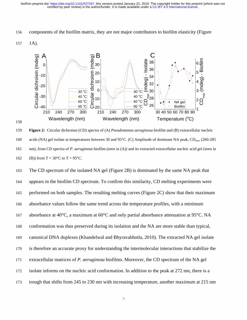
9
components of the biofilm matrix they are not major contributors to biofilm elasticity (Figure 156
1A) 157
158
Figure 2 Circular dichroism (CD) spectra of (A) Pseudomonas aeruginosa biofilm and (B) extracellular nucleic 159
acids (NA) gel isolate at temperatures between 30 and 95degC (C) Amplitude of dominant NA peak CDmax (260-285 160
nm) from CD spectra of P aeruginosa biofilm (seen in (A)) and its extracted extracellular nucleic acid gel (seen in 161
(B)) from T = 30degC to T = 95degC 162
The CD spectrum of the isolated NA gel (Figure 2B) is dominated by the same NA peak that 163
appears in the biofilm CD spectrum To confirm this similarity CD melting experiments were 164
performed on both samples The resulting melting curves (Figure 2C) show that their maximum 165
absorbance values follow the same trend across the temperature profiles with a minimum 166
absorbance at 40degC a maximum at 60degC and only partial absorbance attenuation at 95degC NA 167
conformation was thus preserved during its isolation and the NA are more stable than typical 168
canonical DNA duplexes (Khandelwal and Bhyravabhotla 2010) The extracted NA gel isolate 169
is therefore an accurate proxy for understanding the intermolecular interactions that stabilize the 170
extracellular matrices of P aeruginosa biofilms Moreover the CD spectrum of the NA gel 171
isolate informs on the nucleic acid conformation In addition to the peak at 272 nm there is a 172
trough that shifts from 245 to 230 nm with increasing temperature another maximum at 215 nm 173
210 240 270 300
-40
-30
-20
-10
0
210 240 270 300-20
-10
0
10
20
30
30 oC
40 oC
60 oC
95 oCC
ircu
lar
dic
hro
ism
(m
de
g)
Circu
lar
dic
hro
ism
(m
de
g)
Wavelength (nm)
30 oC
40 oC
60 oC
95 oC
CB
Wavelength (nm)
A
30 40 50 60 70 80 90
26
28
30
32
34
36
38
NA gel
CD
max (
md
eg
) -
Iso
late
Temperature (oC)
1
2
3
4
5
6
Biofilm
CD
max (
md
eg
) -
Bio
film
CC-BY 40 International licensecertified by peer review) is the authorfunder It is made available under aThe copyright holder for this preprint (which was notthis version posted January 22 2019 httpsdoiorg101101527267doi bioRxiv preprint
10
that remains constant with temperature and other sub-maxima at 260 and 253 nm that change 174
with temperature The absence of a trough at 200-215 nm precludes the possibility of NA in A- 175
or Z- conformations (Kypr et al 2009) The dominant maximum and minimum could indicate 176
either B-DNA or G-quadruplex conformations (del Villar-Guerra et al 2018) although the peak 177
at 215 nm is not a feature of B-DNA CD spectra and is slightly higher than the characteristic G-178
quadruplex low wavelength peak of 210 nm Nonetheless the appearance of several peaks in the 179
NA region of 250-285 nm indicates that while the dominant NA conformation is unclear several 180
conformations likely contribute to phase separation of the NA 181
Purine-rich ribonucleotides and eDNA are present in isolated gel 182
Total correlation spectroscopy (TOCSY) and 1H-
13C heteronuclear single quantum coherence 183
(HSQC) are nuclear magnetic resonance (NMR) techniques that can identify proton NMR 184
correlations within individual ribose sugars and their proton-carbon single bond correlations 185
respectively Raising the pH can solubilize P aeruginosa biofilms (Friedman and Kolter 2004) 186
and here we show that after alkalinization the NA peaks dominated the solution 1H-
13C HSQC 187
spectra for the isolated material with no indication that proteins or polysaccharides were present 188
(Supplementary Figure 8 (Wuumlthrich 2008)) The 1H-
13C HSQC-TOCSY spectrum of the 189
isolated gel when dissolved by alkalinization shows two clusters of sugar proton peaks (C1rsquo-H1rsquo) 190
with correlations to neighboring carbons (C2rsquo-H1rsquo) (Figure 3A) 191
The first cluster (rectangles) has C2rsquo chemical shift values of ~40 ppm and the second cluster 192
(ovals) has C2rsquo values of ~70 ppm The 1H-
13C correlations denoted by the rectangles therefore 193
arise from deoxyribose and those denoted by the ovals from ribose sugar conformations The 194
broadened form of the deoxyribonucleotide peaks is consistent with high molecular weight 195
(MW) molecules while the sharpness of the ribonucleotide peaks suggests that they could arise 196
CC-BY 40 International licensecertified by peer review) is the authorfunder It is made available under aThe copyright holder for this preprint (which was notthis version posted January 22 2019 httpsdoiorg101101527267doi bioRxiv preprint
11
from small possibly even single nucleotides Hence eDNA and small ribonucleotides are both 197
likely to be present in the gel dissolved by alkalinization 198
Spin-systems for the eight sharp ribonucleotide peaks (Figure 3B) were assigned by HSQC-199
TOCSY and correlation spectroscopy (COSY) (Supplementary Figure 9) with absolute 200
identification achieved by comparing the heteronuclear multiple bond correlation HMBC spectra 201
of samples and monoribonucleotide standards (Supplementary Figure 10) The 31
P NMR 202
spectrum of the NA gel isolate at elevated pH revealed the presence of a mixture of 203
monoesterified (34 to 41 ppm) and diesterified phosphates (-08 to -12 ppm) indicating the 204
coexistence of monoribonucleotides and DNA respectively (Figure 3C (i)) Long-range 31
P-1H 205
correlations (ie from NA phosphorous to adjoined ribose proton) were observed in the 31
P-1H 206
heteronuclear correlation (HETCOR) spectrum (Figure 3C (ii)) for both monoribonucleotides 207
and DNA The broad 31
P-1H cross peaks at ~-10 ppm (Figure 3C (ii)) correspond to long-range 208
correlations between DNA phosphorous to H3rsquo and H5rsquoH5rsquorsquo protons The eight 209
monoribonucleotides were assigned to 2rsquo- and 3rsquo-(A U G and C)-monophosphates We 210
therefore have both 2rsquo and 3rsquo monoribonucleotides present with the eDNA in the gel solubilized 211
under alkaline conditions 212
However the monoribonucleotide peaks only became resolved upon alkaline dissolution of the 213
biofilm in contrast to the broad peaks evident at pH 7 (Supplementary Figure 11) This indicates 214
that the monoribonucleotides are derived from chain structures that exist at biological pH and 215
assemble with DNA chains into higher-order networked structures which is consistent with the 216
observation in Figure 1A that RNA is a major contributor to P aeruginosa biofilm elasticity 217
218
219
CC-BY 40 International licensecertified by peer review) is the authorfunder It is made available under aThe copyright holder for this preprint (which was notthis version posted January 22 2019 httpsdoiorg101101527267doi bioRxiv preprint
12
220
221
222
223
224
225
226
227
Figure 3 (A) Nuclear magnetic resonance (NMR) 1H-13C 228
heteronuclear single quantum coherence (HSQC)-total 229
correlation spectroscopy (TOCSY) spectrum at 25degC of 230
extracellular nucleic acids (NA) gel isolate after 231
alkalinization showing the C1rsquo-H1rsquo cross peaks of RNA 232
(blue ovals) and DNA (red rectangles) and their correlations 233
to the neighboring carbon C2rsquo-H1rsquo Schematics (right) 234
illustrate these correlations (B) 1H-13C HSQC spectrum of 235
extracellular NA gel isolate at 25degC after alkalinization 236
identifying eight monoribonucleic acid ribose spin systems 237
(C) 1-D 31P NMR of NA isolate gel and Pseudomomonas 238
aeruginosa biofilm with proton decoupling showing the 239
presence of monoesterified (ie monoribonucleotides) and 240
diesterified (ie DNA) phosphate peaks (i) and 2-D 1H-31P heteronuclear correlation (HETCOR) spectrum of 241
extracellular NA showing the 31P-1H cross-peaks of monoribonucleotides and DNA (ii) Couplings of 242
monoesterified phosphates to H2rsquo or H3rsquo of eight monoesterified monoribonucleotides (from left to right 3rsquo 243
AMP3rsquo GMP 3rsquo CMP3rsquo UMP 2rsquo CMP2rsquo UMP 2rsquo AMP 2rsquo GMP) and diesterified phosphate to DNA H3rsquo and 244
H5rsquoH5rsquorsquo protons are denoted by the dashed lines There is a discontinuity (wavy line) in the 31P axis due to the 245
different thresholds required to illustrate the 31P-1H correlations in the mono-esterified and di-esterified regions All 246
samples were prepared in 01 M NaOD (10 mgmL) and preheated to 55degC for 2 h 247
CC-BY 40 International licensecertified by peer review) is the authorfunder It is made available under aThe copyright holder for this preprint (which was notthis version posted January 22 2019 httpsdoiorg101101527267doi bioRxiv preprint
13
The molar ratio of the individual ribonucleotides could be determined from the 31
P spectrum of 248
the gel dissolved at high pH (Table 1) While several peaks could not be separated it was 249
possible to deduce that the RNA is purine rich (ie 57 mol A+G) and that the G+C mol 250
content of 46-50 differs from that of the P aeruginosa genome (ie 67 mol) (Shen et al 251
2006) The same peaks were also observed in the biofilm 31
P spectrum after alkalinization 252
(Figure 3C(i)) 253
Table 1 Relative abundances of monoribonucleotides in extracellular NA gel isolate from Pseudomonas 254 aeruginosa biofilm as determined by integrating 31P NMR spectrum following alkalinization 255
31P shift
(ppm)
Ribonucleotide Relative
abundance
403 3rsquoAMP 180
401 3rsquo GMP 172
390 3rsquo CMP3rsquo UMP 89
387 3rsquo CMP3rsquo UMP 113
368 2rsquo CMP2rsquo UMP 95
366 2rsquo CMP2rsquo UMP 117
350 2rsquo AMP 104
342 2rsquo GMP 130
256
Extracellular DNA and RNA interact to form a network 257
We applied Magic-angle spinning (MAS) solid-state NMR (SSNMR) to confirm that the 258
networks in the gel isolate and biofilm are comprised of DNA and RNA in chain form This 259
technique is ideal for intractable systems such as biofilms to eliminate solubility and extraction 260
biases (Reichhardt and Cegelski 2014) and to analyze inter-molecular H-bond interactions to 261
describe for example inter-nucleotide base pairing in RNA (Marchanka et al 2015) MAS 262
SSNMR averages anisotropic interactions to provide high-resolution spectral characterization of 263
insoluble and large biomolecular systems By analyzing dipolar interactions through-space 264
heteronuclear correlations (eg N∙∙∙H) can be detected In contrast to the liquid state 31
P NMR 265
spectrum of the alkali-dissolved isolate (Figure 3C (i)) the 31
P SSNMR spectrum of the gel 266
CC-BY 40 International licensecertified by peer review) is the authorfunder It is made available under aThe copyright holder for this preprint (which was notthis version posted January 22 2019 httpsdoiorg101101527267doi bioRxiv preprint
14
isolate (ie no alkali treatment Figure 4A (i)) showed a single peak in the diesterified phosphate 267
region consistent with the presence of both DNA and RNA chains There are no sharp peaks 268
present in the monoesterified phosphate region further demonstrating that the 269
monoribonucleotides are a consequence of alkali transesterification (Radak et al 2013) 270
Only the diesterified phosphate peak was observed in the 31
P SSNMR spectrum of the biofilm 271
(Figure 4A (iii)) while both RNA-derived monoesterified and DNA-derived diesterified 272
phosphate peaks were present in the 31
P SSNMR spectrum of alkali digested NA gel-isolate and 273
biofilm (lyophilized) (Figure 4A (ii) and Figure 4A (iv) respectively) Complete alkali RNA 274
transesterification was confirmed by the full conversion of diesterified to monoesterified 275
phosphates (from liquid state 31
P NMR spectra) for the RNA standard when dissolved in 01 M 276
NaOH (Supplementary Figure 12A) Conversely RNA diesterified phosphate peaks were 277
preserved in the RNA standard spectrum following dissolution in EMIM-Ac and recovery by 278
perchloric acid (Supplementary Figure 12B) Hence the alkaline conditions break down the 279
RNA chains into individual monoribonucleotides while the ionic liquid-based extraction does 280
not This suggests that the conventional method for P aeruginosa biofilm dissolution in alkali 281
(Friedman and Kolter 2004) may in fact work by transesterifying RNA as a primary structural 282
component and illustrates the importance of the ionic liquid-based extraction protocol described 283
here as a non-destructive method for interrogating biofilm structural polymers 284
CC-BY 40 International licensecertified by peer review) is the authorfunder It is made available under aThe copyright holder for this preprint (which was notthis version posted January 22 2019 httpsdoiorg101101527267doi bioRxiv preprint
15
285
Figure 4 (A) Solid-state 31P NMR spectra at T = 25degC of extracellular NA gel isolate (i) alkalinized and 286
lyophilized NA gel isolate (ii) Pseudomonas aeruginosa biofilm (iii) and alkalinized and lyophilized P aeruginosa 287
biofilm (iv) showing the presence of diesterified phosphate peaks and the absence of monoesterified phosphate 288
peaks for both NA gel isolate and biofilm in double distilled water and the coexistence of diesterified and 289
monoesterified phosphate peaks for both samples after alkalinization This indicates that alkalinization of the matrix 290
results in RNA transesterification Micrographs of P aeruginosa NA gel isolate stained green with (B) SYTO 291
RNASelect showing RNA fibers and (C) TOTO-1 showing DNA fibers (scale bar 10 microm) 292
293
To visualize the DNA-RNA network organization we used nucleic acid-specific stains ie RNA 294
specific SYTO RNASelect dye (Figure 4B) and the eDNA-specific TOTO-1 dye (Figure 4C) 295
(Okshevsky and Meyer 2014) Fibrous structures which are typical of networked polymer gels 296
(Cornwell and Smith 2015) were observed in the NA gel isolate stained with both nucleic acid 297
dyes as well as in the biofilm stained with the eDNA specific dye (Figure 1C) This is consistent 298
with our observation that the biofilm is readily degraded by DNase I (Figure 1A) (Chen et al 299
2004) Furthermore the NA gel isolate was stained positively with the RNA-specific dye even 300
after DNase I digestion providing further evidence that RNA contributes to networking 301
BeRNA fibers
C
eDNA fibers
A
CC-BY 40 International licensecertified by peer review) is the authorfunder It is made available under aThe copyright holder for this preprint (which was notthis version posted January 22 2019 httpsdoiorg101101527267doi bioRxiv preprint
16
(Supplementary Figure 13) However digestion with RNases III A and H did not degrade the 302
biofilm (Figure 1A Supplementary Figure 14) This may suggest that the RNase binding was 303
shielded by hairpins in the network or the presence of non-canonical DNA-RNA interactions 304
(Geerts-Dimitriadou et al 2012 Nakamura et al 1991) 305
Non-canonical and Watson-Crick base pairs and tetrads support extracellular network 306
Sequence analysis of the extracted material indicated that the gene coverage was even for both 307
chromosomal and extracellular DNA with the exception of bacteriophage Pf1 genes 308
(Supplementary Figure 15) However the Pf1 knockout mutant of P aeruginosa also displayed 309
an elastic response when dissolved in EMIM-Ac (Supplementary Figure 16 Supplementary 310
Tables 1 and 2) indicating that Pf1 DNA is unlikely to be responsible for the phase-separating 311
behavior of P aeruginosa biofilms DNA and RNA interaction therefore cannot be explained by 312
uneven gene coverage To elucidate the mechanism of DNA-RNA gelation we generated a 2D 313
through-space 15
N-1H HETCOR spectrum of
15N
labeled DNA-RNA gel isolated from a P 314
aeruginosa biofilm matrix (Figure 5A) This spectrum was run with longer cross polarization 315
contact times so that correlations between distant 1H and
15N could be observed This confirmed 316
the complete absence of proteins and supported our conclusion that proteins do not contribute to 317
the DNA-RNA interaction The HETCOR spectrum showed four signal clusters at δH 10-14 318
ppm and δN 140-160 ppm (Figure 5A) which arose from direct N-H couplings of TU and G 319
nucleobase imino groups Two of these clusters (δH 12-14 ppm) resulted from imino protons 320
hydrogen-bonded to a nucleobase nitrogen (ie N-H∙∙∙N) and the other two (δH 10 -12 ppm) 321
from imino protons hydrogen-bonded to a nucleobase carbonyl oxygen (ie N-H∙∙∙O) Due to the 322
longer cross polarization times we were able to observe a strong long-range and indirect (ie 323
intermolecular) correlation at δN 196 ppm arising from G-C Watson-Crick base pairing ie 324
CC-BY 40 International licensecertified by peer review) is the authorfunder It is made available under aThe copyright holder for this preprint (which was notthis version posted January 22 2019 httpsdoiorg101101527267doi bioRxiv preprint
17
C(N3)-G(H1) There was also a weak and indirect correlation at δN 220 ppm resulting from A-325
TU Watson-Crick base pairing ie A(N1) to UT(H1) 326
327
328
Figure 5 (A) Solid-state 2D 1H-15N through-space heteronuclear correlation (HETCOR) spectrum of extracellular 329
nucleic acid (NA) gel isolate in double distilled water (2 mg) T = 25degC showing direct N-H couplings for G imino 330
N (N3) and TU imino N (N3) bonded to nucleobase nitrogen (NHN) This indicates Watson-Crick (WC) G-C and 331
UT-A base pairings respectively and for G imino N (N1) and TU imino N (N3) bonded to nucleobase carbonyl 332
oxygen (ie NHO) indicating non-canonical G-G G-TU and UT-UT base pairings or tetrads The spectrum also 333
displayed indirect N-H correlations for the WC base pairs (ie C(N3) to G(H1) and A(N1) to UT(H1)) and no 334
indirect correlations for the non-canonical bonded nucleobases The threshold for the A(N1)-TU(H3) correlation 335
was increased due to lower signal intensity for that particular coupling (B) Pseudomonas aeruginosa biofilm 336
growth yield at 37degC (5 d) as a function of [K+] [Na+] or [Li+] showing the growth dependence on monovalent 337
cation concentration (K+gtNa+gtLi+) Error bars indicate standard deviation (C) Illustration of how structural 338
variability of RNA may allow for the formation of extracellular RNA and DNA networks through hairpins and a 339
mixture of canonical and non-canonical base pairs or tetrads (ie Hoogsteen and G-quadruplexes) 340
341
The absence of long-range correlations between the clusters at δH 10 -12 ppm is consistent with 342
non-Watson-Crick pairings for the G and TU nucleobases The observation of NH to O 343
interactions suggests the formation of G-G TU-TU G-TU Hoogsteen base pairs or tetrads 344
CA B C
CC-BY 40 International licensecertified by peer review) is the authorfunder It is made available under aThe copyright holder for this preprint (which was notthis version posted January 22 2019 httpsdoiorg101101527267doi bioRxiv preprint
18
Hoogsteen H-bonded G-bases can assemble planarly into tetrads and self-stack to form G-345
quadruplexes (Haumlnsel-Hertsch et al 2017) G-quadruplexes are stabilized by the presence of a 346
monovalent cation with potassium providing the greatest stability We experimentally confirmed 347
that potassium had a similar effect in a static biofilm growth assay We found that potassium 348
promoted P aeruginosa biofilm growth to a greater extent than either sodium or lithium (Figure 349
5B) Additionally this was a biofilm-specific effect as there was no effect of monovalent cation 350
on planktonic growth (Supplementary Figure 17) Based on the SSNMR data coupled with the 351
potassium effect and the earlier observation of several maxima in the CD spectrum of the NA gel 352
isolate across the characteristic NA region of 250-285 nm which suggested the coincidence of 353
several NA conformations contributing to phase separation it is proposed that G-quadruplexes 354
contribute to extracellular DNA-RNA networking 355
Discussion 356
We extracted a gel-forming complex composed of DNA and RNA from the extracellular matrix 357
of P aeruginosa biofilms without disrupting its fundamental structural or chemical organization 358
We report the novel observation that extracellular RNA contributes structurally to biofilms 359
While gel-forming exopolymers have been detected in other biofilms (Seviour et al 2009) no 360
functional DNA gel has been described for any biological system much less one also including 361
extracellular RNA 362
While it is not possible to identify the precise nature of the DNA-RNA interaction based on the 363
information presented here purine-rich RNA as described here for the extracellular NA gel 364
isolate (57 mol A-G) is a characteristic of DNA-RNA duplexes because RNA purines bind 365
more strongly to their DNA pyrimidine complement than vice versa (Nadel et al 2015) Both 366
Watson-Crick and non-canonical interactions were present in the NA gel isolate which is also 367
CC-BY 40 International licensecertified by peer review) is the authorfunder It is made available under aThe copyright holder for this preprint (which was notthis version posted January 22 2019 httpsdoiorg101101527267doi bioRxiv preprint
19
consistent with the discrepancy observed between RNA and DNA G+C mol contents (Table 368
1) It is possible therefore that interactions between DNA and RNA as described in this study 369
account for the formation of a highly stable nucleic acid gel 370
Non-canonical base pairing and hairpinning are commonly seen with RNA and cause multivalent 371
intermolecular interactions between RNA strands that lead to a sol-gel transition (Jain and Vale 372
2017) The greater structural versatility of RNA could be a key to the ability of the eDNA to 373
form networks with hairpins as well as canonical and non-canonical base pair interactions all 374
contributing to the assembly of higher order NA structures (Figure 5C) RNA has recently been 375
found to promote intracellular phase separations although this has not been extended to the 376
extracellular domain of prokaryotic cells (Shin and Brangwynne 2017) Additionally G-rich 377
RNA sequences are more predisposed to forming G-quadruplexes because they do not have a 378
complementary strand and in fact form more stable G-quadruplexes than DNA (Guo and Bartel 379
2016) It is therefore probable that the RNA but not DNA enables DNA-RNA networking and 380
gelation 381
The results presented here provide unprecedented resolution of the biofilm exopolymeric matrix 382
and its key foundation structural components We have developed a methodology that preserved 383
the molecular organization of the foundational polymer in its native state upon extraction and 384
isolation This enabled us to use SSNMR to describe intermolecular associations at the atomic 385
level 386
Our findings that a DNA-RNA gel network can provide the foundation for biofilm matrices in P 387
aeruginosa mark a departure from the prevailing paradigm that biofilm gelation is only due to 388
polysaccharides The elastic and film-forming properties were also observed for Pseudomonas 389
putida and Pseudomonas protegens (Figs S16 and S18 Supplementary Tables 1 and 2) 390
CC-BY 40 International licensecertified by peer review) is the authorfunder It is made available under aThe copyright holder for this preprint (which was notthis version posted January 22 2019 httpsdoiorg101101527267doi bioRxiv preprint
20
suggesting that eDNA is broadly important for biofilm formation in members of this genus In 391
addition to Pseudomonas spp other organisms are known to have eDNA including clinical 392
organisms Staphylococcus aureus (Mann et al 2009) Staphylococcus epidermidis (Adam et al 393
2002) and Mycobacterium abscessus (Rose et al 2015) as well as environmental isolates such 394
as strain F8 from the South Saskatchewan River (Boumlckelmann et al 2006) 395
While eDNA is commonly thought to be the product of cell lysis and has been shown to be 396
released by a sub-population of lytic P aeruginosa cells (Turnbull et al 2016) there is an 397
increasing awareness that it serves an important structural function such as in activated sludge 398
granules (Cheng et al 2011) Additionally Bockelmann et al (2006) observed that stable 399
filamentous networks produced by the aquatic strain F8 were comprised of DNA This is the first 400
study that provides an explanation for how eDNA is organized structurally However the 401
environmental factors contributing to eDNA release and how this is regulated (ie whether it is 402
an active or passive process) are still unclear It is possible that DNA-RNA foundational gel 403
networks as described here for the Pseudomonads are broad-scoping phenomena in biofilms 404
and that interactions with RNA enable an extracellular structural function for DNA Thus 405
elucidating how nucleic acids including RNA are integral to the biophysical and other emergent 406
properties imparted on the biofilm via the extracellular matrix will inform the regulation and 407
control of extracellular nucleic acid release across environmental and clinical biofilms 408
Materials and Methods 409
Bacterial strains 410
Unless otherwise stated all experiments were undertaken directly on P aeruginosa PAO1 411
biofilms grown in lysogeny broth at 37degC The P aeruginosa PAO1 Δpf4 knockout mutant is a 412
CC-BY 40 International licensecertified by peer review) is the authorfunder It is made available under aThe copyright holder for this preprint (which was notthis version posted January 22 2019 httpsdoiorg101101527267doi bioRxiv preprint
21
defined Pf4 chromosomal deletion mutant of the entire Pf4 prophage genome(Rice et al 413
2009) 414
P aeruginosa PDO300 PDO300(Δpel) and PDO300(Δpsl) mutant strains were gratefully 415
received from Professor Bernd H A Rehm Institute of Molecular Biosciences Massey 416
University New Zealand (Ghafoor et al 2011) PDO300 PDO300(Δpel) PDO300(Δpsl) and 417
PAO1 (Δpf4) were also grown in LB at 37degC P protogens Pf-5 and P putida ATCC BAA-477 418
and S12 strain were were grown in LB at 30degC 419
Biofilm growth assay 420
Ten milliliter aliquots of P aeruginosa planktonic pre-cultures (LB 200 rpm 37degC OD600 240 421
16 h) were diluted 50 times with LB in 2 L Erlenmeyer flasks and incubated for 5 d under static 422
conditions Supplementary Figure 15A displays an image of 5-d old biofilm in LB The cultures 423
were centrifuged at 10000 x g for 15 min the supernatant removed by decanting and the 424
biofilm then collected and lyophilized (LabConco) Supplementary Figure 15B displays an 425
image of the culture after centrifugation showing clear separation of biofilm and supernatant 426
Enzymatic digestions 427
Twenty milligrams of lyophilized biofilm was resuspended in 1 mL of either i) RNase buffer (50 428
mM Tris HCl 10 mM EDTA pH8) with 02 mg RNaseA from bovine pancreas (Sigma Aldrich) 429
ii) storage buffer (10 mM NaCl 10 mM Tris-HCl) with 01 mg Pronase E from Streptomyces 430
grisens (Sigma Aldrich) with 05 (vv) iii) 1X RNase H reaction buffer (20 mM Tris-HCl pH 431
78 40 mM KCl 8 mM MgCl2 1 mM DTT) with 04 mg RNase H (Thermo Fisher Scientific) 432
iv) 1X RNase III reaction buffer (500 mM NaCl 100 mM Tris pH 79 100 mM MgCl2 10 mM 433
DTT) with 06 mg RNase II (Thermo Fisher Scientific) or v) DNase I buffer (100mM Tris (pH 434
75) 25 mM MgCl2 and CaCl2) with 02 mg DNAse I from bovine pancreas (Sigma Aldrich) All 435
CC-BY 40 International licensecertified by peer review) is the authorfunder It is made available under aThe copyright holder for this preprint (which was notthis version posted January 22 2019 httpsdoiorg101101527267doi bioRxiv preprint
22
digestions were performed with shaking at 200 rpm at 37degC for 16 h The suspensions were then 436
centrifuged (10000 x g 15 min) the supernatant was discarded and the pellets of the biofilm 437
materials were lyophilized 438
Normal force measurement 439
Forty milligrams per milliliter solutions of lyophilized biofilms were added to 1 mL 1-ethyl-3-440
methylimidazolium acetate and incubated in 55degC for either 2 h A Haake Mars 3 (Thermo 441
Fisher Scientific) stress-controlled rotational rheometer with Peltier controlled element at 25degC 442
was used for rheological measurements Thirty five-illimeter diameter parallel plate geometry 443
was used with smooth titanium plates to measure viscosity and normal stress difference (N1 ndash 444
N2) Prior to measurement the gap error was zeroed at 4 N and gap error calculated as previously 445
described (Bird et al 1987 Davies and Stokes 2008 Kravchuk and Stokes 2013) One 446
hundred microliters of sample was deposited on the plates The plates were closed to 100 microm the 447
sample trimmed and the sample allowed to sit for 5 min prior to measurement All measurements 448
with Normal force (FN) less than the resolution of the rheometer (ie lt 01 N) were set to 0 449
before calculation of N1-N2 using equations 9-11 and viscosity from equation 3-5 in Davies and 450
Stokes(Davies and Stokes 2008) for the parallel plate geometry 451
Only the linearly increasing portion of the normal stress difference curves are presented Above 452
this range normal stress difference begins to decrease again which may be due to elastic 453
instabilities or associating polymers (Annable et al 1993) Corrections were made to N1 and N1 454
ndash N2 to account for inertia using equation 17 in Davies and Stokes (Davies and Stokes 2008) and 455
to correct for the baseline residual force in the samples Except for the DNase I-treated biofilm 456
the shear rheology for all treatments could be modelled using the finitely extensible non-linear 457
elastic with Gaussian closure proposed by Peterlin (FENE-P) constitutive model by varying four 458
CC-BY 40 International licensecertified by peer review) is the authorfunder It is made available under aThe copyright holder for this preprint (which was notthis version posted January 22 2019 httpsdoiorg101101527267doi bioRxiv preprint
23
parameters to fit shear viscosity and normal stress difference as a function of shear rate 459
(Supplementary Table 2) Fitting parameters for the FENE-P model include 1 = relaxation time 460
b = a measure of the relative extensibility of the model spring s = solvent viscosity p = 461
polymer contribution to the viscosity The FENE-P equations can be written in the following 462
format as shown by Bird et al(1987) 463
120578 = 120578119904 +120578119901
1205821((1198622 + 1198621)
1
3 minus (1198622 minus 1198621)1
3) (Equation 1) 464
465
1198731 = 2120578119901
1205821((1198622 + 1198621)
1
3 minus (1198622 minus 1198621)1
3)2
(Equation 2) 466
Where 467
1198621 = 119887
41205821 (Equation 3) 468
1198622 = (11986212 + (
119887+3
6)
3
)12
(Equation 4) 469
470
All measurements were performed in triplicate For clarity one representative data set is 471
presented in Figure 1A-B and Supplementary Figure 1A-B with the respective Power Law and 472
FENE-P model fit to that data set Averaged values for the FENE-P and Power law fits with the 473
standard deviation across three replicates are shown in Supplementary Tables 1 and 2 474
Oscillatory measurements were carried out with controlled frequency 01 rads across an 475
amplitude range of 001 to 10 476
Extracellular polymeric substances (EPS) extraction 477
Lyophilized biofilms were dissolved in ionic liquid mixture (40 (vv) 1-ethyl-3-478
methylimidazolium acetate (EMIM Ac) 60 (vv) NN-dimethyl acetamide (DMAc)) at 55degC 479
CC-BY 40 International licensecertified by peer review) is the authorfunder It is made available under aThe copyright holder for this preprint (which was notthis version posted January 22 2019 httpsdoiorg101101527267doi bioRxiv preprint
24
for 16 h The solution was centrifuged (10000 x g) to remove any undissolved material 480
Perchloric acid (70) was added (005 vv) to the viscosified centrate (on ice) After 15 min 481
incubation the solution was centrifuged at 10000 x g at 4degC for 15 min and the pellet recovered 482
This was repeated on the centrate two to four times until the solution was not viscous The 483
precipitate was dialysed against double distilled water for 2 d at 4degC (SnakeSkintrade Dialysis 484
Tubing 35K MWCO 22 mm) and the retentate lyophilized (FreeZone Plus 45 Liter Cascade 485
Benchtop Freeze Dry System) The same procedure was performed on calf thymus DNA lipase 486
cytochrome C for the purposes of determining recovery yield of representative exoproteins and 487
on RNA from torula yeast for assessing by 31
P NMR whether the extraction procedure 488
contributed to RNA transesterification (all from Sigma Aldrich) 489
Extracellular nucleic acid isolation 490
Twenty milligrams of lyophilized retentate (ie post perchloric acid precipitation) were dissolved 491
in 1 mL of 40 (vv) EMIM-Ac 60 DMAc (vv) (55degC 16 h) Chromatographic separation 492
was achieved in a Shimadzu system comprising DGU-20A 3r Prominence Degasser and LC-493
20AD Solvent Delivery Unit fitted with two Agilent PLgel 10 μm column of 105Aring pore size for 494
separation across the MW range 200 kDa to 2000 kDa The eluent flow rate was 30 mLmin-1
495
and the injection volume 1 mL The fractions with molecular weight range of 2000-800kDa and 496
800-200kDa were pooled and dialyzed for 2 d at 4degC (SnakeSkintrade Dialysis Tubing 35K 497
MWCO 22 mm) against double distilled water to induce gelation The gel was then collected 498
from the dialysis tubing 499
Solution-state nuclear magnetic resonance (NMR) 500
Solution-state NMR experiments were performed on an 800 MHz Bruker Avance III 501
spectrometer at 25oC Sample concentration was 10 mg (dry weight)mL
-1 unless otherwise 502
CC-BY 40 International licensecertified by peer review) is the authorfunder It is made available under aThe copyright holder for this preprint (which was notthis version posted January 22 2019 httpsdoiorg101101527267doi bioRxiv preprint
25
specified Spectra were recorded either under conditions of neutral pH in 100 D2O (Cambridge 503
Isotope Laboratories) or following alkalinization (ie transesterification 01 M NaOD 55oC 2 504
h) 1-D NMR experiments include 1H and
31P direct detection while 2-D NMR analysis include 505
13C-HSQC
13C-HSQC-TOCSY
1H-
31P HETCOR HMBC and COSY All spectral analyses 506
were performed using Topspin and SPARKY software 507
Asolectin (Sigma Aldrich) standard (10 mgmL) and lyophilized Pseudomonas aeruginosa 508
PAO1 biofilm (10mgmL) were dissolved in 40 (vv) EMIM Ac 60 (vv) DMAc at (55oC 2 509
h) P aeruginosa PAO1 pre-culture cell lysate was prepared by lysing pre-culture cells with 510
lysozyme in PBS 10 (vv) of D2O was added to all samples for locking purposes 511
Solid-state NMR 512
For solid-state NMR experiments performed on the NA gel isolate 15
N labeled NH4Cl-513
supplemented M9 minimal media was used for biofilm growth M9 consisted of 9552 gL-1
514
Na2HPO42H2O 441g litre-1
KH2PO4 171 gL-1
NaCl 1 gL-1
15
NH4Cl 024 gL-1
MgSO4 515
0011 gL-1
CaCl2 2 gL-1
casamino acids and 04 gL-1
glucose The NA gel was prepared from 516
the M9-grown P aeruginosa PAO1 biofilm as described above 517
Solid-state NMR experiments were performed on 141 T Bruker Advance III instruments 518
equipped with a 19 mm MAS probe operated in double mode The typical 1H
15N and
31P π2 519
pulse lengths were 23 37 and 45 μs respectively 2D dipolar-based 15
N-1H heteronulcear-520
correlation (HETCOR) experiments were conducted on the 15
N-labelled NA gel isolate at 37 kHz 521
MAS spinning frequency Variable temperature was regulated at -20ordmC and the sample 522
temperature was 12ordmC (calibrated using ethylene glycol) In the 15
N-1H HETCOR experiments 523
the initially excited 1H magnetization was transferred to
15N through a cross polarization step 524
followed by t1 evolution Then the 15
N magnetization was flipped to longitudinal axis and 400 525
CC-BY 40 International licensecertified by peer review) is the authorfunder It is made available under aThe copyright holder for this preprint (which was notthis version posted January 22 2019 httpsdoiorg101101527267doi bioRxiv preprint
26
ms proton saturation pulses were applied for water suppression Subsequently the 15
N 526
magnetization was flipped to the transverse plane and transferred to 1H via a second CP step for 527
signal acquisition Two 15
N-1H HETCOR experiments were collected one with 400 s and the 528
other with 2 ms contact times applied for both of the CP steps Low power XiX 1H decoupling 529
(~10 kHz) was employed during 15
N evolution and WALTZ-16 decoupling (10 kHz) was 530
implemented on 15
N channel during 1H acquisition 531
1-D 31
P experiments were performed on 15
N-labelled NA gel isolate and 15
N-labelled P 532
aeruginosa PAO biofilm both directly after dialysis against double distilled water at 4degC for 2 d 533
(SnakeSkintrade Dialysis Tubing 35K MWCO 22 mm) and following alkalinization (01 M 534
NaOD 55ordmC 15 min) and lyophilization (FreeZone Plus 45 Liter Cascade Benchtop Freeze Dry 535
System) 15 kHz MAS spinning frequency and a sample temperature of 27ordmC 75 kHz 536
SPINAL64 1H decoupling was applied during
31P acquisition time All chemical shifts were 537
indirectly referenced using adamantane as a secondary standard (downfield peak is at 4048 ppm 538
DSS scale) 539
Monovalent cation-dependent biofilm growth 540
Ten milliliters aliquots of P aeruginosa pre-cultures (supplemented M9 200 rpm OD600 240 541
16 h) were transferred into 500 mL M9 minimal media (85 gL-1
Na2HPO42H2O 20 gL-1
of 542
NaH2PO4H2O 10 gL-1
KH2PO4 10 gL-1
NH4Cl 048 gL-1
MgSO4 0011 gL-1
CaCl2 2 gL-1
543
casamino acids and 04 gL-1
glucose) supplemented with either KCl LiCl or NaCl at either of 544
three different concentrations (0 002 mM and 008 mM) Biofilms were collected by 545
centrifugation (10000 x g 15 min) as described above lyophilized (LabConco) and weighed To 546
describe the growth curve P aeruginosa wild type PAO1 pre-culture was incubated in M9 547
media at 37degC 200 rpm for 16 h PAO1 WT pre-culture was then diluted with supplemented M9 548
CC-BY 40 International licensecertified by peer review) is the authorfunder It is made available under aThe copyright holder for this preprint (which was notthis version posted January 22 2019 httpsdoiorg101101527267doi bioRxiv preprint
27
media to a volume of 75 mL and a starting OD600 of 001 in a 250 mL Erlenmeyer flask The 549
mixture was incubated at 37degC with shaking (200rpm) The OD600 of the bacteria was measured 550
hourly for 9 h followed by final time point at 24 h 551
DNA Sequencing 552
Genomic DNA was extracted from the biofilm using FastDNA SPIN Kit for soil (MP 553
Biomedicals USA) as per the standard protocol Briefly biofilm was resuspended in Sodium 554
Phosphate Buffer in was lysed (Lysing Matrix) homogenized (FastPrepreg
40 seconds speed 555
setting 60) and the cell debris removed by centrifugation (14000 x g 5 min) Proteins were 556
removed by precipitation (250 μl Protein Precipitation Solution) the supernatant mixed with 557
DNA Binding Matrix which was then homogenized and transferred to a SPINtrade Filter Excess 558
supernatant was removed by centrifugation (14000 x g 5 min) DNA was then eluted from air 559
dried DNA Binding Matrix with DNase Pyrogen-Free Water 560
The NA gel isolate was resuspended in 500 uL of 1x Protease K solution (10times Protease K 561
solution 10 mM Tris HCl 1 SDS and 10 mM EDTA pH 8 buffer (10times protease K buffer 562
containing 500 mM Tris-HCl 10 SDS 10 mM CaCl2) and 10 L of Protease K (20 mgmL-1
563
Thermo Fisher Scientific) was added and the mixture incubated at 56degC for 2-16 h after which 564
DNA was extracted as previously described in the phenol-chloroform method(Ausubel 2002) 565
Samples before sequencing were further purified to remove any remaining protein and RNA by 566
RNase and Proteinase K treatment The DNA was then isolated using phenol-chloroform 567
precipitation as described above The DNA precipitate was dissolved in TE buffer the purity 568
confirmed by 260280 value in Nanodrop (acceptable range value 18-20) and Qubitreg
20 569
fluorometer 570
CC-BY 40 International licensecertified by peer review) is the authorfunder It is made available under aThe copyright holder for this preprint (which was notthis version posted January 22 2019 httpsdoiorg101101527267doi bioRxiv preprint
28
The molecular weight distributions of extracellular and genomic DNA were measured on a 1 571
agarose gel which was prepared from Viviantis LE grade agarose using 1x TAE buffer (40 mM 572
Tris 20 mM Acetate and 1 mM EDTA pH 86) Gels were run horizontally After 573
electrophoresis the gel was stained for 05 h with ethidium bromide and visualized under 574
UV(Lee et al 2012) 575
Three replicates were used for each DNA sequence analysis Library was produced using 576
Illumina DNA sample preparation kit The libraries were sequenced using Illumina MiSeq 577
platform (Illumina San Diego Ca) with paired-end protocol to read lengths of 600 nt generating 578
a total of 1614106 and 1848846 paired end reads Raw reads were quality filtered (reads 579
remaining after trimming PPG1-1549104 PBLC1-1666280) and aligned to the P aeruginosa 580
PAO1 (AE004091) genome using CLC Genomics Workbench 90 (CLC bio Cambridge MA) 581
Circular Dichroism CD 582
Five-day old biofilm and NA gel isolate were resuspended in double distilled water to achieve 583
UV absorbance reading 1 and ddH2O served as a blank The heat-treated samples were analyzed 584
by JASCO-815 spectropolarimeter in a 1 cm path length quartz cuvette containing a solution 585
volume 500 μL Spectra (200-320 nm) were measured at 1degC increments from 30 - 90degC For 586
each measurement an average of three scans was taken and the buffer spectra subtracted Each 587
spectrum presented is the rolling average across 5 temperatures 588
Staining and microscopy 589
Microscopic imaging was conducted on a confocal microscope Zeiss LSM 780 with a 63times 590
objective Extracellular RNA in the gel isolate were stained using SYTO RNASelect (Thermo 591
Fisher Scientific) green fluorescent cell stain (5 mM solution in DMSO) Five microM stain solution 592
was prepared from 1 microL of stock in 1X PBS solution (137 mM NaCl 27 mM KCl 43 mM 593
CC-BY 40 International licensecertified by peer review) is the authorfunder It is made available under aThe copyright holder for this preprint (which was notthis version posted January 22 2019 httpsdoiorg101101527267doi bioRxiv preprint
29
Na2HPO4 147 mM KH2PO4) The gel isolate was labelled with 5 microM stain solution kept at 594
37degC for 20 min and then transferred to glass side for imaging 595
eDNA staining was achieved by depositing biofilm or NA gel isolate on a glass slide air-drying 596
overnight and incubating with 2 microM TOTO-1 iodide (1 mM solution in DMSO Thermo Fisher 597
Scientific) for 15 min 598
Acknowledgments 599
We acknowledge Prof Bernd Rehm for supplying polysaccharide deletion mutants of P 600
aeruginosa Dan Roizman for providing P putida Long Yu for assistance with rheology Dr 601
Gleb Yakubov for coordinating sample preparation for rheological measurements Ravi 602
Jagadeeshan for discussion regarding DNA normal force analysis and Florentin Constancias for 603
analyzing the sequencing data SCELSE is funded by Singaporersquos Ministry of Education 604
National Research Federation Nanyang Technological University (NTU) and National 605
University of Singapore (NUS) and hosted by NTU in partnership with NUS 606
Author Contributions 607
TS FRW WLL SM HMS and XS performed experiments TS and ATP designed 608
the experiments TS FRW ATP HMS GSK and JRS analysed the data TS 609
FRW WLL HMS SAR ATP and SK wrote the manuscript 610
Competing Interests 611
All authors have no competing interests 612
References 613
Mixed species biofilms of Candida albicans and Staphylococcus epidermidis 614 B Adam GS Baillie LJ Douglas (2002) 615
CC-BY 40 International licensecertified by peer review) is the authorfunder It is made available under aThe copyright holder for this preprint (which was notthis version posted January 22 2019 httpsdoiorg101101527267doi bioRxiv preprint
30
Journal of Medical Microbiology 51344-349 616
doi1010990022-1317-51-4-344 617
A characterization of DNA release in Pseudomonas aeruginosa cultures and biofilms 618
M Allesen-Holm KB Barken L Yang M Klausen JS Webb S Kjelleberg S Molin M Givskov T Tolker-Nielsen 619
(2006) 620
Molecular Microbiology 591114-1128 621
doi101111j1365-2958200505008x 622
The rheology of solutions of associating polymers Comparison of experimental behavior with transient 623
network theory 624
T Annable R Buscall R Ettelaie D Whittlestone (1993) 625
Journal of Rheology 37695-726 626
doi1011221550391 627
Short protocols in molecular biology a compendium of methods from Current protocols in molecular 628
biology 629
FM Ausubel (2002) 630
5th ed New York Wiley 631
Dynamics of Polymeric Liquids 632
RB Bird RC Armstrong O Hassager (1987) 633
2nd ed 634
Bacterial extracellular DNA forming a defined network-like structure 635
U Boumlckelmann A Janke R Kuhn TR Neu J Wecke JR Lawrence U Szewzyk (2006) 636
FEMS Microbiology Letters 26231-38 637
doi101111j1574-6968200600361x 638
Infections caused by Pseudomonas aeruginosa 639
GP Bodey R Bolivar V Fainstein L Jadeja (1983) 640
Review of Infectious Diseases 5279-313 641
doi101093clinids52279 642
Pseudomonas aeruginosa uses a cyclic-di-GMP-regulated adhesin to reinforce the biofilm extracellular matrix 643 BR Borlee AD Goldman K Murakami R Samudrala DJ Wozniak MR Parsek (2010) 644
Molecular Microbiology 75827-842 645
doi101111j1365-2958200906991x 646
DNARNA helicase gene mutations in a form of juvenile amyotrophic lateral sclerosis (ALS4) 647
YZ Chen CL Bennett HM Huynh IP Blair I Puls J Irobi I Dierick A Abel ML Kennerson BA Rabin GA 648
Nicholson M Auer-Grumbach K Wagner P De Jonghe JW Griffin KH Fischbeck V Timmerman DR Cornblath 649
PF Chance (2004) 650
Am J Hum Genet 741128-1135 651
doi101086421054 652
Evidence of compositional differences between the extracellular and intracellular DNA of a granular sludge 653
biofilm 654 M Cheng AE Cook T Fukushima PL Bond (2011) 655
Letters in Applied Microbiology 531-7 656
doidoi101111j1472-765X201103074x 657
Dynamic Remodeling of Microbial Biofilms by Functionally Distinct Exopolysaccharides 658
SC Chew B Kundukad T Seviour JRC Van Der Maarel L Yang SA Rice P Doyle S Kjelleberg (2014) 659
mBio 5e01536-01514 660
doi101128mBio01536-14 661
The Pel and Psl polysaccharides provide Pseudomonas aeruginosa structural redundancy within the biofilm 662
matrix 663 KM Colvin Y Irie CS Tart R Urbano JC Whitney C Ryder PL Howell DJ Wozniak MR Parsek (2012) 664
Environmental Microbiology 141913-1928 665
doi101111j1462-2920201102657x 666
Expanding the scope of gels - Combining polymers with low-molecular-weight gelators to yield modified self-667
assembling smart materials with high-tech applications 668 DJ Cornwell DK Smith (2015) 669
Materials Horizons 2279-293 670
doi101039c4mh00245h 671
CC-BY 40 International licensecertified by peer review) is the authorfunder It is made available under aThe copyright holder for this preprint (which was notthis version posted January 22 2019 httpsdoiorg101101527267doi bioRxiv preprint
31
Thin film and high shear rheology of multiphase complex fluids 672
GA Davies JR Stokes (2008) 673
Journal of Non-Newtonian Fluid Mechanics 14873-87 674
doi101016jjnnfm200704013 675
Kinetic model of a granular sludge SBR Influences on nutrient removal 676 MK De Kreuk C Picioreanu M Hosseini JB Xavier MCM Van Loosdrecht (2007) 677
Biotechnology and Bioengineering 97801-815 678
doi101002bit21196 679
G-Quadruplex secondary structure obtained from circular dichroism spectroscopy 680
R Del Villar-Guerra JO Trent JB Chaires (2018) 681
Angewandte Chemie International Edition 577171-7175 682
doidoi101002anie201709184 683
Biofilm streamers cause catastrophic disruption of flow with consequences for environmental and medical 684
systems 685
K Drescher Y Shen BL Bassler HA Stone (2013) 686
Proceedings of the National Academy of Sciences of the United States of America 1104345-4350 687
doi101073pnas1300321110 688
The biofilm matrix 689
H-C Flemming J Wingender (2010) 690
Nature Reviews Microbiology 8623-633 691
doi101038nrmicro2415 692
Two genetic loci produce distinct carbohydrate-rich structural components of the Pseudomonas aeruginosa 693
biofilm matrix 694
L Friedman R Kolter (2004) 695
Journal of Bacteriology 1864457-4465 696
doi101128jb186144457-44652004 697
Analysis of the Tomato spotted wilt virus Ambisense S RNA-Encoded Hairpin Structure in Translation 698
C Geerts-Dimitriadou Y-Y Lu C Geertsema R Goldbach R Kormelink (2012) 699
PLOS ONE 7e31013 700
doi101371journalpone0031013 701
Role of exopolysaccharides in Pseudomonas aeruginosa biofilm formation and architecture 702 A Ghafoor ID Hay BHA Rehm (2011) 703
Applied and Environmental Microbiology 775238-5246 704
doi101128aem00637-11 705
Using circular dichroism spectra to estimate protein secondary structure 706
NJ Greenfield (2006) 707
Nature protocols 12876-2890 708
doi101038nprot2006202 709
RNA G-quadruplexes are globally unfolded in eukaryotic cells and depleted in bacteria 710 JU Guo DP Bartel (2016) 711
Science 353aaf5371 712
doi101126scienceaaf5371 713
DNA G-quadruplexes in the human genome detection functions and therapeutic potential 714
R Haumlnsel-Hertsch M Di Antonio S Balasubramanian (2017) 715
Nature Reviews Molecular Cell Biology 18279 716
doi101038nrm20173 717
Major proteomic changes associated with amyloid-induced biofilm formation in Pseudomonas aeruginosa 718
PAO1 719 F-A Herbst MT Soslashndergaard H Kjeldal A Stensballe PH Nielsen MS Dueholm (2015) 720
Journal of Proteome Research 1472-81 721
doi101021pr500938x 722
Antibiotic resistance of bacterial biofilms 723
N Hoslashiby T Bjarnsholt M Givskov SR Molin O Ciofu (2010) 724
International Journal of Antimicrobial Agents 35322-332 725
doi101016jijantimicag200912011 726
Self-produced exopolysaccharide is a signal that stimulates biofilm formation in Pseudomonas aeruginosa 727
CC-BY 40 International licensecertified by peer review) is the authorfunder It is made available under aThe copyright holder for this preprint (which was notthis version posted January 22 2019 httpsdoiorg101101527267doi bioRxiv preprint
32
Y Irie BR Borlee JR Oconnor PJ Hill CS Harwood DJ Wozniak MR Parsek (2012) 728
Proceedings of the National Academy of Sciences of the United States of America 10920632-20636 729
doi101073pnas1217993109 730
RNA phase transitions in repeat expansion disorders 731
A Jain RD Vale (2017) 732
Nature 546243-247 733
doi101038nature22386 734
Pel is a cationic exopolysaccharide that cross-links extracellular DNA in the Pseudomonas aeruginosa biofilm 735
matrix 736
LK Jennings KM Storek HE Ledvina C Coulon LS Marmont I Sadovskaya PR Secor BS Tseng M Scian A 737
Filloux DJ Wozniak PL Howell MR Parsek (2015) 738
Proceedings of the National Academy of Sciences of the United States of America 11211353-11358 739
doi101073pnas1503058112 740
A phenomenological model for predicting melting temperatures of DNA sequences 741
G Khandelwal J Bhyravabhotla (2010) 742
PLOS ONE 5e12433 743
doi101371journalpone0012433 744
Review of algorithms for estimating the gap error correction in narrow gap parallel plate rheology 745
O Kravchuk JR Stokes (2013) 746
Journal of Rheology 57365-375 747
doi10112214774323 748
Analysis of the ion adsorptionndashdesorption characteristics of biofilm matrices 749 A Kurniawan T Yamamoto Y Tsuchiya H Morisaki (2012) 750
Microbes and Environments 27399-406 751
doi101264jsme2ME11339 752
Circular dichroism and conformational polymorphism of DNA 753
J Kypr I Kejnovskaacute D Renčiuk M Vorliacutečkovaacute (2009) 754
Nucleic Acids Research 371713-1725 755
doi101093nargkp026 756
Agarose gel electrophoresis for the separation of DNA fragments 757
PY Lee J Costumbrado C-Y Hsu YH Kim (2012) 758
Journal of Visualized Experiments JoVE 3923 759
doi1037913923 760
Modulation of eDNA release and degradation affects Staphylococcus aureus biofilm maturation 761
EE Mann KC Rice BR Boles JL Endres D Ranjit L Chandramohan LH Tsang MS Smeltzer AR Horswill KW 762
Bayles (2009) 763
PLoS One 4e5822 764
doi101371journalpone0005822 765
Persistent draining crossover in DNA and other semi-flexible polymers Evidence from hydrodynamic models 766
and extensive measurements on DNA solutions 767
ML Mansfield A Tsortos JF Douglas (2015) 768
The Journal of Chemical Physics 143124903 769
doi10106314930918 770
RNA structure determination by solid-state NMR spectroscopy 771
A Marchanka B Simon G Althoff-Ospelt T Carlomagno (2015) 772
Nature Communications 67024 773
doi101038ncomms8024 774
RNADNA hybrids in the human genome have distinctive nucleotide characteristics chromatin composition 775
and transcriptional relationships 776
J Nadel R Athanasiadou C Lemetre NA Wijetunga P Oacute Broin H Sato Z Zhang J Jeddeloh C Montagna A 777
Golden C Seoighe JM Greally (2015) 778
Epigenetics amp Chromatin 846 779
doi101186s13072-015-0040-6 780
How does RNase H recognize a DNARNA hybrid 781
H Nakamura Y Oda S Iwai H Inoue E Ohtsuka S Kanaya S Kimura C Katsuda K Katayanagi K Morikawa 782
(1991) 783
CC-BY 40 International licensecertified by peer review) is the authorfunder It is made available under aThe copyright holder for this preprint (which was notthis version posted January 22 2019 httpsdoiorg101101527267doi bioRxiv preprint
33
Proceedings of the National Academy of Sciences 8811535-11539 784
Evaluation of fluorescent stains for visualizing extracellular DNA in biofilms 785 M Okshevsky RL Meyer (2014) 786
Journal of Microbiological Methods 105102-104 787
doi101016jmimet201407010 788
The role of extracellular DNA in the establishment maintenance and perpetuation of bacterial biofilms 789
M Okshevsky RL Meyer (2015) 790
Critical Reviews in Microbiology 41341-352 791
doi1031091040841x2013841639 792
Molecular mechanisms of biofilm infection biofilm virulence factors 793
PL Phillips GS Schultz (2012) 794
Advances in Wound Care 1109-114 795
doi101089wound20110301 796
Molecular simulations of RNA 2prime-o-transesterification reaction models in solution 797
BK Radak ME Harris DM York (2013) 798
The Journal of Physical Chemistry B 11794-103 799
doi101021jp3084277 800
Solid-state NMR for bacterial biofilms 801 C Reichhardt L Cegelski (2014) 802
Molecular physics 112887-894 803
doi101080002689762013837983 804
The biofilm life cycle and virulence of Pseudomonas aeruginosa are dependent on a filamentous prophage 805
SA Rice CH Tan PJ Mikkelsen V Kung J Woo M Tay A Hauser D Mcdougald JS Webb S Kjelleberg (2009) 806
The ISME journal 3271-282 807
doi101038ismej2008109 808
Mycobacterium avium possesses extracellular DNA that contributes to biofilm formation structural integrity 809
and tolerance to antibiotics 810
SJ Rose LM Babrak LE Bermudez (2015) 811
PLOS ONE 10e0128772 812
doi101371journalpone0128772 813
Shear thickening in filled Boger fluids 814
R Scirocco J Vermant J Mewis (2005) 815
Journal of Rheology 49551-567 816
doi10112211849185 817
Functional amyloids keep quorum-sensing molecules in check 818 T Seviour SH Hansen L Yang YH Yau VB Wang MR Stenvang G Christiansen E Marsili M Givskov Y Chen 819
DE Otzen PH Nielsen S Geifman-Shochat S Kjelleberg MS Dueholm (2015a) 820
Journal of Biological Chemistry 2906457-6469 821
doi101074jbcM114613810 822
Selectively inducing the synthesis of a key structural exopolysaccharide in aerobic granules by enriching for 823
Candidatus Competibacter phosphatis 824 T Seviour LK Lambert M Pijuan Z Yuan (2011) 825
Applied Microbiology and Biotechnology 921297-1305 826
doi101007s00253-011-3385-1 827
Molecular dynamics unlocks atomic level self-assembly of the exopolysaccharide matrix of water-treatment 828
granular biofilms 829 T Seviour AK Malde S Kjelleberg Z Yuan AE Mark (2012) 830
Biomacromolecules 131965-1972 831
doi101021bm3005808 832
Gel-forming exopolysaccharides explain basic differences between structures of aerobic sludge granules and 833
floccular sludges 834
T Seviour M Pijuan T Nicholson J Keller Z Yuan (2009) 835
Water Research 434469-4478 836
doi101016jwatres200907018 837
Solvent optimization for bacterial extracellular matrices a solution for the insoluble 838 T Seviour P Weerachanchai J Hinks D Roizman SA Rice L Bai J-M Lee S Kjelleberg (2015b) 839
CC-BY 40 International licensecertified by peer review) is the authorfunder It is made available under aThe copyright holder for this preprint (which was notthis version posted January 22 2019 httpsdoiorg101101527267doi bioRxiv preprint
34
RSC Advances 57469-7478 840
doi101039c4ra10930a 841
Extensive genomic plasticity in Pseudomonas aeruginosa revealed by identification and distribution studies of 842
novel genes among clinical isolates 843
K Shen S Sayeed P Antalis J Gladitz A Ahmed B Dice B Janto R Dopico R Keefe J Hayes S Johnson S Yu 844
N Ehrlich J Jocz L Kropp R Wong RM Wadowsky M Slifkin RA Preston G Erdos JC Post GD Ehrlich FZ Hu 845
(2006) 846
Infection and Immunity 745272-5283 847
doi101128IAI00546-06 848
Liquid phase condensation in cell physiology and disease 849
Y Shin CP Brangwynne (2017) 850
Science 357doi101126scienceaaf4382 851
Swirling flow of viscoelastic fluids Part 2 Elastic effects 852 JR Stokes LJW Graham NJ Lawson DV Boger (2001) 853
Journal of Fluid Mechanics 429117-153 854
doi101017S0022112000002901 855
Explosive cell lysis as a mechanism for the biogenesis of bacterial membrane vesicles and biofilms 856
L Turnbull M Toyofuku AL Hynen M Kurosawa G Pessi NK Petty SR Osvath G Carcamo-Oyarce ES Gloag R 857
Shimoni U Omasits S Ito X Yap LG Monahan R Cavaliere CH Ahrens IG Charles N Nomura L Eberl CB 858
Whitchurch (2016) 859
Nature Communications 711220 860
doi101038ncomms11220 861
Extended dissolution studies of cellulose in imidazolium based ionic liquids 862
J Vitz T Erdmenger C Haensch US Schubert (2009) 863
Green Chemistry 11417-424 864
doi101039b818061j 865
NMR of proteins and nucleic acids 866
K Wuumlthrich (2008) 867
2nd ed New York John Wiley and Sons 868
DNA stability in ionic liquids and deep eutectic solvents 869
H Zhao (2015) 870
Journal of Chemical Technology amp Biotechnology 9019-25 871
doidoi101002jctb4511 872
873
CC-BY 40 International licensecertified by peer review) is the authorfunder It is made available under aThe copyright holder for this preprint (which was notthis version posted January 22 2019 httpsdoiorg101101527267doi bioRxiv preprint
Figure 1 (A) N1-N2 and (B) viscosity against shear rate for Pseudomonas aeruginosa biofilm wild type PDO300
ΔPsl ΔPel and pronase RNase A and DNase I digested wild type biofilm immediately following dissolution in 1-
ethyl-3-methylimidazolium acetate (40 mgmL) at 25 degC 100 microm gap This is measured as a function of shear stress
from 10 to 1000 Pa (N1 ndashN2) is not described for DNase I digested biofilm in Figure 1A and Supplementary Figure
1B as their normal force (FN) is less than the resolution of the rheometer (ie 01 N) and set to zero for calculating
(N1 ndashN2) Both the N1-N2 and viscosity data are fitted with the FENE-P model a rigid dumbell model for polymer
solutions Fitting parameters are shown in Supplementary Table 2 (C) Micrograph of P aeruginosa biofilm DNA
stained green with TOTO-1 (scale bar 10 microm) (D) Phase separation of extracellular nucleic acids extracted from P
aeruginosa biofilms into a gel occurs upon transfer from 1-ethyl-3-methylimidazolium acetate into water
CC-BY 40 International licensecertified by peer review) is the authorfunder It is made available under aThe copyright holder for this preprint (which was notthis version posted January 22 2019 httpsdoiorg101101527267doi bioRxiv preprint
Figure 2 Circular dichroism (CD) spectra of (A) Pseudomonas aeruginosa biofilm and (B) extracellular nucleic
acids (NA) gel isolate at temperatures between 30 and 95degC (C) Amplitude of dominant NA peak CDmax (260-285
nm) from CD spectra of P aeruginosa biofilm (seen in (A)) and its extracted extracellular nucleic acid gel (seen in
(B)) from T = 30degC to T = 95degC
210 240 270 300
-40
-30
-20
-10
0
210 240 270 300-20
-10
0
10
20
30
30 oC
40 oC
60 oC
95 oCC
ircu
lar
dic
hro
ism
(m
de
g)
Circu
lar
dic
hro
ism
(m
de
g)
Wavelength (nm)
30 oC
40 oC
60 oC
95 oC
CB
Wavelength (nm)
A
30 40 50 60 70 80 90
26
28
30
32
34
36
38
NA gel
CD
max (
md
eg
) -
Iso
late
Temperature (oC)
1
2
3
4
5
6
Biofilm
CD
max (
md
eg
) -
Bio
film
CC-BY 40 International licensecertified by peer review) is the authorfunder It is made available under aThe copyright holder for this preprint (which was notthis version posted January 22 2019 httpsdoiorg101101527267doi bioRxiv preprint
Figure 3 (A) Nuclear magnetic resonance (NMR) 1H-13C
heteronuclear single quantum coherence (HSQC)-total
correlation spectroscopy (TOCSY) spectrum at 25degC of
extracellular nucleic acids (NA) gel isolate after
alkalinization showing the C1rsquo-H1rsquo cross peaks of RNA
(blue ovals) and DNA (red rectangles) and their correlations
to the neighboring carbon C2rsquo-H1rsquo Schematics (right)
illustrate these correlations (B) 1H-13C HSQC spectrum of
extracellular NA gel isolate at 25degC after alkalinization
identifying eight monoribonucleic acid ribose spin systems
(C) 1-D 31P NMR of NA isolate gel and Pseudomomonas
aeruginosa biofilm with proton decoupling showing the
presence of monoesterified (ie monoribonucleotides) and
diesterified (ie DNA) phosphate peaks (i) and 2-D 1H-31P
heteronuclear correlation (HETCOR) spectrum of
extracellular NA showing the 31P-1H cross-peaks of
monoribonucleotides and DNA (ii) Couplings of
monoesterified phosphates to H2rsquo or H3rsquo of eight
monoesterified monoribonucleotides (from left to right 3rsquo
AMP3rsquo GMP 3rsquo CMP3rsquo UMP 2rsquo CMP2rsquo UMP 2rsquo AMP 2rsquo GMP) and diesterified phosphate to DNA H3rsquo and
H5rsquoH5rsquorsquo protons are denoted by the dashed lines There is a discontinuity (wavy line) in the 31P axis due to the
different thresholds required to illustrate the 31P-1H correlations in the mono-esterified and di-esterified regions All
samples were prepared in 01 M NaOD (10 mgmL) and preheated to 55degC for 2 h
CC-BY 40 International licensecertified by peer review) is the authorfunder It is made available under aThe copyright holder for this preprint (which was notthis version posted January 22 2019 httpsdoiorg101101527267doi bioRxiv preprint
Figure 4 (A) Solid-state 31P NMR spectra at T = 25degC of extracellular NA gel isolate (i) alkalinized and
lyophilized NA gel isolate (ii) Pseudomonas aeruginosa biofilm (iii) and alkalinized and lyophilized P aeruginosa
biofilm (iv) showing the presence of diesterified phosphate peaks and the absence of monoesterified phosphate
peaks for both NA gel isolate and biofilm in double distilled water and the coexistence of diesterified and
monoesterified phosphate peaks for both samples after alkalinization This indicates that alkalinization of the matrix
results in RNA transesterification Micrographs of P aeruginosa NA gel isolate stained green with (B) SYTO
RNASelect showing RNA fibers and (C) TOTO-1 showing DNA fibers (scale bar 10 microm)
BeRNA fibers
C
eDNA fibers
A
CC-BY 40 International licensecertified by peer review) is the authorfunder It is made available under aThe copyright holder for this preprint (which was notthis version posted January 22 2019 httpsdoiorg101101527267doi bioRxiv preprint
Figure 5 (A) Solid-state 2D 1H-15N through-space heteronuclear correlation (HETCOR) spectrum of extracellular
nucleic acid (NA) gel isolate in double distilled water (2 mg) T = 25degC showing direct N-H couplings for G imino
N (N3) and TU imino N (N3) bonded to nucleobase nitrogen (NHN) This indicates Watson-Crick (WC) G-C and
UT-A base pairings respectively and for G imino N (N1) and TU imino N (N3) bonded to nucleobase carbonyl
oxygen (ie NHO) indicating non-canonical G-G G-TU and UT-UT base pairings or tetrads The spectrum also
displayed indirect N-H correlations for the WC base pairs (ie C(N3) to G(H1) and A(N1) to UT(H1)) and no
indirect correlations for the non-canonical bonded nucleobases The threshold for the A(N1)-TU(H3) correlation
was increased due to lower signal intensity for that particular coupling (B) Pseudomonas aeruginosa biofilm
growth yield at 37degC (5 d) as a function of [K+] [Na+] or [Li+] showing the growth dependence on monovalent
cation concentration (K+gtNa+gtLi+) Error bars indicate standard deviation (C) Illustration of how structural
variability of RNA may allow for the formation of extracellular RNA and DNA networks through hairpins and a
mixture of canonical and non-canonical base pairs or tetrads (ie Hoogsteen and G-quadruplexes)
CA B C
CC-BY 40 International licensecertified by peer review) is the authorfunder It is made available under aThe copyright holder for this preprint (which was notthis version posted January 22 2019 httpsdoiorg101101527267doi bioRxiv preprint
1
Supplementary Information for 5
The biofilm matrix scaffold of Pseudomonas species consists of non-
canonically base paired extracellular DNA and RNA
Thomas Seviour1
Fernaldo Richtia Winnerdy2 Wong Lan Li
1 Xiangyan Shi
2 Sudarsan
Mugunthan1 Gurjeet Singh Kohli
1 Heather M Shewan
3 Jason R Stokes
3 Scott A Rice
14 Anh
Tuacircn Phan2 Staffan Kjelleberg
1 5 6 10
1Singapore Centre for Environmental Life Sciences Engineering Nanyang Technological
University 637551 Singapore 2School of Physical and Mathematical Sciences Nanyang
Technological University 637371 Singapore 3School of Chemical Engineering The University
of Queensland 4072 Brisbane Australia 4
The iThree Institute The University of Technology 15
Sydney Sydney 2007 Australia 5 School of Biological Sciences Nanyang Technological
University 637551 Singapore 6
Centre for Marine Bio-Innovation School of Biological Earth
and Environmental Sciences University of New South Wales Sydney 2052 Australia
20
Correspondence to twseviourntuedusg
This PDF file includes
25
Supplementary Figs 1 to 19
Supplementary Tables 1-2
30
35
CC-BY 40 International licensecertified by peer review) is the authorfunder It is made available under aThe copyright holder for this preprint (which was notthis version posted January 22 2019 httpsdoiorg101101527267doi bioRxiv preprint
2
Supplementary Fig 1 (A) Shear stress and (B) normal stress differences (N1 ndashN2) as a function of shear rate for P
aeruginosa biofilm wild type PDO300 ΔPsl ΔPel pronase digested RNase A digested and DNase I digested
wild type biofilm dissolved in 1-ethyl-3-methylimidazolium acetate (40 mgmL) at 25 degC 100 microm rheometer
measurement gap shear stress sweep from 10 to 1000 Pa (N1 ndashN2) is not described for DNase I digested biofilm as 5
its normal force (FN) is less than the resolution of the rheometer (ie 01 N) and is set to zero for calculating (N1 ndash
N2) Lines indicate power-law fits to the data The power law dependences of shear stress on shear rate (m) (see
Table S1) indicate Newtonian-like rheological properties
10
A B
CC-BY 40 International licensecertified by peer review) is the authorfunder It is made available under aThe copyright holder for this preprint (which was notthis version posted January 22 2019 httpsdoiorg101101527267doi bioRxiv preprint
3
Supplementary Fig 2 Storage modulus (Grsquo) loss modulus (Grsquorsquo) and complex viscosity (η) of P aeruginosa wild
type biofilm in amplitude sweep at 25degC 50 microm gap 01 s-1 frequency showing that the biofilm behaves like a gel
at low amplitude (ie Grsquo gt Grsquorsquo) Note Grsquo is not measurable in the DNase I treated biofilm 5
10
01 1 10 100
001
01
1 G
G
G and G
P
a
1
10
100
P
as
CC-BY 40 International licensecertified by peer review) is the authorfunder It is made available under aThe copyright holder for this preprint (which was notthis version posted January 22 2019 httpsdoiorg101101527267doi bioRxiv preprint
4
Supplementary Fig 3
31P NMR spectrum of H3PO4 solution in EMIM-Ac ( upper) lyophilized P aeruginosa
wild type biofilm in EMIM-Ac (10 mgmL-1) ( second upper) asolectin standard in EMIM-Ac ( second lower) 5
and SDS and lysozyme-treated P aeruginosa wild type planktonic cells ( lower) at 25degC showing phospholipid
peaks for asolectin in EMIM-Ac for lysed P aeruginosa cells (SDS lysozyme) in water and the absence of
phospholipid peaks in the spectrum of P aeruginosa treated with EMIM-Ac
CC-BY 40 International licensecertified by peer review) is the authorfunder It is made available under aThe copyright holder for this preprint (which was notthis version posted January 22 2019 httpsdoiorg101101527267doi bioRxiv preprint
5
Supplementary Fig 4 Recovery yield of calf thymus DNA lipase and cytochrome c standards following EMIM-
Ac solubilization and recovery with perchloric acid (upper) followed by ethanol (lower) Error bars indicate standard
deviation
5
DNA Lipase Cyt C0
10
20
30
40
50
60
Ethanol Precipitate
Re
co
ve
ry y
ield
(
ww
)Perchloric Precipitate
DNA Lipase Cyt C0
10
20
30
40
50
60
70
Sample
CC-BY 40 International licensecertified by peer review) is the authorfunder It is made available under aThe copyright holder for this preprint (which was notthis version posted January 22 2019 httpsdoiorg101101527267doi bioRxiv preprint
6
Supplementary Fig 5 Storage modulus (Grsquo) loss modulus (Grsquorsquo) and complex viscosity (η) of P aeruginosa wild
type biofilm extracellular nucleic acid gel isolate in amplitude sweep at 25degC 50 microm gap 01 s-1 frequency showing
that the biofilm behaves like a gel at low amplitude (ie Grsquo gt Grsquorsquo) 5
01 10
1
10
100
1000 G
G
GG
(P
a)
1000
10000
100000
1000000
(
Pas
)
CC-BY 40 International licensecertified by peer review) is the authorfunder It is made available under aThe copyright holder for this preprint (which was notthis version posted January 22 2019 httpsdoiorg101101527267doi bioRxiv preprint
7
Supplementary Fig 6 Agarose electrophoretic gel showing P aeruginosa eDNA (Lanes 2 and 3) and digested
with DNase (Lane 4) Lane 1 is the GeneRuler 1 kbp ladder 5
Recovered
fractions
DNase I
treated
bp
10000
1000
250
1 2 3 4
CC-BY 40 International licensecertified by peer review) is the authorfunder It is made available under aThe copyright holder for this preprint (which was notthis version posted January 22 2019 httpsdoiorg101101527267doi bioRxiv preprint
8
5
10
15
20
Supplementary Fig 7 (A) Photograph of P aeruginosa biofilm extracellular polymeric substances recovered by
ethanol precipitation (70 vv) from 1-ethyl-3-methylimidazolium dialyzed against double distilled water and then 25
pelleted by centrifugation (10000 g 4degC) after it was dissolved (20 mgmL-1) and eDNA first removed by
perchloric acid precipitation (5 vv) showing that gelation was not observed once the nucleic acids were
removed (B) Photograph of calf thymus DNA recovered from 1-ethyl-3-methylimidazolium after dissolution (6
mgmL-1) perchloric acid precipitation and dialysis against double distilled water showing that gelation is not a
universal feature of all DNA following dissolution in 1-ethyl-3-methylimidazolium acetate and perchloric acid 30
precipitation
A B
CC-BY 40 International licensecertified by peer review) is the authorfunder It is made available under aThe copyright holder for this preprint (which was notthis version posted January 22 2019 httpsdoiorg101101527267doi bioRxiv preprint
9
5
10
15
20
Supplementary Fig 8 1H-13C HSQC-TOCSY and 1-D 1H NMR spectra of P aeruginosa biofilm extracellular NA
gel isolate following alkalinization (01 M NaOD 10 mgmL-1 55degC 2 h) at 25degC showing a distribution of 1H-13C
HSQC-TOCSY cross peaks that is consistent with the presence of nucleic acids and the absence of proteins and
hexose-based sugars
25
13
C
1
H
CC-BY 40 International licensecertified by peer review) is the authorfunder It is made available under aThe copyright holder for this preprint (which was notthis version posted January 22 2019 httpsdoiorg101101527267doi bioRxiv preprint
10
Supplementary Fig 9
1H-1H COSY NMR spectrum of P aeruginosa biofilm extracellular NA gel isolate
following alkalinization (01 M NaOD 10 mgmL-1 55degC 2 h)
5
CC-BY 40 International licensecertified by peer review) is the authorfunder It is made available under aThe copyright holder for this preprint (which was notthis version posted January 22 2019 httpsdoiorg101101527267doi bioRxiv preprint
11
Supplementary Fig 10 1H-13C HMBC NMR spectrum of P aeruginosa biofilm extracellular NA gel isolate at
25degC following alkalinization (01 M NaOD 10 mgmL-1 55degC 2 h)
5
CC-BY 40 International licensecertified by peer review) is the authorfunder It is made available under aThe copyright holder for this preprint (which was notthis version posted January 22 2019 httpsdoiorg101101527267doi bioRxiv preprint
12
Supplementary Fig 11 1H NMR spectra of P aeruginosa biofilm NA gel isolate in D2O at 25degC
10 8 6 4 2 0
1H (ppm)
CC-BY 40 International licensecertified by peer review) is the authorfunder It is made available under aThe copyright holder for this preprint (which was notthis version posted January 22 2019 httpsdoiorg101101527267doi bioRxiv preprint
13
5
Supplementary Fig 12 31P NMR spectrum of RNA standard (torula yeast) at 25degC following alkalinization (01 M
NaOD 10 mgmL-1 55degC 2 h) (A) and in D2O following EMIM-Ac solubilisation and perchloric acid recovery (10
mgmL-1) (B) demonstrating that alkalinization only leads to RNA transesterification
000E+000
100E+008
200E+008
300E+008
400E+008
500E+008
Inte
nsity (
au
)
5 4 3 2 1 0 -1 -2 -3-600E+008
-400E+008
-200E+008
000E+000
200E+008 B
Inte
nsity (
au
)
31P (ppm)
A
CC-BY 40 International licensecertified by peer review) is the authorfunder It is made available under aThe copyright holder for this preprint (which was notthis version posted January 22 2019 httpsdoiorg101101527267doi bioRxiv preprint
14
5
10
15
20
25
Supplementary Fig 13 Micrograph of P aeruginosa NA gel isolate digested with DNase I and stained green with
SYTO RNASelect showing that SYTO RNASelect is binding to extracellular RNA (scale bar 10 microm) 30
CC-BY 40 International licensecertified by peer review) is the authorfunder It is made available under aThe copyright holder for this preprint (which was notthis version posted January 22 2019 httpsdoiorg101101527267doi bioRxiv preprint
15
Supplementary Fig 14 Storage modulus (Grsquo) loss modulus (Grsquorsquo) and complex viscosity (η) of P aeruginosa
wild type biofilm extracellular nucleic acid gel isolate in amplitude sweep at 25degC 50 microm gap 01 s-1 frequency
showing that the biofilm behaves like a gel at low amplitude (ie Grsquo gt Grsquorsquo) even after digestion with RNase III (A)
and RNase H (B) 5
001 01 1 10 100
001
01
1
G
G
GG
P
a
10
100
(m
Pas
)
001 01 1 10 100
001
01
1
GG
P
a
10
100
B
(m
Pas
)
A
CC-BY 40 International licensecertified by peer review) is the authorfunder It is made available under aThe copyright holder for this preprint (which was notthis version posted January 22 2019 httpsdoiorg101101527267doi bioRxiv preprint
16
Supplementary Fig 15 Agarose gel loaded with chromosomal DNA extracted from P aeruginosa pre-culture
planktonic cells (upper) and extracellular NA gel isolate (lower) (A) Gene coverage of P aeruginosa biofilm
chromosomal (upper) and extracellular (lower) DNA normalized against rpoB numbers The red oval denotes the 5
peak resulting from bacteriophage Pf1 genes (B)
115866E6 239371E6 364123E6 486767E6 608308E6
0
2
4
6
8
10
Extracellular DNA
Mea
n g
en
e c
overa
ge
Chromosomal DNA
115866E6 239371E6 364123E6 486767E6 608308E6
0
2
4
6
8
10
Region
Chromosomal DNA
Extracellular NA
A B
CC-BY 40 International licensecertified by peer review) is the authorfunder It is made available under aThe copyright holder for this preprint (which was notthis version posted January 22 2019 httpsdoiorg101101527267doi bioRxiv preprint
17
Supplementary Fig 16 (A) Normal stress differences (N1 ndashN2) and (B) shear stress as a function of shear rate for
Pseudomonas biofilms P putida P protogens and P aeruginosa ΔPf4 dissolved in 1-ethyl-3-methylimidazolium
acetate (40 mgmL-1) at 25 degC Lines indicate FENE-P fits to the data 5
A B
CC-BY 40 International licensecertified by peer review) is the authorfunder It is made available under aThe copyright holder for this preprint (which was notthis version posted January 22 2019 httpsdoiorg101101527267doi bioRxiv preprint
18
Supplementary Fig 17 Overnight growth curve for P aeruginosa cells grown in lithium and potassium enhanced
modified M9 media showing that potassium does not enhance planktonic growth over lithium
5
CC-BY 40 International licensecertified by peer review) is the authorfunder It is made available under aThe copyright holder for this preprint (which was notthis version posted January 22 2019 httpsdoiorg101101527267doi bioRxiv preprint
19
5
10
15
Supplementary Fig 18 Photograph of nucleic acid gel extracted from (A) P protegens and (B) P putida
A B
CC-BY 40 International licensecertified by peer review) is the authorfunder It is made available under aThe copyright holder for this preprint (which was notthis version posted January 22 2019 httpsdoiorg101101527267doi bioRxiv preprint
20
5
10
15
20
Supplementary Fig 19 Photographs of P aeruginosa biofilm after 5-d static growth in lysogeny broth medium at
37degC in 2 L Erlenmeyer flask (A) before centrifugation and (B) in 50 mL centrifuge tube after centrifugation
showing the separation of biofilm from supernatant 25
Biofilm
Supernatant
A B
CC-BY 40 International licensecertified by peer review) is the authorfunder It is made available under aThe copyright holder for this preprint (which was notthis version posted January 22 2019 httpsdoiorg101101527267doi bioRxiv preprint
21
Supplementary Table 1 Power law exponent (ie n in 1198731 = 119870119899 and m in 120590 = 119870120590119898) of P aeruginosa biofilms
following dissolution in EMIM-Ac m values approaching unity indicate a Newtonian-like fluid property Viscosity 5
is slightly shear thinning (m = 08 to 09) for all samples except RNase DNase and EMIM-Ac which are Newtonian
like (m ge 093) which would be expected from dilute polymer solutions in viscous fluids (ie Boger fluids)
120590 = 119870120590119898 1198731 = 1198701198731
119899
Sample K m KN1 n
Ave Std Dev Ave Std Dev Ave Std Dev Ave Std Dev
P
aeruginosa
wild type 063
008
084 001 144 045 136 0
Psl 126 035 076 004 781 518 121 015
Pel 035 0 089 0 058 015 134 002
Pronase 055 003 089 0 018 011 164 008
RNase 034 001 093 0 211 009 091 001
P putida 132 001 091 001 056 016 148 008
PDO300 073 035 089 001 233 059 110 003
P protegens 114 001 090 001 275 130 110 011
Pf4 057 002 087 001 159 125 133 024
DNase 020 001 099 001 - -
EMIM-Ac 013 0 098 0 - -
10
Supplementary Table 2 Fitting parameters for the FENE-P model including 1 = relaxation time b = a measure of
the relative extensibility of the model spring s = solvent viscosity p = polymer contribution to the
viscosity Molecular extensibility and relaxation times as predicted by FENE-P decrease in accordance with
elasticity
Average Std Deviation
Sample b 1 p s b 1 p s
P
aeruginosa
wild type 22485 0322 0260 0145
6431 0066 0040 00026
Psl 35115 0676 0381 0161 10009 0219 0113 00114
Pel 20008 0227 0110 0133 6378 0056 0000 00025
Pronase 18718 0103 0197 0167 6931 0047 0015 00339
RNase 3992 0102 0095 0183 176 0008 0003 00077
PDO300 9094 0453 0138 0147 1342 0023 0026 00007
DNase 1357 0005 0020 0129 1068 0001 0011 00112
Pf4 8654 0230 0243 0166 1299 0034 0022 00072
P protegens 1372 0175 0501 0414 192 0028 0025 00013
P putida 3369 0071 0544 0440 535 0002 0018 00199 15
CC-BY 40 International licensecertified by peer review) is the authorfunder It is made available under aThe copyright holder for this preprint (which was notthis version posted January 22 2019 httpsdoiorg101101527267doi bioRxiv preprint
10
that remains constant with temperature and other sub-maxima at 260 and 253 nm that change 174
with temperature The absence of a trough at 200-215 nm precludes the possibility of NA in A- 175
or Z- conformations (Kypr et al 2009) The dominant maximum and minimum could indicate 176
either B-DNA or G-quadruplex conformations (del Villar-Guerra et al 2018) although the peak 177
at 215 nm is not a feature of B-DNA CD spectra and is slightly higher than the characteristic G-178
quadruplex low wavelength peak of 210 nm Nonetheless the appearance of several peaks in the 179
NA region of 250-285 nm indicates that while the dominant NA conformation is unclear several 180
conformations likely contribute to phase separation of the NA 181
Purine-rich ribonucleotides and eDNA are present in isolated gel 182
Total correlation spectroscopy (TOCSY) and 1H-
13C heteronuclear single quantum coherence 183
(HSQC) are nuclear magnetic resonance (NMR) techniques that can identify proton NMR 184
correlations within individual ribose sugars and their proton-carbon single bond correlations 185
respectively Raising the pH can solubilize P aeruginosa biofilms (Friedman and Kolter 2004) 186
and here we show that after alkalinization the NA peaks dominated the solution 1H-
13C HSQC 187
spectra for the isolated material with no indication that proteins or polysaccharides were present 188
(Supplementary Figure 8 (Wuumlthrich 2008)) The 1H-
13C HSQC-TOCSY spectrum of the 189
isolated gel when dissolved by alkalinization shows two clusters of sugar proton peaks (C1rsquo-H1rsquo) 190
with correlations to neighboring carbons (C2rsquo-H1rsquo) (Figure 3A) 191
The first cluster (rectangles) has C2rsquo chemical shift values of ~40 ppm and the second cluster 192
(ovals) has C2rsquo values of ~70 ppm The 1H-
13C correlations denoted by the rectangles therefore 193
arise from deoxyribose and those denoted by the ovals from ribose sugar conformations The 194
broadened form of the deoxyribonucleotide peaks is consistent with high molecular weight 195
(MW) molecules while the sharpness of the ribonucleotide peaks suggests that they could arise 196
CC-BY 40 International licensecertified by peer review) is the authorfunder It is made available under aThe copyright holder for this preprint (which was notthis version posted January 22 2019 httpsdoiorg101101527267doi bioRxiv preprint
11
from small possibly even single nucleotides Hence eDNA and small ribonucleotides are both 197
likely to be present in the gel dissolved by alkalinization 198
Spin-systems for the eight sharp ribonucleotide peaks (Figure 3B) were assigned by HSQC-199
TOCSY and correlation spectroscopy (COSY) (Supplementary Figure 9) with absolute 200
identification achieved by comparing the heteronuclear multiple bond correlation HMBC spectra 201
of samples and monoribonucleotide standards (Supplementary Figure 10) The 31
P NMR 202
spectrum of the NA gel isolate at elevated pH revealed the presence of a mixture of 203
monoesterified (34 to 41 ppm) and diesterified phosphates (-08 to -12 ppm) indicating the 204
coexistence of monoribonucleotides and DNA respectively (Figure 3C (i)) Long-range 31
P-1H 205
correlations (ie from NA phosphorous to adjoined ribose proton) were observed in the 31
P-1H 206
heteronuclear correlation (HETCOR) spectrum (Figure 3C (ii)) for both monoribonucleotides 207
and DNA The broad 31
P-1H cross peaks at ~-10 ppm (Figure 3C (ii)) correspond to long-range 208
correlations between DNA phosphorous to H3rsquo and H5rsquoH5rsquorsquo protons The eight 209
monoribonucleotides were assigned to 2rsquo- and 3rsquo-(A U G and C)-monophosphates We 210
therefore have both 2rsquo and 3rsquo monoribonucleotides present with the eDNA in the gel solubilized 211
under alkaline conditions 212
However the monoribonucleotide peaks only became resolved upon alkaline dissolution of the 213
biofilm in contrast to the broad peaks evident at pH 7 (Supplementary Figure 11) This indicates 214
that the monoribonucleotides are derived from chain structures that exist at biological pH and 215
assemble with DNA chains into higher-order networked structures which is consistent with the 216
observation in Figure 1A that RNA is a major contributor to P aeruginosa biofilm elasticity 217
218
219
CC-BY 40 International licensecertified by peer review) is the authorfunder It is made available under aThe copyright holder for this preprint (which was notthis version posted January 22 2019 httpsdoiorg101101527267doi bioRxiv preprint
12
220
221
222
223
224
225
226
227
Figure 3 (A) Nuclear magnetic resonance (NMR) 1H-13C 228
heteronuclear single quantum coherence (HSQC)-total 229
correlation spectroscopy (TOCSY) spectrum at 25degC of 230
extracellular nucleic acids (NA) gel isolate after 231
alkalinization showing the C1rsquo-H1rsquo cross peaks of RNA 232
(blue ovals) and DNA (red rectangles) and their correlations 233
to the neighboring carbon C2rsquo-H1rsquo Schematics (right) 234
illustrate these correlations (B) 1H-13C HSQC spectrum of 235
extracellular NA gel isolate at 25degC after alkalinization 236
identifying eight monoribonucleic acid ribose spin systems 237
(C) 1-D 31P NMR of NA isolate gel and Pseudomomonas 238
aeruginosa biofilm with proton decoupling showing the 239
presence of monoesterified (ie monoribonucleotides) and 240
diesterified (ie DNA) phosphate peaks (i) and 2-D 1H-31P heteronuclear correlation (HETCOR) spectrum of 241
extracellular NA showing the 31P-1H cross-peaks of monoribonucleotides and DNA (ii) Couplings of 242
monoesterified phosphates to H2rsquo or H3rsquo of eight monoesterified monoribonucleotides (from left to right 3rsquo 243
AMP3rsquo GMP 3rsquo CMP3rsquo UMP 2rsquo CMP2rsquo UMP 2rsquo AMP 2rsquo GMP) and diesterified phosphate to DNA H3rsquo and 244
H5rsquoH5rsquorsquo protons are denoted by the dashed lines There is a discontinuity (wavy line) in the 31P axis due to the 245
different thresholds required to illustrate the 31P-1H correlations in the mono-esterified and di-esterified regions All 246
samples were prepared in 01 M NaOD (10 mgmL) and preheated to 55degC for 2 h 247
CC-BY 40 International licensecertified by peer review) is the authorfunder It is made available under aThe copyright holder for this preprint (which was notthis version posted January 22 2019 httpsdoiorg101101527267doi bioRxiv preprint
13
The molar ratio of the individual ribonucleotides could be determined from the 31
P spectrum of 248
the gel dissolved at high pH (Table 1) While several peaks could not be separated it was 249
possible to deduce that the RNA is purine rich (ie 57 mol A+G) and that the G+C mol 250
content of 46-50 differs from that of the P aeruginosa genome (ie 67 mol) (Shen et al 251
2006) The same peaks were also observed in the biofilm 31
P spectrum after alkalinization 252
(Figure 3C(i)) 253
Table 1 Relative abundances of monoribonucleotides in extracellular NA gel isolate from Pseudomonas 254 aeruginosa biofilm as determined by integrating 31P NMR spectrum following alkalinization 255
31P shift
(ppm)
Ribonucleotide Relative
abundance
403 3rsquoAMP 180
401 3rsquo GMP 172
390 3rsquo CMP3rsquo UMP 89
387 3rsquo CMP3rsquo UMP 113
368 2rsquo CMP2rsquo UMP 95
366 2rsquo CMP2rsquo UMP 117
350 2rsquo AMP 104
342 2rsquo GMP 130
256
Extracellular DNA and RNA interact to form a network 257
We applied Magic-angle spinning (MAS) solid-state NMR (SSNMR) to confirm that the 258
networks in the gel isolate and biofilm are comprised of DNA and RNA in chain form This 259
technique is ideal for intractable systems such as biofilms to eliminate solubility and extraction 260
biases (Reichhardt and Cegelski 2014) and to analyze inter-molecular H-bond interactions to 261
describe for example inter-nucleotide base pairing in RNA (Marchanka et al 2015) MAS 262
SSNMR averages anisotropic interactions to provide high-resolution spectral characterization of 263
insoluble and large biomolecular systems By analyzing dipolar interactions through-space 264
heteronuclear correlations (eg N∙∙∙H) can be detected In contrast to the liquid state 31
P NMR 265
spectrum of the alkali-dissolved isolate (Figure 3C (i)) the 31
P SSNMR spectrum of the gel 266
CC-BY 40 International licensecertified by peer review) is the authorfunder It is made available under aThe copyright holder for this preprint (which was notthis version posted January 22 2019 httpsdoiorg101101527267doi bioRxiv preprint
14
isolate (ie no alkali treatment Figure 4A (i)) showed a single peak in the diesterified phosphate 267
region consistent with the presence of both DNA and RNA chains There are no sharp peaks 268
present in the monoesterified phosphate region further demonstrating that the 269
monoribonucleotides are a consequence of alkali transesterification (Radak et al 2013) 270
Only the diesterified phosphate peak was observed in the 31
P SSNMR spectrum of the biofilm 271
(Figure 4A (iii)) while both RNA-derived monoesterified and DNA-derived diesterified 272
phosphate peaks were present in the 31
P SSNMR spectrum of alkali digested NA gel-isolate and 273
biofilm (lyophilized) (Figure 4A (ii) and Figure 4A (iv) respectively) Complete alkali RNA 274
transesterification was confirmed by the full conversion of diesterified to monoesterified 275
phosphates (from liquid state 31
P NMR spectra) for the RNA standard when dissolved in 01 M 276
NaOH (Supplementary Figure 12A) Conversely RNA diesterified phosphate peaks were 277
preserved in the RNA standard spectrum following dissolution in EMIM-Ac and recovery by 278
perchloric acid (Supplementary Figure 12B) Hence the alkaline conditions break down the 279
RNA chains into individual monoribonucleotides while the ionic liquid-based extraction does 280
not This suggests that the conventional method for P aeruginosa biofilm dissolution in alkali 281
(Friedman and Kolter 2004) may in fact work by transesterifying RNA as a primary structural 282
component and illustrates the importance of the ionic liquid-based extraction protocol described 283
here as a non-destructive method for interrogating biofilm structural polymers 284
CC-BY 40 International licensecertified by peer review) is the authorfunder It is made available under aThe copyright holder for this preprint (which was notthis version posted January 22 2019 httpsdoiorg101101527267doi bioRxiv preprint
15
285
Figure 4 (A) Solid-state 31P NMR spectra at T = 25degC of extracellular NA gel isolate (i) alkalinized and 286
lyophilized NA gel isolate (ii) Pseudomonas aeruginosa biofilm (iii) and alkalinized and lyophilized P aeruginosa 287
biofilm (iv) showing the presence of diesterified phosphate peaks and the absence of monoesterified phosphate 288
peaks for both NA gel isolate and biofilm in double distilled water and the coexistence of diesterified and 289
monoesterified phosphate peaks for both samples after alkalinization This indicates that alkalinization of the matrix 290
results in RNA transesterification Micrographs of P aeruginosa NA gel isolate stained green with (B) SYTO 291
RNASelect showing RNA fibers and (C) TOTO-1 showing DNA fibers (scale bar 10 microm) 292
293
To visualize the DNA-RNA network organization we used nucleic acid-specific stains ie RNA 294
specific SYTO RNASelect dye (Figure 4B) and the eDNA-specific TOTO-1 dye (Figure 4C) 295
(Okshevsky and Meyer 2014) Fibrous structures which are typical of networked polymer gels 296
(Cornwell and Smith 2015) were observed in the NA gel isolate stained with both nucleic acid 297
dyes as well as in the biofilm stained with the eDNA specific dye (Figure 1C) This is consistent 298
with our observation that the biofilm is readily degraded by DNase I (Figure 1A) (Chen et al 299
2004) Furthermore the NA gel isolate was stained positively with the RNA-specific dye even 300
after DNase I digestion providing further evidence that RNA contributes to networking 301
BeRNA fibers
C
eDNA fibers
A
CC-BY 40 International licensecertified by peer review) is the authorfunder It is made available under aThe copyright holder for this preprint (which was notthis version posted January 22 2019 httpsdoiorg101101527267doi bioRxiv preprint
16
(Supplementary Figure 13) However digestion with RNases III A and H did not degrade the 302
biofilm (Figure 1A Supplementary Figure 14) This may suggest that the RNase binding was 303
shielded by hairpins in the network or the presence of non-canonical DNA-RNA interactions 304
(Geerts-Dimitriadou et al 2012 Nakamura et al 1991) 305
Non-canonical and Watson-Crick base pairs and tetrads support extracellular network 306
Sequence analysis of the extracted material indicated that the gene coverage was even for both 307
chromosomal and extracellular DNA with the exception of bacteriophage Pf1 genes 308
(Supplementary Figure 15) However the Pf1 knockout mutant of P aeruginosa also displayed 309
an elastic response when dissolved in EMIM-Ac (Supplementary Figure 16 Supplementary 310
Tables 1 and 2) indicating that Pf1 DNA is unlikely to be responsible for the phase-separating 311
behavior of P aeruginosa biofilms DNA and RNA interaction therefore cannot be explained by 312
uneven gene coverage To elucidate the mechanism of DNA-RNA gelation we generated a 2D 313
through-space 15
N-1H HETCOR spectrum of
15N
labeled DNA-RNA gel isolated from a P 314
aeruginosa biofilm matrix (Figure 5A) This spectrum was run with longer cross polarization 315
contact times so that correlations between distant 1H and
15N could be observed This confirmed 316
the complete absence of proteins and supported our conclusion that proteins do not contribute to 317
the DNA-RNA interaction The HETCOR spectrum showed four signal clusters at δH 10-14 318
ppm and δN 140-160 ppm (Figure 5A) which arose from direct N-H couplings of TU and G 319
nucleobase imino groups Two of these clusters (δH 12-14 ppm) resulted from imino protons 320
hydrogen-bonded to a nucleobase nitrogen (ie N-H∙∙∙N) and the other two (δH 10 -12 ppm) 321
from imino protons hydrogen-bonded to a nucleobase carbonyl oxygen (ie N-H∙∙∙O) Due to the 322
longer cross polarization times we were able to observe a strong long-range and indirect (ie 323
intermolecular) correlation at δN 196 ppm arising from G-C Watson-Crick base pairing ie 324
CC-BY 40 International licensecertified by peer review) is the authorfunder It is made available under aThe copyright holder for this preprint (which was notthis version posted January 22 2019 httpsdoiorg101101527267doi bioRxiv preprint
17
C(N3)-G(H1) There was also a weak and indirect correlation at δN 220 ppm resulting from A-325
TU Watson-Crick base pairing ie A(N1) to UT(H1) 326
327
328
Figure 5 (A) Solid-state 2D 1H-15N through-space heteronuclear correlation (HETCOR) spectrum of extracellular 329
nucleic acid (NA) gel isolate in double distilled water (2 mg) T = 25degC showing direct N-H couplings for G imino 330
N (N3) and TU imino N (N3) bonded to nucleobase nitrogen (NHN) This indicates Watson-Crick (WC) G-C and 331
UT-A base pairings respectively and for G imino N (N1) and TU imino N (N3) bonded to nucleobase carbonyl 332
oxygen (ie NHO) indicating non-canonical G-G G-TU and UT-UT base pairings or tetrads The spectrum also 333
displayed indirect N-H correlations for the WC base pairs (ie C(N3) to G(H1) and A(N1) to UT(H1)) and no 334
indirect correlations for the non-canonical bonded nucleobases The threshold for the A(N1)-TU(H3) correlation 335
was increased due to lower signal intensity for that particular coupling (B) Pseudomonas aeruginosa biofilm 336
growth yield at 37degC (5 d) as a function of [K+] [Na+] or [Li+] showing the growth dependence on monovalent 337
cation concentration (K+gtNa+gtLi+) Error bars indicate standard deviation (C) Illustration of how structural 338
variability of RNA may allow for the formation of extracellular RNA and DNA networks through hairpins and a 339
mixture of canonical and non-canonical base pairs or tetrads (ie Hoogsteen and G-quadruplexes) 340
341
The absence of long-range correlations between the clusters at δH 10 -12 ppm is consistent with 342
non-Watson-Crick pairings for the G and TU nucleobases The observation of NH to O 343
interactions suggests the formation of G-G TU-TU G-TU Hoogsteen base pairs or tetrads 344
CA B C
CC-BY 40 International licensecertified by peer review) is the authorfunder It is made available under aThe copyright holder for this preprint (which was notthis version posted January 22 2019 httpsdoiorg101101527267doi bioRxiv preprint
18
Hoogsteen H-bonded G-bases can assemble planarly into tetrads and self-stack to form G-345
quadruplexes (Haumlnsel-Hertsch et al 2017) G-quadruplexes are stabilized by the presence of a 346
monovalent cation with potassium providing the greatest stability We experimentally confirmed 347
that potassium had a similar effect in a static biofilm growth assay We found that potassium 348
promoted P aeruginosa biofilm growth to a greater extent than either sodium or lithium (Figure 349
5B) Additionally this was a biofilm-specific effect as there was no effect of monovalent cation 350
on planktonic growth (Supplementary Figure 17) Based on the SSNMR data coupled with the 351
potassium effect and the earlier observation of several maxima in the CD spectrum of the NA gel 352
isolate across the characteristic NA region of 250-285 nm which suggested the coincidence of 353
several NA conformations contributing to phase separation it is proposed that G-quadruplexes 354
contribute to extracellular DNA-RNA networking 355
Discussion 356
We extracted a gel-forming complex composed of DNA and RNA from the extracellular matrix 357
of P aeruginosa biofilms without disrupting its fundamental structural or chemical organization 358
We report the novel observation that extracellular RNA contributes structurally to biofilms 359
While gel-forming exopolymers have been detected in other biofilms (Seviour et al 2009) no 360
functional DNA gel has been described for any biological system much less one also including 361
extracellular RNA 362
While it is not possible to identify the precise nature of the DNA-RNA interaction based on the 363
information presented here purine-rich RNA as described here for the extracellular NA gel 364
isolate (57 mol A-G) is a characteristic of DNA-RNA duplexes because RNA purines bind 365
more strongly to their DNA pyrimidine complement than vice versa (Nadel et al 2015) Both 366
Watson-Crick and non-canonical interactions were present in the NA gel isolate which is also 367
CC-BY 40 International licensecertified by peer review) is the authorfunder It is made available under aThe copyright holder for this preprint (which was notthis version posted January 22 2019 httpsdoiorg101101527267doi bioRxiv preprint
19
consistent with the discrepancy observed between RNA and DNA G+C mol contents (Table 368
1) It is possible therefore that interactions between DNA and RNA as described in this study 369
account for the formation of a highly stable nucleic acid gel 370
Non-canonical base pairing and hairpinning are commonly seen with RNA and cause multivalent 371
intermolecular interactions between RNA strands that lead to a sol-gel transition (Jain and Vale 372
2017) The greater structural versatility of RNA could be a key to the ability of the eDNA to 373
form networks with hairpins as well as canonical and non-canonical base pair interactions all 374
contributing to the assembly of higher order NA structures (Figure 5C) RNA has recently been 375
found to promote intracellular phase separations although this has not been extended to the 376
extracellular domain of prokaryotic cells (Shin and Brangwynne 2017) Additionally G-rich 377
RNA sequences are more predisposed to forming G-quadruplexes because they do not have a 378
complementary strand and in fact form more stable G-quadruplexes than DNA (Guo and Bartel 379
2016) It is therefore probable that the RNA but not DNA enables DNA-RNA networking and 380
gelation 381
The results presented here provide unprecedented resolution of the biofilm exopolymeric matrix 382
and its key foundation structural components We have developed a methodology that preserved 383
the molecular organization of the foundational polymer in its native state upon extraction and 384
isolation This enabled us to use SSNMR to describe intermolecular associations at the atomic 385
level 386
Our findings that a DNA-RNA gel network can provide the foundation for biofilm matrices in P 387
aeruginosa mark a departure from the prevailing paradigm that biofilm gelation is only due to 388
polysaccharides The elastic and film-forming properties were also observed for Pseudomonas 389
putida and Pseudomonas protegens (Figs S16 and S18 Supplementary Tables 1 and 2) 390
CC-BY 40 International licensecertified by peer review) is the authorfunder It is made available under aThe copyright holder for this preprint (which was notthis version posted January 22 2019 httpsdoiorg101101527267doi bioRxiv preprint
20
suggesting that eDNA is broadly important for biofilm formation in members of this genus In 391
addition to Pseudomonas spp other organisms are known to have eDNA including clinical 392
organisms Staphylococcus aureus (Mann et al 2009) Staphylococcus epidermidis (Adam et al 393
2002) and Mycobacterium abscessus (Rose et al 2015) as well as environmental isolates such 394
as strain F8 from the South Saskatchewan River (Boumlckelmann et al 2006) 395
While eDNA is commonly thought to be the product of cell lysis and has been shown to be 396
released by a sub-population of lytic P aeruginosa cells (Turnbull et al 2016) there is an 397
increasing awareness that it serves an important structural function such as in activated sludge 398
granules (Cheng et al 2011) Additionally Bockelmann et al (2006) observed that stable 399
filamentous networks produced by the aquatic strain F8 were comprised of DNA This is the first 400
study that provides an explanation for how eDNA is organized structurally However the 401
environmental factors contributing to eDNA release and how this is regulated (ie whether it is 402
an active or passive process) are still unclear It is possible that DNA-RNA foundational gel 403
networks as described here for the Pseudomonads are broad-scoping phenomena in biofilms 404
and that interactions with RNA enable an extracellular structural function for DNA Thus 405
elucidating how nucleic acids including RNA are integral to the biophysical and other emergent 406
properties imparted on the biofilm via the extracellular matrix will inform the regulation and 407
control of extracellular nucleic acid release across environmental and clinical biofilms 408
Materials and Methods 409
Bacterial strains 410
Unless otherwise stated all experiments were undertaken directly on P aeruginosa PAO1 411
biofilms grown in lysogeny broth at 37degC The P aeruginosa PAO1 Δpf4 knockout mutant is a 412
CC-BY 40 International licensecertified by peer review) is the authorfunder It is made available under aThe copyright holder for this preprint (which was notthis version posted January 22 2019 httpsdoiorg101101527267doi bioRxiv preprint
21
defined Pf4 chromosomal deletion mutant of the entire Pf4 prophage genome(Rice et al 413
2009) 414
P aeruginosa PDO300 PDO300(Δpel) and PDO300(Δpsl) mutant strains were gratefully 415
received from Professor Bernd H A Rehm Institute of Molecular Biosciences Massey 416
University New Zealand (Ghafoor et al 2011) PDO300 PDO300(Δpel) PDO300(Δpsl) and 417
PAO1 (Δpf4) were also grown in LB at 37degC P protogens Pf-5 and P putida ATCC BAA-477 418
and S12 strain were were grown in LB at 30degC 419
Biofilm growth assay 420
Ten milliliter aliquots of P aeruginosa planktonic pre-cultures (LB 200 rpm 37degC OD600 240 421
16 h) were diluted 50 times with LB in 2 L Erlenmeyer flasks and incubated for 5 d under static 422
conditions Supplementary Figure 15A displays an image of 5-d old biofilm in LB The cultures 423
were centrifuged at 10000 x g for 15 min the supernatant removed by decanting and the 424
biofilm then collected and lyophilized (LabConco) Supplementary Figure 15B displays an 425
image of the culture after centrifugation showing clear separation of biofilm and supernatant 426
Enzymatic digestions 427
Twenty milligrams of lyophilized biofilm was resuspended in 1 mL of either i) RNase buffer (50 428
mM Tris HCl 10 mM EDTA pH8) with 02 mg RNaseA from bovine pancreas (Sigma Aldrich) 429
ii) storage buffer (10 mM NaCl 10 mM Tris-HCl) with 01 mg Pronase E from Streptomyces 430
grisens (Sigma Aldrich) with 05 (vv) iii) 1X RNase H reaction buffer (20 mM Tris-HCl pH 431
78 40 mM KCl 8 mM MgCl2 1 mM DTT) with 04 mg RNase H (Thermo Fisher Scientific) 432
iv) 1X RNase III reaction buffer (500 mM NaCl 100 mM Tris pH 79 100 mM MgCl2 10 mM 433
DTT) with 06 mg RNase II (Thermo Fisher Scientific) or v) DNase I buffer (100mM Tris (pH 434
75) 25 mM MgCl2 and CaCl2) with 02 mg DNAse I from bovine pancreas (Sigma Aldrich) All 435
CC-BY 40 International licensecertified by peer review) is the authorfunder It is made available under aThe copyright holder for this preprint (which was notthis version posted January 22 2019 httpsdoiorg101101527267doi bioRxiv preprint
22
digestions were performed with shaking at 200 rpm at 37degC for 16 h The suspensions were then 436
centrifuged (10000 x g 15 min) the supernatant was discarded and the pellets of the biofilm 437
materials were lyophilized 438
Normal force measurement 439
Forty milligrams per milliliter solutions of lyophilized biofilms were added to 1 mL 1-ethyl-3-440
methylimidazolium acetate and incubated in 55degC for either 2 h A Haake Mars 3 (Thermo 441
Fisher Scientific) stress-controlled rotational rheometer with Peltier controlled element at 25degC 442
was used for rheological measurements Thirty five-illimeter diameter parallel plate geometry 443
was used with smooth titanium plates to measure viscosity and normal stress difference (N1 ndash 444
N2) Prior to measurement the gap error was zeroed at 4 N and gap error calculated as previously 445
described (Bird et al 1987 Davies and Stokes 2008 Kravchuk and Stokes 2013) One 446
hundred microliters of sample was deposited on the plates The plates were closed to 100 microm the 447
sample trimmed and the sample allowed to sit for 5 min prior to measurement All measurements 448
with Normal force (FN) less than the resolution of the rheometer (ie lt 01 N) were set to 0 449
before calculation of N1-N2 using equations 9-11 and viscosity from equation 3-5 in Davies and 450
Stokes(Davies and Stokes 2008) for the parallel plate geometry 451
Only the linearly increasing portion of the normal stress difference curves are presented Above 452
this range normal stress difference begins to decrease again which may be due to elastic 453
instabilities or associating polymers (Annable et al 1993) Corrections were made to N1 and N1 454
ndash N2 to account for inertia using equation 17 in Davies and Stokes (Davies and Stokes 2008) and 455
to correct for the baseline residual force in the samples Except for the DNase I-treated biofilm 456
the shear rheology for all treatments could be modelled using the finitely extensible non-linear 457
elastic with Gaussian closure proposed by Peterlin (FENE-P) constitutive model by varying four 458
CC-BY 40 International licensecertified by peer review) is the authorfunder It is made available under aThe copyright holder for this preprint (which was notthis version posted January 22 2019 httpsdoiorg101101527267doi bioRxiv preprint
23
parameters to fit shear viscosity and normal stress difference as a function of shear rate 459
(Supplementary Table 2) Fitting parameters for the FENE-P model include 1 = relaxation time 460
b = a measure of the relative extensibility of the model spring s = solvent viscosity p = 461
polymer contribution to the viscosity The FENE-P equations can be written in the following 462
format as shown by Bird et al(1987) 463
120578 = 120578119904 +120578119901
1205821((1198622 + 1198621)
1
3 minus (1198622 minus 1198621)1
3) (Equation 1) 464
465
1198731 = 2120578119901
1205821((1198622 + 1198621)
1
3 minus (1198622 minus 1198621)1
3)2
(Equation 2) 466
Where 467
1198621 = 119887
41205821 (Equation 3) 468
1198622 = (11986212 + (
119887+3
6)
3
)12
(Equation 4) 469
470
All measurements were performed in triplicate For clarity one representative data set is 471
presented in Figure 1A-B and Supplementary Figure 1A-B with the respective Power Law and 472
FENE-P model fit to that data set Averaged values for the FENE-P and Power law fits with the 473
standard deviation across three replicates are shown in Supplementary Tables 1 and 2 474
Oscillatory measurements were carried out with controlled frequency 01 rads across an 475
amplitude range of 001 to 10 476
Extracellular polymeric substances (EPS) extraction 477
Lyophilized biofilms were dissolved in ionic liquid mixture (40 (vv) 1-ethyl-3-478
methylimidazolium acetate (EMIM Ac) 60 (vv) NN-dimethyl acetamide (DMAc)) at 55degC 479
CC-BY 40 International licensecertified by peer review) is the authorfunder It is made available under aThe copyright holder for this preprint (which was notthis version posted January 22 2019 httpsdoiorg101101527267doi bioRxiv preprint
24
for 16 h The solution was centrifuged (10000 x g) to remove any undissolved material 480
Perchloric acid (70) was added (005 vv) to the viscosified centrate (on ice) After 15 min 481
incubation the solution was centrifuged at 10000 x g at 4degC for 15 min and the pellet recovered 482
This was repeated on the centrate two to four times until the solution was not viscous The 483
precipitate was dialysed against double distilled water for 2 d at 4degC (SnakeSkintrade Dialysis 484
Tubing 35K MWCO 22 mm) and the retentate lyophilized (FreeZone Plus 45 Liter Cascade 485
Benchtop Freeze Dry System) The same procedure was performed on calf thymus DNA lipase 486
cytochrome C for the purposes of determining recovery yield of representative exoproteins and 487
on RNA from torula yeast for assessing by 31
P NMR whether the extraction procedure 488
contributed to RNA transesterification (all from Sigma Aldrich) 489
Extracellular nucleic acid isolation 490
Twenty milligrams of lyophilized retentate (ie post perchloric acid precipitation) were dissolved 491
in 1 mL of 40 (vv) EMIM-Ac 60 DMAc (vv) (55degC 16 h) Chromatographic separation 492
was achieved in a Shimadzu system comprising DGU-20A 3r Prominence Degasser and LC-493
20AD Solvent Delivery Unit fitted with two Agilent PLgel 10 μm column of 105Aring pore size for 494
separation across the MW range 200 kDa to 2000 kDa The eluent flow rate was 30 mLmin-1
495
and the injection volume 1 mL The fractions with molecular weight range of 2000-800kDa and 496
800-200kDa were pooled and dialyzed for 2 d at 4degC (SnakeSkintrade Dialysis Tubing 35K 497
MWCO 22 mm) against double distilled water to induce gelation The gel was then collected 498
from the dialysis tubing 499
Solution-state nuclear magnetic resonance (NMR) 500
Solution-state NMR experiments were performed on an 800 MHz Bruker Avance III 501
spectrometer at 25oC Sample concentration was 10 mg (dry weight)mL
-1 unless otherwise 502
CC-BY 40 International licensecertified by peer review) is the authorfunder It is made available under aThe copyright holder for this preprint (which was notthis version posted January 22 2019 httpsdoiorg101101527267doi bioRxiv preprint
25
specified Spectra were recorded either under conditions of neutral pH in 100 D2O (Cambridge 503
Isotope Laboratories) or following alkalinization (ie transesterification 01 M NaOD 55oC 2 504
h) 1-D NMR experiments include 1H and
31P direct detection while 2-D NMR analysis include 505
13C-HSQC
13C-HSQC-TOCSY
1H-
31P HETCOR HMBC and COSY All spectral analyses 506
were performed using Topspin and SPARKY software 507
Asolectin (Sigma Aldrich) standard (10 mgmL) and lyophilized Pseudomonas aeruginosa 508
PAO1 biofilm (10mgmL) were dissolved in 40 (vv) EMIM Ac 60 (vv) DMAc at (55oC 2 509
h) P aeruginosa PAO1 pre-culture cell lysate was prepared by lysing pre-culture cells with 510
lysozyme in PBS 10 (vv) of D2O was added to all samples for locking purposes 511
Solid-state NMR 512
For solid-state NMR experiments performed on the NA gel isolate 15
N labeled NH4Cl-513
supplemented M9 minimal media was used for biofilm growth M9 consisted of 9552 gL-1
514
Na2HPO42H2O 441g litre-1
KH2PO4 171 gL-1
NaCl 1 gL-1
15
NH4Cl 024 gL-1
MgSO4 515
0011 gL-1
CaCl2 2 gL-1
casamino acids and 04 gL-1
glucose The NA gel was prepared from 516
the M9-grown P aeruginosa PAO1 biofilm as described above 517
Solid-state NMR experiments were performed on 141 T Bruker Advance III instruments 518
equipped with a 19 mm MAS probe operated in double mode The typical 1H
15N and
31P π2 519
pulse lengths were 23 37 and 45 μs respectively 2D dipolar-based 15
N-1H heteronulcear-520
correlation (HETCOR) experiments were conducted on the 15
N-labelled NA gel isolate at 37 kHz 521
MAS spinning frequency Variable temperature was regulated at -20ordmC and the sample 522
temperature was 12ordmC (calibrated using ethylene glycol) In the 15
N-1H HETCOR experiments 523
the initially excited 1H magnetization was transferred to
15N through a cross polarization step 524
followed by t1 evolution Then the 15
N magnetization was flipped to longitudinal axis and 400 525
CC-BY 40 International licensecertified by peer review) is the authorfunder It is made available under aThe copyright holder for this preprint (which was notthis version posted January 22 2019 httpsdoiorg101101527267doi bioRxiv preprint
26
ms proton saturation pulses were applied for water suppression Subsequently the 15
N 526
magnetization was flipped to the transverse plane and transferred to 1H via a second CP step for 527
signal acquisition Two 15
N-1H HETCOR experiments were collected one with 400 s and the 528
other with 2 ms contact times applied for both of the CP steps Low power XiX 1H decoupling 529
(~10 kHz) was employed during 15
N evolution and WALTZ-16 decoupling (10 kHz) was 530
implemented on 15
N channel during 1H acquisition 531
1-D 31
P experiments were performed on 15
N-labelled NA gel isolate and 15
N-labelled P 532
aeruginosa PAO biofilm both directly after dialysis against double distilled water at 4degC for 2 d 533
(SnakeSkintrade Dialysis Tubing 35K MWCO 22 mm) and following alkalinization (01 M 534
NaOD 55ordmC 15 min) and lyophilization (FreeZone Plus 45 Liter Cascade Benchtop Freeze Dry 535
System) 15 kHz MAS spinning frequency and a sample temperature of 27ordmC 75 kHz 536
SPINAL64 1H decoupling was applied during
31P acquisition time All chemical shifts were 537
indirectly referenced using adamantane as a secondary standard (downfield peak is at 4048 ppm 538
DSS scale) 539
Monovalent cation-dependent biofilm growth 540
Ten milliliters aliquots of P aeruginosa pre-cultures (supplemented M9 200 rpm OD600 240 541
16 h) were transferred into 500 mL M9 minimal media (85 gL-1
Na2HPO42H2O 20 gL-1
of 542
NaH2PO4H2O 10 gL-1
KH2PO4 10 gL-1
NH4Cl 048 gL-1
MgSO4 0011 gL-1
CaCl2 2 gL-1
543
casamino acids and 04 gL-1
glucose) supplemented with either KCl LiCl or NaCl at either of 544
three different concentrations (0 002 mM and 008 mM) Biofilms were collected by 545
centrifugation (10000 x g 15 min) as described above lyophilized (LabConco) and weighed To 546
describe the growth curve P aeruginosa wild type PAO1 pre-culture was incubated in M9 547
media at 37degC 200 rpm for 16 h PAO1 WT pre-culture was then diluted with supplemented M9 548
CC-BY 40 International licensecertified by peer review) is the authorfunder It is made available under aThe copyright holder for this preprint (which was notthis version posted January 22 2019 httpsdoiorg101101527267doi bioRxiv preprint
27
media to a volume of 75 mL and a starting OD600 of 001 in a 250 mL Erlenmeyer flask The 549
mixture was incubated at 37degC with shaking (200rpm) The OD600 of the bacteria was measured 550
hourly for 9 h followed by final time point at 24 h 551
DNA Sequencing 552
Genomic DNA was extracted from the biofilm using FastDNA SPIN Kit for soil (MP 553
Biomedicals USA) as per the standard protocol Briefly biofilm was resuspended in Sodium 554
Phosphate Buffer in was lysed (Lysing Matrix) homogenized (FastPrepreg
40 seconds speed 555
setting 60) and the cell debris removed by centrifugation (14000 x g 5 min) Proteins were 556
removed by precipitation (250 μl Protein Precipitation Solution) the supernatant mixed with 557
DNA Binding Matrix which was then homogenized and transferred to a SPINtrade Filter Excess 558
supernatant was removed by centrifugation (14000 x g 5 min) DNA was then eluted from air 559
dried DNA Binding Matrix with DNase Pyrogen-Free Water 560
The NA gel isolate was resuspended in 500 uL of 1x Protease K solution (10times Protease K 561
solution 10 mM Tris HCl 1 SDS and 10 mM EDTA pH 8 buffer (10times protease K buffer 562
containing 500 mM Tris-HCl 10 SDS 10 mM CaCl2) and 10 L of Protease K (20 mgmL-1
563
Thermo Fisher Scientific) was added and the mixture incubated at 56degC for 2-16 h after which 564
DNA was extracted as previously described in the phenol-chloroform method(Ausubel 2002) 565
Samples before sequencing were further purified to remove any remaining protein and RNA by 566
RNase and Proteinase K treatment The DNA was then isolated using phenol-chloroform 567
precipitation as described above The DNA precipitate was dissolved in TE buffer the purity 568
confirmed by 260280 value in Nanodrop (acceptable range value 18-20) and Qubitreg
20 569
fluorometer 570
CC-BY 40 International licensecertified by peer review) is the authorfunder It is made available under aThe copyright holder for this preprint (which was notthis version posted January 22 2019 httpsdoiorg101101527267doi bioRxiv preprint
28
The molecular weight distributions of extracellular and genomic DNA were measured on a 1 571
agarose gel which was prepared from Viviantis LE grade agarose using 1x TAE buffer (40 mM 572
Tris 20 mM Acetate and 1 mM EDTA pH 86) Gels were run horizontally After 573
electrophoresis the gel was stained for 05 h with ethidium bromide and visualized under 574
UV(Lee et al 2012) 575
Three replicates were used for each DNA sequence analysis Library was produced using 576
Illumina DNA sample preparation kit The libraries were sequenced using Illumina MiSeq 577
platform (Illumina San Diego Ca) with paired-end protocol to read lengths of 600 nt generating 578
a total of 1614106 and 1848846 paired end reads Raw reads were quality filtered (reads 579
remaining after trimming PPG1-1549104 PBLC1-1666280) and aligned to the P aeruginosa 580
PAO1 (AE004091) genome using CLC Genomics Workbench 90 (CLC bio Cambridge MA) 581
Circular Dichroism CD 582
Five-day old biofilm and NA gel isolate were resuspended in double distilled water to achieve 583
UV absorbance reading 1 and ddH2O served as a blank The heat-treated samples were analyzed 584
by JASCO-815 spectropolarimeter in a 1 cm path length quartz cuvette containing a solution 585
volume 500 μL Spectra (200-320 nm) were measured at 1degC increments from 30 - 90degC For 586
each measurement an average of three scans was taken and the buffer spectra subtracted Each 587
spectrum presented is the rolling average across 5 temperatures 588
Staining and microscopy 589
Microscopic imaging was conducted on a confocal microscope Zeiss LSM 780 with a 63times 590
objective Extracellular RNA in the gel isolate were stained using SYTO RNASelect (Thermo 591
Fisher Scientific) green fluorescent cell stain (5 mM solution in DMSO) Five microM stain solution 592
was prepared from 1 microL of stock in 1X PBS solution (137 mM NaCl 27 mM KCl 43 mM 593
CC-BY 40 International licensecertified by peer review) is the authorfunder It is made available under aThe copyright holder for this preprint (which was notthis version posted January 22 2019 httpsdoiorg101101527267doi bioRxiv preprint
29
Na2HPO4 147 mM KH2PO4) The gel isolate was labelled with 5 microM stain solution kept at 594
37degC for 20 min and then transferred to glass side for imaging 595
eDNA staining was achieved by depositing biofilm or NA gel isolate on a glass slide air-drying 596
overnight and incubating with 2 microM TOTO-1 iodide (1 mM solution in DMSO Thermo Fisher 597
Scientific) for 15 min 598
Acknowledgments 599
We acknowledge Prof Bernd Rehm for supplying polysaccharide deletion mutants of P 600
aeruginosa Dan Roizman for providing P putida Long Yu for assistance with rheology Dr 601
Gleb Yakubov for coordinating sample preparation for rheological measurements Ravi 602
Jagadeeshan for discussion regarding DNA normal force analysis and Florentin Constancias for 603
analyzing the sequencing data SCELSE is funded by Singaporersquos Ministry of Education 604
National Research Federation Nanyang Technological University (NTU) and National 605
University of Singapore (NUS) and hosted by NTU in partnership with NUS 606
Author Contributions 607
TS FRW WLL SM HMS and XS performed experiments TS and ATP designed 608
the experiments TS FRW ATP HMS GSK and JRS analysed the data TS 609
FRW WLL HMS SAR ATP and SK wrote the manuscript 610
Competing Interests 611
All authors have no competing interests 612
References 613
Mixed species biofilms of Candida albicans and Staphylococcus epidermidis 614 B Adam GS Baillie LJ Douglas (2002) 615
CC-BY 40 International licensecertified by peer review) is the authorfunder It is made available under aThe copyright holder for this preprint (which was notthis version posted January 22 2019 httpsdoiorg101101527267doi bioRxiv preprint
30
Journal of Medical Microbiology 51344-349 616
doi1010990022-1317-51-4-344 617
A characterization of DNA release in Pseudomonas aeruginosa cultures and biofilms 618
M Allesen-Holm KB Barken L Yang M Klausen JS Webb S Kjelleberg S Molin M Givskov T Tolker-Nielsen 619
(2006) 620
Molecular Microbiology 591114-1128 621
doi101111j1365-2958200505008x 622
The rheology of solutions of associating polymers Comparison of experimental behavior with transient 623
network theory 624
T Annable R Buscall R Ettelaie D Whittlestone (1993) 625
Journal of Rheology 37695-726 626
doi1011221550391 627
Short protocols in molecular biology a compendium of methods from Current protocols in molecular 628
biology 629
FM Ausubel (2002) 630
5th ed New York Wiley 631
Dynamics of Polymeric Liquids 632
RB Bird RC Armstrong O Hassager (1987) 633
2nd ed 634
Bacterial extracellular DNA forming a defined network-like structure 635
U Boumlckelmann A Janke R Kuhn TR Neu J Wecke JR Lawrence U Szewzyk (2006) 636
FEMS Microbiology Letters 26231-38 637
doi101111j1574-6968200600361x 638
Infections caused by Pseudomonas aeruginosa 639
GP Bodey R Bolivar V Fainstein L Jadeja (1983) 640
Review of Infectious Diseases 5279-313 641
doi101093clinids52279 642
Pseudomonas aeruginosa uses a cyclic-di-GMP-regulated adhesin to reinforce the biofilm extracellular matrix 643 BR Borlee AD Goldman K Murakami R Samudrala DJ Wozniak MR Parsek (2010) 644
Molecular Microbiology 75827-842 645
doi101111j1365-2958200906991x 646
DNARNA helicase gene mutations in a form of juvenile amyotrophic lateral sclerosis (ALS4) 647
YZ Chen CL Bennett HM Huynh IP Blair I Puls J Irobi I Dierick A Abel ML Kennerson BA Rabin GA 648
Nicholson M Auer-Grumbach K Wagner P De Jonghe JW Griffin KH Fischbeck V Timmerman DR Cornblath 649
PF Chance (2004) 650
Am J Hum Genet 741128-1135 651
doi101086421054 652
Evidence of compositional differences between the extracellular and intracellular DNA of a granular sludge 653
biofilm 654 M Cheng AE Cook T Fukushima PL Bond (2011) 655
Letters in Applied Microbiology 531-7 656
doidoi101111j1472-765X201103074x 657
Dynamic Remodeling of Microbial Biofilms by Functionally Distinct Exopolysaccharides 658
SC Chew B Kundukad T Seviour JRC Van Der Maarel L Yang SA Rice P Doyle S Kjelleberg (2014) 659
mBio 5e01536-01514 660
doi101128mBio01536-14 661
The Pel and Psl polysaccharides provide Pseudomonas aeruginosa structural redundancy within the biofilm 662
matrix 663 KM Colvin Y Irie CS Tart R Urbano JC Whitney C Ryder PL Howell DJ Wozniak MR Parsek (2012) 664
Environmental Microbiology 141913-1928 665
doi101111j1462-2920201102657x 666
Expanding the scope of gels - Combining polymers with low-molecular-weight gelators to yield modified self-667
assembling smart materials with high-tech applications 668 DJ Cornwell DK Smith (2015) 669
Materials Horizons 2279-293 670
doi101039c4mh00245h 671
CC-BY 40 International licensecertified by peer review) is the authorfunder It is made available under aThe copyright holder for this preprint (which was notthis version posted January 22 2019 httpsdoiorg101101527267doi bioRxiv preprint
31
Thin film and high shear rheology of multiphase complex fluids 672
GA Davies JR Stokes (2008) 673
Journal of Non-Newtonian Fluid Mechanics 14873-87 674
doi101016jjnnfm200704013 675
Kinetic model of a granular sludge SBR Influences on nutrient removal 676 MK De Kreuk C Picioreanu M Hosseini JB Xavier MCM Van Loosdrecht (2007) 677
Biotechnology and Bioengineering 97801-815 678
doi101002bit21196 679
G-Quadruplex secondary structure obtained from circular dichroism spectroscopy 680
R Del Villar-Guerra JO Trent JB Chaires (2018) 681
Angewandte Chemie International Edition 577171-7175 682
doidoi101002anie201709184 683
Biofilm streamers cause catastrophic disruption of flow with consequences for environmental and medical 684
systems 685
K Drescher Y Shen BL Bassler HA Stone (2013) 686
Proceedings of the National Academy of Sciences of the United States of America 1104345-4350 687
doi101073pnas1300321110 688
The biofilm matrix 689
H-C Flemming J Wingender (2010) 690
Nature Reviews Microbiology 8623-633 691
doi101038nrmicro2415 692
Two genetic loci produce distinct carbohydrate-rich structural components of the Pseudomonas aeruginosa 693
biofilm matrix 694
L Friedman R Kolter (2004) 695
Journal of Bacteriology 1864457-4465 696
doi101128jb186144457-44652004 697
Analysis of the Tomato spotted wilt virus Ambisense S RNA-Encoded Hairpin Structure in Translation 698
C Geerts-Dimitriadou Y-Y Lu C Geertsema R Goldbach R Kormelink (2012) 699
PLOS ONE 7e31013 700
doi101371journalpone0031013 701
Role of exopolysaccharides in Pseudomonas aeruginosa biofilm formation and architecture 702 A Ghafoor ID Hay BHA Rehm (2011) 703
Applied and Environmental Microbiology 775238-5246 704
doi101128aem00637-11 705
Using circular dichroism spectra to estimate protein secondary structure 706
NJ Greenfield (2006) 707
Nature protocols 12876-2890 708
doi101038nprot2006202 709
RNA G-quadruplexes are globally unfolded in eukaryotic cells and depleted in bacteria 710 JU Guo DP Bartel (2016) 711
Science 353aaf5371 712
doi101126scienceaaf5371 713
DNA G-quadruplexes in the human genome detection functions and therapeutic potential 714
R Haumlnsel-Hertsch M Di Antonio S Balasubramanian (2017) 715
Nature Reviews Molecular Cell Biology 18279 716
doi101038nrm20173 717
Major proteomic changes associated with amyloid-induced biofilm formation in Pseudomonas aeruginosa 718
PAO1 719 F-A Herbst MT Soslashndergaard H Kjeldal A Stensballe PH Nielsen MS Dueholm (2015) 720
Journal of Proteome Research 1472-81 721
doi101021pr500938x 722
Antibiotic resistance of bacterial biofilms 723
N Hoslashiby T Bjarnsholt M Givskov SR Molin O Ciofu (2010) 724
International Journal of Antimicrobial Agents 35322-332 725
doi101016jijantimicag200912011 726
Self-produced exopolysaccharide is a signal that stimulates biofilm formation in Pseudomonas aeruginosa 727
CC-BY 40 International licensecertified by peer review) is the authorfunder It is made available under aThe copyright holder for this preprint (which was notthis version posted January 22 2019 httpsdoiorg101101527267doi bioRxiv preprint
32
Y Irie BR Borlee JR Oconnor PJ Hill CS Harwood DJ Wozniak MR Parsek (2012) 728
Proceedings of the National Academy of Sciences of the United States of America 10920632-20636 729
doi101073pnas1217993109 730
RNA phase transitions in repeat expansion disorders 731
A Jain RD Vale (2017) 732
Nature 546243-247 733
doi101038nature22386 734
Pel is a cationic exopolysaccharide that cross-links extracellular DNA in the Pseudomonas aeruginosa biofilm 735
matrix 736
LK Jennings KM Storek HE Ledvina C Coulon LS Marmont I Sadovskaya PR Secor BS Tseng M Scian A 737
Filloux DJ Wozniak PL Howell MR Parsek (2015) 738
Proceedings of the National Academy of Sciences of the United States of America 11211353-11358 739
doi101073pnas1503058112 740
A phenomenological model for predicting melting temperatures of DNA sequences 741
G Khandelwal J Bhyravabhotla (2010) 742
PLOS ONE 5e12433 743
doi101371journalpone0012433 744
Review of algorithms for estimating the gap error correction in narrow gap parallel plate rheology 745
O Kravchuk JR Stokes (2013) 746
Journal of Rheology 57365-375 747
doi10112214774323 748
Analysis of the ion adsorptionndashdesorption characteristics of biofilm matrices 749 A Kurniawan T Yamamoto Y Tsuchiya H Morisaki (2012) 750
Microbes and Environments 27399-406 751
doi101264jsme2ME11339 752
Circular dichroism and conformational polymorphism of DNA 753
J Kypr I Kejnovskaacute D Renčiuk M Vorliacutečkovaacute (2009) 754
Nucleic Acids Research 371713-1725 755
doi101093nargkp026 756
Agarose gel electrophoresis for the separation of DNA fragments 757
PY Lee J Costumbrado C-Y Hsu YH Kim (2012) 758
Journal of Visualized Experiments JoVE 3923 759
doi1037913923 760
Modulation of eDNA release and degradation affects Staphylococcus aureus biofilm maturation 761
EE Mann KC Rice BR Boles JL Endres D Ranjit L Chandramohan LH Tsang MS Smeltzer AR Horswill KW 762
Bayles (2009) 763
PLoS One 4e5822 764
doi101371journalpone0005822 765
Persistent draining crossover in DNA and other semi-flexible polymers Evidence from hydrodynamic models 766
and extensive measurements on DNA solutions 767
ML Mansfield A Tsortos JF Douglas (2015) 768
The Journal of Chemical Physics 143124903 769
doi10106314930918 770
RNA structure determination by solid-state NMR spectroscopy 771
A Marchanka B Simon G Althoff-Ospelt T Carlomagno (2015) 772
Nature Communications 67024 773
doi101038ncomms8024 774
RNADNA hybrids in the human genome have distinctive nucleotide characteristics chromatin composition 775
and transcriptional relationships 776
J Nadel R Athanasiadou C Lemetre NA Wijetunga P Oacute Broin H Sato Z Zhang J Jeddeloh C Montagna A 777
Golden C Seoighe JM Greally (2015) 778
Epigenetics amp Chromatin 846 779
doi101186s13072-015-0040-6 780
How does RNase H recognize a DNARNA hybrid 781
H Nakamura Y Oda S Iwai H Inoue E Ohtsuka S Kanaya S Kimura C Katsuda K Katayanagi K Morikawa 782
(1991) 783
CC-BY 40 International licensecertified by peer review) is the authorfunder It is made available under aThe copyright holder for this preprint (which was notthis version posted January 22 2019 httpsdoiorg101101527267doi bioRxiv preprint
33
Proceedings of the National Academy of Sciences 8811535-11539 784
Evaluation of fluorescent stains for visualizing extracellular DNA in biofilms 785 M Okshevsky RL Meyer (2014) 786
Journal of Microbiological Methods 105102-104 787
doi101016jmimet201407010 788
The role of extracellular DNA in the establishment maintenance and perpetuation of bacterial biofilms 789
M Okshevsky RL Meyer (2015) 790
Critical Reviews in Microbiology 41341-352 791
doi1031091040841x2013841639 792
Molecular mechanisms of biofilm infection biofilm virulence factors 793
PL Phillips GS Schultz (2012) 794
Advances in Wound Care 1109-114 795
doi101089wound20110301 796
Molecular simulations of RNA 2prime-o-transesterification reaction models in solution 797
BK Radak ME Harris DM York (2013) 798
The Journal of Physical Chemistry B 11794-103 799
doi101021jp3084277 800
Solid-state NMR for bacterial biofilms 801 C Reichhardt L Cegelski (2014) 802
Molecular physics 112887-894 803
doi101080002689762013837983 804
The biofilm life cycle and virulence of Pseudomonas aeruginosa are dependent on a filamentous prophage 805
SA Rice CH Tan PJ Mikkelsen V Kung J Woo M Tay A Hauser D Mcdougald JS Webb S Kjelleberg (2009) 806
The ISME journal 3271-282 807
doi101038ismej2008109 808
Mycobacterium avium possesses extracellular DNA that contributes to biofilm formation structural integrity 809
and tolerance to antibiotics 810
SJ Rose LM Babrak LE Bermudez (2015) 811
PLOS ONE 10e0128772 812
doi101371journalpone0128772 813
Shear thickening in filled Boger fluids 814
R Scirocco J Vermant J Mewis (2005) 815
Journal of Rheology 49551-567 816
doi10112211849185 817
Functional amyloids keep quorum-sensing molecules in check 818 T Seviour SH Hansen L Yang YH Yau VB Wang MR Stenvang G Christiansen E Marsili M Givskov Y Chen 819
DE Otzen PH Nielsen S Geifman-Shochat S Kjelleberg MS Dueholm (2015a) 820
Journal of Biological Chemistry 2906457-6469 821
doi101074jbcM114613810 822
Selectively inducing the synthesis of a key structural exopolysaccharide in aerobic granules by enriching for 823
Candidatus Competibacter phosphatis 824 T Seviour LK Lambert M Pijuan Z Yuan (2011) 825
Applied Microbiology and Biotechnology 921297-1305 826
doi101007s00253-011-3385-1 827
Molecular dynamics unlocks atomic level self-assembly of the exopolysaccharide matrix of water-treatment 828
granular biofilms 829 T Seviour AK Malde S Kjelleberg Z Yuan AE Mark (2012) 830
Biomacromolecules 131965-1972 831
doi101021bm3005808 832
Gel-forming exopolysaccharides explain basic differences between structures of aerobic sludge granules and 833
floccular sludges 834
T Seviour M Pijuan T Nicholson J Keller Z Yuan (2009) 835
Water Research 434469-4478 836
doi101016jwatres200907018 837
Solvent optimization for bacterial extracellular matrices a solution for the insoluble 838 T Seviour P Weerachanchai J Hinks D Roizman SA Rice L Bai J-M Lee S Kjelleberg (2015b) 839
CC-BY 40 International licensecertified by peer review) is the authorfunder It is made available under aThe copyright holder for this preprint (which was notthis version posted January 22 2019 httpsdoiorg101101527267doi bioRxiv preprint
34
RSC Advances 57469-7478 840
doi101039c4ra10930a 841
Extensive genomic plasticity in Pseudomonas aeruginosa revealed by identification and distribution studies of 842
novel genes among clinical isolates 843
K Shen S Sayeed P Antalis J Gladitz A Ahmed B Dice B Janto R Dopico R Keefe J Hayes S Johnson S Yu 844
N Ehrlich J Jocz L Kropp R Wong RM Wadowsky M Slifkin RA Preston G Erdos JC Post GD Ehrlich FZ Hu 845
(2006) 846
Infection and Immunity 745272-5283 847
doi101128IAI00546-06 848
Liquid phase condensation in cell physiology and disease 849
Y Shin CP Brangwynne (2017) 850
Science 357doi101126scienceaaf4382 851
Swirling flow of viscoelastic fluids Part 2 Elastic effects 852 JR Stokes LJW Graham NJ Lawson DV Boger (2001) 853
Journal of Fluid Mechanics 429117-153 854
doi101017S0022112000002901 855
Explosive cell lysis as a mechanism for the biogenesis of bacterial membrane vesicles and biofilms 856
L Turnbull M Toyofuku AL Hynen M Kurosawa G Pessi NK Petty SR Osvath G Carcamo-Oyarce ES Gloag R 857
Shimoni U Omasits S Ito X Yap LG Monahan R Cavaliere CH Ahrens IG Charles N Nomura L Eberl CB 858
Whitchurch (2016) 859
Nature Communications 711220 860
doi101038ncomms11220 861
Extended dissolution studies of cellulose in imidazolium based ionic liquids 862
J Vitz T Erdmenger C Haensch US Schubert (2009) 863
Green Chemistry 11417-424 864
doi101039b818061j 865
NMR of proteins and nucleic acids 866
K Wuumlthrich (2008) 867
2nd ed New York John Wiley and Sons 868
DNA stability in ionic liquids and deep eutectic solvents 869
H Zhao (2015) 870
Journal of Chemical Technology amp Biotechnology 9019-25 871
doidoi101002jctb4511 872
873
CC-BY 40 International licensecertified by peer review) is the authorfunder It is made available under aThe copyright holder for this preprint (which was notthis version posted January 22 2019 httpsdoiorg101101527267doi bioRxiv preprint
Figure 1 (A) N1-N2 and (B) viscosity against shear rate for Pseudomonas aeruginosa biofilm wild type PDO300
ΔPsl ΔPel and pronase RNase A and DNase I digested wild type biofilm immediately following dissolution in 1-
ethyl-3-methylimidazolium acetate (40 mgmL) at 25 degC 100 microm gap This is measured as a function of shear stress
from 10 to 1000 Pa (N1 ndashN2) is not described for DNase I digested biofilm in Figure 1A and Supplementary Figure
1B as their normal force (FN) is less than the resolution of the rheometer (ie 01 N) and set to zero for calculating
(N1 ndashN2) Both the N1-N2 and viscosity data are fitted with the FENE-P model a rigid dumbell model for polymer
solutions Fitting parameters are shown in Supplementary Table 2 (C) Micrograph of P aeruginosa biofilm DNA
stained green with TOTO-1 (scale bar 10 microm) (D) Phase separation of extracellular nucleic acids extracted from P
aeruginosa biofilms into a gel occurs upon transfer from 1-ethyl-3-methylimidazolium acetate into water
CC-BY 40 International licensecertified by peer review) is the authorfunder It is made available under aThe copyright holder for this preprint (which was notthis version posted January 22 2019 httpsdoiorg101101527267doi bioRxiv preprint
Figure 2 Circular dichroism (CD) spectra of (A) Pseudomonas aeruginosa biofilm and (B) extracellular nucleic
acids (NA) gel isolate at temperatures between 30 and 95degC (C) Amplitude of dominant NA peak CDmax (260-285
nm) from CD spectra of P aeruginosa biofilm (seen in (A)) and its extracted extracellular nucleic acid gel (seen in
(B)) from T = 30degC to T = 95degC
210 240 270 300
-40
-30
-20
-10
0
210 240 270 300-20
-10
0
10
20
30
30 oC
40 oC
60 oC
95 oCC
ircu
lar
dic
hro
ism
(m
de
g)
Circu
lar
dic
hro
ism
(m
de
g)
Wavelength (nm)
30 oC
40 oC
60 oC
95 oC
CB
Wavelength (nm)
A
30 40 50 60 70 80 90
26
28
30
32
34
36
38
NA gel
CD
max (
md
eg
) -
Iso
late
Temperature (oC)
1
2
3
4
5
6
Biofilm
CD
max (
md
eg
) -
Bio
film
CC-BY 40 International licensecertified by peer review) is the authorfunder It is made available under aThe copyright holder for this preprint (which was notthis version posted January 22 2019 httpsdoiorg101101527267doi bioRxiv preprint
Figure 3 (A) Nuclear magnetic resonance (NMR) 1H-13C
heteronuclear single quantum coherence (HSQC)-total
correlation spectroscopy (TOCSY) spectrum at 25degC of
extracellular nucleic acids (NA) gel isolate after
alkalinization showing the C1rsquo-H1rsquo cross peaks of RNA
(blue ovals) and DNA (red rectangles) and their correlations
to the neighboring carbon C2rsquo-H1rsquo Schematics (right)
illustrate these correlations (B) 1H-13C HSQC spectrum of
extracellular NA gel isolate at 25degC after alkalinization
identifying eight monoribonucleic acid ribose spin systems
(C) 1-D 31P NMR of NA isolate gel and Pseudomomonas
aeruginosa biofilm with proton decoupling showing the
presence of monoesterified (ie monoribonucleotides) and
diesterified (ie DNA) phosphate peaks (i) and 2-D 1H-31P
heteronuclear correlation (HETCOR) spectrum of
extracellular NA showing the 31P-1H cross-peaks of
monoribonucleotides and DNA (ii) Couplings of
monoesterified phosphates to H2rsquo or H3rsquo of eight
monoesterified monoribonucleotides (from left to right 3rsquo
AMP3rsquo GMP 3rsquo CMP3rsquo UMP 2rsquo CMP2rsquo UMP 2rsquo AMP 2rsquo GMP) and diesterified phosphate to DNA H3rsquo and
H5rsquoH5rsquorsquo protons are denoted by the dashed lines There is a discontinuity (wavy line) in the 31P axis due to the
different thresholds required to illustrate the 31P-1H correlations in the mono-esterified and di-esterified regions All
samples were prepared in 01 M NaOD (10 mgmL) and preheated to 55degC for 2 h
CC-BY 40 International licensecertified by peer review) is the authorfunder It is made available under aThe copyright holder for this preprint (which was notthis version posted January 22 2019 httpsdoiorg101101527267doi bioRxiv preprint
Figure 4 (A) Solid-state 31P NMR spectra at T = 25degC of extracellular NA gel isolate (i) alkalinized and
lyophilized NA gel isolate (ii) Pseudomonas aeruginosa biofilm (iii) and alkalinized and lyophilized P aeruginosa
biofilm (iv) showing the presence of diesterified phosphate peaks and the absence of monoesterified phosphate
peaks for both NA gel isolate and biofilm in double distilled water and the coexistence of diesterified and
monoesterified phosphate peaks for both samples after alkalinization This indicates that alkalinization of the matrix
results in RNA transesterification Micrographs of P aeruginosa NA gel isolate stained green with (B) SYTO
RNASelect showing RNA fibers and (C) TOTO-1 showing DNA fibers (scale bar 10 microm)
BeRNA fibers
C
eDNA fibers
A
CC-BY 40 International licensecertified by peer review) is the authorfunder It is made available under aThe copyright holder for this preprint (which was notthis version posted January 22 2019 httpsdoiorg101101527267doi bioRxiv preprint
Figure 5 (A) Solid-state 2D 1H-15N through-space heteronuclear correlation (HETCOR) spectrum of extracellular
nucleic acid (NA) gel isolate in double distilled water (2 mg) T = 25degC showing direct N-H couplings for G imino
N (N3) and TU imino N (N3) bonded to nucleobase nitrogen (NHN) This indicates Watson-Crick (WC) G-C and
UT-A base pairings respectively and for G imino N (N1) and TU imino N (N3) bonded to nucleobase carbonyl
oxygen (ie NHO) indicating non-canonical G-G G-TU and UT-UT base pairings or tetrads The spectrum also
displayed indirect N-H correlations for the WC base pairs (ie C(N3) to G(H1) and A(N1) to UT(H1)) and no
indirect correlations for the non-canonical bonded nucleobases The threshold for the A(N1)-TU(H3) correlation
was increased due to lower signal intensity for that particular coupling (B) Pseudomonas aeruginosa biofilm
growth yield at 37degC (5 d) as a function of [K+] [Na+] or [Li+] showing the growth dependence on monovalent
cation concentration (K+gtNa+gtLi+) Error bars indicate standard deviation (C) Illustration of how structural
variability of RNA may allow for the formation of extracellular RNA and DNA networks through hairpins and a
mixture of canonical and non-canonical base pairs or tetrads (ie Hoogsteen and G-quadruplexes)
CA B C
CC-BY 40 International licensecertified by peer review) is the authorfunder It is made available under aThe copyright holder for this preprint (which was notthis version posted January 22 2019 httpsdoiorg101101527267doi bioRxiv preprint
1
Supplementary Information for 5
The biofilm matrix scaffold of Pseudomonas species consists of non-
canonically base paired extracellular DNA and RNA
Thomas Seviour1
Fernaldo Richtia Winnerdy2 Wong Lan Li
1 Xiangyan Shi
2 Sudarsan
Mugunthan1 Gurjeet Singh Kohli
1 Heather M Shewan
3 Jason R Stokes
3 Scott A Rice
14 Anh
Tuacircn Phan2 Staffan Kjelleberg
1 5 6 10
1Singapore Centre for Environmental Life Sciences Engineering Nanyang Technological
University 637551 Singapore 2School of Physical and Mathematical Sciences Nanyang
Technological University 637371 Singapore 3School of Chemical Engineering The University
of Queensland 4072 Brisbane Australia 4
The iThree Institute The University of Technology 15
Sydney Sydney 2007 Australia 5 School of Biological Sciences Nanyang Technological
University 637551 Singapore 6
Centre for Marine Bio-Innovation School of Biological Earth
and Environmental Sciences University of New South Wales Sydney 2052 Australia
20
Correspondence to twseviourntuedusg
This PDF file includes
25
Supplementary Figs 1 to 19
Supplementary Tables 1-2
30
35
CC-BY 40 International licensecertified by peer review) is the authorfunder It is made available under aThe copyright holder for this preprint (which was notthis version posted January 22 2019 httpsdoiorg101101527267doi bioRxiv preprint
2
Supplementary Fig 1 (A) Shear stress and (B) normal stress differences (N1 ndashN2) as a function of shear rate for P
aeruginosa biofilm wild type PDO300 ΔPsl ΔPel pronase digested RNase A digested and DNase I digested
wild type biofilm dissolved in 1-ethyl-3-methylimidazolium acetate (40 mgmL) at 25 degC 100 microm rheometer
measurement gap shear stress sweep from 10 to 1000 Pa (N1 ndashN2) is not described for DNase I digested biofilm as 5
its normal force (FN) is less than the resolution of the rheometer (ie 01 N) and is set to zero for calculating (N1 ndash
N2) Lines indicate power-law fits to the data The power law dependences of shear stress on shear rate (m) (see
Table S1) indicate Newtonian-like rheological properties
10
A B
CC-BY 40 International licensecertified by peer review) is the authorfunder It is made available under aThe copyright holder for this preprint (which was notthis version posted January 22 2019 httpsdoiorg101101527267doi bioRxiv preprint
3
Supplementary Fig 2 Storage modulus (Grsquo) loss modulus (Grsquorsquo) and complex viscosity (η) of P aeruginosa wild
type biofilm in amplitude sweep at 25degC 50 microm gap 01 s-1 frequency showing that the biofilm behaves like a gel
at low amplitude (ie Grsquo gt Grsquorsquo) Note Grsquo is not measurable in the DNase I treated biofilm 5
10
01 1 10 100
001
01
1 G
G
G and G
P
a
1
10
100
P
as
CC-BY 40 International licensecertified by peer review) is the authorfunder It is made available under aThe copyright holder for this preprint (which was notthis version posted January 22 2019 httpsdoiorg101101527267doi bioRxiv preprint
4
Supplementary Fig 3
31P NMR spectrum of H3PO4 solution in EMIM-Ac ( upper) lyophilized P aeruginosa
wild type biofilm in EMIM-Ac (10 mgmL-1) ( second upper) asolectin standard in EMIM-Ac ( second lower) 5
and SDS and lysozyme-treated P aeruginosa wild type planktonic cells ( lower) at 25degC showing phospholipid
peaks for asolectin in EMIM-Ac for lysed P aeruginosa cells (SDS lysozyme) in water and the absence of
phospholipid peaks in the spectrum of P aeruginosa treated with EMIM-Ac
CC-BY 40 International licensecertified by peer review) is the authorfunder It is made available under aThe copyright holder for this preprint (which was notthis version posted January 22 2019 httpsdoiorg101101527267doi bioRxiv preprint
5
Supplementary Fig 4 Recovery yield of calf thymus DNA lipase and cytochrome c standards following EMIM-
Ac solubilization and recovery with perchloric acid (upper) followed by ethanol (lower) Error bars indicate standard
deviation
5
DNA Lipase Cyt C0
10
20
30
40
50
60
Ethanol Precipitate
Re
co
ve
ry y
ield
(
ww
)Perchloric Precipitate
DNA Lipase Cyt C0
10
20
30
40
50
60
70
Sample
CC-BY 40 International licensecertified by peer review) is the authorfunder It is made available under aThe copyright holder for this preprint (which was notthis version posted January 22 2019 httpsdoiorg101101527267doi bioRxiv preprint
6
Supplementary Fig 5 Storage modulus (Grsquo) loss modulus (Grsquorsquo) and complex viscosity (η) of P aeruginosa wild
type biofilm extracellular nucleic acid gel isolate in amplitude sweep at 25degC 50 microm gap 01 s-1 frequency showing
that the biofilm behaves like a gel at low amplitude (ie Grsquo gt Grsquorsquo) 5
01 10
1
10
100
1000 G
G
GG
(P
a)
1000
10000
100000
1000000
(
Pas
)
CC-BY 40 International licensecertified by peer review) is the authorfunder It is made available under aThe copyright holder for this preprint (which was notthis version posted January 22 2019 httpsdoiorg101101527267doi bioRxiv preprint
7
Supplementary Fig 6 Agarose electrophoretic gel showing P aeruginosa eDNA (Lanes 2 and 3) and digested
with DNase (Lane 4) Lane 1 is the GeneRuler 1 kbp ladder 5
Recovered
fractions
DNase I
treated
bp
10000
1000
250
1 2 3 4
CC-BY 40 International licensecertified by peer review) is the authorfunder It is made available under aThe copyright holder for this preprint (which was notthis version posted January 22 2019 httpsdoiorg101101527267doi bioRxiv preprint
8
5
10
15
20
Supplementary Fig 7 (A) Photograph of P aeruginosa biofilm extracellular polymeric substances recovered by
ethanol precipitation (70 vv) from 1-ethyl-3-methylimidazolium dialyzed against double distilled water and then 25
pelleted by centrifugation (10000 g 4degC) after it was dissolved (20 mgmL-1) and eDNA first removed by
perchloric acid precipitation (5 vv) showing that gelation was not observed once the nucleic acids were
removed (B) Photograph of calf thymus DNA recovered from 1-ethyl-3-methylimidazolium after dissolution (6
mgmL-1) perchloric acid precipitation and dialysis against double distilled water showing that gelation is not a
universal feature of all DNA following dissolution in 1-ethyl-3-methylimidazolium acetate and perchloric acid 30
precipitation
A B
CC-BY 40 International licensecertified by peer review) is the authorfunder It is made available under aThe copyright holder for this preprint (which was notthis version posted January 22 2019 httpsdoiorg101101527267doi bioRxiv preprint
9
5
10
15
20
Supplementary Fig 8 1H-13C HSQC-TOCSY and 1-D 1H NMR spectra of P aeruginosa biofilm extracellular NA
gel isolate following alkalinization (01 M NaOD 10 mgmL-1 55degC 2 h) at 25degC showing a distribution of 1H-13C
HSQC-TOCSY cross peaks that is consistent with the presence of nucleic acids and the absence of proteins and
hexose-based sugars
25
13
C
1
H
CC-BY 40 International licensecertified by peer review) is the authorfunder It is made available under aThe copyright holder for this preprint (which was notthis version posted January 22 2019 httpsdoiorg101101527267doi bioRxiv preprint
10
Supplementary Fig 9
1H-1H COSY NMR spectrum of P aeruginosa biofilm extracellular NA gel isolate
following alkalinization (01 M NaOD 10 mgmL-1 55degC 2 h)
5
CC-BY 40 International licensecertified by peer review) is the authorfunder It is made available under aThe copyright holder for this preprint (which was notthis version posted January 22 2019 httpsdoiorg101101527267doi bioRxiv preprint
11
Supplementary Fig 10 1H-13C HMBC NMR spectrum of P aeruginosa biofilm extracellular NA gel isolate at
25degC following alkalinization (01 M NaOD 10 mgmL-1 55degC 2 h)
5
CC-BY 40 International licensecertified by peer review) is the authorfunder It is made available under aThe copyright holder for this preprint (which was notthis version posted January 22 2019 httpsdoiorg101101527267doi bioRxiv preprint
12
Supplementary Fig 11 1H NMR spectra of P aeruginosa biofilm NA gel isolate in D2O at 25degC
10 8 6 4 2 0
1H (ppm)
CC-BY 40 International licensecertified by peer review) is the authorfunder It is made available under aThe copyright holder for this preprint (which was notthis version posted January 22 2019 httpsdoiorg101101527267doi bioRxiv preprint
13
5
Supplementary Fig 12 31P NMR spectrum of RNA standard (torula yeast) at 25degC following alkalinization (01 M
NaOD 10 mgmL-1 55degC 2 h) (A) and in D2O following EMIM-Ac solubilisation and perchloric acid recovery (10
mgmL-1) (B) demonstrating that alkalinization only leads to RNA transesterification
000E+000
100E+008
200E+008
300E+008
400E+008
500E+008
Inte
nsity (
au
)
5 4 3 2 1 0 -1 -2 -3-600E+008
-400E+008
-200E+008
000E+000
200E+008 B
Inte
nsity (
au
)
31P (ppm)
A
CC-BY 40 International licensecertified by peer review) is the authorfunder It is made available under aThe copyright holder for this preprint (which was notthis version posted January 22 2019 httpsdoiorg101101527267doi bioRxiv preprint
14
5
10
15
20
25
Supplementary Fig 13 Micrograph of P aeruginosa NA gel isolate digested with DNase I and stained green with
SYTO RNASelect showing that SYTO RNASelect is binding to extracellular RNA (scale bar 10 microm) 30
CC-BY 40 International licensecertified by peer review) is the authorfunder It is made available under aThe copyright holder for this preprint (which was notthis version posted January 22 2019 httpsdoiorg101101527267doi bioRxiv preprint
15
Supplementary Fig 14 Storage modulus (Grsquo) loss modulus (Grsquorsquo) and complex viscosity (η) of P aeruginosa
wild type biofilm extracellular nucleic acid gel isolate in amplitude sweep at 25degC 50 microm gap 01 s-1 frequency
showing that the biofilm behaves like a gel at low amplitude (ie Grsquo gt Grsquorsquo) even after digestion with RNase III (A)
and RNase H (B) 5
001 01 1 10 100
001
01
1
G
G
GG
P
a
10
100
(m
Pas
)
001 01 1 10 100
001
01
1
GG
P
a
10
100
B
(m
Pas
)
A
CC-BY 40 International licensecertified by peer review) is the authorfunder It is made available under aThe copyright holder for this preprint (which was notthis version posted January 22 2019 httpsdoiorg101101527267doi bioRxiv preprint
16
Supplementary Fig 15 Agarose gel loaded with chromosomal DNA extracted from P aeruginosa pre-culture
planktonic cells (upper) and extracellular NA gel isolate (lower) (A) Gene coverage of P aeruginosa biofilm
chromosomal (upper) and extracellular (lower) DNA normalized against rpoB numbers The red oval denotes the 5
peak resulting from bacteriophage Pf1 genes (B)
115866E6 239371E6 364123E6 486767E6 608308E6
0
2
4
6
8
10
Extracellular DNA
Mea
n g
en
e c
overa
ge
Chromosomal DNA
115866E6 239371E6 364123E6 486767E6 608308E6
0
2
4
6
8
10
Region
Chromosomal DNA
Extracellular NA
A B
CC-BY 40 International licensecertified by peer review) is the authorfunder It is made available under aThe copyright holder for this preprint (which was notthis version posted January 22 2019 httpsdoiorg101101527267doi bioRxiv preprint
17
Supplementary Fig 16 (A) Normal stress differences (N1 ndashN2) and (B) shear stress as a function of shear rate for
Pseudomonas biofilms P putida P protogens and P aeruginosa ΔPf4 dissolved in 1-ethyl-3-methylimidazolium
acetate (40 mgmL-1) at 25 degC Lines indicate FENE-P fits to the data 5
A B
CC-BY 40 International licensecertified by peer review) is the authorfunder It is made available under aThe copyright holder for this preprint (which was notthis version posted January 22 2019 httpsdoiorg101101527267doi bioRxiv preprint
18
Supplementary Fig 17 Overnight growth curve for P aeruginosa cells grown in lithium and potassium enhanced
modified M9 media showing that potassium does not enhance planktonic growth over lithium
5
CC-BY 40 International licensecertified by peer review) is the authorfunder It is made available under aThe copyright holder for this preprint (which was notthis version posted January 22 2019 httpsdoiorg101101527267doi bioRxiv preprint
19
5
10
15
Supplementary Fig 18 Photograph of nucleic acid gel extracted from (A) P protegens and (B) P putida
A B
CC-BY 40 International licensecertified by peer review) is the authorfunder It is made available under aThe copyright holder for this preprint (which was notthis version posted January 22 2019 httpsdoiorg101101527267doi bioRxiv preprint
20
5
10
15
20
Supplementary Fig 19 Photographs of P aeruginosa biofilm after 5-d static growth in lysogeny broth medium at
37degC in 2 L Erlenmeyer flask (A) before centrifugation and (B) in 50 mL centrifuge tube after centrifugation
showing the separation of biofilm from supernatant 25
Biofilm
Supernatant
A B
CC-BY 40 International licensecertified by peer review) is the authorfunder It is made available under aThe copyright holder for this preprint (which was notthis version posted January 22 2019 httpsdoiorg101101527267doi bioRxiv preprint
21
Supplementary Table 1 Power law exponent (ie n in 1198731 = 119870119899 and m in 120590 = 119870120590119898) of P aeruginosa biofilms
following dissolution in EMIM-Ac m values approaching unity indicate a Newtonian-like fluid property Viscosity 5
is slightly shear thinning (m = 08 to 09) for all samples except RNase DNase and EMIM-Ac which are Newtonian
like (m ge 093) which would be expected from dilute polymer solutions in viscous fluids (ie Boger fluids)
120590 = 119870120590119898 1198731 = 1198701198731
119899
Sample K m KN1 n
Ave Std Dev Ave Std Dev Ave Std Dev Ave Std Dev
P
aeruginosa
wild type 063
008
084 001 144 045 136 0
Psl 126 035 076 004 781 518 121 015
Pel 035 0 089 0 058 015 134 002
Pronase 055 003 089 0 018 011 164 008
RNase 034 001 093 0 211 009 091 001
P putida 132 001 091 001 056 016 148 008
PDO300 073 035 089 001 233 059 110 003
P protegens 114 001 090 001 275 130 110 011
Pf4 057 002 087 001 159 125 133 024
DNase 020 001 099 001 - -
EMIM-Ac 013 0 098 0 - -
10
Supplementary Table 2 Fitting parameters for the FENE-P model including 1 = relaxation time b = a measure of
the relative extensibility of the model spring s = solvent viscosity p = polymer contribution to the
viscosity Molecular extensibility and relaxation times as predicted by FENE-P decrease in accordance with
elasticity
Average Std Deviation
Sample b 1 p s b 1 p s
P
aeruginosa
wild type 22485 0322 0260 0145
6431 0066 0040 00026
Psl 35115 0676 0381 0161 10009 0219 0113 00114
Pel 20008 0227 0110 0133 6378 0056 0000 00025
Pronase 18718 0103 0197 0167 6931 0047 0015 00339
RNase 3992 0102 0095 0183 176 0008 0003 00077
PDO300 9094 0453 0138 0147 1342 0023 0026 00007
DNase 1357 0005 0020 0129 1068 0001 0011 00112
Pf4 8654 0230 0243 0166 1299 0034 0022 00072
P protegens 1372 0175 0501 0414 192 0028 0025 00013
P putida 3369 0071 0544 0440 535 0002 0018 00199 15
CC-BY 40 International licensecertified by peer review) is the authorfunder It is made available under aThe copyright holder for this preprint (which was notthis version posted January 22 2019 httpsdoiorg101101527267doi bioRxiv preprint
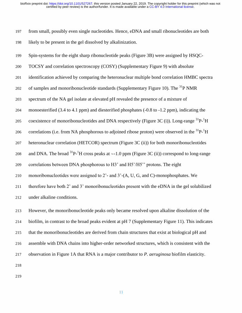
11
from small possibly even single nucleotides Hence eDNA and small ribonucleotides are both 197
likely to be present in the gel dissolved by alkalinization 198
Spin-systems for the eight sharp ribonucleotide peaks (Figure 3B) were assigned by HSQC-199
TOCSY and correlation spectroscopy (COSY) (Supplementary Figure 9) with absolute 200
identification achieved by comparing the heteronuclear multiple bond correlation HMBC spectra 201
of samples and monoribonucleotide standards (Supplementary Figure 10) The 31
P NMR 202
spectrum of the NA gel isolate at elevated pH revealed the presence of a mixture of 203
monoesterified (34 to 41 ppm) and diesterified phosphates (-08 to -12 ppm) indicating the 204
coexistence of monoribonucleotides and DNA respectively (Figure 3C (i)) Long-range 31
P-1H 205
correlations (ie from NA phosphorous to adjoined ribose proton) were observed in the 31
P-1H 206
heteronuclear correlation (HETCOR) spectrum (Figure 3C (ii)) for both monoribonucleotides 207
and DNA The broad 31
P-1H cross peaks at ~-10 ppm (Figure 3C (ii)) correspond to long-range 208
correlations between DNA phosphorous to H3rsquo and H5rsquoH5rsquorsquo protons The eight 209
monoribonucleotides were assigned to 2rsquo- and 3rsquo-(A U G and C)-monophosphates We 210
therefore have both 2rsquo and 3rsquo monoribonucleotides present with the eDNA in the gel solubilized 211
under alkaline conditions 212
However the monoribonucleotide peaks only became resolved upon alkaline dissolution of the 213
biofilm in contrast to the broad peaks evident at pH 7 (Supplementary Figure 11) This indicates 214
that the monoribonucleotides are derived from chain structures that exist at biological pH and 215
assemble with DNA chains into higher-order networked structures which is consistent with the 216
observation in Figure 1A that RNA is a major contributor to P aeruginosa biofilm elasticity 217
218
219
CC-BY 40 International licensecertified by peer review) is the authorfunder It is made available under aThe copyright holder for this preprint (which was notthis version posted January 22 2019 httpsdoiorg101101527267doi bioRxiv preprint
12
220
221
222
223
224
225
226
227
Figure 3 (A) Nuclear magnetic resonance (NMR) 1H-13C 228
heteronuclear single quantum coherence (HSQC)-total 229
correlation spectroscopy (TOCSY) spectrum at 25degC of 230
extracellular nucleic acids (NA) gel isolate after 231
alkalinization showing the C1rsquo-H1rsquo cross peaks of RNA 232
(blue ovals) and DNA (red rectangles) and their correlations 233
to the neighboring carbon C2rsquo-H1rsquo Schematics (right) 234
illustrate these correlations (B) 1H-13C HSQC spectrum of 235
extracellular NA gel isolate at 25degC after alkalinization 236
identifying eight monoribonucleic acid ribose spin systems 237
(C) 1-D 31P NMR of NA isolate gel and Pseudomomonas 238
aeruginosa biofilm with proton decoupling showing the 239
presence of monoesterified (ie monoribonucleotides) and 240
diesterified (ie DNA) phosphate peaks (i) and 2-D 1H-31P heteronuclear correlation (HETCOR) spectrum of 241
extracellular NA showing the 31P-1H cross-peaks of monoribonucleotides and DNA (ii) Couplings of 242
monoesterified phosphates to H2rsquo or H3rsquo of eight monoesterified monoribonucleotides (from left to right 3rsquo 243
AMP3rsquo GMP 3rsquo CMP3rsquo UMP 2rsquo CMP2rsquo UMP 2rsquo AMP 2rsquo GMP) and diesterified phosphate to DNA H3rsquo and 244
H5rsquoH5rsquorsquo protons are denoted by the dashed lines There is a discontinuity (wavy line) in the 31P axis due to the 245
different thresholds required to illustrate the 31P-1H correlations in the mono-esterified and di-esterified regions All 246
samples were prepared in 01 M NaOD (10 mgmL) and preheated to 55degC for 2 h 247
CC-BY 40 International licensecertified by peer review) is the authorfunder It is made available under aThe copyright holder for this preprint (which was notthis version posted January 22 2019 httpsdoiorg101101527267doi bioRxiv preprint
13
The molar ratio of the individual ribonucleotides could be determined from the 31
P spectrum of 248
the gel dissolved at high pH (Table 1) While several peaks could not be separated it was 249
possible to deduce that the RNA is purine rich (ie 57 mol A+G) and that the G+C mol 250
content of 46-50 differs from that of the P aeruginosa genome (ie 67 mol) (Shen et al 251
2006) The same peaks were also observed in the biofilm 31
P spectrum after alkalinization 252
(Figure 3C(i)) 253
Table 1 Relative abundances of monoribonucleotides in extracellular NA gel isolate from Pseudomonas 254 aeruginosa biofilm as determined by integrating 31P NMR spectrum following alkalinization 255
31P shift
(ppm)
Ribonucleotide Relative
abundance
403 3rsquoAMP 180
401 3rsquo GMP 172
390 3rsquo CMP3rsquo UMP 89
387 3rsquo CMP3rsquo UMP 113
368 2rsquo CMP2rsquo UMP 95
366 2rsquo CMP2rsquo UMP 117
350 2rsquo AMP 104
342 2rsquo GMP 130
256
Extracellular DNA and RNA interact to form a network 257
We applied Magic-angle spinning (MAS) solid-state NMR (SSNMR) to confirm that the 258
networks in the gel isolate and biofilm are comprised of DNA and RNA in chain form This 259
technique is ideal for intractable systems such as biofilms to eliminate solubility and extraction 260
biases (Reichhardt and Cegelski 2014) and to analyze inter-molecular H-bond interactions to 261
describe for example inter-nucleotide base pairing in RNA (Marchanka et al 2015) MAS 262
SSNMR averages anisotropic interactions to provide high-resolution spectral characterization of 263
insoluble and large biomolecular systems By analyzing dipolar interactions through-space 264
heteronuclear correlations (eg N∙∙∙H) can be detected In contrast to the liquid state 31
P NMR 265
spectrum of the alkali-dissolved isolate (Figure 3C (i)) the 31
P SSNMR spectrum of the gel 266
CC-BY 40 International licensecertified by peer review) is the authorfunder It is made available under aThe copyright holder for this preprint (which was notthis version posted January 22 2019 httpsdoiorg101101527267doi bioRxiv preprint
14
isolate (ie no alkali treatment Figure 4A (i)) showed a single peak in the diesterified phosphate 267
region consistent with the presence of both DNA and RNA chains There are no sharp peaks 268
present in the monoesterified phosphate region further demonstrating that the 269
monoribonucleotides are a consequence of alkali transesterification (Radak et al 2013) 270
Only the diesterified phosphate peak was observed in the 31
P SSNMR spectrum of the biofilm 271
(Figure 4A (iii)) while both RNA-derived monoesterified and DNA-derived diesterified 272
phosphate peaks were present in the 31
P SSNMR spectrum of alkali digested NA gel-isolate and 273
biofilm (lyophilized) (Figure 4A (ii) and Figure 4A (iv) respectively) Complete alkali RNA 274
transesterification was confirmed by the full conversion of diesterified to monoesterified 275
phosphates (from liquid state 31
P NMR spectra) for the RNA standard when dissolved in 01 M 276
NaOH (Supplementary Figure 12A) Conversely RNA diesterified phosphate peaks were 277
preserved in the RNA standard spectrum following dissolution in EMIM-Ac and recovery by 278
perchloric acid (Supplementary Figure 12B) Hence the alkaline conditions break down the 279
RNA chains into individual monoribonucleotides while the ionic liquid-based extraction does 280
not This suggests that the conventional method for P aeruginosa biofilm dissolution in alkali 281
(Friedman and Kolter 2004) may in fact work by transesterifying RNA as a primary structural 282
component and illustrates the importance of the ionic liquid-based extraction protocol described 283
here as a non-destructive method for interrogating biofilm structural polymers 284
CC-BY 40 International licensecertified by peer review) is the authorfunder It is made available under aThe copyright holder for this preprint (which was notthis version posted January 22 2019 httpsdoiorg101101527267doi bioRxiv preprint
15
285
Figure 4 (A) Solid-state 31P NMR spectra at T = 25degC of extracellular NA gel isolate (i) alkalinized and 286
lyophilized NA gel isolate (ii) Pseudomonas aeruginosa biofilm (iii) and alkalinized and lyophilized P aeruginosa 287
biofilm (iv) showing the presence of diesterified phosphate peaks and the absence of monoesterified phosphate 288
peaks for both NA gel isolate and biofilm in double distilled water and the coexistence of diesterified and 289
monoesterified phosphate peaks for both samples after alkalinization This indicates that alkalinization of the matrix 290
results in RNA transesterification Micrographs of P aeruginosa NA gel isolate stained green with (B) SYTO 291
RNASelect showing RNA fibers and (C) TOTO-1 showing DNA fibers (scale bar 10 microm) 292
293
To visualize the DNA-RNA network organization we used nucleic acid-specific stains ie RNA 294
specific SYTO RNASelect dye (Figure 4B) and the eDNA-specific TOTO-1 dye (Figure 4C) 295
(Okshevsky and Meyer 2014) Fibrous structures which are typical of networked polymer gels 296
(Cornwell and Smith 2015) were observed in the NA gel isolate stained with both nucleic acid 297
dyes as well as in the biofilm stained with the eDNA specific dye (Figure 1C) This is consistent 298
with our observation that the biofilm is readily degraded by DNase I (Figure 1A) (Chen et al 299
2004) Furthermore the NA gel isolate was stained positively with the RNA-specific dye even 300
after DNase I digestion providing further evidence that RNA contributes to networking 301
BeRNA fibers
C
eDNA fibers
A
CC-BY 40 International licensecertified by peer review) is the authorfunder It is made available under aThe copyright holder for this preprint (which was notthis version posted January 22 2019 httpsdoiorg101101527267doi bioRxiv preprint
16
(Supplementary Figure 13) However digestion with RNases III A and H did not degrade the 302
biofilm (Figure 1A Supplementary Figure 14) This may suggest that the RNase binding was 303
shielded by hairpins in the network or the presence of non-canonical DNA-RNA interactions 304
(Geerts-Dimitriadou et al 2012 Nakamura et al 1991) 305
Non-canonical and Watson-Crick base pairs and tetrads support extracellular network 306
Sequence analysis of the extracted material indicated that the gene coverage was even for both 307
chromosomal and extracellular DNA with the exception of bacteriophage Pf1 genes 308
(Supplementary Figure 15) However the Pf1 knockout mutant of P aeruginosa also displayed 309
an elastic response when dissolved in EMIM-Ac (Supplementary Figure 16 Supplementary 310
Tables 1 and 2) indicating that Pf1 DNA is unlikely to be responsible for the phase-separating 311
behavior of P aeruginosa biofilms DNA and RNA interaction therefore cannot be explained by 312
uneven gene coverage To elucidate the mechanism of DNA-RNA gelation we generated a 2D 313
through-space 15
N-1H HETCOR spectrum of
15N
labeled DNA-RNA gel isolated from a P 314
aeruginosa biofilm matrix (Figure 5A) This spectrum was run with longer cross polarization 315
contact times so that correlations between distant 1H and
15N could be observed This confirmed 316
the complete absence of proteins and supported our conclusion that proteins do not contribute to 317
the DNA-RNA interaction The HETCOR spectrum showed four signal clusters at δH 10-14 318
ppm and δN 140-160 ppm (Figure 5A) which arose from direct N-H couplings of TU and G 319
nucleobase imino groups Two of these clusters (δH 12-14 ppm) resulted from imino protons 320
hydrogen-bonded to a nucleobase nitrogen (ie N-H∙∙∙N) and the other two (δH 10 -12 ppm) 321
from imino protons hydrogen-bonded to a nucleobase carbonyl oxygen (ie N-H∙∙∙O) Due to the 322
longer cross polarization times we were able to observe a strong long-range and indirect (ie 323
intermolecular) correlation at δN 196 ppm arising from G-C Watson-Crick base pairing ie 324
CC-BY 40 International licensecertified by peer review) is the authorfunder It is made available under aThe copyright holder for this preprint (which was notthis version posted January 22 2019 httpsdoiorg101101527267doi bioRxiv preprint
17
C(N3)-G(H1) There was also a weak and indirect correlation at δN 220 ppm resulting from A-325
TU Watson-Crick base pairing ie A(N1) to UT(H1) 326
327
328
Figure 5 (A) Solid-state 2D 1H-15N through-space heteronuclear correlation (HETCOR) spectrum of extracellular 329
nucleic acid (NA) gel isolate in double distilled water (2 mg) T = 25degC showing direct N-H couplings for G imino 330
N (N3) and TU imino N (N3) bonded to nucleobase nitrogen (NHN) This indicates Watson-Crick (WC) G-C and 331
UT-A base pairings respectively and for G imino N (N1) and TU imino N (N3) bonded to nucleobase carbonyl 332
oxygen (ie NHO) indicating non-canonical G-G G-TU and UT-UT base pairings or tetrads The spectrum also 333
displayed indirect N-H correlations for the WC base pairs (ie C(N3) to G(H1) and A(N1) to UT(H1)) and no 334
indirect correlations for the non-canonical bonded nucleobases The threshold for the A(N1)-TU(H3) correlation 335
was increased due to lower signal intensity for that particular coupling (B) Pseudomonas aeruginosa biofilm 336
growth yield at 37degC (5 d) as a function of [K+] [Na+] or [Li+] showing the growth dependence on monovalent 337
cation concentration (K+gtNa+gtLi+) Error bars indicate standard deviation (C) Illustration of how structural 338
variability of RNA may allow for the formation of extracellular RNA and DNA networks through hairpins and a 339
mixture of canonical and non-canonical base pairs or tetrads (ie Hoogsteen and G-quadruplexes) 340
341
The absence of long-range correlations between the clusters at δH 10 -12 ppm is consistent with 342
non-Watson-Crick pairings for the G and TU nucleobases The observation of NH to O 343
interactions suggests the formation of G-G TU-TU G-TU Hoogsteen base pairs or tetrads 344
CA B C
CC-BY 40 International licensecertified by peer review) is the authorfunder It is made available under aThe copyright holder for this preprint (which was notthis version posted January 22 2019 httpsdoiorg101101527267doi bioRxiv preprint
18
Hoogsteen H-bonded G-bases can assemble planarly into tetrads and self-stack to form G-345
quadruplexes (Haumlnsel-Hertsch et al 2017) G-quadruplexes are stabilized by the presence of a 346
monovalent cation with potassium providing the greatest stability We experimentally confirmed 347
that potassium had a similar effect in a static biofilm growth assay We found that potassium 348
promoted P aeruginosa biofilm growth to a greater extent than either sodium or lithium (Figure 349
5B) Additionally this was a biofilm-specific effect as there was no effect of monovalent cation 350
on planktonic growth (Supplementary Figure 17) Based on the SSNMR data coupled with the 351
potassium effect and the earlier observation of several maxima in the CD spectrum of the NA gel 352
isolate across the characteristic NA region of 250-285 nm which suggested the coincidence of 353
several NA conformations contributing to phase separation it is proposed that G-quadruplexes 354
contribute to extracellular DNA-RNA networking 355
Discussion 356
We extracted a gel-forming complex composed of DNA and RNA from the extracellular matrix 357
of P aeruginosa biofilms without disrupting its fundamental structural or chemical organization 358
We report the novel observation that extracellular RNA contributes structurally to biofilms 359
While gel-forming exopolymers have been detected in other biofilms (Seviour et al 2009) no 360
functional DNA gel has been described for any biological system much less one also including 361
extracellular RNA 362
While it is not possible to identify the precise nature of the DNA-RNA interaction based on the 363
information presented here purine-rich RNA as described here for the extracellular NA gel 364
isolate (57 mol A-G) is a characteristic of DNA-RNA duplexes because RNA purines bind 365
more strongly to their DNA pyrimidine complement than vice versa (Nadel et al 2015) Both 366
Watson-Crick and non-canonical interactions were present in the NA gel isolate which is also 367
CC-BY 40 International licensecertified by peer review) is the authorfunder It is made available under aThe copyright holder for this preprint (which was notthis version posted January 22 2019 httpsdoiorg101101527267doi bioRxiv preprint
19
consistent with the discrepancy observed between RNA and DNA G+C mol contents (Table 368
1) It is possible therefore that interactions between DNA and RNA as described in this study 369
account for the formation of a highly stable nucleic acid gel 370
Non-canonical base pairing and hairpinning are commonly seen with RNA and cause multivalent 371
intermolecular interactions between RNA strands that lead to a sol-gel transition (Jain and Vale 372
2017) The greater structural versatility of RNA could be a key to the ability of the eDNA to 373
form networks with hairpins as well as canonical and non-canonical base pair interactions all 374
contributing to the assembly of higher order NA structures (Figure 5C) RNA has recently been 375
found to promote intracellular phase separations although this has not been extended to the 376
extracellular domain of prokaryotic cells (Shin and Brangwynne 2017) Additionally G-rich 377
RNA sequences are more predisposed to forming G-quadruplexes because they do not have a 378
complementary strand and in fact form more stable G-quadruplexes than DNA (Guo and Bartel 379
2016) It is therefore probable that the RNA but not DNA enables DNA-RNA networking and 380
gelation 381
The results presented here provide unprecedented resolution of the biofilm exopolymeric matrix 382
and its key foundation structural components We have developed a methodology that preserved 383
the molecular organization of the foundational polymer in its native state upon extraction and 384
isolation This enabled us to use SSNMR to describe intermolecular associations at the atomic 385
level 386
Our findings that a DNA-RNA gel network can provide the foundation for biofilm matrices in P 387
aeruginosa mark a departure from the prevailing paradigm that biofilm gelation is only due to 388
polysaccharides The elastic and film-forming properties were also observed for Pseudomonas 389
putida and Pseudomonas protegens (Figs S16 and S18 Supplementary Tables 1 and 2) 390
CC-BY 40 International licensecertified by peer review) is the authorfunder It is made available under aThe copyright holder for this preprint (which was notthis version posted January 22 2019 httpsdoiorg101101527267doi bioRxiv preprint
20
suggesting that eDNA is broadly important for biofilm formation in members of this genus In 391
addition to Pseudomonas spp other organisms are known to have eDNA including clinical 392
organisms Staphylococcus aureus (Mann et al 2009) Staphylococcus epidermidis (Adam et al 393
2002) and Mycobacterium abscessus (Rose et al 2015) as well as environmental isolates such 394
as strain F8 from the South Saskatchewan River (Boumlckelmann et al 2006) 395
While eDNA is commonly thought to be the product of cell lysis and has been shown to be 396
released by a sub-population of lytic P aeruginosa cells (Turnbull et al 2016) there is an 397
increasing awareness that it serves an important structural function such as in activated sludge 398
granules (Cheng et al 2011) Additionally Bockelmann et al (2006) observed that stable 399
filamentous networks produced by the aquatic strain F8 were comprised of DNA This is the first 400
study that provides an explanation for how eDNA is organized structurally However the 401
environmental factors contributing to eDNA release and how this is regulated (ie whether it is 402
an active or passive process) are still unclear It is possible that DNA-RNA foundational gel 403
networks as described here for the Pseudomonads are broad-scoping phenomena in biofilms 404
and that interactions with RNA enable an extracellular structural function for DNA Thus 405
elucidating how nucleic acids including RNA are integral to the biophysical and other emergent 406
properties imparted on the biofilm via the extracellular matrix will inform the regulation and 407
control of extracellular nucleic acid release across environmental and clinical biofilms 408
Materials and Methods 409
Bacterial strains 410
Unless otherwise stated all experiments were undertaken directly on P aeruginosa PAO1 411
biofilms grown in lysogeny broth at 37degC The P aeruginosa PAO1 Δpf4 knockout mutant is a 412
CC-BY 40 International licensecertified by peer review) is the authorfunder It is made available under aThe copyright holder for this preprint (which was notthis version posted January 22 2019 httpsdoiorg101101527267doi bioRxiv preprint
21
defined Pf4 chromosomal deletion mutant of the entire Pf4 prophage genome(Rice et al 413
2009) 414
P aeruginosa PDO300 PDO300(Δpel) and PDO300(Δpsl) mutant strains were gratefully 415
received from Professor Bernd H A Rehm Institute of Molecular Biosciences Massey 416
University New Zealand (Ghafoor et al 2011) PDO300 PDO300(Δpel) PDO300(Δpsl) and 417
PAO1 (Δpf4) were also grown in LB at 37degC P protogens Pf-5 and P putida ATCC BAA-477 418
and S12 strain were were grown in LB at 30degC 419
Biofilm growth assay 420
Ten milliliter aliquots of P aeruginosa planktonic pre-cultures (LB 200 rpm 37degC OD600 240 421
16 h) were diluted 50 times with LB in 2 L Erlenmeyer flasks and incubated for 5 d under static 422
conditions Supplementary Figure 15A displays an image of 5-d old biofilm in LB The cultures 423
were centrifuged at 10000 x g for 15 min the supernatant removed by decanting and the 424
biofilm then collected and lyophilized (LabConco) Supplementary Figure 15B displays an 425
image of the culture after centrifugation showing clear separation of biofilm and supernatant 426
Enzymatic digestions 427
Twenty milligrams of lyophilized biofilm was resuspended in 1 mL of either i) RNase buffer (50 428
mM Tris HCl 10 mM EDTA pH8) with 02 mg RNaseA from bovine pancreas (Sigma Aldrich) 429
ii) storage buffer (10 mM NaCl 10 mM Tris-HCl) with 01 mg Pronase E from Streptomyces 430
grisens (Sigma Aldrich) with 05 (vv) iii) 1X RNase H reaction buffer (20 mM Tris-HCl pH 431
78 40 mM KCl 8 mM MgCl2 1 mM DTT) with 04 mg RNase H (Thermo Fisher Scientific) 432
iv) 1X RNase III reaction buffer (500 mM NaCl 100 mM Tris pH 79 100 mM MgCl2 10 mM 433
DTT) with 06 mg RNase II (Thermo Fisher Scientific) or v) DNase I buffer (100mM Tris (pH 434
75) 25 mM MgCl2 and CaCl2) with 02 mg DNAse I from bovine pancreas (Sigma Aldrich) All 435
CC-BY 40 International licensecertified by peer review) is the authorfunder It is made available under aThe copyright holder for this preprint (which was notthis version posted January 22 2019 httpsdoiorg101101527267doi bioRxiv preprint
22
digestions were performed with shaking at 200 rpm at 37degC for 16 h The suspensions were then 436
centrifuged (10000 x g 15 min) the supernatant was discarded and the pellets of the biofilm 437
materials were lyophilized 438
Normal force measurement 439
Forty milligrams per milliliter solutions of lyophilized biofilms were added to 1 mL 1-ethyl-3-440
methylimidazolium acetate and incubated in 55degC for either 2 h A Haake Mars 3 (Thermo 441
Fisher Scientific) stress-controlled rotational rheometer with Peltier controlled element at 25degC 442
was used for rheological measurements Thirty five-illimeter diameter parallel plate geometry 443
was used with smooth titanium plates to measure viscosity and normal stress difference (N1 ndash 444
N2) Prior to measurement the gap error was zeroed at 4 N and gap error calculated as previously 445
described (Bird et al 1987 Davies and Stokes 2008 Kravchuk and Stokes 2013) One 446
hundred microliters of sample was deposited on the plates The plates were closed to 100 microm the 447
sample trimmed and the sample allowed to sit for 5 min prior to measurement All measurements 448
with Normal force (FN) less than the resolution of the rheometer (ie lt 01 N) were set to 0 449
before calculation of N1-N2 using equations 9-11 and viscosity from equation 3-5 in Davies and 450
Stokes(Davies and Stokes 2008) for the parallel plate geometry 451
Only the linearly increasing portion of the normal stress difference curves are presented Above 452
this range normal stress difference begins to decrease again which may be due to elastic 453
instabilities or associating polymers (Annable et al 1993) Corrections were made to N1 and N1 454
ndash N2 to account for inertia using equation 17 in Davies and Stokes (Davies and Stokes 2008) and 455
to correct for the baseline residual force in the samples Except for the DNase I-treated biofilm 456
the shear rheology for all treatments could be modelled using the finitely extensible non-linear 457
elastic with Gaussian closure proposed by Peterlin (FENE-P) constitutive model by varying four 458
CC-BY 40 International licensecertified by peer review) is the authorfunder It is made available under aThe copyright holder for this preprint (which was notthis version posted January 22 2019 httpsdoiorg101101527267doi bioRxiv preprint
23
parameters to fit shear viscosity and normal stress difference as a function of shear rate 459
(Supplementary Table 2) Fitting parameters for the FENE-P model include 1 = relaxation time 460
b = a measure of the relative extensibility of the model spring s = solvent viscosity p = 461
polymer contribution to the viscosity The FENE-P equations can be written in the following 462
format as shown by Bird et al(1987) 463
120578 = 120578119904 +120578119901
1205821((1198622 + 1198621)
1
3 minus (1198622 minus 1198621)1
3) (Equation 1) 464
465
1198731 = 2120578119901
1205821((1198622 + 1198621)
1
3 minus (1198622 minus 1198621)1
3)2
(Equation 2) 466
Where 467
1198621 = 119887
41205821 (Equation 3) 468
1198622 = (11986212 + (
119887+3
6)
3
)12
(Equation 4) 469
470
All measurements were performed in triplicate For clarity one representative data set is 471
presented in Figure 1A-B and Supplementary Figure 1A-B with the respective Power Law and 472
FENE-P model fit to that data set Averaged values for the FENE-P and Power law fits with the 473
standard deviation across three replicates are shown in Supplementary Tables 1 and 2 474
Oscillatory measurements were carried out with controlled frequency 01 rads across an 475
amplitude range of 001 to 10 476
Extracellular polymeric substances (EPS) extraction 477
Lyophilized biofilms were dissolved in ionic liquid mixture (40 (vv) 1-ethyl-3-478
methylimidazolium acetate (EMIM Ac) 60 (vv) NN-dimethyl acetamide (DMAc)) at 55degC 479
CC-BY 40 International licensecertified by peer review) is the authorfunder It is made available under aThe copyright holder for this preprint (which was notthis version posted January 22 2019 httpsdoiorg101101527267doi bioRxiv preprint
24
for 16 h The solution was centrifuged (10000 x g) to remove any undissolved material 480
Perchloric acid (70) was added (005 vv) to the viscosified centrate (on ice) After 15 min 481
incubation the solution was centrifuged at 10000 x g at 4degC for 15 min and the pellet recovered 482
This was repeated on the centrate two to four times until the solution was not viscous The 483
precipitate was dialysed against double distilled water for 2 d at 4degC (SnakeSkintrade Dialysis 484
Tubing 35K MWCO 22 mm) and the retentate lyophilized (FreeZone Plus 45 Liter Cascade 485
Benchtop Freeze Dry System) The same procedure was performed on calf thymus DNA lipase 486
cytochrome C for the purposes of determining recovery yield of representative exoproteins and 487
on RNA from torula yeast for assessing by 31
P NMR whether the extraction procedure 488
contributed to RNA transesterification (all from Sigma Aldrich) 489
Extracellular nucleic acid isolation 490
Twenty milligrams of lyophilized retentate (ie post perchloric acid precipitation) were dissolved 491
in 1 mL of 40 (vv) EMIM-Ac 60 DMAc (vv) (55degC 16 h) Chromatographic separation 492
was achieved in a Shimadzu system comprising DGU-20A 3r Prominence Degasser and LC-493
20AD Solvent Delivery Unit fitted with two Agilent PLgel 10 μm column of 105Aring pore size for 494
separation across the MW range 200 kDa to 2000 kDa The eluent flow rate was 30 mLmin-1
495
and the injection volume 1 mL The fractions with molecular weight range of 2000-800kDa and 496
800-200kDa were pooled and dialyzed for 2 d at 4degC (SnakeSkintrade Dialysis Tubing 35K 497
MWCO 22 mm) against double distilled water to induce gelation The gel was then collected 498
from the dialysis tubing 499
Solution-state nuclear magnetic resonance (NMR) 500
Solution-state NMR experiments were performed on an 800 MHz Bruker Avance III 501
spectrometer at 25oC Sample concentration was 10 mg (dry weight)mL
-1 unless otherwise 502
CC-BY 40 International licensecertified by peer review) is the authorfunder It is made available under aThe copyright holder for this preprint (which was notthis version posted January 22 2019 httpsdoiorg101101527267doi bioRxiv preprint
25
specified Spectra were recorded either under conditions of neutral pH in 100 D2O (Cambridge 503
Isotope Laboratories) or following alkalinization (ie transesterification 01 M NaOD 55oC 2 504
h) 1-D NMR experiments include 1H and
31P direct detection while 2-D NMR analysis include 505
13C-HSQC
13C-HSQC-TOCSY
1H-
31P HETCOR HMBC and COSY All spectral analyses 506
were performed using Topspin and SPARKY software 507
Asolectin (Sigma Aldrich) standard (10 mgmL) and lyophilized Pseudomonas aeruginosa 508
PAO1 biofilm (10mgmL) were dissolved in 40 (vv) EMIM Ac 60 (vv) DMAc at (55oC 2 509
h) P aeruginosa PAO1 pre-culture cell lysate was prepared by lysing pre-culture cells with 510
lysozyme in PBS 10 (vv) of D2O was added to all samples for locking purposes 511
Solid-state NMR 512
For solid-state NMR experiments performed on the NA gel isolate 15
N labeled NH4Cl-513
supplemented M9 minimal media was used for biofilm growth M9 consisted of 9552 gL-1
514
Na2HPO42H2O 441g litre-1
KH2PO4 171 gL-1
NaCl 1 gL-1
15
NH4Cl 024 gL-1
MgSO4 515
0011 gL-1
CaCl2 2 gL-1
casamino acids and 04 gL-1
glucose The NA gel was prepared from 516
the M9-grown P aeruginosa PAO1 biofilm as described above 517
Solid-state NMR experiments were performed on 141 T Bruker Advance III instruments 518
equipped with a 19 mm MAS probe operated in double mode The typical 1H
15N and
31P π2 519
pulse lengths were 23 37 and 45 μs respectively 2D dipolar-based 15
N-1H heteronulcear-520
correlation (HETCOR) experiments were conducted on the 15
N-labelled NA gel isolate at 37 kHz 521
MAS spinning frequency Variable temperature was regulated at -20ordmC and the sample 522
temperature was 12ordmC (calibrated using ethylene glycol) In the 15
N-1H HETCOR experiments 523
the initially excited 1H magnetization was transferred to
15N through a cross polarization step 524
followed by t1 evolution Then the 15
N magnetization was flipped to longitudinal axis and 400 525
CC-BY 40 International licensecertified by peer review) is the authorfunder It is made available under aThe copyright holder for this preprint (which was notthis version posted January 22 2019 httpsdoiorg101101527267doi bioRxiv preprint
26
ms proton saturation pulses were applied for water suppression Subsequently the 15
N 526
magnetization was flipped to the transverse plane and transferred to 1H via a second CP step for 527
signal acquisition Two 15
N-1H HETCOR experiments were collected one with 400 s and the 528
other with 2 ms contact times applied for both of the CP steps Low power XiX 1H decoupling 529
(~10 kHz) was employed during 15
N evolution and WALTZ-16 decoupling (10 kHz) was 530
implemented on 15
N channel during 1H acquisition 531
1-D 31
P experiments were performed on 15
N-labelled NA gel isolate and 15
N-labelled P 532
aeruginosa PAO biofilm both directly after dialysis against double distilled water at 4degC for 2 d 533
(SnakeSkintrade Dialysis Tubing 35K MWCO 22 mm) and following alkalinization (01 M 534
NaOD 55ordmC 15 min) and lyophilization (FreeZone Plus 45 Liter Cascade Benchtop Freeze Dry 535
System) 15 kHz MAS spinning frequency and a sample temperature of 27ordmC 75 kHz 536
SPINAL64 1H decoupling was applied during
31P acquisition time All chemical shifts were 537
indirectly referenced using adamantane as a secondary standard (downfield peak is at 4048 ppm 538
DSS scale) 539
Monovalent cation-dependent biofilm growth 540
Ten milliliters aliquots of P aeruginosa pre-cultures (supplemented M9 200 rpm OD600 240 541
16 h) were transferred into 500 mL M9 minimal media (85 gL-1
Na2HPO42H2O 20 gL-1
of 542
NaH2PO4H2O 10 gL-1
KH2PO4 10 gL-1
NH4Cl 048 gL-1
MgSO4 0011 gL-1
CaCl2 2 gL-1
543
casamino acids and 04 gL-1
glucose) supplemented with either KCl LiCl or NaCl at either of 544
three different concentrations (0 002 mM and 008 mM) Biofilms were collected by 545
centrifugation (10000 x g 15 min) as described above lyophilized (LabConco) and weighed To 546
describe the growth curve P aeruginosa wild type PAO1 pre-culture was incubated in M9 547
media at 37degC 200 rpm for 16 h PAO1 WT pre-culture was then diluted with supplemented M9 548
CC-BY 40 International licensecertified by peer review) is the authorfunder It is made available under aThe copyright holder for this preprint (which was notthis version posted January 22 2019 httpsdoiorg101101527267doi bioRxiv preprint
27
media to a volume of 75 mL and a starting OD600 of 001 in a 250 mL Erlenmeyer flask The 549
mixture was incubated at 37degC with shaking (200rpm) The OD600 of the bacteria was measured 550
hourly for 9 h followed by final time point at 24 h 551
DNA Sequencing 552
Genomic DNA was extracted from the biofilm using FastDNA SPIN Kit for soil (MP 553
Biomedicals USA) as per the standard protocol Briefly biofilm was resuspended in Sodium 554
Phosphate Buffer in was lysed (Lysing Matrix) homogenized (FastPrepreg
40 seconds speed 555
setting 60) and the cell debris removed by centrifugation (14000 x g 5 min) Proteins were 556
removed by precipitation (250 μl Protein Precipitation Solution) the supernatant mixed with 557
DNA Binding Matrix which was then homogenized and transferred to a SPINtrade Filter Excess 558
supernatant was removed by centrifugation (14000 x g 5 min) DNA was then eluted from air 559
dried DNA Binding Matrix with DNase Pyrogen-Free Water 560
The NA gel isolate was resuspended in 500 uL of 1x Protease K solution (10times Protease K 561
solution 10 mM Tris HCl 1 SDS and 10 mM EDTA pH 8 buffer (10times protease K buffer 562
containing 500 mM Tris-HCl 10 SDS 10 mM CaCl2) and 10 L of Protease K (20 mgmL-1
563
Thermo Fisher Scientific) was added and the mixture incubated at 56degC for 2-16 h after which 564
DNA was extracted as previously described in the phenol-chloroform method(Ausubel 2002) 565
Samples before sequencing were further purified to remove any remaining protein and RNA by 566
RNase and Proteinase K treatment The DNA was then isolated using phenol-chloroform 567
precipitation as described above The DNA precipitate was dissolved in TE buffer the purity 568
confirmed by 260280 value in Nanodrop (acceptable range value 18-20) and Qubitreg
20 569
fluorometer 570
CC-BY 40 International licensecertified by peer review) is the authorfunder It is made available under aThe copyright holder for this preprint (which was notthis version posted January 22 2019 httpsdoiorg101101527267doi bioRxiv preprint
28
The molecular weight distributions of extracellular and genomic DNA were measured on a 1 571
agarose gel which was prepared from Viviantis LE grade agarose using 1x TAE buffer (40 mM 572
Tris 20 mM Acetate and 1 mM EDTA pH 86) Gels were run horizontally After 573
electrophoresis the gel was stained for 05 h with ethidium bromide and visualized under 574
UV(Lee et al 2012) 575
Three replicates were used for each DNA sequence analysis Library was produced using 576
Illumina DNA sample preparation kit The libraries were sequenced using Illumina MiSeq 577
platform (Illumina San Diego Ca) with paired-end protocol to read lengths of 600 nt generating 578
a total of 1614106 and 1848846 paired end reads Raw reads were quality filtered (reads 579
remaining after trimming PPG1-1549104 PBLC1-1666280) and aligned to the P aeruginosa 580
PAO1 (AE004091) genome using CLC Genomics Workbench 90 (CLC bio Cambridge MA) 581
Circular Dichroism CD 582
Five-day old biofilm and NA gel isolate were resuspended in double distilled water to achieve 583
UV absorbance reading 1 and ddH2O served as a blank The heat-treated samples were analyzed 584
by JASCO-815 spectropolarimeter in a 1 cm path length quartz cuvette containing a solution 585
volume 500 μL Spectra (200-320 nm) were measured at 1degC increments from 30 - 90degC For 586
each measurement an average of three scans was taken and the buffer spectra subtracted Each 587
spectrum presented is the rolling average across 5 temperatures 588
Staining and microscopy 589
Microscopic imaging was conducted on a confocal microscope Zeiss LSM 780 with a 63times 590
objective Extracellular RNA in the gel isolate were stained using SYTO RNASelect (Thermo 591
Fisher Scientific) green fluorescent cell stain (5 mM solution in DMSO) Five microM stain solution 592
was prepared from 1 microL of stock in 1X PBS solution (137 mM NaCl 27 mM KCl 43 mM 593
CC-BY 40 International licensecertified by peer review) is the authorfunder It is made available under aThe copyright holder for this preprint (which was notthis version posted January 22 2019 httpsdoiorg101101527267doi bioRxiv preprint
29
Na2HPO4 147 mM KH2PO4) The gel isolate was labelled with 5 microM stain solution kept at 594
37degC for 20 min and then transferred to glass side for imaging 595
eDNA staining was achieved by depositing biofilm or NA gel isolate on a glass slide air-drying 596
overnight and incubating with 2 microM TOTO-1 iodide (1 mM solution in DMSO Thermo Fisher 597
Scientific) for 15 min 598
Acknowledgments 599
We acknowledge Prof Bernd Rehm for supplying polysaccharide deletion mutants of P 600
aeruginosa Dan Roizman for providing P putida Long Yu for assistance with rheology Dr 601
Gleb Yakubov for coordinating sample preparation for rheological measurements Ravi 602
Jagadeeshan for discussion regarding DNA normal force analysis and Florentin Constancias for 603
analyzing the sequencing data SCELSE is funded by Singaporersquos Ministry of Education 604
National Research Federation Nanyang Technological University (NTU) and National 605
University of Singapore (NUS) and hosted by NTU in partnership with NUS 606
Author Contributions 607
TS FRW WLL SM HMS and XS performed experiments TS and ATP designed 608
the experiments TS FRW ATP HMS GSK and JRS analysed the data TS 609
FRW WLL HMS SAR ATP and SK wrote the manuscript 610
Competing Interests 611
All authors have no competing interests 612
References 613
Mixed species biofilms of Candida albicans and Staphylococcus epidermidis 614 B Adam GS Baillie LJ Douglas (2002) 615
CC-BY 40 International licensecertified by peer review) is the authorfunder It is made available under aThe copyright holder for this preprint (which was notthis version posted January 22 2019 httpsdoiorg101101527267doi bioRxiv preprint
30
Journal of Medical Microbiology 51344-349 616
doi1010990022-1317-51-4-344 617
A characterization of DNA release in Pseudomonas aeruginosa cultures and biofilms 618
M Allesen-Holm KB Barken L Yang M Klausen JS Webb S Kjelleberg S Molin M Givskov T Tolker-Nielsen 619
(2006) 620
Molecular Microbiology 591114-1128 621
doi101111j1365-2958200505008x 622
The rheology of solutions of associating polymers Comparison of experimental behavior with transient 623
network theory 624
T Annable R Buscall R Ettelaie D Whittlestone (1993) 625
Journal of Rheology 37695-726 626
doi1011221550391 627
Short protocols in molecular biology a compendium of methods from Current protocols in molecular 628
biology 629
FM Ausubel (2002) 630
5th ed New York Wiley 631
Dynamics of Polymeric Liquids 632
RB Bird RC Armstrong O Hassager (1987) 633
2nd ed 634
Bacterial extracellular DNA forming a defined network-like structure 635
U Boumlckelmann A Janke R Kuhn TR Neu J Wecke JR Lawrence U Szewzyk (2006) 636
FEMS Microbiology Letters 26231-38 637
doi101111j1574-6968200600361x 638
Infections caused by Pseudomonas aeruginosa 639
GP Bodey R Bolivar V Fainstein L Jadeja (1983) 640
Review of Infectious Diseases 5279-313 641
doi101093clinids52279 642
Pseudomonas aeruginosa uses a cyclic-di-GMP-regulated adhesin to reinforce the biofilm extracellular matrix 643 BR Borlee AD Goldman K Murakami R Samudrala DJ Wozniak MR Parsek (2010) 644
Molecular Microbiology 75827-842 645
doi101111j1365-2958200906991x 646
DNARNA helicase gene mutations in a form of juvenile amyotrophic lateral sclerosis (ALS4) 647
YZ Chen CL Bennett HM Huynh IP Blair I Puls J Irobi I Dierick A Abel ML Kennerson BA Rabin GA 648
Nicholson M Auer-Grumbach K Wagner P De Jonghe JW Griffin KH Fischbeck V Timmerman DR Cornblath 649
PF Chance (2004) 650
Am J Hum Genet 741128-1135 651
doi101086421054 652
Evidence of compositional differences between the extracellular and intracellular DNA of a granular sludge 653
biofilm 654 M Cheng AE Cook T Fukushima PL Bond (2011) 655
Letters in Applied Microbiology 531-7 656
doidoi101111j1472-765X201103074x 657
Dynamic Remodeling of Microbial Biofilms by Functionally Distinct Exopolysaccharides 658
SC Chew B Kundukad T Seviour JRC Van Der Maarel L Yang SA Rice P Doyle S Kjelleberg (2014) 659
mBio 5e01536-01514 660
doi101128mBio01536-14 661
The Pel and Psl polysaccharides provide Pseudomonas aeruginosa structural redundancy within the biofilm 662
matrix 663 KM Colvin Y Irie CS Tart R Urbano JC Whitney C Ryder PL Howell DJ Wozniak MR Parsek (2012) 664
Environmental Microbiology 141913-1928 665
doi101111j1462-2920201102657x 666
Expanding the scope of gels - Combining polymers with low-molecular-weight gelators to yield modified self-667
assembling smart materials with high-tech applications 668 DJ Cornwell DK Smith (2015) 669
Materials Horizons 2279-293 670
doi101039c4mh00245h 671
CC-BY 40 International licensecertified by peer review) is the authorfunder It is made available under aThe copyright holder for this preprint (which was notthis version posted January 22 2019 httpsdoiorg101101527267doi bioRxiv preprint
31
Thin film and high shear rheology of multiphase complex fluids 672
GA Davies JR Stokes (2008) 673
Journal of Non-Newtonian Fluid Mechanics 14873-87 674
doi101016jjnnfm200704013 675
Kinetic model of a granular sludge SBR Influences on nutrient removal 676 MK De Kreuk C Picioreanu M Hosseini JB Xavier MCM Van Loosdrecht (2007) 677
Biotechnology and Bioengineering 97801-815 678
doi101002bit21196 679
G-Quadruplex secondary structure obtained from circular dichroism spectroscopy 680
R Del Villar-Guerra JO Trent JB Chaires (2018) 681
Angewandte Chemie International Edition 577171-7175 682
doidoi101002anie201709184 683
Biofilm streamers cause catastrophic disruption of flow with consequences for environmental and medical 684
systems 685
K Drescher Y Shen BL Bassler HA Stone (2013) 686
Proceedings of the National Academy of Sciences of the United States of America 1104345-4350 687
doi101073pnas1300321110 688
The biofilm matrix 689
H-C Flemming J Wingender (2010) 690
Nature Reviews Microbiology 8623-633 691
doi101038nrmicro2415 692
Two genetic loci produce distinct carbohydrate-rich structural components of the Pseudomonas aeruginosa 693
biofilm matrix 694
L Friedman R Kolter (2004) 695
Journal of Bacteriology 1864457-4465 696
doi101128jb186144457-44652004 697
Analysis of the Tomato spotted wilt virus Ambisense S RNA-Encoded Hairpin Structure in Translation 698
C Geerts-Dimitriadou Y-Y Lu C Geertsema R Goldbach R Kormelink (2012) 699
PLOS ONE 7e31013 700
doi101371journalpone0031013 701
Role of exopolysaccharides in Pseudomonas aeruginosa biofilm formation and architecture 702 A Ghafoor ID Hay BHA Rehm (2011) 703
Applied and Environmental Microbiology 775238-5246 704
doi101128aem00637-11 705
Using circular dichroism spectra to estimate protein secondary structure 706
NJ Greenfield (2006) 707
Nature protocols 12876-2890 708
doi101038nprot2006202 709
RNA G-quadruplexes are globally unfolded in eukaryotic cells and depleted in bacteria 710 JU Guo DP Bartel (2016) 711
Science 353aaf5371 712
doi101126scienceaaf5371 713
DNA G-quadruplexes in the human genome detection functions and therapeutic potential 714
R Haumlnsel-Hertsch M Di Antonio S Balasubramanian (2017) 715
Nature Reviews Molecular Cell Biology 18279 716
doi101038nrm20173 717
Major proteomic changes associated with amyloid-induced biofilm formation in Pseudomonas aeruginosa 718
PAO1 719 F-A Herbst MT Soslashndergaard H Kjeldal A Stensballe PH Nielsen MS Dueholm (2015) 720
Journal of Proteome Research 1472-81 721
doi101021pr500938x 722
Antibiotic resistance of bacterial biofilms 723
N Hoslashiby T Bjarnsholt M Givskov SR Molin O Ciofu (2010) 724
International Journal of Antimicrobial Agents 35322-332 725
doi101016jijantimicag200912011 726
Self-produced exopolysaccharide is a signal that stimulates biofilm formation in Pseudomonas aeruginosa 727
CC-BY 40 International licensecertified by peer review) is the authorfunder It is made available under aThe copyright holder for this preprint (which was notthis version posted January 22 2019 httpsdoiorg101101527267doi bioRxiv preprint
32
Y Irie BR Borlee JR Oconnor PJ Hill CS Harwood DJ Wozniak MR Parsek (2012) 728
Proceedings of the National Academy of Sciences of the United States of America 10920632-20636 729
doi101073pnas1217993109 730
RNA phase transitions in repeat expansion disorders 731
A Jain RD Vale (2017) 732
Nature 546243-247 733
doi101038nature22386 734
Pel is a cationic exopolysaccharide that cross-links extracellular DNA in the Pseudomonas aeruginosa biofilm 735
matrix 736
LK Jennings KM Storek HE Ledvina C Coulon LS Marmont I Sadovskaya PR Secor BS Tseng M Scian A 737
Filloux DJ Wozniak PL Howell MR Parsek (2015) 738
Proceedings of the National Academy of Sciences of the United States of America 11211353-11358 739
doi101073pnas1503058112 740
A phenomenological model for predicting melting temperatures of DNA sequences 741
G Khandelwal J Bhyravabhotla (2010) 742
PLOS ONE 5e12433 743
doi101371journalpone0012433 744
Review of algorithms for estimating the gap error correction in narrow gap parallel plate rheology 745
O Kravchuk JR Stokes (2013) 746
Journal of Rheology 57365-375 747
doi10112214774323 748
Analysis of the ion adsorptionndashdesorption characteristics of biofilm matrices 749 A Kurniawan T Yamamoto Y Tsuchiya H Morisaki (2012) 750
Microbes and Environments 27399-406 751
doi101264jsme2ME11339 752
Circular dichroism and conformational polymorphism of DNA 753
J Kypr I Kejnovskaacute D Renčiuk M Vorliacutečkovaacute (2009) 754
Nucleic Acids Research 371713-1725 755
doi101093nargkp026 756
Agarose gel electrophoresis for the separation of DNA fragments 757
PY Lee J Costumbrado C-Y Hsu YH Kim (2012) 758
Journal of Visualized Experiments JoVE 3923 759
doi1037913923 760
Modulation of eDNA release and degradation affects Staphylococcus aureus biofilm maturation 761
EE Mann KC Rice BR Boles JL Endres D Ranjit L Chandramohan LH Tsang MS Smeltzer AR Horswill KW 762
Bayles (2009) 763
PLoS One 4e5822 764
doi101371journalpone0005822 765
Persistent draining crossover in DNA and other semi-flexible polymers Evidence from hydrodynamic models 766
and extensive measurements on DNA solutions 767
ML Mansfield A Tsortos JF Douglas (2015) 768
The Journal of Chemical Physics 143124903 769
doi10106314930918 770
RNA structure determination by solid-state NMR spectroscopy 771
A Marchanka B Simon G Althoff-Ospelt T Carlomagno (2015) 772
Nature Communications 67024 773
doi101038ncomms8024 774
RNADNA hybrids in the human genome have distinctive nucleotide characteristics chromatin composition 775
and transcriptional relationships 776
J Nadel R Athanasiadou C Lemetre NA Wijetunga P Oacute Broin H Sato Z Zhang J Jeddeloh C Montagna A 777
Golden C Seoighe JM Greally (2015) 778
Epigenetics amp Chromatin 846 779
doi101186s13072-015-0040-6 780
How does RNase H recognize a DNARNA hybrid 781
H Nakamura Y Oda S Iwai H Inoue E Ohtsuka S Kanaya S Kimura C Katsuda K Katayanagi K Morikawa 782
(1991) 783
CC-BY 40 International licensecertified by peer review) is the authorfunder It is made available under aThe copyright holder for this preprint (which was notthis version posted January 22 2019 httpsdoiorg101101527267doi bioRxiv preprint
33
Proceedings of the National Academy of Sciences 8811535-11539 784
Evaluation of fluorescent stains for visualizing extracellular DNA in biofilms 785 M Okshevsky RL Meyer (2014) 786
Journal of Microbiological Methods 105102-104 787
doi101016jmimet201407010 788
The role of extracellular DNA in the establishment maintenance and perpetuation of bacterial biofilms 789
M Okshevsky RL Meyer (2015) 790
Critical Reviews in Microbiology 41341-352 791
doi1031091040841x2013841639 792
Molecular mechanisms of biofilm infection biofilm virulence factors 793
PL Phillips GS Schultz (2012) 794
Advances in Wound Care 1109-114 795
doi101089wound20110301 796
Molecular simulations of RNA 2prime-o-transesterification reaction models in solution 797
BK Radak ME Harris DM York (2013) 798
The Journal of Physical Chemistry B 11794-103 799
doi101021jp3084277 800
Solid-state NMR for bacterial biofilms 801 C Reichhardt L Cegelski (2014) 802
Molecular physics 112887-894 803
doi101080002689762013837983 804
The biofilm life cycle and virulence of Pseudomonas aeruginosa are dependent on a filamentous prophage 805
SA Rice CH Tan PJ Mikkelsen V Kung J Woo M Tay A Hauser D Mcdougald JS Webb S Kjelleberg (2009) 806
The ISME journal 3271-282 807
doi101038ismej2008109 808
Mycobacterium avium possesses extracellular DNA that contributes to biofilm formation structural integrity 809
and tolerance to antibiotics 810
SJ Rose LM Babrak LE Bermudez (2015) 811
PLOS ONE 10e0128772 812
doi101371journalpone0128772 813
Shear thickening in filled Boger fluids 814
R Scirocco J Vermant J Mewis (2005) 815
Journal of Rheology 49551-567 816
doi10112211849185 817
Functional amyloids keep quorum-sensing molecules in check 818 T Seviour SH Hansen L Yang YH Yau VB Wang MR Stenvang G Christiansen E Marsili M Givskov Y Chen 819
DE Otzen PH Nielsen S Geifman-Shochat S Kjelleberg MS Dueholm (2015a) 820
Journal of Biological Chemistry 2906457-6469 821
doi101074jbcM114613810 822
Selectively inducing the synthesis of a key structural exopolysaccharide in aerobic granules by enriching for 823
Candidatus Competibacter phosphatis 824 T Seviour LK Lambert M Pijuan Z Yuan (2011) 825
Applied Microbiology and Biotechnology 921297-1305 826
doi101007s00253-011-3385-1 827
Molecular dynamics unlocks atomic level self-assembly of the exopolysaccharide matrix of water-treatment 828
granular biofilms 829 T Seviour AK Malde S Kjelleberg Z Yuan AE Mark (2012) 830
Biomacromolecules 131965-1972 831
doi101021bm3005808 832
Gel-forming exopolysaccharides explain basic differences between structures of aerobic sludge granules and 833
floccular sludges 834
T Seviour M Pijuan T Nicholson J Keller Z Yuan (2009) 835
Water Research 434469-4478 836
doi101016jwatres200907018 837
Solvent optimization for bacterial extracellular matrices a solution for the insoluble 838 T Seviour P Weerachanchai J Hinks D Roizman SA Rice L Bai J-M Lee S Kjelleberg (2015b) 839
CC-BY 40 International licensecertified by peer review) is the authorfunder It is made available under aThe copyright holder for this preprint (which was notthis version posted January 22 2019 httpsdoiorg101101527267doi bioRxiv preprint
34
RSC Advances 57469-7478 840
doi101039c4ra10930a 841
Extensive genomic plasticity in Pseudomonas aeruginosa revealed by identification and distribution studies of 842
novel genes among clinical isolates 843
K Shen S Sayeed P Antalis J Gladitz A Ahmed B Dice B Janto R Dopico R Keefe J Hayes S Johnson S Yu 844
N Ehrlich J Jocz L Kropp R Wong RM Wadowsky M Slifkin RA Preston G Erdos JC Post GD Ehrlich FZ Hu 845
(2006) 846
Infection and Immunity 745272-5283 847
doi101128IAI00546-06 848
Liquid phase condensation in cell physiology and disease 849
Y Shin CP Brangwynne (2017) 850
Science 357doi101126scienceaaf4382 851
Swirling flow of viscoelastic fluids Part 2 Elastic effects 852 JR Stokes LJW Graham NJ Lawson DV Boger (2001) 853
Journal of Fluid Mechanics 429117-153 854
doi101017S0022112000002901 855
Explosive cell lysis as a mechanism for the biogenesis of bacterial membrane vesicles and biofilms 856
L Turnbull M Toyofuku AL Hynen M Kurosawa G Pessi NK Petty SR Osvath G Carcamo-Oyarce ES Gloag R 857
Shimoni U Omasits S Ito X Yap LG Monahan R Cavaliere CH Ahrens IG Charles N Nomura L Eberl CB 858
Whitchurch (2016) 859
Nature Communications 711220 860
doi101038ncomms11220 861
Extended dissolution studies of cellulose in imidazolium based ionic liquids 862
J Vitz T Erdmenger C Haensch US Schubert (2009) 863
Green Chemistry 11417-424 864
doi101039b818061j 865
NMR of proteins and nucleic acids 866
K Wuumlthrich (2008) 867
2nd ed New York John Wiley and Sons 868
DNA stability in ionic liquids and deep eutectic solvents 869
H Zhao (2015) 870
Journal of Chemical Technology amp Biotechnology 9019-25 871
doidoi101002jctb4511 872
873
CC-BY 40 International licensecertified by peer review) is the authorfunder It is made available under aThe copyright holder for this preprint (which was notthis version posted January 22 2019 httpsdoiorg101101527267doi bioRxiv preprint
Figure 1 (A) N1-N2 and (B) viscosity against shear rate for Pseudomonas aeruginosa biofilm wild type PDO300
ΔPsl ΔPel and pronase RNase A and DNase I digested wild type biofilm immediately following dissolution in 1-
ethyl-3-methylimidazolium acetate (40 mgmL) at 25 degC 100 microm gap This is measured as a function of shear stress
from 10 to 1000 Pa (N1 ndashN2) is not described for DNase I digested biofilm in Figure 1A and Supplementary Figure
1B as their normal force (FN) is less than the resolution of the rheometer (ie 01 N) and set to zero for calculating
(N1 ndashN2) Both the N1-N2 and viscosity data are fitted with the FENE-P model a rigid dumbell model for polymer
solutions Fitting parameters are shown in Supplementary Table 2 (C) Micrograph of P aeruginosa biofilm DNA
stained green with TOTO-1 (scale bar 10 microm) (D) Phase separation of extracellular nucleic acids extracted from P
aeruginosa biofilms into a gel occurs upon transfer from 1-ethyl-3-methylimidazolium acetate into water
CC-BY 40 International licensecertified by peer review) is the authorfunder It is made available under aThe copyright holder for this preprint (which was notthis version posted January 22 2019 httpsdoiorg101101527267doi bioRxiv preprint
Figure 2 Circular dichroism (CD) spectra of (A) Pseudomonas aeruginosa biofilm and (B) extracellular nucleic
acids (NA) gel isolate at temperatures between 30 and 95degC (C) Amplitude of dominant NA peak CDmax (260-285
nm) from CD spectra of P aeruginosa biofilm (seen in (A)) and its extracted extracellular nucleic acid gel (seen in
(B)) from T = 30degC to T = 95degC
210 240 270 300
-40
-30
-20
-10
0
210 240 270 300-20
-10
0
10
20
30
30 oC
40 oC
60 oC
95 oCC
ircu
lar
dic
hro
ism
(m
de
g)
Circu
lar
dic
hro
ism
(m
de
g)
Wavelength (nm)
30 oC
40 oC
60 oC
95 oC
CB
Wavelength (nm)
A
30 40 50 60 70 80 90
26
28
30
32
34
36
38
NA gel
CD
max (
md
eg
) -
Iso
late
Temperature (oC)
1
2
3
4
5
6
Biofilm
CD
max (
md
eg
) -
Bio
film
CC-BY 40 International licensecertified by peer review) is the authorfunder It is made available under aThe copyright holder for this preprint (which was notthis version posted January 22 2019 httpsdoiorg101101527267doi bioRxiv preprint
Figure 3 (A) Nuclear magnetic resonance (NMR) 1H-13C
heteronuclear single quantum coherence (HSQC)-total
correlation spectroscopy (TOCSY) spectrum at 25degC of
extracellular nucleic acids (NA) gel isolate after
alkalinization showing the C1rsquo-H1rsquo cross peaks of RNA
(blue ovals) and DNA (red rectangles) and their correlations
to the neighboring carbon C2rsquo-H1rsquo Schematics (right)
illustrate these correlations (B) 1H-13C HSQC spectrum of
extracellular NA gel isolate at 25degC after alkalinization
identifying eight monoribonucleic acid ribose spin systems
(C) 1-D 31P NMR of NA isolate gel and Pseudomomonas
aeruginosa biofilm with proton decoupling showing the
presence of monoesterified (ie monoribonucleotides) and
diesterified (ie DNA) phosphate peaks (i) and 2-D 1H-31P
heteronuclear correlation (HETCOR) spectrum of
extracellular NA showing the 31P-1H cross-peaks of
monoribonucleotides and DNA (ii) Couplings of
monoesterified phosphates to H2rsquo or H3rsquo of eight
monoesterified monoribonucleotides (from left to right 3rsquo
AMP3rsquo GMP 3rsquo CMP3rsquo UMP 2rsquo CMP2rsquo UMP 2rsquo AMP 2rsquo GMP) and diesterified phosphate to DNA H3rsquo and
H5rsquoH5rsquorsquo protons are denoted by the dashed lines There is a discontinuity (wavy line) in the 31P axis due to the
different thresholds required to illustrate the 31P-1H correlations in the mono-esterified and di-esterified regions All
samples were prepared in 01 M NaOD (10 mgmL) and preheated to 55degC for 2 h
CC-BY 40 International licensecertified by peer review) is the authorfunder It is made available under aThe copyright holder for this preprint (which was notthis version posted January 22 2019 httpsdoiorg101101527267doi bioRxiv preprint
Figure 4 (A) Solid-state 31P NMR spectra at T = 25degC of extracellular NA gel isolate (i) alkalinized and
lyophilized NA gel isolate (ii) Pseudomonas aeruginosa biofilm (iii) and alkalinized and lyophilized P aeruginosa
biofilm (iv) showing the presence of diesterified phosphate peaks and the absence of monoesterified phosphate
peaks for both NA gel isolate and biofilm in double distilled water and the coexistence of diesterified and
monoesterified phosphate peaks for both samples after alkalinization This indicates that alkalinization of the matrix
results in RNA transesterification Micrographs of P aeruginosa NA gel isolate stained green with (B) SYTO
RNASelect showing RNA fibers and (C) TOTO-1 showing DNA fibers (scale bar 10 microm)
BeRNA fibers
C
eDNA fibers
A
CC-BY 40 International licensecertified by peer review) is the authorfunder It is made available under aThe copyright holder for this preprint (which was notthis version posted January 22 2019 httpsdoiorg101101527267doi bioRxiv preprint
Figure 5 (A) Solid-state 2D 1H-15N through-space heteronuclear correlation (HETCOR) spectrum of extracellular
nucleic acid (NA) gel isolate in double distilled water (2 mg) T = 25degC showing direct N-H couplings for G imino
N (N3) and TU imino N (N3) bonded to nucleobase nitrogen (NHN) This indicates Watson-Crick (WC) G-C and
UT-A base pairings respectively and for G imino N (N1) and TU imino N (N3) bonded to nucleobase carbonyl
oxygen (ie NHO) indicating non-canonical G-G G-TU and UT-UT base pairings or tetrads The spectrum also
displayed indirect N-H correlations for the WC base pairs (ie C(N3) to G(H1) and A(N1) to UT(H1)) and no
indirect correlations for the non-canonical bonded nucleobases The threshold for the A(N1)-TU(H3) correlation
was increased due to lower signal intensity for that particular coupling (B) Pseudomonas aeruginosa biofilm
growth yield at 37degC (5 d) as a function of [K+] [Na+] or [Li+] showing the growth dependence on monovalent
cation concentration (K+gtNa+gtLi+) Error bars indicate standard deviation (C) Illustration of how structural
variability of RNA may allow for the formation of extracellular RNA and DNA networks through hairpins and a
mixture of canonical and non-canonical base pairs or tetrads (ie Hoogsteen and G-quadruplexes)
CA B C
CC-BY 40 International licensecertified by peer review) is the authorfunder It is made available under aThe copyright holder for this preprint (which was notthis version posted January 22 2019 httpsdoiorg101101527267doi bioRxiv preprint
1
Supplementary Information for 5
The biofilm matrix scaffold of Pseudomonas species consists of non-
canonically base paired extracellular DNA and RNA
Thomas Seviour1
Fernaldo Richtia Winnerdy2 Wong Lan Li
1 Xiangyan Shi
2 Sudarsan
Mugunthan1 Gurjeet Singh Kohli
1 Heather M Shewan
3 Jason R Stokes
3 Scott A Rice
14 Anh
Tuacircn Phan2 Staffan Kjelleberg
1 5 6 10
1Singapore Centre for Environmental Life Sciences Engineering Nanyang Technological
University 637551 Singapore 2School of Physical and Mathematical Sciences Nanyang
Technological University 637371 Singapore 3School of Chemical Engineering The University
of Queensland 4072 Brisbane Australia 4
The iThree Institute The University of Technology 15
Sydney Sydney 2007 Australia 5 School of Biological Sciences Nanyang Technological
University 637551 Singapore 6
Centre for Marine Bio-Innovation School of Biological Earth
and Environmental Sciences University of New South Wales Sydney 2052 Australia
20
Correspondence to twseviourntuedusg
This PDF file includes
25
Supplementary Figs 1 to 19
Supplementary Tables 1-2
30
35
CC-BY 40 International licensecertified by peer review) is the authorfunder It is made available under aThe copyright holder for this preprint (which was notthis version posted January 22 2019 httpsdoiorg101101527267doi bioRxiv preprint
2
Supplementary Fig 1 (A) Shear stress and (B) normal stress differences (N1 ndashN2) as a function of shear rate for P
aeruginosa biofilm wild type PDO300 ΔPsl ΔPel pronase digested RNase A digested and DNase I digested
wild type biofilm dissolved in 1-ethyl-3-methylimidazolium acetate (40 mgmL) at 25 degC 100 microm rheometer
measurement gap shear stress sweep from 10 to 1000 Pa (N1 ndashN2) is not described for DNase I digested biofilm as 5
its normal force (FN) is less than the resolution of the rheometer (ie 01 N) and is set to zero for calculating (N1 ndash
N2) Lines indicate power-law fits to the data The power law dependences of shear stress on shear rate (m) (see
Table S1) indicate Newtonian-like rheological properties
10
A B
CC-BY 40 International licensecertified by peer review) is the authorfunder It is made available under aThe copyright holder for this preprint (which was notthis version posted January 22 2019 httpsdoiorg101101527267doi bioRxiv preprint
3
Supplementary Fig 2 Storage modulus (Grsquo) loss modulus (Grsquorsquo) and complex viscosity (η) of P aeruginosa wild
type biofilm in amplitude sweep at 25degC 50 microm gap 01 s-1 frequency showing that the biofilm behaves like a gel
at low amplitude (ie Grsquo gt Grsquorsquo) Note Grsquo is not measurable in the DNase I treated biofilm 5
10
01 1 10 100
001
01
1 G
G
G and G
P
a
1
10
100
P
as
CC-BY 40 International licensecertified by peer review) is the authorfunder It is made available under aThe copyright holder for this preprint (which was notthis version posted January 22 2019 httpsdoiorg101101527267doi bioRxiv preprint
4
Supplementary Fig 3
31P NMR spectrum of H3PO4 solution in EMIM-Ac ( upper) lyophilized P aeruginosa
wild type biofilm in EMIM-Ac (10 mgmL-1) ( second upper) asolectin standard in EMIM-Ac ( second lower) 5
and SDS and lysozyme-treated P aeruginosa wild type planktonic cells ( lower) at 25degC showing phospholipid
peaks for asolectin in EMIM-Ac for lysed P aeruginosa cells (SDS lysozyme) in water and the absence of
phospholipid peaks in the spectrum of P aeruginosa treated with EMIM-Ac
CC-BY 40 International licensecertified by peer review) is the authorfunder It is made available under aThe copyright holder for this preprint (which was notthis version posted January 22 2019 httpsdoiorg101101527267doi bioRxiv preprint
5
Supplementary Fig 4 Recovery yield of calf thymus DNA lipase and cytochrome c standards following EMIM-
Ac solubilization and recovery with perchloric acid (upper) followed by ethanol (lower) Error bars indicate standard
deviation
5
DNA Lipase Cyt C0
10
20
30
40
50
60
Ethanol Precipitate
Re
co
ve
ry y
ield
(
ww
)Perchloric Precipitate
DNA Lipase Cyt C0
10
20
30
40
50
60
70
Sample
CC-BY 40 International licensecertified by peer review) is the authorfunder It is made available under aThe copyright holder for this preprint (which was notthis version posted January 22 2019 httpsdoiorg101101527267doi bioRxiv preprint
6
Supplementary Fig 5 Storage modulus (Grsquo) loss modulus (Grsquorsquo) and complex viscosity (η) of P aeruginosa wild
type biofilm extracellular nucleic acid gel isolate in amplitude sweep at 25degC 50 microm gap 01 s-1 frequency showing
that the biofilm behaves like a gel at low amplitude (ie Grsquo gt Grsquorsquo) 5
01 10
1
10
100
1000 G
G
GG
(P
a)
1000
10000
100000
1000000
(
Pas
)
CC-BY 40 International licensecertified by peer review) is the authorfunder It is made available under aThe copyright holder for this preprint (which was notthis version posted January 22 2019 httpsdoiorg101101527267doi bioRxiv preprint
7
Supplementary Fig 6 Agarose electrophoretic gel showing P aeruginosa eDNA (Lanes 2 and 3) and digested
with DNase (Lane 4) Lane 1 is the GeneRuler 1 kbp ladder 5
Recovered
fractions
DNase I
treated
bp
10000
1000
250
1 2 3 4
CC-BY 40 International licensecertified by peer review) is the authorfunder It is made available under aThe copyright holder for this preprint (which was notthis version posted January 22 2019 httpsdoiorg101101527267doi bioRxiv preprint
8
5
10
15
20
Supplementary Fig 7 (A) Photograph of P aeruginosa biofilm extracellular polymeric substances recovered by
ethanol precipitation (70 vv) from 1-ethyl-3-methylimidazolium dialyzed against double distilled water and then 25
pelleted by centrifugation (10000 g 4degC) after it was dissolved (20 mgmL-1) and eDNA first removed by
perchloric acid precipitation (5 vv) showing that gelation was not observed once the nucleic acids were
removed (B) Photograph of calf thymus DNA recovered from 1-ethyl-3-methylimidazolium after dissolution (6
mgmL-1) perchloric acid precipitation and dialysis against double distilled water showing that gelation is not a
universal feature of all DNA following dissolution in 1-ethyl-3-methylimidazolium acetate and perchloric acid 30
precipitation
A B
CC-BY 40 International licensecertified by peer review) is the authorfunder It is made available under aThe copyright holder for this preprint (which was notthis version posted January 22 2019 httpsdoiorg101101527267doi bioRxiv preprint
9
5
10
15
20
Supplementary Fig 8 1H-13C HSQC-TOCSY and 1-D 1H NMR spectra of P aeruginosa biofilm extracellular NA
gel isolate following alkalinization (01 M NaOD 10 mgmL-1 55degC 2 h) at 25degC showing a distribution of 1H-13C
HSQC-TOCSY cross peaks that is consistent with the presence of nucleic acids and the absence of proteins and
hexose-based sugars
25
13
C
1
H
CC-BY 40 International licensecertified by peer review) is the authorfunder It is made available under aThe copyright holder for this preprint (which was notthis version posted January 22 2019 httpsdoiorg101101527267doi bioRxiv preprint
10
Supplementary Fig 9
1H-1H COSY NMR spectrum of P aeruginosa biofilm extracellular NA gel isolate
following alkalinization (01 M NaOD 10 mgmL-1 55degC 2 h)
5
CC-BY 40 International licensecertified by peer review) is the authorfunder It is made available under aThe copyright holder for this preprint (which was notthis version posted January 22 2019 httpsdoiorg101101527267doi bioRxiv preprint
11
Supplementary Fig 10 1H-13C HMBC NMR spectrum of P aeruginosa biofilm extracellular NA gel isolate at
25degC following alkalinization (01 M NaOD 10 mgmL-1 55degC 2 h)
5
CC-BY 40 International licensecertified by peer review) is the authorfunder It is made available under aThe copyright holder for this preprint (which was notthis version posted January 22 2019 httpsdoiorg101101527267doi bioRxiv preprint
12
Supplementary Fig 11 1H NMR spectra of P aeruginosa biofilm NA gel isolate in D2O at 25degC
10 8 6 4 2 0
1H (ppm)
CC-BY 40 International licensecertified by peer review) is the authorfunder It is made available under aThe copyright holder for this preprint (which was notthis version posted January 22 2019 httpsdoiorg101101527267doi bioRxiv preprint
13
5
Supplementary Fig 12 31P NMR spectrum of RNA standard (torula yeast) at 25degC following alkalinization (01 M
NaOD 10 mgmL-1 55degC 2 h) (A) and in D2O following EMIM-Ac solubilisation and perchloric acid recovery (10
mgmL-1) (B) demonstrating that alkalinization only leads to RNA transesterification
000E+000
100E+008
200E+008
300E+008
400E+008
500E+008
Inte
nsity (
au
)
5 4 3 2 1 0 -1 -2 -3-600E+008
-400E+008
-200E+008
000E+000
200E+008 B
Inte
nsity (
au
)
31P (ppm)
A
CC-BY 40 International licensecertified by peer review) is the authorfunder It is made available under aThe copyright holder for this preprint (which was notthis version posted January 22 2019 httpsdoiorg101101527267doi bioRxiv preprint
14
5
10
15
20
25
Supplementary Fig 13 Micrograph of P aeruginosa NA gel isolate digested with DNase I and stained green with
SYTO RNASelect showing that SYTO RNASelect is binding to extracellular RNA (scale bar 10 microm) 30
CC-BY 40 International licensecertified by peer review) is the authorfunder It is made available under aThe copyright holder for this preprint (which was notthis version posted January 22 2019 httpsdoiorg101101527267doi bioRxiv preprint
15
Supplementary Fig 14 Storage modulus (Grsquo) loss modulus (Grsquorsquo) and complex viscosity (η) of P aeruginosa
wild type biofilm extracellular nucleic acid gel isolate in amplitude sweep at 25degC 50 microm gap 01 s-1 frequency
showing that the biofilm behaves like a gel at low amplitude (ie Grsquo gt Grsquorsquo) even after digestion with RNase III (A)
and RNase H (B) 5
001 01 1 10 100
001
01
1
G
G
GG
P
a
10
100
(m
Pas
)
001 01 1 10 100
001
01
1
GG
P
a
10
100
B
(m
Pas
)
A
CC-BY 40 International licensecertified by peer review) is the authorfunder It is made available under aThe copyright holder for this preprint (which was notthis version posted January 22 2019 httpsdoiorg101101527267doi bioRxiv preprint
16
Supplementary Fig 15 Agarose gel loaded with chromosomal DNA extracted from P aeruginosa pre-culture
planktonic cells (upper) and extracellular NA gel isolate (lower) (A) Gene coverage of P aeruginosa biofilm
chromosomal (upper) and extracellular (lower) DNA normalized against rpoB numbers The red oval denotes the 5
peak resulting from bacteriophage Pf1 genes (B)
115866E6 239371E6 364123E6 486767E6 608308E6
0
2
4
6
8
10
Extracellular DNA
Mea
n g
en
e c
overa
ge
Chromosomal DNA
115866E6 239371E6 364123E6 486767E6 608308E6
0
2
4
6
8
10
Region
Chromosomal DNA
Extracellular NA
A B
CC-BY 40 International licensecertified by peer review) is the authorfunder It is made available under aThe copyright holder for this preprint (which was notthis version posted January 22 2019 httpsdoiorg101101527267doi bioRxiv preprint
17
Supplementary Fig 16 (A) Normal stress differences (N1 ndashN2) and (B) shear stress as a function of shear rate for
Pseudomonas biofilms P putida P protogens and P aeruginosa ΔPf4 dissolved in 1-ethyl-3-methylimidazolium
acetate (40 mgmL-1) at 25 degC Lines indicate FENE-P fits to the data 5
A B
CC-BY 40 International licensecertified by peer review) is the authorfunder It is made available under aThe copyright holder for this preprint (which was notthis version posted January 22 2019 httpsdoiorg101101527267doi bioRxiv preprint
18
Supplementary Fig 17 Overnight growth curve for P aeruginosa cells grown in lithium and potassium enhanced
modified M9 media showing that potassium does not enhance planktonic growth over lithium
5
CC-BY 40 International licensecertified by peer review) is the authorfunder It is made available under aThe copyright holder for this preprint (which was notthis version posted January 22 2019 httpsdoiorg101101527267doi bioRxiv preprint
19
5
10
15
Supplementary Fig 18 Photograph of nucleic acid gel extracted from (A) P protegens and (B) P putida
A B
CC-BY 40 International licensecertified by peer review) is the authorfunder It is made available under aThe copyright holder for this preprint (which was notthis version posted January 22 2019 httpsdoiorg101101527267doi bioRxiv preprint
20
5
10
15
20
Supplementary Fig 19 Photographs of P aeruginosa biofilm after 5-d static growth in lysogeny broth medium at
37degC in 2 L Erlenmeyer flask (A) before centrifugation and (B) in 50 mL centrifuge tube after centrifugation
showing the separation of biofilm from supernatant 25
Biofilm
Supernatant
A B
CC-BY 40 International licensecertified by peer review) is the authorfunder It is made available under aThe copyright holder for this preprint (which was notthis version posted January 22 2019 httpsdoiorg101101527267doi bioRxiv preprint
21
Supplementary Table 1 Power law exponent (ie n in 1198731 = 119870119899 and m in 120590 = 119870120590119898) of P aeruginosa biofilms
following dissolution in EMIM-Ac m values approaching unity indicate a Newtonian-like fluid property Viscosity 5
is slightly shear thinning (m = 08 to 09) for all samples except RNase DNase and EMIM-Ac which are Newtonian
like (m ge 093) which would be expected from dilute polymer solutions in viscous fluids (ie Boger fluids)
120590 = 119870120590119898 1198731 = 1198701198731
119899
Sample K m KN1 n
Ave Std Dev Ave Std Dev Ave Std Dev Ave Std Dev
P
aeruginosa
wild type 063
008
084 001 144 045 136 0
Psl 126 035 076 004 781 518 121 015
Pel 035 0 089 0 058 015 134 002
Pronase 055 003 089 0 018 011 164 008
RNase 034 001 093 0 211 009 091 001
P putida 132 001 091 001 056 016 148 008
PDO300 073 035 089 001 233 059 110 003
P protegens 114 001 090 001 275 130 110 011
Pf4 057 002 087 001 159 125 133 024
DNase 020 001 099 001 - -
EMIM-Ac 013 0 098 0 - -
10
Supplementary Table 2 Fitting parameters for the FENE-P model including 1 = relaxation time b = a measure of
the relative extensibility of the model spring s = solvent viscosity p = polymer contribution to the
viscosity Molecular extensibility and relaxation times as predicted by FENE-P decrease in accordance with
elasticity
Average Std Deviation
Sample b 1 p s b 1 p s
P
aeruginosa
wild type 22485 0322 0260 0145
6431 0066 0040 00026
Psl 35115 0676 0381 0161 10009 0219 0113 00114
Pel 20008 0227 0110 0133 6378 0056 0000 00025
Pronase 18718 0103 0197 0167 6931 0047 0015 00339
RNase 3992 0102 0095 0183 176 0008 0003 00077
PDO300 9094 0453 0138 0147 1342 0023 0026 00007
DNase 1357 0005 0020 0129 1068 0001 0011 00112
Pf4 8654 0230 0243 0166 1299 0034 0022 00072
P protegens 1372 0175 0501 0414 192 0028 0025 00013
P putida 3369 0071 0544 0440 535 0002 0018 00199 15
CC-BY 40 International licensecertified by peer review) is the authorfunder It is made available under aThe copyright holder for this preprint (which was notthis version posted January 22 2019 httpsdoiorg101101527267doi bioRxiv preprint
12
220
221
222
223
224
225
226
227
Figure 3 (A) Nuclear magnetic resonance (NMR) 1H-13C 228
heteronuclear single quantum coherence (HSQC)-total 229
correlation spectroscopy (TOCSY) spectrum at 25degC of 230
extracellular nucleic acids (NA) gel isolate after 231
alkalinization showing the C1rsquo-H1rsquo cross peaks of RNA 232
(blue ovals) and DNA (red rectangles) and their correlations 233
to the neighboring carbon C2rsquo-H1rsquo Schematics (right) 234
illustrate these correlations (B) 1H-13C HSQC spectrum of 235
extracellular NA gel isolate at 25degC after alkalinization 236
identifying eight monoribonucleic acid ribose spin systems 237
(C) 1-D 31P NMR of NA isolate gel and Pseudomomonas 238
aeruginosa biofilm with proton decoupling showing the 239
presence of monoesterified (ie monoribonucleotides) and 240
diesterified (ie DNA) phosphate peaks (i) and 2-D 1H-31P heteronuclear correlation (HETCOR) spectrum of 241
extracellular NA showing the 31P-1H cross-peaks of monoribonucleotides and DNA (ii) Couplings of 242
monoesterified phosphates to H2rsquo or H3rsquo of eight monoesterified monoribonucleotides (from left to right 3rsquo 243
AMP3rsquo GMP 3rsquo CMP3rsquo UMP 2rsquo CMP2rsquo UMP 2rsquo AMP 2rsquo GMP) and diesterified phosphate to DNA H3rsquo and 244
H5rsquoH5rsquorsquo protons are denoted by the dashed lines There is a discontinuity (wavy line) in the 31P axis due to the 245
different thresholds required to illustrate the 31P-1H correlations in the mono-esterified and di-esterified regions All 246
samples were prepared in 01 M NaOD (10 mgmL) and preheated to 55degC for 2 h 247
CC-BY 40 International licensecertified by peer review) is the authorfunder It is made available under aThe copyright holder for this preprint (which was notthis version posted January 22 2019 httpsdoiorg101101527267doi bioRxiv preprint
13
The molar ratio of the individual ribonucleotides could be determined from the 31
P spectrum of 248
the gel dissolved at high pH (Table 1) While several peaks could not be separated it was 249
possible to deduce that the RNA is purine rich (ie 57 mol A+G) and that the G+C mol 250
content of 46-50 differs from that of the P aeruginosa genome (ie 67 mol) (Shen et al 251
2006) The same peaks were also observed in the biofilm 31
P spectrum after alkalinization 252
(Figure 3C(i)) 253
Table 1 Relative abundances of monoribonucleotides in extracellular NA gel isolate from Pseudomonas 254 aeruginosa biofilm as determined by integrating 31P NMR spectrum following alkalinization 255
31P shift
(ppm)
Ribonucleotide Relative
abundance
403 3rsquoAMP 180
401 3rsquo GMP 172
390 3rsquo CMP3rsquo UMP 89
387 3rsquo CMP3rsquo UMP 113
368 2rsquo CMP2rsquo UMP 95
366 2rsquo CMP2rsquo UMP 117
350 2rsquo AMP 104
342 2rsquo GMP 130
256
Extracellular DNA and RNA interact to form a network 257
We applied Magic-angle spinning (MAS) solid-state NMR (SSNMR) to confirm that the 258
networks in the gel isolate and biofilm are comprised of DNA and RNA in chain form This 259
technique is ideal for intractable systems such as biofilms to eliminate solubility and extraction 260
biases (Reichhardt and Cegelski 2014) and to analyze inter-molecular H-bond interactions to 261
describe for example inter-nucleotide base pairing in RNA (Marchanka et al 2015) MAS 262
SSNMR averages anisotropic interactions to provide high-resolution spectral characterization of 263
insoluble and large biomolecular systems By analyzing dipolar interactions through-space 264
heteronuclear correlations (eg N∙∙∙H) can be detected In contrast to the liquid state 31
P NMR 265
spectrum of the alkali-dissolved isolate (Figure 3C (i)) the 31
P SSNMR spectrum of the gel 266
CC-BY 40 International licensecertified by peer review) is the authorfunder It is made available under aThe copyright holder for this preprint (which was notthis version posted January 22 2019 httpsdoiorg101101527267doi bioRxiv preprint
14
isolate (ie no alkali treatment Figure 4A (i)) showed a single peak in the diesterified phosphate 267
region consistent with the presence of both DNA and RNA chains There are no sharp peaks 268
present in the monoesterified phosphate region further demonstrating that the 269
monoribonucleotides are a consequence of alkali transesterification (Radak et al 2013) 270
Only the diesterified phosphate peak was observed in the 31
P SSNMR spectrum of the biofilm 271
(Figure 4A (iii)) while both RNA-derived monoesterified and DNA-derived diesterified 272
phosphate peaks were present in the 31
P SSNMR spectrum of alkali digested NA gel-isolate and 273
biofilm (lyophilized) (Figure 4A (ii) and Figure 4A (iv) respectively) Complete alkali RNA 274
transesterification was confirmed by the full conversion of diesterified to monoesterified 275
phosphates (from liquid state 31
P NMR spectra) for the RNA standard when dissolved in 01 M 276
NaOH (Supplementary Figure 12A) Conversely RNA diesterified phosphate peaks were 277
preserved in the RNA standard spectrum following dissolution in EMIM-Ac and recovery by 278
perchloric acid (Supplementary Figure 12B) Hence the alkaline conditions break down the 279
RNA chains into individual monoribonucleotides while the ionic liquid-based extraction does 280
not This suggests that the conventional method for P aeruginosa biofilm dissolution in alkali 281
(Friedman and Kolter 2004) may in fact work by transesterifying RNA as a primary structural 282
component and illustrates the importance of the ionic liquid-based extraction protocol described 283
here as a non-destructive method for interrogating biofilm structural polymers 284
CC-BY 40 International licensecertified by peer review) is the authorfunder It is made available under aThe copyright holder for this preprint (which was notthis version posted January 22 2019 httpsdoiorg101101527267doi bioRxiv preprint
15
285
Figure 4 (A) Solid-state 31P NMR spectra at T = 25degC of extracellular NA gel isolate (i) alkalinized and 286
lyophilized NA gel isolate (ii) Pseudomonas aeruginosa biofilm (iii) and alkalinized and lyophilized P aeruginosa 287
biofilm (iv) showing the presence of diesterified phosphate peaks and the absence of monoesterified phosphate 288
peaks for both NA gel isolate and biofilm in double distilled water and the coexistence of diesterified and 289
monoesterified phosphate peaks for both samples after alkalinization This indicates that alkalinization of the matrix 290
results in RNA transesterification Micrographs of P aeruginosa NA gel isolate stained green with (B) SYTO 291
RNASelect showing RNA fibers and (C) TOTO-1 showing DNA fibers (scale bar 10 microm) 292
293
To visualize the DNA-RNA network organization we used nucleic acid-specific stains ie RNA 294
specific SYTO RNASelect dye (Figure 4B) and the eDNA-specific TOTO-1 dye (Figure 4C) 295
(Okshevsky and Meyer 2014) Fibrous structures which are typical of networked polymer gels 296
(Cornwell and Smith 2015) were observed in the NA gel isolate stained with both nucleic acid 297
dyes as well as in the biofilm stained with the eDNA specific dye (Figure 1C) This is consistent 298
with our observation that the biofilm is readily degraded by DNase I (Figure 1A) (Chen et al 299
2004) Furthermore the NA gel isolate was stained positively with the RNA-specific dye even 300
after DNase I digestion providing further evidence that RNA contributes to networking 301
BeRNA fibers
C
eDNA fibers
A
CC-BY 40 International licensecertified by peer review) is the authorfunder It is made available under aThe copyright holder for this preprint (which was notthis version posted January 22 2019 httpsdoiorg101101527267doi bioRxiv preprint
16
(Supplementary Figure 13) However digestion with RNases III A and H did not degrade the 302
biofilm (Figure 1A Supplementary Figure 14) This may suggest that the RNase binding was 303
shielded by hairpins in the network or the presence of non-canonical DNA-RNA interactions 304
(Geerts-Dimitriadou et al 2012 Nakamura et al 1991) 305
Non-canonical and Watson-Crick base pairs and tetrads support extracellular network 306
Sequence analysis of the extracted material indicated that the gene coverage was even for both 307
chromosomal and extracellular DNA with the exception of bacteriophage Pf1 genes 308
(Supplementary Figure 15) However the Pf1 knockout mutant of P aeruginosa also displayed 309
an elastic response when dissolved in EMIM-Ac (Supplementary Figure 16 Supplementary 310
Tables 1 and 2) indicating that Pf1 DNA is unlikely to be responsible for the phase-separating 311
behavior of P aeruginosa biofilms DNA and RNA interaction therefore cannot be explained by 312
uneven gene coverage To elucidate the mechanism of DNA-RNA gelation we generated a 2D 313
through-space 15
N-1H HETCOR spectrum of
15N
labeled DNA-RNA gel isolated from a P 314
aeruginosa biofilm matrix (Figure 5A) This spectrum was run with longer cross polarization 315
contact times so that correlations between distant 1H and
15N could be observed This confirmed 316
the complete absence of proteins and supported our conclusion that proteins do not contribute to 317
the DNA-RNA interaction The HETCOR spectrum showed four signal clusters at δH 10-14 318
ppm and δN 140-160 ppm (Figure 5A) which arose from direct N-H couplings of TU and G 319
nucleobase imino groups Two of these clusters (δH 12-14 ppm) resulted from imino protons 320
hydrogen-bonded to a nucleobase nitrogen (ie N-H∙∙∙N) and the other two (δH 10 -12 ppm) 321
from imino protons hydrogen-bonded to a nucleobase carbonyl oxygen (ie N-H∙∙∙O) Due to the 322
longer cross polarization times we were able to observe a strong long-range and indirect (ie 323
intermolecular) correlation at δN 196 ppm arising from G-C Watson-Crick base pairing ie 324
CC-BY 40 International licensecertified by peer review) is the authorfunder It is made available under aThe copyright holder for this preprint (which was notthis version posted January 22 2019 httpsdoiorg101101527267doi bioRxiv preprint
17
C(N3)-G(H1) There was also a weak and indirect correlation at δN 220 ppm resulting from A-325
TU Watson-Crick base pairing ie A(N1) to UT(H1) 326
327
328
Figure 5 (A) Solid-state 2D 1H-15N through-space heteronuclear correlation (HETCOR) spectrum of extracellular 329
nucleic acid (NA) gel isolate in double distilled water (2 mg) T = 25degC showing direct N-H couplings for G imino 330
N (N3) and TU imino N (N3) bonded to nucleobase nitrogen (NHN) This indicates Watson-Crick (WC) G-C and 331
UT-A base pairings respectively and for G imino N (N1) and TU imino N (N3) bonded to nucleobase carbonyl 332
oxygen (ie NHO) indicating non-canonical G-G G-TU and UT-UT base pairings or tetrads The spectrum also 333
displayed indirect N-H correlations for the WC base pairs (ie C(N3) to G(H1) and A(N1) to UT(H1)) and no 334
indirect correlations for the non-canonical bonded nucleobases The threshold for the A(N1)-TU(H3) correlation 335
was increased due to lower signal intensity for that particular coupling (B) Pseudomonas aeruginosa biofilm 336
growth yield at 37degC (5 d) as a function of [K+] [Na+] or [Li+] showing the growth dependence on monovalent 337
cation concentration (K+gtNa+gtLi+) Error bars indicate standard deviation (C) Illustration of how structural 338
variability of RNA may allow for the formation of extracellular RNA and DNA networks through hairpins and a 339
mixture of canonical and non-canonical base pairs or tetrads (ie Hoogsteen and G-quadruplexes) 340
341
The absence of long-range correlations between the clusters at δH 10 -12 ppm is consistent with 342
non-Watson-Crick pairings for the G and TU nucleobases The observation of NH to O 343
interactions suggests the formation of G-G TU-TU G-TU Hoogsteen base pairs or tetrads 344
CA B C
CC-BY 40 International licensecertified by peer review) is the authorfunder It is made available under aThe copyright holder for this preprint (which was notthis version posted January 22 2019 httpsdoiorg101101527267doi bioRxiv preprint
18
Hoogsteen H-bonded G-bases can assemble planarly into tetrads and self-stack to form G-345
quadruplexes (Haumlnsel-Hertsch et al 2017) G-quadruplexes are stabilized by the presence of a 346
monovalent cation with potassium providing the greatest stability We experimentally confirmed 347
that potassium had a similar effect in a static biofilm growth assay We found that potassium 348
promoted P aeruginosa biofilm growth to a greater extent than either sodium or lithium (Figure 349
5B) Additionally this was a biofilm-specific effect as there was no effect of monovalent cation 350
on planktonic growth (Supplementary Figure 17) Based on the SSNMR data coupled with the 351
potassium effect and the earlier observation of several maxima in the CD spectrum of the NA gel 352
isolate across the characteristic NA region of 250-285 nm which suggested the coincidence of 353
several NA conformations contributing to phase separation it is proposed that G-quadruplexes 354
contribute to extracellular DNA-RNA networking 355
Discussion 356
We extracted a gel-forming complex composed of DNA and RNA from the extracellular matrix 357
of P aeruginosa biofilms without disrupting its fundamental structural or chemical organization 358
We report the novel observation that extracellular RNA contributes structurally to biofilms 359
While gel-forming exopolymers have been detected in other biofilms (Seviour et al 2009) no 360
functional DNA gel has been described for any biological system much less one also including 361
extracellular RNA 362
While it is not possible to identify the precise nature of the DNA-RNA interaction based on the 363
information presented here purine-rich RNA as described here for the extracellular NA gel 364
isolate (57 mol A-G) is a characteristic of DNA-RNA duplexes because RNA purines bind 365
more strongly to their DNA pyrimidine complement than vice versa (Nadel et al 2015) Both 366
Watson-Crick and non-canonical interactions were present in the NA gel isolate which is also 367
CC-BY 40 International licensecertified by peer review) is the authorfunder It is made available under aThe copyright holder for this preprint (which was notthis version posted January 22 2019 httpsdoiorg101101527267doi bioRxiv preprint
19
consistent with the discrepancy observed between RNA and DNA G+C mol contents (Table 368
1) It is possible therefore that interactions between DNA and RNA as described in this study 369
account for the formation of a highly stable nucleic acid gel 370
Non-canonical base pairing and hairpinning are commonly seen with RNA and cause multivalent 371
intermolecular interactions between RNA strands that lead to a sol-gel transition (Jain and Vale 372
2017) The greater structural versatility of RNA could be a key to the ability of the eDNA to 373
form networks with hairpins as well as canonical and non-canonical base pair interactions all 374
contributing to the assembly of higher order NA structures (Figure 5C) RNA has recently been 375
found to promote intracellular phase separations although this has not been extended to the 376
extracellular domain of prokaryotic cells (Shin and Brangwynne 2017) Additionally G-rich 377
RNA sequences are more predisposed to forming G-quadruplexes because they do not have a 378
complementary strand and in fact form more stable G-quadruplexes than DNA (Guo and Bartel 379
2016) It is therefore probable that the RNA but not DNA enables DNA-RNA networking and 380
gelation 381
The results presented here provide unprecedented resolution of the biofilm exopolymeric matrix 382
and its key foundation structural components We have developed a methodology that preserved 383
the molecular organization of the foundational polymer in its native state upon extraction and 384
isolation This enabled us to use SSNMR to describe intermolecular associations at the atomic 385
level 386
Our findings that a DNA-RNA gel network can provide the foundation for biofilm matrices in P 387
aeruginosa mark a departure from the prevailing paradigm that biofilm gelation is only due to 388
polysaccharides The elastic and film-forming properties were also observed for Pseudomonas 389
putida and Pseudomonas protegens (Figs S16 and S18 Supplementary Tables 1 and 2) 390
CC-BY 40 International licensecertified by peer review) is the authorfunder It is made available under aThe copyright holder for this preprint (which was notthis version posted January 22 2019 httpsdoiorg101101527267doi bioRxiv preprint
20
suggesting that eDNA is broadly important for biofilm formation in members of this genus In 391
addition to Pseudomonas spp other organisms are known to have eDNA including clinical 392
organisms Staphylococcus aureus (Mann et al 2009) Staphylococcus epidermidis (Adam et al 393
2002) and Mycobacterium abscessus (Rose et al 2015) as well as environmental isolates such 394
as strain F8 from the South Saskatchewan River (Boumlckelmann et al 2006) 395
While eDNA is commonly thought to be the product of cell lysis and has been shown to be 396
released by a sub-population of lytic P aeruginosa cells (Turnbull et al 2016) there is an 397
increasing awareness that it serves an important structural function such as in activated sludge 398
granules (Cheng et al 2011) Additionally Bockelmann et al (2006) observed that stable 399
filamentous networks produced by the aquatic strain F8 were comprised of DNA This is the first 400
study that provides an explanation for how eDNA is organized structurally However the 401
environmental factors contributing to eDNA release and how this is regulated (ie whether it is 402
an active or passive process) are still unclear It is possible that DNA-RNA foundational gel 403
networks as described here for the Pseudomonads are broad-scoping phenomena in biofilms 404
and that interactions with RNA enable an extracellular structural function for DNA Thus 405
elucidating how nucleic acids including RNA are integral to the biophysical and other emergent 406
properties imparted on the biofilm via the extracellular matrix will inform the regulation and 407
control of extracellular nucleic acid release across environmental and clinical biofilms 408
Materials and Methods 409
Bacterial strains 410
Unless otherwise stated all experiments were undertaken directly on P aeruginosa PAO1 411
biofilms grown in lysogeny broth at 37degC The P aeruginosa PAO1 Δpf4 knockout mutant is a 412
CC-BY 40 International licensecertified by peer review) is the authorfunder It is made available under aThe copyright holder for this preprint (which was notthis version posted January 22 2019 httpsdoiorg101101527267doi bioRxiv preprint
21
defined Pf4 chromosomal deletion mutant of the entire Pf4 prophage genome(Rice et al 413
2009) 414
P aeruginosa PDO300 PDO300(Δpel) and PDO300(Δpsl) mutant strains were gratefully 415
received from Professor Bernd H A Rehm Institute of Molecular Biosciences Massey 416
University New Zealand (Ghafoor et al 2011) PDO300 PDO300(Δpel) PDO300(Δpsl) and 417
PAO1 (Δpf4) were also grown in LB at 37degC P protogens Pf-5 and P putida ATCC BAA-477 418
and S12 strain were were grown in LB at 30degC 419
Biofilm growth assay 420
Ten milliliter aliquots of P aeruginosa planktonic pre-cultures (LB 200 rpm 37degC OD600 240 421
16 h) were diluted 50 times with LB in 2 L Erlenmeyer flasks and incubated for 5 d under static 422
conditions Supplementary Figure 15A displays an image of 5-d old biofilm in LB The cultures 423
were centrifuged at 10000 x g for 15 min the supernatant removed by decanting and the 424
biofilm then collected and lyophilized (LabConco) Supplementary Figure 15B displays an 425
image of the culture after centrifugation showing clear separation of biofilm and supernatant 426
Enzymatic digestions 427
Twenty milligrams of lyophilized biofilm was resuspended in 1 mL of either i) RNase buffer (50 428
mM Tris HCl 10 mM EDTA pH8) with 02 mg RNaseA from bovine pancreas (Sigma Aldrich) 429
ii) storage buffer (10 mM NaCl 10 mM Tris-HCl) with 01 mg Pronase E from Streptomyces 430
grisens (Sigma Aldrich) with 05 (vv) iii) 1X RNase H reaction buffer (20 mM Tris-HCl pH 431
78 40 mM KCl 8 mM MgCl2 1 mM DTT) with 04 mg RNase H (Thermo Fisher Scientific) 432
iv) 1X RNase III reaction buffer (500 mM NaCl 100 mM Tris pH 79 100 mM MgCl2 10 mM 433
DTT) with 06 mg RNase II (Thermo Fisher Scientific) or v) DNase I buffer (100mM Tris (pH 434
75) 25 mM MgCl2 and CaCl2) with 02 mg DNAse I from bovine pancreas (Sigma Aldrich) All 435
CC-BY 40 International licensecertified by peer review) is the authorfunder It is made available under aThe copyright holder for this preprint (which was notthis version posted January 22 2019 httpsdoiorg101101527267doi bioRxiv preprint
22
digestions were performed with shaking at 200 rpm at 37degC for 16 h The suspensions were then 436
centrifuged (10000 x g 15 min) the supernatant was discarded and the pellets of the biofilm 437
materials were lyophilized 438
Normal force measurement 439
Forty milligrams per milliliter solutions of lyophilized biofilms were added to 1 mL 1-ethyl-3-440
methylimidazolium acetate and incubated in 55degC for either 2 h A Haake Mars 3 (Thermo 441
Fisher Scientific) stress-controlled rotational rheometer with Peltier controlled element at 25degC 442
was used for rheological measurements Thirty five-illimeter diameter parallel plate geometry 443
was used with smooth titanium plates to measure viscosity and normal stress difference (N1 ndash 444
N2) Prior to measurement the gap error was zeroed at 4 N and gap error calculated as previously 445
described (Bird et al 1987 Davies and Stokes 2008 Kravchuk and Stokes 2013) One 446
hundred microliters of sample was deposited on the plates The plates were closed to 100 microm the 447
sample trimmed and the sample allowed to sit for 5 min prior to measurement All measurements 448
with Normal force (FN) less than the resolution of the rheometer (ie lt 01 N) were set to 0 449
before calculation of N1-N2 using equations 9-11 and viscosity from equation 3-5 in Davies and 450
Stokes(Davies and Stokes 2008) for the parallel plate geometry 451
Only the linearly increasing portion of the normal stress difference curves are presented Above 452
this range normal stress difference begins to decrease again which may be due to elastic 453
instabilities or associating polymers (Annable et al 1993) Corrections were made to N1 and N1 454
ndash N2 to account for inertia using equation 17 in Davies and Stokes (Davies and Stokes 2008) and 455
to correct for the baseline residual force in the samples Except for the DNase I-treated biofilm 456
the shear rheology for all treatments could be modelled using the finitely extensible non-linear 457
elastic with Gaussian closure proposed by Peterlin (FENE-P) constitutive model by varying four 458
CC-BY 40 International licensecertified by peer review) is the authorfunder It is made available under aThe copyright holder for this preprint (which was notthis version posted January 22 2019 httpsdoiorg101101527267doi bioRxiv preprint
23
parameters to fit shear viscosity and normal stress difference as a function of shear rate 459
(Supplementary Table 2) Fitting parameters for the FENE-P model include 1 = relaxation time 460
b = a measure of the relative extensibility of the model spring s = solvent viscosity p = 461
polymer contribution to the viscosity The FENE-P equations can be written in the following 462
format as shown by Bird et al(1987) 463
120578 = 120578119904 +120578119901
1205821((1198622 + 1198621)
1
3 minus (1198622 minus 1198621)1
3) (Equation 1) 464
465
1198731 = 2120578119901
1205821((1198622 + 1198621)
1
3 minus (1198622 minus 1198621)1
3)2
(Equation 2) 466
Where 467
1198621 = 119887
41205821 (Equation 3) 468
1198622 = (11986212 + (
119887+3
6)
3
)12
(Equation 4) 469
470
All measurements were performed in triplicate For clarity one representative data set is 471
presented in Figure 1A-B and Supplementary Figure 1A-B with the respective Power Law and 472
FENE-P model fit to that data set Averaged values for the FENE-P and Power law fits with the 473
standard deviation across three replicates are shown in Supplementary Tables 1 and 2 474
Oscillatory measurements were carried out with controlled frequency 01 rads across an 475
amplitude range of 001 to 10 476
Extracellular polymeric substances (EPS) extraction 477
Lyophilized biofilms were dissolved in ionic liquid mixture (40 (vv) 1-ethyl-3-478
methylimidazolium acetate (EMIM Ac) 60 (vv) NN-dimethyl acetamide (DMAc)) at 55degC 479
CC-BY 40 International licensecertified by peer review) is the authorfunder It is made available under aThe copyright holder for this preprint (which was notthis version posted January 22 2019 httpsdoiorg101101527267doi bioRxiv preprint
24
for 16 h The solution was centrifuged (10000 x g) to remove any undissolved material 480
Perchloric acid (70) was added (005 vv) to the viscosified centrate (on ice) After 15 min 481
incubation the solution was centrifuged at 10000 x g at 4degC for 15 min and the pellet recovered 482
This was repeated on the centrate two to four times until the solution was not viscous The 483
precipitate was dialysed against double distilled water for 2 d at 4degC (SnakeSkintrade Dialysis 484
Tubing 35K MWCO 22 mm) and the retentate lyophilized (FreeZone Plus 45 Liter Cascade 485
Benchtop Freeze Dry System) The same procedure was performed on calf thymus DNA lipase 486
cytochrome C for the purposes of determining recovery yield of representative exoproteins and 487
on RNA from torula yeast for assessing by 31
P NMR whether the extraction procedure 488
contributed to RNA transesterification (all from Sigma Aldrich) 489
Extracellular nucleic acid isolation 490
Twenty milligrams of lyophilized retentate (ie post perchloric acid precipitation) were dissolved 491
in 1 mL of 40 (vv) EMIM-Ac 60 DMAc (vv) (55degC 16 h) Chromatographic separation 492
was achieved in a Shimadzu system comprising DGU-20A 3r Prominence Degasser and LC-493
20AD Solvent Delivery Unit fitted with two Agilent PLgel 10 μm column of 105Aring pore size for 494
separation across the MW range 200 kDa to 2000 kDa The eluent flow rate was 30 mLmin-1
495
and the injection volume 1 mL The fractions with molecular weight range of 2000-800kDa and 496
800-200kDa were pooled and dialyzed for 2 d at 4degC (SnakeSkintrade Dialysis Tubing 35K 497
MWCO 22 mm) against double distilled water to induce gelation The gel was then collected 498
from the dialysis tubing 499
Solution-state nuclear magnetic resonance (NMR) 500
Solution-state NMR experiments were performed on an 800 MHz Bruker Avance III 501
spectrometer at 25oC Sample concentration was 10 mg (dry weight)mL
-1 unless otherwise 502
CC-BY 40 International licensecertified by peer review) is the authorfunder It is made available under aThe copyright holder for this preprint (which was notthis version posted January 22 2019 httpsdoiorg101101527267doi bioRxiv preprint
25
specified Spectra were recorded either under conditions of neutral pH in 100 D2O (Cambridge 503
Isotope Laboratories) or following alkalinization (ie transesterification 01 M NaOD 55oC 2 504
h) 1-D NMR experiments include 1H and
31P direct detection while 2-D NMR analysis include 505
13C-HSQC
13C-HSQC-TOCSY
1H-
31P HETCOR HMBC and COSY All spectral analyses 506
were performed using Topspin and SPARKY software 507
Asolectin (Sigma Aldrich) standard (10 mgmL) and lyophilized Pseudomonas aeruginosa 508
PAO1 biofilm (10mgmL) were dissolved in 40 (vv) EMIM Ac 60 (vv) DMAc at (55oC 2 509
h) P aeruginosa PAO1 pre-culture cell lysate was prepared by lysing pre-culture cells with 510
lysozyme in PBS 10 (vv) of D2O was added to all samples for locking purposes 511
Solid-state NMR 512
For solid-state NMR experiments performed on the NA gel isolate 15
N labeled NH4Cl-513
supplemented M9 minimal media was used for biofilm growth M9 consisted of 9552 gL-1
514
Na2HPO42H2O 441g litre-1
KH2PO4 171 gL-1
NaCl 1 gL-1
15
NH4Cl 024 gL-1
MgSO4 515
0011 gL-1
CaCl2 2 gL-1
casamino acids and 04 gL-1
glucose The NA gel was prepared from 516
the M9-grown P aeruginosa PAO1 biofilm as described above 517
Solid-state NMR experiments were performed on 141 T Bruker Advance III instruments 518
equipped with a 19 mm MAS probe operated in double mode The typical 1H
15N and
31P π2 519
pulse lengths were 23 37 and 45 μs respectively 2D dipolar-based 15
N-1H heteronulcear-520
correlation (HETCOR) experiments were conducted on the 15
N-labelled NA gel isolate at 37 kHz 521
MAS spinning frequency Variable temperature was regulated at -20ordmC and the sample 522
temperature was 12ordmC (calibrated using ethylene glycol) In the 15
N-1H HETCOR experiments 523
the initially excited 1H magnetization was transferred to
15N through a cross polarization step 524
followed by t1 evolution Then the 15
N magnetization was flipped to longitudinal axis and 400 525
CC-BY 40 International licensecertified by peer review) is the authorfunder It is made available under aThe copyright holder for this preprint (which was notthis version posted January 22 2019 httpsdoiorg101101527267doi bioRxiv preprint
26
ms proton saturation pulses were applied for water suppression Subsequently the 15
N 526
magnetization was flipped to the transverse plane and transferred to 1H via a second CP step for 527
signal acquisition Two 15
N-1H HETCOR experiments were collected one with 400 s and the 528
other with 2 ms contact times applied for both of the CP steps Low power XiX 1H decoupling 529
(~10 kHz) was employed during 15
N evolution and WALTZ-16 decoupling (10 kHz) was 530
implemented on 15
N channel during 1H acquisition 531
1-D 31
P experiments were performed on 15
N-labelled NA gel isolate and 15
N-labelled P 532
aeruginosa PAO biofilm both directly after dialysis against double distilled water at 4degC for 2 d 533
(SnakeSkintrade Dialysis Tubing 35K MWCO 22 mm) and following alkalinization (01 M 534
NaOD 55ordmC 15 min) and lyophilization (FreeZone Plus 45 Liter Cascade Benchtop Freeze Dry 535
System) 15 kHz MAS spinning frequency and a sample temperature of 27ordmC 75 kHz 536
SPINAL64 1H decoupling was applied during
31P acquisition time All chemical shifts were 537
indirectly referenced using adamantane as a secondary standard (downfield peak is at 4048 ppm 538
DSS scale) 539
Monovalent cation-dependent biofilm growth 540
Ten milliliters aliquots of P aeruginosa pre-cultures (supplemented M9 200 rpm OD600 240 541
16 h) were transferred into 500 mL M9 minimal media (85 gL-1
Na2HPO42H2O 20 gL-1
of 542
NaH2PO4H2O 10 gL-1
KH2PO4 10 gL-1
NH4Cl 048 gL-1
MgSO4 0011 gL-1
CaCl2 2 gL-1
543
casamino acids and 04 gL-1
glucose) supplemented with either KCl LiCl or NaCl at either of 544
three different concentrations (0 002 mM and 008 mM) Biofilms were collected by 545
centrifugation (10000 x g 15 min) as described above lyophilized (LabConco) and weighed To 546
describe the growth curve P aeruginosa wild type PAO1 pre-culture was incubated in M9 547
media at 37degC 200 rpm for 16 h PAO1 WT pre-culture was then diluted with supplemented M9 548
CC-BY 40 International licensecertified by peer review) is the authorfunder It is made available under aThe copyright holder for this preprint (which was notthis version posted January 22 2019 httpsdoiorg101101527267doi bioRxiv preprint
27
media to a volume of 75 mL and a starting OD600 of 001 in a 250 mL Erlenmeyer flask The 549
mixture was incubated at 37degC with shaking (200rpm) The OD600 of the bacteria was measured 550
hourly for 9 h followed by final time point at 24 h 551
DNA Sequencing 552
Genomic DNA was extracted from the biofilm using FastDNA SPIN Kit for soil (MP 553
Biomedicals USA) as per the standard protocol Briefly biofilm was resuspended in Sodium 554
Phosphate Buffer in was lysed (Lysing Matrix) homogenized (FastPrepreg
40 seconds speed 555
setting 60) and the cell debris removed by centrifugation (14000 x g 5 min) Proteins were 556
removed by precipitation (250 μl Protein Precipitation Solution) the supernatant mixed with 557
DNA Binding Matrix which was then homogenized and transferred to a SPINtrade Filter Excess 558
supernatant was removed by centrifugation (14000 x g 5 min) DNA was then eluted from air 559
dried DNA Binding Matrix with DNase Pyrogen-Free Water 560
The NA gel isolate was resuspended in 500 uL of 1x Protease K solution (10times Protease K 561
solution 10 mM Tris HCl 1 SDS and 10 mM EDTA pH 8 buffer (10times protease K buffer 562
containing 500 mM Tris-HCl 10 SDS 10 mM CaCl2) and 10 L of Protease K (20 mgmL-1
563
Thermo Fisher Scientific) was added and the mixture incubated at 56degC for 2-16 h after which 564
DNA was extracted as previously described in the phenol-chloroform method(Ausubel 2002) 565
Samples before sequencing were further purified to remove any remaining protein and RNA by 566
RNase and Proteinase K treatment The DNA was then isolated using phenol-chloroform 567
precipitation as described above The DNA precipitate was dissolved in TE buffer the purity 568
confirmed by 260280 value in Nanodrop (acceptable range value 18-20) and Qubitreg
20 569
fluorometer 570
CC-BY 40 International licensecertified by peer review) is the authorfunder It is made available under aThe copyright holder for this preprint (which was notthis version posted January 22 2019 httpsdoiorg101101527267doi bioRxiv preprint
28
The molecular weight distributions of extracellular and genomic DNA were measured on a 1 571
agarose gel which was prepared from Viviantis LE grade agarose using 1x TAE buffer (40 mM 572
Tris 20 mM Acetate and 1 mM EDTA pH 86) Gels were run horizontally After 573
electrophoresis the gel was stained for 05 h with ethidium bromide and visualized under 574
UV(Lee et al 2012) 575
Three replicates were used for each DNA sequence analysis Library was produced using 576
Illumina DNA sample preparation kit The libraries were sequenced using Illumina MiSeq 577
platform (Illumina San Diego Ca) with paired-end protocol to read lengths of 600 nt generating 578
a total of 1614106 and 1848846 paired end reads Raw reads were quality filtered (reads 579
remaining after trimming PPG1-1549104 PBLC1-1666280) and aligned to the P aeruginosa 580
PAO1 (AE004091) genome using CLC Genomics Workbench 90 (CLC bio Cambridge MA) 581
Circular Dichroism CD 582
Five-day old biofilm and NA gel isolate were resuspended in double distilled water to achieve 583
UV absorbance reading 1 and ddH2O served as a blank The heat-treated samples were analyzed 584
by JASCO-815 spectropolarimeter in a 1 cm path length quartz cuvette containing a solution 585
volume 500 μL Spectra (200-320 nm) were measured at 1degC increments from 30 - 90degC For 586
each measurement an average of three scans was taken and the buffer spectra subtracted Each 587
spectrum presented is the rolling average across 5 temperatures 588
Staining and microscopy 589
Microscopic imaging was conducted on a confocal microscope Zeiss LSM 780 with a 63times 590
objective Extracellular RNA in the gel isolate were stained using SYTO RNASelect (Thermo 591
Fisher Scientific) green fluorescent cell stain (5 mM solution in DMSO) Five microM stain solution 592
was prepared from 1 microL of stock in 1X PBS solution (137 mM NaCl 27 mM KCl 43 mM 593
CC-BY 40 International licensecertified by peer review) is the authorfunder It is made available under aThe copyright holder for this preprint (which was notthis version posted January 22 2019 httpsdoiorg101101527267doi bioRxiv preprint
29
Na2HPO4 147 mM KH2PO4) The gel isolate was labelled with 5 microM stain solution kept at 594
37degC for 20 min and then transferred to glass side for imaging 595
eDNA staining was achieved by depositing biofilm or NA gel isolate on a glass slide air-drying 596
overnight and incubating with 2 microM TOTO-1 iodide (1 mM solution in DMSO Thermo Fisher 597
Scientific) for 15 min 598
Acknowledgments 599
We acknowledge Prof Bernd Rehm for supplying polysaccharide deletion mutants of P 600
aeruginosa Dan Roizman for providing P putida Long Yu for assistance with rheology Dr 601
Gleb Yakubov for coordinating sample preparation for rheological measurements Ravi 602
Jagadeeshan for discussion regarding DNA normal force analysis and Florentin Constancias for 603
analyzing the sequencing data SCELSE is funded by Singaporersquos Ministry of Education 604
National Research Federation Nanyang Technological University (NTU) and National 605
University of Singapore (NUS) and hosted by NTU in partnership with NUS 606
Author Contributions 607
TS FRW WLL SM HMS and XS performed experiments TS and ATP designed 608
the experiments TS FRW ATP HMS GSK and JRS analysed the data TS 609
FRW WLL HMS SAR ATP and SK wrote the manuscript 610
Competing Interests 611
All authors have no competing interests 612
References 613
Mixed species biofilms of Candida albicans and Staphylococcus epidermidis 614 B Adam GS Baillie LJ Douglas (2002) 615
CC-BY 40 International licensecertified by peer review) is the authorfunder It is made available under aThe copyright holder for this preprint (which was notthis version posted January 22 2019 httpsdoiorg101101527267doi bioRxiv preprint
30
Journal of Medical Microbiology 51344-349 616
doi1010990022-1317-51-4-344 617
A characterization of DNA release in Pseudomonas aeruginosa cultures and biofilms 618
M Allesen-Holm KB Barken L Yang M Klausen JS Webb S Kjelleberg S Molin M Givskov T Tolker-Nielsen 619
(2006) 620
Molecular Microbiology 591114-1128 621
doi101111j1365-2958200505008x 622
The rheology of solutions of associating polymers Comparison of experimental behavior with transient 623
network theory 624
T Annable R Buscall R Ettelaie D Whittlestone (1993) 625
Journal of Rheology 37695-726 626
doi1011221550391 627
Short protocols in molecular biology a compendium of methods from Current protocols in molecular 628
biology 629
FM Ausubel (2002) 630
5th ed New York Wiley 631
Dynamics of Polymeric Liquids 632
RB Bird RC Armstrong O Hassager (1987) 633
2nd ed 634
Bacterial extracellular DNA forming a defined network-like structure 635
U Boumlckelmann A Janke R Kuhn TR Neu J Wecke JR Lawrence U Szewzyk (2006) 636
FEMS Microbiology Letters 26231-38 637
doi101111j1574-6968200600361x 638
Infections caused by Pseudomonas aeruginosa 639
GP Bodey R Bolivar V Fainstein L Jadeja (1983) 640
Review of Infectious Diseases 5279-313 641
doi101093clinids52279 642
Pseudomonas aeruginosa uses a cyclic-di-GMP-regulated adhesin to reinforce the biofilm extracellular matrix 643 BR Borlee AD Goldman K Murakami R Samudrala DJ Wozniak MR Parsek (2010) 644
Molecular Microbiology 75827-842 645
doi101111j1365-2958200906991x 646
DNARNA helicase gene mutations in a form of juvenile amyotrophic lateral sclerosis (ALS4) 647
YZ Chen CL Bennett HM Huynh IP Blair I Puls J Irobi I Dierick A Abel ML Kennerson BA Rabin GA 648
Nicholson M Auer-Grumbach K Wagner P De Jonghe JW Griffin KH Fischbeck V Timmerman DR Cornblath 649
PF Chance (2004) 650
Am J Hum Genet 741128-1135 651
doi101086421054 652
Evidence of compositional differences between the extracellular and intracellular DNA of a granular sludge 653
biofilm 654 M Cheng AE Cook T Fukushima PL Bond (2011) 655
Letters in Applied Microbiology 531-7 656
doidoi101111j1472-765X201103074x 657
Dynamic Remodeling of Microbial Biofilms by Functionally Distinct Exopolysaccharides 658
SC Chew B Kundukad T Seviour JRC Van Der Maarel L Yang SA Rice P Doyle S Kjelleberg (2014) 659
mBio 5e01536-01514 660
doi101128mBio01536-14 661
The Pel and Psl polysaccharides provide Pseudomonas aeruginosa structural redundancy within the biofilm 662
matrix 663 KM Colvin Y Irie CS Tart R Urbano JC Whitney C Ryder PL Howell DJ Wozniak MR Parsek (2012) 664
Environmental Microbiology 141913-1928 665
doi101111j1462-2920201102657x 666
Expanding the scope of gels - Combining polymers with low-molecular-weight gelators to yield modified self-667
assembling smart materials with high-tech applications 668 DJ Cornwell DK Smith (2015) 669
Materials Horizons 2279-293 670
doi101039c4mh00245h 671
CC-BY 40 International licensecertified by peer review) is the authorfunder It is made available under aThe copyright holder for this preprint (which was notthis version posted January 22 2019 httpsdoiorg101101527267doi bioRxiv preprint
31
Thin film and high shear rheology of multiphase complex fluids 672
GA Davies JR Stokes (2008) 673
Journal of Non-Newtonian Fluid Mechanics 14873-87 674
doi101016jjnnfm200704013 675
Kinetic model of a granular sludge SBR Influences on nutrient removal 676 MK De Kreuk C Picioreanu M Hosseini JB Xavier MCM Van Loosdrecht (2007) 677
Biotechnology and Bioengineering 97801-815 678
doi101002bit21196 679
G-Quadruplex secondary structure obtained from circular dichroism spectroscopy 680
R Del Villar-Guerra JO Trent JB Chaires (2018) 681
Angewandte Chemie International Edition 577171-7175 682
doidoi101002anie201709184 683
Biofilm streamers cause catastrophic disruption of flow with consequences for environmental and medical 684
systems 685
K Drescher Y Shen BL Bassler HA Stone (2013) 686
Proceedings of the National Academy of Sciences of the United States of America 1104345-4350 687
doi101073pnas1300321110 688
The biofilm matrix 689
H-C Flemming J Wingender (2010) 690
Nature Reviews Microbiology 8623-633 691
doi101038nrmicro2415 692
Two genetic loci produce distinct carbohydrate-rich structural components of the Pseudomonas aeruginosa 693
biofilm matrix 694
L Friedman R Kolter (2004) 695
Journal of Bacteriology 1864457-4465 696
doi101128jb186144457-44652004 697
Analysis of the Tomato spotted wilt virus Ambisense S RNA-Encoded Hairpin Structure in Translation 698
C Geerts-Dimitriadou Y-Y Lu C Geertsema R Goldbach R Kormelink (2012) 699
PLOS ONE 7e31013 700
doi101371journalpone0031013 701
Role of exopolysaccharides in Pseudomonas aeruginosa biofilm formation and architecture 702 A Ghafoor ID Hay BHA Rehm (2011) 703
Applied and Environmental Microbiology 775238-5246 704
doi101128aem00637-11 705
Using circular dichroism spectra to estimate protein secondary structure 706
NJ Greenfield (2006) 707
Nature protocols 12876-2890 708
doi101038nprot2006202 709
RNA G-quadruplexes are globally unfolded in eukaryotic cells and depleted in bacteria 710 JU Guo DP Bartel (2016) 711
Science 353aaf5371 712
doi101126scienceaaf5371 713
DNA G-quadruplexes in the human genome detection functions and therapeutic potential 714
R Haumlnsel-Hertsch M Di Antonio S Balasubramanian (2017) 715
Nature Reviews Molecular Cell Biology 18279 716
doi101038nrm20173 717
Major proteomic changes associated with amyloid-induced biofilm formation in Pseudomonas aeruginosa 718
PAO1 719 F-A Herbst MT Soslashndergaard H Kjeldal A Stensballe PH Nielsen MS Dueholm (2015) 720
Journal of Proteome Research 1472-81 721
doi101021pr500938x 722
Antibiotic resistance of bacterial biofilms 723
N Hoslashiby T Bjarnsholt M Givskov SR Molin O Ciofu (2010) 724
International Journal of Antimicrobial Agents 35322-332 725
doi101016jijantimicag200912011 726
Self-produced exopolysaccharide is a signal that stimulates biofilm formation in Pseudomonas aeruginosa 727
CC-BY 40 International licensecertified by peer review) is the authorfunder It is made available under aThe copyright holder for this preprint (which was notthis version posted January 22 2019 httpsdoiorg101101527267doi bioRxiv preprint
32
Y Irie BR Borlee JR Oconnor PJ Hill CS Harwood DJ Wozniak MR Parsek (2012) 728
Proceedings of the National Academy of Sciences of the United States of America 10920632-20636 729
doi101073pnas1217993109 730
RNA phase transitions in repeat expansion disorders 731
A Jain RD Vale (2017) 732
Nature 546243-247 733
doi101038nature22386 734
Pel is a cationic exopolysaccharide that cross-links extracellular DNA in the Pseudomonas aeruginosa biofilm 735
matrix 736
LK Jennings KM Storek HE Ledvina C Coulon LS Marmont I Sadovskaya PR Secor BS Tseng M Scian A 737
Filloux DJ Wozniak PL Howell MR Parsek (2015) 738
Proceedings of the National Academy of Sciences of the United States of America 11211353-11358 739
doi101073pnas1503058112 740
A phenomenological model for predicting melting temperatures of DNA sequences 741
G Khandelwal J Bhyravabhotla (2010) 742
PLOS ONE 5e12433 743
doi101371journalpone0012433 744
Review of algorithms for estimating the gap error correction in narrow gap parallel plate rheology 745
O Kravchuk JR Stokes (2013) 746
Journal of Rheology 57365-375 747
doi10112214774323 748
Analysis of the ion adsorptionndashdesorption characteristics of biofilm matrices 749 A Kurniawan T Yamamoto Y Tsuchiya H Morisaki (2012) 750
Microbes and Environments 27399-406 751
doi101264jsme2ME11339 752
Circular dichroism and conformational polymorphism of DNA 753
J Kypr I Kejnovskaacute D Renčiuk M Vorliacutečkovaacute (2009) 754
Nucleic Acids Research 371713-1725 755
doi101093nargkp026 756
Agarose gel electrophoresis for the separation of DNA fragments 757
PY Lee J Costumbrado C-Y Hsu YH Kim (2012) 758
Journal of Visualized Experiments JoVE 3923 759
doi1037913923 760
Modulation of eDNA release and degradation affects Staphylococcus aureus biofilm maturation 761
EE Mann KC Rice BR Boles JL Endres D Ranjit L Chandramohan LH Tsang MS Smeltzer AR Horswill KW 762
Bayles (2009) 763
PLoS One 4e5822 764
doi101371journalpone0005822 765
Persistent draining crossover in DNA and other semi-flexible polymers Evidence from hydrodynamic models 766
and extensive measurements on DNA solutions 767
ML Mansfield A Tsortos JF Douglas (2015) 768
The Journal of Chemical Physics 143124903 769
doi10106314930918 770
RNA structure determination by solid-state NMR spectroscopy 771
A Marchanka B Simon G Althoff-Ospelt T Carlomagno (2015) 772
Nature Communications 67024 773
doi101038ncomms8024 774
RNADNA hybrids in the human genome have distinctive nucleotide characteristics chromatin composition 775
and transcriptional relationships 776
J Nadel R Athanasiadou C Lemetre NA Wijetunga P Oacute Broin H Sato Z Zhang J Jeddeloh C Montagna A 777
Golden C Seoighe JM Greally (2015) 778
Epigenetics amp Chromatin 846 779
doi101186s13072-015-0040-6 780
How does RNase H recognize a DNARNA hybrid 781
H Nakamura Y Oda S Iwai H Inoue E Ohtsuka S Kanaya S Kimura C Katsuda K Katayanagi K Morikawa 782
(1991) 783
CC-BY 40 International licensecertified by peer review) is the authorfunder It is made available under aThe copyright holder for this preprint (which was notthis version posted January 22 2019 httpsdoiorg101101527267doi bioRxiv preprint
33
Proceedings of the National Academy of Sciences 8811535-11539 784
Evaluation of fluorescent stains for visualizing extracellular DNA in biofilms 785 M Okshevsky RL Meyer (2014) 786
Journal of Microbiological Methods 105102-104 787
doi101016jmimet201407010 788
The role of extracellular DNA in the establishment maintenance and perpetuation of bacterial biofilms 789
M Okshevsky RL Meyer (2015) 790
Critical Reviews in Microbiology 41341-352 791
doi1031091040841x2013841639 792
Molecular mechanisms of biofilm infection biofilm virulence factors 793
PL Phillips GS Schultz (2012) 794
Advances in Wound Care 1109-114 795
doi101089wound20110301 796
Molecular simulations of RNA 2prime-o-transesterification reaction models in solution 797
BK Radak ME Harris DM York (2013) 798
The Journal of Physical Chemistry B 11794-103 799
doi101021jp3084277 800
Solid-state NMR for bacterial biofilms 801 C Reichhardt L Cegelski (2014) 802
Molecular physics 112887-894 803
doi101080002689762013837983 804
The biofilm life cycle and virulence of Pseudomonas aeruginosa are dependent on a filamentous prophage 805
SA Rice CH Tan PJ Mikkelsen V Kung J Woo M Tay A Hauser D Mcdougald JS Webb S Kjelleberg (2009) 806
The ISME journal 3271-282 807
doi101038ismej2008109 808
Mycobacterium avium possesses extracellular DNA that contributes to biofilm formation structural integrity 809
and tolerance to antibiotics 810
SJ Rose LM Babrak LE Bermudez (2015) 811
PLOS ONE 10e0128772 812
doi101371journalpone0128772 813
Shear thickening in filled Boger fluids 814
R Scirocco J Vermant J Mewis (2005) 815
Journal of Rheology 49551-567 816
doi10112211849185 817
Functional amyloids keep quorum-sensing molecules in check 818 T Seviour SH Hansen L Yang YH Yau VB Wang MR Stenvang G Christiansen E Marsili M Givskov Y Chen 819
DE Otzen PH Nielsen S Geifman-Shochat S Kjelleberg MS Dueholm (2015a) 820
Journal of Biological Chemistry 2906457-6469 821
doi101074jbcM114613810 822
Selectively inducing the synthesis of a key structural exopolysaccharide in aerobic granules by enriching for 823
Candidatus Competibacter phosphatis 824 T Seviour LK Lambert M Pijuan Z Yuan (2011) 825
Applied Microbiology and Biotechnology 921297-1305 826
doi101007s00253-011-3385-1 827
Molecular dynamics unlocks atomic level self-assembly of the exopolysaccharide matrix of water-treatment 828
granular biofilms 829 T Seviour AK Malde S Kjelleberg Z Yuan AE Mark (2012) 830
Biomacromolecules 131965-1972 831
doi101021bm3005808 832
Gel-forming exopolysaccharides explain basic differences between structures of aerobic sludge granules and 833
floccular sludges 834
T Seviour M Pijuan T Nicholson J Keller Z Yuan (2009) 835
Water Research 434469-4478 836
doi101016jwatres200907018 837
Solvent optimization for bacterial extracellular matrices a solution for the insoluble 838 T Seviour P Weerachanchai J Hinks D Roizman SA Rice L Bai J-M Lee S Kjelleberg (2015b) 839
CC-BY 40 International licensecertified by peer review) is the authorfunder It is made available under aThe copyright holder for this preprint (which was notthis version posted January 22 2019 httpsdoiorg101101527267doi bioRxiv preprint
34
RSC Advances 57469-7478 840
doi101039c4ra10930a 841
Extensive genomic plasticity in Pseudomonas aeruginosa revealed by identification and distribution studies of 842
novel genes among clinical isolates 843
K Shen S Sayeed P Antalis J Gladitz A Ahmed B Dice B Janto R Dopico R Keefe J Hayes S Johnson S Yu 844
N Ehrlich J Jocz L Kropp R Wong RM Wadowsky M Slifkin RA Preston G Erdos JC Post GD Ehrlich FZ Hu 845
(2006) 846
Infection and Immunity 745272-5283 847
doi101128IAI00546-06 848
Liquid phase condensation in cell physiology and disease 849
Y Shin CP Brangwynne (2017) 850
Science 357doi101126scienceaaf4382 851
Swirling flow of viscoelastic fluids Part 2 Elastic effects 852 JR Stokes LJW Graham NJ Lawson DV Boger (2001) 853
Journal of Fluid Mechanics 429117-153 854
doi101017S0022112000002901 855
Explosive cell lysis as a mechanism for the biogenesis of bacterial membrane vesicles and biofilms 856
L Turnbull M Toyofuku AL Hynen M Kurosawa G Pessi NK Petty SR Osvath G Carcamo-Oyarce ES Gloag R 857
Shimoni U Omasits S Ito X Yap LG Monahan R Cavaliere CH Ahrens IG Charles N Nomura L Eberl CB 858
Whitchurch (2016) 859
Nature Communications 711220 860
doi101038ncomms11220 861
Extended dissolution studies of cellulose in imidazolium based ionic liquids 862
J Vitz T Erdmenger C Haensch US Schubert (2009) 863
Green Chemistry 11417-424 864
doi101039b818061j 865
NMR of proteins and nucleic acids 866
K Wuumlthrich (2008) 867
2nd ed New York John Wiley and Sons 868
DNA stability in ionic liquids and deep eutectic solvents 869
H Zhao (2015) 870
Journal of Chemical Technology amp Biotechnology 9019-25 871
doidoi101002jctb4511 872
873
CC-BY 40 International licensecertified by peer review) is the authorfunder It is made available under aThe copyright holder for this preprint (which was notthis version posted January 22 2019 httpsdoiorg101101527267doi bioRxiv preprint
Figure 1 (A) N1-N2 and (B) viscosity against shear rate for Pseudomonas aeruginosa biofilm wild type PDO300
ΔPsl ΔPel and pronase RNase A and DNase I digested wild type biofilm immediately following dissolution in 1-
ethyl-3-methylimidazolium acetate (40 mgmL) at 25 degC 100 microm gap This is measured as a function of shear stress
from 10 to 1000 Pa (N1 ndashN2) is not described for DNase I digested biofilm in Figure 1A and Supplementary Figure
1B as their normal force (FN) is less than the resolution of the rheometer (ie 01 N) and set to zero for calculating
(N1 ndashN2) Both the N1-N2 and viscosity data are fitted with the FENE-P model a rigid dumbell model for polymer
solutions Fitting parameters are shown in Supplementary Table 2 (C) Micrograph of P aeruginosa biofilm DNA
stained green with TOTO-1 (scale bar 10 microm) (D) Phase separation of extracellular nucleic acids extracted from P
aeruginosa biofilms into a gel occurs upon transfer from 1-ethyl-3-methylimidazolium acetate into water
CC-BY 40 International licensecertified by peer review) is the authorfunder It is made available under aThe copyright holder for this preprint (which was notthis version posted January 22 2019 httpsdoiorg101101527267doi bioRxiv preprint
Figure 2 Circular dichroism (CD) spectra of (A) Pseudomonas aeruginosa biofilm and (B) extracellular nucleic
acids (NA) gel isolate at temperatures between 30 and 95degC (C) Amplitude of dominant NA peak CDmax (260-285
nm) from CD spectra of P aeruginosa biofilm (seen in (A)) and its extracted extracellular nucleic acid gel (seen in
(B)) from T = 30degC to T = 95degC
210 240 270 300
-40
-30
-20
-10
0
210 240 270 300-20
-10
0
10
20
30
30 oC
40 oC
60 oC
95 oCC
ircu
lar
dic
hro
ism
(m
de
g)
Circu
lar
dic
hro
ism
(m
de
g)
Wavelength (nm)
30 oC
40 oC
60 oC
95 oC
CB
Wavelength (nm)
A
30 40 50 60 70 80 90
26
28
30
32
34
36
38
NA gel
CD
max (
md
eg
) -
Iso
late
Temperature (oC)
1
2
3
4
5
6
Biofilm
CD
max (
md
eg
) -
Bio
film
CC-BY 40 International licensecertified by peer review) is the authorfunder It is made available under aThe copyright holder for this preprint (which was notthis version posted January 22 2019 httpsdoiorg101101527267doi bioRxiv preprint
Figure 3 (A) Nuclear magnetic resonance (NMR) 1H-13C
heteronuclear single quantum coherence (HSQC)-total
correlation spectroscopy (TOCSY) spectrum at 25degC of
extracellular nucleic acids (NA) gel isolate after
alkalinization showing the C1rsquo-H1rsquo cross peaks of RNA
(blue ovals) and DNA (red rectangles) and their correlations
to the neighboring carbon C2rsquo-H1rsquo Schematics (right)
illustrate these correlations (B) 1H-13C HSQC spectrum of
extracellular NA gel isolate at 25degC after alkalinization
identifying eight monoribonucleic acid ribose spin systems
(C) 1-D 31P NMR of NA isolate gel and Pseudomomonas
aeruginosa biofilm with proton decoupling showing the
presence of monoesterified (ie monoribonucleotides) and
diesterified (ie DNA) phosphate peaks (i) and 2-D 1H-31P
heteronuclear correlation (HETCOR) spectrum of
extracellular NA showing the 31P-1H cross-peaks of
monoribonucleotides and DNA (ii) Couplings of
monoesterified phosphates to H2rsquo or H3rsquo of eight
monoesterified monoribonucleotides (from left to right 3rsquo
AMP3rsquo GMP 3rsquo CMP3rsquo UMP 2rsquo CMP2rsquo UMP 2rsquo AMP 2rsquo GMP) and diesterified phosphate to DNA H3rsquo and
H5rsquoH5rsquorsquo protons are denoted by the dashed lines There is a discontinuity (wavy line) in the 31P axis due to the
different thresholds required to illustrate the 31P-1H correlations in the mono-esterified and di-esterified regions All
samples were prepared in 01 M NaOD (10 mgmL) and preheated to 55degC for 2 h
CC-BY 40 International licensecertified by peer review) is the authorfunder It is made available under aThe copyright holder for this preprint (which was notthis version posted January 22 2019 httpsdoiorg101101527267doi bioRxiv preprint
Figure 4 (A) Solid-state 31P NMR spectra at T = 25degC of extracellular NA gel isolate (i) alkalinized and
lyophilized NA gel isolate (ii) Pseudomonas aeruginosa biofilm (iii) and alkalinized and lyophilized P aeruginosa
biofilm (iv) showing the presence of diesterified phosphate peaks and the absence of monoesterified phosphate
peaks for both NA gel isolate and biofilm in double distilled water and the coexistence of diesterified and
monoesterified phosphate peaks for both samples after alkalinization This indicates that alkalinization of the matrix
results in RNA transesterification Micrographs of P aeruginosa NA gel isolate stained green with (B) SYTO
RNASelect showing RNA fibers and (C) TOTO-1 showing DNA fibers (scale bar 10 microm)
BeRNA fibers
C
eDNA fibers
A
CC-BY 40 International licensecertified by peer review) is the authorfunder It is made available under aThe copyright holder for this preprint (which was notthis version posted January 22 2019 httpsdoiorg101101527267doi bioRxiv preprint
Figure 5 (A) Solid-state 2D 1H-15N through-space heteronuclear correlation (HETCOR) spectrum of extracellular
nucleic acid (NA) gel isolate in double distilled water (2 mg) T = 25degC showing direct N-H couplings for G imino
N (N3) and TU imino N (N3) bonded to nucleobase nitrogen (NHN) This indicates Watson-Crick (WC) G-C and
UT-A base pairings respectively and for G imino N (N1) and TU imino N (N3) bonded to nucleobase carbonyl
oxygen (ie NHO) indicating non-canonical G-G G-TU and UT-UT base pairings or tetrads The spectrum also
displayed indirect N-H correlations for the WC base pairs (ie C(N3) to G(H1) and A(N1) to UT(H1)) and no
indirect correlations for the non-canonical bonded nucleobases The threshold for the A(N1)-TU(H3) correlation
was increased due to lower signal intensity for that particular coupling (B) Pseudomonas aeruginosa biofilm
growth yield at 37degC (5 d) as a function of [K+] [Na+] or [Li+] showing the growth dependence on monovalent
cation concentration (K+gtNa+gtLi+) Error bars indicate standard deviation (C) Illustration of how structural
variability of RNA may allow for the formation of extracellular RNA and DNA networks through hairpins and a
mixture of canonical and non-canonical base pairs or tetrads (ie Hoogsteen and G-quadruplexes)
CA B C
CC-BY 40 International licensecertified by peer review) is the authorfunder It is made available under aThe copyright holder for this preprint (which was notthis version posted January 22 2019 httpsdoiorg101101527267doi bioRxiv preprint
1
Supplementary Information for 5
The biofilm matrix scaffold of Pseudomonas species consists of non-
canonically base paired extracellular DNA and RNA
Thomas Seviour1
Fernaldo Richtia Winnerdy2 Wong Lan Li
1 Xiangyan Shi
2 Sudarsan
Mugunthan1 Gurjeet Singh Kohli
1 Heather M Shewan
3 Jason R Stokes
3 Scott A Rice
14 Anh
Tuacircn Phan2 Staffan Kjelleberg
1 5 6 10
1Singapore Centre for Environmental Life Sciences Engineering Nanyang Technological
University 637551 Singapore 2School of Physical and Mathematical Sciences Nanyang
Technological University 637371 Singapore 3School of Chemical Engineering The University
of Queensland 4072 Brisbane Australia 4
The iThree Institute The University of Technology 15
Sydney Sydney 2007 Australia 5 School of Biological Sciences Nanyang Technological
University 637551 Singapore 6
Centre for Marine Bio-Innovation School of Biological Earth
and Environmental Sciences University of New South Wales Sydney 2052 Australia
20
Correspondence to twseviourntuedusg
This PDF file includes
25
Supplementary Figs 1 to 19
Supplementary Tables 1-2
30
35
CC-BY 40 International licensecertified by peer review) is the authorfunder It is made available under aThe copyright holder for this preprint (which was notthis version posted January 22 2019 httpsdoiorg101101527267doi bioRxiv preprint
2
Supplementary Fig 1 (A) Shear stress and (B) normal stress differences (N1 ndashN2) as a function of shear rate for P
aeruginosa biofilm wild type PDO300 ΔPsl ΔPel pronase digested RNase A digested and DNase I digested
wild type biofilm dissolved in 1-ethyl-3-methylimidazolium acetate (40 mgmL) at 25 degC 100 microm rheometer
measurement gap shear stress sweep from 10 to 1000 Pa (N1 ndashN2) is not described for DNase I digested biofilm as 5
its normal force (FN) is less than the resolution of the rheometer (ie 01 N) and is set to zero for calculating (N1 ndash
N2) Lines indicate power-law fits to the data The power law dependences of shear stress on shear rate (m) (see
Table S1) indicate Newtonian-like rheological properties
10
A B
CC-BY 40 International licensecertified by peer review) is the authorfunder It is made available under aThe copyright holder for this preprint (which was notthis version posted January 22 2019 httpsdoiorg101101527267doi bioRxiv preprint
3
Supplementary Fig 2 Storage modulus (Grsquo) loss modulus (Grsquorsquo) and complex viscosity (η) of P aeruginosa wild
type biofilm in amplitude sweep at 25degC 50 microm gap 01 s-1 frequency showing that the biofilm behaves like a gel
at low amplitude (ie Grsquo gt Grsquorsquo) Note Grsquo is not measurable in the DNase I treated biofilm 5
10
01 1 10 100
001
01
1 G
G
G and G
P
a
1
10
100
P
as
CC-BY 40 International licensecertified by peer review) is the authorfunder It is made available under aThe copyright holder for this preprint (which was notthis version posted January 22 2019 httpsdoiorg101101527267doi bioRxiv preprint
4
Supplementary Fig 3
31P NMR spectrum of H3PO4 solution in EMIM-Ac ( upper) lyophilized P aeruginosa
wild type biofilm in EMIM-Ac (10 mgmL-1) ( second upper) asolectin standard in EMIM-Ac ( second lower) 5
and SDS and lysozyme-treated P aeruginosa wild type planktonic cells ( lower) at 25degC showing phospholipid
peaks for asolectin in EMIM-Ac for lysed P aeruginosa cells (SDS lysozyme) in water and the absence of
phospholipid peaks in the spectrum of P aeruginosa treated with EMIM-Ac
CC-BY 40 International licensecertified by peer review) is the authorfunder It is made available under aThe copyright holder for this preprint (which was notthis version posted January 22 2019 httpsdoiorg101101527267doi bioRxiv preprint
5
Supplementary Fig 4 Recovery yield of calf thymus DNA lipase and cytochrome c standards following EMIM-
Ac solubilization and recovery with perchloric acid (upper) followed by ethanol (lower) Error bars indicate standard
deviation
5
DNA Lipase Cyt C0
10
20
30
40
50
60
Ethanol Precipitate
Re
co
ve
ry y
ield
(
ww
)Perchloric Precipitate
DNA Lipase Cyt C0
10
20
30
40
50
60
70
Sample
CC-BY 40 International licensecertified by peer review) is the authorfunder It is made available under aThe copyright holder for this preprint (which was notthis version posted January 22 2019 httpsdoiorg101101527267doi bioRxiv preprint
6
Supplementary Fig 5 Storage modulus (Grsquo) loss modulus (Grsquorsquo) and complex viscosity (η) of P aeruginosa wild
type biofilm extracellular nucleic acid gel isolate in amplitude sweep at 25degC 50 microm gap 01 s-1 frequency showing
that the biofilm behaves like a gel at low amplitude (ie Grsquo gt Grsquorsquo) 5
01 10
1
10
100
1000 G
G
GG
(P
a)
1000
10000
100000
1000000
(
Pas
)
CC-BY 40 International licensecertified by peer review) is the authorfunder It is made available under aThe copyright holder for this preprint (which was notthis version posted January 22 2019 httpsdoiorg101101527267doi bioRxiv preprint
7
Supplementary Fig 6 Agarose electrophoretic gel showing P aeruginosa eDNA (Lanes 2 and 3) and digested
with DNase (Lane 4) Lane 1 is the GeneRuler 1 kbp ladder 5
Recovered
fractions
DNase I
treated
bp
10000
1000
250
1 2 3 4
CC-BY 40 International licensecertified by peer review) is the authorfunder It is made available under aThe copyright holder for this preprint (which was notthis version posted January 22 2019 httpsdoiorg101101527267doi bioRxiv preprint
8
5
10
15
20
Supplementary Fig 7 (A) Photograph of P aeruginosa biofilm extracellular polymeric substances recovered by
ethanol precipitation (70 vv) from 1-ethyl-3-methylimidazolium dialyzed against double distilled water and then 25
pelleted by centrifugation (10000 g 4degC) after it was dissolved (20 mgmL-1) and eDNA first removed by
perchloric acid precipitation (5 vv) showing that gelation was not observed once the nucleic acids were
removed (B) Photograph of calf thymus DNA recovered from 1-ethyl-3-methylimidazolium after dissolution (6
mgmL-1) perchloric acid precipitation and dialysis against double distilled water showing that gelation is not a
universal feature of all DNA following dissolution in 1-ethyl-3-methylimidazolium acetate and perchloric acid 30
precipitation
A B
CC-BY 40 International licensecertified by peer review) is the authorfunder It is made available under aThe copyright holder for this preprint (which was notthis version posted January 22 2019 httpsdoiorg101101527267doi bioRxiv preprint
9
5
10
15
20
Supplementary Fig 8 1H-13C HSQC-TOCSY and 1-D 1H NMR spectra of P aeruginosa biofilm extracellular NA
gel isolate following alkalinization (01 M NaOD 10 mgmL-1 55degC 2 h) at 25degC showing a distribution of 1H-13C
HSQC-TOCSY cross peaks that is consistent with the presence of nucleic acids and the absence of proteins and
hexose-based sugars
25
13
C
1
H
CC-BY 40 International licensecertified by peer review) is the authorfunder It is made available under aThe copyright holder for this preprint (which was notthis version posted January 22 2019 httpsdoiorg101101527267doi bioRxiv preprint
10
Supplementary Fig 9
1H-1H COSY NMR spectrum of P aeruginosa biofilm extracellular NA gel isolate
following alkalinization (01 M NaOD 10 mgmL-1 55degC 2 h)
5
CC-BY 40 International licensecertified by peer review) is the authorfunder It is made available under aThe copyright holder for this preprint (which was notthis version posted January 22 2019 httpsdoiorg101101527267doi bioRxiv preprint
11
Supplementary Fig 10 1H-13C HMBC NMR spectrum of P aeruginosa biofilm extracellular NA gel isolate at
25degC following alkalinization (01 M NaOD 10 mgmL-1 55degC 2 h)
5
CC-BY 40 International licensecertified by peer review) is the authorfunder It is made available under aThe copyright holder for this preprint (which was notthis version posted January 22 2019 httpsdoiorg101101527267doi bioRxiv preprint
12
Supplementary Fig 11 1H NMR spectra of P aeruginosa biofilm NA gel isolate in D2O at 25degC
10 8 6 4 2 0
1H (ppm)
CC-BY 40 International licensecertified by peer review) is the authorfunder It is made available under aThe copyright holder for this preprint (which was notthis version posted January 22 2019 httpsdoiorg101101527267doi bioRxiv preprint
13
5
Supplementary Fig 12 31P NMR spectrum of RNA standard (torula yeast) at 25degC following alkalinization (01 M
NaOD 10 mgmL-1 55degC 2 h) (A) and in D2O following EMIM-Ac solubilisation and perchloric acid recovery (10
mgmL-1) (B) demonstrating that alkalinization only leads to RNA transesterification
000E+000
100E+008
200E+008
300E+008
400E+008
500E+008
Inte
nsity (
au
)
5 4 3 2 1 0 -1 -2 -3-600E+008
-400E+008
-200E+008
000E+000
200E+008 B
Inte
nsity (
au
)
31P (ppm)
A
CC-BY 40 International licensecertified by peer review) is the authorfunder It is made available under aThe copyright holder for this preprint (which was notthis version posted January 22 2019 httpsdoiorg101101527267doi bioRxiv preprint
14
5
10
15
20
25
Supplementary Fig 13 Micrograph of P aeruginosa NA gel isolate digested with DNase I and stained green with
SYTO RNASelect showing that SYTO RNASelect is binding to extracellular RNA (scale bar 10 microm) 30
CC-BY 40 International licensecertified by peer review) is the authorfunder It is made available under aThe copyright holder for this preprint (which was notthis version posted January 22 2019 httpsdoiorg101101527267doi bioRxiv preprint
15
Supplementary Fig 14 Storage modulus (Grsquo) loss modulus (Grsquorsquo) and complex viscosity (η) of P aeruginosa
wild type biofilm extracellular nucleic acid gel isolate in amplitude sweep at 25degC 50 microm gap 01 s-1 frequency
showing that the biofilm behaves like a gel at low amplitude (ie Grsquo gt Grsquorsquo) even after digestion with RNase III (A)
and RNase H (B) 5
001 01 1 10 100
001
01
1
G
G
GG
P
a
10
100
(m
Pas
)
001 01 1 10 100
001
01
1
GG
P
a
10
100
B
(m
Pas
)
A
CC-BY 40 International licensecertified by peer review) is the authorfunder It is made available under aThe copyright holder for this preprint (which was notthis version posted January 22 2019 httpsdoiorg101101527267doi bioRxiv preprint
16
Supplementary Fig 15 Agarose gel loaded with chromosomal DNA extracted from P aeruginosa pre-culture
planktonic cells (upper) and extracellular NA gel isolate (lower) (A) Gene coverage of P aeruginosa biofilm
chromosomal (upper) and extracellular (lower) DNA normalized against rpoB numbers The red oval denotes the 5
peak resulting from bacteriophage Pf1 genes (B)
115866E6 239371E6 364123E6 486767E6 608308E6
0
2
4
6
8
10
Extracellular DNA
Mea
n g
en
e c
overa
ge
Chromosomal DNA
115866E6 239371E6 364123E6 486767E6 608308E6
0
2
4
6
8
10
Region
Chromosomal DNA
Extracellular NA
A B
CC-BY 40 International licensecertified by peer review) is the authorfunder It is made available under aThe copyright holder for this preprint (which was notthis version posted January 22 2019 httpsdoiorg101101527267doi bioRxiv preprint
17
Supplementary Fig 16 (A) Normal stress differences (N1 ndashN2) and (B) shear stress as a function of shear rate for
Pseudomonas biofilms P putida P protogens and P aeruginosa ΔPf4 dissolved in 1-ethyl-3-methylimidazolium
acetate (40 mgmL-1) at 25 degC Lines indicate FENE-P fits to the data 5
A B
CC-BY 40 International licensecertified by peer review) is the authorfunder It is made available under aThe copyright holder for this preprint (which was notthis version posted January 22 2019 httpsdoiorg101101527267doi bioRxiv preprint
18
Supplementary Fig 17 Overnight growth curve for P aeruginosa cells grown in lithium and potassium enhanced
modified M9 media showing that potassium does not enhance planktonic growth over lithium
5
CC-BY 40 International licensecertified by peer review) is the authorfunder It is made available under aThe copyright holder for this preprint (which was notthis version posted January 22 2019 httpsdoiorg101101527267doi bioRxiv preprint
19
5
10
15
Supplementary Fig 18 Photograph of nucleic acid gel extracted from (A) P protegens and (B) P putida
A B
CC-BY 40 International licensecertified by peer review) is the authorfunder It is made available under aThe copyright holder for this preprint (which was notthis version posted January 22 2019 httpsdoiorg101101527267doi bioRxiv preprint
20
5
10
15
20
Supplementary Fig 19 Photographs of P aeruginosa biofilm after 5-d static growth in lysogeny broth medium at
37degC in 2 L Erlenmeyer flask (A) before centrifugation and (B) in 50 mL centrifuge tube after centrifugation
showing the separation of biofilm from supernatant 25
Biofilm
Supernatant
A B
CC-BY 40 International licensecertified by peer review) is the authorfunder It is made available under aThe copyright holder for this preprint (which was notthis version posted January 22 2019 httpsdoiorg101101527267doi bioRxiv preprint
21
Supplementary Table 1 Power law exponent (ie n in 1198731 = 119870119899 and m in 120590 = 119870120590119898) of P aeruginosa biofilms
following dissolution in EMIM-Ac m values approaching unity indicate a Newtonian-like fluid property Viscosity 5
is slightly shear thinning (m = 08 to 09) for all samples except RNase DNase and EMIM-Ac which are Newtonian
like (m ge 093) which would be expected from dilute polymer solutions in viscous fluids (ie Boger fluids)
120590 = 119870120590119898 1198731 = 1198701198731
119899
Sample K m KN1 n
Ave Std Dev Ave Std Dev Ave Std Dev Ave Std Dev
P
aeruginosa
wild type 063
008
084 001 144 045 136 0
Psl 126 035 076 004 781 518 121 015
Pel 035 0 089 0 058 015 134 002
Pronase 055 003 089 0 018 011 164 008
RNase 034 001 093 0 211 009 091 001
P putida 132 001 091 001 056 016 148 008
PDO300 073 035 089 001 233 059 110 003
P protegens 114 001 090 001 275 130 110 011
Pf4 057 002 087 001 159 125 133 024
DNase 020 001 099 001 - -
EMIM-Ac 013 0 098 0 - -
10
Supplementary Table 2 Fitting parameters for the FENE-P model including 1 = relaxation time b = a measure of
the relative extensibility of the model spring s = solvent viscosity p = polymer contribution to the
viscosity Molecular extensibility and relaxation times as predicted by FENE-P decrease in accordance with
elasticity
Average Std Deviation
Sample b 1 p s b 1 p s
P
aeruginosa
wild type 22485 0322 0260 0145
6431 0066 0040 00026
Psl 35115 0676 0381 0161 10009 0219 0113 00114
Pel 20008 0227 0110 0133 6378 0056 0000 00025
Pronase 18718 0103 0197 0167 6931 0047 0015 00339
RNase 3992 0102 0095 0183 176 0008 0003 00077
PDO300 9094 0453 0138 0147 1342 0023 0026 00007
DNase 1357 0005 0020 0129 1068 0001 0011 00112
Pf4 8654 0230 0243 0166 1299 0034 0022 00072
P protegens 1372 0175 0501 0414 192 0028 0025 00013
P putida 3369 0071 0544 0440 535 0002 0018 00199 15
CC-BY 40 International licensecertified by peer review) is the authorfunder It is made available under aThe copyright holder for this preprint (which was notthis version posted January 22 2019 httpsdoiorg101101527267doi bioRxiv preprint
13
The molar ratio of the individual ribonucleotides could be determined from the 31
P spectrum of 248
the gel dissolved at high pH (Table 1) While several peaks could not be separated it was 249
possible to deduce that the RNA is purine rich (ie 57 mol A+G) and that the G+C mol 250
content of 46-50 differs from that of the P aeruginosa genome (ie 67 mol) (Shen et al 251
2006) The same peaks were also observed in the biofilm 31
P spectrum after alkalinization 252
(Figure 3C(i)) 253
Table 1 Relative abundances of monoribonucleotides in extracellular NA gel isolate from Pseudomonas 254 aeruginosa biofilm as determined by integrating 31P NMR spectrum following alkalinization 255
31P shift
(ppm)
Ribonucleotide Relative
abundance
403 3rsquoAMP 180
401 3rsquo GMP 172
390 3rsquo CMP3rsquo UMP 89
387 3rsquo CMP3rsquo UMP 113
368 2rsquo CMP2rsquo UMP 95
366 2rsquo CMP2rsquo UMP 117
350 2rsquo AMP 104
342 2rsquo GMP 130
256
Extracellular DNA and RNA interact to form a network 257
We applied Magic-angle spinning (MAS) solid-state NMR (SSNMR) to confirm that the 258
networks in the gel isolate and biofilm are comprised of DNA and RNA in chain form This 259
technique is ideal for intractable systems such as biofilms to eliminate solubility and extraction 260
biases (Reichhardt and Cegelski 2014) and to analyze inter-molecular H-bond interactions to 261
describe for example inter-nucleotide base pairing in RNA (Marchanka et al 2015) MAS 262
SSNMR averages anisotropic interactions to provide high-resolution spectral characterization of 263
insoluble and large biomolecular systems By analyzing dipolar interactions through-space 264
heteronuclear correlations (eg N∙∙∙H) can be detected In contrast to the liquid state 31
P NMR 265
spectrum of the alkali-dissolved isolate (Figure 3C (i)) the 31
P SSNMR spectrum of the gel 266
CC-BY 40 International licensecertified by peer review) is the authorfunder It is made available under aThe copyright holder for this preprint (which was notthis version posted January 22 2019 httpsdoiorg101101527267doi bioRxiv preprint
14
isolate (ie no alkali treatment Figure 4A (i)) showed a single peak in the diesterified phosphate 267
region consistent with the presence of both DNA and RNA chains There are no sharp peaks 268
present in the monoesterified phosphate region further demonstrating that the 269
monoribonucleotides are a consequence of alkali transesterification (Radak et al 2013) 270
Only the diesterified phosphate peak was observed in the 31
P SSNMR spectrum of the biofilm 271
(Figure 4A (iii)) while both RNA-derived monoesterified and DNA-derived diesterified 272
phosphate peaks were present in the 31
P SSNMR spectrum of alkali digested NA gel-isolate and 273
biofilm (lyophilized) (Figure 4A (ii) and Figure 4A (iv) respectively) Complete alkali RNA 274
transesterification was confirmed by the full conversion of diesterified to monoesterified 275
phosphates (from liquid state 31
P NMR spectra) for the RNA standard when dissolved in 01 M 276
NaOH (Supplementary Figure 12A) Conversely RNA diesterified phosphate peaks were 277
preserved in the RNA standard spectrum following dissolution in EMIM-Ac and recovery by 278
perchloric acid (Supplementary Figure 12B) Hence the alkaline conditions break down the 279
RNA chains into individual monoribonucleotides while the ionic liquid-based extraction does 280
not This suggests that the conventional method for P aeruginosa biofilm dissolution in alkali 281
(Friedman and Kolter 2004) may in fact work by transesterifying RNA as a primary structural 282
component and illustrates the importance of the ionic liquid-based extraction protocol described 283
here as a non-destructive method for interrogating biofilm structural polymers 284
CC-BY 40 International licensecertified by peer review) is the authorfunder It is made available under aThe copyright holder for this preprint (which was notthis version posted January 22 2019 httpsdoiorg101101527267doi bioRxiv preprint
15
285
Figure 4 (A) Solid-state 31P NMR spectra at T = 25degC of extracellular NA gel isolate (i) alkalinized and 286
lyophilized NA gel isolate (ii) Pseudomonas aeruginosa biofilm (iii) and alkalinized and lyophilized P aeruginosa 287
biofilm (iv) showing the presence of diesterified phosphate peaks and the absence of monoesterified phosphate 288
peaks for both NA gel isolate and biofilm in double distilled water and the coexistence of diesterified and 289
monoesterified phosphate peaks for both samples after alkalinization This indicates that alkalinization of the matrix 290
results in RNA transesterification Micrographs of P aeruginosa NA gel isolate stained green with (B) SYTO 291
RNASelect showing RNA fibers and (C) TOTO-1 showing DNA fibers (scale bar 10 microm) 292
293
To visualize the DNA-RNA network organization we used nucleic acid-specific stains ie RNA 294
specific SYTO RNASelect dye (Figure 4B) and the eDNA-specific TOTO-1 dye (Figure 4C) 295
(Okshevsky and Meyer 2014) Fibrous structures which are typical of networked polymer gels 296
(Cornwell and Smith 2015) were observed in the NA gel isolate stained with both nucleic acid 297
dyes as well as in the biofilm stained with the eDNA specific dye (Figure 1C) This is consistent 298
with our observation that the biofilm is readily degraded by DNase I (Figure 1A) (Chen et al 299
2004) Furthermore the NA gel isolate was stained positively with the RNA-specific dye even 300
after DNase I digestion providing further evidence that RNA contributes to networking 301
BeRNA fibers
C
eDNA fibers
A
CC-BY 40 International licensecertified by peer review) is the authorfunder It is made available under aThe copyright holder for this preprint (which was notthis version posted January 22 2019 httpsdoiorg101101527267doi bioRxiv preprint
16
(Supplementary Figure 13) However digestion with RNases III A and H did not degrade the 302
biofilm (Figure 1A Supplementary Figure 14) This may suggest that the RNase binding was 303
shielded by hairpins in the network or the presence of non-canonical DNA-RNA interactions 304
(Geerts-Dimitriadou et al 2012 Nakamura et al 1991) 305
Non-canonical and Watson-Crick base pairs and tetrads support extracellular network 306
Sequence analysis of the extracted material indicated that the gene coverage was even for both 307
chromosomal and extracellular DNA with the exception of bacteriophage Pf1 genes 308
(Supplementary Figure 15) However the Pf1 knockout mutant of P aeruginosa also displayed 309
an elastic response when dissolved in EMIM-Ac (Supplementary Figure 16 Supplementary 310
Tables 1 and 2) indicating that Pf1 DNA is unlikely to be responsible for the phase-separating 311
behavior of P aeruginosa biofilms DNA and RNA interaction therefore cannot be explained by 312
uneven gene coverage To elucidate the mechanism of DNA-RNA gelation we generated a 2D 313
through-space 15
N-1H HETCOR spectrum of
15N
labeled DNA-RNA gel isolated from a P 314
aeruginosa biofilm matrix (Figure 5A) This spectrum was run with longer cross polarization 315
contact times so that correlations between distant 1H and
15N could be observed This confirmed 316
the complete absence of proteins and supported our conclusion that proteins do not contribute to 317
the DNA-RNA interaction The HETCOR spectrum showed four signal clusters at δH 10-14 318
ppm and δN 140-160 ppm (Figure 5A) which arose from direct N-H couplings of TU and G 319
nucleobase imino groups Two of these clusters (δH 12-14 ppm) resulted from imino protons 320
hydrogen-bonded to a nucleobase nitrogen (ie N-H∙∙∙N) and the other two (δH 10 -12 ppm) 321
from imino protons hydrogen-bonded to a nucleobase carbonyl oxygen (ie N-H∙∙∙O) Due to the 322
longer cross polarization times we were able to observe a strong long-range and indirect (ie 323
intermolecular) correlation at δN 196 ppm arising from G-C Watson-Crick base pairing ie 324
CC-BY 40 International licensecertified by peer review) is the authorfunder It is made available under aThe copyright holder for this preprint (which was notthis version posted January 22 2019 httpsdoiorg101101527267doi bioRxiv preprint
17
C(N3)-G(H1) There was also a weak and indirect correlation at δN 220 ppm resulting from A-325
TU Watson-Crick base pairing ie A(N1) to UT(H1) 326
327
328
Figure 5 (A) Solid-state 2D 1H-15N through-space heteronuclear correlation (HETCOR) spectrum of extracellular 329
nucleic acid (NA) gel isolate in double distilled water (2 mg) T = 25degC showing direct N-H couplings for G imino 330
N (N3) and TU imino N (N3) bonded to nucleobase nitrogen (NHN) This indicates Watson-Crick (WC) G-C and 331
UT-A base pairings respectively and for G imino N (N1) and TU imino N (N3) bonded to nucleobase carbonyl 332
oxygen (ie NHO) indicating non-canonical G-G G-TU and UT-UT base pairings or tetrads The spectrum also 333
displayed indirect N-H correlations for the WC base pairs (ie C(N3) to G(H1) and A(N1) to UT(H1)) and no 334
indirect correlations for the non-canonical bonded nucleobases The threshold for the A(N1)-TU(H3) correlation 335
was increased due to lower signal intensity for that particular coupling (B) Pseudomonas aeruginosa biofilm 336
growth yield at 37degC (5 d) as a function of [K+] [Na+] or [Li+] showing the growth dependence on monovalent 337
cation concentration (K+gtNa+gtLi+) Error bars indicate standard deviation (C) Illustration of how structural 338
variability of RNA may allow for the formation of extracellular RNA and DNA networks through hairpins and a 339
mixture of canonical and non-canonical base pairs or tetrads (ie Hoogsteen and G-quadruplexes) 340
341
The absence of long-range correlations between the clusters at δH 10 -12 ppm is consistent with 342
non-Watson-Crick pairings for the G and TU nucleobases The observation of NH to O 343
interactions suggests the formation of G-G TU-TU G-TU Hoogsteen base pairs or tetrads 344
CA B C
CC-BY 40 International licensecertified by peer review) is the authorfunder It is made available under aThe copyright holder for this preprint (which was notthis version posted January 22 2019 httpsdoiorg101101527267doi bioRxiv preprint
18
Hoogsteen H-bonded G-bases can assemble planarly into tetrads and self-stack to form G-345
quadruplexes (Haumlnsel-Hertsch et al 2017) G-quadruplexes are stabilized by the presence of a 346
monovalent cation with potassium providing the greatest stability We experimentally confirmed 347
that potassium had a similar effect in a static biofilm growth assay We found that potassium 348
promoted P aeruginosa biofilm growth to a greater extent than either sodium or lithium (Figure 349
5B) Additionally this was a biofilm-specific effect as there was no effect of monovalent cation 350
on planktonic growth (Supplementary Figure 17) Based on the SSNMR data coupled with the 351
potassium effect and the earlier observation of several maxima in the CD spectrum of the NA gel 352
isolate across the characteristic NA region of 250-285 nm which suggested the coincidence of 353
several NA conformations contributing to phase separation it is proposed that G-quadruplexes 354
contribute to extracellular DNA-RNA networking 355
Discussion 356
We extracted a gel-forming complex composed of DNA and RNA from the extracellular matrix 357
of P aeruginosa biofilms without disrupting its fundamental structural or chemical organization 358
We report the novel observation that extracellular RNA contributes structurally to biofilms 359
While gel-forming exopolymers have been detected in other biofilms (Seviour et al 2009) no 360
functional DNA gel has been described for any biological system much less one also including 361
extracellular RNA 362
While it is not possible to identify the precise nature of the DNA-RNA interaction based on the 363
information presented here purine-rich RNA as described here for the extracellular NA gel 364
isolate (57 mol A-G) is a characteristic of DNA-RNA duplexes because RNA purines bind 365
more strongly to their DNA pyrimidine complement than vice versa (Nadel et al 2015) Both 366
Watson-Crick and non-canonical interactions were present in the NA gel isolate which is also 367
CC-BY 40 International licensecertified by peer review) is the authorfunder It is made available under aThe copyright holder for this preprint (which was notthis version posted January 22 2019 httpsdoiorg101101527267doi bioRxiv preprint
19
consistent with the discrepancy observed between RNA and DNA G+C mol contents (Table 368
1) It is possible therefore that interactions between DNA and RNA as described in this study 369
account for the formation of a highly stable nucleic acid gel 370
Non-canonical base pairing and hairpinning are commonly seen with RNA and cause multivalent 371
intermolecular interactions between RNA strands that lead to a sol-gel transition (Jain and Vale 372
2017) The greater structural versatility of RNA could be a key to the ability of the eDNA to 373
form networks with hairpins as well as canonical and non-canonical base pair interactions all 374
contributing to the assembly of higher order NA structures (Figure 5C) RNA has recently been 375
found to promote intracellular phase separations although this has not been extended to the 376
extracellular domain of prokaryotic cells (Shin and Brangwynne 2017) Additionally G-rich 377
RNA sequences are more predisposed to forming G-quadruplexes because they do not have a 378
complementary strand and in fact form more stable G-quadruplexes than DNA (Guo and Bartel 379
2016) It is therefore probable that the RNA but not DNA enables DNA-RNA networking and 380
gelation 381
The results presented here provide unprecedented resolution of the biofilm exopolymeric matrix 382
and its key foundation structural components We have developed a methodology that preserved 383
the molecular organization of the foundational polymer in its native state upon extraction and 384
isolation This enabled us to use SSNMR to describe intermolecular associations at the atomic 385
level 386
Our findings that a DNA-RNA gel network can provide the foundation for biofilm matrices in P 387
aeruginosa mark a departure from the prevailing paradigm that biofilm gelation is only due to 388
polysaccharides The elastic and film-forming properties were also observed for Pseudomonas 389
putida and Pseudomonas protegens (Figs S16 and S18 Supplementary Tables 1 and 2) 390
CC-BY 40 International licensecertified by peer review) is the authorfunder It is made available under aThe copyright holder for this preprint (which was notthis version posted January 22 2019 httpsdoiorg101101527267doi bioRxiv preprint
20
suggesting that eDNA is broadly important for biofilm formation in members of this genus In 391
addition to Pseudomonas spp other organisms are known to have eDNA including clinical 392
organisms Staphylococcus aureus (Mann et al 2009) Staphylococcus epidermidis (Adam et al 393
2002) and Mycobacterium abscessus (Rose et al 2015) as well as environmental isolates such 394
as strain F8 from the South Saskatchewan River (Boumlckelmann et al 2006) 395
While eDNA is commonly thought to be the product of cell lysis and has been shown to be 396
released by a sub-population of lytic P aeruginosa cells (Turnbull et al 2016) there is an 397
increasing awareness that it serves an important structural function such as in activated sludge 398
granules (Cheng et al 2011) Additionally Bockelmann et al (2006) observed that stable 399
filamentous networks produced by the aquatic strain F8 were comprised of DNA This is the first 400
study that provides an explanation for how eDNA is organized structurally However the 401
environmental factors contributing to eDNA release and how this is regulated (ie whether it is 402
an active or passive process) are still unclear It is possible that DNA-RNA foundational gel 403
networks as described here for the Pseudomonads are broad-scoping phenomena in biofilms 404
and that interactions with RNA enable an extracellular structural function for DNA Thus 405
elucidating how nucleic acids including RNA are integral to the biophysical and other emergent 406
properties imparted on the biofilm via the extracellular matrix will inform the regulation and 407
control of extracellular nucleic acid release across environmental and clinical biofilms 408
Materials and Methods 409
Bacterial strains 410
Unless otherwise stated all experiments were undertaken directly on P aeruginosa PAO1 411
biofilms grown in lysogeny broth at 37degC The P aeruginosa PAO1 Δpf4 knockout mutant is a 412
CC-BY 40 International licensecertified by peer review) is the authorfunder It is made available under aThe copyright holder for this preprint (which was notthis version posted January 22 2019 httpsdoiorg101101527267doi bioRxiv preprint
21
defined Pf4 chromosomal deletion mutant of the entire Pf4 prophage genome(Rice et al 413
2009) 414
P aeruginosa PDO300 PDO300(Δpel) and PDO300(Δpsl) mutant strains were gratefully 415
received from Professor Bernd H A Rehm Institute of Molecular Biosciences Massey 416
University New Zealand (Ghafoor et al 2011) PDO300 PDO300(Δpel) PDO300(Δpsl) and 417
PAO1 (Δpf4) were also grown in LB at 37degC P protogens Pf-5 and P putida ATCC BAA-477 418
and S12 strain were were grown in LB at 30degC 419
Biofilm growth assay 420
Ten milliliter aliquots of P aeruginosa planktonic pre-cultures (LB 200 rpm 37degC OD600 240 421
16 h) were diluted 50 times with LB in 2 L Erlenmeyer flasks and incubated for 5 d under static 422
conditions Supplementary Figure 15A displays an image of 5-d old biofilm in LB The cultures 423
were centrifuged at 10000 x g for 15 min the supernatant removed by decanting and the 424
biofilm then collected and lyophilized (LabConco) Supplementary Figure 15B displays an 425
image of the culture after centrifugation showing clear separation of biofilm and supernatant 426
Enzymatic digestions 427
Twenty milligrams of lyophilized biofilm was resuspended in 1 mL of either i) RNase buffer (50 428
mM Tris HCl 10 mM EDTA pH8) with 02 mg RNaseA from bovine pancreas (Sigma Aldrich) 429
ii) storage buffer (10 mM NaCl 10 mM Tris-HCl) with 01 mg Pronase E from Streptomyces 430
grisens (Sigma Aldrich) with 05 (vv) iii) 1X RNase H reaction buffer (20 mM Tris-HCl pH 431
78 40 mM KCl 8 mM MgCl2 1 mM DTT) with 04 mg RNase H (Thermo Fisher Scientific) 432
iv) 1X RNase III reaction buffer (500 mM NaCl 100 mM Tris pH 79 100 mM MgCl2 10 mM 433
DTT) with 06 mg RNase II (Thermo Fisher Scientific) or v) DNase I buffer (100mM Tris (pH 434
75) 25 mM MgCl2 and CaCl2) with 02 mg DNAse I from bovine pancreas (Sigma Aldrich) All 435
CC-BY 40 International licensecertified by peer review) is the authorfunder It is made available under aThe copyright holder for this preprint (which was notthis version posted January 22 2019 httpsdoiorg101101527267doi bioRxiv preprint
22
digestions were performed with shaking at 200 rpm at 37degC for 16 h The suspensions were then 436
centrifuged (10000 x g 15 min) the supernatant was discarded and the pellets of the biofilm 437
materials were lyophilized 438
Normal force measurement 439
Forty milligrams per milliliter solutions of lyophilized biofilms were added to 1 mL 1-ethyl-3-440
methylimidazolium acetate and incubated in 55degC for either 2 h A Haake Mars 3 (Thermo 441
Fisher Scientific) stress-controlled rotational rheometer with Peltier controlled element at 25degC 442
was used for rheological measurements Thirty five-illimeter diameter parallel plate geometry 443
was used with smooth titanium plates to measure viscosity and normal stress difference (N1 ndash 444
N2) Prior to measurement the gap error was zeroed at 4 N and gap error calculated as previously 445
described (Bird et al 1987 Davies and Stokes 2008 Kravchuk and Stokes 2013) One 446
hundred microliters of sample was deposited on the plates The plates were closed to 100 microm the 447
sample trimmed and the sample allowed to sit for 5 min prior to measurement All measurements 448
with Normal force (FN) less than the resolution of the rheometer (ie lt 01 N) were set to 0 449
before calculation of N1-N2 using equations 9-11 and viscosity from equation 3-5 in Davies and 450
Stokes(Davies and Stokes 2008) for the parallel plate geometry 451
Only the linearly increasing portion of the normal stress difference curves are presented Above 452
this range normal stress difference begins to decrease again which may be due to elastic 453
instabilities or associating polymers (Annable et al 1993) Corrections were made to N1 and N1 454
ndash N2 to account for inertia using equation 17 in Davies and Stokes (Davies and Stokes 2008) and 455
to correct for the baseline residual force in the samples Except for the DNase I-treated biofilm 456
the shear rheology for all treatments could be modelled using the finitely extensible non-linear 457
elastic with Gaussian closure proposed by Peterlin (FENE-P) constitutive model by varying four 458
CC-BY 40 International licensecertified by peer review) is the authorfunder It is made available under aThe copyright holder for this preprint (which was notthis version posted January 22 2019 httpsdoiorg101101527267doi bioRxiv preprint
23
parameters to fit shear viscosity and normal stress difference as a function of shear rate 459
(Supplementary Table 2) Fitting parameters for the FENE-P model include 1 = relaxation time 460
b = a measure of the relative extensibility of the model spring s = solvent viscosity p = 461
polymer contribution to the viscosity The FENE-P equations can be written in the following 462
format as shown by Bird et al(1987) 463
120578 = 120578119904 +120578119901
1205821((1198622 + 1198621)
1
3 minus (1198622 minus 1198621)1
3) (Equation 1) 464
465
1198731 = 2120578119901
1205821((1198622 + 1198621)
1
3 minus (1198622 minus 1198621)1
3)2
(Equation 2) 466
Where 467
1198621 = 119887
41205821 (Equation 3) 468
1198622 = (11986212 + (
119887+3
6)
3
)12
(Equation 4) 469
470
All measurements were performed in triplicate For clarity one representative data set is 471
presented in Figure 1A-B and Supplementary Figure 1A-B with the respective Power Law and 472
FENE-P model fit to that data set Averaged values for the FENE-P and Power law fits with the 473
standard deviation across three replicates are shown in Supplementary Tables 1 and 2 474
Oscillatory measurements were carried out with controlled frequency 01 rads across an 475
amplitude range of 001 to 10 476
Extracellular polymeric substances (EPS) extraction 477
Lyophilized biofilms were dissolved in ionic liquid mixture (40 (vv) 1-ethyl-3-478
methylimidazolium acetate (EMIM Ac) 60 (vv) NN-dimethyl acetamide (DMAc)) at 55degC 479
CC-BY 40 International licensecertified by peer review) is the authorfunder It is made available under aThe copyright holder for this preprint (which was notthis version posted January 22 2019 httpsdoiorg101101527267doi bioRxiv preprint
24
for 16 h The solution was centrifuged (10000 x g) to remove any undissolved material 480
Perchloric acid (70) was added (005 vv) to the viscosified centrate (on ice) After 15 min 481
incubation the solution was centrifuged at 10000 x g at 4degC for 15 min and the pellet recovered 482
This was repeated on the centrate two to four times until the solution was not viscous The 483
precipitate was dialysed against double distilled water for 2 d at 4degC (SnakeSkintrade Dialysis 484
Tubing 35K MWCO 22 mm) and the retentate lyophilized (FreeZone Plus 45 Liter Cascade 485
Benchtop Freeze Dry System) The same procedure was performed on calf thymus DNA lipase 486
cytochrome C for the purposes of determining recovery yield of representative exoproteins and 487
on RNA from torula yeast for assessing by 31
P NMR whether the extraction procedure 488
contributed to RNA transesterification (all from Sigma Aldrich) 489
Extracellular nucleic acid isolation 490
Twenty milligrams of lyophilized retentate (ie post perchloric acid precipitation) were dissolved 491
in 1 mL of 40 (vv) EMIM-Ac 60 DMAc (vv) (55degC 16 h) Chromatographic separation 492
was achieved in a Shimadzu system comprising DGU-20A 3r Prominence Degasser and LC-493
20AD Solvent Delivery Unit fitted with two Agilent PLgel 10 μm column of 105Aring pore size for 494
separation across the MW range 200 kDa to 2000 kDa The eluent flow rate was 30 mLmin-1
495
and the injection volume 1 mL The fractions with molecular weight range of 2000-800kDa and 496
800-200kDa were pooled and dialyzed for 2 d at 4degC (SnakeSkintrade Dialysis Tubing 35K 497
MWCO 22 mm) against double distilled water to induce gelation The gel was then collected 498
from the dialysis tubing 499
Solution-state nuclear magnetic resonance (NMR) 500
Solution-state NMR experiments were performed on an 800 MHz Bruker Avance III 501
spectrometer at 25oC Sample concentration was 10 mg (dry weight)mL
-1 unless otherwise 502
CC-BY 40 International licensecertified by peer review) is the authorfunder It is made available under aThe copyright holder for this preprint (which was notthis version posted January 22 2019 httpsdoiorg101101527267doi bioRxiv preprint
25
specified Spectra were recorded either under conditions of neutral pH in 100 D2O (Cambridge 503
Isotope Laboratories) or following alkalinization (ie transesterification 01 M NaOD 55oC 2 504
h) 1-D NMR experiments include 1H and
31P direct detection while 2-D NMR analysis include 505
13C-HSQC
13C-HSQC-TOCSY
1H-
31P HETCOR HMBC and COSY All spectral analyses 506
were performed using Topspin and SPARKY software 507
Asolectin (Sigma Aldrich) standard (10 mgmL) and lyophilized Pseudomonas aeruginosa 508
PAO1 biofilm (10mgmL) were dissolved in 40 (vv) EMIM Ac 60 (vv) DMAc at (55oC 2 509
h) P aeruginosa PAO1 pre-culture cell lysate was prepared by lysing pre-culture cells with 510
lysozyme in PBS 10 (vv) of D2O was added to all samples for locking purposes 511
Solid-state NMR 512
For solid-state NMR experiments performed on the NA gel isolate 15
N labeled NH4Cl-513
supplemented M9 minimal media was used for biofilm growth M9 consisted of 9552 gL-1
514
Na2HPO42H2O 441g litre-1
KH2PO4 171 gL-1
NaCl 1 gL-1
15
NH4Cl 024 gL-1
MgSO4 515
0011 gL-1
CaCl2 2 gL-1
casamino acids and 04 gL-1
glucose The NA gel was prepared from 516
the M9-grown P aeruginosa PAO1 biofilm as described above 517
Solid-state NMR experiments were performed on 141 T Bruker Advance III instruments 518
equipped with a 19 mm MAS probe operated in double mode The typical 1H
15N and
31P π2 519
pulse lengths were 23 37 and 45 μs respectively 2D dipolar-based 15
N-1H heteronulcear-520
correlation (HETCOR) experiments were conducted on the 15
N-labelled NA gel isolate at 37 kHz 521
MAS spinning frequency Variable temperature was regulated at -20ordmC and the sample 522
temperature was 12ordmC (calibrated using ethylene glycol) In the 15
N-1H HETCOR experiments 523
the initially excited 1H magnetization was transferred to
15N through a cross polarization step 524
followed by t1 evolution Then the 15
N magnetization was flipped to longitudinal axis and 400 525
CC-BY 40 International licensecertified by peer review) is the authorfunder It is made available under aThe copyright holder for this preprint (which was notthis version posted January 22 2019 httpsdoiorg101101527267doi bioRxiv preprint
26
ms proton saturation pulses were applied for water suppression Subsequently the 15
N 526
magnetization was flipped to the transverse plane and transferred to 1H via a second CP step for 527
signal acquisition Two 15
N-1H HETCOR experiments were collected one with 400 s and the 528
other with 2 ms contact times applied for both of the CP steps Low power XiX 1H decoupling 529
(~10 kHz) was employed during 15
N evolution and WALTZ-16 decoupling (10 kHz) was 530
implemented on 15
N channel during 1H acquisition 531
1-D 31
P experiments were performed on 15
N-labelled NA gel isolate and 15
N-labelled P 532
aeruginosa PAO biofilm both directly after dialysis against double distilled water at 4degC for 2 d 533
(SnakeSkintrade Dialysis Tubing 35K MWCO 22 mm) and following alkalinization (01 M 534
NaOD 55ordmC 15 min) and lyophilization (FreeZone Plus 45 Liter Cascade Benchtop Freeze Dry 535
System) 15 kHz MAS spinning frequency and a sample temperature of 27ordmC 75 kHz 536
SPINAL64 1H decoupling was applied during
31P acquisition time All chemical shifts were 537
indirectly referenced using adamantane as a secondary standard (downfield peak is at 4048 ppm 538
DSS scale) 539
Monovalent cation-dependent biofilm growth 540
Ten milliliters aliquots of P aeruginosa pre-cultures (supplemented M9 200 rpm OD600 240 541
16 h) were transferred into 500 mL M9 minimal media (85 gL-1
Na2HPO42H2O 20 gL-1
of 542
NaH2PO4H2O 10 gL-1
KH2PO4 10 gL-1
NH4Cl 048 gL-1
MgSO4 0011 gL-1
CaCl2 2 gL-1
543
casamino acids and 04 gL-1
glucose) supplemented with either KCl LiCl or NaCl at either of 544
three different concentrations (0 002 mM and 008 mM) Biofilms were collected by 545
centrifugation (10000 x g 15 min) as described above lyophilized (LabConco) and weighed To 546
describe the growth curve P aeruginosa wild type PAO1 pre-culture was incubated in M9 547
media at 37degC 200 rpm for 16 h PAO1 WT pre-culture was then diluted with supplemented M9 548
CC-BY 40 International licensecertified by peer review) is the authorfunder It is made available under aThe copyright holder for this preprint (which was notthis version posted January 22 2019 httpsdoiorg101101527267doi bioRxiv preprint
27
media to a volume of 75 mL and a starting OD600 of 001 in a 250 mL Erlenmeyer flask The 549
mixture was incubated at 37degC with shaking (200rpm) The OD600 of the bacteria was measured 550
hourly for 9 h followed by final time point at 24 h 551
DNA Sequencing 552
Genomic DNA was extracted from the biofilm using FastDNA SPIN Kit for soil (MP 553
Biomedicals USA) as per the standard protocol Briefly biofilm was resuspended in Sodium 554
Phosphate Buffer in was lysed (Lysing Matrix) homogenized (FastPrepreg
40 seconds speed 555
setting 60) and the cell debris removed by centrifugation (14000 x g 5 min) Proteins were 556
removed by precipitation (250 μl Protein Precipitation Solution) the supernatant mixed with 557
DNA Binding Matrix which was then homogenized and transferred to a SPINtrade Filter Excess 558
supernatant was removed by centrifugation (14000 x g 5 min) DNA was then eluted from air 559
dried DNA Binding Matrix with DNase Pyrogen-Free Water 560
The NA gel isolate was resuspended in 500 uL of 1x Protease K solution (10times Protease K 561
solution 10 mM Tris HCl 1 SDS and 10 mM EDTA pH 8 buffer (10times protease K buffer 562
containing 500 mM Tris-HCl 10 SDS 10 mM CaCl2) and 10 L of Protease K (20 mgmL-1
563
Thermo Fisher Scientific) was added and the mixture incubated at 56degC for 2-16 h after which 564
DNA was extracted as previously described in the phenol-chloroform method(Ausubel 2002) 565
Samples before sequencing were further purified to remove any remaining protein and RNA by 566
RNase and Proteinase K treatment The DNA was then isolated using phenol-chloroform 567
precipitation as described above The DNA precipitate was dissolved in TE buffer the purity 568
confirmed by 260280 value in Nanodrop (acceptable range value 18-20) and Qubitreg
20 569
fluorometer 570
CC-BY 40 International licensecertified by peer review) is the authorfunder It is made available under aThe copyright holder for this preprint (which was notthis version posted January 22 2019 httpsdoiorg101101527267doi bioRxiv preprint
28
The molecular weight distributions of extracellular and genomic DNA were measured on a 1 571
agarose gel which was prepared from Viviantis LE grade agarose using 1x TAE buffer (40 mM 572
Tris 20 mM Acetate and 1 mM EDTA pH 86) Gels were run horizontally After 573
electrophoresis the gel was stained for 05 h with ethidium bromide and visualized under 574
UV(Lee et al 2012) 575
Three replicates were used for each DNA sequence analysis Library was produced using 576
Illumina DNA sample preparation kit The libraries were sequenced using Illumina MiSeq 577
platform (Illumina San Diego Ca) with paired-end protocol to read lengths of 600 nt generating 578
a total of 1614106 and 1848846 paired end reads Raw reads were quality filtered (reads 579
remaining after trimming PPG1-1549104 PBLC1-1666280) and aligned to the P aeruginosa 580
PAO1 (AE004091) genome using CLC Genomics Workbench 90 (CLC bio Cambridge MA) 581
Circular Dichroism CD 582
Five-day old biofilm and NA gel isolate were resuspended in double distilled water to achieve 583
UV absorbance reading 1 and ddH2O served as a blank The heat-treated samples were analyzed 584
by JASCO-815 spectropolarimeter in a 1 cm path length quartz cuvette containing a solution 585
volume 500 μL Spectra (200-320 nm) were measured at 1degC increments from 30 - 90degC For 586
each measurement an average of three scans was taken and the buffer spectra subtracted Each 587
spectrum presented is the rolling average across 5 temperatures 588
Staining and microscopy 589
Microscopic imaging was conducted on a confocal microscope Zeiss LSM 780 with a 63times 590
objective Extracellular RNA in the gel isolate were stained using SYTO RNASelect (Thermo 591
Fisher Scientific) green fluorescent cell stain (5 mM solution in DMSO) Five microM stain solution 592
was prepared from 1 microL of stock in 1X PBS solution (137 mM NaCl 27 mM KCl 43 mM 593
CC-BY 40 International licensecertified by peer review) is the authorfunder It is made available under aThe copyright holder for this preprint (which was notthis version posted January 22 2019 httpsdoiorg101101527267doi bioRxiv preprint
29
Na2HPO4 147 mM KH2PO4) The gel isolate was labelled with 5 microM stain solution kept at 594
37degC for 20 min and then transferred to glass side for imaging 595
eDNA staining was achieved by depositing biofilm or NA gel isolate on a glass slide air-drying 596
overnight and incubating with 2 microM TOTO-1 iodide (1 mM solution in DMSO Thermo Fisher 597
Scientific) for 15 min 598
Acknowledgments 599
We acknowledge Prof Bernd Rehm for supplying polysaccharide deletion mutants of P 600
aeruginosa Dan Roizman for providing P putida Long Yu for assistance with rheology Dr 601
Gleb Yakubov for coordinating sample preparation for rheological measurements Ravi 602
Jagadeeshan for discussion regarding DNA normal force analysis and Florentin Constancias for 603
analyzing the sequencing data SCELSE is funded by Singaporersquos Ministry of Education 604
National Research Federation Nanyang Technological University (NTU) and National 605
University of Singapore (NUS) and hosted by NTU in partnership with NUS 606
Author Contributions 607
TS FRW WLL SM HMS and XS performed experiments TS and ATP designed 608
the experiments TS FRW ATP HMS GSK and JRS analysed the data TS 609
FRW WLL HMS SAR ATP and SK wrote the manuscript 610
Competing Interests 611
All authors have no competing interests 612
References 613
Mixed species biofilms of Candida albicans and Staphylococcus epidermidis 614 B Adam GS Baillie LJ Douglas (2002) 615
CC-BY 40 International licensecertified by peer review) is the authorfunder It is made available under aThe copyright holder for this preprint (which was notthis version posted January 22 2019 httpsdoiorg101101527267doi bioRxiv preprint
30
Journal of Medical Microbiology 51344-349 616
doi1010990022-1317-51-4-344 617
A characterization of DNA release in Pseudomonas aeruginosa cultures and biofilms 618
M Allesen-Holm KB Barken L Yang M Klausen JS Webb S Kjelleberg S Molin M Givskov T Tolker-Nielsen 619
(2006) 620
Molecular Microbiology 591114-1128 621
doi101111j1365-2958200505008x 622
The rheology of solutions of associating polymers Comparison of experimental behavior with transient 623
network theory 624
T Annable R Buscall R Ettelaie D Whittlestone (1993) 625
Journal of Rheology 37695-726 626
doi1011221550391 627
Short protocols in molecular biology a compendium of methods from Current protocols in molecular 628
biology 629
FM Ausubel (2002) 630
5th ed New York Wiley 631
Dynamics of Polymeric Liquids 632
RB Bird RC Armstrong O Hassager (1987) 633
2nd ed 634
Bacterial extracellular DNA forming a defined network-like structure 635
U Boumlckelmann A Janke R Kuhn TR Neu J Wecke JR Lawrence U Szewzyk (2006) 636
FEMS Microbiology Letters 26231-38 637
doi101111j1574-6968200600361x 638
Infections caused by Pseudomonas aeruginosa 639
GP Bodey R Bolivar V Fainstein L Jadeja (1983) 640
Review of Infectious Diseases 5279-313 641
doi101093clinids52279 642
Pseudomonas aeruginosa uses a cyclic-di-GMP-regulated adhesin to reinforce the biofilm extracellular matrix 643 BR Borlee AD Goldman K Murakami R Samudrala DJ Wozniak MR Parsek (2010) 644
Molecular Microbiology 75827-842 645
doi101111j1365-2958200906991x 646
DNARNA helicase gene mutations in a form of juvenile amyotrophic lateral sclerosis (ALS4) 647
YZ Chen CL Bennett HM Huynh IP Blair I Puls J Irobi I Dierick A Abel ML Kennerson BA Rabin GA 648
Nicholson M Auer-Grumbach K Wagner P De Jonghe JW Griffin KH Fischbeck V Timmerman DR Cornblath 649
PF Chance (2004) 650
Am J Hum Genet 741128-1135 651
doi101086421054 652
Evidence of compositional differences between the extracellular and intracellular DNA of a granular sludge 653
biofilm 654 M Cheng AE Cook T Fukushima PL Bond (2011) 655
Letters in Applied Microbiology 531-7 656
doidoi101111j1472-765X201103074x 657
Dynamic Remodeling of Microbial Biofilms by Functionally Distinct Exopolysaccharides 658
SC Chew B Kundukad T Seviour JRC Van Der Maarel L Yang SA Rice P Doyle S Kjelleberg (2014) 659
mBio 5e01536-01514 660
doi101128mBio01536-14 661
The Pel and Psl polysaccharides provide Pseudomonas aeruginosa structural redundancy within the biofilm 662
matrix 663 KM Colvin Y Irie CS Tart R Urbano JC Whitney C Ryder PL Howell DJ Wozniak MR Parsek (2012) 664
Environmental Microbiology 141913-1928 665
doi101111j1462-2920201102657x 666
Expanding the scope of gels - Combining polymers with low-molecular-weight gelators to yield modified self-667
assembling smart materials with high-tech applications 668 DJ Cornwell DK Smith (2015) 669
Materials Horizons 2279-293 670
doi101039c4mh00245h 671
CC-BY 40 International licensecertified by peer review) is the authorfunder It is made available under aThe copyright holder for this preprint (which was notthis version posted January 22 2019 httpsdoiorg101101527267doi bioRxiv preprint
31
Thin film and high shear rheology of multiphase complex fluids 672
GA Davies JR Stokes (2008) 673
Journal of Non-Newtonian Fluid Mechanics 14873-87 674
doi101016jjnnfm200704013 675
Kinetic model of a granular sludge SBR Influences on nutrient removal 676 MK De Kreuk C Picioreanu M Hosseini JB Xavier MCM Van Loosdrecht (2007) 677
Biotechnology and Bioengineering 97801-815 678
doi101002bit21196 679
G-Quadruplex secondary structure obtained from circular dichroism spectroscopy 680
R Del Villar-Guerra JO Trent JB Chaires (2018) 681
Angewandte Chemie International Edition 577171-7175 682
doidoi101002anie201709184 683
Biofilm streamers cause catastrophic disruption of flow with consequences for environmental and medical 684
systems 685
K Drescher Y Shen BL Bassler HA Stone (2013) 686
Proceedings of the National Academy of Sciences of the United States of America 1104345-4350 687
doi101073pnas1300321110 688
The biofilm matrix 689
H-C Flemming J Wingender (2010) 690
Nature Reviews Microbiology 8623-633 691
doi101038nrmicro2415 692
Two genetic loci produce distinct carbohydrate-rich structural components of the Pseudomonas aeruginosa 693
biofilm matrix 694
L Friedman R Kolter (2004) 695
Journal of Bacteriology 1864457-4465 696
doi101128jb186144457-44652004 697
Analysis of the Tomato spotted wilt virus Ambisense S RNA-Encoded Hairpin Structure in Translation 698
C Geerts-Dimitriadou Y-Y Lu C Geertsema R Goldbach R Kormelink (2012) 699
PLOS ONE 7e31013 700
doi101371journalpone0031013 701
Role of exopolysaccharides in Pseudomonas aeruginosa biofilm formation and architecture 702 A Ghafoor ID Hay BHA Rehm (2011) 703
Applied and Environmental Microbiology 775238-5246 704
doi101128aem00637-11 705
Using circular dichroism spectra to estimate protein secondary structure 706
NJ Greenfield (2006) 707
Nature protocols 12876-2890 708
doi101038nprot2006202 709
RNA G-quadruplexes are globally unfolded in eukaryotic cells and depleted in bacteria 710 JU Guo DP Bartel (2016) 711
Science 353aaf5371 712
doi101126scienceaaf5371 713
DNA G-quadruplexes in the human genome detection functions and therapeutic potential 714
R Haumlnsel-Hertsch M Di Antonio S Balasubramanian (2017) 715
Nature Reviews Molecular Cell Biology 18279 716
doi101038nrm20173 717
Major proteomic changes associated with amyloid-induced biofilm formation in Pseudomonas aeruginosa 718
PAO1 719 F-A Herbst MT Soslashndergaard H Kjeldal A Stensballe PH Nielsen MS Dueholm (2015) 720
Journal of Proteome Research 1472-81 721
doi101021pr500938x 722
Antibiotic resistance of bacterial biofilms 723
N Hoslashiby T Bjarnsholt M Givskov SR Molin O Ciofu (2010) 724
International Journal of Antimicrobial Agents 35322-332 725
doi101016jijantimicag200912011 726
Self-produced exopolysaccharide is a signal that stimulates biofilm formation in Pseudomonas aeruginosa 727
CC-BY 40 International licensecertified by peer review) is the authorfunder It is made available under aThe copyright holder for this preprint (which was notthis version posted January 22 2019 httpsdoiorg101101527267doi bioRxiv preprint
32
Y Irie BR Borlee JR Oconnor PJ Hill CS Harwood DJ Wozniak MR Parsek (2012) 728
Proceedings of the National Academy of Sciences of the United States of America 10920632-20636 729
doi101073pnas1217993109 730
RNA phase transitions in repeat expansion disorders 731
A Jain RD Vale (2017) 732
Nature 546243-247 733
doi101038nature22386 734
Pel is a cationic exopolysaccharide that cross-links extracellular DNA in the Pseudomonas aeruginosa biofilm 735
matrix 736
LK Jennings KM Storek HE Ledvina C Coulon LS Marmont I Sadovskaya PR Secor BS Tseng M Scian A 737
Filloux DJ Wozniak PL Howell MR Parsek (2015) 738
Proceedings of the National Academy of Sciences of the United States of America 11211353-11358 739
doi101073pnas1503058112 740
A phenomenological model for predicting melting temperatures of DNA sequences 741
G Khandelwal J Bhyravabhotla (2010) 742
PLOS ONE 5e12433 743
doi101371journalpone0012433 744
Review of algorithms for estimating the gap error correction in narrow gap parallel plate rheology 745
O Kravchuk JR Stokes (2013) 746
Journal of Rheology 57365-375 747
doi10112214774323 748
Analysis of the ion adsorptionndashdesorption characteristics of biofilm matrices 749 A Kurniawan T Yamamoto Y Tsuchiya H Morisaki (2012) 750
Microbes and Environments 27399-406 751
doi101264jsme2ME11339 752
Circular dichroism and conformational polymorphism of DNA 753
J Kypr I Kejnovskaacute D Renčiuk M Vorliacutečkovaacute (2009) 754
Nucleic Acids Research 371713-1725 755
doi101093nargkp026 756
Agarose gel electrophoresis for the separation of DNA fragments 757
PY Lee J Costumbrado C-Y Hsu YH Kim (2012) 758
Journal of Visualized Experiments JoVE 3923 759
doi1037913923 760
Modulation of eDNA release and degradation affects Staphylococcus aureus biofilm maturation 761
EE Mann KC Rice BR Boles JL Endres D Ranjit L Chandramohan LH Tsang MS Smeltzer AR Horswill KW 762
Bayles (2009) 763
PLoS One 4e5822 764
doi101371journalpone0005822 765
Persistent draining crossover in DNA and other semi-flexible polymers Evidence from hydrodynamic models 766
and extensive measurements on DNA solutions 767
ML Mansfield A Tsortos JF Douglas (2015) 768
The Journal of Chemical Physics 143124903 769
doi10106314930918 770
RNA structure determination by solid-state NMR spectroscopy 771
A Marchanka B Simon G Althoff-Ospelt T Carlomagno (2015) 772
Nature Communications 67024 773
doi101038ncomms8024 774
RNADNA hybrids in the human genome have distinctive nucleotide characteristics chromatin composition 775
and transcriptional relationships 776
J Nadel R Athanasiadou C Lemetre NA Wijetunga P Oacute Broin H Sato Z Zhang J Jeddeloh C Montagna A 777
Golden C Seoighe JM Greally (2015) 778
Epigenetics amp Chromatin 846 779
doi101186s13072-015-0040-6 780
How does RNase H recognize a DNARNA hybrid 781
H Nakamura Y Oda S Iwai H Inoue E Ohtsuka S Kanaya S Kimura C Katsuda K Katayanagi K Morikawa 782
(1991) 783
CC-BY 40 International licensecertified by peer review) is the authorfunder It is made available under aThe copyright holder for this preprint (which was notthis version posted January 22 2019 httpsdoiorg101101527267doi bioRxiv preprint
33
Proceedings of the National Academy of Sciences 8811535-11539 784
Evaluation of fluorescent stains for visualizing extracellular DNA in biofilms 785 M Okshevsky RL Meyer (2014) 786
Journal of Microbiological Methods 105102-104 787
doi101016jmimet201407010 788
The role of extracellular DNA in the establishment maintenance and perpetuation of bacterial biofilms 789
M Okshevsky RL Meyer (2015) 790
Critical Reviews in Microbiology 41341-352 791
doi1031091040841x2013841639 792
Molecular mechanisms of biofilm infection biofilm virulence factors 793
PL Phillips GS Schultz (2012) 794
Advances in Wound Care 1109-114 795
doi101089wound20110301 796
Molecular simulations of RNA 2prime-o-transesterification reaction models in solution 797
BK Radak ME Harris DM York (2013) 798
The Journal of Physical Chemistry B 11794-103 799
doi101021jp3084277 800
Solid-state NMR for bacterial biofilms 801 C Reichhardt L Cegelski (2014) 802
Molecular physics 112887-894 803
doi101080002689762013837983 804
The biofilm life cycle and virulence of Pseudomonas aeruginosa are dependent on a filamentous prophage 805
SA Rice CH Tan PJ Mikkelsen V Kung J Woo M Tay A Hauser D Mcdougald JS Webb S Kjelleberg (2009) 806
The ISME journal 3271-282 807
doi101038ismej2008109 808
Mycobacterium avium possesses extracellular DNA that contributes to biofilm formation structural integrity 809
and tolerance to antibiotics 810
SJ Rose LM Babrak LE Bermudez (2015) 811
PLOS ONE 10e0128772 812
doi101371journalpone0128772 813
Shear thickening in filled Boger fluids 814
R Scirocco J Vermant J Mewis (2005) 815
Journal of Rheology 49551-567 816
doi10112211849185 817
Functional amyloids keep quorum-sensing molecules in check 818 T Seviour SH Hansen L Yang YH Yau VB Wang MR Stenvang G Christiansen E Marsili M Givskov Y Chen 819
DE Otzen PH Nielsen S Geifman-Shochat S Kjelleberg MS Dueholm (2015a) 820
Journal of Biological Chemistry 2906457-6469 821
doi101074jbcM114613810 822
Selectively inducing the synthesis of a key structural exopolysaccharide in aerobic granules by enriching for 823
Candidatus Competibacter phosphatis 824 T Seviour LK Lambert M Pijuan Z Yuan (2011) 825
Applied Microbiology and Biotechnology 921297-1305 826
doi101007s00253-011-3385-1 827
Molecular dynamics unlocks atomic level self-assembly of the exopolysaccharide matrix of water-treatment 828
granular biofilms 829 T Seviour AK Malde S Kjelleberg Z Yuan AE Mark (2012) 830
Biomacromolecules 131965-1972 831
doi101021bm3005808 832
Gel-forming exopolysaccharides explain basic differences between structures of aerobic sludge granules and 833
floccular sludges 834
T Seviour M Pijuan T Nicholson J Keller Z Yuan (2009) 835
Water Research 434469-4478 836
doi101016jwatres200907018 837
Solvent optimization for bacterial extracellular matrices a solution for the insoluble 838 T Seviour P Weerachanchai J Hinks D Roizman SA Rice L Bai J-M Lee S Kjelleberg (2015b) 839
CC-BY 40 International licensecertified by peer review) is the authorfunder It is made available under aThe copyright holder for this preprint (which was notthis version posted January 22 2019 httpsdoiorg101101527267doi bioRxiv preprint
34
RSC Advances 57469-7478 840
doi101039c4ra10930a 841
Extensive genomic plasticity in Pseudomonas aeruginosa revealed by identification and distribution studies of 842
novel genes among clinical isolates 843
K Shen S Sayeed P Antalis J Gladitz A Ahmed B Dice B Janto R Dopico R Keefe J Hayes S Johnson S Yu 844
N Ehrlich J Jocz L Kropp R Wong RM Wadowsky M Slifkin RA Preston G Erdos JC Post GD Ehrlich FZ Hu 845
(2006) 846
Infection and Immunity 745272-5283 847
doi101128IAI00546-06 848
Liquid phase condensation in cell physiology and disease 849
Y Shin CP Brangwynne (2017) 850
Science 357doi101126scienceaaf4382 851
Swirling flow of viscoelastic fluids Part 2 Elastic effects 852 JR Stokes LJW Graham NJ Lawson DV Boger (2001) 853
Journal of Fluid Mechanics 429117-153 854
doi101017S0022112000002901 855
Explosive cell lysis as a mechanism for the biogenesis of bacterial membrane vesicles and biofilms 856
L Turnbull M Toyofuku AL Hynen M Kurosawa G Pessi NK Petty SR Osvath G Carcamo-Oyarce ES Gloag R 857
Shimoni U Omasits S Ito X Yap LG Monahan R Cavaliere CH Ahrens IG Charles N Nomura L Eberl CB 858
Whitchurch (2016) 859
Nature Communications 711220 860
doi101038ncomms11220 861
Extended dissolution studies of cellulose in imidazolium based ionic liquids 862
J Vitz T Erdmenger C Haensch US Schubert (2009) 863
Green Chemistry 11417-424 864
doi101039b818061j 865
NMR of proteins and nucleic acids 866
K Wuumlthrich (2008) 867
2nd ed New York John Wiley and Sons 868
DNA stability in ionic liquids and deep eutectic solvents 869
H Zhao (2015) 870
Journal of Chemical Technology amp Biotechnology 9019-25 871
doidoi101002jctb4511 872
873
CC-BY 40 International licensecertified by peer review) is the authorfunder It is made available under aThe copyright holder for this preprint (which was notthis version posted January 22 2019 httpsdoiorg101101527267doi bioRxiv preprint
Figure 1 (A) N1-N2 and (B) viscosity against shear rate for Pseudomonas aeruginosa biofilm wild type PDO300
ΔPsl ΔPel and pronase RNase A and DNase I digested wild type biofilm immediately following dissolution in 1-
ethyl-3-methylimidazolium acetate (40 mgmL) at 25 degC 100 microm gap This is measured as a function of shear stress
from 10 to 1000 Pa (N1 ndashN2) is not described for DNase I digested biofilm in Figure 1A and Supplementary Figure
1B as their normal force (FN) is less than the resolution of the rheometer (ie 01 N) and set to zero for calculating
(N1 ndashN2) Both the N1-N2 and viscosity data are fitted with the FENE-P model a rigid dumbell model for polymer
solutions Fitting parameters are shown in Supplementary Table 2 (C) Micrograph of P aeruginosa biofilm DNA
stained green with TOTO-1 (scale bar 10 microm) (D) Phase separation of extracellular nucleic acids extracted from P
aeruginosa biofilms into a gel occurs upon transfer from 1-ethyl-3-methylimidazolium acetate into water
CC-BY 40 International licensecertified by peer review) is the authorfunder It is made available under aThe copyright holder for this preprint (which was notthis version posted January 22 2019 httpsdoiorg101101527267doi bioRxiv preprint
Figure 2 Circular dichroism (CD) spectra of (A) Pseudomonas aeruginosa biofilm and (B) extracellular nucleic
acids (NA) gel isolate at temperatures between 30 and 95degC (C) Amplitude of dominant NA peak CDmax (260-285
nm) from CD spectra of P aeruginosa biofilm (seen in (A)) and its extracted extracellular nucleic acid gel (seen in
(B)) from T = 30degC to T = 95degC
210 240 270 300
-40
-30
-20
-10
0
210 240 270 300-20
-10
0
10
20
30
30 oC
40 oC
60 oC
95 oCC
ircu
lar
dic
hro
ism
(m
de
g)
Circu
lar
dic
hro
ism
(m
de
g)
Wavelength (nm)
30 oC
40 oC
60 oC
95 oC
CB
Wavelength (nm)
A
30 40 50 60 70 80 90
26
28
30
32
34
36
38
NA gel
CD
max (
md
eg
) -
Iso
late
Temperature (oC)
1
2
3
4
5
6
Biofilm
CD
max (
md
eg
) -
Bio
film
CC-BY 40 International licensecertified by peer review) is the authorfunder It is made available under aThe copyright holder for this preprint (which was notthis version posted January 22 2019 httpsdoiorg101101527267doi bioRxiv preprint
Figure 3 (A) Nuclear magnetic resonance (NMR) 1H-13C
heteronuclear single quantum coherence (HSQC)-total
correlation spectroscopy (TOCSY) spectrum at 25degC of
extracellular nucleic acids (NA) gel isolate after
alkalinization showing the C1rsquo-H1rsquo cross peaks of RNA
(blue ovals) and DNA (red rectangles) and their correlations
to the neighboring carbon C2rsquo-H1rsquo Schematics (right)
illustrate these correlations (B) 1H-13C HSQC spectrum of
extracellular NA gel isolate at 25degC after alkalinization
identifying eight monoribonucleic acid ribose spin systems
(C) 1-D 31P NMR of NA isolate gel and Pseudomomonas
aeruginosa biofilm with proton decoupling showing the
presence of monoesterified (ie monoribonucleotides) and
diesterified (ie DNA) phosphate peaks (i) and 2-D 1H-31P
heteronuclear correlation (HETCOR) spectrum of
extracellular NA showing the 31P-1H cross-peaks of
monoribonucleotides and DNA (ii) Couplings of
monoesterified phosphates to H2rsquo or H3rsquo of eight
monoesterified monoribonucleotides (from left to right 3rsquo
AMP3rsquo GMP 3rsquo CMP3rsquo UMP 2rsquo CMP2rsquo UMP 2rsquo AMP 2rsquo GMP) and diesterified phosphate to DNA H3rsquo and
H5rsquoH5rsquorsquo protons are denoted by the dashed lines There is a discontinuity (wavy line) in the 31P axis due to the
different thresholds required to illustrate the 31P-1H correlations in the mono-esterified and di-esterified regions All
samples were prepared in 01 M NaOD (10 mgmL) and preheated to 55degC for 2 h
CC-BY 40 International licensecertified by peer review) is the authorfunder It is made available under aThe copyright holder for this preprint (which was notthis version posted January 22 2019 httpsdoiorg101101527267doi bioRxiv preprint
Figure 4 (A) Solid-state 31P NMR spectra at T = 25degC of extracellular NA gel isolate (i) alkalinized and
lyophilized NA gel isolate (ii) Pseudomonas aeruginosa biofilm (iii) and alkalinized and lyophilized P aeruginosa
biofilm (iv) showing the presence of diesterified phosphate peaks and the absence of monoesterified phosphate
peaks for both NA gel isolate and biofilm in double distilled water and the coexistence of diesterified and
monoesterified phosphate peaks for both samples after alkalinization This indicates that alkalinization of the matrix
results in RNA transesterification Micrographs of P aeruginosa NA gel isolate stained green with (B) SYTO
RNASelect showing RNA fibers and (C) TOTO-1 showing DNA fibers (scale bar 10 microm)
BeRNA fibers
C
eDNA fibers
A
CC-BY 40 International licensecertified by peer review) is the authorfunder It is made available under aThe copyright holder for this preprint (which was notthis version posted January 22 2019 httpsdoiorg101101527267doi bioRxiv preprint
Figure 5 (A) Solid-state 2D 1H-15N through-space heteronuclear correlation (HETCOR) spectrum of extracellular
nucleic acid (NA) gel isolate in double distilled water (2 mg) T = 25degC showing direct N-H couplings for G imino
N (N3) and TU imino N (N3) bonded to nucleobase nitrogen (NHN) This indicates Watson-Crick (WC) G-C and
UT-A base pairings respectively and for G imino N (N1) and TU imino N (N3) bonded to nucleobase carbonyl
oxygen (ie NHO) indicating non-canonical G-G G-TU and UT-UT base pairings or tetrads The spectrum also
displayed indirect N-H correlations for the WC base pairs (ie C(N3) to G(H1) and A(N1) to UT(H1)) and no
indirect correlations for the non-canonical bonded nucleobases The threshold for the A(N1)-TU(H3) correlation
was increased due to lower signal intensity for that particular coupling (B) Pseudomonas aeruginosa biofilm
growth yield at 37degC (5 d) as a function of [K+] [Na+] or [Li+] showing the growth dependence on monovalent
cation concentration (K+gtNa+gtLi+) Error bars indicate standard deviation (C) Illustration of how structural
variability of RNA may allow for the formation of extracellular RNA and DNA networks through hairpins and a
mixture of canonical and non-canonical base pairs or tetrads (ie Hoogsteen and G-quadruplexes)
CA B C
CC-BY 40 International licensecertified by peer review) is the authorfunder It is made available under aThe copyright holder for this preprint (which was notthis version posted January 22 2019 httpsdoiorg101101527267doi bioRxiv preprint
1
Supplementary Information for 5
The biofilm matrix scaffold of Pseudomonas species consists of non-
canonically base paired extracellular DNA and RNA
Thomas Seviour1
Fernaldo Richtia Winnerdy2 Wong Lan Li
1 Xiangyan Shi
2 Sudarsan
Mugunthan1 Gurjeet Singh Kohli
1 Heather M Shewan
3 Jason R Stokes
3 Scott A Rice
14 Anh
Tuacircn Phan2 Staffan Kjelleberg
1 5 6 10
1Singapore Centre for Environmental Life Sciences Engineering Nanyang Technological
University 637551 Singapore 2School of Physical and Mathematical Sciences Nanyang
Technological University 637371 Singapore 3School of Chemical Engineering The University
of Queensland 4072 Brisbane Australia 4
The iThree Institute The University of Technology 15
Sydney Sydney 2007 Australia 5 School of Biological Sciences Nanyang Technological
University 637551 Singapore 6
Centre for Marine Bio-Innovation School of Biological Earth
and Environmental Sciences University of New South Wales Sydney 2052 Australia
20
Correspondence to twseviourntuedusg
This PDF file includes
25
Supplementary Figs 1 to 19
Supplementary Tables 1-2
30
35
CC-BY 40 International licensecertified by peer review) is the authorfunder It is made available under aThe copyright holder for this preprint (which was notthis version posted January 22 2019 httpsdoiorg101101527267doi bioRxiv preprint
2
Supplementary Fig 1 (A) Shear stress and (B) normal stress differences (N1 ndashN2) as a function of shear rate for P
aeruginosa biofilm wild type PDO300 ΔPsl ΔPel pronase digested RNase A digested and DNase I digested
wild type biofilm dissolved in 1-ethyl-3-methylimidazolium acetate (40 mgmL) at 25 degC 100 microm rheometer
measurement gap shear stress sweep from 10 to 1000 Pa (N1 ndashN2) is not described for DNase I digested biofilm as 5
its normal force (FN) is less than the resolution of the rheometer (ie 01 N) and is set to zero for calculating (N1 ndash
N2) Lines indicate power-law fits to the data The power law dependences of shear stress on shear rate (m) (see
Table S1) indicate Newtonian-like rheological properties
10
A B
CC-BY 40 International licensecertified by peer review) is the authorfunder It is made available under aThe copyright holder for this preprint (which was notthis version posted January 22 2019 httpsdoiorg101101527267doi bioRxiv preprint
3
Supplementary Fig 2 Storage modulus (Grsquo) loss modulus (Grsquorsquo) and complex viscosity (η) of P aeruginosa wild
type biofilm in amplitude sweep at 25degC 50 microm gap 01 s-1 frequency showing that the biofilm behaves like a gel
at low amplitude (ie Grsquo gt Grsquorsquo) Note Grsquo is not measurable in the DNase I treated biofilm 5
10
01 1 10 100
001
01
1 G
G
G and G
P
a
1
10
100
P
as
CC-BY 40 International licensecertified by peer review) is the authorfunder It is made available under aThe copyright holder for this preprint (which was notthis version posted January 22 2019 httpsdoiorg101101527267doi bioRxiv preprint
4
Supplementary Fig 3
31P NMR spectrum of H3PO4 solution in EMIM-Ac ( upper) lyophilized P aeruginosa
wild type biofilm in EMIM-Ac (10 mgmL-1) ( second upper) asolectin standard in EMIM-Ac ( second lower) 5
and SDS and lysozyme-treated P aeruginosa wild type planktonic cells ( lower) at 25degC showing phospholipid
peaks for asolectin in EMIM-Ac for lysed P aeruginosa cells (SDS lysozyme) in water and the absence of
phospholipid peaks in the spectrum of P aeruginosa treated with EMIM-Ac
CC-BY 40 International licensecertified by peer review) is the authorfunder It is made available under aThe copyright holder for this preprint (which was notthis version posted January 22 2019 httpsdoiorg101101527267doi bioRxiv preprint
5
Supplementary Fig 4 Recovery yield of calf thymus DNA lipase and cytochrome c standards following EMIM-
Ac solubilization and recovery with perchloric acid (upper) followed by ethanol (lower) Error bars indicate standard
deviation
5
DNA Lipase Cyt C0
10
20
30
40
50
60
Ethanol Precipitate
Re
co
ve
ry y
ield
(
ww
)Perchloric Precipitate
DNA Lipase Cyt C0
10
20
30
40
50
60
70
Sample
CC-BY 40 International licensecertified by peer review) is the authorfunder It is made available under aThe copyright holder for this preprint (which was notthis version posted January 22 2019 httpsdoiorg101101527267doi bioRxiv preprint
6
Supplementary Fig 5 Storage modulus (Grsquo) loss modulus (Grsquorsquo) and complex viscosity (η) of P aeruginosa wild
type biofilm extracellular nucleic acid gel isolate in amplitude sweep at 25degC 50 microm gap 01 s-1 frequency showing
that the biofilm behaves like a gel at low amplitude (ie Grsquo gt Grsquorsquo) 5
01 10
1
10
100
1000 G
G
GG
(P
a)
1000
10000
100000
1000000
(
Pas
)
CC-BY 40 International licensecertified by peer review) is the authorfunder It is made available under aThe copyright holder for this preprint (which was notthis version posted January 22 2019 httpsdoiorg101101527267doi bioRxiv preprint
7
Supplementary Fig 6 Agarose electrophoretic gel showing P aeruginosa eDNA (Lanes 2 and 3) and digested
with DNase (Lane 4) Lane 1 is the GeneRuler 1 kbp ladder 5
Recovered
fractions
DNase I
treated
bp
10000
1000
250
1 2 3 4
CC-BY 40 International licensecertified by peer review) is the authorfunder It is made available under aThe copyright holder for this preprint (which was notthis version posted January 22 2019 httpsdoiorg101101527267doi bioRxiv preprint
8
5
10
15
20
Supplementary Fig 7 (A) Photograph of P aeruginosa biofilm extracellular polymeric substances recovered by
ethanol precipitation (70 vv) from 1-ethyl-3-methylimidazolium dialyzed against double distilled water and then 25
pelleted by centrifugation (10000 g 4degC) after it was dissolved (20 mgmL-1) and eDNA first removed by
perchloric acid precipitation (5 vv) showing that gelation was not observed once the nucleic acids were
removed (B) Photograph of calf thymus DNA recovered from 1-ethyl-3-methylimidazolium after dissolution (6
mgmL-1) perchloric acid precipitation and dialysis against double distilled water showing that gelation is not a
universal feature of all DNA following dissolution in 1-ethyl-3-methylimidazolium acetate and perchloric acid 30
precipitation
A B
CC-BY 40 International licensecertified by peer review) is the authorfunder It is made available under aThe copyright holder for this preprint (which was notthis version posted January 22 2019 httpsdoiorg101101527267doi bioRxiv preprint
9
5
10
15
20
Supplementary Fig 8 1H-13C HSQC-TOCSY and 1-D 1H NMR spectra of P aeruginosa biofilm extracellular NA
gel isolate following alkalinization (01 M NaOD 10 mgmL-1 55degC 2 h) at 25degC showing a distribution of 1H-13C
HSQC-TOCSY cross peaks that is consistent with the presence of nucleic acids and the absence of proteins and
hexose-based sugars
25
13
C
1
H
CC-BY 40 International licensecertified by peer review) is the authorfunder It is made available under aThe copyright holder for this preprint (which was notthis version posted January 22 2019 httpsdoiorg101101527267doi bioRxiv preprint
10
Supplementary Fig 9
1H-1H COSY NMR spectrum of P aeruginosa biofilm extracellular NA gel isolate
following alkalinization (01 M NaOD 10 mgmL-1 55degC 2 h)
5
CC-BY 40 International licensecertified by peer review) is the authorfunder It is made available under aThe copyright holder for this preprint (which was notthis version posted January 22 2019 httpsdoiorg101101527267doi bioRxiv preprint
11
Supplementary Fig 10 1H-13C HMBC NMR spectrum of P aeruginosa biofilm extracellular NA gel isolate at
25degC following alkalinization (01 M NaOD 10 mgmL-1 55degC 2 h)
5
CC-BY 40 International licensecertified by peer review) is the authorfunder It is made available under aThe copyright holder for this preprint (which was notthis version posted January 22 2019 httpsdoiorg101101527267doi bioRxiv preprint
12
Supplementary Fig 11 1H NMR spectra of P aeruginosa biofilm NA gel isolate in D2O at 25degC
10 8 6 4 2 0
1H (ppm)
CC-BY 40 International licensecertified by peer review) is the authorfunder It is made available under aThe copyright holder for this preprint (which was notthis version posted January 22 2019 httpsdoiorg101101527267doi bioRxiv preprint
13
5
Supplementary Fig 12 31P NMR spectrum of RNA standard (torula yeast) at 25degC following alkalinization (01 M
NaOD 10 mgmL-1 55degC 2 h) (A) and in D2O following EMIM-Ac solubilisation and perchloric acid recovery (10
mgmL-1) (B) demonstrating that alkalinization only leads to RNA transesterification
000E+000
100E+008
200E+008
300E+008
400E+008
500E+008
Inte
nsity (
au
)
5 4 3 2 1 0 -1 -2 -3-600E+008
-400E+008
-200E+008
000E+000
200E+008 B
Inte
nsity (
au
)
31P (ppm)
A
CC-BY 40 International licensecertified by peer review) is the authorfunder It is made available under aThe copyright holder for this preprint (which was notthis version posted January 22 2019 httpsdoiorg101101527267doi bioRxiv preprint
14
5
10
15
20
25
Supplementary Fig 13 Micrograph of P aeruginosa NA gel isolate digested with DNase I and stained green with
SYTO RNASelect showing that SYTO RNASelect is binding to extracellular RNA (scale bar 10 microm) 30
CC-BY 40 International licensecertified by peer review) is the authorfunder It is made available under aThe copyright holder for this preprint (which was notthis version posted January 22 2019 httpsdoiorg101101527267doi bioRxiv preprint
15
Supplementary Fig 14 Storage modulus (Grsquo) loss modulus (Grsquorsquo) and complex viscosity (η) of P aeruginosa
wild type biofilm extracellular nucleic acid gel isolate in amplitude sweep at 25degC 50 microm gap 01 s-1 frequency
showing that the biofilm behaves like a gel at low amplitude (ie Grsquo gt Grsquorsquo) even after digestion with RNase III (A)
and RNase H (B) 5
001 01 1 10 100
001
01
1
G
G
GG
P
a
10
100
(m
Pas
)
001 01 1 10 100
001
01
1
GG
P
a
10
100
B
(m
Pas
)
A
CC-BY 40 International licensecertified by peer review) is the authorfunder It is made available under aThe copyright holder for this preprint (which was notthis version posted January 22 2019 httpsdoiorg101101527267doi bioRxiv preprint
16
Supplementary Fig 15 Agarose gel loaded with chromosomal DNA extracted from P aeruginosa pre-culture
planktonic cells (upper) and extracellular NA gel isolate (lower) (A) Gene coverage of P aeruginosa biofilm
chromosomal (upper) and extracellular (lower) DNA normalized against rpoB numbers The red oval denotes the 5
peak resulting from bacteriophage Pf1 genes (B)
115866E6 239371E6 364123E6 486767E6 608308E6
0
2
4
6
8
10
Extracellular DNA
Mea
n g
en
e c
overa
ge
Chromosomal DNA
115866E6 239371E6 364123E6 486767E6 608308E6
0
2
4
6
8
10
Region
Chromosomal DNA
Extracellular NA
A B
CC-BY 40 International licensecertified by peer review) is the authorfunder It is made available under aThe copyright holder for this preprint (which was notthis version posted January 22 2019 httpsdoiorg101101527267doi bioRxiv preprint
17
Supplementary Fig 16 (A) Normal stress differences (N1 ndashN2) and (B) shear stress as a function of shear rate for
Pseudomonas biofilms P putida P protogens and P aeruginosa ΔPf4 dissolved in 1-ethyl-3-methylimidazolium
acetate (40 mgmL-1) at 25 degC Lines indicate FENE-P fits to the data 5
A B
CC-BY 40 International licensecertified by peer review) is the authorfunder It is made available under aThe copyright holder for this preprint (which was notthis version posted January 22 2019 httpsdoiorg101101527267doi bioRxiv preprint
18
Supplementary Fig 17 Overnight growth curve for P aeruginosa cells grown in lithium and potassium enhanced
modified M9 media showing that potassium does not enhance planktonic growth over lithium
5
CC-BY 40 International licensecertified by peer review) is the authorfunder It is made available under aThe copyright holder for this preprint (which was notthis version posted January 22 2019 httpsdoiorg101101527267doi bioRxiv preprint
19
5
10
15
Supplementary Fig 18 Photograph of nucleic acid gel extracted from (A) P protegens and (B) P putida
A B
CC-BY 40 International licensecertified by peer review) is the authorfunder It is made available under aThe copyright holder for this preprint (which was notthis version posted January 22 2019 httpsdoiorg101101527267doi bioRxiv preprint
20
5
10
15
20
Supplementary Fig 19 Photographs of P aeruginosa biofilm after 5-d static growth in lysogeny broth medium at
37degC in 2 L Erlenmeyer flask (A) before centrifugation and (B) in 50 mL centrifuge tube after centrifugation
showing the separation of biofilm from supernatant 25
Biofilm
Supernatant
A B
CC-BY 40 International licensecertified by peer review) is the authorfunder It is made available under aThe copyright holder for this preprint (which was notthis version posted January 22 2019 httpsdoiorg101101527267doi bioRxiv preprint
21
Supplementary Table 1 Power law exponent (ie n in 1198731 = 119870119899 and m in 120590 = 119870120590119898) of P aeruginosa biofilms
following dissolution in EMIM-Ac m values approaching unity indicate a Newtonian-like fluid property Viscosity 5
is slightly shear thinning (m = 08 to 09) for all samples except RNase DNase and EMIM-Ac which are Newtonian
like (m ge 093) which would be expected from dilute polymer solutions in viscous fluids (ie Boger fluids)
120590 = 119870120590119898 1198731 = 1198701198731
119899
Sample K m KN1 n
Ave Std Dev Ave Std Dev Ave Std Dev Ave Std Dev
P
aeruginosa
wild type 063
008
084 001 144 045 136 0
Psl 126 035 076 004 781 518 121 015
Pel 035 0 089 0 058 015 134 002
Pronase 055 003 089 0 018 011 164 008
RNase 034 001 093 0 211 009 091 001
P putida 132 001 091 001 056 016 148 008
PDO300 073 035 089 001 233 059 110 003
P protegens 114 001 090 001 275 130 110 011
Pf4 057 002 087 001 159 125 133 024
DNase 020 001 099 001 - -
EMIM-Ac 013 0 098 0 - -
10
Supplementary Table 2 Fitting parameters for the FENE-P model including 1 = relaxation time b = a measure of
the relative extensibility of the model spring s = solvent viscosity p = polymer contribution to the
viscosity Molecular extensibility and relaxation times as predicted by FENE-P decrease in accordance with
elasticity
Average Std Deviation
Sample b 1 p s b 1 p s
P
aeruginosa
wild type 22485 0322 0260 0145
6431 0066 0040 00026
Psl 35115 0676 0381 0161 10009 0219 0113 00114
Pel 20008 0227 0110 0133 6378 0056 0000 00025
Pronase 18718 0103 0197 0167 6931 0047 0015 00339
RNase 3992 0102 0095 0183 176 0008 0003 00077
PDO300 9094 0453 0138 0147 1342 0023 0026 00007
DNase 1357 0005 0020 0129 1068 0001 0011 00112
Pf4 8654 0230 0243 0166 1299 0034 0022 00072
P protegens 1372 0175 0501 0414 192 0028 0025 00013
P putida 3369 0071 0544 0440 535 0002 0018 00199 15
CC-BY 40 International licensecertified by peer review) is the authorfunder It is made available under aThe copyright holder for this preprint (which was notthis version posted January 22 2019 httpsdoiorg101101527267doi bioRxiv preprint
14
isolate (ie no alkali treatment Figure 4A (i)) showed a single peak in the diesterified phosphate 267
region consistent with the presence of both DNA and RNA chains There are no sharp peaks 268
present in the monoesterified phosphate region further demonstrating that the 269
monoribonucleotides are a consequence of alkali transesterification (Radak et al 2013) 270
Only the diesterified phosphate peak was observed in the 31
P SSNMR spectrum of the biofilm 271
(Figure 4A (iii)) while both RNA-derived monoesterified and DNA-derived diesterified 272
phosphate peaks were present in the 31
P SSNMR spectrum of alkali digested NA gel-isolate and 273
biofilm (lyophilized) (Figure 4A (ii) and Figure 4A (iv) respectively) Complete alkali RNA 274
transesterification was confirmed by the full conversion of diesterified to monoesterified 275
phosphates (from liquid state 31
P NMR spectra) for the RNA standard when dissolved in 01 M 276
NaOH (Supplementary Figure 12A) Conversely RNA diesterified phosphate peaks were 277
preserved in the RNA standard spectrum following dissolution in EMIM-Ac and recovery by 278
perchloric acid (Supplementary Figure 12B) Hence the alkaline conditions break down the 279
RNA chains into individual monoribonucleotides while the ionic liquid-based extraction does 280
not This suggests that the conventional method for P aeruginosa biofilm dissolution in alkali 281
(Friedman and Kolter 2004) may in fact work by transesterifying RNA as a primary structural 282
component and illustrates the importance of the ionic liquid-based extraction protocol described 283
here as a non-destructive method for interrogating biofilm structural polymers 284
CC-BY 40 International licensecertified by peer review) is the authorfunder It is made available under aThe copyright holder for this preprint (which was notthis version posted January 22 2019 httpsdoiorg101101527267doi bioRxiv preprint
15
285
Figure 4 (A) Solid-state 31P NMR spectra at T = 25degC of extracellular NA gel isolate (i) alkalinized and 286
lyophilized NA gel isolate (ii) Pseudomonas aeruginosa biofilm (iii) and alkalinized and lyophilized P aeruginosa 287
biofilm (iv) showing the presence of diesterified phosphate peaks and the absence of monoesterified phosphate 288
peaks for both NA gel isolate and biofilm in double distilled water and the coexistence of diesterified and 289
monoesterified phosphate peaks for both samples after alkalinization This indicates that alkalinization of the matrix 290
results in RNA transesterification Micrographs of P aeruginosa NA gel isolate stained green with (B) SYTO 291
RNASelect showing RNA fibers and (C) TOTO-1 showing DNA fibers (scale bar 10 microm) 292
293
To visualize the DNA-RNA network organization we used nucleic acid-specific stains ie RNA 294
specific SYTO RNASelect dye (Figure 4B) and the eDNA-specific TOTO-1 dye (Figure 4C) 295
(Okshevsky and Meyer 2014) Fibrous structures which are typical of networked polymer gels 296
(Cornwell and Smith 2015) were observed in the NA gel isolate stained with both nucleic acid 297
dyes as well as in the biofilm stained with the eDNA specific dye (Figure 1C) This is consistent 298
with our observation that the biofilm is readily degraded by DNase I (Figure 1A) (Chen et al 299
2004) Furthermore the NA gel isolate was stained positively with the RNA-specific dye even 300
after DNase I digestion providing further evidence that RNA contributes to networking 301
BeRNA fibers
C
eDNA fibers
A
CC-BY 40 International licensecertified by peer review) is the authorfunder It is made available under aThe copyright holder for this preprint (which was notthis version posted January 22 2019 httpsdoiorg101101527267doi bioRxiv preprint
16
(Supplementary Figure 13) However digestion with RNases III A and H did not degrade the 302
biofilm (Figure 1A Supplementary Figure 14) This may suggest that the RNase binding was 303
shielded by hairpins in the network or the presence of non-canonical DNA-RNA interactions 304
(Geerts-Dimitriadou et al 2012 Nakamura et al 1991) 305
Non-canonical and Watson-Crick base pairs and tetrads support extracellular network 306
Sequence analysis of the extracted material indicated that the gene coverage was even for both 307
chromosomal and extracellular DNA with the exception of bacteriophage Pf1 genes 308
(Supplementary Figure 15) However the Pf1 knockout mutant of P aeruginosa also displayed 309
an elastic response when dissolved in EMIM-Ac (Supplementary Figure 16 Supplementary 310
Tables 1 and 2) indicating that Pf1 DNA is unlikely to be responsible for the phase-separating 311
behavior of P aeruginosa biofilms DNA and RNA interaction therefore cannot be explained by 312
uneven gene coverage To elucidate the mechanism of DNA-RNA gelation we generated a 2D 313
through-space 15
N-1H HETCOR spectrum of
15N
labeled DNA-RNA gel isolated from a P 314
aeruginosa biofilm matrix (Figure 5A) This spectrum was run with longer cross polarization 315
contact times so that correlations between distant 1H and
15N could be observed This confirmed 316
the complete absence of proteins and supported our conclusion that proteins do not contribute to 317
the DNA-RNA interaction The HETCOR spectrum showed four signal clusters at δH 10-14 318
ppm and δN 140-160 ppm (Figure 5A) which arose from direct N-H couplings of TU and G 319
nucleobase imino groups Two of these clusters (δH 12-14 ppm) resulted from imino protons 320
hydrogen-bonded to a nucleobase nitrogen (ie N-H∙∙∙N) and the other two (δH 10 -12 ppm) 321
from imino protons hydrogen-bonded to a nucleobase carbonyl oxygen (ie N-H∙∙∙O) Due to the 322
longer cross polarization times we were able to observe a strong long-range and indirect (ie 323
intermolecular) correlation at δN 196 ppm arising from G-C Watson-Crick base pairing ie 324
CC-BY 40 International licensecertified by peer review) is the authorfunder It is made available under aThe copyright holder for this preprint (which was notthis version posted January 22 2019 httpsdoiorg101101527267doi bioRxiv preprint
17
C(N3)-G(H1) There was also a weak and indirect correlation at δN 220 ppm resulting from A-325
TU Watson-Crick base pairing ie A(N1) to UT(H1) 326
327
328
Figure 5 (A) Solid-state 2D 1H-15N through-space heteronuclear correlation (HETCOR) spectrum of extracellular 329
nucleic acid (NA) gel isolate in double distilled water (2 mg) T = 25degC showing direct N-H couplings for G imino 330
N (N3) and TU imino N (N3) bonded to nucleobase nitrogen (NHN) This indicates Watson-Crick (WC) G-C and 331
UT-A base pairings respectively and for G imino N (N1) and TU imino N (N3) bonded to nucleobase carbonyl 332
oxygen (ie NHO) indicating non-canonical G-G G-TU and UT-UT base pairings or tetrads The spectrum also 333
displayed indirect N-H correlations for the WC base pairs (ie C(N3) to G(H1) and A(N1) to UT(H1)) and no 334
indirect correlations for the non-canonical bonded nucleobases The threshold for the A(N1)-TU(H3) correlation 335
was increased due to lower signal intensity for that particular coupling (B) Pseudomonas aeruginosa biofilm 336
growth yield at 37degC (5 d) as a function of [K+] [Na+] or [Li+] showing the growth dependence on monovalent 337
cation concentration (K+gtNa+gtLi+) Error bars indicate standard deviation (C) Illustration of how structural 338
variability of RNA may allow for the formation of extracellular RNA and DNA networks through hairpins and a 339
mixture of canonical and non-canonical base pairs or tetrads (ie Hoogsteen and G-quadruplexes) 340
341
The absence of long-range correlations between the clusters at δH 10 -12 ppm is consistent with 342
non-Watson-Crick pairings for the G and TU nucleobases The observation of NH to O 343
interactions suggests the formation of G-G TU-TU G-TU Hoogsteen base pairs or tetrads 344
CA B C
CC-BY 40 International licensecertified by peer review) is the authorfunder It is made available under aThe copyright holder for this preprint (which was notthis version posted January 22 2019 httpsdoiorg101101527267doi bioRxiv preprint
18
Hoogsteen H-bonded G-bases can assemble planarly into tetrads and self-stack to form G-345
quadruplexes (Haumlnsel-Hertsch et al 2017) G-quadruplexes are stabilized by the presence of a 346
monovalent cation with potassium providing the greatest stability We experimentally confirmed 347
that potassium had a similar effect in a static biofilm growth assay We found that potassium 348
promoted P aeruginosa biofilm growth to a greater extent than either sodium or lithium (Figure 349
5B) Additionally this was a biofilm-specific effect as there was no effect of monovalent cation 350
on planktonic growth (Supplementary Figure 17) Based on the SSNMR data coupled with the 351
potassium effect and the earlier observation of several maxima in the CD spectrum of the NA gel 352
isolate across the characteristic NA region of 250-285 nm which suggested the coincidence of 353
several NA conformations contributing to phase separation it is proposed that G-quadruplexes 354
contribute to extracellular DNA-RNA networking 355
Discussion 356
We extracted a gel-forming complex composed of DNA and RNA from the extracellular matrix 357
of P aeruginosa biofilms without disrupting its fundamental structural or chemical organization 358
We report the novel observation that extracellular RNA contributes structurally to biofilms 359
While gel-forming exopolymers have been detected in other biofilms (Seviour et al 2009) no 360
functional DNA gel has been described for any biological system much less one also including 361
extracellular RNA 362
While it is not possible to identify the precise nature of the DNA-RNA interaction based on the 363
information presented here purine-rich RNA as described here for the extracellular NA gel 364
isolate (57 mol A-G) is a characteristic of DNA-RNA duplexes because RNA purines bind 365
more strongly to their DNA pyrimidine complement than vice versa (Nadel et al 2015) Both 366
Watson-Crick and non-canonical interactions were present in the NA gel isolate which is also 367
CC-BY 40 International licensecertified by peer review) is the authorfunder It is made available under aThe copyright holder for this preprint (which was notthis version posted January 22 2019 httpsdoiorg101101527267doi bioRxiv preprint
19
consistent with the discrepancy observed between RNA and DNA G+C mol contents (Table 368
1) It is possible therefore that interactions between DNA and RNA as described in this study 369
account for the formation of a highly stable nucleic acid gel 370
Non-canonical base pairing and hairpinning are commonly seen with RNA and cause multivalent 371
intermolecular interactions between RNA strands that lead to a sol-gel transition (Jain and Vale 372
2017) The greater structural versatility of RNA could be a key to the ability of the eDNA to 373
form networks with hairpins as well as canonical and non-canonical base pair interactions all 374
contributing to the assembly of higher order NA structures (Figure 5C) RNA has recently been 375
found to promote intracellular phase separations although this has not been extended to the 376
extracellular domain of prokaryotic cells (Shin and Brangwynne 2017) Additionally G-rich 377
RNA sequences are more predisposed to forming G-quadruplexes because they do not have a 378
complementary strand and in fact form more stable G-quadruplexes than DNA (Guo and Bartel 379
2016) It is therefore probable that the RNA but not DNA enables DNA-RNA networking and 380
gelation 381
The results presented here provide unprecedented resolution of the biofilm exopolymeric matrix 382
and its key foundation structural components We have developed a methodology that preserved 383
the molecular organization of the foundational polymer in its native state upon extraction and 384
isolation This enabled us to use SSNMR to describe intermolecular associations at the atomic 385
level 386
Our findings that a DNA-RNA gel network can provide the foundation for biofilm matrices in P 387
aeruginosa mark a departure from the prevailing paradigm that biofilm gelation is only due to 388
polysaccharides The elastic and film-forming properties were also observed for Pseudomonas 389
putida and Pseudomonas protegens (Figs S16 and S18 Supplementary Tables 1 and 2) 390
CC-BY 40 International licensecertified by peer review) is the authorfunder It is made available under aThe copyright holder for this preprint (which was notthis version posted January 22 2019 httpsdoiorg101101527267doi bioRxiv preprint
20
suggesting that eDNA is broadly important for biofilm formation in members of this genus In 391
addition to Pseudomonas spp other organisms are known to have eDNA including clinical 392
organisms Staphylococcus aureus (Mann et al 2009) Staphylococcus epidermidis (Adam et al 393
2002) and Mycobacterium abscessus (Rose et al 2015) as well as environmental isolates such 394
as strain F8 from the South Saskatchewan River (Boumlckelmann et al 2006) 395
While eDNA is commonly thought to be the product of cell lysis and has been shown to be 396
released by a sub-population of lytic P aeruginosa cells (Turnbull et al 2016) there is an 397
increasing awareness that it serves an important structural function such as in activated sludge 398
granules (Cheng et al 2011) Additionally Bockelmann et al (2006) observed that stable 399
filamentous networks produced by the aquatic strain F8 were comprised of DNA This is the first 400
study that provides an explanation for how eDNA is organized structurally However the 401
environmental factors contributing to eDNA release and how this is regulated (ie whether it is 402
an active or passive process) are still unclear It is possible that DNA-RNA foundational gel 403
networks as described here for the Pseudomonads are broad-scoping phenomena in biofilms 404
and that interactions with RNA enable an extracellular structural function for DNA Thus 405
elucidating how nucleic acids including RNA are integral to the biophysical and other emergent 406
properties imparted on the biofilm via the extracellular matrix will inform the regulation and 407
control of extracellular nucleic acid release across environmental and clinical biofilms 408
Materials and Methods 409
Bacterial strains 410
Unless otherwise stated all experiments were undertaken directly on P aeruginosa PAO1 411
biofilms grown in lysogeny broth at 37degC The P aeruginosa PAO1 Δpf4 knockout mutant is a 412
CC-BY 40 International licensecertified by peer review) is the authorfunder It is made available under aThe copyright holder for this preprint (which was notthis version posted January 22 2019 httpsdoiorg101101527267doi bioRxiv preprint
21
defined Pf4 chromosomal deletion mutant of the entire Pf4 prophage genome(Rice et al 413
2009) 414
P aeruginosa PDO300 PDO300(Δpel) and PDO300(Δpsl) mutant strains were gratefully 415
received from Professor Bernd H A Rehm Institute of Molecular Biosciences Massey 416
University New Zealand (Ghafoor et al 2011) PDO300 PDO300(Δpel) PDO300(Δpsl) and 417
PAO1 (Δpf4) were also grown in LB at 37degC P protogens Pf-5 and P putida ATCC BAA-477 418
and S12 strain were were grown in LB at 30degC 419
Biofilm growth assay 420
Ten milliliter aliquots of P aeruginosa planktonic pre-cultures (LB 200 rpm 37degC OD600 240 421
16 h) were diluted 50 times with LB in 2 L Erlenmeyer flasks and incubated for 5 d under static 422
conditions Supplementary Figure 15A displays an image of 5-d old biofilm in LB The cultures 423
were centrifuged at 10000 x g for 15 min the supernatant removed by decanting and the 424
biofilm then collected and lyophilized (LabConco) Supplementary Figure 15B displays an 425
image of the culture after centrifugation showing clear separation of biofilm and supernatant 426
Enzymatic digestions 427
Twenty milligrams of lyophilized biofilm was resuspended in 1 mL of either i) RNase buffer (50 428
mM Tris HCl 10 mM EDTA pH8) with 02 mg RNaseA from bovine pancreas (Sigma Aldrich) 429
ii) storage buffer (10 mM NaCl 10 mM Tris-HCl) with 01 mg Pronase E from Streptomyces 430
grisens (Sigma Aldrich) with 05 (vv) iii) 1X RNase H reaction buffer (20 mM Tris-HCl pH 431
78 40 mM KCl 8 mM MgCl2 1 mM DTT) with 04 mg RNase H (Thermo Fisher Scientific) 432
iv) 1X RNase III reaction buffer (500 mM NaCl 100 mM Tris pH 79 100 mM MgCl2 10 mM 433
DTT) with 06 mg RNase II (Thermo Fisher Scientific) or v) DNase I buffer (100mM Tris (pH 434
75) 25 mM MgCl2 and CaCl2) with 02 mg DNAse I from bovine pancreas (Sigma Aldrich) All 435
CC-BY 40 International licensecertified by peer review) is the authorfunder It is made available under aThe copyright holder for this preprint (which was notthis version posted January 22 2019 httpsdoiorg101101527267doi bioRxiv preprint
22
digestions were performed with shaking at 200 rpm at 37degC for 16 h The suspensions were then 436
centrifuged (10000 x g 15 min) the supernatant was discarded and the pellets of the biofilm 437
materials were lyophilized 438
Normal force measurement 439
Forty milligrams per milliliter solutions of lyophilized biofilms were added to 1 mL 1-ethyl-3-440
methylimidazolium acetate and incubated in 55degC for either 2 h A Haake Mars 3 (Thermo 441
Fisher Scientific) stress-controlled rotational rheometer with Peltier controlled element at 25degC 442
was used for rheological measurements Thirty five-illimeter diameter parallel plate geometry 443
was used with smooth titanium plates to measure viscosity and normal stress difference (N1 ndash 444
N2) Prior to measurement the gap error was zeroed at 4 N and gap error calculated as previously 445
described (Bird et al 1987 Davies and Stokes 2008 Kravchuk and Stokes 2013) One 446
hundred microliters of sample was deposited on the plates The plates were closed to 100 microm the 447
sample trimmed and the sample allowed to sit for 5 min prior to measurement All measurements 448
with Normal force (FN) less than the resolution of the rheometer (ie lt 01 N) were set to 0 449
before calculation of N1-N2 using equations 9-11 and viscosity from equation 3-5 in Davies and 450
Stokes(Davies and Stokes 2008) for the parallel plate geometry 451
Only the linearly increasing portion of the normal stress difference curves are presented Above 452
this range normal stress difference begins to decrease again which may be due to elastic 453
instabilities or associating polymers (Annable et al 1993) Corrections were made to N1 and N1 454
ndash N2 to account for inertia using equation 17 in Davies and Stokes (Davies and Stokes 2008) and 455
to correct for the baseline residual force in the samples Except for the DNase I-treated biofilm 456
the shear rheology for all treatments could be modelled using the finitely extensible non-linear 457
elastic with Gaussian closure proposed by Peterlin (FENE-P) constitutive model by varying four 458
CC-BY 40 International licensecertified by peer review) is the authorfunder It is made available under aThe copyright holder for this preprint (which was notthis version posted January 22 2019 httpsdoiorg101101527267doi bioRxiv preprint
23
parameters to fit shear viscosity and normal stress difference as a function of shear rate 459
(Supplementary Table 2) Fitting parameters for the FENE-P model include 1 = relaxation time 460
b = a measure of the relative extensibility of the model spring s = solvent viscosity p = 461
polymer contribution to the viscosity The FENE-P equations can be written in the following 462
format as shown by Bird et al(1987) 463
120578 = 120578119904 +120578119901
1205821((1198622 + 1198621)
1
3 minus (1198622 minus 1198621)1
3) (Equation 1) 464
465
1198731 = 2120578119901
1205821((1198622 + 1198621)
1
3 minus (1198622 minus 1198621)1
3)2
(Equation 2) 466
Where 467
1198621 = 119887
41205821 (Equation 3) 468
1198622 = (11986212 + (
119887+3
6)
3
)12
(Equation 4) 469
470
All measurements were performed in triplicate For clarity one representative data set is 471
presented in Figure 1A-B and Supplementary Figure 1A-B with the respective Power Law and 472
FENE-P model fit to that data set Averaged values for the FENE-P and Power law fits with the 473
standard deviation across three replicates are shown in Supplementary Tables 1 and 2 474
Oscillatory measurements were carried out with controlled frequency 01 rads across an 475
amplitude range of 001 to 10 476
Extracellular polymeric substances (EPS) extraction 477
Lyophilized biofilms were dissolved in ionic liquid mixture (40 (vv) 1-ethyl-3-478
methylimidazolium acetate (EMIM Ac) 60 (vv) NN-dimethyl acetamide (DMAc)) at 55degC 479
CC-BY 40 International licensecertified by peer review) is the authorfunder It is made available under aThe copyright holder for this preprint (which was notthis version posted January 22 2019 httpsdoiorg101101527267doi bioRxiv preprint
24
for 16 h The solution was centrifuged (10000 x g) to remove any undissolved material 480
Perchloric acid (70) was added (005 vv) to the viscosified centrate (on ice) After 15 min 481
incubation the solution was centrifuged at 10000 x g at 4degC for 15 min and the pellet recovered 482
This was repeated on the centrate two to four times until the solution was not viscous The 483
precipitate was dialysed against double distilled water for 2 d at 4degC (SnakeSkintrade Dialysis 484
Tubing 35K MWCO 22 mm) and the retentate lyophilized (FreeZone Plus 45 Liter Cascade 485
Benchtop Freeze Dry System) The same procedure was performed on calf thymus DNA lipase 486
cytochrome C for the purposes of determining recovery yield of representative exoproteins and 487
on RNA from torula yeast for assessing by 31
P NMR whether the extraction procedure 488
contributed to RNA transesterification (all from Sigma Aldrich) 489
Extracellular nucleic acid isolation 490
Twenty milligrams of lyophilized retentate (ie post perchloric acid precipitation) were dissolved 491
in 1 mL of 40 (vv) EMIM-Ac 60 DMAc (vv) (55degC 16 h) Chromatographic separation 492
was achieved in a Shimadzu system comprising DGU-20A 3r Prominence Degasser and LC-493
20AD Solvent Delivery Unit fitted with two Agilent PLgel 10 μm column of 105Aring pore size for 494
separation across the MW range 200 kDa to 2000 kDa The eluent flow rate was 30 mLmin-1
495
and the injection volume 1 mL The fractions with molecular weight range of 2000-800kDa and 496
800-200kDa were pooled and dialyzed for 2 d at 4degC (SnakeSkintrade Dialysis Tubing 35K 497
MWCO 22 mm) against double distilled water to induce gelation The gel was then collected 498
from the dialysis tubing 499
Solution-state nuclear magnetic resonance (NMR) 500
Solution-state NMR experiments were performed on an 800 MHz Bruker Avance III 501
spectrometer at 25oC Sample concentration was 10 mg (dry weight)mL
-1 unless otherwise 502
CC-BY 40 International licensecertified by peer review) is the authorfunder It is made available under aThe copyright holder for this preprint (which was notthis version posted January 22 2019 httpsdoiorg101101527267doi bioRxiv preprint
25
specified Spectra were recorded either under conditions of neutral pH in 100 D2O (Cambridge 503
Isotope Laboratories) or following alkalinization (ie transesterification 01 M NaOD 55oC 2 504
h) 1-D NMR experiments include 1H and
31P direct detection while 2-D NMR analysis include 505
13C-HSQC
13C-HSQC-TOCSY
1H-
31P HETCOR HMBC and COSY All spectral analyses 506
were performed using Topspin and SPARKY software 507
Asolectin (Sigma Aldrich) standard (10 mgmL) and lyophilized Pseudomonas aeruginosa 508
PAO1 biofilm (10mgmL) were dissolved in 40 (vv) EMIM Ac 60 (vv) DMAc at (55oC 2 509
h) P aeruginosa PAO1 pre-culture cell lysate was prepared by lysing pre-culture cells with 510
lysozyme in PBS 10 (vv) of D2O was added to all samples for locking purposes 511
Solid-state NMR 512
For solid-state NMR experiments performed on the NA gel isolate 15
N labeled NH4Cl-513
supplemented M9 minimal media was used for biofilm growth M9 consisted of 9552 gL-1
514
Na2HPO42H2O 441g litre-1
KH2PO4 171 gL-1
NaCl 1 gL-1
15
NH4Cl 024 gL-1
MgSO4 515
0011 gL-1
CaCl2 2 gL-1
casamino acids and 04 gL-1
glucose The NA gel was prepared from 516
the M9-grown P aeruginosa PAO1 biofilm as described above 517
Solid-state NMR experiments were performed on 141 T Bruker Advance III instruments 518
equipped with a 19 mm MAS probe operated in double mode The typical 1H
15N and
31P π2 519
pulse lengths were 23 37 and 45 μs respectively 2D dipolar-based 15
N-1H heteronulcear-520
correlation (HETCOR) experiments were conducted on the 15
N-labelled NA gel isolate at 37 kHz 521
MAS spinning frequency Variable temperature was regulated at -20ordmC and the sample 522
temperature was 12ordmC (calibrated using ethylene glycol) In the 15
N-1H HETCOR experiments 523
the initially excited 1H magnetization was transferred to
15N through a cross polarization step 524
followed by t1 evolution Then the 15
N magnetization was flipped to longitudinal axis and 400 525
CC-BY 40 International licensecertified by peer review) is the authorfunder It is made available under aThe copyright holder for this preprint (which was notthis version posted January 22 2019 httpsdoiorg101101527267doi bioRxiv preprint
26
ms proton saturation pulses were applied for water suppression Subsequently the 15
N 526
magnetization was flipped to the transverse plane and transferred to 1H via a second CP step for 527
signal acquisition Two 15
N-1H HETCOR experiments were collected one with 400 s and the 528
other with 2 ms contact times applied for both of the CP steps Low power XiX 1H decoupling 529
(~10 kHz) was employed during 15
N evolution and WALTZ-16 decoupling (10 kHz) was 530
implemented on 15
N channel during 1H acquisition 531
1-D 31
P experiments were performed on 15
N-labelled NA gel isolate and 15
N-labelled P 532
aeruginosa PAO biofilm both directly after dialysis against double distilled water at 4degC for 2 d 533
(SnakeSkintrade Dialysis Tubing 35K MWCO 22 mm) and following alkalinization (01 M 534
NaOD 55ordmC 15 min) and lyophilization (FreeZone Plus 45 Liter Cascade Benchtop Freeze Dry 535
System) 15 kHz MAS spinning frequency and a sample temperature of 27ordmC 75 kHz 536
SPINAL64 1H decoupling was applied during
31P acquisition time All chemical shifts were 537
indirectly referenced using adamantane as a secondary standard (downfield peak is at 4048 ppm 538
DSS scale) 539
Monovalent cation-dependent biofilm growth 540
Ten milliliters aliquots of P aeruginosa pre-cultures (supplemented M9 200 rpm OD600 240 541
16 h) were transferred into 500 mL M9 minimal media (85 gL-1
Na2HPO42H2O 20 gL-1
of 542
NaH2PO4H2O 10 gL-1
KH2PO4 10 gL-1
NH4Cl 048 gL-1
MgSO4 0011 gL-1
CaCl2 2 gL-1
543
casamino acids and 04 gL-1
glucose) supplemented with either KCl LiCl or NaCl at either of 544
three different concentrations (0 002 mM and 008 mM) Biofilms were collected by 545
centrifugation (10000 x g 15 min) as described above lyophilized (LabConco) and weighed To 546
describe the growth curve P aeruginosa wild type PAO1 pre-culture was incubated in M9 547
media at 37degC 200 rpm for 16 h PAO1 WT pre-culture was then diluted with supplemented M9 548
CC-BY 40 International licensecertified by peer review) is the authorfunder It is made available under aThe copyright holder for this preprint (which was notthis version posted January 22 2019 httpsdoiorg101101527267doi bioRxiv preprint
27
media to a volume of 75 mL and a starting OD600 of 001 in a 250 mL Erlenmeyer flask The 549
mixture was incubated at 37degC with shaking (200rpm) The OD600 of the bacteria was measured 550
hourly for 9 h followed by final time point at 24 h 551
DNA Sequencing 552
Genomic DNA was extracted from the biofilm using FastDNA SPIN Kit for soil (MP 553
Biomedicals USA) as per the standard protocol Briefly biofilm was resuspended in Sodium 554
Phosphate Buffer in was lysed (Lysing Matrix) homogenized (FastPrepreg
40 seconds speed 555
setting 60) and the cell debris removed by centrifugation (14000 x g 5 min) Proteins were 556
removed by precipitation (250 μl Protein Precipitation Solution) the supernatant mixed with 557
DNA Binding Matrix which was then homogenized and transferred to a SPINtrade Filter Excess 558
supernatant was removed by centrifugation (14000 x g 5 min) DNA was then eluted from air 559
dried DNA Binding Matrix with DNase Pyrogen-Free Water 560
The NA gel isolate was resuspended in 500 uL of 1x Protease K solution (10times Protease K 561
solution 10 mM Tris HCl 1 SDS and 10 mM EDTA pH 8 buffer (10times protease K buffer 562
containing 500 mM Tris-HCl 10 SDS 10 mM CaCl2) and 10 L of Protease K (20 mgmL-1
563
Thermo Fisher Scientific) was added and the mixture incubated at 56degC for 2-16 h after which 564
DNA was extracted as previously described in the phenol-chloroform method(Ausubel 2002) 565
Samples before sequencing were further purified to remove any remaining protein and RNA by 566
RNase and Proteinase K treatment The DNA was then isolated using phenol-chloroform 567
precipitation as described above The DNA precipitate was dissolved in TE buffer the purity 568
confirmed by 260280 value in Nanodrop (acceptable range value 18-20) and Qubitreg
20 569
fluorometer 570
CC-BY 40 International licensecertified by peer review) is the authorfunder It is made available under aThe copyright holder for this preprint (which was notthis version posted January 22 2019 httpsdoiorg101101527267doi bioRxiv preprint
28
The molecular weight distributions of extracellular and genomic DNA were measured on a 1 571
agarose gel which was prepared from Viviantis LE grade agarose using 1x TAE buffer (40 mM 572
Tris 20 mM Acetate and 1 mM EDTA pH 86) Gels were run horizontally After 573
electrophoresis the gel was stained for 05 h with ethidium bromide and visualized under 574
UV(Lee et al 2012) 575
Three replicates were used for each DNA sequence analysis Library was produced using 576
Illumina DNA sample preparation kit The libraries were sequenced using Illumina MiSeq 577
platform (Illumina San Diego Ca) with paired-end protocol to read lengths of 600 nt generating 578
a total of 1614106 and 1848846 paired end reads Raw reads were quality filtered (reads 579
remaining after trimming PPG1-1549104 PBLC1-1666280) and aligned to the P aeruginosa 580
PAO1 (AE004091) genome using CLC Genomics Workbench 90 (CLC bio Cambridge MA) 581
Circular Dichroism CD 582
Five-day old biofilm and NA gel isolate were resuspended in double distilled water to achieve 583
UV absorbance reading 1 and ddH2O served as a blank The heat-treated samples were analyzed 584
by JASCO-815 spectropolarimeter in a 1 cm path length quartz cuvette containing a solution 585
volume 500 μL Spectra (200-320 nm) were measured at 1degC increments from 30 - 90degC For 586
each measurement an average of three scans was taken and the buffer spectra subtracted Each 587
spectrum presented is the rolling average across 5 temperatures 588
Staining and microscopy 589
Microscopic imaging was conducted on a confocal microscope Zeiss LSM 780 with a 63times 590
objective Extracellular RNA in the gel isolate were stained using SYTO RNASelect (Thermo 591
Fisher Scientific) green fluorescent cell stain (5 mM solution in DMSO) Five microM stain solution 592
was prepared from 1 microL of stock in 1X PBS solution (137 mM NaCl 27 mM KCl 43 mM 593
CC-BY 40 International licensecertified by peer review) is the authorfunder It is made available under aThe copyright holder for this preprint (which was notthis version posted January 22 2019 httpsdoiorg101101527267doi bioRxiv preprint
29
Na2HPO4 147 mM KH2PO4) The gel isolate was labelled with 5 microM stain solution kept at 594
37degC for 20 min and then transferred to glass side for imaging 595
eDNA staining was achieved by depositing biofilm or NA gel isolate on a glass slide air-drying 596
overnight and incubating with 2 microM TOTO-1 iodide (1 mM solution in DMSO Thermo Fisher 597
Scientific) for 15 min 598
Acknowledgments 599
We acknowledge Prof Bernd Rehm for supplying polysaccharide deletion mutants of P 600
aeruginosa Dan Roizman for providing P putida Long Yu for assistance with rheology Dr 601
Gleb Yakubov for coordinating sample preparation for rheological measurements Ravi 602
Jagadeeshan for discussion regarding DNA normal force analysis and Florentin Constancias for 603
analyzing the sequencing data SCELSE is funded by Singaporersquos Ministry of Education 604
National Research Federation Nanyang Technological University (NTU) and National 605
University of Singapore (NUS) and hosted by NTU in partnership with NUS 606
Author Contributions 607
TS FRW WLL SM HMS and XS performed experiments TS and ATP designed 608
the experiments TS FRW ATP HMS GSK and JRS analysed the data TS 609
FRW WLL HMS SAR ATP and SK wrote the manuscript 610
Competing Interests 611
All authors have no competing interests 612
References 613
Mixed species biofilms of Candida albicans and Staphylococcus epidermidis 614 B Adam GS Baillie LJ Douglas (2002) 615
CC-BY 40 International licensecertified by peer review) is the authorfunder It is made available under aThe copyright holder for this preprint (which was notthis version posted January 22 2019 httpsdoiorg101101527267doi bioRxiv preprint
30
Journal of Medical Microbiology 51344-349 616
doi1010990022-1317-51-4-344 617
A characterization of DNA release in Pseudomonas aeruginosa cultures and biofilms 618
M Allesen-Holm KB Barken L Yang M Klausen JS Webb S Kjelleberg S Molin M Givskov T Tolker-Nielsen 619
(2006) 620
Molecular Microbiology 591114-1128 621
doi101111j1365-2958200505008x 622
The rheology of solutions of associating polymers Comparison of experimental behavior with transient 623
network theory 624
T Annable R Buscall R Ettelaie D Whittlestone (1993) 625
Journal of Rheology 37695-726 626
doi1011221550391 627
Short protocols in molecular biology a compendium of methods from Current protocols in molecular 628
biology 629
FM Ausubel (2002) 630
5th ed New York Wiley 631
Dynamics of Polymeric Liquids 632
RB Bird RC Armstrong O Hassager (1987) 633
2nd ed 634
Bacterial extracellular DNA forming a defined network-like structure 635
U Boumlckelmann A Janke R Kuhn TR Neu J Wecke JR Lawrence U Szewzyk (2006) 636
FEMS Microbiology Letters 26231-38 637
doi101111j1574-6968200600361x 638
Infections caused by Pseudomonas aeruginosa 639
GP Bodey R Bolivar V Fainstein L Jadeja (1983) 640
Review of Infectious Diseases 5279-313 641
doi101093clinids52279 642
Pseudomonas aeruginosa uses a cyclic-di-GMP-regulated adhesin to reinforce the biofilm extracellular matrix 643 BR Borlee AD Goldman K Murakami R Samudrala DJ Wozniak MR Parsek (2010) 644
Molecular Microbiology 75827-842 645
doi101111j1365-2958200906991x 646
DNARNA helicase gene mutations in a form of juvenile amyotrophic lateral sclerosis (ALS4) 647
YZ Chen CL Bennett HM Huynh IP Blair I Puls J Irobi I Dierick A Abel ML Kennerson BA Rabin GA 648
Nicholson M Auer-Grumbach K Wagner P De Jonghe JW Griffin KH Fischbeck V Timmerman DR Cornblath 649
PF Chance (2004) 650
Am J Hum Genet 741128-1135 651
doi101086421054 652
Evidence of compositional differences between the extracellular and intracellular DNA of a granular sludge 653
biofilm 654 M Cheng AE Cook T Fukushima PL Bond (2011) 655
Letters in Applied Microbiology 531-7 656
doidoi101111j1472-765X201103074x 657
Dynamic Remodeling of Microbial Biofilms by Functionally Distinct Exopolysaccharides 658
SC Chew B Kundukad T Seviour JRC Van Der Maarel L Yang SA Rice P Doyle S Kjelleberg (2014) 659
mBio 5e01536-01514 660
doi101128mBio01536-14 661
The Pel and Psl polysaccharides provide Pseudomonas aeruginosa structural redundancy within the biofilm 662
matrix 663 KM Colvin Y Irie CS Tart R Urbano JC Whitney C Ryder PL Howell DJ Wozniak MR Parsek (2012) 664
Environmental Microbiology 141913-1928 665
doi101111j1462-2920201102657x 666
Expanding the scope of gels - Combining polymers with low-molecular-weight gelators to yield modified self-667
assembling smart materials with high-tech applications 668 DJ Cornwell DK Smith (2015) 669
Materials Horizons 2279-293 670
doi101039c4mh00245h 671
CC-BY 40 International licensecertified by peer review) is the authorfunder It is made available under aThe copyright holder for this preprint (which was notthis version posted January 22 2019 httpsdoiorg101101527267doi bioRxiv preprint
31
Thin film and high shear rheology of multiphase complex fluids 672
GA Davies JR Stokes (2008) 673
Journal of Non-Newtonian Fluid Mechanics 14873-87 674
doi101016jjnnfm200704013 675
Kinetic model of a granular sludge SBR Influences on nutrient removal 676 MK De Kreuk C Picioreanu M Hosseini JB Xavier MCM Van Loosdrecht (2007) 677
Biotechnology and Bioengineering 97801-815 678
doi101002bit21196 679
G-Quadruplex secondary structure obtained from circular dichroism spectroscopy 680
R Del Villar-Guerra JO Trent JB Chaires (2018) 681
Angewandte Chemie International Edition 577171-7175 682
doidoi101002anie201709184 683
Biofilm streamers cause catastrophic disruption of flow with consequences for environmental and medical 684
systems 685
K Drescher Y Shen BL Bassler HA Stone (2013) 686
Proceedings of the National Academy of Sciences of the United States of America 1104345-4350 687
doi101073pnas1300321110 688
The biofilm matrix 689
H-C Flemming J Wingender (2010) 690
Nature Reviews Microbiology 8623-633 691
doi101038nrmicro2415 692
Two genetic loci produce distinct carbohydrate-rich structural components of the Pseudomonas aeruginosa 693
biofilm matrix 694
L Friedman R Kolter (2004) 695
Journal of Bacteriology 1864457-4465 696
doi101128jb186144457-44652004 697
Analysis of the Tomato spotted wilt virus Ambisense S RNA-Encoded Hairpin Structure in Translation 698
C Geerts-Dimitriadou Y-Y Lu C Geertsema R Goldbach R Kormelink (2012) 699
PLOS ONE 7e31013 700
doi101371journalpone0031013 701
Role of exopolysaccharides in Pseudomonas aeruginosa biofilm formation and architecture 702 A Ghafoor ID Hay BHA Rehm (2011) 703
Applied and Environmental Microbiology 775238-5246 704
doi101128aem00637-11 705
Using circular dichroism spectra to estimate protein secondary structure 706
NJ Greenfield (2006) 707
Nature protocols 12876-2890 708
doi101038nprot2006202 709
RNA G-quadruplexes are globally unfolded in eukaryotic cells and depleted in bacteria 710 JU Guo DP Bartel (2016) 711
Science 353aaf5371 712
doi101126scienceaaf5371 713
DNA G-quadruplexes in the human genome detection functions and therapeutic potential 714
R Haumlnsel-Hertsch M Di Antonio S Balasubramanian (2017) 715
Nature Reviews Molecular Cell Biology 18279 716
doi101038nrm20173 717
Major proteomic changes associated with amyloid-induced biofilm formation in Pseudomonas aeruginosa 718
PAO1 719 F-A Herbst MT Soslashndergaard H Kjeldal A Stensballe PH Nielsen MS Dueholm (2015) 720
Journal of Proteome Research 1472-81 721
doi101021pr500938x 722
Antibiotic resistance of bacterial biofilms 723
N Hoslashiby T Bjarnsholt M Givskov SR Molin O Ciofu (2010) 724
International Journal of Antimicrobial Agents 35322-332 725
doi101016jijantimicag200912011 726
Self-produced exopolysaccharide is a signal that stimulates biofilm formation in Pseudomonas aeruginosa 727
CC-BY 40 International licensecertified by peer review) is the authorfunder It is made available under aThe copyright holder for this preprint (which was notthis version posted January 22 2019 httpsdoiorg101101527267doi bioRxiv preprint
32
Y Irie BR Borlee JR Oconnor PJ Hill CS Harwood DJ Wozniak MR Parsek (2012) 728
Proceedings of the National Academy of Sciences of the United States of America 10920632-20636 729
doi101073pnas1217993109 730
RNA phase transitions in repeat expansion disorders 731
A Jain RD Vale (2017) 732
Nature 546243-247 733
doi101038nature22386 734
Pel is a cationic exopolysaccharide that cross-links extracellular DNA in the Pseudomonas aeruginosa biofilm 735
matrix 736
LK Jennings KM Storek HE Ledvina C Coulon LS Marmont I Sadovskaya PR Secor BS Tseng M Scian A 737
Filloux DJ Wozniak PL Howell MR Parsek (2015) 738
Proceedings of the National Academy of Sciences of the United States of America 11211353-11358 739
doi101073pnas1503058112 740
A phenomenological model for predicting melting temperatures of DNA sequences 741
G Khandelwal J Bhyravabhotla (2010) 742
PLOS ONE 5e12433 743
doi101371journalpone0012433 744
Review of algorithms for estimating the gap error correction in narrow gap parallel plate rheology 745
O Kravchuk JR Stokes (2013) 746
Journal of Rheology 57365-375 747
doi10112214774323 748
Analysis of the ion adsorptionndashdesorption characteristics of biofilm matrices 749 A Kurniawan T Yamamoto Y Tsuchiya H Morisaki (2012) 750
Microbes and Environments 27399-406 751
doi101264jsme2ME11339 752
Circular dichroism and conformational polymorphism of DNA 753
J Kypr I Kejnovskaacute D Renčiuk M Vorliacutečkovaacute (2009) 754
Nucleic Acids Research 371713-1725 755
doi101093nargkp026 756
Agarose gel electrophoresis for the separation of DNA fragments 757
PY Lee J Costumbrado C-Y Hsu YH Kim (2012) 758
Journal of Visualized Experiments JoVE 3923 759
doi1037913923 760
Modulation of eDNA release and degradation affects Staphylococcus aureus biofilm maturation 761
EE Mann KC Rice BR Boles JL Endres D Ranjit L Chandramohan LH Tsang MS Smeltzer AR Horswill KW 762
Bayles (2009) 763
PLoS One 4e5822 764
doi101371journalpone0005822 765
Persistent draining crossover in DNA and other semi-flexible polymers Evidence from hydrodynamic models 766
and extensive measurements on DNA solutions 767
ML Mansfield A Tsortos JF Douglas (2015) 768
The Journal of Chemical Physics 143124903 769
doi10106314930918 770
RNA structure determination by solid-state NMR spectroscopy 771
A Marchanka B Simon G Althoff-Ospelt T Carlomagno (2015) 772
Nature Communications 67024 773
doi101038ncomms8024 774
RNADNA hybrids in the human genome have distinctive nucleotide characteristics chromatin composition 775
and transcriptional relationships 776
J Nadel R Athanasiadou C Lemetre NA Wijetunga P Oacute Broin H Sato Z Zhang J Jeddeloh C Montagna A 777
Golden C Seoighe JM Greally (2015) 778
Epigenetics amp Chromatin 846 779
doi101186s13072-015-0040-6 780
How does RNase H recognize a DNARNA hybrid 781
H Nakamura Y Oda S Iwai H Inoue E Ohtsuka S Kanaya S Kimura C Katsuda K Katayanagi K Morikawa 782
(1991) 783
CC-BY 40 International licensecertified by peer review) is the authorfunder It is made available under aThe copyright holder for this preprint (which was notthis version posted January 22 2019 httpsdoiorg101101527267doi bioRxiv preprint
33
Proceedings of the National Academy of Sciences 8811535-11539 784
Evaluation of fluorescent stains for visualizing extracellular DNA in biofilms 785 M Okshevsky RL Meyer (2014) 786
Journal of Microbiological Methods 105102-104 787
doi101016jmimet201407010 788
The role of extracellular DNA in the establishment maintenance and perpetuation of bacterial biofilms 789
M Okshevsky RL Meyer (2015) 790
Critical Reviews in Microbiology 41341-352 791
doi1031091040841x2013841639 792
Molecular mechanisms of biofilm infection biofilm virulence factors 793
PL Phillips GS Schultz (2012) 794
Advances in Wound Care 1109-114 795
doi101089wound20110301 796
Molecular simulations of RNA 2prime-o-transesterification reaction models in solution 797
BK Radak ME Harris DM York (2013) 798
The Journal of Physical Chemistry B 11794-103 799
doi101021jp3084277 800
Solid-state NMR for bacterial biofilms 801 C Reichhardt L Cegelski (2014) 802
Molecular physics 112887-894 803
doi101080002689762013837983 804
The biofilm life cycle and virulence of Pseudomonas aeruginosa are dependent on a filamentous prophage 805
SA Rice CH Tan PJ Mikkelsen V Kung J Woo M Tay A Hauser D Mcdougald JS Webb S Kjelleberg (2009) 806
The ISME journal 3271-282 807
doi101038ismej2008109 808
Mycobacterium avium possesses extracellular DNA that contributes to biofilm formation structural integrity 809
and tolerance to antibiotics 810
SJ Rose LM Babrak LE Bermudez (2015) 811
PLOS ONE 10e0128772 812
doi101371journalpone0128772 813
Shear thickening in filled Boger fluids 814
R Scirocco J Vermant J Mewis (2005) 815
Journal of Rheology 49551-567 816
doi10112211849185 817
Functional amyloids keep quorum-sensing molecules in check 818 T Seviour SH Hansen L Yang YH Yau VB Wang MR Stenvang G Christiansen E Marsili M Givskov Y Chen 819
DE Otzen PH Nielsen S Geifman-Shochat S Kjelleberg MS Dueholm (2015a) 820
Journal of Biological Chemistry 2906457-6469 821
doi101074jbcM114613810 822
Selectively inducing the synthesis of a key structural exopolysaccharide in aerobic granules by enriching for 823
Candidatus Competibacter phosphatis 824 T Seviour LK Lambert M Pijuan Z Yuan (2011) 825
Applied Microbiology and Biotechnology 921297-1305 826
doi101007s00253-011-3385-1 827
Molecular dynamics unlocks atomic level self-assembly of the exopolysaccharide matrix of water-treatment 828
granular biofilms 829 T Seviour AK Malde S Kjelleberg Z Yuan AE Mark (2012) 830
Biomacromolecules 131965-1972 831
doi101021bm3005808 832
Gel-forming exopolysaccharides explain basic differences between structures of aerobic sludge granules and 833
floccular sludges 834
T Seviour M Pijuan T Nicholson J Keller Z Yuan (2009) 835
Water Research 434469-4478 836
doi101016jwatres200907018 837
Solvent optimization for bacterial extracellular matrices a solution for the insoluble 838 T Seviour P Weerachanchai J Hinks D Roizman SA Rice L Bai J-M Lee S Kjelleberg (2015b) 839
CC-BY 40 International licensecertified by peer review) is the authorfunder It is made available under aThe copyright holder for this preprint (which was notthis version posted January 22 2019 httpsdoiorg101101527267doi bioRxiv preprint
34
RSC Advances 57469-7478 840
doi101039c4ra10930a 841
Extensive genomic plasticity in Pseudomonas aeruginosa revealed by identification and distribution studies of 842
novel genes among clinical isolates 843
K Shen S Sayeed P Antalis J Gladitz A Ahmed B Dice B Janto R Dopico R Keefe J Hayes S Johnson S Yu 844
N Ehrlich J Jocz L Kropp R Wong RM Wadowsky M Slifkin RA Preston G Erdos JC Post GD Ehrlich FZ Hu 845
(2006) 846
Infection and Immunity 745272-5283 847
doi101128IAI00546-06 848
Liquid phase condensation in cell physiology and disease 849
Y Shin CP Brangwynne (2017) 850
Science 357doi101126scienceaaf4382 851
Swirling flow of viscoelastic fluids Part 2 Elastic effects 852 JR Stokes LJW Graham NJ Lawson DV Boger (2001) 853
Journal of Fluid Mechanics 429117-153 854
doi101017S0022112000002901 855
Explosive cell lysis as a mechanism for the biogenesis of bacterial membrane vesicles and biofilms 856
L Turnbull M Toyofuku AL Hynen M Kurosawa G Pessi NK Petty SR Osvath G Carcamo-Oyarce ES Gloag R 857
Shimoni U Omasits S Ito X Yap LG Monahan R Cavaliere CH Ahrens IG Charles N Nomura L Eberl CB 858
Whitchurch (2016) 859
Nature Communications 711220 860
doi101038ncomms11220 861
Extended dissolution studies of cellulose in imidazolium based ionic liquids 862
J Vitz T Erdmenger C Haensch US Schubert (2009) 863
Green Chemistry 11417-424 864
doi101039b818061j 865
NMR of proteins and nucleic acids 866
K Wuumlthrich (2008) 867
2nd ed New York John Wiley and Sons 868
DNA stability in ionic liquids and deep eutectic solvents 869
H Zhao (2015) 870
Journal of Chemical Technology amp Biotechnology 9019-25 871
doidoi101002jctb4511 872
873
CC-BY 40 International licensecertified by peer review) is the authorfunder It is made available under aThe copyright holder for this preprint (which was notthis version posted January 22 2019 httpsdoiorg101101527267doi bioRxiv preprint
Figure 1 (A) N1-N2 and (B) viscosity against shear rate for Pseudomonas aeruginosa biofilm wild type PDO300
ΔPsl ΔPel and pronase RNase A and DNase I digested wild type biofilm immediately following dissolution in 1-
ethyl-3-methylimidazolium acetate (40 mgmL) at 25 degC 100 microm gap This is measured as a function of shear stress
from 10 to 1000 Pa (N1 ndashN2) is not described for DNase I digested biofilm in Figure 1A and Supplementary Figure
1B as their normal force (FN) is less than the resolution of the rheometer (ie 01 N) and set to zero for calculating
(N1 ndashN2) Both the N1-N2 and viscosity data are fitted with the FENE-P model a rigid dumbell model for polymer
solutions Fitting parameters are shown in Supplementary Table 2 (C) Micrograph of P aeruginosa biofilm DNA
stained green with TOTO-1 (scale bar 10 microm) (D) Phase separation of extracellular nucleic acids extracted from P
aeruginosa biofilms into a gel occurs upon transfer from 1-ethyl-3-methylimidazolium acetate into water
CC-BY 40 International licensecertified by peer review) is the authorfunder It is made available under aThe copyright holder for this preprint (which was notthis version posted January 22 2019 httpsdoiorg101101527267doi bioRxiv preprint
Figure 2 Circular dichroism (CD) spectra of (A) Pseudomonas aeruginosa biofilm and (B) extracellular nucleic
acids (NA) gel isolate at temperatures between 30 and 95degC (C) Amplitude of dominant NA peak CDmax (260-285
nm) from CD spectra of P aeruginosa biofilm (seen in (A)) and its extracted extracellular nucleic acid gel (seen in
(B)) from T = 30degC to T = 95degC
210 240 270 300
-40
-30
-20
-10
0
210 240 270 300-20
-10
0
10
20
30
30 oC
40 oC
60 oC
95 oCC
ircu
lar
dic
hro
ism
(m
de
g)
Circu
lar
dic
hro
ism
(m
de
g)
Wavelength (nm)
30 oC
40 oC
60 oC
95 oC
CB
Wavelength (nm)
A
30 40 50 60 70 80 90
26
28
30
32
34
36
38
NA gel
CD
max (
md
eg
) -
Iso
late
Temperature (oC)
1
2
3
4
5
6
Biofilm
CD
max (
md
eg
) -
Bio
film
CC-BY 40 International licensecertified by peer review) is the authorfunder It is made available under aThe copyright holder for this preprint (which was notthis version posted January 22 2019 httpsdoiorg101101527267doi bioRxiv preprint
Figure 3 (A) Nuclear magnetic resonance (NMR) 1H-13C
heteronuclear single quantum coherence (HSQC)-total
correlation spectroscopy (TOCSY) spectrum at 25degC of
extracellular nucleic acids (NA) gel isolate after
alkalinization showing the C1rsquo-H1rsquo cross peaks of RNA
(blue ovals) and DNA (red rectangles) and their correlations
to the neighboring carbon C2rsquo-H1rsquo Schematics (right)
illustrate these correlations (B) 1H-13C HSQC spectrum of
extracellular NA gel isolate at 25degC after alkalinization
identifying eight monoribonucleic acid ribose spin systems
(C) 1-D 31P NMR of NA isolate gel and Pseudomomonas
aeruginosa biofilm with proton decoupling showing the
presence of monoesterified (ie monoribonucleotides) and
diesterified (ie DNA) phosphate peaks (i) and 2-D 1H-31P
heteronuclear correlation (HETCOR) spectrum of
extracellular NA showing the 31P-1H cross-peaks of
monoribonucleotides and DNA (ii) Couplings of
monoesterified phosphates to H2rsquo or H3rsquo of eight
monoesterified monoribonucleotides (from left to right 3rsquo
AMP3rsquo GMP 3rsquo CMP3rsquo UMP 2rsquo CMP2rsquo UMP 2rsquo AMP 2rsquo GMP) and diesterified phosphate to DNA H3rsquo and
H5rsquoH5rsquorsquo protons are denoted by the dashed lines There is a discontinuity (wavy line) in the 31P axis due to the
different thresholds required to illustrate the 31P-1H correlations in the mono-esterified and di-esterified regions All
samples were prepared in 01 M NaOD (10 mgmL) and preheated to 55degC for 2 h
CC-BY 40 International licensecertified by peer review) is the authorfunder It is made available under aThe copyright holder for this preprint (which was notthis version posted January 22 2019 httpsdoiorg101101527267doi bioRxiv preprint
Figure 4 (A) Solid-state 31P NMR spectra at T = 25degC of extracellular NA gel isolate (i) alkalinized and
lyophilized NA gel isolate (ii) Pseudomonas aeruginosa biofilm (iii) and alkalinized and lyophilized P aeruginosa
biofilm (iv) showing the presence of diesterified phosphate peaks and the absence of monoesterified phosphate
peaks for both NA gel isolate and biofilm in double distilled water and the coexistence of diesterified and
monoesterified phosphate peaks for both samples after alkalinization This indicates that alkalinization of the matrix
results in RNA transesterification Micrographs of P aeruginosa NA gel isolate stained green with (B) SYTO
RNASelect showing RNA fibers and (C) TOTO-1 showing DNA fibers (scale bar 10 microm)
BeRNA fibers
C
eDNA fibers
A
CC-BY 40 International licensecertified by peer review) is the authorfunder It is made available under aThe copyright holder for this preprint (which was notthis version posted January 22 2019 httpsdoiorg101101527267doi bioRxiv preprint
Figure 5 (A) Solid-state 2D 1H-15N through-space heteronuclear correlation (HETCOR) spectrum of extracellular
nucleic acid (NA) gel isolate in double distilled water (2 mg) T = 25degC showing direct N-H couplings for G imino
N (N3) and TU imino N (N3) bonded to nucleobase nitrogen (NHN) This indicates Watson-Crick (WC) G-C and
UT-A base pairings respectively and for G imino N (N1) and TU imino N (N3) bonded to nucleobase carbonyl
oxygen (ie NHO) indicating non-canonical G-G G-TU and UT-UT base pairings or tetrads The spectrum also
displayed indirect N-H correlations for the WC base pairs (ie C(N3) to G(H1) and A(N1) to UT(H1)) and no
indirect correlations for the non-canonical bonded nucleobases The threshold for the A(N1)-TU(H3) correlation
was increased due to lower signal intensity for that particular coupling (B) Pseudomonas aeruginosa biofilm
growth yield at 37degC (5 d) as a function of [K+] [Na+] or [Li+] showing the growth dependence on monovalent
cation concentration (K+gtNa+gtLi+) Error bars indicate standard deviation (C) Illustration of how structural
variability of RNA may allow for the formation of extracellular RNA and DNA networks through hairpins and a
mixture of canonical and non-canonical base pairs or tetrads (ie Hoogsteen and G-quadruplexes)
CA B C
CC-BY 40 International licensecertified by peer review) is the authorfunder It is made available under aThe copyright holder for this preprint (which was notthis version posted January 22 2019 httpsdoiorg101101527267doi bioRxiv preprint
1
Supplementary Information for 5
The biofilm matrix scaffold of Pseudomonas species consists of non-
canonically base paired extracellular DNA and RNA
Thomas Seviour1
Fernaldo Richtia Winnerdy2 Wong Lan Li
1 Xiangyan Shi
2 Sudarsan
Mugunthan1 Gurjeet Singh Kohli
1 Heather M Shewan
3 Jason R Stokes
3 Scott A Rice
14 Anh
Tuacircn Phan2 Staffan Kjelleberg
1 5 6 10
1Singapore Centre for Environmental Life Sciences Engineering Nanyang Technological
University 637551 Singapore 2School of Physical and Mathematical Sciences Nanyang
Technological University 637371 Singapore 3School of Chemical Engineering The University
of Queensland 4072 Brisbane Australia 4
The iThree Institute The University of Technology 15
Sydney Sydney 2007 Australia 5 School of Biological Sciences Nanyang Technological
University 637551 Singapore 6
Centre for Marine Bio-Innovation School of Biological Earth
and Environmental Sciences University of New South Wales Sydney 2052 Australia
20
Correspondence to twseviourntuedusg
This PDF file includes
25
Supplementary Figs 1 to 19
Supplementary Tables 1-2
30
35
CC-BY 40 International licensecertified by peer review) is the authorfunder It is made available under aThe copyright holder for this preprint (which was notthis version posted January 22 2019 httpsdoiorg101101527267doi bioRxiv preprint
2
Supplementary Fig 1 (A) Shear stress and (B) normal stress differences (N1 ndashN2) as a function of shear rate for P
aeruginosa biofilm wild type PDO300 ΔPsl ΔPel pronase digested RNase A digested and DNase I digested
wild type biofilm dissolved in 1-ethyl-3-methylimidazolium acetate (40 mgmL) at 25 degC 100 microm rheometer
measurement gap shear stress sweep from 10 to 1000 Pa (N1 ndashN2) is not described for DNase I digested biofilm as 5
its normal force (FN) is less than the resolution of the rheometer (ie 01 N) and is set to zero for calculating (N1 ndash
N2) Lines indicate power-law fits to the data The power law dependences of shear stress on shear rate (m) (see
Table S1) indicate Newtonian-like rheological properties
10
A B
CC-BY 40 International licensecertified by peer review) is the authorfunder It is made available under aThe copyright holder for this preprint (which was notthis version posted January 22 2019 httpsdoiorg101101527267doi bioRxiv preprint
3
Supplementary Fig 2 Storage modulus (Grsquo) loss modulus (Grsquorsquo) and complex viscosity (η) of P aeruginosa wild
type biofilm in amplitude sweep at 25degC 50 microm gap 01 s-1 frequency showing that the biofilm behaves like a gel
at low amplitude (ie Grsquo gt Grsquorsquo) Note Grsquo is not measurable in the DNase I treated biofilm 5
10
01 1 10 100
001
01
1 G
G
G and G
P
a
1
10
100
P
as
CC-BY 40 International licensecertified by peer review) is the authorfunder It is made available under aThe copyright holder for this preprint (which was notthis version posted January 22 2019 httpsdoiorg101101527267doi bioRxiv preprint
4
Supplementary Fig 3
31P NMR spectrum of H3PO4 solution in EMIM-Ac ( upper) lyophilized P aeruginosa
wild type biofilm in EMIM-Ac (10 mgmL-1) ( second upper) asolectin standard in EMIM-Ac ( second lower) 5
and SDS and lysozyme-treated P aeruginosa wild type planktonic cells ( lower) at 25degC showing phospholipid
peaks for asolectin in EMIM-Ac for lysed P aeruginosa cells (SDS lysozyme) in water and the absence of
phospholipid peaks in the spectrum of P aeruginosa treated with EMIM-Ac
CC-BY 40 International licensecertified by peer review) is the authorfunder It is made available under aThe copyright holder for this preprint (which was notthis version posted January 22 2019 httpsdoiorg101101527267doi bioRxiv preprint
5
Supplementary Fig 4 Recovery yield of calf thymus DNA lipase and cytochrome c standards following EMIM-
Ac solubilization and recovery with perchloric acid (upper) followed by ethanol (lower) Error bars indicate standard
deviation
5
DNA Lipase Cyt C0
10
20
30
40
50
60
Ethanol Precipitate
Re
co
ve
ry y
ield
(
ww
)Perchloric Precipitate
DNA Lipase Cyt C0
10
20
30
40
50
60
70
Sample
CC-BY 40 International licensecertified by peer review) is the authorfunder It is made available under aThe copyright holder for this preprint (which was notthis version posted January 22 2019 httpsdoiorg101101527267doi bioRxiv preprint
6
Supplementary Fig 5 Storage modulus (Grsquo) loss modulus (Grsquorsquo) and complex viscosity (η) of P aeruginosa wild
type biofilm extracellular nucleic acid gel isolate in amplitude sweep at 25degC 50 microm gap 01 s-1 frequency showing
that the biofilm behaves like a gel at low amplitude (ie Grsquo gt Grsquorsquo) 5
01 10
1
10
100
1000 G
G
GG
(P
a)
1000
10000
100000
1000000
(
Pas
)
CC-BY 40 International licensecertified by peer review) is the authorfunder It is made available under aThe copyright holder for this preprint (which was notthis version posted January 22 2019 httpsdoiorg101101527267doi bioRxiv preprint
7
Supplementary Fig 6 Agarose electrophoretic gel showing P aeruginosa eDNA (Lanes 2 and 3) and digested
with DNase (Lane 4) Lane 1 is the GeneRuler 1 kbp ladder 5
Recovered
fractions
DNase I
treated
bp
10000
1000
250
1 2 3 4
CC-BY 40 International licensecertified by peer review) is the authorfunder It is made available under aThe copyright holder for this preprint (which was notthis version posted January 22 2019 httpsdoiorg101101527267doi bioRxiv preprint
8
5
10
15
20
Supplementary Fig 7 (A) Photograph of P aeruginosa biofilm extracellular polymeric substances recovered by
ethanol precipitation (70 vv) from 1-ethyl-3-methylimidazolium dialyzed against double distilled water and then 25
pelleted by centrifugation (10000 g 4degC) after it was dissolved (20 mgmL-1) and eDNA first removed by
perchloric acid precipitation (5 vv) showing that gelation was not observed once the nucleic acids were
removed (B) Photograph of calf thymus DNA recovered from 1-ethyl-3-methylimidazolium after dissolution (6
mgmL-1) perchloric acid precipitation and dialysis against double distilled water showing that gelation is not a
universal feature of all DNA following dissolution in 1-ethyl-3-methylimidazolium acetate and perchloric acid 30
precipitation
A B
CC-BY 40 International licensecertified by peer review) is the authorfunder It is made available under aThe copyright holder for this preprint (which was notthis version posted January 22 2019 httpsdoiorg101101527267doi bioRxiv preprint
9
5
10
15
20
Supplementary Fig 8 1H-13C HSQC-TOCSY and 1-D 1H NMR spectra of P aeruginosa biofilm extracellular NA
gel isolate following alkalinization (01 M NaOD 10 mgmL-1 55degC 2 h) at 25degC showing a distribution of 1H-13C
HSQC-TOCSY cross peaks that is consistent with the presence of nucleic acids and the absence of proteins and
hexose-based sugars
25
13
C
1
H
CC-BY 40 International licensecertified by peer review) is the authorfunder It is made available under aThe copyright holder for this preprint (which was notthis version posted January 22 2019 httpsdoiorg101101527267doi bioRxiv preprint
10
Supplementary Fig 9
1H-1H COSY NMR spectrum of P aeruginosa biofilm extracellular NA gel isolate
following alkalinization (01 M NaOD 10 mgmL-1 55degC 2 h)
5
CC-BY 40 International licensecertified by peer review) is the authorfunder It is made available under aThe copyright holder for this preprint (which was notthis version posted January 22 2019 httpsdoiorg101101527267doi bioRxiv preprint
11
Supplementary Fig 10 1H-13C HMBC NMR spectrum of P aeruginosa biofilm extracellular NA gel isolate at
25degC following alkalinization (01 M NaOD 10 mgmL-1 55degC 2 h)
5
CC-BY 40 International licensecertified by peer review) is the authorfunder It is made available under aThe copyright holder for this preprint (which was notthis version posted January 22 2019 httpsdoiorg101101527267doi bioRxiv preprint
12
Supplementary Fig 11 1H NMR spectra of P aeruginosa biofilm NA gel isolate in D2O at 25degC
10 8 6 4 2 0
1H (ppm)
CC-BY 40 International licensecertified by peer review) is the authorfunder It is made available under aThe copyright holder for this preprint (which was notthis version posted January 22 2019 httpsdoiorg101101527267doi bioRxiv preprint
13
5
Supplementary Fig 12 31P NMR spectrum of RNA standard (torula yeast) at 25degC following alkalinization (01 M
NaOD 10 mgmL-1 55degC 2 h) (A) and in D2O following EMIM-Ac solubilisation and perchloric acid recovery (10
mgmL-1) (B) demonstrating that alkalinization only leads to RNA transesterification
000E+000
100E+008
200E+008
300E+008
400E+008
500E+008
Inte
nsity (
au
)
5 4 3 2 1 0 -1 -2 -3-600E+008
-400E+008
-200E+008
000E+000
200E+008 B
Inte
nsity (
au
)
31P (ppm)
A
CC-BY 40 International licensecertified by peer review) is the authorfunder It is made available under aThe copyright holder for this preprint (which was notthis version posted January 22 2019 httpsdoiorg101101527267doi bioRxiv preprint
14
5
10
15
20
25
Supplementary Fig 13 Micrograph of P aeruginosa NA gel isolate digested with DNase I and stained green with
SYTO RNASelect showing that SYTO RNASelect is binding to extracellular RNA (scale bar 10 microm) 30
CC-BY 40 International licensecertified by peer review) is the authorfunder It is made available under aThe copyright holder for this preprint (which was notthis version posted January 22 2019 httpsdoiorg101101527267doi bioRxiv preprint
15
Supplementary Fig 14 Storage modulus (Grsquo) loss modulus (Grsquorsquo) and complex viscosity (η) of P aeruginosa
wild type biofilm extracellular nucleic acid gel isolate in amplitude sweep at 25degC 50 microm gap 01 s-1 frequency
showing that the biofilm behaves like a gel at low amplitude (ie Grsquo gt Grsquorsquo) even after digestion with RNase III (A)
and RNase H (B) 5
001 01 1 10 100
001
01
1
G
G
GG
P
a
10
100
(m
Pas
)
001 01 1 10 100
001
01
1
GG
P
a
10
100
B
(m
Pas
)
A
CC-BY 40 International licensecertified by peer review) is the authorfunder It is made available under aThe copyright holder for this preprint (which was notthis version posted January 22 2019 httpsdoiorg101101527267doi bioRxiv preprint
16
Supplementary Fig 15 Agarose gel loaded with chromosomal DNA extracted from P aeruginosa pre-culture
planktonic cells (upper) and extracellular NA gel isolate (lower) (A) Gene coverage of P aeruginosa biofilm
chromosomal (upper) and extracellular (lower) DNA normalized against rpoB numbers The red oval denotes the 5
peak resulting from bacteriophage Pf1 genes (B)
115866E6 239371E6 364123E6 486767E6 608308E6
0
2
4
6
8
10
Extracellular DNA
Mea
n g
en
e c
overa
ge
Chromosomal DNA
115866E6 239371E6 364123E6 486767E6 608308E6
0
2
4
6
8
10
Region
Chromosomal DNA
Extracellular NA
A B
CC-BY 40 International licensecertified by peer review) is the authorfunder It is made available under aThe copyright holder for this preprint (which was notthis version posted January 22 2019 httpsdoiorg101101527267doi bioRxiv preprint
17
Supplementary Fig 16 (A) Normal stress differences (N1 ndashN2) and (B) shear stress as a function of shear rate for
Pseudomonas biofilms P putida P protogens and P aeruginosa ΔPf4 dissolved in 1-ethyl-3-methylimidazolium
acetate (40 mgmL-1) at 25 degC Lines indicate FENE-P fits to the data 5
A B
CC-BY 40 International licensecertified by peer review) is the authorfunder It is made available under aThe copyright holder for this preprint (which was notthis version posted January 22 2019 httpsdoiorg101101527267doi bioRxiv preprint
18
Supplementary Fig 17 Overnight growth curve for P aeruginosa cells grown in lithium and potassium enhanced
modified M9 media showing that potassium does not enhance planktonic growth over lithium
5
CC-BY 40 International licensecertified by peer review) is the authorfunder It is made available under aThe copyright holder for this preprint (which was notthis version posted January 22 2019 httpsdoiorg101101527267doi bioRxiv preprint
19
5
10
15
Supplementary Fig 18 Photograph of nucleic acid gel extracted from (A) P protegens and (B) P putida
A B
CC-BY 40 International licensecertified by peer review) is the authorfunder It is made available under aThe copyright holder for this preprint (which was notthis version posted January 22 2019 httpsdoiorg101101527267doi bioRxiv preprint
20
5
10
15
20
Supplementary Fig 19 Photographs of P aeruginosa biofilm after 5-d static growth in lysogeny broth medium at
37degC in 2 L Erlenmeyer flask (A) before centrifugation and (B) in 50 mL centrifuge tube after centrifugation
showing the separation of biofilm from supernatant 25
Biofilm
Supernatant
A B
CC-BY 40 International licensecertified by peer review) is the authorfunder It is made available under aThe copyright holder for this preprint (which was notthis version posted January 22 2019 httpsdoiorg101101527267doi bioRxiv preprint
21
Supplementary Table 1 Power law exponent (ie n in 1198731 = 119870119899 and m in 120590 = 119870120590119898) of P aeruginosa biofilms
following dissolution in EMIM-Ac m values approaching unity indicate a Newtonian-like fluid property Viscosity 5
is slightly shear thinning (m = 08 to 09) for all samples except RNase DNase and EMIM-Ac which are Newtonian
like (m ge 093) which would be expected from dilute polymer solutions in viscous fluids (ie Boger fluids)
120590 = 119870120590119898 1198731 = 1198701198731
119899
Sample K m KN1 n
Ave Std Dev Ave Std Dev Ave Std Dev Ave Std Dev
P
aeruginosa
wild type 063
008
084 001 144 045 136 0
Psl 126 035 076 004 781 518 121 015
Pel 035 0 089 0 058 015 134 002
Pronase 055 003 089 0 018 011 164 008
RNase 034 001 093 0 211 009 091 001
P putida 132 001 091 001 056 016 148 008
PDO300 073 035 089 001 233 059 110 003
P protegens 114 001 090 001 275 130 110 011
Pf4 057 002 087 001 159 125 133 024
DNase 020 001 099 001 - -
EMIM-Ac 013 0 098 0 - -
10
Supplementary Table 2 Fitting parameters for the FENE-P model including 1 = relaxation time b = a measure of
the relative extensibility of the model spring s = solvent viscosity p = polymer contribution to the
viscosity Molecular extensibility and relaxation times as predicted by FENE-P decrease in accordance with
elasticity
Average Std Deviation
Sample b 1 p s b 1 p s
P
aeruginosa
wild type 22485 0322 0260 0145
6431 0066 0040 00026
Psl 35115 0676 0381 0161 10009 0219 0113 00114
Pel 20008 0227 0110 0133 6378 0056 0000 00025
Pronase 18718 0103 0197 0167 6931 0047 0015 00339
RNase 3992 0102 0095 0183 176 0008 0003 00077
PDO300 9094 0453 0138 0147 1342 0023 0026 00007
DNase 1357 0005 0020 0129 1068 0001 0011 00112
Pf4 8654 0230 0243 0166 1299 0034 0022 00072
P protegens 1372 0175 0501 0414 192 0028 0025 00013
P putida 3369 0071 0544 0440 535 0002 0018 00199 15
CC-BY 40 International licensecertified by peer review) is the authorfunder It is made available under aThe copyright holder for this preprint (which was notthis version posted January 22 2019 httpsdoiorg101101527267doi bioRxiv preprint

15
285
Figure 4 (A) Solid-state 31P NMR spectra at T = 25degC of extracellular NA gel isolate (i) alkalinized and 286
lyophilized NA gel isolate (ii) Pseudomonas aeruginosa biofilm (iii) and alkalinized and lyophilized P aeruginosa 287
biofilm (iv) showing the presence of diesterified phosphate peaks and the absence of monoesterified phosphate 288
peaks for both NA gel isolate and biofilm in double distilled water and the coexistence of diesterified and 289
monoesterified phosphate peaks for both samples after alkalinization This indicates that alkalinization of the matrix 290
results in RNA transesterification Micrographs of P aeruginosa NA gel isolate stained green with (B) SYTO 291
RNASelect showing RNA fibers and (C) TOTO-1 showing DNA fibers (scale bar 10 microm) 292
293
To visualize the DNA-RNA network organization we used nucleic acid-specific stains ie RNA 294
specific SYTO RNASelect dye (Figure 4B) and the eDNA-specific TOTO-1 dye (Figure 4C) 295
(Okshevsky and Meyer 2014) Fibrous structures which are typical of networked polymer gels 296
(Cornwell and Smith 2015) were observed in the NA gel isolate stained with both nucleic acid 297
dyes as well as in the biofilm stained with the eDNA specific dye (Figure 1C) This is consistent 298
with our observation that the biofilm is readily degraded by DNase I (Figure 1A) (Chen et al 299
2004) Furthermore the NA gel isolate was stained positively with the RNA-specific dye even 300
after DNase I digestion providing further evidence that RNA contributes to networking 301
BeRNA fibers
C
eDNA fibers
A
CC-BY 40 International licensecertified by peer review) is the authorfunder It is made available under aThe copyright holder for this preprint (which was notthis version posted January 22 2019 httpsdoiorg101101527267doi bioRxiv preprint
16
(Supplementary Figure 13) However digestion with RNases III A and H did not degrade the 302
biofilm (Figure 1A Supplementary Figure 14) This may suggest that the RNase binding was 303
shielded by hairpins in the network or the presence of non-canonical DNA-RNA interactions 304
(Geerts-Dimitriadou et al 2012 Nakamura et al 1991) 305
Non-canonical and Watson-Crick base pairs and tetrads support extracellular network 306
Sequence analysis of the extracted material indicated that the gene coverage was even for both 307
chromosomal and extracellular DNA with the exception of bacteriophage Pf1 genes 308
(Supplementary Figure 15) However the Pf1 knockout mutant of P aeruginosa also displayed 309
an elastic response when dissolved in EMIM-Ac (Supplementary Figure 16 Supplementary 310
Tables 1 and 2) indicating that Pf1 DNA is unlikely to be responsible for the phase-separating 311
behavior of P aeruginosa biofilms DNA and RNA interaction therefore cannot be explained by 312
uneven gene coverage To elucidate the mechanism of DNA-RNA gelation we generated a 2D 313
through-space 15
N-1H HETCOR spectrum of
15N
labeled DNA-RNA gel isolated from a P 314
aeruginosa biofilm matrix (Figure 5A) This spectrum was run with longer cross polarization 315
contact times so that correlations between distant 1H and
15N could be observed This confirmed 316
the complete absence of proteins and supported our conclusion that proteins do not contribute to 317
the DNA-RNA interaction The HETCOR spectrum showed four signal clusters at δH 10-14 318
ppm and δN 140-160 ppm (Figure 5A) which arose from direct N-H couplings of TU and G 319
nucleobase imino groups Two of these clusters (δH 12-14 ppm) resulted from imino protons 320
hydrogen-bonded to a nucleobase nitrogen (ie N-H∙∙∙N) and the other two (δH 10 -12 ppm) 321
from imino protons hydrogen-bonded to a nucleobase carbonyl oxygen (ie N-H∙∙∙O) Due to the 322
longer cross polarization times we were able to observe a strong long-range and indirect (ie 323
intermolecular) correlation at δN 196 ppm arising from G-C Watson-Crick base pairing ie 324
CC-BY 40 International licensecertified by peer review) is the authorfunder It is made available under aThe copyright holder for this preprint (which was notthis version posted January 22 2019 httpsdoiorg101101527267doi bioRxiv preprint
17
C(N3)-G(H1) There was also a weak and indirect correlation at δN 220 ppm resulting from A-325
TU Watson-Crick base pairing ie A(N1) to UT(H1) 326
327
328
Figure 5 (A) Solid-state 2D 1H-15N through-space heteronuclear correlation (HETCOR) spectrum of extracellular 329
nucleic acid (NA) gel isolate in double distilled water (2 mg) T = 25degC showing direct N-H couplings for G imino 330
N (N3) and TU imino N (N3) bonded to nucleobase nitrogen (NHN) This indicates Watson-Crick (WC) G-C and 331
UT-A base pairings respectively and for G imino N (N1) and TU imino N (N3) bonded to nucleobase carbonyl 332
oxygen (ie NHO) indicating non-canonical G-G G-TU and UT-UT base pairings or tetrads The spectrum also 333
displayed indirect N-H correlations for the WC base pairs (ie C(N3) to G(H1) and A(N1) to UT(H1)) and no 334
indirect correlations for the non-canonical bonded nucleobases The threshold for the A(N1)-TU(H3) correlation 335
was increased due to lower signal intensity for that particular coupling (B) Pseudomonas aeruginosa biofilm 336
growth yield at 37degC (5 d) as a function of [K+] [Na+] or [Li+] showing the growth dependence on monovalent 337
cation concentration (K+gtNa+gtLi+) Error bars indicate standard deviation (C) Illustration of how structural 338
variability of RNA may allow for the formation of extracellular RNA and DNA networks through hairpins and a 339
mixture of canonical and non-canonical base pairs or tetrads (ie Hoogsteen and G-quadruplexes) 340
341
The absence of long-range correlations between the clusters at δH 10 -12 ppm is consistent with 342
non-Watson-Crick pairings for the G and TU nucleobases The observation of NH to O 343
interactions suggests the formation of G-G TU-TU G-TU Hoogsteen base pairs or tetrads 344
CA B C
CC-BY 40 International licensecertified by peer review) is the authorfunder It is made available under aThe copyright holder for this preprint (which was notthis version posted January 22 2019 httpsdoiorg101101527267doi bioRxiv preprint
18
Hoogsteen H-bonded G-bases can assemble planarly into tetrads and self-stack to form G-345
quadruplexes (Haumlnsel-Hertsch et al 2017) G-quadruplexes are stabilized by the presence of a 346
monovalent cation with potassium providing the greatest stability We experimentally confirmed 347
that potassium had a similar effect in a static biofilm growth assay We found that potassium 348
promoted P aeruginosa biofilm growth to a greater extent than either sodium or lithium (Figure 349
5B) Additionally this was a biofilm-specific effect as there was no effect of monovalent cation 350
on planktonic growth (Supplementary Figure 17) Based on the SSNMR data coupled with the 351
potassium effect and the earlier observation of several maxima in the CD spectrum of the NA gel 352
isolate across the characteristic NA region of 250-285 nm which suggested the coincidence of 353
several NA conformations contributing to phase separation it is proposed that G-quadruplexes 354
contribute to extracellular DNA-RNA networking 355
Discussion 356
We extracted a gel-forming complex composed of DNA and RNA from the extracellular matrix 357
of P aeruginosa biofilms without disrupting its fundamental structural or chemical organization 358
We report the novel observation that extracellular RNA contributes structurally to biofilms 359
While gel-forming exopolymers have been detected in other biofilms (Seviour et al 2009) no 360
functional DNA gel has been described for any biological system much less one also including 361
extracellular RNA 362
While it is not possible to identify the precise nature of the DNA-RNA interaction based on the 363
information presented here purine-rich RNA as described here for the extracellular NA gel 364
isolate (57 mol A-G) is a characteristic of DNA-RNA duplexes because RNA purines bind 365
more strongly to their DNA pyrimidine complement than vice versa (Nadel et al 2015) Both 366
Watson-Crick and non-canonical interactions were present in the NA gel isolate which is also 367
CC-BY 40 International licensecertified by peer review) is the authorfunder It is made available under aThe copyright holder for this preprint (which was notthis version posted January 22 2019 httpsdoiorg101101527267doi bioRxiv preprint
19
consistent with the discrepancy observed between RNA and DNA G+C mol contents (Table 368
1) It is possible therefore that interactions between DNA and RNA as described in this study 369
account for the formation of a highly stable nucleic acid gel 370
Non-canonical base pairing and hairpinning are commonly seen with RNA and cause multivalent 371
intermolecular interactions between RNA strands that lead to a sol-gel transition (Jain and Vale 372
2017) The greater structural versatility of RNA could be a key to the ability of the eDNA to 373
form networks with hairpins as well as canonical and non-canonical base pair interactions all 374
contributing to the assembly of higher order NA structures (Figure 5C) RNA has recently been 375
found to promote intracellular phase separations although this has not been extended to the 376
extracellular domain of prokaryotic cells (Shin and Brangwynne 2017) Additionally G-rich 377
RNA sequences are more predisposed to forming G-quadruplexes because they do not have a 378
complementary strand and in fact form more stable G-quadruplexes than DNA (Guo and Bartel 379
2016) It is therefore probable that the RNA but not DNA enables DNA-RNA networking and 380
gelation 381
The results presented here provide unprecedented resolution of the biofilm exopolymeric matrix 382
and its key foundation structural components We have developed a methodology that preserved 383
the molecular organization of the foundational polymer in its native state upon extraction and 384
isolation This enabled us to use SSNMR to describe intermolecular associations at the atomic 385
level 386
Our findings that a DNA-RNA gel network can provide the foundation for biofilm matrices in P 387
aeruginosa mark a departure from the prevailing paradigm that biofilm gelation is only due to 388
polysaccharides The elastic and film-forming properties were also observed for Pseudomonas 389
putida and Pseudomonas protegens (Figs S16 and S18 Supplementary Tables 1 and 2) 390
CC-BY 40 International licensecertified by peer review) is the authorfunder It is made available under aThe copyright holder for this preprint (which was notthis version posted January 22 2019 httpsdoiorg101101527267doi bioRxiv preprint
20
suggesting that eDNA is broadly important for biofilm formation in members of this genus In 391
addition to Pseudomonas spp other organisms are known to have eDNA including clinical 392
organisms Staphylococcus aureus (Mann et al 2009) Staphylococcus epidermidis (Adam et al 393
2002) and Mycobacterium abscessus (Rose et al 2015) as well as environmental isolates such 394
as strain F8 from the South Saskatchewan River (Boumlckelmann et al 2006) 395
While eDNA is commonly thought to be the product of cell lysis and has been shown to be 396
released by a sub-population of lytic P aeruginosa cells (Turnbull et al 2016) there is an 397
increasing awareness that it serves an important structural function such as in activated sludge 398
granules (Cheng et al 2011) Additionally Bockelmann et al (2006) observed that stable 399
filamentous networks produced by the aquatic strain F8 were comprised of DNA This is the first 400
study that provides an explanation for how eDNA is organized structurally However the 401
environmental factors contributing to eDNA release and how this is regulated (ie whether it is 402
an active or passive process) are still unclear It is possible that DNA-RNA foundational gel 403
networks as described here for the Pseudomonads are broad-scoping phenomena in biofilms 404
and that interactions with RNA enable an extracellular structural function for DNA Thus 405
elucidating how nucleic acids including RNA are integral to the biophysical and other emergent 406
properties imparted on the biofilm via the extracellular matrix will inform the regulation and 407
control of extracellular nucleic acid release across environmental and clinical biofilms 408
Materials and Methods 409
Bacterial strains 410
Unless otherwise stated all experiments were undertaken directly on P aeruginosa PAO1 411
biofilms grown in lysogeny broth at 37degC The P aeruginosa PAO1 Δpf4 knockout mutant is a 412
CC-BY 40 International licensecertified by peer review) is the authorfunder It is made available under aThe copyright holder for this preprint (which was notthis version posted January 22 2019 httpsdoiorg101101527267doi bioRxiv preprint
21
defined Pf4 chromosomal deletion mutant of the entire Pf4 prophage genome(Rice et al 413
2009) 414
P aeruginosa PDO300 PDO300(Δpel) and PDO300(Δpsl) mutant strains were gratefully 415
received from Professor Bernd H A Rehm Institute of Molecular Biosciences Massey 416
University New Zealand (Ghafoor et al 2011) PDO300 PDO300(Δpel) PDO300(Δpsl) and 417
PAO1 (Δpf4) were also grown in LB at 37degC P protogens Pf-5 and P putida ATCC BAA-477 418
and S12 strain were were grown in LB at 30degC 419
Biofilm growth assay 420
Ten milliliter aliquots of P aeruginosa planktonic pre-cultures (LB 200 rpm 37degC OD600 240 421
16 h) were diluted 50 times with LB in 2 L Erlenmeyer flasks and incubated for 5 d under static 422
conditions Supplementary Figure 15A displays an image of 5-d old biofilm in LB The cultures 423
were centrifuged at 10000 x g for 15 min the supernatant removed by decanting and the 424
biofilm then collected and lyophilized (LabConco) Supplementary Figure 15B displays an 425
image of the culture after centrifugation showing clear separation of biofilm and supernatant 426
Enzymatic digestions 427
Twenty milligrams of lyophilized biofilm was resuspended in 1 mL of either i) RNase buffer (50 428
mM Tris HCl 10 mM EDTA pH8) with 02 mg RNaseA from bovine pancreas (Sigma Aldrich) 429
ii) storage buffer (10 mM NaCl 10 mM Tris-HCl) with 01 mg Pronase E from Streptomyces 430
grisens (Sigma Aldrich) with 05 (vv) iii) 1X RNase H reaction buffer (20 mM Tris-HCl pH 431
78 40 mM KCl 8 mM MgCl2 1 mM DTT) with 04 mg RNase H (Thermo Fisher Scientific) 432
iv) 1X RNase III reaction buffer (500 mM NaCl 100 mM Tris pH 79 100 mM MgCl2 10 mM 433
DTT) with 06 mg RNase II (Thermo Fisher Scientific) or v) DNase I buffer (100mM Tris (pH 434
75) 25 mM MgCl2 and CaCl2) with 02 mg DNAse I from bovine pancreas (Sigma Aldrich) All 435
CC-BY 40 International licensecertified by peer review) is the authorfunder It is made available under aThe copyright holder for this preprint (which was notthis version posted January 22 2019 httpsdoiorg101101527267doi bioRxiv preprint
22
digestions were performed with shaking at 200 rpm at 37degC for 16 h The suspensions were then 436
centrifuged (10000 x g 15 min) the supernatant was discarded and the pellets of the biofilm 437
materials were lyophilized 438
Normal force measurement 439
Forty milligrams per milliliter solutions of lyophilized biofilms were added to 1 mL 1-ethyl-3-440
methylimidazolium acetate and incubated in 55degC for either 2 h A Haake Mars 3 (Thermo 441
Fisher Scientific) stress-controlled rotational rheometer with Peltier controlled element at 25degC 442
was used for rheological measurements Thirty five-illimeter diameter parallel plate geometry 443
was used with smooth titanium plates to measure viscosity and normal stress difference (N1 ndash 444
N2) Prior to measurement the gap error was zeroed at 4 N and gap error calculated as previously 445
described (Bird et al 1987 Davies and Stokes 2008 Kravchuk and Stokes 2013) One 446
hundred microliters of sample was deposited on the plates The plates were closed to 100 microm the 447
sample trimmed and the sample allowed to sit for 5 min prior to measurement All measurements 448
with Normal force (FN) less than the resolution of the rheometer (ie lt 01 N) were set to 0 449
before calculation of N1-N2 using equations 9-11 and viscosity from equation 3-5 in Davies and 450
Stokes(Davies and Stokes 2008) for the parallel plate geometry 451
Only the linearly increasing portion of the normal stress difference curves are presented Above 452
this range normal stress difference begins to decrease again which may be due to elastic 453
instabilities or associating polymers (Annable et al 1993) Corrections were made to N1 and N1 454
ndash N2 to account for inertia using equation 17 in Davies and Stokes (Davies and Stokes 2008) and 455
to correct for the baseline residual force in the samples Except for the DNase I-treated biofilm 456
the shear rheology for all treatments could be modelled using the finitely extensible non-linear 457
elastic with Gaussian closure proposed by Peterlin (FENE-P) constitutive model by varying four 458
CC-BY 40 International licensecertified by peer review) is the authorfunder It is made available under aThe copyright holder for this preprint (which was notthis version posted January 22 2019 httpsdoiorg101101527267doi bioRxiv preprint
23
parameters to fit shear viscosity and normal stress difference as a function of shear rate 459
(Supplementary Table 2) Fitting parameters for the FENE-P model include 1 = relaxation time 460
b = a measure of the relative extensibility of the model spring s = solvent viscosity p = 461
polymer contribution to the viscosity The FENE-P equations can be written in the following 462
format as shown by Bird et al(1987) 463
120578 = 120578119904 +120578119901
1205821((1198622 + 1198621)
1
3 minus (1198622 minus 1198621)1
3) (Equation 1) 464
465
1198731 = 2120578119901
1205821((1198622 + 1198621)
1
3 minus (1198622 minus 1198621)1
3)2
(Equation 2) 466
Where 467
1198621 = 119887
41205821 (Equation 3) 468
1198622 = (11986212 + (
119887+3
6)
3
)12
(Equation 4) 469
470
All measurements were performed in triplicate For clarity one representative data set is 471
presented in Figure 1A-B and Supplementary Figure 1A-B with the respective Power Law and 472
FENE-P model fit to that data set Averaged values for the FENE-P and Power law fits with the 473
standard deviation across three replicates are shown in Supplementary Tables 1 and 2 474
Oscillatory measurements were carried out with controlled frequency 01 rads across an 475
amplitude range of 001 to 10 476
Extracellular polymeric substances (EPS) extraction 477
Lyophilized biofilms were dissolved in ionic liquid mixture (40 (vv) 1-ethyl-3-478
methylimidazolium acetate (EMIM Ac) 60 (vv) NN-dimethyl acetamide (DMAc)) at 55degC 479
CC-BY 40 International licensecertified by peer review) is the authorfunder It is made available under aThe copyright holder for this preprint (which was notthis version posted January 22 2019 httpsdoiorg101101527267doi bioRxiv preprint
24
for 16 h The solution was centrifuged (10000 x g) to remove any undissolved material 480
Perchloric acid (70) was added (005 vv) to the viscosified centrate (on ice) After 15 min 481
incubation the solution was centrifuged at 10000 x g at 4degC for 15 min and the pellet recovered 482
This was repeated on the centrate two to four times until the solution was not viscous The 483
precipitate was dialysed against double distilled water for 2 d at 4degC (SnakeSkintrade Dialysis 484
Tubing 35K MWCO 22 mm) and the retentate lyophilized (FreeZone Plus 45 Liter Cascade 485
Benchtop Freeze Dry System) The same procedure was performed on calf thymus DNA lipase 486
cytochrome C for the purposes of determining recovery yield of representative exoproteins and 487
on RNA from torula yeast for assessing by 31
P NMR whether the extraction procedure 488
contributed to RNA transesterification (all from Sigma Aldrich) 489
Extracellular nucleic acid isolation 490
Twenty milligrams of lyophilized retentate (ie post perchloric acid precipitation) were dissolved 491
in 1 mL of 40 (vv) EMIM-Ac 60 DMAc (vv) (55degC 16 h) Chromatographic separation 492
was achieved in a Shimadzu system comprising DGU-20A 3r Prominence Degasser and LC-493
20AD Solvent Delivery Unit fitted with two Agilent PLgel 10 μm column of 105Aring pore size for 494
separation across the MW range 200 kDa to 2000 kDa The eluent flow rate was 30 mLmin-1
495
and the injection volume 1 mL The fractions with molecular weight range of 2000-800kDa and 496
800-200kDa were pooled and dialyzed for 2 d at 4degC (SnakeSkintrade Dialysis Tubing 35K 497
MWCO 22 mm) against double distilled water to induce gelation The gel was then collected 498
from the dialysis tubing 499
Solution-state nuclear magnetic resonance (NMR) 500
Solution-state NMR experiments were performed on an 800 MHz Bruker Avance III 501
spectrometer at 25oC Sample concentration was 10 mg (dry weight)mL
-1 unless otherwise 502
CC-BY 40 International licensecertified by peer review) is the authorfunder It is made available under aThe copyright holder for this preprint (which was notthis version posted January 22 2019 httpsdoiorg101101527267doi bioRxiv preprint
25
specified Spectra were recorded either under conditions of neutral pH in 100 D2O (Cambridge 503
Isotope Laboratories) or following alkalinization (ie transesterification 01 M NaOD 55oC 2 504
h) 1-D NMR experiments include 1H and
31P direct detection while 2-D NMR analysis include 505
13C-HSQC
13C-HSQC-TOCSY
1H-
31P HETCOR HMBC and COSY All spectral analyses 506
were performed using Topspin and SPARKY software 507
Asolectin (Sigma Aldrich) standard (10 mgmL) and lyophilized Pseudomonas aeruginosa 508
PAO1 biofilm (10mgmL) were dissolved in 40 (vv) EMIM Ac 60 (vv) DMAc at (55oC 2 509
h) P aeruginosa PAO1 pre-culture cell lysate was prepared by lysing pre-culture cells with 510
lysozyme in PBS 10 (vv) of D2O was added to all samples for locking purposes 511
Solid-state NMR 512
For solid-state NMR experiments performed on the NA gel isolate 15
N labeled NH4Cl-513
supplemented M9 minimal media was used for biofilm growth M9 consisted of 9552 gL-1
514
Na2HPO42H2O 441g litre-1
KH2PO4 171 gL-1
NaCl 1 gL-1
15
NH4Cl 024 gL-1
MgSO4 515
0011 gL-1
CaCl2 2 gL-1
casamino acids and 04 gL-1
glucose The NA gel was prepared from 516
the M9-grown P aeruginosa PAO1 biofilm as described above 517
Solid-state NMR experiments were performed on 141 T Bruker Advance III instruments 518
equipped with a 19 mm MAS probe operated in double mode The typical 1H
15N and
31P π2 519
pulse lengths were 23 37 and 45 μs respectively 2D dipolar-based 15
N-1H heteronulcear-520
correlation (HETCOR) experiments were conducted on the 15
N-labelled NA gel isolate at 37 kHz 521
MAS spinning frequency Variable temperature was regulated at -20ordmC and the sample 522
temperature was 12ordmC (calibrated using ethylene glycol) In the 15
N-1H HETCOR experiments 523
the initially excited 1H magnetization was transferred to
15N through a cross polarization step 524
followed by t1 evolution Then the 15
N magnetization was flipped to longitudinal axis and 400 525
CC-BY 40 International licensecertified by peer review) is the authorfunder It is made available under aThe copyright holder for this preprint (which was notthis version posted January 22 2019 httpsdoiorg101101527267doi bioRxiv preprint
26
ms proton saturation pulses were applied for water suppression Subsequently the 15
N 526
magnetization was flipped to the transverse plane and transferred to 1H via a second CP step for 527
signal acquisition Two 15
N-1H HETCOR experiments were collected one with 400 s and the 528
other with 2 ms contact times applied for both of the CP steps Low power XiX 1H decoupling 529
(~10 kHz) was employed during 15
N evolution and WALTZ-16 decoupling (10 kHz) was 530
implemented on 15
N channel during 1H acquisition 531
1-D 31
P experiments were performed on 15
N-labelled NA gel isolate and 15
N-labelled P 532
aeruginosa PAO biofilm both directly after dialysis against double distilled water at 4degC for 2 d 533
(SnakeSkintrade Dialysis Tubing 35K MWCO 22 mm) and following alkalinization (01 M 534
NaOD 55ordmC 15 min) and lyophilization (FreeZone Plus 45 Liter Cascade Benchtop Freeze Dry 535
System) 15 kHz MAS spinning frequency and a sample temperature of 27ordmC 75 kHz 536
SPINAL64 1H decoupling was applied during
31P acquisition time All chemical shifts were 537
indirectly referenced using adamantane as a secondary standard (downfield peak is at 4048 ppm 538
DSS scale) 539
Monovalent cation-dependent biofilm growth 540
Ten milliliters aliquots of P aeruginosa pre-cultures (supplemented M9 200 rpm OD600 240 541
16 h) were transferred into 500 mL M9 minimal media (85 gL-1
Na2HPO42H2O 20 gL-1
of 542
NaH2PO4H2O 10 gL-1
KH2PO4 10 gL-1
NH4Cl 048 gL-1
MgSO4 0011 gL-1
CaCl2 2 gL-1
543
casamino acids and 04 gL-1
glucose) supplemented with either KCl LiCl or NaCl at either of 544
three different concentrations (0 002 mM and 008 mM) Biofilms were collected by 545
centrifugation (10000 x g 15 min) as described above lyophilized (LabConco) and weighed To 546
describe the growth curve P aeruginosa wild type PAO1 pre-culture was incubated in M9 547
media at 37degC 200 rpm for 16 h PAO1 WT pre-culture was then diluted with supplemented M9 548
CC-BY 40 International licensecertified by peer review) is the authorfunder It is made available under aThe copyright holder for this preprint (which was notthis version posted January 22 2019 httpsdoiorg101101527267doi bioRxiv preprint
27
media to a volume of 75 mL and a starting OD600 of 001 in a 250 mL Erlenmeyer flask The 549
mixture was incubated at 37degC with shaking (200rpm) The OD600 of the bacteria was measured 550
hourly for 9 h followed by final time point at 24 h 551
DNA Sequencing 552
Genomic DNA was extracted from the biofilm using FastDNA SPIN Kit for soil (MP 553
Biomedicals USA) as per the standard protocol Briefly biofilm was resuspended in Sodium 554
Phosphate Buffer in was lysed (Lysing Matrix) homogenized (FastPrepreg
40 seconds speed 555
setting 60) and the cell debris removed by centrifugation (14000 x g 5 min) Proteins were 556
removed by precipitation (250 μl Protein Precipitation Solution) the supernatant mixed with 557
DNA Binding Matrix which was then homogenized and transferred to a SPINtrade Filter Excess 558
supernatant was removed by centrifugation (14000 x g 5 min) DNA was then eluted from air 559
dried DNA Binding Matrix with DNase Pyrogen-Free Water 560
The NA gel isolate was resuspended in 500 uL of 1x Protease K solution (10times Protease K 561
solution 10 mM Tris HCl 1 SDS and 10 mM EDTA pH 8 buffer (10times protease K buffer 562
containing 500 mM Tris-HCl 10 SDS 10 mM CaCl2) and 10 L of Protease K (20 mgmL-1
563
Thermo Fisher Scientific) was added and the mixture incubated at 56degC for 2-16 h after which 564
DNA was extracted as previously described in the phenol-chloroform method(Ausubel 2002) 565
Samples before sequencing were further purified to remove any remaining protein and RNA by 566
RNase and Proteinase K treatment The DNA was then isolated using phenol-chloroform 567
precipitation as described above The DNA precipitate was dissolved in TE buffer the purity 568
confirmed by 260280 value in Nanodrop (acceptable range value 18-20) and Qubitreg
20 569
fluorometer 570
CC-BY 40 International licensecertified by peer review) is the authorfunder It is made available under aThe copyright holder for this preprint (which was notthis version posted January 22 2019 httpsdoiorg101101527267doi bioRxiv preprint
28
The molecular weight distributions of extracellular and genomic DNA were measured on a 1 571
agarose gel which was prepared from Viviantis LE grade agarose using 1x TAE buffer (40 mM 572
Tris 20 mM Acetate and 1 mM EDTA pH 86) Gels were run horizontally After 573
electrophoresis the gel was stained for 05 h with ethidium bromide and visualized under 574
UV(Lee et al 2012) 575
Three replicates were used for each DNA sequence analysis Library was produced using 576
Illumina DNA sample preparation kit The libraries were sequenced using Illumina MiSeq 577
platform (Illumina San Diego Ca) with paired-end protocol to read lengths of 600 nt generating 578
a total of 1614106 and 1848846 paired end reads Raw reads were quality filtered (reads 579
remaining after trimming PPG1-1549104 PBLC1-1666280) and aligned to the P aeruginosa 580
PAO1 (AE004091) genome using CLC Genomics Workbench 90 (CLC bio Cambridge MA) 581
Circular Dichroism CD 582
Five-day old biofilm and NA gel isolate were resuspended in double distilled water to achieve 583
UV absorbance reading 1 and ddH2O served as a blank The heat-treated samples were analyzed 584
by JASCO-815 spectropolarimeter in a 1 cm path length quartz cuvette containing a solution 585
volume 500 μL Spectra (200-320 nm) were measured at 1degC increments from 30 - 90degC For 586
each measurement an average of three scans was taken and the buffer spectra subtracted Each 587
spectrum presented is the rolling average across 5 temperatures 588
Staining and microscopy 589
Microscopic imaging was conducted on a confocal microscope Zeiss LSM 780 with a 63times 590
objective Extracellular RNA in the gel isolate were stained using SYTO RNASelect (Thermo 591
Fisher Scientific) green fluorescent cell stain (5 mM solution in DMSO) Five microM stain solution 592
was prepared from 1 microL of stock in 1X PBS solution (137 mM NaCl 27 mM KCl 43 mM 593
CC-BY 40 International licensecertified by peer review) is the authorfunder It is made available under aThe copyright holder for this preprint (which was notthis version posted January 22 2019 httpsdoiorg101101527267doi bioRxiv preprint
29
Na2HPO4 147 mM KH2PO4) The gel isolate was labelled with 5 microM stain solution kept at 594
37degC for 20 min and then transferred to glass side for imaging 595
eDNA staining was achieved by depositing biofilm or NA gel isolate on a glass slide air-drying 596
overnight and incubating with 2 microM TOTO-1 iodide (1 mM solution in DMSO Thermo Fisher 597
Scientific) for 15 min 598
Acknowledgments 599
We acknowledge Prof Bernd Rehm for supplying polysaccharide deletion mutants of P 600
aeruginosa Dan Roizman for providing P putida Long Yu for assistance with rheology Dr 601
Gleb Yakubov for coordinating sample preparation for rheological measurements Ravi 602
Jagadeeshan for discussion regarding DNA normal force analysis and Florentin Constancias for 603
analyzing the sequencing data SCELSE is funded by Singaporersquos Ministry of Education 604
National Research Federation Nanyang Technological University (NTU) and National 605
University of Singapore (NUS) and hosted by NTU in partnership with NUS 606
Author Contributions 607
TS FRW WLL SM HMS and XS performed experiments TS and ATP designed 608
the experiments TS FRW ATP HMS GSK and JRS analysed the data TS 609
FRW WLL HMS SAR ATP and SK wrote the manuscript 610
Competing Interests 611
All authors have no competing interests 612
References 613
Mixed species biofilms of Candida albicans and Staphylococcus epidermidis 614 B Adam GS Baillie LJ Douglas (2002) 615
CC-BY 40 International licensecertified by peer review) is the authorfunder It is made available under aThe copyright holder for this preprint (which was notthis version posted January 22 2019 httpsdoiorg101101527267doi bioRxiv preprint
30
Journal of Medical Microbiology 51344-349 616
doi1010990022-1317-51-4-344 617
A characterization of DNA release in Pseudomonas aeruginosa cultures and biofilms 618
M Allesen-Holm KB Barken L Yang M Klausen JS Webb S Kjelleberg S Molin M Givskov T Tolker-Nielsen 619
(2006) 620
Molecular Microbiology 591114-1128 621
doi101111j1365-2958200505008x 622
The rheology of solutions of associating polymers Comparison of experimental behavior with transient 623
network theory 624
T Annable R Buscall R Ettelaie D Whittlestone (1993) 625
Journal of Rheology 37695-726 626
doi1011221550391 627
Short protocols in molecular biology a compendium of methods from Current protocols in molecular 628
biology 629
FM Ausubel (2002) 630
5th ed New York Wiley 631
Dynamics of Polymeric Liquids 632
RB Bird RC Armstrong O Hassager (1987) 633
2nd ed 634
Bacterial extracellular DNA forming a defined network-like structure 635
U Boumlckelmann A Janke R Kuhn TR Neu J Wecke JR Lawrence U Szewzyk (2006) 636
FEMS Microbiology Letters 26231-38 637
doi101111j1574-6968200600361x 638
Infections caused by Pseudomonas aeruginosa 639
GP Bodey R Bolivar V Fainstein L Jadeja (1983) 640
Review of Infectious Diseases 5279-313 641
doi101093clinids52279 642
Pseudomonas aeruginosa uses a cyclic-di-GMP-regulated adhesin to reinforce the biofilm extracellular matrix 643 BR Borlee AD Goldman K Murakami R Samudrala DJ Wozniak MR Parsek (2010) 644
Molecular Microbiology 75827-842 645
doi101111j1365-2958200906991x 646
DNARNA helicase gene mutations in a form of juvenile amyotrophic lateral sclerosis (ALS4) 647
YZ Chen CL Bennett HM Huynh IP Blair I Puls J Irobi I Dierick A Abel ML Kennerson BA Rabin GA 648
Nicholson M Auer-Grumbach K Wagner P De Jonghe JW Griffin KH Fischbeck V Timmerman DR Cornblath 649
PF Chance (2004) 650
Am J Hum Genet 741128-1135 651
doi101086421054 652
Evidence of compositional differences between the extracellular and intracellular DNA of a granular sludge 653
biofilm 654 M Cheng AE Cook T Fukushima PL Bond (2011) 655
Letters in Applied Microbiology 531-7 656
doidoi101111j1472-765X201103074x 657
Dynamic Remodeling of Microbial Biofilms by Functionally Distinct Exopolysaccharides 658
SC Chew B Kundukad T Seviour JRC Van Der Maarel L Yang SA Rice P Doyle S Kjelleberg (2014) 659
mBio 5e01536-01514 660
doi101128mBio01536-14 661
The Pel and Psl polysaccharides provide Pseudomonas aeruginosa structural redundancy within the biofilm 662
matrix 663 KM Colvin Y Irie CS Tart R Urbano JC Whitney C Ryder PL Howell DJ Wozniak MR Parsek (2012) 664
Environmental Microbiology 141913-1928 665
doi101111j1462-2920201102657x 666
Expanding the scope of gels - Combining polymers with low-molecular-weight gelators to yield modified self-667
assembling smart materials with high-tech applications 668 DJ Cornwell DK Smith (2015) 669
Materials Horizons 2279-293 670
doi101039c4mh00245h 671
CC-BY 40 International licensecertified by peer review) is the authorfunder It is made available under aThe copyright holder for this preprint (which was notthis version posted January 22 2019 httpsdoiorg101101527267doi bioRxiv preprint
31
Thin film and high shear rheology of multiphase complex fluids 672
GA Davies JR Stokes (2008) 673
Journal of Non-Newtonian Fluid Mechanics 14873-87 674
doi101016jjnnfm200704013 675
Kinetic model of a granular sludge SBR Influences on nutrient removal 676 MK De Kreuk C Picioreanu M Hosseini JB Xavier MCM Van Loosdrecht (2007) 677
Biotechnology and Bioengineering 97801-815 678
doi101002bit21196 679
G-Quadruplex secondary structure obtained from circular dichroism spectroscopy 680
R Del Villar-Guerra JO Trent JB Chaires (2018) 681
Angewandte Chemie International Edition 577171-7175 682
doidoi101002anie201709184 683
Biofilm streamers cause catastrophic disruption of flow with consequences for environmental and medical 684
systems 685
K Drescher Y Shen BL Bassler HA Stone (2013) 686
Proceedings of the National Academy of Sciences of the United States of America 1104345-4350 687
doi101073pnas1300321110 688
The biofilm matrix 689
H-C Flemming J Wingender (2010) 690
Nature Reviews Microbiology 8623-633 691
doi101038nrmicro2415 692
Two genetic loci produce distinct carbohydrate-rich structural components of the Pseudomonas aeruginosa 693
biofilm matrix 694
L Friedman R Kolter (2004) 695
Journal of Bacteriology 1864457-4465 696
doi101128jb186144457-44652004 697
Analysis of the Tomato spotted wilt virus Ambisense S RNA-Encoded Hairpin Structure in Translation 698
C Geerts-Dimitriadou Y-Y Lu C Geertsema R Goldbach R Kormelink (2012) 699
PLOS ONE 7e31013 700
doi101371journalpone0031013 701
Role of exopolysaccharides in Pseudomonas aeruginosa biofilm formation and architecture 702 A Ghafoor ID Hay BHA Rehm (2011) 703
Applied and Environmental Microbiology 775238-5246 704
doi101128aem00637-11 705
Using circular dichroism spectra to estimate protein secondary structure 706
NJ Greenfield (2006) 707
Nature protocols 12876-2890 708
doi101038nprot2006202 709
RNA G-quadruplexes are globally unfolded in eukaryotic cells and depleted in bacteria 710 JU Guo DP Bartel (2016) 711
Science 353aaf5371 712
doi101126scienceaaf5371 713
DNA G-quadruplexes in the human genome detection functions and therapeutic potential 714
R Haumlnsel-Hertsch M Di Antonio S Balasubramanian (2017) 715
Nature Reviews Molecular Cell Biology 18279 716
doi101038nrm20173 717
Major proteomic changes associated with amyloid-induced biofilm formation in Pseudomonas aeruginosa 718
PAO1 719 F-A Herbst MT Soslashndergaard H Kjeldal A Stensballe PH Nielsen MS Dueholm (2015) 720
Journal of Proteome Research 1472-81 721
doi101021pr500938x 722
Antibiotic resistance of bacterial biofilms 723
N Hoslashiby T Bjarnsholt M Givskov SR Molin O Ciofu (2010) 724
International Journal of Antimicrobial Agents 35322-332 725
doi101016jijantimicag200912011 726
Self-produced exopolysaccharide is a signal that stimulates biofilm formation in Pseudomonas aeruginosa 727
CC-BY 40 International licensecertified by peer review) is the authorfunder It is made available under aThe copyright holder for this preprint (which was notthis version posted January 22 2019 httpsdoiorg101101527267doi bioRxiv preprint
32
Y Irie BR Borlee JR Oconnor PJ Hill CS Harwood DJ Wozniak MR Parsek (2012) 728
Proceedings of the National Academy of Sciences of the United States of America 10920632-20636 729
doi101073pnas1217993109 730
RNA phase transitions in repeat expansion disorders 731
A Jain RD Vale (2017) 732
Nature 546243-247 733
doi101038nature22386 734
Pel is a cationic exopolysaccharide that cross-links extracellular DNA in the Pseudomonas aeruginosa biofilm 735
matrix 736
LK Jennings KM Storek HE Ledvina C Coulon LS Marmont I Sadovskaya PR Secor BS Tseng M Scian A 737
Filloux DJ Wozniak PL Howell MR Parsek (2015) 738
Proceedings of the National Academy of Sciences of the United States of America 11211353-11358 739
doi101073pnas1503058112 740
A phenomenological model for predicting melting temperatures of DNA sequences 741
G Khandelwal J Bhyravabhotla (2010) 742
PLOS ONE 5e12433 743
doi101371journalpone0012433 744
Review of algorithms for estimating the gap error correction in narrow gap parallel plate rheology 745
O Kravchuk JR Stokes (2013) 746
Journal of Rheology 57365-375 747
doi10112214774323 748
Analysis of the ion adsorptionndashdesorption characteristics of biofilm matrices 749 A Kurniawan T Yamamoto Y Tsuchiya H Morisaki (2012) 750
Microbes and Environments 27399-406 751
doi101264jsme2ME11339 752
Circular dichroism and conformational polymorphism of DNA 753
J Kypr I Kejnovskaacute D Renčiuk M Vorliacutečkovaacute (2009) 754
Nucleic Acids Research 371713-1725 755
doi101093nargkp026 756
Agarose gel electrophoresis for the separation of DNA fragments 757
PY Lee J Costumbrado C-Y Hsu YH Kim (2012) 758
Journal of Visualized Experiments JoVE 3923 759
doi1037913923 760
Modulation of eDNA release and degradation affects Staphylococcus aureus biofilm maturation 761
EE Mann KC Rice BR Boles JL Endres D Ranjit L Chandramohan LH Tsang MS Smeltzer AR Horswill KW 762
Bayles (2009) 763
PLoS One 4e5822 764
doi101371journalpone0005822 765
Persistent draining crossover in DNA and other semi-flexible polymers Evidence from hydrodynamic models 766
and extensive measurements on DNA solutions 767
ML Mansfield A Tsortos JF Douglas (2015) 768
The Journal of Chemical Physics 143124903 769
doi10106314930918 770
RNA structure determination by solid-state NMR spectroscopy 771
A Marchanka B Simon G Althoff-Ospelt T Carlomagno (2015) 772
Nature Communications 67024 773
doi101038ncomms8024 774
RNADNA hybrids in the human genome have distinctive nucleotide characteristics chromatin composition 775
and transcriptional relationships 776
J Nadel R Athanasiadou C Lemetre NA Wijetunga P Oacute Broin H Sato Z Zhang J Jeddeloh C Montagna A 777
Golden C Seoighe JM Greally (2015) 778
Epigenetics amp Chromatin 846 779
doi101186s13072-015-0040-6 780
How does RNase H recognize a DNARNA hybrid 781
H Nakamura Y Oda S Iwai H Inoue E Ohtsuka S Kanaya S Kimura C Katsuda K Katayanagi K Morikawa 782
(1991) 783
CC-BY 40 International licensecertified by peer review) is the authorfunder It is made available under aThe copyright holder for this preprint (which was notthis version posted January 22 2019 httpsdoiorg101101527267doi bioRxiv preprint
33
Proceedings of the National Academy of Sciences 8811535-11539 784
Evaluation of fluorescent stains for visualizing extracellular DNA in biofilms 785 M Okshevsky RL Meyer (2014) 786
Journal of Microbiological Methods 105102-104 787
doi101016jmimet201407010 788
The role of extracellular DNA in the establishment maintenance and perpetuation of bacterial biofilms 789
M Okshevsky RL Meyer (2015) 790
Critical Reviews in Microbiology 41341-352 791
doi1031091040841x2013841639 792
Molecular mechanisms of biofilm infection biofilm virulence factors 793
PL Phillips GS Schultz (2012) 794
Advances in Wound Care 1109-114 795
doi101089wound20110301 796
Molecular simulations of RNA 2prime-o-transesterification reaction models in solution 797
BK Radak ME Harris DM York (2013) 798
The Journal of Physical Chemistry B 11794-103 799
doi101021jp3084277 800
Solid-state NMR for bacterial biofilms 801 C Reichhardt L Cegelski (2014) 802
Molecular physics 112887-894 803
doi101080002689762013837983 804
The biofilm life cycle and virulence of Pseudomonas aeruginosa are dependent on a filamentous prophage 805
SA Rice CH Tan PJ Mikkelsen V Kung J Woo M Tay A Hauser D Mcdougald JS Webb S Kjelleberg (2009) 806
The ISME journal 3271-282 807
doi101038ismej2008109 808
Mycobacterium avium possesses extracellular DNA that contributes to biofilm formation structural integrity 809
and tolerance to antibiotics 810
SJ Rose LM Babrak LE Bermudez (2015) 811
PLOS ONE 10e0128772 812
doi101371journalpone0128772 813
Shear thickening in filled Boger fluids 814
R Scirocco J Vermant J Mewis (2005) 815
Journal of Rheology 49551-567 816
doi10112211849185 817
Functional amyloids keep quorum-sensing molecules in check 818 T Seviour SH Hansen L Yang YH Yau VB Wang MR Stenvang G Christiansen E Marsili M Givskov Y Chen 819
DE Otzen PH Nielsen S Geifman-Shochat S Kjelleberg MS Dueholm (2015a) 820
Journal of Biological Chemistry 2906457-6469 821
doi101074jbcM114613810 822
Selectively inducing the synthesis of a key structural exopolysaccharide in aerobic granules by enriching for 823
Candidatus Competibacter phosphatis 824 T Seviour LK Lambert M Pijuan Z Yuan (2011) 825
Applied Microbiology and Biotechnology 921297-1305 826
doi101007s00253-011-3385-1 827
Molecular dynamics unlocks atomic level self-assembly of the exopolysaccharide matrix of water-treatment 828
granular biofilms 829 T Seviour AK Malde S Kjelleberg Z Yuan AE Mark (2012) 830
Biomacromolecules 131965-1972 831
doi101021bm3005808 832
Gel-forming exopolysaccharides explain basic differences between structures of aerobic sludge granules and 833
floccular sludges 834
T Seviour M Pijuan T Nicholson J Keller Z Yuan (2009) 835
Water Research 434469-4478 836
doi101016jwatres200907018 837
Solvent optimization for bacterial extracellular matrices a solution for the insoluble 838 T Seviour P Weerachanchai J Hinks D Roizman SA Rice L Bai J-M Lee S Kjelleberg (2015b) 839
CC-BY 40 International licensecertified by peer review) is the authorfunder It is made available under aThe copyright holder for this preprint (which was notthis version posted January 22 2019 httpsdoiorg101101527267doi bioRxiv preprint
34
RSC Advances 57469-7478 840
doi101039c4ra10930a 841
Extensive genomic plasticity in Pseudomonas aeruginosa revealed by identification and distribution studies of 842
novel genes among clinical isolates 843
K Shen S Sayeed P Antalis J Gladitz A Ahmed B Dice B Janto R Dopico R Keefe J Hayes S Johnson S Yu 844
N Ehrlich J Jocz L Kropp R Wong RM Wadowsky M Slifkin RA Preston G Erdos JC Post GD Ehrlich FZ Hu 845
(2006) 846
Infection and Immunity 745272-5283 847
doi101128IAI00546-06 848
Liquid phase condensation in cell physiology and disease 849
Y Shin CP Brangwynne (2017) 850
Science 357doi101126scienceaaf4382 851
Swirling flow of viscoelastic fluids Part 2 Elastic effects 852 JR Stokes LJW Graham NJ Lawson DV Boger (2001) 853
Journal of Fluid Mechanics 429117-153 854
doi101017S0022112000002901 855
Explosive cell lysis as a mechanism for the biogenesis of bacterial membrane vesicles and biofilms 856
L Turnbull M Toyofuku AL Hynen M Kurosawa G Pessi NK Petty SR Osvath G Carcamo-Oyarce ES Gloag R 857
Shimoni U Omasits S Ito X Yap LG Monahan R Cavaliere CH Ahrens IG Charles N Nomura L Eberl CB 858
Whitchurch (2016) 859
Nature Communications 711220 860
doi101038ncomms11220 861
Extended dissolution studies of cellulose in imidazolium based ionic liquids 862
J Vitz T Erdmenger C Haensch US Schubert (2009) 863
Green Chemistry 11417-424 864
doi101039b818061j 865
NMR of proteins and nucleic acids 866
K Wuumlthrich (2008) 867
2nd ed New York John Wiley and Sons 868
DNA stability in ionic liquids and deep eutectic solvents 869
H Zhao (2015) 870
Journal of Chemical Technology amp Biotechnology 9019-25 871
doidoi101002jctb4511 872
873
CC-BY 40 International licensecertified by peer review) is the authorfunder It is made available under aThe copyright holder for this preprint (which was notthis version posted January 22 2019 httpsdoiorg101101527267doi bioRxiv preprint
Figure 1 (A) N1-N2 and (B) viscosity against shear rate for Pseudomonas aeruginosa biofilm wild type PDO300
ΔPsl ΔPel and pronase RNase A and DNase I digested wild type biofilm immediately following dissolution in 1-
ethyl-3-methylimidazolium acetate (40 mgmL) at 25 degC 100 microm gap This is measured as a function of shear stress
from 10 to 1000 Pa (N1 ndashN2) is not described for DNase I digested biofilm in Figure 1A and Supplementary Figure
1B as their normal force (FN) is less than the resolution of the rheometer (ie 01 N) and set to zero for calculating
(N1 ndashN2) Both the N1-N2 and viscosity data are fitted with the FENE-P model a rigid dumbell model for polymer
solutions Fitting parameters are shown in Supplementary Table 2 (C) Micrograph of P aeruginosa biofilm DNA
stained green with TOTO-1 (scale bar 10 microm) (D) Phase separation of extracellular nucleic acids extracted from P
aeruginosa biofilms into a gel occurs upon transfer from 1-ethyl-3-methylimidazolium acetate into water
CC-BY 40 International licensecertified by peer review) is the authorfunder It is made available under aThe copyright holder for this preprint (which was notthis version posted January 22 2019 httpsdoiorg101101527267doi bioRxiv preprint
Figure 2 Circular dichroism (CD) spectra of (A) Pseudomonas aeruginosa biofilm and (B) extracellular nucleic
acids (NA) gel isolate at temperatures between 30 and 95degC (C) Amplitude of dominant NA peak CDmax (260-285
nm) from CD spectra of P aeruginosa biofilm (seen in (A)) and its extracted extracellular nucleic acid gel (seen in
(B)) from T = 30degC to T = 95degC
210 240 270 300
-40
-30
-20
-10
0
210 240 270 300-20
-10
0
10
20
30
30 oC
40 oC
60 oC
95 oCC
ircu
lar
dic
hro
ism
(m
de
g)
Circu
lar
dic
hro
ism
(m
de
g)
Wavelength (nm)
30 oC
40 oC
60 oC
95 oC
CB
Wavelength (nm)
A
30 40 50 60 70 80 90
26
28
30
32
34
36
38
NA gel
CD
max (
md
eg
) -
Iso
late
Temperature (oC)
1
2
3
4
5
6
Biofilm
CD
max (
md
eg
) -
Bio
film
CC-BY 40 International licensecertified by peer review) is the authorfunder It is made available under aThe copyright holder for this preprint (which was notthis version posted January 22 2019 httpsdoiorg101101527267doi bioRxiv preprint
Figure 3 (A) Nuclear magnetic resonance (NMR) 1H-13C
heteronuclear single quantum coherence (HSQC)-total
correlation spectroscopy (TOCSY) spectrum at 25degC of
extracellular nucleic acids (NA) gel isolate after
alkalinization showing the C1rsquo-H1rsquo cross peaks of RNA
(blue ovals) and DNA (red rectangles) and their correlations
to the neighboring carbon C2rsquo-H1rsquo Schematics (right)
illustrate these correlations (B) 1H-13C HSQC spectrum of
extracellular NA gel isolate at 25degC after alkalinization
identifying eight monoribonucleic acid ribose spin systems
(C) 1-D 31P NMR of NA isolate gel and Pseudomomonas
aeruginosa biofilm with proton decoupling showing the
presence of monoesterified (ie monoribonucleotides) and
diesterified (ie DNA) phosphate peaks (i) and 2-D 1H-31P
heteronuclear correlation (HETCOR) spectrum of
extracellular NA showing the 31P-1H cross-peaks of
monoribonucleotides and DNA (ii) Couplings of
monoesterified phosphates to H2rsquo or H3rsquo of eight
monoesterified monoribonucleotides (from left to right 3rsquo
AMP3rsquo GMP 3rsquo CMP3rsquo UMP 2rsquo CMP2rsquo UMP 2rsquo AMP 2rsquo GMP) and diesterified phosphate to DNA H3rsquo and
H5rsquoH5rsquorsquo protons are denoted by the dashed lines There is a discontinuity (wavy line) in the 31P axis due to the
different thresholds required to illustrate the 31P-1H correlations in the mono-esterified and di-esterified regions All
samples were prepared in 01 M NaOD (10 mgmL) and preheated to 55degC for 2 h
CC-BY 40 International licensecertified by peer review) is the authorfunder It is made available under aThe copyright holder for this preprint (which was notthis version posted January 22 2019 httpsdoiorg101101527267doi bioRxiv preprint
Figure 4 (A) Solid-state 31P NMR spectra at T = 25degC of extracellular NA gel isolate (i) alkalinized and
lyophilized NA gel isolate (ii) Pseudomonas aeruginosa biofilm (iii) and alkalinized and lyophilized P aeruginosa
biofilm (iv) showing the presence of diesterified phosphate peaks and the absence of monoesterified phosphate
peaks for both NA gel isolate and biofilm in double distilled water and the coexistence of diesterified and
monoesterified phosphate peaks for both samples after alkalinization This indicates that alkalinization of the matrix
results in RNA transesterification Micrographs of P aeruginosa NA gel isolate stained green with (B) SYTO
RNASelect showing RNA fibers and (C) TOTO-1 showing DNA fibers (scale bar 10 microm)
BeRNA fibers
C
eDNA fibers
A
CC-BY 40 International licensecertified by peer review) is the authorfunder It is made available under aThe copyright holder for this preprint (which was notthis version posted January 22 2019 httpsdoiorg101101527267doi bioRxiv preprint
Figure 5 (A) Solid-state 2D 1H-15N through-space heteronuclear correlation (HETCOR) spectrum of extracellular
nucleic acid (NA) gel isolate in double distilled water (2 mg) T = 25degC showing direct N-H couplings for G imino
N (N3) and TU imino N (N3) bonded to nucleobase nitrogen (NHN) This indicates Watson-Crick (WC) G-C and
UT-A base pairings respectively and for G imino N (N1) and TU imino N (N3) bonded to nucleobase carbonyl
oxygen (ie NHO) indicating non-canonical G-G G-TU and UT-UT base pairings or tetrads The spectrum also
displayed indirect N-H correlations for the WC base pairs (ie C(N3) to G(H1) and A(N1) to UT(H1)) and no
indirect correlations for the non-canonical bonded nucleobases The threshold for the A(N1)-TU(H3) correlation
was increased due to lower signal intensity for that particular coupling (B) Pseudomonas aeruginosa biofilm
growth yield at 37degC (5 d) as a function of [K+] [Na+] or [Li+] showing the growth dependence on monovalent
cation concentration (K+gtNa+gtLi+) Error bars indicate standard deviation (C) Illustration of how structural
variability of RNA may allow for the formation of extracellular RNA and DNA networks through hairpins and a
mixture of canonical and non-canonical base pairs or tetrads (ie Hoogsteen and G-quadruplexes)
CA B C
CC-BY 40 International licensecertified by peer review) is the authorfunder It is made available under aThe copyright holder for this preprint (which was notthis version posted January 22 2019 httpsdoiorg101101527267doi bioRxiv preprint
1
Supplementary Information for 5
The biofilm matrix scaffold of Pseudomonas species consists of non-
canonically base paired extracellular DNA and RNA
Thomas Seviour1
Fernaldo Richtia Winnerdy2 Wong Lan Li
1 Xiangyan Shi
2 Sudarsan
Mugunthan1 Gurjeet Singh Kohli
1 Heather M Shewan
3 Jason R Stokes
3 Scott A Rice
14 Anh
Tuacircn Phan2 Staffan Kjelleberg
1 5 6 10
1Singapore Centre for Environmental Life Sciences Engineering Nanyang Technological
University 637551 Singapore 2School of Physical and Mathematical Sciences Nanyang
Technological University 637371 Singapore 3School of Chemical Engineering The University
of Queensland 4072 Brisbane Australia 4
The iThree Institute The University of Technology 15
Sydney Sydney 2007 Australia 5 School of Biological Sciences Nanyang Technological
University 637551 Singapore 6
Centre for Marine Bio-Innovation School of Biological Earth
and Environmental Sciences University of New South Wales Sydney 2052 Australia
20
Correspondence to twseviourntuedusg
This PDF file includes
25
Supplementary Figs 1 to 19
Supplementary Tables 1-2
30
35
CC-BY 40 International licensecertified by peer review) is the authorfunder It is made available under aThe copyright holder for this preprint (which was notthis version posted January 22 2019 httpsdoiorg101101527267doi bioRxiv preprint
2
Supplementary Fig 1 (A) Shear stress and (B) normal stress differences (N1 ndashN2) as a function of shear rate for P
aeruginosa biofilm wild type PDO300 ΔPsl ΔPel pronase digested RNase A digested and DNase I digested
wild type biofilm dissolved in 1-ethyl-3-methylimidazolium acetate (40 mgmL) at 25 degC 100 microm rheometer
measurement gap shear stress sweep from 10 to 1000 Pa (N1 ndashN2) is not described for DNase I digested biofilm as 5
its normal force (FN) is less than the resolution of the rheometer (ie 01 N) and is set to zero for calculating (N1 ndash
N2) Lines indicate power-law fits to the data The power law dependences of shear stress on shear rate (m) (see
Table S1) indicate Newtonian-like rheological properties
10
A B
CC-BY 40 International licensecertified by peer review) is the authorfunder It is made available under aThe copyright holder for this preprint (which was notthis version posted January 22 2019 httpsdoiorg101101527267doi bioRxiv preprint
3
Supplementary Fig 2 Storage modulus (Grsquo) loss modulus (Grsquorsquo) and complex viscosity (η) of P aeruginosa wild
type biofilm in amplitude sweep at 25degC 50 microm gap 01 s-1 frequency showing that the biofilm behaves like a gel
at low amplitude (ie Grsquo gt Grsquorsquo) Note Grsquo is not measurable in the DNase I treated biofilm 5
10
01 1 10 100
001
01
1 G
G
G and G
P
a
1
10
100
P
as
CC-BY 40 International licensecertified by peer review) is the authorfunder It is made available under aThe copyright holder for this preprint (which was notthis version posted January 22 2019 httpsdoiorg101101527267doi bioRxiv preprint
4
Supplementary Fig 3
31P NMR spectrum of H3PO4 solution in EMIM-Ac ( upper) lyophilized P aeruginosa
wild type biofilm in EMIM-Ac (10 mgmL-1) ( second upper) asolectin standard in EMIM-Ac ( second lower) 5
and SDS and lysozyme-treated P aeruginosa wild type planktonic cells ( lower) at 25degC showing phospholipid
peaks for asolectin in EMIM-Ac for lysed P aeruginosa cells (SDS lysozyme) in water and the absence of
phospholipid peaks in the spectrum of P aeruginosa treated with EMIM-Ac
CC-BY 40 International licensecertified by peer review) is the authorfunder It is made available under aThe copyright holder for this preprint (which was notthis version posted January 22 2019 httpsdoiorg101101527267doi bioRxiv preprint
5
Supplementary Fig 4 Recovery yield of calf thymus DNA lipase and cytochrome c standards following EMIM-
Ac solubilization and recovery with perchloric acid (upper) followed by ethanol (lower) Error bars indicate standard
deviation
5
DNA Lipase Cyt C0
10
20
30
40
50
60
Ethanol Precipitate
Re
co
ve
ry y
ield
(
ww
)Perchloric Precipitate
DNA Lipase Cyt C0
10
20
30
40
50
60
70
Sample
CC-BY 40 International licensecertified by peer review) is the authorfunder It is made available under aThe copyright holder for this preprint (which was notthis version posted January 22 2019 httpsdoiorg101101527267doi bioRxiv preprint
6
Supplementary Fig 5 Storage modulus (Grsquo) loss modulus (Grsquorsquo) and complex viscosity (η) of P aeruginosa wild
type biofilm extracellular nucleic acid gel isolate in amplitude sweep at 25degC 50 microm gap 01 s-1 frequency showing
that the biofilm behaves like a gel at low amplitude (ie Grsquo gt Grsquorsquo) 5
01 10
1
10
100
1000 G
G
GG
(P
a)
1000
10000
100000
1000000
(
Pas
)
CC-BY 40 International licensecertified by peer review) is the authorfunder It is made available under aThe copyright holder for this preprint (which was notthis version posted January 22 2019 httpsdoiorg101101527267doi bioRxiv preprint
7
Supplementary Fig 6 Agarose electrophoretic gel showing P aeruginosa eDNA (Lanes 2 and 3) and digested
with DNase (Lane 4) Lane 1 is the GeneRuler 1 kbp ladder 5
Recovered
fractions
DNase I
treated
bp
10000
1000
250
1 2 3 4
CC-BY 40 International licensecertified by peer review) is the authorfunder It is made available under aThe copyright holder for this preprint (which was notthis version posted January 22 2019 httpsdoiorg101101527267doi bioRxiv preprint
8
5
10
15
20
Supplementary Fig 7 (A) Photograph of P aeruginosa biofilm extracellular polymeric substances recovered by
ethanol precipitation (70 vv) from 1-ethyl-3-methylimidazolium dialyzed against double distilled water and then 25
pelleted by centrifugation (10000 g 4degC) after it was dissolved (20 mgmL-1) and eDNA first removed by
perchloric acid precipitation (5 vv) showing that gelation was not observed once the nucleic acids were
removed (B) Photograph of calf thymus DNA recovered from 1-ethyl-3-methylimidazolium after dissolution (6
mgmL-1) perchloric acid precipitation and dialysis against double distilled water showing that gelation is not a
universal feature of all DNA following dissolution in 1-ethyl-3-methylimidazolium acetate and perchloric acid 30
precipitation
A B
CC-BY 40 International licensecertified by peer review) is the authorfunder It is made available under aThe copyright holder for this preprint (which was notthis version posted January 22 2019 httpsdoiorg101101527267doi bioRxiv preprint
9
5
10
15
20
Supplementary Fig 8 1H-13C HSQC-TOCSY and 1-D 1H NMR spectra of P aeruginosa biofilm extracellular NA
gel isolate following alkalinization (01 M NaOD 10 mgmL-1 55degC 2 h) at 25degC showing a distribution of 1H-13C
HSQC-TOCSY cross peaks that is consistent with the presence of nucleic acids and the absence of proteins and
hexose-based sugars
25
13
C
1
H
CC-BY 40 International licensecertified by peer review) is the authorfunder It is made available under aThe copyright holder for this preprint (which was notthis version posted January 22 2019 httpsdoiorg101101527267doi bioRxiv preprint
10
Supplementary Fig 9
1H-1H COSY NMR spectrum of P aeruginosa biofilm extracellular NA gel isolate
following alkalinization (01 M NaOD 10 mgmL-1 55degC 2 h)
5
CC-BY 40 International licensecertified by peer review) is the authorfunder It is made available under aThe copyright holder for this preprint (which was notthis version posted January 22 2019 httpsdoiorg101101527267doi bioRxiv preprint
11
Supplementary Fig 10 1H-13C HMBC NMR spectrum of P aeruginosa biofilm extracellular NA gel isolate at
25degC following alkalinization (01 M NaOD 10 mgmL-1 55degC 2 h)
5
CC-BY 40 International licensecertified by peer review) is the authorfunder It is made available under aThe copyright holder for this preprint (which was notthis version posted January 22 2019 httpsdoiorg101101527267doi bioRxiv preprint
12
Supplementary Fig 11 1H NMR spectra of P aeruginosa biofilm NA gel isolate in D2O at 25degC
10 8 6 4 2 0
1H (ppm)
CC-BY 40 International licensecertified by peer review) is the authorfunder It is made available under aThe copyright holder for this preprint (which was notthis version posted January 22 2019 httpsdoiorg101101527267doi bioRxiv preprint
13
5
Supplementary Fig 12 31P NMR spectrum of RNA standard (torula yeast) at 25degC following alkalinization (01 M
NaOD 10 mgmL-1 55degC 2 h) (A) and in D2O following EMIM-Ac solubilisation and perchloric acid recovery (10
mgmL-1) (B) demonstrating that alkalinization only leads to RNA transesterification
000E+000
100E+008
200E+008
300E+008
400E+008
500E+008
Inte
nsity (
au
)
5 4 3 2 1 0 -1 -2 -3-600E+008
-400E+008
-200E+008
000E+000
200E+008 B
Inte
nsity (
au
)
31P (ppm)
A
CC-BY 40 International licensecertified by peer review) is the authorfunder It is made available under aThe copyright holder for this preprint (which was notthis version posted January 22 2019 httpsdoiorg101101527267doi bioRxiv preprint
14
5
10
15
20
25
Supplementary Fig 13 Micrograph of P aeruginosa NA gel isolate digested with DNase I and stained green with
SYTO RNASelect showing that SYTO RNASelect is binding to extracellular RNA (scale bar 10 microm) 30
CC-BY 40 International licensecertified by peer review) is the authorfunder It is made available under aThe copyright holder for this preprint (which was notthis version posted January 22 2019 httpsdoiorg101101527267doi bioRxiv preprint
15
Supplementary Fig 14 Storage modulus (Grsquo) loss modulus (Grsquorsquo) and complex viscosity (η) of P aeruginosa
wild type biofilm extracellular nucleic acid gel isolate in amplitude sweep at 25degC 50 microm gap 01 s-1 frequency
showing that the biofilm behaves like a gel at low amplitude (ie Grsquo gt Grsquorsquo) even after digestion with RNase III (A)
and RNase H (B) 5
001 01 1 10 100
001
01
1
G
G
GG
P
a
10
100
(m
Pas
)
001 01 1 10 100
001
01
1
GG
P
a
10
100
B
(m
Pas
)
A
CC-BY 40 International licensecertified by peer review) is the authorfunder It is made available under aThe copyright holder for this preprint (which was notthis version posted January 22 2019 httpsdoiorg101101527267doi bioRxiv preprint
16
Supplementary Fig 15 Agarose gel loaded with chromosomal DNA extracted from P aeruginosa pre-culture
planktonic cells (upper) and extracellular NA gel isolate (lower) (A) Gene coverage of P aeruginosa biofilm
chromosomal (upper) and extracellular (lower) DNA normalized against rpoB numbers The red oval denotes the 5
peak resulting from bacteriophage Pf1 genes (B)
115866E6 239371E6 364123E6 486767E6 608308E6
0
2
4
6
8
10
Extracellular DNA
Mea
n g
en
e c
overa
ge
Chromosomal DNA
115866E6 239371E6 364123E6 486767E6 608308E6
0
2
4
6
8
10
Region
Chromosomal DNA
Extracellular NA
A B
CC-BY 40 International licensecertified by peer review) is the authorfunder It is made available under aThe copyright holder for this preprint (which was notthis version posted January 22 2019 httpsdoiorg101101527267doi bioRxiv preprint
17
Supplementary Fig 16 (A) Normal stress differences (N1 ndashN2) and (B) shear stress as a function of shear rate for
Pseudomonas biofilms P putida P protogens and P aeruginosa ΔPf4 dissolved in 1-ethyl-3-methylimidazolium
acetate (40 mgmL-1) at 25 degC Lines indicate FENE-P fits to the data 5
A B
CC-BY 40 International licensecertified by peer review) is the authorfunder It is made available under aThe copyright holder for this preprint (which was notthis version posted January 22 2019 httpsdoiorg101101527267doi bioRxiv preprint
18
Supplementary Fig 17 Overnight growth curve for P aeruginosa cells grown in lithium and potassium enhanced
modified M9 media showing that potassium does not enhance planktonic growth over lithium
5
CC-BY 40 International licensecertified by peer review) is the authorfunder It is made available under aThe copyright holder for this preprint (which was notthis version posted January 22 2019 httpsdoiorg101101527267doi bioRxiv preprint
19
5
10
15
Supplementary Fig 18 Photograph of nucleic acid gel extracted from (A) P protegens and (B) P putida
A B
CC-BY 40 International licensecertified by peer review) is the authorfunder It is made available under aThe copyright holder for this preprint (which was notthis version posted January 22 2019 httpsdoiorg101101527267doi bioRxiv preprint
20
5
10
15
20
Supplementary Fig 19 Photographs of P aeruginosa biofilm after 5-d static growth in lysogeny broth medium at
37degC in 2 L Erlenmeyer flask (A) before centrifugation and (B) in 50 mL centrifuge tube after centrifugation
showing the separation of biofilm from supernatant 25
Biofilm
Supernatant
A B
CC-BY 40 International licensecertified by peer review) is the authorfunder It is made available under aThe copyright holder for this preprint (which was notthis version posted January 22 2019 httpsdoiorg101101527267doi bioRxiv preprint
21
Supplementary Table 1 Power law exponent (ie n in 1198731 = 119870119899 and m in 120590 = 119870120590119898) of P aeruginosa biofilms
following dissolution in EMIM-Ac m values approaching unity indicate a Newtonian-like fluid property Viscosity 5
is slightly shear thinning (m = 08 to 09) for all samples except RNase DNase and EMIM-Ac which are Newtonian
like (m ge 093) which would be expected from dilute polymer solutions in viscous fluids (ie Boger fluids)
120590 = 119870120590119898 1198731 = 1198701198731
119899
Sample K m KN1 n
Ave Std Dev Ave Std Dev Ave Std Dev Ave Std Dev
P
aeruginosa
wild type 063
008
084 001 144 045 136 0
Psl 126 035 076 004 781 518 121 015
Pel 035 0 089 0 058 015 134 002
Pronase 055 003 089 0 018 011 164 008
RNase 034 001 093 0 211 009 091 001
P putida 132 001 091 001 056 016 148 008
PDO300 073 035 089 001 233 059 110 003
P protegens 114 001 090 001 275 130 110 011
Pf4 057 002 087 001 159 125 133 024
DNase 020 001 099 001 - -
EMIM-Ac 013 0 098 0 - -
10
Supplementary Table 2 Fitting parameters for the FENE-P model including 1 = relaxation time b = a measure of
the relative extensibility of the model spring s = solvent viscosity p = polymer contribution to the
viscosity Molecular extensibility and relaxation times as predicted by FENE-P decrease in accordance with
elasticity
Average Std Deviation
Sample b 1 p s b 1 p s
P
aeruginosa
wild type 22485 0322 0260 0145
6431 0066 0040 00026
Psl 35115 0676 0381 0161 10009 0219 0113 00114
Pel 20008 0227 0110 0133 6378 0056 0000 00025
Pronase 18718 0103 0197 0167 6931 0047 0015 00339
RNase 3992 0102 0095 0183 176 0008 0003 00077
PDO300 9094 0453 0138 0147 1342 0023 0026 00007
DNase 1357 0005 0020 0129 1068 0001 0011 00112
Pf4 8654 0230 0243 0166 1299 0034 0022 00072
P protegens 1372 0175 0501 0414 192 0028 0025 00013
P putida 3369 0071 0544 0440 535 0002 0018 00199 15
CC-BY 40 International licensecertified by peer review) is the authorfunder It is made available under aThe copyright holder for this preprint (which was notthis version posted January 22 2019 httpsdoiorg101101527267doi bioRxiv preprint

16
(Supplementary Figure 13) However digestion with RNases III A and H did not degrade the 302
biofilm (Figure 1A Supplementary Figure 14) This may suggest that the RNase binding was 303
shielded by hairpins in the network or the presence of non-canonical DNA-RNA interactions 304
(Geerts-Dimitriadou et al 2012 Nakamura et al 1991) 305
Non-canonical and Watson-Crick base pairs and tetrads support extracellular network 306
Sequence analysis of the extracted material indicated that the gene coverage was even for both 307
chromosomal and extracellular DNA with the exception of bacteriophage Pf1 genes 308
(Supplementary Figure 15) However the Pf1 knockout mutant of P aeruginosa also displayed 309
an elastic response when dissolved in EMIM-Ac (Supplementary Figure 16 Supplementary 310
Tables 1 and 2) indicating that Pf1 DNA is unlikely to be responsible for the phase-separating 311
behavior of P aeruginosa biofilms DNA and RNA interaction therefore cannot be explained by 312
uneven gene coverage To elucidate the mechanism of DNA-RNA gelation we generated a 2D 313
through-space 15
N-1H HETCOR spectrum of
15N
labeled DNA-RNA gel isolated from a P 314
aeruginosa biofilm matrix (Figure 5A) This spectrum was run with longer cross polarization 315
contact times so that correlations between distant 1H and
15N could be observed This confirmed 316
the complete absence of proteins and supported our conclusion that proteins do not contribute to 317
the DNA-RNA interaction The HETCOR spectrum showed four signal clusters at δH 10-14 318
ppm and δN 140-160 ppm (Figure 5A) which arose from direct N-H couplings of TU and G 319
nucleobase imino groups Two of these clusters (δH 12-14 ppm) resulted from imino protons 320
hydrogen-bonded to a nucleobase nitrogen (ie N-H∙∙∙N) and the other two (δH 10 -12 ppm) 321
from imino protons hydrogen-bonded to a nucleobase carbonyl oxygen (ie N-H∙∙∙O) Due to the 322
longer cross polarization times we were able to observe a strong long-range and indirect (ie 323
intermolecular) correlation at δN 196 ppm arising from G-C Watson-Crick base pairing ie 324
CC-BY 40 International licensecertified by peer review) is the authorfunder It is made available under aThe copyright holder for this preprint (which was notthis version posted January 22 2019 httpsdoiorg101101527267doi bioRxiv preprint
17
C(N3)-G(H1) There was also a weak and indirect correlation at δN 220 ppm resulting from A-325
TU Watson-Crick base pairing ie A(N1) to UT(H1) 326
327
328
Figure 5 (A) Solid-state 2D 1H-15N through-space heteronuclear correlation (HETCOR) spectrum of extracellular 329
nucleic acid (NA) gel isolate in double distilled water (2 mg) T = 25degC showing direct N-H couplings for G imino 330
N (N3) and TU imino N (N3) bonded to nucleobase nitrogen (NHN) This indicates Watson-Crick (WC) G-C and 331
UT-A base pairings respectively and for G imino N (N1) and TU imino N (N3) bonded to nucleobase carbonyl 332
oxygen (ie NHO) indicating non-canonical G-G G-TU and UT-UT base pairings or tetrads The spectrum also 333
displayed indirect N-H correlations for the WC base pairs (ie C(N3) to G(H1) and A(N1) to UT(H1)) and no 334
indirect correlations for the non-canonical bonded nucleobases The threshold for the A(N1)-TU(H3) correlation 335
was increased due to lower signal intensity for that particular coupling (B) Pseudomonas aeruginosa biofilm 336
growth yield at 37degC (5 d) as a function of [K+] [Na+] or [Li+] showing the growth dependence on monovalent 337
cation concentration (K+gtNa+gtLi+) Error bars indicate standard deviation (C) Illustration of how structural 338
variability of RNA may allow for the formation of extracellular RNA and DNA networks through hairpins and a 339
mixture of canonical and non-canonical base pairs or tetrads (ie Hoogsteen and G-quadruplexes) 340
341
The absence of long-range correlations between the clusters at δH 10 -12 ppm is consistent with 342
non-Watson-Crick pairings for the G and TU nucleobases The observation of NH to O 343
interactions suggests the formation of G-G TU-TU G-TU Hoogsteen base pairs or tetrads 344
CA B C
CC-BY 40 International licensecertified by peer review) is the authorfunder It is made available under aThe copyright holder for this preprint (which was notthis version posted January 22 2019 httpsdoiorg101101527267doi bioRxiv preprint
18
Hoogsteen H-bonded G-bases can assemble planarly into tetrads and self-stack to form G-345
quadruplexes (Haumlnsel-Hertsch et al 2017) G-quadruplexes are stabilized by the presence of a 346
monovalent cation with potassium providing the greatest stability We experimentally confirmed 347
that potassium had a similar effect in a static biofilm growth assay We found that potassium 348
promoted P aeruginosa biofilm growth to a greater extent than either sodium or lithium (Figure 349
5B) Additionally this was a biofilm-specific effect as there was no effect of monovalent cation 350
on planktonic growth (Supplementary Figure 17) Based on the SSNMR data coupled with the 351
potassium effect and the earlier observation of several maxima in the CD spectrum of the NA gel 352
isolate across the characteristic NA region of 250-285 nm which suggested the coincidence of 353
several NA conformations contributing to phase separation it is proposed that G-quadruplexes 354
contribute to extracellular DNA-RNA networking 355
Discussion 356
We extracted a gel-forming complex composed of DNA and RNA from the extracellular matrix 357
of P aeruginosa biofilms without disrupting its fundamental structural or chemical organization 358
We report the novel observation that extracellular RNA contributes structurally to biofilms 359
While gel-forming exopolymers have been detected in other biofilms (Seviour et al 2009) no 360
functional DNA gel has been described for any biological system much less one also including 361
extracellular RNA 362
While it is not possible to identify the precise nature of the DNA-RNA interaction based on the 363
information presented here purine-rich RNA as described here for the extracellular NA gel 364
isolate (57 mol A-G) is a characteristic of DNA-RNA duplexes because RNA purines bind 365
more strongly to their DNA pyrimidine complement than vice versa (Nadel et al 2015) Both 366
Watson-Crick and non-canonical interactions were present in the NA gel isolate which is also 367
CC-BY 40 International licensecertified by peer review) is the authorfunder It is made available under aThe copyright holder for this preprint (which was notthis version posted January 22 2019 httpsdoiorg101101527267doi bioRxiv preprint
19
consistent with the discrepancy observed between RNA and DNA G+C mol contents (Table 368
1) It is possible therefore that interactions between DNA and RNA as described in this study 369
account for the formation of a highly stable nucleic acid gel 370
Non-canonical base pairing and hairpinning are commonly seen with RNA and cause multivalent 371
intermolecular interactions between RNA strands that lead to a sol-gel transition (Jain and Vale 372
2017) The greater structural versatility of RNA could be a key to the ability of the eDNA to 373
form networks with hairpins as well as canonical and non-canonical base pair interactions all 374
contributing to the assembly of higher order NA structures (Figure 5C) RNA has recently been 375
found to promote intracellular phase separations although this has not been extended to the 376
extracellular domain of prokaryotic cells (Shin and Brangwynne 2017) Additionally G-rich 377
RNA sequences are more predisposed to forming G-quadruplexes because they do not have a 378
complementary strand and in fact form more stable G-quadruplexes than DNA (Guo and Bartel 379
2016) It is therefore probable that the RNA but not DNA enables DNA-RNA networking and 380
gelation 381
The results presented here provide unprecedented resolution of the biofilm exopolymeric matrix 382
and its key foundation structural components We have developed a methodology that preserved 383
the molecular organization of the foundational polymer in its native state upon extraction and 384
isolation This enabled us to use SSNMR to describe intermolecular associations at the atomic 385
level 386
Our findings that a DNA-RNA gel network can provide the foundation for biofilm matrices in P 387
aeruginosa mark a departure from the prevailing paradigm that biofilm gelation is only due to 388
polysaccharides The elastic and film-forming properties were also observed for Pseudomonas 389
putida and Pseudomonas protegens (Figs S16 and S18 Supplementary Tables 1 and 2) 390
CC-BY 40 International licensecertified by peer review) is the authorfunder It is made available under aThe copyright holder for this preprint (which was notthis version posted January 22 2019 httpsdoiorg101101527267doi bioRxiv preprint
20
suggesting that eDNA is broadly important for biofilm formation in members of this genus In 391
addition to Pseudomonas spp other organisms are known to have eDNA including clinical 392
organisms Staphylococcus aureus (Mann et al 2009) Staphylococcus epidermidis (Adam et al 393
2002) and Mycobacterium abscessus (Rose et al 2015) as well as environmental isolates such 394
as strain F8 from the South Saskatchewan River (Boumlckelmann et al 2006) 395
While eDNA is commonly thought to be the product of cell lysis and has been shown to be 396
released by a sub-population of lytic P aeruginosa cells (Turnbull et al 2016) there is an 397
increasing awareness that it serves an important structural function such as in activated sludge 398
granules (Cheng et al 2011) Additionally Bockelmann et al (2006) observed that stable 399
filamentous networks produced by the aquatic strain F8 were comprised of DNA This is the first 400
study that provides an explanation for how eDNA is organized structurally However the 401
environmental factors contributing to eDNA release and how this is regulated (ie whether it is 402
an active or passive process) are still unclear It is possible that DNA-RNA foundational gel 403
networks as described here for the Pseudomonads are broad-scoping phenomena in biofilms 404
and that interactions with RNA enable an extracellular structural function for DNA Thus 405
elucidating how nucleic acids including RNA are integral to the biophysical and other emergent 406
properties imparted on the biofilm via the extracellular matrix will inform the regulation and 407
control of extracellular nucleic acid release across environmental and clinical biofilms 408
Materials and Methods 409
Bacterial strains 410
Unless otherwise stated all experiments were undertaken directly on P aeruginosa PAO1 411
biofilms grown in lysogeny broth at 37degC The P aeruginosa PAO1 Δpf4 knockout mutant is a 412
CC-BY 40 International licensecertified by peer review) is the authorfunder It is made available under aThe copyright holder for this preprint (which was notthis version posted January 22 2019 httpsdoiorg101101527267doi bioRxiv preprint
21
defined Pf4 chromosomal deletion mutant of the entire Pf4 prophage genome(Rice et al 413
2009) 414
P aeruginosa PDO300 PDO300(Δpel) and PDO300(Δpsl) mutant strains were gratefully 415
received from Professor Bernd H A Rehm Institute of Molecular Biosciences Massey 416
University New Zealand (Ghafoor et al 2011) PDO300 PDO300(Δpel) PDO300(Δpsl) and 417
PAO1 (Δpf4) were also grown in LB at 37degC P protogens Pf-5 and P putida ATCC BAA-477 418
and S12 strain were were grown in LB at 30degC 419
Biofilm growth assay 420
Ten milliliter aliquots of P aeruginosa planktonic pre-cultures (LB 200 rpm 37degC OD600 240 421
16 h) were diluted 50 times with LB in 2 L Erlenmeyer flasks and incubated for 5 d under static 422
conditions Supplementary Figure 15A displays an image of 5-d old biofilm in LB The cultures 423
were centrifuged at 10000 x g for 15 min the supernatant removed by decanting and the 424
biofilm then collected and lyophilized (LabConco) Supplementary Figure 15B displays an 425
image of the culture after centrifugation showing clear separation of biofilm and supernatant 426
Enzymatic digestions 427
Twenty milligrams of lyophilized biofilm was resuspended in 1 mL of either i) RNase buffer (50 428
mM Tris HCl 10 mM EDTA pH8) with 02 mg RNaseA from bovine pancreas (Sigma Aldrich) 429
ii) storage buffer (10 mM NaCl 10 mM Tris-HCl) with 01 mg Pronase E from Streptomyces 430
grisens (Sigma Aldrich) with 05 (vv) iii) 1X RNase H reaction buffer (20 mM Tris-HCl pH 431
78 40 mM KCl 8 mM MgCl2 1 mM DTT) with 04 mg RNase H (Thermo Fisher Scientific) 432
iv) 1X RNase III reaction buffer (500 mM NaCl 100 mM Tris pH 79 100 mM MgCl2 10 mM 433
DTT) with 06 mg RNase II (Thermo Fisher Scientific) or v) DNase I buffer (100mM Tris (pH 434
75) 25 mM MgCl2 and CaCl2) with 02 mg DNAse I from bovine pancreas (Sigma Aldrich) All 435
CC-BY 40 International licensecertified by peer review) is the authorfunder It is made available under aThe copyright holder for this preprint (which was notthis version posted January 22 2019 httpsdoiorg101101527267doi bioRxiv preprint
22
digestions were performed with shaking at 200 rpm at 37degC for 16 h The suspensions were then 436
centrifuged (10000 x g 15 min) the supernatant was discarded and the pellets of the biofilm 437
materials were lyophilized 438
Normal force measurement 439
Forty milligrams per milliliter solutions of lyophilized biofilms were added to 1 mL 1-ethyl-3-440
methylimidazolium acetate and incubated in 55degC for either 2 h A Haake Mars 3 (Thermo 441
Fisher Scientific) stress-controlled rotational rheometer with Peltier controlled element at 25degC 442
was used for rheological measurements Thirty five-illimeter diameter parallel plate geometry 443
was used with smooth titanium plates to measure viscosity and normal stress difference (N1 ndash 444
N2) Prior to measurement the gap error was zeroed at 4 N and gap error calculated as previously 445
described (Bird et al 1987 Davies and Stokes 2008 Kravchuk and Stokes 2013) One 446
hundred microliters of sample was deposited on the plates The plates were closed to 100 microm the 447
sample trimmed and the sample allowed to sit for 5 min prior to measurement All measurements 448
with Normal force (FN) less than the resolution of the rheometer (ie lt 01 N) were set to 0 449
before calculation of N1-N2 using equations 9-11 and viscosity from equation 3-5 in Davies and 450
Stokes(Davies and Stokes 2008) for the parallel plate geometry 451
Only the linearly increasing portion of the normal stress difference curves are presented Above 452
this range normal stress difference begins to decrease again which may be due to elastic 453
instabilities or associating polymers (Annable et al 1993) Corrections were made to N1 and N1 454
ndash N2 to account for inertia using equation 17 in Davies and Stokes (Davies and Stokes 2008) and 455
to correct for the baseline residual force in the samples Except for the DNase I-treated biofilm 456
the shear rheology for all treatments could be modelled using the finitely extensible non-linear 457
elastic with Gaussian closure proposed by Peterlin (FENE-P) constitutive model by varying four 458
CC-BY 40 International licensecertified by peer review) is the authorfunder It is made available under aThe copyright holder for this preprint (which was notthis version posted January 22 2019 httpsdoiorg101101527267doi bioRxiv preprint
23
parameters to fit shear viscosity and normal stress difference as a function of shear rate 459
(Supplementary Table 2) Fitting parameters for the FENE-P model include 1 = relaxation time 460
b = a measure of the relative extensibility of the model spring s = solvent viscosity p = 461
polymer contribution to the viscosity The FENE-P equations can be written in the following 462
format as shown by Bird et al(1987) 463
120578 = 120578119904 +120578119901
1205821((1198622 + 1198621)
1
3 minus (1198622 minus 1198621)1
3) (Equation 1) 464
465
1198731 = 2120578119901
1205821((1198622 + 1198621)
1
3 minus (1198622 minus 1198621)1
3)2
(Equation 2) 466
Where 467
1198621 = 119887
41205821 (Equation 3) 468
1198622 = (11986212 + (
119887+3
6)
3
)12
(Equation 4) 469
470
All measurements were performed in triplicate For clarity one representative data set is 471
presented in Figure 1A-B and Supplementary Figure 1A-B with the respective Power Law and 472
FENE-P model fit to that data set Averaged values for the FENE-P and Power law fits with the 473
standard deviation across three replicates are shown in Supplementary Tables 1 and 2 474
Oscillatory measurements were carried out with controlled frequency 01 rads across an 475
amplitude range of 001 to 10 476
Extracellular polymeric substances (EPS) extraction 477
Lyophilized biofilms were dissolved in ionic liquid mixture (40 (vv) 1-ethyl-3-478
methylimidazolium acetate (EMIM Ac) 60 (vv) NN-dimethyl acetamide (DMAc)) at 55degC 479
CC-BY 40 International licensecertified by peer review) is the authorfunder It is made available under aThe copyright holder for this preprint (which was notthis version posted January 22 2019 httpsdoiorg101101527267doi bioRxiv preprint
24
for 16 h The solution was centrifuged (10000 x g) to remove any undissolved material 480
Perchloric acid (70) was added (005 vv) to the viscosified centrate (on ice) After 15 min 481
incubation the solution was centrifuged at 10000 x g at 4degC for 15 min and the pellet recovered 482
This was repeated on the centrate two to four times until the solution was not viscous The 483
precipitate was dialysed against double distilled water for 2 d at 4degC (SnakeSkintrade Dialysis 484
Tubing 35K MWCO 22 mm) and the retentate lyophilized (FreeZone Plus 45 Liter Cascade 485
Benchtop Freeze Dry System) The same procedure was performed on calf thymus DNA lipase 486
cytochrome C for the purposes of determining recovery yield of representative exoproteins and 487
on RNA from torula yeast for assessing by 31
P NMR whether the extraction procedure 488
contributed to RNA transesterification (all from Sigma Aldrich) 489
Extracellular nucleic acid isolation 490
Twenty milligrams of lyophilized retentate (ie post perchloric acid precipitation) were dissolved 491
in 1 mL of 40 (vv) EMIM-Ac 60 DMAc (vv) (55degC 16 h) Chromatographic separation 492
was achieved in a Shimadzu system comprising DGU-20A 3r Prominence Degasser and LC-493
20AD Solvent Delivery Unit fitted with two Agilent PLgel 10 μm column of 105Aring pore size for 494
separation across the MW range 200 kDa to 2000 kDa The eluent flow rate was 30 mLmin-1
495
and the injection volume 1 mL The fractions with molecular weight range of 2000-800kDa and 496
800-200kDa were pooled and dialyzed for 2 d at 4degC (SnakeSkintrade Dialysis Tubing 35K 497
MWCO 22 mm) against double distilled water to induce gelation The gel was then collected 498
from the dialysis tubing 499
Solution-state nuclear magnetic resonance (NMR) 500
Solution-state NMR experiments were performed on an 800 MHz Bruker Avance III 501
spectrometer at 25oC Sample concentration was 10 mg (dry weight)mL
-1 unless otherwise 502
CC-BY 40 International licensecertified by peer review) is the authorfunder It is made available under aThe copyright holder for this preprint (which was notthis version posted January 22 2019 httpsdoiorg101101527267doi bioRxiv preprint
25
specified Spectra were recorded either under conditions of neutral pH in 100 D2O (Cambridge 503
Isotope Laboratories) or following alkalinization (ie transesterification 01 M NaOD 55oC 2 504
h) 1-D NMR experiments include 1H and
31P direct detection while 2-D NMR analysis include 505
13C-HSQC
13C-HSQC-TOCSY
1H-
31P HETCOR HMBC and COSY All spectral analyses 506
were performed using Topspin and SPARKY software 507
Asolectin (Sigma Aldrich) standard (10 mgmL) and lyophilized Pseudomonas aeruginosa 508
PAO1 biofilm (10mgmL) were dissolved in 40 (vv) EMIM Ac 60 (vv) DMAc at (55oC 2 509
h) P aeruginosa PAO1 pre-culture cell lysate was prepared by lysing pre-culture cells with 510
lysozyme in PBS 10 (vv) of D2O was added to all samples for locking purposes 511
Solid-state NMR 512
For solid-state NMR experiments performed on the NA gel isolate 15
N labeled NH4Cl-513
supplemented M9 minimal media was used for biofilm growth M9 consisted of 9552 gL-1
514
Na2HPO42H2O 441g litre-1
KH2PO4 171 gL-1
NaCl 1 gL-1
15
NH4Cl 024 gL-1
MgSO4 515
0011 gL-1
CaCl2 2 gL-1
casamino acids and 04 gL-1
glucose The NA gel was prepared from 516
the M9-grown P aeruginosa PAO1 biofilm as described above 517
Solid-state NMR experiments were performed on 141 T Bruker Advance III instruments 518
equipped with a 19 mm MAS probe operated in double mode The typical 1H
15N and
31P π2 519
pulse lengths were 23 37 and 45 μs respectively 2D dipolar-based 15
N-1H heteronulcear-520
correlation (HETCOR) experiments were conducted on the 15
N-labelled NA gel isolate at 37 kHz 521
MAS spinning frequency Variable temperature was regulated at -20ordmC and the sample 522
temperature was 12ordmC (calibrated using ethylene glycol) In the 15
N-1H HETCOR experiments 523
the initially excited 1H magnetization was transferred to
15N through a cross polarization step 524
followed by t1 evolution Then the 15
N magnetization was flipped to longitudinal axis and 400 525
CC-BY 40 International licensecertified by peer review) is the authorfunder It is made available under aThe copyright holder for this preprint (which was notthis version posted January 22 2019 httpsdoiorg101101527267doi bioRxiv preprint
26
ms proton saturation pulses were applied for water suppression Subsequently the 15
N 526
magnetization was flipped to the transverse plane and transferred to 1H via a second CP step for 527
signal acquisition Two 15
N-1H HETCOR experiments were collected one with 400 s and the 528
other with 2 ms contact times applied for both of the CP steps Low power XiX 1H decoupling 529
(~10 kHz) was employed during 15
N evolution and WALTZ-16 decoupling (10 kHz) was 530
implemented on 15
N channel during 1H acquisition 531
1-D 31
P experiments were performed on 15
N-labelled NA gel isolate and 15
N-labelled P 532
aeruginosa PAO biofilm both directly after dialysis against double distilled water at 4degC for 2 d 533
(SnakeSkintrade Dialysis Tubing 35K MWCO 22 mm) and following alkalinization (01 M 534
NaOD 55ordmC 15 min) and lyophilization (FreeZone Plus 45 Liter Cascade Benchtop Freeze Dry 535
System) 15 kHz MAS spinning frequency and a sample temperature of 27ordmC 75 kHz 536
SPINAL64 1H decoupling was applied during
31P acquisition time All chemical shifts were 537
indirectly referenced using adamantane as a secondary standard (downfield peak is at 4048 ppm 538
DSS scale) 539
Monovalent cation-dependent biofilm growth 540
Ten milliliters aliquots of P aeruginosa pre-cultures (supplemented M9 200 rpm OD600 240 541
16 h) were transferred into 500 mL M9 minimal media (85 gL-1
Na2HPO42H2O 20 gL-1
of 542
NaH2PO4H2O 10 gL-1
KH2PO4 10 gL-1
NH4Cl 048 gL-1
MgSO4 0011 gL-1
CaCl2 2 gL-1
543
casamino acids and 04 gL-1
glucose) supplemented with either KCl LiCl or NaCl at either of 544
three different concentrations (0 002 mM and 008 mM) Biofilms were collected by 545
centrifugation (10000 x g 15 min) as described above lyophilized (LabConco) and weighed To 546
describe the growth curve P aeruginosa wild type PAO1 pre-culture was incubated in M9 547
media at 37degC 200 rpm for 16 h PAO1 WT pre-culture was then diluted with supplemented M9 548
CC-BY 40 International licensecertified by peer review) is the authorfunder It is made available under aThe copyright holder for this preprint (which was notthis version posted January 22 2019 httpsdoiorg101101527267doi bioRxiv preprint
27
media to a volume of 75 mL and a starting OD600 of 001 in a 250 mL Erlenmeyer flask The 549
mixture was incubated at 37degC with shaking (200rpm) The OD600 of the bacteria was measured 550
hourly for 9 h followed by final time point at 24 h 551
DNA Sequencing 552
Genomic DNA was extracted from the biofilm using FastDNA SPIN Kit for soil (MP 553
Biomedicals USA) as per the standard protocol Briefly biofilm was resuspended in Sodium 554
Phosphate Buffer in was lysed (Lysing Matrix) homogenized (FastPrepreg
40 seconds speed 555
setting 60) and the cell debris removed by centrifugation (14000 x g 5 min) Proteins were 556
removed by precipitation (250 μl Protein Precipitation Solution) the supernatant mixed with 557
DNA Binding Matrix which was then homogenized and transferred to a SPINtrade Filter Excess 558
supernatant was removed by centrifugation (14000 x g 5 min) DNA was then eluted from air 559
dried DNA Binding Matrix with DNase Pyrogen-Free Water 560
The NA gel isolate was resuspended in 500 uL of 1x Protease K solution (10times Protease K 561
solution 10 mM Tris HCl 1 SDS and 10 mM EDTA pH 8 buffer (10times protease K buffer 562
containing 500 mM Tris-HCl 10 SDS 10 mM CaCl2) and 10 L of Protease K (20 mgmL-1
563
Thermo Fisher Scientific) was added and the mixture incubated at 56degC for 2-16 h after which 564
DNA was extracted as previously described in the phenol-chloroform method(Ausubel 2002) 565
Samples before sequencing were further purified to remove any remaining protein and RNA by 566
RNase and Proteinase K treatment The DNA was then isolated using phenol-chloroform 567
precipitation as described above The DNA precipitate was dissolved in TE buffer the purity 568
confirmed by 260280 value in Nanodrop (acceptable range value 18-20) and Qubitreg
20 569
fluorometer 570
CC-BY 40 International licensecertified by peer review) is the authorfunder It is made available under aThe copyright holder for this preprint (which was notthis version posted January 22 2019 httpsdoiorg101101527267doi bioRxiv preprint
28
The molecular weight distributions of extracellular and genomic DNA were measured on a 1 571
agarose gel which was prepared from Viviantis LE grade agarose using 1x TAE buffer (40 mM 572
Tris 20 mM Acetate and 1 mM EDTA pH 86) Gels were run horizontally After 573
electrophoresis the gel was stained for 05 h with ethidium bromide and visualized under 574
UV(Lee et al 2012) 575
Three replicates were used for each DNA sequence analysis Library was produced using 576
Illumina DNA sample preparation kit The libraries were sequenced using Illumina MiSeq 577
platform (Illumina San Diego Ca) with paired-end protocol to read lengths of 600 nt generating 578
a total of 1614106 and 1848846 paired end reads Raw reads were quality filtered (reads 579
remaining after trimming PPG1-1549104 PBLC1-1666280) and aligned to the P aeruginosa 580
PAO1 (AE004091) genome using CLC Genomics Workbench 90 (CLC bio Cambridge MA) 581
Circular Dichroism CD 582
Five-day old biofilm and NA gel isolate were resuspended in double distilled water to achieve 583
UV absorbance reading 1 and ddH2O served as a blank The heat-treated samples were analyzed 584
by JASCO-815 spectropolarimeter in a 1 cm path length quartz cuvette containing a solution 585
volume 500 μL Spectra (200-320 nm) were measured at 1degC increments from 30 - 90degC For 586
each measurement an average of three scans was taken and the buffer spectra subtracted Each 587
spectrum presented is the rolling average across 5 temperatures 588
Staining and microscopy 589
Microscopic imaging was conducted on a confocal microscope Zeiss LSM 780 with a 63times 590
objective Extracellular RNA in the gel isolate were stained using SYTO RNASelect (Thermo 591
Fisher Scientific) green fluorescent cell stain (5 mM solution in DMSO) Five microM stain solution 592
was prepared from 1 microL of stock in 1X PBS solution (137 mM NaCl 27 mM KCl 43 mM 593
CC-BY 40 International licensecertified by peer review) is the authorfunder It is made available under aThe copyright holder for this preprint (which was notthis version posted January 22 2019 httpsdoiorg101101527267doi bioRxiv preprint
29
Na2HPO4 147 mM KH2PO4) The gel isolate was labelled with 5 microM stain solution kept at 594
37degC for 20 min and then transferred to glass side for imaging 595
eDNA staining was achieved by depositing biofilm or NA gel isolate on a glass slide air-drying 596
overnight and incubating with 2 microM TOTO-1 iodide (1 mM solution in DMSO Thermo Fisher 597
Scientific) for 15 min 598
Acknowledgments 599
We acknowledge Prof Bernd Rehm for supplying polysaccharide deletion mutants of P 600
aeruginosa Dan Roizman for providing P putida Long Yu for assistance with rheology Dr 601
Gleb Yakubov for coordinating sample preparation for rheological measurements Ravi 602
Jagadeeshan for discussion regarding DNA normal force analysis and Florentin Constancias for 603
analyzing the sequencing data SCELSE is funded by Singaporersquos Ministry of Education 604
National Research Federation Nanyang Technological University (NTU) and National 605
University of Singapore (NUS) and hosted by NTU in partnership with NUS 606
Author Contributions 607
TS FRW WLL SM HMS and XS performed experiments TS and ATP designed 608
the experiments TS FRW ATP HMS GSK and JRS analysed the data TS 609
FRW WLL HMS SAR ATP and SK wrote the manuscript 610
Competing Interests 611
All authors have no competing interests 612
References 613
Mixed species biofilms of Candida albicans and Staphylococcus epidermidis 614 B Adam GS Baillie LJ Douglas (2002) 615
CC-BY 40 International licensecertified by peer review) is the authorfunder It is made available under aThe copyright holder for this preprint (which was notthis version posted January 22 2019 httpsdoiorg101101527267doi bioRxiv preprint
30
Journal of Medical Microbiology 51344-349 616
doi1010990022-1317-51-4-344 617
A characterization of DNA release in Pseudomonas aeruginosa cultures and biofilms 618
M Allesen-Holm KB Barken L Yang M Klausen JS Webb S Kjelleberg S Molin M Givskov T Tolker-Nielsen 619
(2006) 620
Molecular Microbiology 591114-1128 621
doi101111j1365-2958200505008x 622
The rheology of solutions of associating polymers Comparison of experimental behavior with transient 623
network theory 624
T Annable R Buscall R Ettelaie D Whittlestone (1993) 625
Journal of Rheology 37695-726 626
doi1011221550391 627
Short protocols in molecular biology a compendium of methods from Current protocols in molecular 628
biology 629
FM Ausubel (2002) 630
5th ed New York Wiley 631
Dynamics of Polymeric Liquids 632
RB Bird RC Armstrong O Hassager (1987) 633
2nd ed 634
Bacterial extracellular DNA forming a defined network-like structure 635
U Boumlckelmann A Janke R Kuhn TR Neu J Wecke JR Lawrence U Szewzyk (2006) 636
FEMS Microbiology Letters 26231-38 637
doi101111j1574-6968200600361x 638
Infections caused by Pseudomonas aeruginosa 639
GP Bodey R Bolivar V Fainstein L Jadeja (1983) 640
Review of Infectious Diseases 5279-313 641
doi101093clinids52279 642
Pseudomonas aeruginosa uses a cyclic-di-GMP-regulated adhesin to reinforce the biofilm extracellular matrix 643 BR Borlee AD Goldman K Murakami R Samudrala DJ Wozniak MR Parsek (2010) 644
Molecular Microbiology 75827-842 645
doi101111j1365-2958200906991x 646
DNARNA helicase gene mutations in a form of juvenile amyotrophic lateral sclerosis (ALS4) 647
YZ Chen CL Bennett HM Huynh IP Blair I Puls J Irobi I Dierick A Abel ML Kennerson BA Rabin GA 648
Nicholson M Auer-Grumbach K Wagner P De Jonghe JW Griffin KH Fischbeck V Timmerman DR Cornblath 649
PF Chance (2004) 650
Am J Hum Genet 741128-1135 651
doi101086421054 652
Evidence of compositional differences between the extracellular and intracellular DNA of a granular sludge 653
biofilm 654 M Cheng AE Cook T Fukushima PL Bond (2011) 655
Letters in Applied Microbiology 531-7 656
doidoi101111j1472-765X201103074x 657
Dynamic Remodeling of Microbial Biofilms by Functionally Distinct Exopolysaccharides 658
SC Chew B Kundukad T Seviour JRC Van Der Maarel L Yang SA Rice P Doyle S Kjelleberg (2014) 659
mBio 5e01536-01514 660
doi101128mBio01536-14 661
The Pel and Psl polysaccharides provide Pseudomonas aeruginosa structural redundancy within the biofilm 662
matrix 663 KM Colvin Y Irie CS Tart R Urbano JC Whitney C Ryder PL Howell DJ Wozniak MR Parsek (2012) 664
Environmental Microbiology 141913-1928 665
doi101111j1462-2920201102657x 666
Expanding the scope of gels - Combining polymers with low-molecular-weight gelators to yield modified self-667
assembling smart materials with high-tech applications 668 DJ Cornwell DK Smith (2015) 669
Materials Horizons 2279-293 670
doi101039c4mh00245h 671
CC-BY 40 International licensecertified by peer review) is the authorfunder It is made available under aThe copyright holder for this preprint (which was notthis version posted January 22 2019 httpsdoiorg101101527267doi bioRxiv preprint
31
Thin film and high shear rheology of multiphase complex fluids 672
GA Davies JR Stokes (2008) 673
Journal of Non-Newtonian Fluid Mechanics 14873-87 674
doi101016jjnnfm200704013 675
Kinetic model of a granular sludge SBR Influences on nutrient removal 676 MK De Kreuk C Picioreanu M Hosseini JB Xavier MCM Van Loosdrecht (2007) 677
Biotechnology and Bioengineering 97801-815 678
doi101002bit21196 679
G-Quadruplex secondary structure obtained from circular dichroism spectroscopy 680
R Del Villar-Guerra JO Trent JB Chaires (2018) 681
Angewandte Chemie International Edition 577171-7175 682
doidoi101002anie201709184 683
Biofilm streamers cause catastrophic disruption of flow with consequences for environmental and medical 684
systems 685
K Drescher Y Shen BL Bassler HA Stone (2013) 686
Proceedings of the National Academy of Sciences of the United States of America 1104345-4350 687
doi101073pnas1300321110 688
The biofilm matrix 689
H-C Flemming J Wingender (2010) 690
Nature Reviews Microbiology 8623-633 691
doi101038nrmicro2415 692
Two genetic loci produce distinct carbohydrate-rich structural components of the Pseudomonas aeruginosa 693
biofilm matrix 694
L Friedman R Kolter (2004) 695
Journal of Bacteriology 1864457-4465 696
doi101128jb186144457-44652004 697
Analysis of the Tomato spotted wilt virus Ambisense S RNA-Encoded Hairpin Structure in Translation 698
C Geerts-Dimitriadou Y-Y Lu C Geertsema R Goldbach R Kormelink (2012) 699
PLOS ONE 7e31013 700
doi101371journalpone0031013 701
Role of exopolysaccharides in Pseudomonas aeruginosa biofilm formation and architecture 702 A Ghafoor ID Hay BHA Rehm (2011) 703
Applied and Environmental Microbiology 775238-5246 704
doi101128aem00637-11 705
Using circular dichroism spectra to estimate protein secondary structure 706
NJ Greenfield (2006) 707
Nature protocols 12876-2890 708
doi101038nprot2006202 709
RNA G-quadruplexes are globally unfolded in eukaryotic cells and depleted in bacteria 710 JU Guo DP Bartel (2016) 711
Science 353aaf5371 712
doi101126scienceaaf5371 713
DNA G-quadruplexes in the human genome detection functions and therapeutic potential 714
R Haumlnsel-Hertsch M Di Antonio S Balasubramanian (2017) 715
Nature Reviews Molecular Cell Biology 18279 716
doi101038nrm20173 717
Major proteomic changes associated with amyloid-induced biofilm formation in Pseudomonas aeruginosa 718
PAO1 719 F-A Herbst MT Soslashndergaard H Kjeldal A Stensballe PH Nielsen MS Dueholm (2015) 720
Journal of Proteome Research 1472-81 721
doi101021pr500938x 722
Antibiotic resistance of bacterial biofilms 723
N Hoslashiby T Bjarnsholt M Givskov SR Molin O Ciofu (2010) 724
International Journal of Antimicrobial Agents 35322-332 725
doi101016jijantimicag200912011 726
Self-produced exopolysaccharide is a signal that stimulates biofilm formation in Pseudomonas aeruginosa 727
CC-BY 40 International licensecertified by peer review) is the authorfunder It is made available under aThe copyright holder for this preprint (which was notthis version posted January 22 2019 httpsdoiorg101101527267doi bioRxiv preprint
32
Y Irie BR Borlee JR Oconnor PJ Hill CS Harwood DJ Wozniak MR Parsek (2012) 728
Proceedings of the National Academy of Sciences of the United States of America 10920632-20636 729
doi101073pnas1217993109 730
RNA phase transitions in repeat expansion disorders 731
A Jain RD Vale (2017) 732
Nature 546243-247 733
doi101038nature22386 734
Pel is a cationic exopolysaccharide that cross-links extracellular DNA in the Pseudomonas aeruginosa biofilm 735
matrix 736
LK Jennings KM Storek HE Ledvina C Coulon LS Marmont I Sadovskaya PR Secor BS Tseng M Scian A 737
Filloux DJ Wozniak PL Howell MR Parsek (2015) 738
Proceedings of the National Academy of Sciences of the United States of America 11211353-11358 739
doi101073pnas1503058112 740
A phenomenological model for predicting melting temperatures of DNA sequences 741
G Khandelwal J Bhyravabhotla (2010) 742
PLOS ONE 5e12433 743
doi101371journalpone0012433 744
Review of algorithms for estimating the gap error correction in narrow gap parallel plate rheology 745
O Kravchuk JR Stokes (2013) 746
Journal of Rheology 57365-375 747
doi10112214774323 748
Analysis of the ion adsorptionndashdesorption characteristics of biofilm matrices 749 A Kurniawan T Yamamoto Y Tsuchiya H Morisaki (2012) 750
Microbes and Environments 27399-406 751
doi101264jsme2ME11339 752
Circular dichroism and conformational polymorphism of DNA 753
J Kypr I Kejnovskaacute D Renčiuk M Vorliacutečkovaacute (2009) 754
Nucleic Acids Research 371713-1725 755
doi101093nargkp026 756
Agarose gel electrophoresis for the separation of DNA fragments 757
PY Lee J Costumbrado C-Y Hsu YH Kim (2012) 758
Journal of Visualized Experiments JoVE 3923 759
doi1037913923 760
Modulation of eDNA release and degradation affects Staphylococcus aureus biofilm maturation 761
EE Mann KC Rice BR Boles JL Endres D Ranjit L Chandramohan LH Tsang MS Smeltzer AR Horswill KW 762
Bayles (2009) 763
PLoS One 4e5822 764
doi101371journalpone0005822 765
Persistent draining crossover in DNA and other semi-flexible polymers Evidence from hydrodynamic models 766
and extensive measurements on DNA solutions 767
ML Mansfield A Tsortos JF Douglas (2015) 768
The Journal of Chemical Physics 143124903 769
doi10106314930918 770
RNA structure determination by solid-state NMR spectroscopy 771
A Marchanka B Simon G Althoff-Ospelt T Carlomagno (2015) 772
Nature Communications 67024 773
doi101038ncomms8024 774
RNADNA hybrids in the human genome have distinctive nucleotide characteristics chromatin composition 775
and transcriptional relationships 776
J Nadel R Athanasiadou C Lemetre NA Wijetunga P Oacute Broin H Sato Z Zhang J Jeddeloh C Montagna A 777
Golden C Seoighe JM Greally (2015) 778
Epigenetics amp Chromatin 846 779
doi101186s13072-015-0040-6 780
How does RNase H recognize a DNARNA hybrid 781
H Nakamura Y Oda S Iwai H Inoue E Ohtsuka S Kanaya S Kimura C Katsuda K Katayanagi K Morikawa 782
(1991) 783
CC-BY 40 International licensecertified by peer review) is the authorfunder It is made available under aThe copyright holder for this preprint (which was notthis version posted January 22 2019 httpsdoiorg101101527267doi bioRxiv preprint
33
Proceedings of the National Academy of Sciences 8811535-11539 784
Evaluation of fluorescent stains for visualizing extracellular DNA in biofilms 785 M Okshevsky RL Meyer (2014) 786
Journal of Microbiological Methods 105102-104 787
doi101016jmimet201407010 788
The role of extracellular DNA in the establishment maintenance and perpetuation of bacterial biofilms 789
M Okshevsky RL Meyer (2015) 790
Critical Reviews in Microbiology 41341-352 791
doi1031091040841x2013841639 792
Molecular mechanisms of biofilm infection biofilm virulence factors 793
PL Phillips GS Schultz (2012) 794
Advances in Wound Care 1109-114 795
doi101089wound20110301 796
Molecular simulations of RNA 2prime-o-transesterification reaction models in solution 797
BK Radak ME Harris DM York (2013) 798
The Journal of Physical Chemistry B 11794-103 799
doi101021jp3084277 800
Solid-state NMR for bacterial biofilms 801 C Reichhardt L Cegelski (2014) 802
Molecular physics 112887-894 803
doi101080002689762013837983 804
The biofilm life cycle and virulence of Pseudomonas aeruginosa are dependent on a filamentous prophage 805
SA Rice CH Tan PJ Mikkelsen V Kung J Woo M Tay A Hauser D Mcdougald JS Webb S Kjelleberg (2009) 806
The ISME journal 3271-282 807
doi101038ismej2008109 808
Mycobacterium avium possesses extracellular DNA that contributes to biofilm formation structural integrity 809
and tolerance to antibiotics 810
SJ Rose LM Babrak LE Bermudez (2015) 811
PLOS ONE 10e0128772 812
doi101371journalpone0128772 813
Shear thickening in filled Boger fluids 814
R Scirocco J Vermant J Mewis (2005) 815
Journal of Rheology 49551-567 816
doi10112211849185 817
Functional amyloids keep quorum-sensing molecules in check 818 T Seviour SH Hansen L Yang YH Yau VB Wang MR Stenvang G Christiansen E Marsili M Givskov Y Chen 819
DE Otzen PH Nielsen S Geifman-Shochat S Kjelleberg MS Dueholm (2015a) 820
Journal of Biological Chemistry 2906457-6469 821
doi101074jbcM114613810 822
Selectively inducing the synthesis of a key structural exopolysaccharide in aerobic granules by enriching for 823
Candidatus Competibacter phosphatis 824 T Seviour LK Lambert M Pijuan Z Yuan (2011) 825
Applied Microbiology and Biotechnology 921297-1305 826
doi101007s00253-011-3385-1 827
Molecular dynamics unlocks atomic level self-assembly of the exopolysaccharide matrix of water-treatment 828
granular biofilms 829 T Seviour AK Malde S Kjelleberg Z Yuan AE Mark (2012) 830
Biomacromolecules 131965-1972 831
doi101021bm3005808 832
Gel-forming exopolysaccharides explain basic differences between structures of aerobic sludge granules and 833
floccular sludges 834
T Seviour M Pijuan T Nicholson J Keller Z Yuan (2009) 835
Water Research 434469-4478 836
doi101016jwatres200907018 837
Solvent optimization for bacterial extracellular matrices a solution for the insoluble 838 T Seviour P Weerachanchai J Hinks D Roizman SA Rice L Bai J-M Lee S Kjelleberg (2015b) 839
CC-BY 40 International licensecertified by peer review) is the authorfunder It is made available under aThe copyright holder for this preprint (which was notthis version posted January 22 2019 httpsdoiorg101101527267doi bioRxiv preprint
34
RSC Advances 57469-7478 840
doi101039c4ra10930a 841
Extensive genomic plasticity in Pseudomonas aeruginosa revealed by identification and distribution studies of 842
novel genes among clinical isolates 843
K Shen S Sayeed P Antalis J Gladitz A Ahmed B Dice B Janto R Dopico R Keefe J Hayes S Johnson S Yu 844
N Ehrlich J Jocz L Kropp R Wong RM Wadowsky M Slifkin RA Preston G Erdos JC Post GD Ehrlich FZ Hu 845
(2006) 846
Infection and Immunity 745272-5283 847
doi101128IAI00546-06 848
Liquid phase condensation in cell physiology and disease 849
Y Shin CP Brangwynne (2017) 850
Science 357doi101126scienceaaf4382 851
Swirling flow of viscoelastic fluids Part 2 Elastic effects 852 JR Stokes LJW Graham NJ Lawson DV Boger (2001) 853
Journal of Fluid Mechanics 429117-153 854
doi101017S0022112000002901 855
Explosive cell lysis as a mechanism for the biogenesis of bacterial membrane vesicles and biofilms 856
L Turnbull M Toyofuku AL Hynen M Kurosawa G Pessi NK Petty SR Osvath G Carcamo-Oyarce ES Gloag R 857
Shimoni U Omasits S Ito X Yap LG Monahan R Cavaliere CH Ahrens IG Charles N Nomura L Eberl CB 858
Whitchurch (2016) 859
Nature Communications 711220 860
doi101038ncomms11220 861
Extended dissolution studies of cellulose in imidazolium based ionic liquids 862
J Vitz T Erdmenger C Haensch US Schubert (2009) 863
Green Chemistry 11417-424 864
doi101039b818061j 865
NMR of proteins and nucleic acids 866
K Wuumlthrich (2008) 867
2nd ed New York John Wiley and Sons 868
DNA stability in ionic liquids and deep eutectic solvents 869
H Zhao (2015) 870
Journal of Chemical Technology amp Biotechnology 9019-25 871
doidoi101002jctb4511 872
873
CC-BY 40 International licensecertified by peer review) is the authorfunder It is made available under aThe copyright holder for this preprint (which was notthis version posted January 22 2019 httpsdoiorg101101527267doi bioRxiv preprint
Figure 1 (A) N1-N2 and (B) viscosity against shear rate for Pseudomonas aeruginosa biofilm wild type PDO300
ΔPsl ΔPel and pronase RNase A and DNase I digested wild type biofilm immediately following dissolution in 1-
ethyl-3-methylimidazolium acetate (40 mgmL) at 25 degC 100 microm gap This is measured as a function of shear stress
from 10 to 1000 Pa (N1 ndashN2) is not described for DNase I digested biofilm in Figure 1A and Supplementary Figure
1B as their normal force (FN) is less than the resolution of the rheometer (ie 01 N) and set to zero for calculating
(N1 ndashN2) Both the N1-N2 and viscosity data are fitted with the FENE-P model a rigid dumbell model for polymer
solutions Fitting parameters are shown in Supplementary Table 2 (C) Micrograph of P aeruginosa biofilm DNA
stained green with TOTO-1 (scale bar 10 microm) (D) Phase separation of extracellular nucleic acids extracted from P
aeruginosa biofilms into a gel occurs upon transfer from 1-ethyl-3-methylimidazolium acetate into water
CC-BY 40 International licensecertified by peer review) is the authorfunder It is made available under aThe copyright holder for this preprint (which was notthis version posted January 22 2019 httpsdoiorg101101527267doi bioRxiv preprint
Figure 2 Circular dichroism (CD) spectra of (A) Pseudomonas aeruginosa biofilm and (B) extracellular nucleic
acids (NA) gel isolate at temperatures between 30 and 95degC (C) Amplitude of dominant NA peak CDmax (260-285
nm) from CD spectra of P aeruginosa biofilm (seen in (A)) and its extracted extracellular nucleic acid gel (seen in
(B)) from T = 30degC to T = 95degC
210 240 270 300
-40
-30
-20
-10
0
210 240 270 300-20
-10
0
10
20
30
30 oC
40 oC
60 oC
95 oCC
ircu
lar
dic
hro
ism
(m
de
g)
Circu
lar
dic
hro
ism
(m
de
g)
Wavelength (nm)
30 oC
40 oC
60 oC
95 oC
CB
Wavelength (nm)
A
30 40 50 60 70 80 90
26
28
30
32
34
36
38
NA gel
CD
max (
md
eg
) -
Iso
late
Temperature (oC)
1
2
3
4
5
6
Biofilm
CD
max (
md
eg
) -
Bio
film
CC-BY 40 International licensecertified by peer review) is the authorfunder It is made available under aThe copyright holder for this preprint (which was notthis version posted January 22 2019 httpsdoiorg101101527267doi bioRxiv preprint
Figure 3 (A) Nuclear magnetic resonance (NMR) 1H-13C
heteronuclear single quantum coherence (HSQC)-total
correlation spectroscopy (TOCSY) spectrum at 25degC of
extracellular nucleic acids (NA) gel isolate after
alkalinization showing the C1rsquo-H1rsquo cross peaks of RNA
(blue ovals) and DNA (red rectangles) and their correlations
to the neighboring carbon C2rsquo-H1rsquo Schematics (right)
illustrate these correlations (B) 1H-13C HSQC spectrum of
extracellular NA gel isolate at 25degC after alkalinization
identifying eight monoribonucleic acid ribose spin systems
(C) 1-D 31P NMR of NA isolate gel and Pseudomomonas
aeruginosa biofilm with proton decoupling showing the
presence of monoesterified (ie monoribonucleotides) and
diesterified (ie DNA) phosphate peaks (i) and 2-D 1H-31P
heteronuclear correlation (HETCOR) spectrum of
extracellular NA showing the 31P-1H cross-peaks of
monoribonucleotides and DNA (ii) Couplings of
monoesterified phosphates to H2rsquo or H3rsquo of eight
monoesterified monoribonucleotides (from left to right 3rsquo
AMP3rsquo GMP 3rsquo CMP3rsquo UMP 2rsquo CMP2rsquo UMP 2rsquo AMP 2rsquo GMP) and diesterified phosphate to DNA H3rsquo and
H5rsquoH5rsquorsquo protons are denoted by the dashed lines There is a discontinuity (wavy line) in the 31P axis due to the
different thresholds required to illustrate the 31P-1H correlations in the mono-esterified and di-esterified regions All
samples were prepared in 01 M NaOD (10 mgmL) and preheated to 55degC for 2 h
CC-BY 40 International licensecertified by peer review) is the authorfunder It is made available under aThe copyright holder for this preprint (which was notthis version posted January 22 2019 httpsdoiorg101101527267doi bioRxiv preprint
Figure 4 (A) Solid-state 31P NMR spectra at T = 25degC of extracellular NA gel isolate (i) alkalinized and
lyophilized NA gel isolate (ii) Pseudomonas aeruginosa biofilm (iii) and alkalinized and lyophilized P aeruginosa
biofilm (iv) showing the presence of diesterified phosphate peaks and the absence of monoesterified phosphate
peaks for both NA gel isolate and biofilm in double distilled water and the coexistence of diesterified and
monoesterified phosphate peaks for both samples after alkalinization This indicates that alkalinization of the matrix
results in RNA transesterification Micrographs of P aeruginosa NA gel isolate stained green with (B) SYTO
RNASelect showing RNA fibers and (C) TOTO-1 showing DNA fibers (scale bar 10 microm)
BeRNA fibers
C
eDNA fibers
A
CC-BY 40 International licensecertified by peer review) is the authorfunder It is made available under aThe copyright holder for this preprint (which was notthis version posted January 22 2019 httpsdoiorg101101527267doi bioRxiv preprint
Figure 5 (A) Solid-state 2D 1H-15N through-space heteronuclear correlation (HETCOR) spectrum of extracellular
nucleic acid (NA) gel isolate in double distilled water (2 mg) T = 25degC showing direct N-H couplings for G imino
N (N3) and TU imino N (N3) bonded to nucleobase nitrogen (NHN) This indicates Watson-Crick (WC) G-C and
UT-A base pairings respectively and for G imino N (N1) and TU imino N (N3) bonded to nucleobase carbonyl
oxygen (ie NHO) indicating non-canonical G-G G-TU and UT-UT base pairings or tetrads The spectrum also
displayed indirect N-H correlations for the WC base pairs (ie C(N3) to G(H1) and A(N1) to UT(H1)) and no
indirect correlations for the non-canonical bonded nucleobases The threshold for the A(N1)-TU(H3) correlation
was increased due to lower signal intensity for that particular coupling (B) Pseudomonas aeruginosa biofilm
growth yield at 37degC (5 d) as a function of [K+] [Na+] or [Li+] showing the growth dependence on monovalent
cation concentration (K+gtNa+gtLi+) Error bars indicate standard deviation (C) Illustration of how structural
variability of RNA may allow for the formation of extracellular RNA and DNA networks through hairpins and a
mixture of canonical and non-canonical base pairs or tetrads (ie Hoogsteen and G-quadruplexes)
CA B C
CC-BY 40 International licensecertified by peer review) is the authorfunder It is made available under aThe copyright holder for this preprint (which was notthis version posted January 22 2019 httpsdoiorg101101527267doi bioRxiv preprint
1
Supplementary Information for 5
The biofilm matrix scaffold of Pseudomonas species consists of non-
canonically base paired extracellular DNA and RNA
Thomas Seviour1
Fernaldo Richtia Winnerdy2 Wong Lan Li
1 Xiangyan Shi
2 Sudarsan
Mugunthan1 Gurjeet Singh Kohli
1 Heather M Shewan
3 Jason R Stokes
3 Scott A Rice
14 Anh
Tuacircn Phan2 Staffan Kjelleberg
1 5 6 10
1Singapore Centre for Environmental Life Sciences Engineering Nanyang Technological
University 637551 Singapore 2School of Physical and Mathematical Sciences Nanyang
Technological University 637371 Singapore 3School of Chemical Engineering The University
of Queensland 4072 Brisbane Australia 4
The iThree Institute The University of Technology 15
Sydney Sydney 2007 Australia 5 School of Biological Sciences Nanyang Technological
University 637551 Singapore 6
Centre for Marine Bio-Innovation School of Biological Earth
and Environmental Sciences University of New South Wales Sydney 2052 Australia
20
Correspondence to twseviourntuedusg
This PDF file includes
25
Supplementary Figs 1 to 19
Supplementary Tables 1-2
30
35
CC-BY 40 International licensecertified by peer review) is the authorfunder It is made available under aThe copyright holder for this preprint (which was notthis version posted January 22 2019 httpsdoiorg101101527267doi bioRxiv preprint
2
Supplementary Fig 1 (A) Shear stress and (B) normal stress differences (N1 ndashN2) as a function of shear rate for P
aeruginosa biofilm wild type PDO300 ΔPsl ΔPel pronase digested RNase A digested and DNase I digested
wild type biofilm dissolved in 1-ethyl-3-methylimidazolium acetate (40 mgmL) at 25 degC 100 microm rheometer
measurement gap shear stress sweep from 10 to 1000 Pa (N1 ndashN2) is not described for DNase I digested biofilm as 5
its normal force (FN) is less than the resolution of the rheometer (ie 01 N) and is set to zero for calculating (N1 ndash
N2) Lines indicate power-law fits to the data The power law dependences of shear stress on shear rate (m) (see
Table S1) indicate Newtonian-like rheological properties
10
A B
CC-BY 40 International licensecertified by peer review) is the authorfunder It is made available under aThe copyright holder for this preprint (which was notthis version posted January 22 2019 httpsdoiorg101101527267doi bioRxiv preprint
3
Supplementary Fig 2 Storage modulus (Grsquo) loss modulus (Grsquorsquo) and complex viscosity (η) of P aeruginosa wild
type biofilm in amplitude sweep at 25degC 50 microm gap 01 s-1 frequency showing that the biofilm behaves like a gel
at low amplitude (ie Grsquo gt Grsquorsquo) Note Grsquo is not measurable in the DNase I treated biofilm 5
10
01 1 10 100
001
01
1 G
G
G and G
P
a
1
10
100
P
as
CC-BY 40 International licensecertified by peer review) is the authorfunder It is made available under aThe copyright holder for this preprint (which was notthis version posted January 22 2019 httpsdoiorg101101527267doi bioRxiv preprint
4
Supplementary Fig 3
31P NMR spectrum of H3PO4 solution in EMIM-Ac ( upper) lyophilized P aeruginosa
wild type biofilm in EMIM-Ac (10 mgmL-1) ( second upper) asolectin standard in EMIM-Ac ( second lower) 5
and SDS and lysozyme-treated P aeruginosa wild type planktonic cells ( lower) at 25degC showing phospholipid
peaks for asolectin in EMIM-Ac for lysed P aeruginosa cells (SDS lysozyme) in water and the absence of
phospholipid peaks in the spectrum of P aeruginosa treated with EMIM-Ac
CC-BY 40 International licensecertified by peer review) is the authorfunder It is made available under aThe copyright holder for this preprint (which was notthis version posted January 22 2019 httpsdoiorg101101527267doi bioRxiv preprint
5
Supplementary Fig 4 Recovery yield of calf thymus DNA lipase and cytochrome c standards following EMIM-
Ac solubilization and recovery with perchloric acid (upper) followed by ethanol (lower) Error bars indicate standard
deviation
5
DNA Lipase Cyt C0
10
20
30
40
50
60
Ethanol Precipitate
Re
co
ve
ry y
ield
(
ww
)Perchloric Precipitate
DNA Lipase Cyt C0
10
20
30
40
50
60
70
Sample
CC-BY 40 International licensecertified by peer review) is the authorfunder It is made available under aThe copyright holder for this preprint (which was notthis version posted January 22 2019 httpsdoiorg101101527267doi bioRxiv preprint
6
Supplementary Fig 5 Storage modulus (Grsquo) loss modulus (Grsquorsquo) and complex viscosity (η) of P aeruginosa wild
type biofilm extracellular nucleic acid gel isolate in amplitude sweep at 25degC 50 microm gap 01 s-1 frequency showing
that the biofilm behaves like a gel at low amplitude (ie Grsquo gt Grsquorsquo) 5
01 10
1
10
100
1000 G
G
GG
(P
a)
1000
10000
100000
1000000
(
Pas
)
CC-BY 40 International licensecertified by peer review) is the authorfunder It is made available under aThe copyright holder for this preprint (which was notthis version posted January 22 2019 httpsdoiorg101101527267doi bioRxiv preprint
7
Supplementary Fig 6 Agarose electrophoretic gel showing P aeruginosa eDNA (Lanes 2 and 3) and digested
with DNase (Lane 4) Lane 1 is the GeneRuler 1 kbp ladder 5
Recovered
fractions
DNase I
treated
bp
10000
1000
250
1 2 3 4
CC-BY 40 International licensecertified by peer review) is the authorfunder It is made available under aThe copyright holder for this preprint (which was notthis version posted January 22 2019 httpsdoiorg101101527267doi bioRxiv preprint
8
5
10
15
20
Supplementary Fig 7 (A) Photograph of P aeruginosa biofilm extracellular polymeric substances recovered by
ethanol precipitation (70 vv) from 1-ethyl-3-methylimidazolium dialyzed against double distilled water and then 25
pelleted by centrifugation (10000 g 4degC) after it was dissolved (20 mgmL-1) and eDNA first removed by
perchloric acid precipitation (5 vv) showing that gelation was not observed once the nucleic acids were
removed (B) Photograph of calf thymus DNA recovered from 1-ethyl-3-methylimidazolium after dissolution (6
mgmL-1) perchloric acid precipitation and dialysis against double distilled water showing that gelation is not a
universal feature of all DNA following dissolution in 1-ethyl-3-methylimidazolium acetate and perchloric acid 30
precipitation
A B
CC-BY 40 International licensecertified by peer review) is the authorfunder It is made available under aThe copyright holder for this preprint (which was notthis version posted January 22 2019 httpsdoiorg101101527267doi bioRxiv preprint
9
5
10
15
20
Supplementary Fig 8 1H-13C HSQC-TOCSY and 1-D 1H NMR spectra of P aeruginosa biofilm extracellular NA
gel isolate following alkalinization (01 M NaOD 10 mgmL-1 55degC 2 h) at 25degC showing a distribution of 1H-13C
HSQC-TOCSY cross peaks that is consistent with the presence of nucleic acids and the absence of proteins and
hexose-based sugars
25
13
C
1
H
CC-BY 40 International licensecertified by peer review) is the authorfunder It is made available under aThe copyright holder for this preprint (which was notthis version posted January 22 2019 httpsdoiorg101101527267doi bioRxiv preprint
10
Supplementary Fig 9
1H-1H COSY NMR spectrum of P aeruginosa biofilm extracellular NA gel isolate
following alkalinization (01 M NaOD 10 mgmL-1 55degC 2 h)
5
CC-BY 40 International licensecertified by peer review) is the authorfunder It is made available under aThe copyright holder for this preprint (which was notthis version posted January 22 2019 httpsdoiorg101101527267doi bioRxiv preprint
11
Supplementary Fig 10 1H-13C HMBC NMR spectrum of P aeruginosa biofilm extracellular NA gel isolate at
25degC following alkalinization (01 M NaOD 10 mgmL-1 55degC 2 h)
5
CC-BY 40 International licensecertified by peer review) is the authorfunder It is made available under aThe copyright holder for this preprint (which was notthis version posted January 22 2019 httpsdoiorg101101527267doi bioRxiv preprint
12
Supplementary Fig 11 1H NMR spectra of P aeruginosa biofilm NA gel isolate in D2O at 25degC
10 8 6 4 2 0
1H (ppm)
CC-BY 40 International licensecertified by peer review) is the authorfunder It is made available under aThe copyright holder for this preprint (which was notthis version posted January 22 2019 httpsdoiorg101101527267doi bioRxiv preprint
13
5
Supplementary Fig 12 31P NMR spectrum of RNA standard (torula yeast) at 25degC following alkalinization (01 M
NaOD 10 mgmL-1 55degC 2 h) (A) and in D2O following EMIM-Ac solubilisation and perchloric acid recovery (10
mgmL-1) (B) demonstrating that alkalinization only leads to RNA transesterification
000E+000
100E+008
200E+008
300E+008
400E+008
500E+008
Inte
nsity (
au
)
5 4 3 2 1 0 -1 -2 -3-600E+008
-400E+008
-200E+008
000E+000
200E+008 B
Inte
nsity (
au
)
31P (ppm)
A
CC-BY 40 International licensecertified by peer review) is the authorfunder It is made available under aThe copyright holder for this preprint (which was notthis version posted January 22 2019 httpsdoiorg101101527267doi bioRxiv preprint
14
5
10
15
20
25
Supplementary Fig 13 Micrograph of P aeruginosa NA gel isolate digested with DNase I and stained green with
SYTO RNASelect showing that SYTO RNASelect is binding to extracellular RNA (scale bar 10 microm) 30
CC-BY 40 International licensecertified by peer review) is the authorfunder It is made available under aThe copyright holder for this preprint (which was notthis version posted January 22 2019 httpsdoiorg101101527267doi bioRxiv preprint
15
Supplementary Fig 14 Storage modulus (Grsquo) loss modulus (Grsquorsquo) and complex viscosity (η) of P aeruginosa
wild type biofilm extracellular nucleic acid gel isolate in amplitude sweep at 25degC 50 microm gap 01 s-1 frequency
showing that the biofilm behaves like a gel at low amplitude (ie Grsquo gt Grsquorsquo) even after digestion with RNase III (A)
and RNase H (B) 5
001 01 1 10 100
001
01
1
G
G
GG
P
a
10
100
(m
Pas
)
001 01 1 10 100
001
01
1
GG
P
a
10
100
B
(m
Pas
)
A
CC-BY 40 International licensecertified by peer review) is the authorfunder It is made available under aThe copyright holder for this preprint (which was notthis version posted January 22 2019 httpsdoiorg101101527267doi bioRxiv preprint
16
Supplementary Fig 15 Agarose gel loaded with chromosomal DNA extracted from P aeruginosa pre-culture
planktonic cells (upper) and extracellular NA gel isolate (lower) (A) Gene coverage of P aeruginosa biofilm
chromosomal (upper) and extracellular (lower) DNA normalized against rpoB numbers The red oval denotes the 5
peak resulting from bacteriophage Pf1 genes (B)
115866E6 239371E6 364123E6 486767E6 608308E6
0
2
4
6
8
10
Extracellular DNA
Mea
n g
en
e c
overa
ge
Chromosomal DNA
115866E6 239371E6 364123E6 486767E6 608308E6
0
2
4
6
8
10
Region
Chromosomal DNA
Extracellular NA
A B
CC-BY 40 International licensecertified by peer review) is the authorfunder It is made available under aThe copyright holder for this preprint (which was notthis version posted January 22 2019 httpsdoiorg101101527267doi bioRxiv preprint
17
Supplementary Fig 16 (A) Normal stress differences (N1 ndashN2) and (B) shear stress as a function of shear rate for
Pseudomonas biofilms P putida P protogens and P aeruginosa ΔPf4 dissolved in 1-ethyl-3-methylimidazolium
acetate (40 mgmL-1) at 25 degC Lines indicate FENE-P fits to the data 5
A B
CC-BY 40 International licensecertified by peer review) is the authorfunder It is made available under aThe copyright holder for this preprint (which was notthis version posted January 22 2019 httpsdoiorg101101527267doi bioRxiv preprint
18
Supplementary Fig 17 Overnight growth curve for P aeruginosa cells grown in lithium and potassium enhanced
modified M9 media showing that potassium does not enhance planktonic growth over lithium
5
CC-BY 40 International licensecertified by peer review) is the authorfunder It is made available under aThe copyright holder for this preprint (which was notthis version posted January 22 2019 httpsdoiorg101101527267doi bioRxiv preprint
19
5
10
15
Supplementary Fig 18 Photograph of nucleic acid gel extracted from (A) P protegens and (B) P putida
A B
CC-BY 40 International licensecertified by peer review) is the authorfunder It is made available under aThe copyright holder for this preprint (which was notthis version posted January 22 2019 httpsdoiorg101101527267doi bioRxiv preprint
20
5
10
15
20
Supplementary Fig 19 Photographs of P aeruginosa biofilm after 5-d static growth in lysogeny broth medium at
37degC in 2 L Erlenmeyer flask (A) before centrifugation and (B) in 50 mL centrifuge tube after centrifugation
showing the separation of biofilm from supernatant 25
Biofilm
Supernatant
A B
CC-BY 40 International licensecertified by peer review) is the authorfunder It is made available under aThe copyright holder for this preprint (which was notthis version posted January 22 2019 httpsdoiorg101101527267doi bioRxiv preprint
21
Supplementary Table 1 Power law exponent (ie n in 1198731 = 119870119899 and m in 120590 = 119870120590119898) of P aeruginosa biofilms
following dissolution in EMIM-Ac m values approaching unity indicate a Newtonian-like fluid property Viscosity 5
is slightly shear thinning (m = 08 to 09) for all samples except RNase DNase and EMIM-Ac which are Newtonian
like (m ge 093) which would be expected from dilute polymer solutions in viscous fluids (ie Boger fluids)
120590 = 119870120590119898 1198731 = 1198701198731
119899
Sample K m KN1 n
Ave Std Dev Ave Std Dev Ave Std Dev Ave Std Dev
P
aeruginosa
wild type 063
008
084 001 144 045 136 0
Psl 126 035 076 004 781 518 121 015
Pel 035 0 089 0 058 015 134 002
Pronase 055 003 089 0 018 011 164 008
RNase 034 001 093 0 211 009 091 001
P putida 132 001 091 001 056 016 148 008
PDO300 073 035 089 001 233 059 110 003
P protegens 114 001 090 001 275 130 110 011
Pf4 057 002 087 001 159 125 133 024
DNase 020 001 099 001 - -
EMIM-Ac 013 0 098 0 - -
10
Supplementary Table 2 Fitting parameters for the FENE-P model including 1 = relaxation time b = a measure of
the relative extensibility of the model spring s = solvent viscosity p = polymer contribution to the
viscosity Molecular extensibility and relaxation times as predicted by FENE-P decrease in accordance with
elasticity
Average Std Deviation
Sample b 1 p s b 1 p s
P
aeruginosa
wild type 22485 0322 0260 0145
6431 0066 0040 00026
Psl 35115 0676 0381 0161 10009 0219 0113 00114
Pel 20008 0227 0110 0133 6378 0056 0000 00025
Pronase 18718 0103 0197 0167 6931 0047 0015 00339
RNase 3992 0102 0095 0183 176 0008 0003 00077
PDO300 9094 0453 0138 0147 1342 0023 0026 00007
DNase 1357 0005 0020 0129 1068 0001 0011 00112
Pf4 8654 0230 0243 0166 1299 0034 0022 00072
P protegens 1372 0175 0501 0414 192 0028 0025 00013
P putida 3369 0071 0544 0440 535 0002 0018 00199 15
CC-BY 40 International licensecertified by peer review) is the authorfunder It is made available under aThe copyright holder for this preprint (which was notthis version posted January 22 2019 httpsdoiorg101101527267doi bioRxiv preprint

17
C(N3)-G(H1) There was also a weak and indirect correlation at δN 220 ppm resulting from A-325
TU Watson-Crick base pairing ie A(N1) to UT(H1) 326
327
328
Figure 5 (A) Solid-state 2D 1H-15N through-space heteronuclear correlation (HETCOR) spectrum of extracellular 329
nucleic acid (NA) gel isolate in double distilled water (2 mg) T = 25degC showing direct N-H couplings for G imino 330
N (N3) and TU imino N (N3) bonded to nucleobase nitrogen (NHN) This indicates Watson-Crick (WC) G-C and 331
UT-A base pairings respectively and for G imino N (N1) and TU imino N (N3) bonded to nucleobase carbonyl 332
oxygen (ie NHO) indicating non-canonical G-G G-TU and UT-UT base pairings or tetrads The spectrum also 333
displayed indirect N-H correlations for the WC base pairs (ie C(N3) to G(H1) and A(N1) to UT(H1)) and no 334
indirect correlations for the non-canonical bonded nucleobases The threshold for the A(N1)-TU(H3) correlation 335
was increased due to lower signal intensity for that particular coupling (B) Pseudomonas aeruginosa biofilm 336
growth yield at 37degC (5 d) as a function of [K+] [Na+] or [Li+] showing the growth dependence on monovalent 337
cation concentration (K+gtNa+gtLi+) Error bars indicate standard deviation (C) Illustration of how structural 338
variability of RNA may allow for the formation of extracellular RNA and DNA networks through hairpins and a 339
mixture of canonical and non-canonical base pairs or tetrads (ie Hoogsteen and G-quadruplexes) 340
341
The absence of long-range correlations between the clusters at δH 10 -12 ppm is consistent with 342
non-Watson-Crick pairings for the G and TU nucleobases The observation of NH to O 343
interactions suggests the formation of G-G TU-TU G-TU Hoogsteen base pairs or tetrads 344
CA B C
CC-BY 40 International licensecertified by peer review) is the authorfunder It is made available under aThe copyright holder for this preprint (which was notthis version posted January 22 2019 httpsdoiorg101101527267doi bioRxiv preprint
18
Hoogsteen H-bonded G-bases can assemble planarly into tetrads and self-stack to form G-345
quadruplexes (Haumlnsel-Hertsch et al 2017) G-quadruplexes are stabilized by the presence of a 346
monovalent cation with potassium providing the greatest stability We experimentally confirmed 347
that potassium had a similar effect in a static biofilm growth assay We found that potassium 348
promoted P aeruginosa biofilm growth to a greater extent than either sodium or lithium (Figure 349
5B) Additionally this was a biofilm-specific effect as there was no effect of monovalent cation 350
on planktonic growth (Supplementary Figure 17) Based on the SSNMR data coupled with the 351
potassium effect and the earlier observation of several maxima in the CD spectrum of the NA gel 352
isolate across the characteristic NA region of 250-285 nm which suggested the coincidence of 353
several NA conformations contributing to phase separation it is proposed that G-quadruplexes 354
contribute to extracellular DNA-RNA networking 355
Discussion 356
We extracted a gel-forming complex composed of DNA and RNA from the extracellular matrix 357
of P aeruginosa biofilms without disrupting its fundamental structural or chemical organization 358
We report the novel observation that extracellular RNA contributes structurally to biofilms 359
While gel-forming exopolymers have been detected in other biofilms (Seviour et al 2009) no 360
functional DNA gel has been described for any biological system much less one also including 361
extracellular RNA 362
While it is not possible to identify the precise nature of the DNA-RNA interaction based on the 363
information presented here purine-rich RNA as described here for the extracellular NA gel 364
isolate (57 mol A-G) is a characteristic of DNA-RNA duplexes because RNA purines bind 365
more strongly to their DNA pyrimidine complement than vice versa (Nadel et al 2015) Both 366
Watson-Crick and non-canonical interactions were present in the NA gel isolate which is also 367
CC-BY 40 International licensecertified by peer review) is the authorfunder It is made available under aThe copyright holder for this preprint (which was notthis version posted January 22 2019 httpsdoiorg101101527267doi bioRxiv preprint
19
consistent with the discrepancy observed between RNA and DNA G+C mol contents (Table 368
1) It is possible therefore that interactions between DNA and RNA as described in this study 369
account for the formation of a highly stable nucleic acid gel 370
Non-canonical base pairing and hairpinning are commonly seen with RNA and cause multivalent 371
intermolecular interactions between RNA strands that lead to a sol-gel transition (Jain and Vale 372
2017) The greater structural versatility of RNA could be a key to the ability of the eDNA to 373
form networks with hairpins as well as canonical and non-canonical base pair interactions all 374
contributing to the assembly of higher order NA structures (Figure 5C) RNA has recently been 375
found to promote intracellular phase separations although this has not been extended to the 376
extracellular domain of prokaryotic cells (Shin and Brangwynne 2017) Additionally G-rich 377
RNA sequences are more predisposed to forming G-quadruplexes because they do not have a 378
complementary strand and in fact form more stable G-quadruplexes than DNA (Guo and Bartel 379
2016) It is therefore probable that the RNA but not DNA enables DNA-RNA networking and 380
gelation 381
The results presented here provide unprecedented resolution of the biofilm exopolymeric matrix 382
and its key foundation structural components We have developed a methodology that preserved 383
the molecular organization of the foundational polymer in its native state upon extraction and 384
isolation This enabled us to use SSNMR to describe intermolecular associations at the atomic 385
level 386
Our findings that a DNA-RNA gel network can provide the foundation for biofilm matrices in P 387
aeruginosa mark a departure from the prevailing paradigm that biofilm gelation is only due to 388
polysaccharides The elastic and film-forming properties were also observed for Pseudomonas 389
putida and Pseudomonas protegens (Figs S16 and S18 Supplementary Tables 1 and 2) 390
CC-BY 40 International licensecertified by peer review) is the authorfunder It is made available under aThe copyright holder for this preprint (which was notthis version posted January 22 2019 httpsdoiorg101101527267doi bioRxiv preprint
20
suggesting that eDNA is broadly important for biofilm formation in members of this genus In 391
addition to Pseudomonas spp other organisms are known to have eDNA including clinical 392
organisms Staphylococcus aureus (Mann et al 2009) Staphylococcus epidermidis (Adam et al 393
2002) and Mycobacterium abscessus (Rose et al 2015) as well as environmental isolates such 394
as strain F8 from the South Saskatchewan River (Boumlckelmann et al 2006) 395
While eDNA is commonly thought to be the product of cell lysis and has been shown to be 396
released by a sub-population of lytic P aeruginosa cells (Turnbull et al 2016) there is an 397
increasing awareness that it serves an important structural function such as in activated sludge 398
granules (Cheng et al 2011) Additionally Bockelmann et al (2006) observed that stable 399
filamentous networks produced by the aquatic strain F8 were comprised of DNA This is the first 400
study that provides an explanation for how eDNA is organized structurally However the 401
environmental factors contributing to eDNA release and how this is regulated (ie whether it is 402
an active or passive process) are still unclear It is possible that DNA-RNA foundational gel 403
networks as described here for the Pseudomonads are broad-scoping phenomena in biofilms 404
and that interactions with RNA enable an extracellular structural function for DNA Thus 405
elucidating how nucleic acids including RNA are integral to the biophysical and other emergent 406
properties imparted on the biofilm via the extracellular matrix will inform the regulation and 407
control of extracellular nucleic acid release across environmental and clinical biofilms 408
Materials and Methods 409
Bacterial strains 410
Unless otherwise stated all experiments were undertaken directly on P aeruginosa PAO1 411
biofilms grown in lysogeny broth at 37degC The P aeruginosa PAO1 Δpf4 knockout mutant is a 412
CC-BY 40 International licensecertified by peer review) is the authorfunder It is made available under aThe copyright holder for this preprint (which was notthis version posted January 22 2019 httpsdoiorg101101527267doi bioRxiv preprint
21
defined Pf4 chromosomal deletion mutant of the entire Pf4 prophage genome(Rice et al 413
2009) 414
P aeruginosa PDO300 PDO300(Δpel) and PDO300(Δpsl) mutant strains were gratefully 415
received from Professor Bernd H A Rehm Institute of Molecular Biosciences Massey 416
University New Zealand (Ghafoor et al 2011) PDO300 PDO300(Δpel) PDO300(Δpsl) and 417
PAO1 (Δpf4) were also grown in LB at 37degC P protogens Pf-5 and P putida ATCC BAA-477 418
and S12 strain were were grown in LB at 30degC 419
Biofilm growth assay 420
Ten milliliter aliquots of P aeruginosa planktonic pre-cultures (LB 200 rpm 37degC OD600 240 421
16 h) were diluted 50 times with LB in 2 L Erlenmeyer flasks and incubated for 5 d under static 422
conditions Supplementary Figure 15A displays an image of 5-d old biofilm in LB The cultures 423
were centrifuged at 10000 x g for 15 min the supernatant removed by decanting and the 424
biofilm then collected and lyophilized (LabConco) Supplementary Figure 15B displays an 425
image of the culture after centrifugation showing clear separation of biofilm and supernatant 426
Enzymatic digestions 427
Twenty milligrams of lyophilized biofilm was resuspended in 1 mL of either i) RNase buffer (50 428
mM Tris HCl 10 mM EDTA pH8) with 02 mg RNaseA from bovine pancreas (Sigma Aldrich) 429
ii) storage buffer (10 mM NaCl 10 mM Tris-HCl) with 01 mg Pronase E from Streptomyces 430
grisens (Sigma Aldrich) with 05 (vv) iii) 1X RNase H reaction buffer (20 mM Tris-HCl pH 431
78 40 mM KCl 8 mM MgCl2 1 mM DTT) with 04 mg RNase H (Thermo Fisher Scientific) 432
iv) 1X RNase III reaction buffer (500 mM NaCl 100 mM Tris pH 79 100 mM MgCl2 10 mM 433
DTT) with 06 mg RNase II (Thermo Fisher Scientific) or v) DNase I buffer (100mM Tris (pH 434
75) 25 mM MgCl2 and CaCl2) with 02 mg DNAse I from bovine pancreas (Sigma Aldrich) All 435
CC-BY 40 International licensecertified by peer review) is the authorfunder It is made available under aThe copyright holder for this preprint (which was notthis version posted January 22 2019 httpsdoiorg101101527267doi bioRxiv preprint
22
digestions were performed with shaking at 200 rpm at 37degC for 16 h The suspensions were then 436
centrifuged (10000 x g 15 min) the supernatant was discarded and the pellets of the biofilm 437
materials were lyophilized 438
Normal force measurement 439
Forty milligrams per milliliter solutions of lyophilized biofilms were added to 1 mL 1-ethyl-3-440
methylimidazolium acetate and incubated in 55degC for either 2 h A Haake Mars 3 (Thermo 441
Fisher Scientific) stress-controlled rotational rheometer with Peltier controlled element at 25degC 442
was used for rheological measurements Thirty five-illimeter diameter parallel plate geometry 443
was used with smooth titanium plates to measure viscosity and normal stress difference (N1 ndash 444
N2) Prior to measurement the gap error was zeroed at 4 N and gap error calculated as previously 445
described (Bird et al 1987 Davies and Stokes 2008 Kravchuk and Stokes 2013) One 446
hundred microliters of sample was deposited on the plates The plates were closed to 100 microm the 447
sample trimmed and the sample allowed to sit for 5 min prior to measurement All measurements 448
with Normal force (FN) less than the resolution of the rheometer (ie lt 01 N) were set to 0 449
before calculation of N1-N2 using equations 9-11 and viscosity from equation 3-5 in Davies and 450
Stokes(Davies and Stokes 2008) for the parallel plate geometry 451
Only the linearly increasing portion of the normal stress difference curves are presented Above 452
this range normal stress difference begins to decrease again which may be due to elastic 453
instabilities or associating polymers (Annable et al 1993) Corrections were made to N1 and N1 454
ndash N2 to account for inertia using equation 17 in Davies and Stokes (Davies and Stokes 2008) and 455
to correct for the baseline residual force in the samples Except for the DNase I-treated biofilm 456
the shear rheology for all treatments could be modelled using the finitely extensible non-linear 457
elastic with Gaussian closure proposed by Peterlin (FENE-P) constitutive model by varying four 458
CC-BY 40 International licensecertified by peer review) is the authorfunder It is made available under aThe copyright holder for this preprint (which was notthis version posted January 22 2019 httpsdoiorg101101527267doi bioRxiv preprint
23
parameters to fit shear viscosity and normal stress difference as a function of shear rate 459
(Supplementary Table 2) Fitting parameters for the FENE-P model include 1 = relaxation time 460
b = a measure of the relative extensibility of the model spring s = solvent viscosity p = 461
polymer contribution to the viscosity The FENE-P equations can be written in the following 462
format as shown by Bird et al(1987) 463
120578 = 120578119904 +120578119901
1205821((1198622 + 1198621)
1
3 minus (1198622 minus 1198621)1
3) (Equation 1) 464
465
1198731 = 2120578119901
1205821((1198622 + 1198621)
1
3 minus (1198622 minus 1198621)1
3)2
(Equation 2) 466
Where 467
1198621 = 119887
41205821 (Equation 3) 468
1198622 = (11986212 + (
119887+3
6)
3
)12
(Equation 4) 469
470
All measurements were performed in triplicate For clarity one representative data set is 471
presented in Figure 1A-B and Supplementary Figure 1A-B with the respective Power Law and 472
FENE-P model fit to that data set Averaged values for the FENE-P and Power law fits with the 473
standard deviation across three replicates are shown in Supplementary Tables 1 and 2 474
Oscillatory measurements were carried out with controlled frequency 01 rads across an 475
amplitude range of 001 to 10 476
Extracellular polymeric substances (EPS) extraction 477
Lyophilized biofilms were dissolved in ionic liquid mixture (40 (vv) 1-ethyl-3-478
methylimidazolium acetate (EMIM Ac) 60 (vv) NN-dimethyl acetamide (DMAc)) at 55degC 479
CC-BY 40 International licensecertified by peer review) is the authorfunder It is made available under aThe copyright holder for this preprint (which was notthis version posted January 22 2019 httpsdoiorg101101527267doi bioRxiv preprint
24
for 16 h The solution was centrifuged (10000 x g) to remove any undissolved material 480
Perchloric acid (70) was added (005 vv) to the viscosified centrate (on ice) After 15 min 481
incubation the solution was centrifuged at 10000 x g at 4degC for 15 min and the pellet recovered 482
This was repeated on the centrate two to four times until the solution was not viscous The 483
precipitate was dialysed against double distilled water for 2 d at 4degC (SnakeSkintrade Dialysis 484
Tubing 35K MWCO 22 mm) and the retentate lyophilized (FreeZone Plus 45 Liter Cascade 485
Benchtop Freeze Dry System) The same procedure was performed on calf thymus DNA lipase 486
cytochrome C for the purposes of determining recovery yield of representative exoproteins and 487
on RNA from torula yeast for assessing by 31
P NMR whether the extraction procedure 488
contributed to RNA transesterification (all from Sigma Aldrich) 489
Extracellular nucleic acid isolation 490
Twenty milligrams of lyophilized retentate (ie post perchloric acid precipitation) were dissolved 491
in 1 mL of 40 (vv) EMIM-Ac 60 DMAc (vv) (55degC 16 h) Chromatographic separation 492
was achieved in a Shimadzu system comprising DGU-20A 3r Prominence Degasser and LC-493
20AD Solvent Delivery Unit fitted with two Agilent PLgel 10 μm column of 105Aring pore size for 494
separation across the MW range 200 kDa to 2000 kDa The eluent flow rate was 30 mLmin-1
495
and the injection volume 1 mL The fractions with molecular weight range of 2000-800kDa and 496
800-200kDa were pooled and dialyzed for 2 d at 4degC (SnakeSkintrade Dialysis Tubing 35K 497
MWCO 22 mm) against double distilled water to induce gelation The gel was then collected 498
from the dialysis tubing 499
Solution-state nuclear magnetic resonance (NMR) 500
Solution-state NMR experiments were performed on an 800 MHz Bruker Avance III 501
spectrometer at 25oC Sample concentration was 10 mg (dry weight)mL
-1 unless otherwise 502
CC-BY 40 International licensecertified by peer review) is the authorfunder It is made available under aThe copyright holder for this preprint (which was notthis version posted January 22 2019 httpsdoiorg101101527267doi bioRxiv preprint
25
specified Spectra were recorded either under conditions of neutral pH in 100 D2O (Cambridge 503
Isotope Laboratories) or following alkalinization (ie transesterification 01 M NaOD 55oC 2 504
h) 1-D NMR experiments include 1H and
31P direct detection while 2-D NMR analysis include 505
13C-HSQC
13C-HSQC-TOCSY
1H-
31P HETCOR HMBC and COSY All spectral analyses 506
were performed using Topspin and SPARKY software 507
Asolectin (Sigma Aldrich) standard (10 mgmL) and lyophilized Pseudomonas aeruginosa 508
PAO1 biofilm (10mgmL) were dissolved in 40 (vv) EMIM Ac 60 (vv) DMAc at (55oC 2 509
h) P aeruginosa PAO1 pre-culture cell lysate was prepared by lysing pre-culture cells with 510
lysozyme in PBS 10 (vv) of D2O was added to all samples for locking purposes 511
Solid-state NMR 512
For solid-state NMR experiments performed on the NA gel isolate 15
N labeled NH4Cl-513
supplemented M9 minimal media was used for biofilm growth M9 consisted of 9552 gL-1
514
Na2HPO42H2O 441g litre-1
KH2PO4 171 gL-1
NaCl 1 gL-1
15
NH4Cl 024 gL-1
MgSO4 515
0011 gL-1
CaCl2 2 gL-1
casamino acids and 04 gL-1
glucose The NA gel was prepared from 516
the M9-grown P aeruginosa PAO1 biofilm as described above 517
Solid-state NMR experiments were performed on 141 T Bruker Advance III instruments 518
equipped with a 19 mm MAS probe operated in double mode The typical 1H
15N and
31P π2 519
pulse lengths were 23 37 and 45 μs respectively 2D dipolar-based 15
N-1H heteronulcear-520
correlation (HETCOR) experiments were conducted on the 15
N-labelled NA gel isolate at 37 kHz 521
MAS spinning frequency Variable temperature was regulated at -20ordmC and the sample 522
temperature was 12ordmC (calibrated using ethylene glycol) In the 15
N-1H HETCOR experiments 523
the initially excited 1H magnetization was transferred to
15N through a cross polarization step 524
followed by t1 evolution Then the 15
N magnetization was flipped to longitudinal axis and 400 525
CC-BY 40 International licensecertified by peer review) is the authorfunder It is made available under aThe copyright holder for this preprint (which was notthis version posted January 22 2019 httpsdoiorg101101527267doi bioRxiv preprint
26
ms proton saturation pulses were applied for water suppression Subsequently the 15
N 526
magnetization was flipped to the transverse plane and transferred to 1H via a second CP step for 527
signal acquisition Two 15
N-1H HETCOR experiments were collected one with 400 s and the 528
other with 2 ms contact times applied for both of the CP steps Low power XiX 1H decoupling 529
(~10 kHz) was employed during 15
N evolution and WALTZ-16 decoupling (10 kHz) was 530
implemented on 15
N channel during 1H acquisition 531
1-D 31
P experiments were performed on 15
N-labelled NA gel isolate and 15
N-labelled P 532
aeruginosa PAO biofilm both directly after dialysis against double distilled water at 4degC for 2 d 533
(SnakeSkintrade Dialysis Tubing 35K MWCO 22 mm) and following alkalinization (01 M 534
NaOD 55ordmC 15 min) and lyophilization (FreeZone Plus 45 Liter Cascade Benchtop Freeze Dry 535
System) 15 kHz MAS spinning frequency and a sample temperature of 27ordmC 75 kHz 536
SPINAL64 1H decoupling was applied during
31P acquisition time All chemical shifts were 537
indirectly referenced using adamantane as a secondary standard (downfield peak is at 4048 ppm 538
DSS scale) 539
Monovalent cation-dependent biofilm growth 540
Ten milliliters aliquots of P aeruginosa pre-cultures (supplemented M9 200 rpm OD600 240 541
16 h) were transferred into 500 mL M9 minimal media (85 gL-1
Na2HPO42H2O 20 gL-1
of 542
NaH2PO4H2O 10 gL-1
KH2PO4 10 gL-1
NH4Cl 048 gL-1
MgSO4 0011 gL-1
CaCl2 2 gL-1
543
casamino acids and 04 gL-1
glucose) supplemented with either KCl LiCl or NaCl at either of 544
three different concentrations (0 002 mM and 008 mM) Biofilms were collected by 545
centrifugation (10000 x g 15 min) as described above lyophilized (LabConco) and weighed To 546
describe the growth curve P aeruginosa wild type PAO1 pre-culture was incubated in M9 547
media at 37degC 200 rpm for 16 h PAO1 WT pre-culture was then diluted with supplemented M9 548
CC-BY 40 International licensecertified by peer review) is the authorfunder It is made available under aThe copyright holder for this preprint (which was notthis version posted January 22 2019 httpsdoiorg101101527267doi bioRxiv preprint
27
media to a volume of 75 mL and a starting OD600 of 001 in a 250 mL Erlenmeyer flask The 549
mixture was incubated at 37degC with shaking (200rpm) The OD600 of the bacteria was measured 550
hourly for 9 h followed by final time point at 24 h 551
DNA Sequencing 552
Genomic DNA was extracted from the biofilm using FastDNA SPIN Kit for soil (MP 553
Biomedicals USA) as per the standard protocol Briefly biofilm was resuspended in Sodium 554
Phosphate Buffer in was lysed (Lysing Matrix) homogenized (FastPrepreg
40 seconds speed 555
setting 60) and the cell debris removed by centrifugation (14000 x g 5 min) Proteins were 556
removed by precipitation (250 μl Protein Precipitation Solution) the supernatant mixed with 557
DNA Binding Matrix which was then homogenized and transferred to a SPINtrade Filter Excess 558
supernatant was removed by centrifugation (14000 x g 5 min) DNA was then eluted from air 559
dried DNA Binding Matrix with DNase Pyrogen-Free Water 560
The NA gel isolate was resuspended in 500 uL of 1x Protease K solution (10times Protease K 561
solution 10 mM Tris HCl 1 SDS and 10 mM EDTA pH 8 buffer (10times protease K buffer 562
containing 500 mM Tris-HCl 10 SDS 10 mM CaCl2) and 10 L of Protease K (20 mgmL-1
563
Thermo Fisher Scientific) was added and the mixture incubated at 56degC for 2-16 h after which 564
DNA was extracted as previously described in the phenol-chloroform method(Ausubel 2002) 565
Samples before sequencing were further purified to remove any remaining protein and RNA by 566
RNase and Proteinase K treatment The DNA was then isolated using phenol-chloroform 567
precipitation as described above The DNA precipitate was dissolved in TE buffer the purity 568
confirmed by 260280 value in Nanodrop (acceptable range value 18-20) and Qubitreg
20 569
fluorometer 570
CC-BY 40 International licensecertified by peer review) is the authorfunder It is made available under aThe copyright holder for this preprint (which was notthis version posted January 22 2019 httpsdoiorg101101527267doi bioRxiv preprint
28
The molecular weight distributions of extracellular and genomic DNA were measured on a 1 571
agarose gel which was prepared from Viviantis LE grade agarose using 1x TAE buffer (40 mM 572
Tris 20 mM Acetate and 1 mM EDTA pH 86) Gels were run horizontally After 573
electrophoresis the gel was stained for 05 h with ethidium bromide and visualized under 574
UV(Lee et al 2012) 575
Three replicates were used for each DNA sequence analysis Library was produced using 576
Illumina DNA sample preparation kit The libraries were sequenced using Illumina MiSeq 577
platform (Illumina San Diego Ca) with paired-end protocol to read lengths of 600 nt generating 578
a total of 1614106 and 1848846 paired end reads Raw reads were quality filtered (reads 579
remaining after trimming PPG1-1549104 PBLC1-1666280) and aligned to the P aeruginosa 580
PAO1 (AE004091) genome using CLC Genomics Workbench 90 (CLC bio Cambridge MA) 581
Circular Dichroism CD 582
Five-day old biofilm and NA gel isolate were resuspended in double distilled water to achieve 583
UV absorbance reading 1 and ddH2O served as a blank The heat-treated samples were analyzed 584
by JASCO-815 spectropolarimeter in a 1 cm path length quartz cuvette containing a solution 585
volume 500 μL Spectra (200-320 nm) were measured at 1degC increments from 30 - 90degC For 586
each measurement an average of three scans was taken and the buffer spectra subtracted Each 587
spectrum presented is the rolling average across 5 temperatures 588
Staining and microscopy 589
Microscopic imaging was conducted on a confocal microscope Zeiss LSM 780 with a 63times 590
objective Extracellular RNA in the gel isolate were stained using SYTO RNASelect (Thermo 591
Fisher Scientific) green fluorescent cell stain (5 mM solution in DMSO) Five microM stain solution 592
was prepared from 1 microL of stock in 1X PBS solution (137 mM NaCl 27 mM KCl 43 mM 593
CC-BY 40 International licensecertified by peer review) is the authorfunder It is made available under aThe copyright holder for this preprint (which was notthis version posted January 22 2019 httpsdoiorg101101527267doi bioRxiv preprint
29
Na2HPO4 147 mM KH2PO4) The gel isolate was labelled with 5 microM stain solution kept at 594
37degC for 20 min and then transferred to glass side for imaging 595
eDNA staining was achieved by depositing biofilm or NA gel isolate on a glass slide air-drying 596
overnight and incubating with 2 microM TOTO-1 iodide (1 mM solution in DMSO Thermo Fisher 597
Scientific) for 15 min 598
Acknowledgments 599
We acknowledge Prof Bernd Rehm for supplying polysaccharide deletion mutants of P 600
aeruginosa Dan Roizman for providing P putida Long Yu for assistance with rheology Dr 601
Gleb Yakubov for coordinating sample preparation for rheological measurements Ravi 602
Jagadeeshan for discussion regarding DNA normal force analysis and Florentin Constancias for 603
analyzing the sequencing data SCELSE is funded by Singaporersquos Ministry of Education 604
National Research Federation Nanyang Technological University (NTU) and National 605
University of Singapore (NUS) and hosted by NTU in partnership with NUS 606
Author Contributions 607
TS FRW WLL SM HMS and XS performed experiments TS and ATP designed 608
the experiments TS FRW ATP HMS GSK and JRS analysed the data TS 609
FRW WLL HMS SAR ATP and SK wrote the manuscript 610
Competing Interests 611
All authors have no competing interests 612
References 613
Mixed species biofilms of Candida albicans and Staphylococcus epidermidis 614 B Adam GS Baillie LJ Douglas (2002) 615
CC-BY 40 International licensecertified by peer review) is the authorfunder It is made available under aThe copyright holder for this preprint (which was notthis version posted January 22 2019 httpsdoiorg101101527267doi bioRxiv preprint
30
Journal of Medical Microbiology 51344-349 616
doi1010990022-1317-51-4-344 617
A characterization of DNA release in Pseudomonas aeruginosa cultures and biofilms 618
M Allesen-Holm KB Barken L Yang M Klausen JS Webb S Kjelleberg S Molin M Givskov T Tolker-Nielsen 619
(2006) 620
Molecular Microbiology 591114-1128 621
doi101111j1365-2958200505008x 622
The rheology of solutions of associating polymers Comparison of experimental behavior with transient 623
network theory 624
T Annable R Buscall R Ettelaie D Whittlestone (1993) 625
Journal of Rheology 37695-726 626
doi1011221550391 627
Short protocols in molecular biology a compendium of methods from Current protocols in molecular 628
biology 629
FM Ausubel (2002) 630
5th ed New York Wiley 631
Dynamics of Polymeric Liquids 632
RB Bird RC Armstrong O Hassager (1987) 633
2nd ed 634
Bacterial extracellular DNA forming a defined network-like structure 635
U Boumlckelmann A Janke R Kuhn TR Neu J Wecke JR Lawrence U Szewzyk (2006) 636
FEMS Microbiology Letters 26231-38 637
doi101111j1574-6968200600361x 638
Infections caused by Pseudomonas aeruginosa 639
GP Bodey R Bolivar V Fainstein L Jadeja (1983) 640
Review of Infectious Diseases 5279-313 641
doi101093clinids52279 642
Pseudomonas aeruginosa uses a cyclic-di-GMP-regulated adhesin to reinforce the biofilm extracellular matrix 643 BR Borlee AD Goldman K Murakami R Samudrala DJ Wozniak MR Parsek (2010) 644
Molecular Microbiology 75827-842 645
doi101111j1365-2958200906991x 646
DNARNA helicase gene mutations in a form of juvenile amyotrophic lateral sclerosis (ALS4) 647
YZ Chen CL Bennett HM Huynh IP Blair I Puls J Irobi I Dierick A Abel ML Kennerson BA Rabin GA 648
Nicholson M Auer-Grumbach K Wagner P De Jonghe JW Griffin KH Fischbeck V Timmerman DR Cornblath 649
PF Chance (2004) 650
Am J Hum Genet 741128-1135 651
doi101086421054 652
Evidence of compositional differences between the extracellular and intracellular DNA of a granular sludge 653
biofilm 654 M Cheng AE Cook T Fukushima PL Bond (2011) 655
Letters in Applied Microbiology 531-7 656
doidoi101111j1472-765X201103074x 657
Dynamic Remodeling of Microbial Biofilms by Functionally Distinct Exopolysaccharides 658
SC Chew B Kundukad T Seviour JRC Van Der Maarel L Yang SA Rice P Doyle S Kjelleberg (2014) 659
mBio 5e01536-01514 660
doi101128mBio01536-14 661
The Pel and Psl polysaccharides provide Pseudomonas aeruginosa structural redundancy within the biofilm 662
matrix 663 KM Colvin Y Irie CS Tart R Urbano JC Whitney C Ryder PL Howell DJ Wozniak MR Parsek (2012) 664
Environmental Microbiology 141913-1928 665
doi101111j1462-2920201102657x 666
Expanding the scope of gels - Combining polymers with low-molecular-weight gelators to yield modified self-667
assembling smart materials with high-tech applications 668 DJ Cornwell DK Smith (2015) 669
Materials Horizons 2279-293 670
doi101039c4mh00245h 671
CC-BY 40 International licensecertified by peer review) is the authorfunder It is made available under aThe copyright holder for this preprint (which was notthis version posted January 22 2019 httpsdoiorg101101527267doi bioRxiv preprint
31
Thin film and high shear rheology of multiphase complex fluids 672
GA Davies JR Stokes (2008) 673
Journal of Non-Newtonian Fluid Mechanics 14873-87 674
doi101016jjnnfm200704013 675
Kinetic model of a granular sludge SBR Influences on nutrient removal 676 MK De Kreuk C Picioreanu M Hosseini JB Xavier MCM Van Loosdrecht (2007) 677
Biotechnology and Bioengineering 97801-815 678
doi101002bit21196 679
G-Quadruplex secondary structure obtained from circular dichroism spectroscopy 680
R Del Villar-Guerra JO Trent JB Chaires (2018) 681
Angewandte Chemie International Edition 577171-7175 682
doidoi101002anie201709184 683
Biofilm streamers cause catastrophic disruption of flow with consequences for environmental and medical 684
systems 685
K Drescher Y Shen BL Bassler HA Stone (2013) 686
Proceedings of the National Academy of Sciences of the United States of America 1104345-4350 687
doi101073pnas1300321110 688
The biofilm matrix 689
H-C Flemming J Wingender (2010) 690
Nature Reviews Microbiology 8623-633 691
doi101038nrmicro2415 692
Two genetic loci produce distinct carbohydrate-rich structural components of the Pseudomonas aeruginosa 693
biofilm matrix 694
L Friedman R Kolter (2004) 695
Journal of Bacteriology 1864457-4465 696
doi101128jb186144457-44652004 697
Analysis of the Tomato spotted wilt virus Ambisense S RNA-Encoded Hairpin Structure in Translation 698
C Geerts-Dimitriadou Y-Y Lu C Geertsema R Goldbach R Kormelink (2012) 699
PLOS ONE 7e31013 700
doi101371journalpone0031013 701
Role of exopolysaccharides in Pseudomonas aeruginosa biofilm formation and architecture 702 A Ghafoor ID Hay BHA Rehm (2011) 703
Applied and Environmental Microbiology 775238-5246 704
doi101128aem00637-11 705
Using circular dichroism spectra to estimate protein secondary structure 706
NJ Greenfield (2006) 707
Nature protocols 12876-2890 708
doi101038nprot2006202 709
RNA G-quadruplexes are globally unfolded in eukaryotic cells and depleted in bacteria 710 JU Guo DP Bartel (2016) 711
Science 353aaf5371 712
doi101126scienceaaf5371 713
DNA G-quadruplexes in the human genome detection functions and therapeutic potential 714
R Haumlnsel-Hertsch M Di Antonio S Balasubramanian (2017) 715
Nature Reviews Molecular Cell Biology 18279 716
doi101038nrm20173 717
Major proteomic changes associated with amyloid-induced biofilm formation in Pseudomonas aeruginosa 718
PAO1 719 F-A Herbst MT Soslashndergaard H Kjeldal A Stensballe PH Nielsen MS Dueholm (2015) 720
Journal of Proteome Research 1472-81 721
doi101021pr500938x 722
Antibiotic resistance of bacterial biofilms 723
N Hoslashiby T Bjarnsholt M Givskov SR Molin O Ciofu (2010) 724
International Journal of Antimicrobial Agents 35322-332 725
doi101016jijantimicag200912011 726
Self-produced exopolysaccharide is a signal that stimulates biofilm formation in Pseudomonas aeruginosa 727
CC-BY 40 International licensecertified by peer review) is the authorfunder It is made available under aThe copyright holder for this preprint (which was notthis version posted January 22 2019 httpsdoiorg101101527267doi bioRxiv preprint
32
Y Irie BR Borlee JR Oconnor PJ Hill CS Harwood DJ Wozniak MR Parsek (2012) 728
Proceedings of the National Academy of Sciences of the United States of America 10920632-20636 729
doi101073pnas1217993109 730
RNA phase transitions in repeat expansion disorders 731
A Jain RD Vale (2017) 732
Nature 546243-247 733
doi101038nature22386 734
Pel is a cationic exopolysaccharide that cross-links extracellular DNA in the Pseudomonas aeruginosa biofilm 735
matrix 736
LK Jennings KM Storek HE Ledvina C Coulon LS Marmont I Sadovskaya PR Secor BS Tseng M Scian A 737
Filloux DJ Wozniak PL Howell MR Parsek (2015) 738
Proceedings of the National Academy of Sciences of the United States of America 11211353-11358 739
doi101073pnas1503058112 740
A phenomenological model for predicting melting temperatures of DNA sequences 741
G Khandelwal J Bhyravabhotla (2010) 742
PLOS ONE 5e12433 743
doi101371journalpone0012433 744
Review of algorithms for estimating the gap error correction in narrow gap parallel plate rheology 745
O Kravchuk JR Stokes (2013) 746
Journal of Rheology 57365-375 747
doi10112214774323 748
Analysis of the ion adsorptionndashdesorption characteristics of biofilm matrices 749 A Kurniawan T Yamamoto Y Tsuchiya H Morisaki (2012) 750
Microbes and Environments 27399-406 751
doi101264jsme2ME11339 752
Circular dichroism and conformational polymorphism of DNA 753
J Kypr I Kejnovskaacute D Renčiuk M Vorliacutečkovaacute (2009) 754
Nucleic Acids Research 371713-1725 755
doi101093nargkp026 756
Agarose gel electrophoresis for the separation of DNA fragments 757
PY Lee J Costumbrado C-Y Hsu YH Kim (2012) 758
Journal of Visualized Experiments JoVE 3923 759
doi1037913923 760
Modulation of eDNA release and degradation affects Staphylococcus aureus biofilm maturation 761
EE Mann KC Rice BR Boles JL Endres D Ranjit L Chandramohan LH Tsang MS Smeltzer AR Horswill KW 762
Bayles (2009) 763
PLoS One 4e5822 764
doi101371journalpone0005822 765
Persistent draining crossover in DNA and other semi-flexible polymers Evidence from hydrodynamic models 766
and extensive measurements on DNA solutions 767
ML Mansfield A Tsortos JF Douglas (2015) 768
The Journal of Chemical Physics 143124903 769
doi10106314930918 770
RNA structure determination by solid-state NMR spectroscopy 771
A Marchanka B Simon G Althoff-Ospelt T Carlomagno (2015) 772
Nature Communications 67024 773
doi101038ncomms8024 774
RNADNA hybrids in the human genome have distinctive nucleotide characteristics chromatin composition 775
and transcriptional relationships 776
J Nadel R Athanasiadou C Lemetre NA Wijetunga P Oacute Broin H Sato Z Zhang J Jeddeloh C Montagna A 777
Golden C Seoighe JM Greally (2015) 778
Epigenetics amp Chromatin 846 779
doi101186s13072-015-0040-6 780
How does RNase H recognize a DNARNA hybrid 781
H Nakamura Y Oda S Iwai H Inoue E Ohtsuka S Kanaya S Kimura C Katsuda K Katayanagi K Morikawa 782
(1991) 783
CC-BY 40 International licensecertified by peer review) is the authorfunder It is made available under aThe copyright holder for this preprint (which was notthis version posted January 22 2019 httpsdoiorg101101527267doi bioRxiv preprint
33
Proceedings of the National Academy of Sciences 8811535-11539 784
Evaluation of fluorescent stains for visualizing extracellular DNA in biofilms 785 M Okshevsky RL Meyer (2014) 786
Journal of Microbiological Methods 105102-104 787
doi101016jmimet201407010 788
The role of extracellular DNA in the establishment maintenance and perpetuation of bacterial biofilms 789
M Okshevsky RL Meyer (2015) 790
Critical Reviews in Microbiology 41341-352 791
doi1031091040841x2013841639 792
Molecular mechanisms of biofilm infection biofilm virulence factors 793
PL Phillips GS Schultz (2012) 794
Advances in Wound Care 1109-114 795
doi101089wound20110301 796
Molecular simulations of RNA 2prime-o-transesterification reaction models in solution 797
BK Radak ME Harris DM York (2013) 798
The Journal of Physical Chemistry B 11794-103 799
doi101021jp3084277 800
Solid-state NMR for bacterial biofilms 801 C Reichhardt L Cegelski (2014) 802
Molecular physics 112887-894 803
doi101080002689762013837983 804
The biofilm life cycle and virulence of Pseudomonas aeruginosa are dependent on a filamentous prophage 805
SA Rice CH Tan PJ Mikkelsen V Kung J Woo M Tay A Hauser D Mcdougald JS Webb S Kjelleberg (2009) 806
The ISME journal 3271-282 807
doi101038ismej2008109 808
Mycobacterium avium possesses extracellular DNA that contributes to biofilm formation structural integrity 809
and tolerance to antibiotics 810
SJ Rose LM Babrak LE Bermudez (2015) 811
PLOS ONE 10e0128772 812
doi101371journalpone0128772 813
Shear thickening in filled Boger fluids 814
R Scirocco J Vermant J Mewis (2005) 815
Journal of Rheology 49551-567 816
doi10112211849185 817
Functional amyloids keep quorum-sensing molecules in check 818 T Seviour SH Hansen L Yang YH Yau VB Wang MR Stenvang G Christiansen E Marsili M Givskov Y Chen 819
DE Otzen PH Nielsen S Geifman-Shochat S Kjelleberg MS Dueholm (2015a) 820
Journal of Biological Chemistry 2906457-6469 821
doi101074jbcM114613810 822
Selectively inducing the synthesis of a key structural exopolysaccharide in aerobic granules by enriching for 823
Candidatus Competibacter phosphatis 824 T Seviour LK Lambert M Pijuan Z Yuan (2011) 825
Applied Microbiology and Biotechnology 921297-1305 826
doi101007s00253-011-3385-1 827
Molecular dynamics unlocks atomic level self-assembly of the exopolysaccharide matrix of water-treatment 828
granular biofilms 829 T Seviour AK Malde S Kjelleberg Z Yuan AE Mark (2012) 830
Biomacromolecules 131965-1972 831
doi101021bm3005808 832
Gel-forming exopolysaccharides explain basic differences between structures of aerobic sludge granules and 833
floccular sludges 834
T Seviour M Pijuan T Nicholson J Keller Z Yuan (2009) 835
Water Research 434469-4478 836
doi101016jwatres200907018 837
Solvent optimization for bacterial extracellular matrices a solution for the insoluble 838 T Seviour P Weerachanchai J Hinks D Roizman SA Rice L Bai J-M Lee S Kjelleberg (2015b) 839
CC-BY 40 International licensecertified by peer review) is the authorfunder It is made available under aThe copyright holder for this preprint (which was notthis version posted January 22 2019 httpsdoiorg101101527267doi bioRxiv preprint
34
RSC Advances 57469-7478 840
doi101039c4ra10930a 841
Extensive genomic plasticity in Pseudomonas aeruginosa revealed by identification and distribution studies of 842
novel genes among clinical isolates 843
K Shen S Sayeed P Antalis J Gladitz A Ahmed B Dice B Janto R Dopico R Keefe J Hayes S Johnson S Yu 844
N Ehrlich J Jocz L Kropp R Wong RM Wadowsky M Slifkin RA Preston G Erdos JC Post GD Ehrlich FZ Hu 845
(2006) 846
Infection and Immunity 745272-5283 847
doi101128IAI00546-06 848
Liquid phase condensation in cell physiology and disease 849
Y Shin CP Brangwynne (2017) 850
Science 357doi101126scienceaaf4382 851
Swirling flow of viscoelastic fluids Part 2 Elastic effects 852 JR Stokes LJW Graham NJ Lawson DV Boger (2001) 853
Journal of Fluid Mechanics 429117-153 854
doi101017S0022112000002901 855
Explosive cell lysis as a mechanism for the biogenesis of bacterial membrane vesicles and biofilms 856
L Turnbull M Toyofuku AL Hynen M Kurosawa G Pessi NK Petty SR Osvath G Carcamo-Oyarce ES Gloag R 857
Shimoni U Omasits S Ito X Yap LG Monahan R Cavaliere CH Ahrens IG Charles N Nomura L Eberl CB 858
Whitchurch (2016) 859
Nature Communications 711220 860
doi101038ncomms11220 861
Extended dissolution studies of cellulose in imidazolium based ionic liquids 862
J Vitz T Erdmenger C Haensch US Schubert (2009) 863
Green Chemistry 11417-424 864
doi101039b818061j 865
NMR of proteins and nucleic acids 866
K Wuumlthrich (2008) 867
2nd ed New York John Wiley and Sons 868
DNA stability in ionic liquids and deep eutectic solvents 869
H Zhao (2015) 870
Journal of Chemical Technology amp Biotechnology 9019-25 871
doidoi101002jctb4511 872
873
CC-BY 40 International licensecertified by peer review) is the authorfunder It is made available under aThe copyright holder for this preprint (which was notthis version posted January 22 2019 httpsdoiorg101101527267doi bioRxiv preprint
Figure 1 (A) N1-N2 and (B) viscosity against shear rate for Pseudomonas aeruginosa biofilm wild type PDO300
ΔPsl ΔPel and pronase RNase A and DNase I digested wild type biofilm immediately following dissolution in 1-
ethyl-3-methylimidazolium acetate (40 mgmL) at 25 degC 100 microm gap This is measured as a function of shear stress
from 10 to 1000 Pa (N1 ndashN2) is not described for DNase I digested biofilm in Figure 1A and Supplementary Figure
1B as their normal force (FN) is less than the resolution of the rheometer (ie 01 N) and set to zero for calculating
(N1 ndashN2) Both the N1-N2 and viscosity data are fitted with the FENE-P model a rigid dumbell model for polymer
solutions Fitting parameters are shown in Supplementary Table 2 (C) Micrograph of P aeruginosa biofilm DNA
stained green with TOTO-1 (scale bar 10 microm) (D) Phase separation of extracellular nucleic acids extracted from P
aeruginosa biofilms into a gel occurs upon transfer from 1-ethyl-3-methylimidazolium acetate into water
CC-BY 40 International licensecertified by peer review) is the authorfunder It is made available under aThe copyright holder for this preprint (which was notthis version posted January 22 2019 httpsdoiorg101101527267doi bioRxiv preprint
Figure 2 Circular dichroism (CD) spectra of (A) Pseudomonas aeruginosa biofilm and (B) extracellular nucleic
acids (NA) gel isolate at temperatures between 30 and 95degC (C) Amplitude of dominant NA peak CDmax (260-285
nm) from CD spectra of P aeruginosa biofilm (seen in (A)) and its extracted extracellular nucleic acid gel (seen in
(B)) from T = 30degC to T = 95degC
210 240 270 300
-40
-30
-20
-10
0
210 240 270 300-20
-10
0
10
20
30
30 oC
40 oC
60 oC
95 oCC
ircu
lar
dic
hro
ism
(m
de
g)
Circu
lar
dic
hro
ism
(m
de
g)
Wavelength (nm)
30 oC
40 oC
60 oC
95 oC
CB
Wavelength (nm)
A
30 40 50 60 70 80 90
26
28
30
32
34
36
38
NA gel
CD
max (
md
eg
) -
Iso
late
Temperature (oC)
1
2
3
4
5
6
Biofilm
CD
max (
md
eg
) -
Bio
film
CC-BY 40 International licensecertified by peer review) is the authorfunder It is made available under aThe copyright holder for this preprint (which was notthis version posted January 22 2019 httpsdoiorg101101527267doi bioRxiv preprint
Figure 3 (A) Nuclear magnetic resonance (NMR) 1H-13C
heteronuclear single quantum coherence (HSQC)-total
correlation spectroscopy (TOCSY) spectrum at 25degC of
extracellular nucleic acids (NA) gel isolate after
alkalinization showing the C1rsquo-H1rsquo cross peaks of RNA
(blue ovals) and DNA (red rectangles) and their correlations
to the neighboring carbon C2rsquo-H1rsquo Schematics (right)
illustrate these correlations (B) 1H-13C HSQC spectrum of
extracellular NA gel isolate at 25degC after alkalinization
identifying eight monoribonucleic acid ribose spin systems
(C) 1-D 31P NMR of NA isolate gel and Pseudomomonas
aeruginosa biofilm with proton decoupling showing the
presence of monoesterified (ie monoribonucleotides) and
diesterified (ie DNA) phosphate peaks (i) and 2-D 1H-31P
heteronuclear correlation (HETCOR) spectrum of
extracellular NA showing the 31P-1H cross-peaks of
monoribonucleotides and DNA (ii) Couplings of
monoesterified phosphates to H2rsquo or H3rsquo of eight
monoesterified monoribonucleotides (from left to right 3rsquo
AMP3rsquo GMP 3rsquo CMP3rsquo UMP 2rsquo CMP2rsquo UMP 2rsquo AMP 2rsquo GMP) and diesterified phosphate to DNA H3rsquo and
H5rsquoH5rsquorsquo protons are denoted by the dashed lines There is a discontinuity (wavy line) in the 31P axis due to the
different thresholds required to illustrate the 31P-1H correlations in the mono-esterified and di-esterified regions All
samples were prepared in 01 M NaOD (10 mgmL) and preheated to 55degC for 2 h
CC-BY 40 International licensecertified by peer review) is the authorfunder It is made available under aThe copyright holder for this preprint (which was notthis version posted January 22 2019 httpsdoiorg101101527267doi bioRxiv preprint
Figure 4 (A) Solid-state 31P NMR spectra at T = 25degC of extracellular NA gel isolate (i) alkalinized and
lyophilized NA gel isolate (ii) Pseudomonas aeruginosa biofilm (iii) and alkalinized and lyophilized P aeruginosa
biofilm (iv) showing the presence of diesterified phosphate peaks and the absence of monoesterified phosphate
peaks for both NA gel isolate and biofilm in double distilled water and the coexistence of diesterified and
monoesterified phosphate peaks for both samples after alkalinization This indicates that alkalinization of the matrix
results in RNA transesterification Micrographs of P aeruginosa NA gel isolate stained green with (B) SYTO
RNASelect showing RNA fibers and (C) TOTO-1 showing DNA fibers (scale bar 10 microm)
BeRNA fibers
C
eDNA fibers
A
CC-BY 40 International licensecertified by peer review) is the authorfunder It is made available under aThe copyright holder for this preprint (which was notthis version posted January 22 2019 httpsdoiorg101101527267doi bioRxiv preprint
Figure 5 (A) Solid-state 2D 1H-15N through-space heteronuclear correlation (HETCOR) spectrum of extracellular
nucleic acid (NA) gel isolate in double distilled water (2 mg) T = 25degC showing direct N-H couplings for G imino
N (N3) and TU imino N (N3) bonded to nucleobase nitrogen (NHN) This indicates Watson-Crick (WC) G-C and
UT-A base pairings respectively and for G imino N (N1) and TU imino N (N3) bonded to nucleobase carbonyl
oxygen (ie NHO) indicating non-canonical G-G G-TU and UT-UT base pairings or tetrads The spectrum also
displayed indirect N-H correlations for the WC base pairs (ie C(N3) to G(H1) and A(N1) to UT(H1)) and no
indirect correlations for the non-canonical bonded nucleobases The threshold for the A(N1)-TU(H3) correlation
was increased due to lower signal intensity for that particular coupling (B) Pseudomonas aeruginosa biofilm
growth yield at 37degC (5 d) as a function of [K+] [Na+] or [Li+] showing the growth dependence on monovalent
cation concentration (K+gtNa+gtLi+) Error bars indicate standard deviation (C) Illustration of how structural
variability of RNA may allow for the formation of extracellular RNA and DNA networks through hairpins and a
mixture of canonical and non-canonical base pairs or tetrads (ie Hoogsteen and G-quadruplexes)
CA B C
CC-BY 40 International licensecertified by peer review) is the authorfunder It is made available under aThe copyright holder for this preprint (which was notthis version posted January 22 2019 httpsdoiorg101101527267doi bioRxiv preprint
1
Supplementary Information for 5
The biofilm matrix scaffold of Pseudomonas species consists of non-
canonically base paired extracellular DNA and RNA
Thomas Seviour1
Fernaldo Richtia Winnerdy2 Wong Lan Li
1 Xiangyan Shi
2 Sudarsan
Mugunthan1 Gurjeet Singh Kohli
1 Heather M Shewan
3 Jason R Stokes
3 Scott A Rice
14 Anh
Tuacircn Phan2 Staffan Kjelleberg
1 5 6 10
1Singapore Centre for Environmental Life Sciences Engineering Nanyang Technological
University 637551 Singapore 2School of Physical and Mathematical Sciences Nanyang
Technological University 637371 Singapore 3School of Chemical Engineering The University
of Queensland 4072 Brisbane Australia 4
The iThree Institute The University of Technology 15
Sydney Sydney 2007 Australia 5 School of Biological Sciences Nanyang Technological
University 637551 Singapore 6
Centre for Marine Bio-Innovation School of Biological Earth
and Environmental Sciences University of New South Wales Sydney 2052 Australia
20
Correspondence to twseviourntuedusg
This PDF file includes
25
Supplementary Figs 1 to 19
Supplementary Tables 1-2
30
35
CC-BY 40 International licensecertified by peer review) is the authorfunder It is made available under aThe copyright holder for this preprint (which was notthis version posted January 22 2019 httpsdoiorg101101527267doi bioRxiv preprint
2
Supplementary Fig 1 (A) Shear stress and (B) normal stress differences (N1 ndashN2) as a function of shear rate for P
aeruginosa biofilm wild type PDO300 ΔPsl ΔPel pronase digested RNase A digested and DNase I digested
wild type biofilm dissolved in 1-ethyl-3-methylimidazolium acetate (40 mgmL) at 25 degC 100 microm rheometer
measurement gap shear stress sweep from 10 to 1000 Pa (N1 ndashN2) is not described for DNase I digested biofilm as 5
its normal force (FN) is less than the resolution of the rheometer (ie 01 N) and is set to zero for calculating (N1 ndash
N2) Lines indicate power-law fits to the data The power law dependences of shear stress on shear rate (m) (see
Table S1) indicate Newtonian-like rheological properties
10
A B
CC-BY 40 International licensecertified by peer review) is the authorfunder It is made available under aThe copyright holder for this preprint (which was notthis version posted January 22 2019 httpsdoiorg101101527267doi bioRxiv preprint
3
Supplementary Fig 2 Storage modulus (Grsquo) loss modulus (Grsquorsquo) and complex viscosity (η) of P aeruginosa wild
type biofilm in amplitude sweep at 25degC 50 microm gap 01 s-1 frequency showing that the biofilm behaves like a gel
at low amplitude (ie Grsquo gt Grsquorsquo) Note Grsquo is not measurable in the DNase I treated biofilm 5
10
01 1 10 100
001
01
1 G
G
G and G
P
a
1
10
100
P
as
CC-BY 40 International licensecertified by peer review) is the authorfunder It is made available under aThe copyright holder for this preprint (which was notthis version posted January 22 2019 httpsdoiorg101101527267doi bioRxiv preprint
4
Supplementary Fig 3
31P NMR spectrum of H3PO4 solution in EMIM-Ac ( upper) lyophilized P aeruginosa
wild type biofilm in EMIM-Ac (10 mgmL-1) ( second upper) asolectin standard in EMIM-Ac ( second lower) 5
and SDS and lysozyme-treated P aeruginosa wild type planktonic cells ( lower) at 25degC showing phospholipid
peaks for asolectin in EMIM-Ac for lysed P aeruginosa cells (SDS lysozyme) in water and the absence of
phospholipid peaks in the spectrum of P aeruginosa treated with EMIM-Ac
CC-BY 40 International licensecertified by peer review) is the authorfunder It is made available under aThe copyright holder for this preprint (which was notthis version posted January 22 2019 httpsdoiorg101101527267doi bioRxiv preprint
5
Supplementary Fig 4 Recovery yield of calf thymus DNA lipase and cytochrome c standards following EMIM-
Ac solubilization and recovery with perchloric acid (upper) followed by ethanol (lower) Error bars indicate standard
deviation
5
DNA Lipase Cyt C0
10
20
30
40
50
60
Ethanol Precipitate
Re
co
ve
ry y
ield
(
ww
)Perchloric Precipitate
DNA Lipase Cyt C0
10
20
30
40
50
60
70
Sample
CC-BY 40 International licensecertified by peer review) is the authorfunder It is made available under aThe copyright holder for this preprint (which was notthis version posted January 22 2019 httpsdoiorg101101527267doi bioRxiv preprint
6
Supplementary Fig 5 Storage modulus (Grsquo) loss modulus (Grsquorsquo) and complex viscosity (η) of P aeruginosa wild
type biofilm extracellular nucleic acid gel isolate in amplitude sweep at 25degC 50 microm gap 01 s-1 frequency showing
that the biofilm behaves like a gel at low amplitude (ie Grsquo gt Grsquorsquo) 5
01 10
1
10
100
1000 G
G
GG
(P
a)
1000
10000
100000
1000000
(
Pas
)
CC-BY 40 International licensecertified by peer review) is the authorfunder It is made available under aThe copyright holder for this preprint (which was notthis version posted January 22 2019 httpsdoiorg101101527267doi bioRxiv preprint
7
Supplementary Fig 6 Agarose electrophoretic gel showing P aeruginosa eDNA (Lanes 2 and 3) and digested
with DNase (Lane 4) Lane 1 is the GeneRuler 1 kbp ladder 5
Recovered
fractions
DNase I
treated
bp
10000
1000
250
1 2 3 4
CC-BY 40 International licensecertified by peer review) is the authorfunder It is made available under aThe copyright holder for this preprint (which was notthis version posted January 22 2019 httpsdoiorg101101527267doi bioRxiv preprint
8
5
10
15
20
Supplementary Fig 7 (A) Photograph of P aeruginosa biofilm extracellular polymeric substances recovered by
ethanol precipitation (70 vv) from 1-ethyl-3-methylimidazolium dialyzed against double distilled water and then 25
pelleted by centrifugation (10000 g 4degC) after it was dissolved (20 mgmL-1) and eDNA first removed by
perchloric acid precipitation (5 vv) showing that gelation was not observed once the nucleic acids were
removed (B) Photograph of calf thymus DNA recovered from 1-ethyl-3-methylimidazolium after dissolution (6
mgmL-1) perchloric acid precipitation and dialysis against double distilled water showing that gelation is not a
universal feature of all DNA following dissolution in 1-ethyl-3-methylimidazolium acetate and perchloric acid 30
precipitation
A B
CC-BY 40 International licensecertified by peer review) is the authorfunder It is made available under aThe copyright holder for this preprint (which was notthis version posted January 22 2019 httpsdoiorg101101527267doi bioRxiv preprint
9
5
10
15
20
Supplementary Fig 8 1H-13C HSQC-TOCSY and 1-D 1H NMR spectra of P aeruginosa biofilm extracellular NA
gel isolate following alkalinization (01 M NaOD 10 mgmL-1 55degC 2 h) at 25degC showing a distribution of 1H-13C
HSQC-TOCSY cross peaks that is consistent with the presence of nucleic acids and the absence of proteins and
hexose-based sugars
25
13
C
1
H
CC-BY 40 International licensecertified by peer review) is the authorfunder It is made available under aThe copyright holder for this preprint (which was notthis version posted January 22 2019 httpsdoiorg101101527267doi bioRxiv preprint
10
Supplementary Fig 9
1H-1H COSY NMR spectrum of P aeruginosa biofilm extracellular NA gel isolate
following alkalinization (01 M NaOD 10 mgmL-1 55degC 2 h)
5
CC-BY 40 International licensecertified by peer review) is the authorfunder It is made available under aThe copyright holder for this preprint (which was notthis version posted January 22 2019 httpsdoiorg101101527267doi bioRxiv preprint
11
Supplementary Fig 10 1H-13C HMBC NMR spectrum of P aeruginosa biofilm extracellular NA gel isolate at
25degC following alkalinization (01 M NaOD 10 mgmL-1 55degC 2 h)
5
CC-BY 40 International licensecertified by peer review) is the authorfunder It is made available under aThe copyright holder for this preprint (which was notthis version posted January 22 2019 httpsdoiorg101101527267doi bioRxiv preprint
12
Supplementary Fig 11 1H NMR spectra of P aeruginosa biofilm NA gel isolate in D2O at 25degC
10 8 6 4 2 0
1H (ppm)
CC-BY 40 International licensecertified by peer review) is the authorfunder It is made available under aThe copyright holder for this preprint (which was notthis version posted January 22 2019 httpsdoiorg101101527267doi bioRxiv preprint
13
5
Supplementary Fig 12 31P NMR spectrum of RNA standard (torula yeast) at 25degC following alkalinization (01 M
NaOD 10 mgmL-1 55degC 2 h) (A) and in D2O following EMIM-Ac solubilisation and perchloric acid recovery (10
mgmL-1) (B) demonstrating that alkalinization only leads to RNA transesterification
000E+000
100E+008
200E+008
300E+008
400E+008
500E+008
Inte
nsity (
au
)
5 4 3 2 1 0 -1 -2 -3-600E+008
-400E+008
-200E+008
000E+000
200E+008 B
Inte
nsity (
au
)
31P (ppm)
A
CC-BY 40 International licensecertified by peer review) is the authorfunder It is made available under aThe copyright holder for this preprint (which was notthis version posted January 22 2019 httpsdoiorg101101527267doi bioRxiv preprint
14
5
10
15
20
25
Supplementary Fig 13 Micrograph of P aeruginosa NA gel isolate digested with DNase I and stained green with
SYTO RNASelect showing that SYTO RNASelect is binding to extracellular RNA (scale bar 10 microm) 30
CC-BY 40 International licensecertified by peer review) is the authorfunder It is made available under aThe copyright holder for this preprint (which was notthis version posted January 22 2019 httpsdoiorg101101527267doi bioRxiv preprint
15
Supplementary Fig 14 Storage modulus (Grsquo) loss modulus (Grsquorsquo) and complex viscosity (η) of P aeruginosa
wild type biofilm extracellular nucleic acid gel isolate in amplitude sweep at 25degC 50 microm gap 01 s-1 frequency
showing that the biofilm behaves like a gel at low amplitude (ie Grsquo gt Grsquorsquo) even after digestion with RNase III (A)
and RNase H (B) 5
001 01 1 10 100
001
01
1
G
G
GG
P
a
10
100
(m
Pas
)
001 01 1 10 100
001
01
1
GG
P
a
10
100
B
(m
Pas
)
A
CC-BY 40 International licensecertified by peer review) is the authorfunder It is made available under aThe copyright holder for this preprint (which was notthis version posted January 22 2019 httpsdoiorg101101527267doi bioRxiv preprint
16
Supplementary Fig 15 Agarose gel loaded with chromosomal DNA extracted from P aeruginosa pre-culture
planktonic cells (upper) and extracellular NA gel isolate (lower) (A) Gene coverage of P aeruginosa biofilm
chromosomal (upper) and extracellular (lower) DNA normalized against rpoB numbers The red oval denotes the 5
peak resulting from bacteriophage Pf1 genes (B)
115866E6 239371E6 364123E6 486767E6 608308E6
0
2
4
6
8
10
Extracellular DNA
Mea
n g
en
e c
overa
ge
Chromosomal DNA
115866E6 239371E6 364123E6 486767E6 608308E6
0
2
4
6
8
10
Region
Chromosomal DNA
Extracellular NA
A B
CC-BY 40 International licensecertified by peer review) is the authorfunder It is made available under aThe copyright holder for this preprint (which was notthis version posted January 22 2019 httpsdoiorg101101527267doi bioRxiv preprint
17
Supplementary Fig 16 (A) Normal stress differences (N1 ndashN2) and (B) shear stress as a function of shear rate for
Pseudomonas biofilms P putida P protogens and P aeruginosa ΔPf4 dissolved in 1-ethyl-3-methylimidazolium
acetate (40 mgmL-1) at 25 degC Lines indicate FENE-P fits to the data 5
A B
CC-BY 40 International licensecertified by peer review) is the authorfunder It is made available under aThe copyright holder for this preprint (which was notthis version posted January 22 2019 httpsdoiorg101101527267doi bioRxiv preprint
18
Supplementary Fig 17 Overnight growth curve for P aeruginosa cells grown in lithium and potassium enhanced
modified M9 media showing that potassium does not enhance planktonic growth over lithium
5
CC-BY 40 International licensecertified by peer review) is the authorfunder It is made available under aThe copyright holder for this preprint (which was notthis version posted January 22 2019 httpsdoiorg101101527267doi bioRxiv preprint
19
5
10
15
Supplementary Fig 18 Photograph of nucleic acid gel extracted from (A) P protegens and (B) P putida
A B
CC-BY 40 International licensecertified by peer review) is the authorfunder It is made available under aThe copyright holder for this preprint (which was notthis version posted January 22 2019 httpsdoiorg101101527267doi bioRxiv preprint
20
5
10
15
20
Supplementary Fig 19 Photographs of P aeruginosa biofilm after 5-d static growth in lysogeny broth medium at
37degC in 2 L Erlenmeyer flask (A) before centrifugation and (B) in 50 mL centrifuge tube after centrifugation
showing the separation of biofilm from supernatant 25
Biofilm
Supernatant
A B
CC-BY 40 International licensecertified by peer review) is the authorfunder It is made available under aThe copyright holder for this preprint (which was notthis version posted January 22 2019 httpsdoiorg101101527267doi bioRxiv preprint
21
Supplementary Table 1 Power law exponent (ie n in 1198731 = 119870119899 and m in 120590 = 119870120590119898) of P aeruginosa biofilms
following dissolution in EMIM-Ac m values approaching unity indicate a Newtonian-like fluid property Viscosity 5
is slightly shear thinning (m = 08 to 09) for all samples except RNase DNase and EMIM-Ac which are Newtonian
like (m ge 093) which would be expected from dilute polymer solutions in viscous fluids (ie Boger fluids)
120590 = 119870120590119898 1198731 = 1198701198731
119899
Sample K m KN1 n
Ave Std Dev Ave Std Dev Ave Std Dev Ave Std Dev
P
aeruginosa
wild type 063
008
084 001 144 045 136 0
Psl 126 035 076 004 781 518 121 015
Pel 035 0 089 0 058 015 134 002
Pronase 055 003 089 0 018 011 164 008
RNase 034 001 093 0 211 009 091 001
P putida 132 001 091 001 056 016 148 008
PDO300 073 035 089 001 233 059 110 003
P protegens 114 001 090 001 275 130 110 011
Pf4 057 002 087 001 159 125 133 024
DNase 020 001 099 001 - -
EMIM-Ac 013 0 098 0 - -
10
Supplementary Table 2 Fitting parameters for the FENE-P model including 1 = relaxation time b = a measure of
the relative extensibility of the model spring s = solvent viscosity p = polymer contribution to the
viscosity Molecular extensibility and relaxation times as predicted by FENE-P decrease in accordance with
elasticity
Average Std Deviation
Sample b 1 p s b 1 p s
P
aeruginosa
wild type 22485 0322 0260 0145
6431 0066 0040 00026
Psl 35115 0676 0381 0161 10009 0219 0113 00114
Pel 20008 0227 0110 0133 6378 0056 0000 00025
Pronase 18718 0103 0197 0167 6931 0047 0015 00339
RNase 3992 0102 0095 0183 176 0008 0003 00077
PDO300 9094 0453 0138 0147 1342 0023 0026 00007
DNase 1357 0005 0020 0129 1068 0001 0011 00112
Pf4 8654 0230 0243 0166 1299 0034 0022 00072
P protegens 1372 0175 0501 0414 192 0028 0025 00013
P putida 3369 0071 0544 0440 535 0002 0018 00199 15
CC-BY 40 International licensecertified by peer review) is the authorfunder It is made available under aThe copyright holder for this preprint (which was notthis version posted January 22 2019 httpsdoiorg101101527267doi bioRxiv preprint
18
Hoogsteen H-bonded G-bases can assemble planarly into tetrads and self-stack to form G-345
quadruplexes (Haumlnsel-Hertsch et al 2017) G-quadruplexes are stabilized by the presence of a 346
monovalent cation with potassium providing the greatest stability We experimentally confirmed 347
that potassium had a similar effect in a static biofilm growth assay We found that potassium 348
promoted P aeruginosa biofilm growth to a greater extent than either sodium or lithium (Figure 349
5B) Additionally this was a biofilm-specific effect as there was no effect of monovalent cation 350
on planktonic growth (Supplementary Figure 17) Based on the SSNMR data coupled with the 351
potassium effect and the earlier observation of several maxima in the CD spectrum of the NA gel 352
isolate across the characteristic NA region of 250-285 nm which suggested the coincidence of 353
several NA conformations contributing to phase separation it is proposed that G-quadruplexes 354
contribute to extracellular DNA-RNA networking 355
Discussion 356
We extracted a gel-forming complex composed of DNA and RNA from the extracellular matrix 357
of P aeruginosa biofilms without disrupting its fundamental structural or chemical organization 358
We report the novel observation that extracellular RNA contributes structurally to biofilms 359
While gel-forming exopolymers have been detected in other biofilms (Seviour et al 2009) no 360
functional DNA gel has been described for any biological system much less one also including 361
extracellular RNA 362
While it is not possible to identify the precise nature of the DNA-RNA interaction based on the 363
information presented here purine-rich RNA as described here for the extracellular NA gel 364
isolate (57 mol A-G) is a characteristic of DNA-RNA duplexes because RNA purines bind 365
more strongly to their DNA pyrimidine complement than vice versa (Nadel et al 2015) Both 366
Watson-Crick and non-canonical interactions were present in the NA gel isolate which is also 367
CC-BY 40 International licensecertified by peer review) is the authorfunder It is made available under aThe copyright holder for this preprint (which was notthis version posted January 22 2019 httpsdoiorg101101527267doi bioRxiv preprint
19
consistent with the discrepancy observed between RNA and DNA G+C mol contents (Table 368
1) It is possible therefore that interactions between DNA and RNA as described in this study 369
account for the formation of a highly stable nucleic acid gel 370
Non-canonical base pairing and hairpinning are commonly seen with RNA and cause multivalent 371
intermolecular interactions between RNA strands that lead to a sol-gel transition (Jain and Vale 372
2017) The greater structural versatility of RNA could be a key to the ability of the eDNA to 373
form networks with hairpins as well as canonical and non-canonical base pair interactions all 374
contributing to the assembly of higher order NA structures (Figure 5C) RNA has recently been 375
found to promote intracellular phase separations although this has not been extended to the 376
extracellular domain of prokaryotic cells (Shin and Brangwynne 2017) Additionally G-rich 377
RNA sequences are more predisposed to forming G-quadruplexes because they do not have a 378
complementary strand and in fact form more stable G-quadruplexes than DNA (Guo and Bartel 379
2016) It is therefore probable that the RNA but not DNA enables DNA-RNA networking and 380
gelation 381
The results presented here provide unprecedented resolution of the biofilm exopolymeric matrix 382
and its key foundation structural components We have developed a methodology that preserved 383
the molecular organization of the foundational polymer in its native state upon extraction and 384
isolation This enabled us to use SSNMR to describe intermolecular associations at the atomic 385
level 386
Our findings that a DNA-RNA gel network can provide the foundation for biofilm matrices in P 387
aeruginosa mark a departure from the prevailing paradigm that biofilm gelation is only due to 388
polysaccharides The elastic and film-forming properties were also observed for Pseudomonas 389
putida and Pseudomonas protegens (Figs S16 and S18 Supplementary Tables 1 and 2) 390
CC-BY 40 International licensecertified by peer review) is the authorfunder It is made available under aThe copyright holder for this preprint (which was notthis version posted January 22 2019 httpsdoiorg101101527267doi bioRxiv preprint
20
suggesting that eDNA is broadly important for biofilm formation in members of this genus In 391
addition to Pseudomonas spp other organisms are known to have eDNA including clinical 392
organisms Staphylococcus aureus (Mann et al 2009) Staphylococcus epidermidis (Adam et al 393
2002) and Mycobacterium abscessus (Rose et al 2015) as well as environmental isolates such 394
as strain F8 from the South Saskatchewan River (Boumlckelmann et al 2006) 395
While eDNA is commonly thought to be the product of cell lysis and has been shown to be 396
released by a sub-population of lytic P aeruginosa cells (Turnbull et al 2016) there is an 397
increasing awareness that it serves an important structural function such as in activated sludge 398
granules (Cheng et al 2011) Additionally Bockelmann et al (2006) observed that stable 399
filamentous networks produced by the aquatic strain F8 were comprised of DNA This is the first 400
study that provides an explanation for how eDNA is organized structurally However the 401
environmental factors contributing to eDNA release and how this is regulated (ie whether it is 402
an active or passive process) are still unclear It is possible that DNA-RNA foundational gel 403
networks as described here for the Pseudomonads are broad-scoping phenomena in biofilms 404
and that interactions with RNA enable an extracellular structural function for DNA Thus 405
elucidating how nucleic acids including RNA are integral to the biophysical and other emergent 406
properties imparted on the biofilm via the extracellular matrix will inform the regulation and 407
control of extracellular nucleic acid release across environmental and clinical biofilms 408
Materials and Methods 409
Bacterial strains 410
Unless otherwise stated all experiments were undertaken directly on P aeruginosa PAO1 411
biofilms grown in lysogeny broth at 37degC The P aeruginosa PAO1 Δpf4 knockout mutant is a 412
CC-BY 40 International licensecertified by peer review) is the authorfunder It is made available under aThe copyright holder for this preprint (which was notthis version posted January 22 2019 httpsdoiorg101101527267doi bioRxiv preprint
21
defined Pf4 chromosomal deletion mutant of the entire Pf4 prophage genome(Rice et al 413
2009) 414
P aeruginosa PDO300 PDO300(Δpel) and PDO300(Δpsl) mutant strains were gratefully 415
received from Professor Bernd H A Rehm Institute of Molecular Biosciences Massey 416
University New Zealand (Ghafoor et al 2011) PDO300 PDO300(Δpel) PDO300(Δpsl) and 417
PAO1 (Δpf4) were also grown in LB at 37degC P protogens Pf-5 and P putida ATCC BAA-477 418
and S12 strain were were grown in LB at 30degC 419
Biofilm growth assay 420
Ten milliliter aliquots of P aeruginosa planktonic pre-cultures (LB 200 rpm 37degC OD600 240 421
16 h) were diluted 50 times with LB in 2 L Erlenmeyer flasks and incubated for 5 d under static 422
conditions Supplementary Figure 15A displays an image of 5-d old biofilm in LB The cultures 423
were centrifuged at 10000 x g for 15 min the supernatant removed by decanting and the 424
biofilm then collected and lyophilized (LabConco) Supplementary Figure 15B displays an 425
image of the culture after centrifugation showing clear separation of biofilm and supernatant 426
Enzymatic digestions 427
Twenty milligrams of lyophilized biofilm was resuspended in 1 mL of either i) RNase buffer (50 428
mM Tris HCl 10 mM EDTA pH8) with 02 mg RNaseA from bovine pancreas (Sigma Aldrich) 429
ii) storage buffer (10 mM NaCl 10 mM Tris-HCl) with 01 mg Pronase E from Streptomyces 430
grisens (Sigma Aldrich) with 05 (vv) iii) 1X RNase H reaction buffer (20 mM Tris-HCl pH 431
78 40 mM KCl 8 mM MgCl2 1 mM DTT) with 04 mg RNase H (Thermo Fisher Scientific) 432
iv) 1X RNase III reaction buffer (500 mM NaCl 100 mM Tris pH 79 100 mM MgCl2 10 mM 433
DTT) with 06 mg RNase II (Thermo Fisher Scientific) or v) DNase I buffer (100mM Tris (pH 434
75) 25 mM MgCl2 and CaCl2) with 02 mg DNAse I from bovine pancreas (Sigma Aldrich) All 435
CC-BY 40 International licensecertified by peer review) is the authorfunder It is made available under aThe copyright holder for this preprint (which was notthis version posted January 22 2019 httpsdoiorg101101527267doi bioRxiv preprint
22
digestions were performed with shaking at 200 rpm at 37degC for 16 h The suspensions were then 436
centrifuged (10000 x g 15 min) the supernatant was discarded and the pellets of the biofilm 437
materials were lyophilized 438
Normal force measurement 439
Forty milligrams per milliliter solutions of lyophilized biofilms were added to 1 mL 1-ethyl-3-440
methylimidazolium acetate and incubated in 55degC for either 2 h A Haake Mars 3 (Thermo 441
Fisher Scientific) stress-controlled rotational rheometer with Peltier controlled element at 25degC 442
was used for rheological measurements Thirty five-illimeter diameter parallel plate geometry 443
was used with smooth titanium plates to measure viscosity and normal stress difference (N1 ndash 444
N2) Prior to measurement the gap error was zeroed at 4 N and gap error calculated as previously 445
described (Bird et al 1987 Davies and Stokes 2008 Kravchuk and Stokes 2013) One 446
hundred microliters of sample was deposited on the plates The plates were closed to 100 microm the 447
sample trimmed and the sample allowed to sit for 5 min prior to measurement All measurements 448
with Normal force (FN) less than the resolution of the rheometer (ie lt 01 N) were set to 0 449
before calculation of N1-N2 using equations 9-11 and viscosity from equation 3-5 in Davies and 450
Stokes(Davies and Stokes 2008) for the parallel plate geometry 451
Only the linearly increasing portion of the normal stress difference curves are presented Above 452
this range normal stress difference begins to decrease again which may be due to elastic 453
instabilities or associating polymers (Annable et al 1993) Corrections were made to N1 and N1 454
ndash N2 to account for inertia using equation 17 in Davies and Stokes (Davies and Stokes 2008) and 455
to correct for the baseline residual force in the samples Except for the DNase I-treated biofilm 456
the shear rheology for all treatments could be modelled using the finitely extensible non-linear 457
elastic with Gaussian closure proposed by Peterlin (FENE-P) constitutive model by varying four 458
CC-BY 40 International licensecertified by peer review) is the authorfunder It is made available under aThe copyright holder for this preprint (which was notthis version posted January 22 2019 httpsdoiorg101101527267doi bioRxiv preprint
23
parameters to fit shear viscosity and normal stress difference as a function of shear rate 459
(Supplementary Table 2) Fitting parameters for the FENE-P model include 1 = relaxation time 460
b = a measure of the relative extensibility of the model spring s = solvent viscosity p = 461
polymer contribution to the viscosity The FENE-P equations can be written in the following 462
format as shown by Bird et al(1987) 463
120578 = 120578119904 +120578119901
1205821((1198622 + 1198621)
1
3 minus (1198622 minus 1198621)1
3) (Equation 1) 464
465
1198731 = 2120578119901
1205821((1198622 + 1198621)
1
3 minus (1198622 minus 1198621)1
3)2
(Equation 2) 466
Where 467
1198621 = 119887
41205821 (Equation 3) 468
1198622 = (11986212 + (
119887+3
6)
3
)12
(Equation 4) 469
470
All measurements were performed in triplicate For clarity one representative data set is 471
presented in Figure 1A-B and Supplementary Figure 1A-B with the respective Power Law and 472
FENE-P model fit to that data set Averaged values for the FENE-P and Power law fits with the 473
standard deviation across three replicates are shown in Supplementary Tables 1 and 2 474
Oscillatory measurements were carried out with controlled frequency 01 rads across an 475
amplitude range of 001 to 10 476
Extracellular polymeric substances (EPS) extraction 477
Lyophilized biofilms were dissolved in ionic liquid mixture (40 (vv) 1-ethyl-3-478
methylimidazolium acetate (EMIM Ac) 60 (vv) NN-dimethyl acetamide (DMAc)) at 55degC 479
CC-BY 40 International licensecertified by peer review) is the authorfunder It is made available under aThe copyright holder for this preprint (which was notthis version posted January 22 2019 httpsdoiorg101101527267doi bioRxiv preprint
24
for 16 h The solution was centrifuged (10000 x g) to remove any undissolved material 480
Perchloric acid (70) was added (005 vv) to the viscosified centrate (on ice) After 15 min 481
incubation the solution was centrifuged at 10000 x g at 4degC for 15 min and the pellet recovered 482
This was repeated on the centrate two to four times until the solution was not viscous The 483
precipitate was dialysed against double distilled water for 2 d at 4degC (SnakeSkintrade Dialysis 484
Tubing 35K MWCO 22 mm) and the retentate lyophilized (FreeZone Plus 45 Liter Cascade 485
Benchtop Freeze Dry System) The same procedure was performed on calf thymus DNA lipase 486
cytochrome C for the purposes of determining recovery yield of representative exoproteins and 487
on RNA from torula yeast for assessing by 31
P NMR whether the extraction procedure 488
contributed to RNA transesterification (all from Sigma Aldrich) 489
Extracellular nucleic acid isolation 490
Twenty milligrams of lyophilized retentate (ie post perchloric acid precipitation) were dissolved 491
in 1 mL of 40 (vv) EMIM-Ac 60 DMAc (vv) (55degC 16 h) Chromatographic separation 492
was achieved in a Shimadzu system comprising DGU-20A 3r Prominence Degasser and LC-493
20AD Solvent Delivery Unit fitted with two Agilent PLgel 10 μm column of 105Aring pore size for 494
separation across the MW range 200 kDa to 2000 kDa The eluent flow rate was 30 mLmin-1
495
and the injection volume 1 mL The fractions with molecular weight range of 2000-800kDa and 496
800-200kDa were pooled and dialyzed for 2 d at 4degC (SnakeSkintrade Dialysis Tubing 35K 497
MWCO 22 mm) against double distilled water to induce gelation The gel was then collected 498
from the dialysis tubing 499
Solution-state nuclear magnetic resonance (NMR) 500
Solution-state NMR experiments were performed on an 800 MHz Bruker Avance III 501
spectrometer at 25oC Sample concentration was 10 mg (dry weight)mL
-1 unless otherwise 502
CC-BY 40 International licensecertified by peer review) is the authorfunder It is made available under aThe copyright holder for this preprint (which was notthis version posted January 22 2019 httpsdoiorg101101527267doi bioRxiv preprint
25
specified Spectra were recorded either under conditions of neutral pH in 100 D2O (Cambridge 503
Isotope Laboratories) or following alkalinization (ie transesterification 01 M NaOD 55oC 2 504
h) 1-D NMR experiments include 1H and
31P direct detection while 2-D NMR analysis include 505
13C-HSQC
13C-HSQC-TOCSY
1H-
31P HETCOR HMBC and COSY All spectral analyses 506
were performed using Topspin and SPARKY software 507
Asolectin (Sigma Aldrich) standard (10 mgmL) and lyophilized Pseudomonas aeruginosa 508
PAO1 biofilm (10mgmL) were dissolved in 40 (vv) EMIM Ac 60 (vv) DMAc at (55oC 2 509
h) P aeruginosa PAO1 pre-culture cell lysate was prepared by lysing pre-culture cells with 510
lysozyme in PBS 10 (vv) of D2O was added to all samples for locking purposes 511
Solid-state NMR 512
For solid-state NMR experiments performed on the NA gel isolate 15
N labeled NH4Cl-513
supplemented M9 minimal media was used for biofilm growth M9 consisted of 9552 gL-1
514
Na2HPO42H2O 441g litre-1
KH2PO4 171 gL-1
NaCl 1 gL-1
15
NH4Cl 024 gL-1
MgSO4 515
0011 gL-1
CaCl2 2 gL-1
casamino acids and 04 gL-1
glucose The NA gel was prepared from 516
the M9-grown P aeruginosa PAO1 biofilm as described above 517
Solid-state NMR experiments were performed on 141 T Bruker Advance III instruments 518
equipped with a 19 mm MAS probe operated in double mode The typical 1H
15N and
31P π2 519
pulse lengths were 23 37 and 45 μs respectively 2D dipolar-based 15
N-1H heteronulcear-520
correlation (HETCOR) experiments were conducted on the 15
N-labelled NA gel isolate at 37 kHz 521
MAS spinning frequency Variable temperature was regulated at -20ordmC and the sample 522
temperature was 12ordmC (calibrated using ethylene glycol) In the 15
N-1H HETCOR experiments 523
the initially excited 1H magnetization was transferred to
15N through a cross polarization step 524
followed by t1 evolution Then the 15
N magnetization was flipped to longitudinal axis and 400 525
CC-BY 40 International licensecertified by peer review) is the authorfunder It is made available under aThe copyright holder for this preprint (which was notthis version posted January 22 2019 httpsdoiorg101101527267doi bioRxiv preprint
26
ms proton saturation pulses were applied for water suppression Subsequently the 15
N 526
magnetization was flipped to the transverse plane and transferred to 1H via a second CP step for 527
signal acquisition Two 15
N-1H HETCOR experiments were collected one with 400 s and the 528
other with 2 ms contact times applied for both of the CP steps Low power XiX 1H decoupling 529
(~10 kHz) was employed during 15
N evolution and WALTZ-16 decoupling (10 kHz) was 530
implemented on 15
N channel during 1H acquisition 531
1-D 31
P experiments were performed on 15
N-labelled NA gel isolate and 15
N-labelled P 532
aeruginosa PAO biofilm both directly after dialysis against double distilled water at 4degC for 2 d 533
(SnakeSkintrade Dialysis Tubing 35K MWCO 22 mm) and following alkalinization (01 M 534
NaOD 55ordmC 15 min) and lyophilization (FreeZone Plus 45 Liter Cascade Benchtop Freeze Dry 535
System) 15 kHz MAS spinning frequency and a sample temperature of 27ordmC 75 kHz 536
SPINAL64 1H decoupling was applied during
31P acquisition time All chemical shifts were 537
indirectly referenced using adamantane as a secondary standard (downfield peak is at 4048 ppm 538
DSS scale) 539
Monovalent cation-dependent biofilm growth 540
Ten milliliters aliquots of P aeruginosa pre-cultures (supplemented M9 200 rpm OD600 240 541
16 h) were transferred into 500 mL M9 minimal media (85 gL-1
Na2HPO42H2O 20 gL-1
of 542
NaH2PO4H2O 10 gL-1
KH2PO4 10 gL-1
NH4Cl 048 gL-1
MgSO4 0011 gL-1
CaCl2 2 gL-1
543
casamino acids and 04 gL-1
glucose) supplemented with either KCl LiCl or NaCl at either of 544
three different concentrations (0 002 mM and 008 mM) Biofilms were collected by 545
centrifugation (10000 x g 15 min) as described above lyophilized (LabConco) and weighed To 546
describe the growth curve P aeruginosa wild type PAO1 pre-culture was incubated in M9 547
media at 37degC 200 rpm for 16 h PAO1 WT pre-culture was then diluted with supplemented M9 548
CC-BY 40 International licensecertified by peer review) is the authorfunder It is made available under aThe copyright holder for this preprint (which was notthis version posted January 22 2019 httpsdoiorg101101527267doi bioRxiv preprint
27
media to a volume of 75 mL and a starting OD600 of 001 in a 250 mL Erlenmeyer flask The 549
mixture was incubated at 37degC with shaking (200rpm) The OD600 of the bacteria was measured 550
hourly for 9 h followed by final time point at 24 h 551
DNA Sequencing 552
Genomic DNA was extracted from the biofilm using FastDNA SPIN Kit for soil (MP 553
Biomedicals USA) as per the standard protocol Briefly biofilm was resuspended in Sodium 554
Phosphate Buffer in was lysed (Lysing Matrix) homogenized (FastPrepreg
40 seconds speed 555
setting 60) and the cell debris removed by centrifugation (14000 x g 5 min) Proteins were 556
removed by precipitation (250 μl Protein Precipitation Solution) the supernatant mixed with 557
DNA Binding Matrix which was then homogenized and transferred to a SPINtrade Filter Excess 558
supernatant was removed by centrifugation (14000 x g 5 min) DNA was then eluted from air 559
dried DNA Binding Matrix with DNase Pyrogen-Free Water 560
The NA gel isolate was resuspended in 500 uL of 1x Protease K solution (10times Protease K 561
solution 10 mM Tris HCl 1 SDS and 10 mM EDTA pH 8 buffer (10times protease K buffer 562
containing 500 mM Tris-HCl 10 SDS 10 mM CaCl2) and 10 L of Protease K (20 mgmL-1
563
Thermo Fisher Scientific) was added and the mixture incubated at 56degC for 2-16 h after which 564
DNA was extracted as previously described in the phenol-chloroform method(Ausubel 2002) 565
Samples before sequencing were further purified to remove any remaining protein and RNA by 566
RNase and Proteinase K treatment The DNA was then isolated using phenol-chloroform 567
precipitation as described above The DNA precipitate was dissolved in TE buffer the purity 568
confirmed by 260280 value in Nanodrop (acceptable range value 18-20) and Qubitreg
20 569
fluorometer 570
CC-BY 40 International licensecertified by peer review) is the authorfunder It is made available under aThe copyright holder for this preprint (which was notthis version posted January 22 2019 httpsdoiorg101101527267doi bioRxiv preprint
28
The molecular weight distributions of extracellular and genomic DNA were measured on a 1 571
agarose gel which was prepared from Viviantis LE grade agarose using 1x TAE buffer (40 mM 572
Tris 20 mM Acetate and 1 mM EDTA pH 86) Gels were run horizontally After 573
electrophoresis the gel was stained for 05 h with ethidium bromide and visualized under 574
UV(Lee et al 2012) 575
Three replicates were used for each DNA sequence analysis Library was produced using 576
Illumina DNA sample preparation kit The libraries were sequenced using Illumina MiSeq 577
platform (Illumina San Diego Ca) with paired-end protocol to read lengths of 600 nt generating 578
a total of 1614106 and 1848846 paired end reads Raw reads were quality filtered (reads 579
remaining after trimming PPG1-1549104 PBLC1-1666280) and aligned to the P aeruginosa 580
PAO1 (AE004091) genome using CLC Genomics Workbench 90 (CLC bio Cambridge MA) 581
Circular Dichroism CD 582
Five-day old biofilm and NA gel isolate were resuspended in double distilled water to achieve 583
UV absorbance reading 1 and ddH2O served as a blank The heat-treated samples were analyzed 584
by JASCO-815 spectropolarimeter in a 1 cm path length quartz cuvette containing a solution 585
volume 500 μL Spectra (200-320 nm) were measured at 1degC increments from 30 - 90degC For 586
each measurement an average of three scans was taken and the buffer spectra subtracted Each 587
spectrum presented is the rolling average across 5 temperatures 588
Staining and microscopy 589
Microscopic imaging was conducted on a confocal microscope Zeiss LSM 780 with a 63times 590
objective Extracellular RNA in the gel isolate were stained using SYTO RNASelect (Thermo 591
Fisher Scientific) green fluorescent cell stain (5 mM solution in DMSO) Five microM stain solution 592
was prepared from 1 microL of stock in 1X PBS solution (137 mM NaCl 27 mM KCl 43 mM 593
CC-BY 40 International licensecertified by peer review) is the authorfunder It is made available under aThe copyright holder for this preprint (which was notthis version posted January 22 2019 httpsdoiorg101101527267doi bioRxiv preprint
29
Na2HPO4 147 mM KH2PO4) The gel isolate was labelled with 5 microM stain solution kept at 594
37degC for 20 min and then transferred to glass side for imaging 595
eDNA staining was achieved by depositing biofilm or NA gel isolate on a glass slide air-drying 596
overnight and incubating with 2 microM TOTO-1 iodide (1 mM solution in DMSO Thermo Fisher 597
Scientific) for 15 min 598
Acknowledgments 599
We acknowledge Prof Bernd Rehm for supplying polysaccharide deletion mutants of P 600
aeruginosa Dan Roizman for providing P putida Long Yu for assistance with rheology Dr 601
Gleb Yakubov for coordinating sample preparation for rheological measurements Ravi 602
Jagadeeshan for discussion regarding DNA normal force analysis and Florentin Constancias for 603
analyzing the sequencing data SCELSE is funded by Singaporersquos Ministry of Education 604
National Research Federation Nanyang Technological University (NTU) and National 605
University of Singapore (NUS) and hosted by NTU in partnership with NUS 606
Author Contributions 607
TS FRW WLL SM HMS and XS performed experiments TS and ATP designed 608
the experiments TS FRW ATP HMS GSK and JRS analysed the data TS 609
FRW WLL HMS SAR ATP and SK wrote the manuscript 610
Competing Interests 611
All authors have no competing interests 612
References 613
Mixed species biofilms of Candida albicans and Staphylococcus epidermidis 614 B Adam GS Baillie LJ Douglas (2002) 615
CC-BY 40 International licensecertified by peer review) is the authorfunder It is made available under aThe copyright holder for this preprint (which was notthis version posted January 22 2019 httpsdoiorg101101527267doi bioRxiv preprint
30
Journal of Medical Microbiology 51344-349 616
doi1010990022-1317-51-4-344 617
A characterization of DNA release in Pseudomonas aeruginosa cultures and biofilms 618
M Allesen-Holm KB Barken L Yang M Klausen JS Webb S Kjelleberg S Molin M Givskov T Tolker-Nielsen 619
(2006) 620
Molecular Microbiology 591114-1128 621
doi101111j1365-2958200505008x 622
The rheology of solutions of associating polymers Comparison of experimental behavior with transient 623
network theory 624
T Annable R Buscall R Ettelaie D Whittlestone (1993) 625
Journal of Rheology 37695-726 626
doi1011221550391 627
Short protocols in molecular biology a compendium of methods from Current protocols in molecular 628
biology 629
FM Ausubel (2002) 630
5th ed New York Wiley 631
Dynamics of Polymeric Liquids 632
RB Bird RC Armstrong O Hassager (1987) 633
2nd ed 634
Bacterial extracellular DNA forming a defined network-like structure 635
U Boumlckelmann A Janke R Kuhn TR Neu J Wecke JR Lawrence U Szewzyk (2006) 636
FEMS Microbiology Letters 26231-38 637
doi101111j1574-6968200600361x 638
Infections caused by Pseudomonas aeruginosa 639
GP Bodey R Bolivar V Fainstein L Jadeja (1983) 640
Review of Infectious Diseases 5279-313 641
doi101093clinids52279 642
Pseudomonas aeruginosa uses a cyclic-di-GMP-regulated adhesin to reinforce the biofilm extracellular matrix 643 BR Borlee AD Goldman K Murakami R Samudrala DJ Wozniak MR Parsek (2010) 644
Molecular Microbiology 75827-842 645
doi101111j1365-2958200906991x 646
DNARNA helicase gene mutations in a form of juvenile amyotrophic lateral sclerosis (ALS4) 647
YZ Chen CL Bennett HM Huynh IP Blair I Puls J Irobi I Dierick A Abel ML Kennerson BA Rabin GA 648
Nicholson M Auer-Grumbach K Wagner P De Jonghe JW Griffin KH Fischbeck V Timmerman DR Cornblath 649
PF Chance (2004) 650
Am J Hum Genet 741128-1135 651
doi101086421054 652
Evidence of compositional differences between the extracellular and intracellular DNA of a granular sludge 653
biofilm 654 M Cheng AE Cook T Fukushima PL Bond (2011) 655
Letters in Applied Microbiology 531-7 656
doidoi101111j1472-765X201103074x 657
Dynamic Remodeling of Microbial Biofilms by Functionally Distinct Exopolysaccharides 658
SC Chew B Kundukad T Seviour JRC Van Der Maarel L Yang SA Rice P Doyle S Kjelleberg (2014) 659
mBio 5e01536-01514 660
doi101128mBio01536-14 661
The Pel and Psl polysaccharides provide Pseudomonas aeruginosa structural redundancy within the biofilm 662
matrix 663 KM Colvin Y Irie CS Tart R Urbano JC Whitney C Ryder PL Howell DJ Wozniak MR Parsek (2012) 664
Environmental Microbiology 141913-1928 665
doi101111j1462-2920201102657x 666
Expanding the scope of gels - Combining polymers with low-molecular-weight gelators to yield modified self-667
assembling smart materials with high-tech applications 668 DJ Cornwell DK Smith (2015) 669
Materials Horizons 2279-293 670
doi101039c4mh00245h 671
CC-BY 40 International licensecertified by peer review) is the authorfunder It is made available under aThe copyright holder for this preprint (which was notthis version posted January 22 2019 httpsdoiorg101101527267doi bioRxiv preprint
31
Thin film and high shear rheology of multiphase complex fluids 672
GA Davies JR Stokes (2008) 673
Journal of Non-Newtonian Fluid Mechanics 14873-87 674
doi101016jjnnfm200704013 675
Kinetic model of a granular sludge SBR Influences on nutrient removal 676 MK De Kreuk C Picioreanu M Hosseini JB Xavier MCM Van Loosdrecht (2007) 677
Biotechnology and Bioengineering 97801-815 678
doi101002bit21196 679
G-Quadruplex secondary structure obtained from circular dichroism spectroscopy 680
R Del Villar-Guerra JO Trent JB Chaires (2018) 681
Angewandte Chemie International Edition 577171-7175 682
doidoi101002anie201709184 683
Biofilm streamers cause catastrophic disruption of flow with consequences for environmental and medical 684
systems 685
K Drescher Y Shen BL Bassler HA Stone (2013) 686
Proceedings of the National Academy of Sciences of the United States of America 1104345-4350 687
doi101073pnas1300321110 688
The biofilm matrix 689
H-C Flemming J Wingender (2010) 690
Nature Reviews Microbiology 8623-633 691
doi101038nrmicro2415 692
Two genetic loci produce distinct carbohydrate-rich structural components of the Pseudomonas aeruginosa 693
biofilm matrix 694
L Friedman R Kolter (2004) 695
Journal of Bacteriology 1864457-4465 696
doi101128jb186144457-44652004 697
Analysis of the Tomato spotted wilt virus Ambisense S RNA-Encoded Hairpin Structure in Translation 698
C Geerts-Dimitriadou Y-Y Lu C Geertsema R Goldbach R Kormelink (2012) 699
PLOS ONE 7e31013 700
doi101371journalpone0031013 701
Role of exopolysaccharides in Pseudomonas aeruginosa biofilm formation and architecture 702 A Ghafoor ID Hay BHA Rehm (2011) 703
Applied and Environmental Microbiology 775238-5246 704
doi101128aem00637-11 705
Using circular dichroism spectra to estimate protein secondary structure 706
NJ Greenfield (2006) 707
Nature protocols 12876-2890 708
doi101038nprot2006202 709
RNA G-quadruplexes are globally unfolded in eukaryotic cells and depleted in bacteria 710 JU Guo DP Bartel (2016) 711
Science 353aaf5371 712
doi101126scienceaaf5371 713
DNA G-quadruplexes in the human genome detection functions and therapeutic potential 714
R Haumlnsel-Hertsch M Di Antonio S Balasubramanian (2017) 715
Nature Reviews Molecular Cell Biology 18279 716
doi101038nrm20173 717
Major proteomic changes associated with amyloid-induced biofilm formation in Pseudomonas aeruginosa 718
PAO1 719 F-A Herbst MT Soslashndergaard H Kjeldal A Stensballe PH Nielsen MS Dueholm (2015) 720
Journal of Proteome Research 1472-81 721
doi101021pr500938x 722
Antibiotic resistance of bacterial biofilms 723
N Hoslashiby T Bjarnsholt M Givskov SR Molin O Ciofu (2010) 724
International Journal of Antimicrobial Agents 35322-332 725
doi101016jijantimicag200912011 726
Self-produced exopolysaccharide is a signal that stimulates biofilm formation in Pseudomonas aeruginosa 727
CC-BY 40 International licensecertified by peer review) is the authorfunder It is made available under aThe copyright holder for this preprint (which was notthis version posted January 22 2019 httpsdoiorg101101527267doi bioRxiv preprint
32
Y Irie BR Borlee JR Oconnor PJ Hill CS Harwood DJ Wozniak MR Parsek (2012) 728
Proceedings of the National Academy of Sciences of the United States of America 10920632-20636 729
doi101073pnas1217993109 730
RNA phase transitions in repeat expansion disorders 731
A Jain RD Vale (2017) 732
Nature 546243-247 733
doi101038nature22386 734
Pel is a cationic exopolysaccharide that cross-links extracellular DNA in the Pseudomonas aeruginosa biofilm 735
matrix 736
LK Jennings KM Storek HE Ledvina C Coulon LS Marmont I Sadovskaya PR Secor BS Tseng M Scian A 737
Filloux DJ Wozniak PL Howell MR Parsek (2015) 738
Proceedings of the National Academy of Sciences of the United States of America 11211353-11358 739
doi101073pnas1503058112 740
A phenomenological model for predicting melting temperatures of DNA sequences 741
G Khandelwal J Bhyravabhotla (2010) 742
PLOS ONE 5e12433 743
doi101371journalpone0012433 744
Review of algorithms for estimating the gap error correction in narrow gap parallel plate rheology 745
O Kravchuk JR Stokes (2013) 746
Journal of Rheology 57365-375 747
doi10112214774323 748
Analysis of the ion adsorptionndashdesorption characteristics of biofilm matrices 749 A Kurniawan T Yamamoto Y Tsuchiya H Morisaki (2012) 750
Microbes and Environments 27399-406 751
doi101264jsme2ME11339 752
Circular dichroism and conformational polymorphism of DNA 753
J Kypr I Kejnovskaacute D Renčiuk M Vorliacutečkovaacute (2009) 754
Nucleic Acids Research 371713-1725 755
doi101093nargkp026 756
Agarose gel electrophoresis for the separation of DNA fragments 757
PY Lee J Costumbrado C-Y Hsu YH Kim (2012) 758
Journal of Visualized Experiments JoVE 3923 759
doi1037913923 760
Modulation of eDNA release and degradation affects Staphylococcus aureus biofilm maturation 761
EE Mann KC Rice BR Boles JL Endres D Ranjit L Chandramohan LH Tsang MS Smeltzer AR Horswill KW 762
Bayles (2009) 763
PLoS One 4e5822 764
doi101371journalpone0005822 765
Persistent draining crossover in DNA and other semi-flexible polymers Evidence from hydrodynamic models 766
and extensive measurements on DNA solutions 767
ML Mansfield A Tsortos JF Douglas (2015) 768
The Journal of Chemical Physics 143124903 769
doi10106314930918 770
RNA structure determination by solid-state NMR spectroscopy 771
A Marchanka B Simon G Althoff-Ospelt T Carlomagno (2015) 772
Nature Communications 67024 773
doi101038ncomms8024 774
RNADNA hybrids in the human genome have distinctive nucleotide characteristics chromatin composition 775
and transcriptional relationships 776
J Nadel R Athanasiadou C Lemetre NA Wijetunga P Oacute Broin H Sato Z Zhang J Jeddeloh C Montagna A 777
Golden C Seoighe JM Greally (2015) 778
Epigenetics amp Chromatin 846 779
doi101186s13072-015-0040-6 780
How does RNase H recognize a DNARNA hybrid 781
H Nakamura Y Oda S Iwai H Inoue E Ohtsuka S Kanaya S Kimura C Katsuda K Katayanagi K Morikawa 782
(1991) 783
CC-BY 40 International licensecertified by peer review) is the authorfunder It is made available under aThe copyright holder for this preprint (which was notthis version posted January 22 2019 httpsdoiorg101101527267doi bioRxiv preprint
33
Proceedings of the National Academy of Sciences 8811535-11539 784
Evaluation of fluorescent stains for visualizing extracellular DNA in biofilms 785 M Okshevsky RL Meyer (2014) 786
Journal of Microbiological Methods 105102-104 787
doi101016jmimet201407010 788
The role of extracellular DNA in the establishment maintenance and perpetuation of bacterial biofilms 789
M Okshevsky RL Meyer (2015) 790
Critical Reviews in Microbiology 41341-352 791
doi1031091040841x2013841639 792
Molecular mechanisms of biofilm infection biofilm virulence factors 793
PL Phillips GS Schultz (2012) 794
Advances in Wound Care 1109-114 795
doi101089wound20110301 796
Molecular simulations of RNA 2prime-o-transesterification reaction models in solution 797
BK Radak ME Harris DM York (2013) 798
The Journal of Physical Chemistry B 11794-103 799
doi101021jp3084277 800
Solid-state NMR for bacterial biofilms 801 C Reichhardt L Cegelski (2014) 802
Molecular physics 112887-894 803
doi101080002689762013837983 804
The biofilm life cycle and virulence of Pseudomonas aeruginosa are dependent on a filamentous prophage 805
SA Rice CH Tan PJ Mikkelsen V Kung J Woo M Tay A Hauser D Mcdougald JS Webb S Kjelleberg (2009) 806
The ISME journal 3271-282 807
doi101038ismej2008109 808
Mycobacterium avium possesses extracellular DNA that contributes to biofilm formation structural integrity 809
and tolerance to antibiotics 810
SJ Rose LM Babrak LE Bermudez (2015) 811
PLOS ONE 10e0128772 812
doi101371journalpone0128772 813
Shear thickening in filled Boger fluids 814
R Scirocco J Vermant J Mewis (2005) 815
Journal of Rheology 49551-567 816
doi10112211849185 817
Functional amyloids keep quorum-sensing molecules in check 818 T Seviour SH Hansen L Yang YH Yau VB Wang MR Stenvang G Christiansen E Marsili M Givskov Y Chen 819
DE Otzen PH Nielsen S Geifman-Shochat S Kjelleberg MS Dueholm (2015a) 820
Journal of Biological Chemistry 2906457-6469 821
doi101074jbcM114613810 822
Selectively inducing the synthesis of a key structural exopolysaccharide in aerobic granules by enriching for 823
Candidatus Competibacter phosphatis 824 T Seviour LK Lambert M Pijuan Z Yuan (2011) 825
Applied Microbiology and Biotechnology 921297-1305 826
doi101007s00253-011-3385-1 827
Molecular dynamics unlocks atomic level self-assembly of the exopolysaccharide matrix of water-treatment 828
granular biofilms 829 T Seviour AK Malde S Kjelleberg Z Yuan AE Mark (2012) 830
Biomacromolecules 131965-1972 831
doi101021bm3005808 832
Gel-forming exopolysaccharides explain basic differences between structures of aerobic sludge granules and 833
floccular sludges 834
T Seviour M Pijuan T Nicholson J Keller Z Yuan (2009) 835
Water Research 434469-4478 836
doi101016jwatres200907018 837
Solvent optimization for bacterial extracellular matrices a solution for the insoluble 838 T Seviour P Weerachanchai J Hinks D Roizman SA Rice L Bai J-M Lee S Kjelleberg (2015b) 839
CC-BY 40 International licensecertified by peer review) is the authorfunder It is made available under aThe copyright holder for this preprint (which was notthis version posted January 22 2019 httpsdoiorg101101527267doi bioRxiv preprint
34
RSC Advances 57469-7478 840
doi101039c4ra10930a 841
Extensive genomic plasticity in Pseudomonas aeruginosa revealed by identification and distribution studies of 842
novel genes among clinical isolates 843
K Shen S Sayeed P Antalis J Gladitz A Ahmed B Dice B Janto R Dopico R Keefe J Hayes S Johnson S Yu 844
N Ehrlich J Jocz L Kropp R Wong RM Wadowsky M Slifkin RA Preston G Erdos JC Post GD Ehrlich FZ Hu 845
(2006) 846
Infection and Immunity 745272-5283 847
doi101128IAI00546-06 848
Liquid phase condensation in cell physiology and disease 849
Y Shin CP Brangwynne (2017) 850
Science 357doi101126scienceaaf4382 851
Swirling flow of viscoelastic fluids Part 2 Elastic effects 852 JR Stokes LJW Graham NJ Lawson DV Boger (2001) 853
Journal of Fluid Mechanics 429117-153 854
doi101017S0022112000002901 855
Explosive cell lysis as a mechanism for the biogenesis of bacterial membrane vesicles and biofilms 856
L Turnbull M Toyofuku AL Hynen M Kurosawa G Pessi NK Petty SR Osvath G Carcamo-Oyarce ES Gloag R 857
Shimoni U Omasits S Ito X Yap LG Monahan R Cavaliere CH Ahrens IG Charles N Nomura L Eberl CB 858
Whitchurch (2016) 859
Nature Communications 711220 860
doi101038ncomms11220 861
Extended dissolution studies of cellulose in imidazolium based ionic liquids 862
J Vitz T Erdmenger C Haensch US Schubert (2009) 863
Green Chemistry 11417-424 864
doi101039b818061j 865
NMR of proteins and nucleic acids 866
K Wuumlthrich (2008) 867
2nd ed New York John Wiley and Sons 868
DNA stability in ionic liquids and deep eutectic solvents 869
H Zhao (2015) 870
Journal of Chemical Technology amp Biotechnology 9019-25 871
doidoi101002jctb4511 872
873
CC-BY 40 International licensecertified by peer review) is the authorfunder It is made available under aThe copyright holder for this preprint (which was notthis version posted January 22 2019 httpsdoiorg101101527267doi bioRxiv preprint
Figure 1 (A) N1-N2 and (B) viscosity against shear rate for Pseudomonas aeruginosa biofilm wild type PDO300
ΔPsl ΔPel and pronase RNase A and DNase I digested wild type biofilm immediately following dissolution in 1-
ethyl-3-methylimidazolium acetate (40 mgmL) at 25 degC 100 microm gap This is measured as a function of shear stress
from 10 to 1000 Pa (N1 ndashN2) is not described for DNase I digested biofilm in Figure 1A and Supplementary Figure
1B as their normal force (FN) is less than the resolution of the rheometer (ie 01 N) and set to zero for calculating
(N1 ndashN2) Both the N1-N2 and viscosity data are fitted with the FENE-P model a rigid dumbell model for polymer
solutions Fitting parameters are shown in Supplementary Table 2 (C) Micrograph of P aeruginosa biofilm DNA
stained green with TOTO-1 (scale bar 10 microm) (D) Phase separation of extracellular nucleic acids extracted from P
aeruginosa biofilms into a gel occurs upon transfer from 1-ethyl-3-methylimidazolium acetate into water
CC-BY 40 International licensecertified by peer review) is the authorfunder It is made available under aThe copyright holder for this preprint (which was notthis version posted January 22 2019 httpsdoiorg101101527267doi bioRxiv preprint
Figure 2 Circular dichroism (CD) spectra of (A) Pseudomonas aeruginosa biofilm and (B) extracellular nucleic
acids (NA) gel isolate at temperatures between 30 and 95degC (C) Amplitude of dominant NA peak CDmax (260-285
nm) from CD spectra of P aeruginosa biofilm (seen in (A)) and its extracted extracellular nucleic acid gel (seen in
(B)) from T = 30degC to T = 95degC
210 240 270 300
-40
-30
-20
-10
0
210 240 270 300-20
-10
0
10
20
30
30 oC
40 oC
60 oC
95 oCC
ircu
lar
dic
hro
ism
(m
de
g)
Circu
lar
dic
hro
ism
(m
de
g)
Wavelength (nm)
30 oC
40 oC
60 oC
95 oC
CB
Wavelength (nm)
A
30 40 50 60 70 80 90
26
28
30
32
34
36
38
NA gel
CD
max (
md
eg
) -
Iso
late
Temperature (oC)
1
2
3
4
5
6
Biofilm
CD
max (
md
eg
) -
Bio
film
CC-BY 40 International licensecertified by peer review) is the authorfunder It is made available under aThe copyright holder for this preprint (which was notthis version posted January 22 2019 httpsdoiorg101101527267doi bioRxiv preprint
Figure 3 (A) Nuclear magnetic resonance (NMR) 1H-13C
heteronuclear single quantum coherence (HSQC)-total
correlation spectroscopy (TOCSY) spectrum at 25degC of
extracellular nucleic acids (NA) gel isolate after
alkalinization showing the C1rsquo-H1rsquo cross peaks of RNA
(blue ovals) and DNA (red rectangles) and their correlations
to the neighboring carbon C2rsquo-H1rsquo Schematics (right)
illustrate these correlations (B) 1H-13C HSQC spectrum of
extracellular NA gel isolate at 25degC after alkalinization
identifying eight monoribonucleic acid ribose spin systems
(C) 1-D 31P NMR of NA isolate gel and Pseudomomonas
aeruginosa biofilm with proton decoupling showing the
presence of monoesterified (ie monoribonucleotides) and
diesterified (ie DNA) phosphate peaks (i) and 2-D 1H-31P
heteronuclear correlation (HETCOR) spectrum of
extracellular NA showing the 31P-1H cross-peaks of
monoribonucleotides and DNA (ii) Couplings of
monoesterified phosphates to H2rsquo or H3rsquo of eight
monoesterified monoribonucleotides (from left to right 3rsquo
AMP3rsquo GMP 3rsquo CMP3rsquo UMP 2rsquo CMP2rsquo UMP 2rsquo AMP 2rsquo GMP) and diesterified phosphate to DNA H3rsquo and
H5rsquoH5rsquorsquo protons are denoted by the dashed lines There is a discontinuity (wavy line) in the 31P axis due to the
different thresholds required to illustrate the 31P-1H correlations in the mono-esterified and di-esterified regions All
samples were prepared in 01 M NaOD (10 mgmL) and preheated to 55degC for 2 h
CC-BY 40 International licensecertified by peer review) is the authorfunder It is made available under aThe copyright holder for this preprint (which was notthis version posted January 22 2019 httpsdoiorg101101527267doi bioRxiv preprint
Figure 4 (A) Solid-state 31P NMR spectra at T = 25degC of extracellular NA gel isolate (i) alkalinized and
lyophilized NA gel isolate (ii) Pseudomonas aeruginosa biofilm (iii) and alkalinized and lyophilized P aeruginosa
biofilm (iv) showing the presence of diesterified phosphate peaks and the absence of monoesterified phosphate
peaks for both NA gel isolate and biofilm in double distilled water and the coexistence of diesterified and
monoesterified phosphate peaks for both samples after alkalinization This indicates that alkalinization of the matrix
results in RNA transesterification Micrographs of P aeruginosa NA gel isolate stained green with (B) SYTO
RNASelect showing RNA fibers and (C) TOTO-1 showing DNA fibers (scale bar 10 microm)
BeRNA fibers
C
eDNA fibers
A
CC-BY 40 International licensecertified by peer review) is the authorfunder It is made available under aThe copyright holder for this preprint (which was notthis version posted January 22 2019 httpsdoiorg101101527267doi bioRxiv preprint
Figure 5 (A) Solid-state 2D 1H-15N through-space heteronuclear correlation (HETCOR) spectrum of extracellular
nucleic acid (NA) gel isolate in double distilled water (2 mg) T = 25degC showing direct N-H couplings for G imino
N (N3) and TU imino N (N3) bonded to nucleobase nitrogen (NHN) This indicates Watson-Crick (WC) G-C and
UT-A base pairings respectively and for G imino N (N1) and TU imino N (N3) bonded to nucleobase carbonyl
oxygen (ie NHO) indicating non-canonical G-G G-TU and UT-UT base pairings or tetrads The spectrum also
displayed indirect N-H correlations for the WC base pairs (ie C(N3) to G(H1) and A(N1) to UT(H1)) and no
indirect correlations for the non-canonical bonded nucleobases The threshold for the A(N1)-TU(H3) correlation
was increased due to lower signal intensity for that particular coupling (B) Pseudomonas aeruginosa biofilm
growth yield at 37degC (5 d) as a function of [K+] [Na+] or [Li+] showing the growth dependence on monovalent
cation concentration (K+gtNa+gtLi+) Error bars indicate standard deviation (C) Illustration of how structural
variability of RNA may allow for the formation of extracellular RNA and DNA networks through hairpins and a
mixture of canonical and non-canonical base pairs or tetrads (ie Hoogsteen and G-quadruplexes)
CA B C
CC-BY 40 International licensecertified by peer review) is the authorfunder It is made available under aThe copyright holder for this preprint (which was notthis version posted January 22 2019 httpsdoiorg101101527267doi bioRxiv preprint
1
Supplementary Information for 5
The biofilm matrix scaffold of Pseudomonas species consists of non-
canonically base paired extracellular DNA and RNA
Thomas Seviour1
Fernaldo Richtia Winnerdy2 Wong Lan Li
1 Xiangyan Shi
2 Sudarsan
Mugunthan1 Gurjeet Singh Kohli
1 Heather M Shewan
3 Jason R Stokes
3 Scott A Rice
14 Anh
Tuacircn Phan2 Staffan Kjelleberg
1 5 6 10
1Singapore Centre for Environmental Life Sciences Engineering Nanyang Technological
University 637551 Singapore 2School of Physical and Mathematical Sciences Nanyang
Technological University 637371 Singapore 3School of Chemical Engineering The University
of Queensland 4072 Brisbane Australia 4
The iThree Institute The University of Technology 15
Sydney Sydney 2007 Australia 5 School of Biological Sciences Nanyang Technological
University 637551 Singapore 6
Centre for Marine Bio-Innovation School of Biological Earth
and Environmental Sciences University of New South Wales Sydney 2052 Australia
20
Correspondence to twseviourntuedusg
This PDF file includes
25
Supplementary Figs 1 to 19
Supplementary Tables 1-2
30
35
CC-BY 40 International licensecertified by peer review) is the authorfunder It is made available under aThe copyright holder for this preprint (which was notthis version posted January 22 2019 httpsdoiorg101101527267doi bioRxiv preprint
2
Supplementary Fig 1 (A) Shear stress and (B) normal stress differences (N1 ndashN2) as a function of shear rate for P
aeruginosa biofilm wild type PDO300 ΔPsl ΔPel pronase digested RNase A digested and DNase I digested
wild type biofilm dissolved in 1-ethyl-3-methylimidazolium acetate (40 mgmL) at 25 degC 100 microm rheometer
measurement gap shear stress sweep from 10 to 1000 Pa (N1 ndashN2) is not described for DNase I digested biofilm as 5
its normal force (FN) is less than the resolution of the rheometer (ie 01 N) and is set to zero for calculating (N1 ndash
N2) Lines indicate power-law fits to the data The power law dependences of shear stress on shear rate (m) (see
Table S1) indicate Newtonian-like rheological properties
10
A B
CC-BY 40 International licensecertified by peer review) is the authorfunder It is made available under aThe copyright holder for this preprint (which was notthis version posted January 22 2019 httpsdoiorg101101527267doi bioRxiv preprint
3
Supplementary Fig 2 Storage modulus (Grsquo) loss modulus (Grsquorsquo) and complex viscosity (η) of P aeruginosa wild
type biofilm in amplitude sweep at 25degC 50 microm gap 01 s-1 frequency showing that the biofilm behaves like a gel
at low amplitude (ie Grsquo gt Grsquorsquo) Note Grsquo is not measurable in the DNase I treated biofilm 5
10
01 1 10 100
001
01
1 G
G
G and G
P
a
1
10
100
P
as
CC-BY 40 International licensecertified by peer review) is the authorfunder It is made available under aThe copyright holder for this preprint (which was notthis version posted January 22 2019 httpsdoiorg101101527267doi bioRxiv preprint
4
Supplementary Fig 3
31P NMR spectrum of H3PO4 solution in EMIM-Ac ( upper) lyophilized P aeruginosa
wild type biofilm in EMIM-Ac (10 mgmL-1) ( second upper) asolectin standard in EMIM-Ac ( second lower) 5
and SDS and lysozyme-treated P aeruginosa wild type planktonic cells ( lower) at 25degC showing phospholipid
peaks for asolectin in EMIM-Ac for lysed P aeruginosa cells (SDS lysozyme) in water and the absence of
phospholipid peaks in the spectrum of P aeruginosa treated with EMIM-Ac
CC-BY 40 International licensecertified by peer review) is the authorfunder It is made available under aThe copyright holder for this preprint (which was notthis version posted January 22 2019 httpsdoiorg101101527267doi bioRxiv preprint
5
Supplementary Fig 4 Recovery yield of calf thymus DNA lipase and cytochrome c standards following EMIM-
Ac solubilization and recovery with perchloric acid (upper) followed by ethanol (lower) Error bars indicate standard
deviation
5
DNA Lipase Cyt C0
10
20
30
40
50
60
Ethanol Precipitate
Re
co
ve
ry y
ield
(
ww
)Perchloric Precipitate
DNA Lipase Cyt C0
10
20
30
40
50
60
70
Sample
CC-BY 40 International licensecertified by peer review) is the authorfunder It is made available under aThe copyright holder for this preprint (which was notthis version posted January 22 2019 httpsdoiorg101101527267doi bioRxiv preprint
6
Supplementary Fig 5 Storage modulus (Grsquo) loss modulus (Grsquorsquo) and complex viscosity (η) of P aeruginosa wild
type biofilm extracellular nucleic acid gel isolate in amplitude sweep at 25degC 50 microm gap 01 s-1 frequency showing
that the biofilm behaves like a gel at low amplitude (ie Grsquo gt Grsquorsquo) 5
01 10
1
10
100
1000 G
G
GG
(P
a)
1000
10000
100000
1000000
(
Pas
)
CC-BY 40 International licensecertified by peer review) is the authorfunder It is made available under aThe copyright holder for this preprint (which was notthis version posted January 22 2019 httpsdoiorg101101527267doi bioRxiv preprint
7
Supplementary Fig 6 Agarose electrophoretic gel showing P aeruginosa eDNA (Lanes 2 and 3) and digested
with DNase (Lane 4) Lane 1 is the GeneRuler 1 kbp ladder 5
Recovered
fractions
DNase I
treated
bp
10000
1000
250
1 2 3 4
CC-BY 40 International licensecertified by peer review) is the authorfunder It is made available under aThe copyright holder for this preprint (which was notthis version posted January 22 2019 httpsdoiorg101101527267doi bioRxiv preprint
8
5
10
15
20
Supplementary Fig 7 (A) Photograph of P aeruginosa biofilm extracellular polymeric substances recovered by
ethanol precipitation (70 vv) from 1-ethyl-3-methylimidazolium dialyzed against double distilled water and then 25
pelleted by centrifugation (10000 g 4degC) after it was dissolved (20 mgmL-1) and eDNA first removed by
perchloric acid precipitation (5 vv) showing that gelation was not observed once the nucleic acids were
removed (B) Photograph of calf thymus DNA recovered from 1-ethyl-3-methylimidazolium after dissolution (6
mgmL-1) perchloric acid precipitation and dialysis against double distilled water showing that gelation is not a
universal feature of all DNA following dissolution in 1-ethyl-3-methylimidazolium acetate and perchloric acid 30
precipitation
A B
CC-BY 40 International licensecertified by peer review) is the authorfunder It is made available under aThe copyright holder for this preprint (which was notthis version posted January 22 2019 httpsdoiorg101101527267doi bioRxiv preprint
9
5
10
15
20
Supplementary Fig 8 1H-13C HSQC-TOCSY and 1-D 1H NMR spectra of P aeruginosa biofilm extracellular NA
gel isolate following alkalinization (01 M NaOD 10 mgmL-1 55degC 2 h) at 25degC showing a distribution of 1H-13C
HSQC-TOCSY cross peaks that is consistent with the presence of nucleic acids and the absence of proteins and
hexose-based sugars
25
13
C
1
H
CC-BY 40 International licensecertified by peer review) is the authorfunder It is made available under aThe copyright holder for this preprint (which was notthis version posted January 22 2019 httpsdoiorg101101527267doi bioRxiv preprint
10
Supplementary Fig 9
1H-1H COSY NMR spectrum of P aeruginosa biofilm extracellular NA gel isolate
following alkalinization (01 M NaOD 10 mgmL-1 55degC 2 h)
5
CC-BY 40 International licensecertified by peer review) is the authorfunder It is made available under aThe copyright holder for this preprint (which was notthis version posted January 22 2019 httpsdoiorg101101527267doi bioRxiv preprint
11
Supplementary Fig 10 1H-13C HMBC NMR spectrum of P aeruginosa biofilm extracellular NA gel isolate at
25degC following alkalinization (01 M NaOD 10 mgmL-1 55degC 2 h)
5
CC-BY 40 International licensecertified by peer review) is the authorfunder It is made available under aThe copyright holder for this preprint (which was notthis version posted January 22 2019 httpsdoiorg101101527267doi bioRxiv preprint
12
Supplementary Fig 11 1H NMR spectra of P aeruginosa biofilm NA gel isolate in D2O at 25degC
10 8 6 4 2 0
1H (ppm)
CC-BY 40 International licensecertified by peer review) is the authorfunder It is made available under aThe copyright holder for this preprint (which was notthis version posted January 22 2019 httpsdoiorg101101527267doi bioRxiv preprint
13
5
Supplementary Fig 12 31P NMR spectrum of RNA standard (torula yeast) at 25degC following alkalinization (01 M
NaOD 10 mgmL-1 55degC 2 h) (A) and in D2O following EMIM-Ac solubilisation and perchloric acid recovery (10
mgmL-1) (B) demonstrating that alkalinization only leads to RNA transesterification
000E+000
100E+008
200E+008
300E+008
400E+008
500E+008
Inte
nsity (
au
)
5 4 3 2 1 0 -1 -2 -3-600E+008
-400E+008
-200E+008
000E+000
200E+008 B
Inte
nsity (
au
)
31P (ppm)
A
CC-BY 40 International licensecertified by peer review) is the authorfunder It is made available under aThe copyright holder for this preprint (which was notthis version posted January 22 2019 httpsdoiorg101101527267doi bioRxiv preprint
14
5
10
15
20
25
Supplementary Fig 13 Micrograph of P aeruginosa NA gel isolate digested with DNase I and stained green with
SYTO RNASelect showing that SYTO RNASelect is binding to extracellular RNA (scale bar 10 microm) 30
CC-BY 40 International licensecertified by peer review) is the authorfunder It is made available under aThe copyright holder for this preprint (which was notthis version posted January 22 2019 httpsdoiorg101101527267doi bioRxiv preprint
15
Supplementary Fig 14 Storage modulus (Grsquo) loss modulus (Grsquorsquo) and complex viscosity (η) of P aeruginosa
wild type biofilm extracellular nucleic acid gel isolate in amplitude sweep at 25degC 50 microm gap 01 s-1 frequency
showing that the biofilm behaves like a gel at low amplitude (ie Grsquo gt Grsquorsquo) even after digestion with RNase III (A)
and RNase H (B) 5
001 01 1 10 100
001
01
1
G
G
GG
P
a
10
100
(m
Pas
)
001 01 1 10 100
001
01
1
GG
P
a
10
100
B
(m
Pas
)
A
CC-BY 40 International licensecertified by peer review) is the authorfunder It is made available under aThe copyright holder for this preprint (which was notthis version posted January 22 2019 httpsdoiorg101101527267doi bioRxiv preprint
16
Supplementary Fig 15 Agarose gel loaded with chromosomal DNA extracted from P aeruginosa pre-culture
planktonic cells (upper) and extracellular NA gel isolate (lower) (A) Gene coverage of P aeruginosa biofilm
chromosomal (upper) and extracellular (lower) DNA normalized against rpoB numbers The red oval denotes the 5
peak resulting from bacteriophage Pf1 genes (B)
115866E6 239371E6 364123E6 486767E6 608308E6
0
2
4
6
8
10
Extracellular DNA
Mea
n g
en
e c
overa
ge
Chromosomal DNA
115866E6 239371E6 364123E6 486767E6 608308E6
0
2
4
6
8
10
Region
Chromosomal DNA
Extracellular NA
A B
CC-BY 40 International licensecertified by peer review) is the authorfunder It is made available under aThe copyright holder for this preprint (which was notthis version posted January 22 2019 httpsdoiorg101101527267doi bioRxiv preprint
17
Supplementary Fig 16 (A) Normal stress differences (N1 ndashN2) and (B) shear stress as a function of shear rate for
Pseudomonas biofilms P putida P protogens and P aeruginosa ΔPf4 dissolved in 1-ethyl-3-methylimidazolium
acetate (40 mgmL-1) at 25 degC Lines indicate FENE-P fits to the data 5
A B
CC-BY 40 International licensecertified by peer review) is the authorfunder It is made available under aThe copyright holder for this preprint (which was notthis version posted January 22 2019 httpsdoiorg101101527267doi bioRxiv preprint
18
Supplementary Fig 17 Overnight growth curve for P aeruginosa cells grown in lithium and potassium enhanced
modified M9 media showing that potassium does not enhance planktonic growth over lithium
5
CC-BY 40 International licensecertified by peer review) is the authorfunder It is made available under aThe copyright holder for this preprint (which was notthis version posted January 22 2019 httpsdoiorg101101527267doi bioRxiv preprint
19
5
10
15
Supplementary Fig 18 Photograph of nucleic acid gel extracted from (A) P protegens and (B) P putida
A B
CC-BY 40 International licensecertified by peer review) is the authorfunder It is made available under aThe copyright holder for this preprint (which was notthis version posted January 22 2019 httpsdoiorg101101527267doi bioRxiv preprint
20
5
10
15
20
Supplementary Fig 19 Photographs of P aeruginosa biofilm after 5-d static growth in lysogeny broth medium at
37degC in 2 L Erlenmeyer flask (A) before centrifugation and (B) in 50 mL centrifuge tube after centrifugation
showing the separation of biofilm from supernatant 25
Biofilm
Supernatant
A B
CC-BY 40 International licensecertified by peer review) is the authorfunder It is made available under aThe copyright holder for this preprint (which was notthis version posted January 22 2019 httpsdoiorg101101527267doi bioRxiv preprint
21
Supplementary Table 1 Power law exponent (ie n in 1198731 = 119870119899 and m in 120590 = 119870120590119898) of P aeruginosa biofilms
following dissolution in EMIM-Ac m values approaching unity indicate a Newtonian-like fluid property Viscosity 5
is slightly shear thinning (m = 08 to 09) for all samples except RNase DNase and EMIM-Ac which are Newtonian
like (m ge 093) which would be expected from dilute polymer solutions in viscous fluids (ie Boger fluids)
120590 = 119870120590119898 1198731 = 1198701198731
119899
Sample K m KN1 n
Ave Std Dev Ave Std Dev Ave Std Dev Ave Std Dev
P
aeruginosa
wild type 063
008
084 001 144 045 136 0
Psl 126 035 076 004 781 518 121 015
Pel 035 0 089 0 058 015 134 002
Pronase 055 003 089 0 018 011 164 008
RNase 034 001 093 0 211 009 091 001
P putida 132 001 091 001 056 016 148 008
PDO300 073 035 089 001 233 059 110 003
P protegens 114 001 090 001 275 130 110 011
Pf4 057 002 087 001 159 125 133 024
DNase 020 001 099 001 - -
EMIM-Ac 013 0 098 0 - -
10
Supplementary Table 2 Fitting parameters for the FENE-P model including 1 = relaxation time b = a measure of
the relative extensibility of the model spring s = solvent viscosity p = polymer contribution to the
viscosity Molecular extensibility and relaxation times as predicted by FENE-P decrease in accordance with
elasticity
Average Std Deviation
Sample b 1 p s b 1 p s
P
aeruginosa
wild type 22485 0322 0260 0145
6431 0066 0040 00026
Psl 35115 0676 0381 0161 10009 0219 0113 00114
Pel 20008 0227 0110 0133 6378 0056 0000 00025
Pronase 18718 0103 0197 0167 6931 0047 0015 00339
RNase 3992 0102 0095 0183 176 0008 0003 00077
PDO300 9094 0453 0138 0147 1342 0023 0026 00007
DNase 1357 0005 0020 0129 1068 0001 0011 00112
Pf4 8654 0230 0243 0166 1299 0034 0022 00072
P protegens 1372 0175 0501 0414 192 0028 0025 00013
P putida 3369 0071 0544 0440 535 0002 0018 00199 15
CC-BY 40 International licensecertified by peer review) is the authorfunder It is made available under aThe copyright holder for this preprint (which was notthis version posted January 22 2019 httpsdoiorg101101527267doi bioRxiv preprint
19
consistent with the discrepancy observed between RNA and DNA G+C mol contents (Table 368
1) It is possible therefore that interactions between DNA and RNA as described in this study 369
account for the formation of a highly stable nucleic acid gel 370
Non-canonical base pairing and hairpinning are commonly seen with RNA and cause multivalent 371
intermolecular interactions between RNA strands that lead to a sol-gel transition (Jain and Vale 372
2017) The greater structural versatility of RNA could be a key to the ability of the eDNA to 373
form networks with hairpins as well as canonical and non-canonical base pair interactions all 374
contributing to the assembly of higher order NA structures (Figure 5C) RNA has recently been 375
found to promote intracellular phase separations although this has not been extended to the 376
extracellular domain of prokaryotic cells (Shin and Brangwynne 2017) Additionally G-rich 377
RNA sequences are more predisposed to forming G-quadruplexes because they do not have a 378
complementary strand and in fact form more stable G-quadruplexes than DNA (Guo and Bartel 379
2016) It is therefore probable that the RNA but not DNA enables DNA-RNA networking and 380
gelation 381
The results presented here provide unprecedented resolution of the biofilm exopolymeric matrix 382
and its key foundation structural components We have developed a methodology that preserved 383
the molecular organization of the foundational polymer in its native state upon extraction and 384
isolation This enabled us to use SSNMR to describe intermolecular associations at the atomic 385
level 386
Our findings that a DNA-RNA gel network can provide the foundation for biofilm matrices in P 387
aeruginosa mark a departure from the prevailing paradigm that biofilm gelation is only due to 388
polysaccharides The elastic and film-forming properties were also observed for Pseudomonas 389
putida and Pseudomonas protegens (Figs S16 and S18 Supplementary Tables 1 and 2) 390
CC-BY 40 International licensecertified by peer review) is the authorfunder It is made available under aThe copyright holder for this preprint (which was notthis version posted January 22 2019 httpsdoiorg101101527267doi bioRxiv preprint
20
suggesting that eDNA is broadly important for biofilm formation in members of this genus In 391
addition to Pseudomonas spp other organisms are known to have eDNA including clinical 392
organisms Staphylococcus aureus (Mann et al 2009) Staphylococcus epidermidis (Adam et al 393
2002) and Mycobacterium abscessus (Rose et al 2015) as well as environmental isolates such 394
as strain F8 from the South Saskatchewan River (Boumlckelmann et al 2006) 395
While eDNA is commonly thought to be the product of cell lysis and has been shown to be 396
released by a sub-population of lytic P aeruginosa cells (Turnbull et al 2016) there is an 397
increasing awareness that it serves an important structural function such as in activated sludge 398
granules (Cheng et al 2011) Additionally Bockelmann et al (2006) observed that stable 399
filamentous networks produced by the aquatic strain F8 were comprised of DNA This is the first 400
study that provides an explanation for how eDNA is organized structurally However the 401
environmental factors contributing to eDNA release and how this is regulated (ie whether it is 402
an active or passive process) are still unclear It is possible that DNA-RNA foundational gel 403
networks as described here for the Pseudomonads are broad-scoping phenomena in biofilms 404
and that interactions with RNA enable an extracellular structural function for DNA Thus 405
elucidating how nucleic acids including RNA are integral to the biophysical and other emergent 406
properties imparted on the biofilm via the extracellular matrix will inform the regulation and 407
control of extracellular nucleic acid release across environmental and clinical biofilms 408
Materials and Methods 409
Bacterial strains 410
Unless otherwise stated all experiments were undertaken directly on P aeruginosa PAO1 411
biofilms grown in lysogeny broth at 37degC The P aeruginosa PAO1 Δpf4 knockout mutant is a 412
CC-BY 40 International licensecertified by peer review) is the authorfunder It is made available under aThe copyright holder for this preprint (which was notthis version posted January 22 2019 httpsdoiorg101101527267doi bioRxiv preprint
21
defined Pf4 chromosomal deletion mutant of the entire Pf4 prophage genome(Rice et al 413
2009) 414
P aeruginosa PDO300 PDO300(Δpel) and PDO300(Δpsl) mutant strains were gratefully 415
received from Professor Bernd H A Rehm Institute of Molecular Biosciences Massey 416
University New Zealand (Ghafoor et al 2011) PDO300 PDO300(Δpel) PDO300(Δpsl) and 417
PAO1 (Δpf4) were also grown in LB at 37degC P protogens Pf-5 and P putida ATCC BAA-477 418
and S12 strain were were grown in LB at 30degC 419
Biofilm growth assay 420
Ten milliliter aliquots of P aeruginosa planktonic pre-cultures (LB 200 rpm 37degC OD600 240 421
16 h) were diluted 50 times with LB in 2 L Erlenmeyer flasks and incubated for 5 d under static 422
conditions Supplementary Figure 15A displays an image of 5-d old biofilm in LB The cultures 423
were centrifuged at 10000 x g for 15 min the supernatant removed by decanting and the 424
biofilm then collected and lyophilized (LabConco) Supplementary Figure 15B displays an 425
image of the culture after centrifugation showing clear separation of biofilm and supernatant 426
Enzymatic digestions 427
Twenty milligrams of lyophilized biofilm was resuspended in 1 mL of either i) RNase buffer (50 428
mM Tris HCl 10 mM EDTA pH8) with 02 mg RNaseA from bovine pancreas (Sigma Aldrich) 429
ii) storage buffer (10 mM NaCl 10 mM Tris-HCl) with 01 mg Pronase E from Streptomyces 430
grisens (Sigma Aldrich) with 05 (vv) iii) 1X RNase H reaction buffer (20 mM Tris-HCl pH 431
78 40 mM KCl 8 mM MgCl2 1 mM DTT) with 04 mg RNase H (Thermo Fisher Scientific) 432
iv) 1X RNase III reaction buffer (500 mM NaCl 100 mM Tris pH 79 100 mM MgCl2 10 mM 433
DTT) with 06 mg RNase II (Thermo Fisher Scientific) or v) DNase I buffer (100mM Tris (pH 434
75) 25 mM MgCl2 and CaCl2) with 02 mg DNAse I from bovine pancreas (Sigma Aldrich) All 435
CC-BY 40 International licensecertified by peer review) is the authorfunder It is made available under aThe copyright holder for this preprint (which was notthis version posted January 22 2019 httpsdoiorg101101527267doi bioRxiv preprint
22
digestions were performed with shaking at 200 rpm at 37degC for 16 h The suspensions were then 436
centrifuged (10000 x g 15 min) the supernatant was discarded and the pellets of the biofilm 437
materials were lyophilized 438
Normal force measurement 439
Forty milligrams per milliliter solutions of lyophilized biofilms were added to 1 mL 1-ethyl-3-440
methylimidazolium acetate and incubated in 55degC for either 2 h A Haake Mars 3 (Thermo 441
Fisher Scientific) stress-controlled rotational rheometer with Peltier controlled element at 25degC 442
was used for rheological measurements Thirty five-illimeter diameter parallel plate geometry 443
was used with smooth titanium plates to measure viscosity and normal stress difference (N1 ndash 444
N2) Prior to measurement the gap error was zeroed at 4 N and gap error calculated as previously 445
described (Bird et al 1987 Davies and Stokes 2008 Kravchuk and Stokes 2013) One 446
hundred microliters of sample was deposited on the plates The plates were closed to 100 microm the 447
sample trimmed and the sample allowed to sit for 5 min prior to measurement All measurements 448
with Normal force (FN) less than the resolution of the rheometer (ie lt 01 N) were set to 0 449
before calculation of N1-N2 using equations 9-11 and viscosity from equation 3-5 in Davies and 450
Stokes(Davies and Stokes 2008) for the parallel plate geometry 451
Only the linearly increasing portion of the normal stress difference curves are presented Above 452
this range normal stress difference begins to decrease again which may be due to elastic 453
instabilities or associating polymers (Annable et al 1993) Corrections were made to N1 and N1 454
ndash N2 to account for inertia using equation 17 in Davies and Stokes (Davies and Stokes 2008) and 455
to correct for the baseline residual force in the samples Except for the DNase I-treated biofilm 456
the shear rheology for all treatments could be modelled using the finitely extensible non-linear 457
elastic with Gaussian closure proposed by Peterlin (FENE-P) constitutive model by varying four 458
CC-BY 40 International licensecertified by peer review) is the authorfunder It is made available under aThe copyright holder for this preprint (which was notthis version posted January 22 2019 httpsdoiorg101101527267doi bioRxiv preprint
23
parameters to fit shear viscosity and normal stress difference as a function of shear rate 459
(Supplementary Table 2) Fitting parameters for the FENE-P model include 1 = relaxation time 460
b = a measure of the relative extensibility of the model spring s = solvent viscosity p = 461
polymer contribution to the viscosity The FENE-P equations can be written in the following 462
format as shown by Bird et al(1987) 463
120578 = 120578119904 +120578119901
1205821((1198622 + 1198621)
1
3 minus (1198622 minus 1198621)1
3) (Equation 1) 464
465
1198731 = 2120578119901
1205821((1198622 + 1198621)
1
3 minus (1198622 minus 1198621)1
3)2
(Equation 2) 466
Where 467
1198621 = 119887
41205821 (Equation 3) 468
1198622 = (11986212 + (
119887+3
6)
3
)12
(Equation 4) 469
470
All measurements were performed in triplicate For clarity one representative data set is 471
presented in Figure 1A-B and Supplementary Figure 1A-B with the respective Power Law and 472
FENE-P model fit to that data set Averaged values for the FENE-P and Power law fits with the 473
standard deviation across three replicates are shown in Supplementary Tables 1 and 2 474
Oscillatory measurements were carried out with controlled frequency 01 rads across an 475
amplitude range of 001 to 10 476
Extracellular polymeric substances (EPS) extraction 477
Lyophilized biofilms were dissolved in ionic liquid mixture (40 (vv) 1-ethyl-3-478
methylimidazolium acetate (EMIM Ac) 60 (vv) NN-dimethyl acetamide (DMAc)) at 55degC 479
CC-BY 40 International licensecertified by peer review) is the authorfunder It is made available under aThe copyright holder for this preprint (which was notthis version posted January 22 2019 httpsdoiorg101101527267doi bioRxiv preprint
24
for 16 h The solution was centrifuged (10000 x g) to remove any undissolved material 480
Perchloric acid (70) was added (005 vv) to the viscosified centrate (on ice) After 15 min 481
incubation the solution was centrifuged at 10000 x g at 4degC for 15 min and the pellet recovered 482
This was repeated on the centrate two to four times until the solution was not viscous The 483
precipitate was dialysed against double distilled water for 2 d at 4degC (SnakeSkintrade Dialysis 484
Tubing 35K MWCO 22 mm) and the retentate lyophilized (FreeZone Plus 45 Liter Cascade 485
Benchtop Freeze Dry System) The same procedure was performed on calf thymus DNA lipase 486
cytochrome C for the purposes of determining recovery yield of representative exoproteins and 487
on RNA from torula yeast for assessing by 31
P NMR whether the extraction procedure 488
contributed to RNA transesterification (all from Sigma Aldrich) 489
Extracellular nucleic acid isolation 490
Twenty milligrams of lyophilized retentate (ie post perchloric acid precipitation) were dissolved 491
in 1 mL of 40 (vv) EMIM-Ac 60 DMAc (vv) (55degC 16 h) Chromatographic separation 492
was achieved in a Shimadzu system comprising DGU-20A 3r Prominence Degasser and LC-493
20AD Solvent Delivery Unit fitted with two Agilent PLgel 10 μm column of 105Aring pore size for 494
separation across the MW range 200 kDa to 2000 kDa The eluent flow rate was 30 mLmin-1
495
and the injection volume 1 mL The fractions with molecular weight range of 2000-800kDa and 496
800-200kDa were pooled and dialyzed for 2 d at 4degC (SnakeSkintrade Dialysis Tubing 35K 497
MWCO 22 mm) against double distilled water to induce gelation The gel was then collected 498
from the dialysis tubing 499
Solution-state nuclear magnetic resonance (NMR) 500
Solution-state NMR experiments were performed on an 800 MHz Bruker Avance III 501
spectrometer at 25oC Sample concentration was 10 mg (dry weight)mL
-1 unless otherwise 502
CC-BY 40 International licensecertified by peer review) is the authorfunder It is made available under aThe copyright holder for this preprint (which was notthis version posted January 22 2019 httpsdoiorg101101527267doi bioRxiv preprint
25
specified Spectra were recorded either under conditions of neutral pH in 100 D2O (Cambridge 503
Isotope Laboratories) or following alkalinization (ie transesterification 01 M NaOD 55oC 2 504
h) 1-D NMR experiments include 1H and
31P direct detection while 2-D NMR analysis include 505
13C-HSQC
13C-HSQC-TOCSY
1H-
31P HETCOR HMBC and COSY All spectral analyses 506
were performed using Topspin and SPARKY software 507
Asolectin (Sigma Aldrich) standard (10 mgmL) and lyophilized Pseudomonas aeruginosa 508
PAO1 biofilm (10mgmL) were dissolved in 40 (vv) EMIM Ac 60 (vv) DMAc at (55oC 2 509
h) P aeruginosa PAO1 pre-culture cell lysate was prepared by lysing pre-culture cells with 510
lysozyme in PBS 10 (vv) of D2O was added to all samples for locking purposes 511
Solid-state NMR 512
For solid-state NMR experiments performed on the NA gel isolate 15
N labeled NH4Cl-513
supplemented M9 minimal media was used for biofilm growth M9 consisted of 9552 gL-1
514
Na2HPO42H2O 441g litre-1
KH2PO4 171 gL-1
NaCl 1 gL-1
15
NH4Cl 024 gL-1
MgSO4 515
0011 gL-1
CaCl2 2 gL-1
casamino acids and 04 gL-1
glucose The NA gel was prepared from 516
the M9-grown P aeruginosa PAO1 biofilm as described above 517
Solid-state NMR experiments were performed on 141 T Bruker Advance III instruments 518
equipped with a 19 mm MAS probe operated in double mode The typical 1H
15N and
31P π2 519
pulse lengths were 23 37 and 45 μs respectively 2D dipolar-based 15
N-1H heteronulcear-520
correlation (HETCOR) experiments were conducted on the 15
N-labelled NA gel isolate at 37 kHz 521
MAS spinning frequency Variable temperature was regulated at -20ordmC and the sample 522
temperature was 12ordmC (calibrated using ethylene glycol) In the 15
N-1H HETCOR experiments 523
the initially excited 1H magnetization was transferred to
15N through a cross polarization step 524
followed by t1 evolution Then the 15
N magnetization was flipped to longitudinal axis and 400 525
CC-BY 40 International licensecertified by peer review) is the authorfunder It is made available under aThe copyright holder for this preprint (which was notthis version posted January 22 2019 httpsdoiorg101101527267doi bioRxiv preprint
26
ms proton saturation pulses were applied for water suppression Subsequently the 15
N 526
magnetization was flipped to the transverse plane and transferred to 1H via a second CP step for 527
signal acquisition Two 15
N-1H HETCOR experiments were collected one with 400 s and the 528
other with 2 ms contact times applied for both of the CP steps Low power XiX 1H decoupling 529
(~10 kHz) was employed during 15
N evolution and WALTZ-16 decoupling (10 kHz) was 530
implemented on 15
N channel during 1H acquisition 531
1-D 31
P experiments were performed on 15
N-labelled NA gel isolate and 15
N-labelled P 532
aeruginosa PAO biofilm both directly after dialysis against double distilled water at 4degC for 2 d 533
(SnakeSkintrade Dialysis Tubing 35K MWCO 22 mm) and following alkalinization (01 M 534
NaOD 55ordmC 15 min) and lyophilization (FreeZone Plus 45 Liter Cascade Benchtop Freeze Dry 535
System) 15 kHz MAS spinning frequency and a sample temperature of 27ordmC 75 kHz 536
SPINAL64 1H decoupling was applied during
31P acquisition time All chemical shifts were 537
indirectly referenced using adamantane as a secondary standard (downfield peak is at 4048 ppm 538
DSS scale) 539
Monovalent cation-dependent biofilm growth 540
Ten milliliters aliquots of P aeruginosa pre-cultures (supplemented M9 200 rpm OD600 240 541
16 h) were transferred into 500 mL M9 minimal media (85 gL-1
Na2HPO42H2O 20 gL-1
of 542
NaH2PO4H2O 10 gL-1
KH2PO4 10 gL-1
NH4Cl 048 gL-1
MgSO4 0011 gL-1
CaCl2 2 gL-1
543
casamino acids and 04 gL-1
glucose) supplemented with either KCl LiCl or NaCl at either of 544
three different concentrations (0 002 mM and 008 mM) Biofilms were collected by 545
centrifugation (10000 x g 15 min) as described above lyophilized (LabConco) and weighed To 546
describe the growth curve P aeruginosa wild type PAO1 pre-culture was incubated in M9 547
media at 37degC 200 rpm for 16 h PAO1 WT pre-culture was then diluted with supplemented M9 548
CC-BY 40 International licensecertified by peer review) is the authorfunder It is made available under aThe copyright holder for this preprint (which was notthis version posted January 22 2019 httpsdoiorg101101527267doi bioRxiv preprint
27
media to a volume of 75 mL and a starting OD600 of 001 in a 250 mL Erlenmeyer flask The 549
mixture was incubated at 37degC with shaking (200rpm) The OD600 of the bacteria was measured 550
hourly for 9 h followed by final time point at 24 h 551
DNA Sequencing 552
Genomic DNA was extracted from the biofilm using FastDNA SPIN Kit for soil (MP 553
Biomedicals USA) as per the standard protocol Briefly biofilm was resuspended in Sodium 554
Phosphate Buffer in was lysed (Lysing Matrix) homogenized (FastPrepreg
40 seconds speed 555
setting 60) and the cell debris removed by centrifugation (14000 x g 5 min) Proteins were 556
removed by precipitation (250 μl Protein Precipitation Solution) the supernatant mixed with 557
DNA Binding Matrix which was then homogenized and transferred to a SPINtrade Filter Excess 558
supernatant was removed by centrifugation (14000 x g 5 min) DNA was then eluted from air 559
dried DNA Binding Matrix with DNase Pyrogen-Free Water 560
The NA gel isolate was resuspended in 500 uL of 1x Protease K solution (10times Protease K 561
solution 10 mM Tris HCl 1 SDS and 10 mM EDTA pH 8 buffer (10times protease K buffer 562
containing 500 mM Tris-HCl 10 SDS 10 mM CaCl2) and 10 L of Protease K (20 mgmL-1
563
Thermo Fisher Scientific) was added and the mixture incubated at 56degC for 2-16 h after which 564
DNA was extracted as previously described in the phenol-chloroform method(Ausubel 2002) 565
Samples before sequencing were further purified to remove any remaining protein and RNA by 566
RNase and Proteinase K treatment The DNA was then isolated using phenol-chloroform 567
precipitation as described above The DNA precipitate was dissolved in TE buffer the purity 568
confirmed by 260280 value in Nanodrop (acceptable range value 18-20) and Qubitreg
20 569
fluorometer 570
CC-BY 40 International licensecertified by peer review) is the authorfunder It is made available under aThe copyright holder for this preprint (which was notthis version posted January 22 2019 httpsdoiorg101101527267doi bioRxiv preprint
28
The molecular weight distributions of extracellular and genomic DNA were measured on a 1 571
agarose gel which was prepared from Viviantis LE grade agarose using 1x TAE buffer (40 mM 572
Tris 20 mM Acetate and 1 mM EDTA pH 86) Gels were run horizontally After 573
electrophoresis the gel was stained for 05 h with ethidium bromide and visualized under 574
UV(Lee et al 2012) 575
Three replicates were used for each DNA sequence analysis Library was produced using 576
Illumina DNA sample preparation kit The libraries were sequenced using Illumina MiSeq 577
platform (Illumina San Diego Ca) with paired-end protocol to read lengths of 600 nt generating 578
a total of 1614106 and 1848846 paired end reads Raw reads were quality filtered (reads 579
remaining after trimming PPG1-1549104 PBLC1-1666280) and aligned to the P aeruginosa 580
PAO1 (AE004091) genome using CLC Genomics Workbench 90 (CLC bio Cambridge MA) 581
Circular Dichroism CD 582
Five-day old biofilm and NA gel isolate were resuspended in double distilled water to achieve 583
UV absorbance reading 1 and ddH2O served as a blank The heat-treated samples were analyzed 584
by JASCO-815 spectropolarimeter in a 1 cm path length quartz cuvette containing a solution 585
volume 500 μL Spectra (200-320 nm) were measured at 1degC increments from 30 - 90degC For 586
each measurement an average of three scans was taken and the buffer spectra subtracted Each 587
spectrum presented is the rolling average across 5 temperatures 588
Staining and microscopy 589
Microscopic imaging was conducted on a confocal microscope Zeiss LSM 780 with a 63times 590
objective Extracellular RNA in the gel isolate were stained using SYTO RNASelect (Thermo 591
Fisher Scientific) green fluorescent cell stain (5 mM solution in DMSO) Five microM stain solution 592
was prepared from 1 microL of stock in 1X PBS solution (137 mM NaCl 27 mM KCl 43 mM 593
CC-BY 40 International licensecertified by peer review) is the authorfunder It is made available under aThe copyright holder for this preprint (which was notthis version posted January 22 2019 httpsdoiorg101101527267doi bioRxiv preprint
29
Na2HPO4 147 mM KH2PO4) The gel isolate was labelled with 5 microM stain solution kept at 594
37degC for 20 min and then transferred to glass side for imaging 595
eDNA staining was achieved by depositing biofilm or NA gel isolate on a glass slide air-drying 596
overnight and incubating with 2 microM TOTO-1 iodide (1 mM solution in DMSO Thermo Fisher 597
Scientific) for 15 min 598
Acknowledgments 599
We acknowledge Prof Bernd Rehm for supplying polysaccharide deletion mutants of P 600
aeruginosa Dan Roizman for providing P putida Long Yu for assistance with rheology Dr 601
Gleb Yakubov for coordinating sample preparation for rheological measurements Ravi 602
Jagadeeshan for discussion regarding DNA normal force analysis and Florentin Constancias for 603
analyzing the sequencing data SCELSE is funded by Singaporersquos Ministry of Education 604
National Research Federation Nanyang Technological University (NTU) and National 605
University of Singapore (NUS) and hosted by NTU in partnership with NUS 606
Author Contributions 607
TS FRW WLL SM HMS and XS performed experiments TS and ATP designed 608
the experiments TS FRW ATP HMS GSK and JRS analysed the data TS 609
FRW WLL HMS SAR ATP and SK wrote the manuscript 610
Competing Interests 611
All authors have no competing interests 612
References 613
Mixed species biofilms of Candida albicans and Staphylococcus epidermidis 614 B Adam GS Baillie LJ Douglas (2002) 615
CC-BY 40 International licensecertified by peer review) is the authorfunder It is made available under aThe copyright holder for this preprint (which was notthis version posted January 22 2019 httpsdoiorg101101527267doi bioRxiv preprint
30
Journal of Medical Microbiology 51344-349 616
doi1010990022-1317-51-4-344 617
A characterization of DNA release in Pseudomonas aeruginosa cultures and biofilms 618
M Allesen-Holm KB Barken L Yang M Klausen JS Webb S Kjelleberg S Molin M Givskov T Tolker-Nielsen 619
(2006) 620
Molecular Microbiology 591114-1128 621
doi101111j1365-2958200505008x 622
The rheology of solutions of associating polymers Comparison of experimental behavior with transient 623
network theory 624
T Annable R Buscall R Ettelaie D Whittlestone (1993) 625
Journal of Rheology 37695-726 626
doi1011221550391 627
Short protocols in molecular biology a compendium of methods from Current protocols in molecular 628
biology 629
FM Ausubel (2002) 630
5th ed New York Wiley 631
Dynamics of Polymeric Liquids 632
RB Bird RC Armstrong O Hassager (1987) 633
2nd ed 634
Bacterial extracellular DNA forming a defined network-like structure 635
U Boumlckelmann A Janke R Kuhn TR Neu J Wecke JR Lawrence U Szewzyk (2006) 636
FEMS Microbiology Letters 26231-38 637
doi101111j1574-6968200600361x 638
Infections caused by Pseudomonas aeruginosa 639
GP Bodey R Bolivar V Fainstein L Jadeja (1983) 640
Review of Infectious Diseases 5279-313 641
doi101093clinids52279 642
Pseudomonas aeruginosa uses a cyclic-di-GMP-regulated adhesin to reinforce the biofilm extracellular matrix 643 BR Borlee AD Goldman K Murakami R Samudrala DJ Wozniak MR Parsek (2010) 644
Molecular Microbiology 75827-842 645
doi101111j1365-2958200906991x 646
DNARNA helicase gene mutations in a form of juvenile amyotrophic lateral sclerosis (ALS4) 647
YZ Chen CL Bennett HM Huynh IP Blair I Puls J Irobi I Dierick A Abel ML Kennerson BA Rabin GA 648
Nicholson M Auer-Grumbach K Wagner P De Jonghe JW Griffin KH Fischbeck V Timmerman DR Cornblath 649
PF Chance (2004) 650
Am J Hum Genet 741128-1135 651
doi101086421054 652
Evidence of compositional differences between the extracellular and intracellular DNA of a granular sludge 653
biofilm 654 M Cheng AE Cook T Fukushima PL Bond (2011) 655
Letters in Applied Microbiology 531-7 656
doidoi101111j1472-765X201103074x 657
Dynamic Remodeling of Microbial Biofilms by Functionally Distinct Exopolysaccharides 658
SC Chew B Kundukad T Seviour JRC Van Der Maarel L Yang SA Rice P Doyle S Kjelleberg (2014) 659
mBio 5e01536-01514 660
doi101128mBio01536-14 661
The Pel and Psl polysaccharides provide Pseudomonas aeruginosa structural redundancy within the biofilm 662
matrix 663 KM Colvin Y Irie CS Tart R Urbano JC Whitney C Ryder PL Howell DJ Wozniak MR Parsek (2012) 664
Environmental Microbiology 141913-1928 665
doi101111j1462-2920201102657x 666
Expanding the scope of gels - Combining polymers with low-molecular-weight gelators to yield modified self-667
assembling smart materials with high-tech applications 668 DJ Cornwell DK Smith (2015) 669
Materials Horizons 2279-293 670
doi101039c4mh00245h 671
CC-BY 40 International licensecertified by peer review) is the authorfunder It is made available under aThe copyright holder for this preprint (which was notthis version posted January 22 2019 httpsdoiorg101101527267doi bioRxiv preprint
31
Thin film and high shear rheology of multiphase complex fluids 672
GA Davies JR Stokes (2008) 673
Journal of Non-Newtonian Fluid Mechanics 14873-87 674
doi101016jjnnfm200704013 675
Kinetic model of a granular sludge SBR Influences on nutrient removal 676 MK De Kreuk C Picioreanu M Hosseini JB Xavier MCM Van Loosdrecht (2007) 677
Biotechnology and Bioengineering 97801-815 678
doi101002bit21196 679
G-Quadruplex secondary structure obtained from circular dichroism spectroscopy 680
R Del Villar-Guerra JO Trent JB Chaires (2018) 681
Angewandte Chemie International Edition 577171-7175 682
doidoi101002anie201709184 683
Biofilm streamers cause catastrophic disruption of flow with consequences for environmental and medical 684
systems 685
K Drescher Y Shen BL Bassler HA Stone (2013) 686
Proceedings of the National Academy of Sciences of the United States of America 1104345-4350 687
doi101073pnas1300321110 688
The biofilm matrix 689
H-C Flemming J Wingender (2010) 690
Nature Reviews Microbiology 8623-633 691
doi101038nrmicro2415 692
Two genetic loci produce distinct carbohydrate-rich structural components of the Pseudomonas aeruginosa 693
biofilm matrix 694
L Friedman R Kolter (2004) 695
Journal of Bacteriology 1864457-4465 696
doi101128jb186144457-44652004 697
Analysis of the Tomato spotted wilt virus Ambisense S RNA-Encoded Hairpin Structure in Translation 698
C Geerts-Dimitriadou Y-Y Lu C Geertsema R Goldbach R Kormelink (2012) 699
PLOS ONE 7e31013 700
doi101371journalpone0031013 701
Role of exopolysaccharides in Pseudomonas aeruginosa biofilm formation and architecture 702 A Ghafoor ID Hay BHA Rehm (2011) 703
Applied and Environmental Microbiology 775238-5246 704
doi101128aem00637-11 705
Using circular dichroism spectra to estimate protein secondary structure 706
NJ Greenfield (2006) 707
Nature protocols 12876-2890 708
doi101038nprot2006202 709
RNA G-quadruplexes are globally unfolded in eukaryotic cells and depleted in bacteria 710 JU Guo DP Bartel (2016) 711
Science 353aaf5371 712
doi101126scienceaaf5371 713
DNA G-quadruplexes in the human genome detection functions and therapeutic potential 714
R Haumlnsel-Hertsch M Di Antonio S Balasubramanian (2017) 715
Nature Reviews Molecular Cell Biology 18279 716
doi101038nrm20173 717
Major proteomic changes associated with amyloid-induced biofilm formation in Pseudomonas aeruginosa 718
PAO1 719 F-A Herbst MT Soslashndergaard H Kjeldal A Stensballe PH Nielsen MS Dueholm (2015) 720
Journal of Proteome Research 1472-81 721
doi101021pr500938x 722
Antibiotic resistance of bacterial biofilms 723
N Hoslashiby T Bjarnsholt M Givskov SR Molin O Ciofu (2010) 724
International Journal of Antimicrobial Agents 35322-332 725
doi101016jijantimicag200912011 726
Self-produced exopolysaccharide is a signal that stimulates biofilm formation in Pseudomonas aeruginosa 727
CC-BY 40 International licensecertified by peer review) is the authorfunder It is made available under aThe copyright holder for this preprint (which was notthis version posted January 22 2019 httpsdoiorg101101527267doi bioRxiv preprint
32
Y Irie BR Borlee JR Oconnor PJ Hill CS Harwood DJ Wozniak MR Parsek (2012) 728
Proceedings of the National Academy of Sciences of the United States of America 10920632-20636 729
doi101073pnas1217993109 730
RNA phase transitions in repeat expansion disorders 731
A Jain RD Vale (2017) 732
Nature 546243-247 733
doi101038nature22386 734
Pel is a cationic exopolysaccharide that cross-links extracellular DNA in the Pseudomonas aeruginosa biofilm 735
matrix 736
LK Jennings KM Storek HE Ledvina C Coulon LS Marmont I Sadovskaya PR Secor BS Tseng M Scian A 737
Filloux DJ Wozniak PL Howell MR Parsek (2015) 738
Proceedings of the National Academy of Sciences of the United States of America 11211353-11358 739
doi101073pnas1503058112 740
A phenomenological model for predicting melting temperatures of DNA sequences 741
G Khandelwal J Bhyravabhotla (2010) 742
PLOS ONE 5e12433 743
doi101371journalpone0012433 744
Review of algorithms for estimating the gap error correction in narrow gap parallel plate rheology 745
O Kravchuk JR Stokes (2013) 746
Journal of Rheology 57365-375 747
doi10112214774323 748
Analysis of the ion adsorptionndashdesorption characteristics of biofilm matrices 749 A Kurniawan T Yamamoto Y Tsuchiya H Morisaki (2012) 750
Microbes and Environments 27399-406 751
doi101264jsme2ME11339 752
Circular dichroism and conformational polymorphism of DNA 753
J Kypr I Kejnovskaacute D Renčiuk M Vorliacutečkovaacute (2009) 754
Nucleic Acids Research 371713-1725 755
doi101093nargkp026 756
Agarose gel electrophoresis for the separation of DNA fragments 757
PY Lee J Costumbrado C-Y Hsu YH Kim (2012) 758
Journal of Visualized Experiments JoVE 3923 759
doi1037913923 760
Modulation of eDNA release and degradation affects Staphylococcus aureus biofilm maturation 761
EE Mann KC Rice BR Boles JL Endres D Ranjit L Chandramohan LH Tsang MS Smeltzer AR Horswill KW 762
Bayles (2009) 763
PLoS One 4e5822 764
doi101371journalpone0005822 765
Persistent draining crossover in DNA and other semi-flexible polymers Evidence from hydrodynamic models 766
and extensive measurements on DNA solutions 767
ML Mansfield A Tsortos JF Douglas (2015) 768
The Journal of Chemical Physics 143124903 769
doi10106314930918 770
RNA structure determination by solid-state NMR spectroscopy 771
A Marchanka B Simon G Althoff-Ospelt T Carlomagno (2015) 772
Nature Communications 67024 773
doi101038ncomms8024 774
RNADNA hybrids in the human genome have distinctive nucleotide characteristics chromatin composition 775
and transcriptional relationships 776
J Nadel R Athanasiadou C Lemetre NA Wijetunga P Oacute Broin H Sato Z Zhang J Jeddeloh C Montagna A 777
Golden C Seoighe JM Greally (2015) 778
Epigenetics amp Chromatin 846 779
doi101186s13072-015-0040-6 780
How does RNase H recognize a DNARNA hybrid 781
H Nakamura Y Oda S Iwai H Inoue E Ohtsuka S Kanaya S Kimura C Katsuda K Katayanagi K Morikawa 782
(1991) 783
CC-BY 40 International licensecertified by peer review) is the authorfunder It is made available under aThe copyright holder for this preprint (which was notthis version posted January 22 2019 httpsdoiorg101101527267doi bioRxiv preprint
33
Proceedings of the National Academy of Sciences 8811535-11539 784
Evaluation of fluorescent stains for visualizing extracellular DNA in biofilms 785 M Okshevsky RL Meyer (2014) 786
Journal of Microbiological Methods 105102-104 787
doi101016jmimet201407010 788
The role of extracellular DNA in the establishment maintenance and perpetuation of bacterial biofilms 789
M Okshevsky RL Meyer (2015) 790
Critical Reviews in Microbiology 41341-352 791
doi1031091040841x2013841639 792
Molecular mechanisms of biofilm infection biofilm virulence factors 793
PL Phillips GS Schultz (2012) 794
Advances in Wound Care 1109-114 795
doi101089wound20110301 796
Molecular simulations of RNA 2prime-o-transesterification reaction models in solution 797
BK Radak ME Harris DM York (2013) 798
The Journal of Physical Chemistry B 11794-103 799
doi101021jp3084277 800
Solid-state NMR for bacterial biofilms 801 C Reichhardt L Cegelski (2014) 802
Molecular physics 112887-894 803
doi101080002689762013837983 804
The biofilm life cycle and virulence of Pseudomonas aeruginosa are dependent on a filamentous prophage 805
SA Rice CH Tan PJ Mikkelsen V Kung J Woo M Tay A Hauser D Mcdougald JS Webb S Kjelleberg (2009) 806
The ISME journal 3271-282 807
doi101038ismej2008109 808
Mycobacterium avium possesses extracellular DNA that contributes to biofilm formation structural integrity 809
and tolerance to antibiotics 810
SJ Rose LM Babrak LE Bermudez (2015) 811
PLOS ONE 10e0128772 812
doi101371journalpone0128772 813
Shear thickening in filled Boger fluids 814
R Scirocco J Vermant J Mewis (2005) 815
Journal of Rheology 49551-567 816
doi10112211849185 817
Functional amyloids keep quorum-sensing molecules in check 818 T Seviour SH Hansen L Yang YH Yau VB Wang MR Stenvang G Christiansen E Marsili M Givskov Y Chen 819
DE Otzen PH Nielsen S Geifman-Shochat S Kjelleberg MS Dueholm (2015a) 820
Journal of Biological Chemistry 2906457-6469 821
doi101074jbcM114613810 822
Selectively inducing the synthesis of a key structural exopolysaccharide in aerobic granules by enriching for 823
Candidatus Competibacter phosphatis 824 T Seviour LK Lambert M Pijuan Z Yuan (2011) 825
Applied Microbiology and Biotechnology 921297-1305 826
doi101007s00253-011-3385-1 827
Molecular dynamics unlocks atomic level self-assembly of the exopolysaccharide matrix of water-treatment 828
granular biofilms 829 T Seviour AK Malde S Kjelleberg Z Yuan AE Mark (2012) 830
Biomacromolecules 131965-1972 831
doi101021bm3005808 832
Gel-forming exopolysaccharides explain basic differences between structures of aerobic sludge granules and 833
floccular sludges 834
T Seviour M Pijuan T Nicholson J Keller Z Yuan (2009) 835
Water Research 434469-4478 836
doi101016jwatres200907018 837
Solvent optimization for bacterial extracellular matrices a solution for the insoluble 838 T Seviour P Weerachanchai J Hinks D Roizman SA Rice L Bai J-M Lee S Kjelleberg (2015b) 839
CC-BY 40 International licensecertified by peer review) is the authorfunder It is made available under aThe copyright holder for this preprint (which was notthis version posted January 22 2019 httpsdoiorg101101527267doi bioRxiv preprint
34
RSC Advances 57469-7478 840
doi101039c4ra10930a 841
Extensive genomic plasticity in Pseudomonas aeruginosa revealed by identification and distribution studies of 842
novel genes among clinical isolates 843
K Shen S Sayeed P Antalis J Gladitz A Ahmed B Dice B Janto R Dopico R Keefe J Hayes S Johnson S Yu 844
N Ehrlich J Jocz L Kropp R Wong RM Wadowsky M Slifkin RA Preston G Erdos JC Post GD Ehrlich FZ Hu 845
(2006) 846
Infection and Immunity 745272-5283 847
doi101128IAI00546-06 848
Liquid phase condensation in cell physiology and disease 849
Y Shin CP Brangwynne (2017) 850
Science 357doi101126scienceaaf4382 851
Swirling flow of viscoelastic fluids Part 2 Elastic effects 852 JR Stokes LJW Graham NJ Lawson DV Boger (2001) 853
Journal of Fluid Mechanics 429117-153 854
doi101017S0022112000002901 855
Explosive cell lysis as a mechanism for the biogenesis of bacterial membrane vesicles and biofilms 856
L Turnbull M Toyofuku AL Hynen M Kurosawa G Pessi NK Petty SR Osvath G Carcamo-Oyarce ES Gloag R 857
Shimoni U Omasits S Ito X Yap LG Monahan R Cavaliere CH Ahrens IG Charles N Nomura L Eberl CB 858
Whitchurch (2016) 859
Nature Communications 711220 860
doi101038ncomms11220 861
Extended dissolution studies of cellulose in imidazolium based ionic liquids 862
J Vitz T Erdmenger C Haensch US Schubert (2009) 863
Green Chemistry 11417-424 864
doi101039b818061j 865
NMR of proteins and nucleic acids 866
K Wuumlthrich (2008) 867
2nd ed New York John Wiley and Sons 868
DNA stability in ionic liquids and deep eutectic solvents 869
H Zhao (2015) 870
Journal of Chemical Technology amp Biotechnology 9019-25 871
doidoi101002jctb4511 872
873
CC-BY 40 International licensecertified by peer review) is the authorfunder It is made available under aThe copyright holder for this preprint (which was notthis version posted January 22 2019 httpsdoiorg101101527267doi bioRxiv preprint
Figure 1 (A) N1-N2 and (B) viscosity against shear rate for Pseudomonas aeruginosa biofilm wild type PDO300
ΔPsl ΔPel and pronase RNase A and DNase I digested wild type biofilm immediately following dissolution in 1-
ethyl-3-methylimidazolium acetate (40 mgmL) at 25 degC 100 microm gap This is measured as a function of shear stress
from 10 to 1000 Pa (N1 ndashN2) is not described for DNase I digested biofilm in Figure 1A and Supplementary Figure
1B as their normal force (FN) is less than the resolution of the rheometer (ie 01 N) and set to zero for calculating
(N1 ndashN2) Both the N1-N2 and viscosity data are fitted with the FENE-P model a rigid dumbell model for polymer
solutions Fitting parameters are shown in Supplementary Table 2 (C) Micrograph of P aeruginosa biofilm DNA
stained green with TOTO-1 (scale bar 10 microm) (D) Phase separation of extracellular nucleic acids extracted from P
aeruginosa biofilms into a gel occurs upon transfer from 1-ethyl-3-methylimidazolium acetate into water
CC-BY 40 International licensecertified by peer review) is the authorfunder It is made available under aThe copyright holder for this preprint (which was notthis version posted January 22 2019 httpsdoiorg101101527267doi bioRxiv preprint
Figure 2 Circular dichroism (CD) spectra of (A) Pseudomonas aeruginosa biofilm and (B) extracellular nucleic
acids (NA) gel isolate at temperatures between 30 and 95degC (C) Amplitude of dominant NA peak CDmax (260-285
nm) from CD spectra of P aeruginosa biofilm (seen in (A)) and its extracted extracellular nucleic acid gel (seen in
(B)) from T = 30degC to T = 95degC
210 240 270 300
-40
-30
-20
-10
0
210 240 270 300-20
-10
0
10
20
30
30 oC
40 oC
60 oC
95 oCC
ircu
lar
dic
hro
ism
(m
de
g)
Circu
lar
dic
hro
ism
(m
de
g)
Wavelength (nm)
30 oC
40 oC
60 oC
95 oC
CB
Wavelength (nm)
A
30 40 50 60 70 80 90
26
28
30
32
34
36
38
NA gel
CD
max (
md
eg
) -
Iso
late
Temperature (oC)
1
2
3
4
5
6
Biofilm
CD
max (
md
eg
) -
Bio
film
CC-BY 40 International licensecertified by peer review) is the authorfunder It is made available under aThe copyright holder for this preprint (which was notthis version posted January 22 2019 httpsdoiorg101101527267doi bioRxiv preprint
Figure 3 (A) Nuclear magnetic resonance (NMR) 1H-13C
heteronuclear single quantum coherence (HSQC)-total
correlation spectroscopy (TOCSY) spectrum at 25degC of
extracellular nucleic acids (NA) gel isolate after
alkalinization showing the C1rsquo-H1rsquo cross peaks of RNA
(blue ovals) and DNA (red rectangles) and their correlations
to the neighboring carbon C2rsquo-H1rsquo Schematics (right)
illustrate these correlations (B) 1H-13C HSQC spectrum of
extracellular NA gel isolate at 25degC after alkalinization
identifying eight monoribonucleic acid ribose spin systems
(C) 1-D 31P NMR of NA isolate gel and Pseudomomonas
aeruginosa biofilm with proton decoupling showing the
presence of monoesterified (ie monoribonucleotides) and
diesterified (ie DNA) phosphate peaks (i) and 2-D 1H-31P
heteronuclear correlation (HETCOR) spectrum of
extracellular NA showing the 31P-1H cross-peaks of
monoribonucleotides and DNA (ii) Couplings of
monoesterified phosphates to H2rsquo or H3rsquo of eight
monoesterified monoribonucleotides (from left to right 3rsquo
AMP3rsquo GMP 3rsquo CMP3rsquo UMP 2rsquo CMP2rsquo UMP 2rsquo AMP 2rsquo GMP) and diesterified phosphate to DNA H3rsquo and
H5rsquoH5rsquorsquo protons are denoted by the dashed lines There is a discontinuity (wavy line) in the 31P axis due to the
different thresholds required to illustrate the 31P-1H correlations in the mono-esterified and di-esterified regions All
samples were prepared in 01 M NaOD (10 mgmL) and preheated to 55degC for 2 h
CC-BY 40 International licensecertified by peer review) is the authorfunder It is made available under aThe copyright holder for this preprint (which was notthis version posted January 22 2019 httpsdoiorg101101527267doi bioRxiv preprint
Figure 4 (A) Solid-state 31P NMR spectra at T = 25degC of extracellular NA gel isolate (i) alkalinized and
lyophilized NA gel isolate (ii) Pseudomonas aeruginosa biofilm (iii) and alkalinized and lyophilized P aeruginosa
biofilm (iv) showing the presence of diesterified phosphate peaks and the absence of monoesterified phosphate
peaks for both NA gel isolate and biofilm in double distilled water and the coexistence of diesterified and
monoesterified phosphate peaks for both samples after alkalinization This indicates that alkalinization of the matrix
results in RNA transesterification Micrographs of P aeruginosa NA gel isolate stained green with (B) SYTO
RNASelect showing RNA fibers and (C) TOTO-1 showing DNA fibers (scale bar 10 microm)
BeRNA fibers
C
eDNA fibers
A
CC-BY 40 International licensecertified by peer review) is the authorfunder It is made available under aThe copyright holder for this preprint (which was notthis version posted January 22 2019 httpsdoiorg101101527267doi bioRxiv preprint
Figure 5 (A) Solid-state 2D 1H-15N through-space heteronuclear correlation (HETCOR) spectrum of extracellular
nucleic acid (NA) gel isolate in double distilled water (2 mg) T = 25degC showing direct N-H couplings for G imino
N (N3) and TU imino N (N3) bonded to nucleobase nitrogen (NHN) This indicates Watson-Crick (WC) G-C and
UT-A base pairings respectively and for G imino N (N1) and TU imino N (N3) bonded to nucleobase carbonyl
oxygen (ie NHO) indicating non-canonical G-G G-TU and UT-UT base pairings or tetrads The spectrum also
displayed indirect N-H correlations for the WC base pairs (ie C(N3) to G(H1) and A(N1) to UT(H1)) and no
indirect correlations for the non-canonical bonded nucleobases The threshold for the A(N1)-TU(H3) correlation
was increased due to lower signal intensity for that particular coupling (B) Pseudomonas aeruginosa biofilm
growth yield at 37degC (5 d) as a function of [K+] [Na+] or [Li+] showing the growth dependence on monovalent
cation concentration (K+gtNa+gtLi+) Error bars indicate standard deviation (C) Illustration of how structural
variability of RNA may allow for the formation of extracellular RNA and DNA networks through hairpins and a
mixture of canonical and non-canonical base pairs or tetrads (ie Hoogsteen and G-quadruplexes)
CA B C
CC-BY 40 International licensecertified by peer review) is the authorfunder It is made available under aThe copyright holder for this preprint (which was notthis version posted January 22 2019 httpsdoiorg101101527267doi bioRxiv preprint
1
Supplementary Information for 5
The biofilm matrix scaffold of Pseudomonas species consists of non-
canonically base paired extracellular DNA and RNA
Thomas Seviour1
Fernaldo Richtia Winnerdy2 Wong Lan Li
1 Xiangyan Shi
2 Sudarsan
Mugunthan1 Gurjeet Singh Kohli
1 Heather M Shewan
3 Jason R Stokes
3 Scott A Rice
14 Anh
Tuacircn Phan2 Staffan Kjelleberg
1 5 6 10
1Singapore Centre for Environmental Life Sciences Engineering Nanyang Technological
University 637551 Singapore 2School of Physical and Mathematical Sciences Nanyang
Technological University 637371 Singapore 3School of Chemical Engineering The University
of Queensland 4072 Brisbane Australia 4
The iThree Institute The University of Technology 15
Sydney Sydney 2007 Australia 5 School of Biological Sciences Nanyang Technological
University 637551 Singapore 6
Centre for Marine Bio-Innovation School of Biological Earth
and Environmental Sciences University of New South Wales Sydney 2052 Australia
20
Correspondence to twseviourntuedusg
This PDF file includes
25
Supplementary Figs 1 to 19
Supplementary Tables 1-2
30
35
CC-BY 40 International licensecertified by peer review) is the authorfunder It is made available under aThe copyright holder for this preprint (which was notthis version posted January 22 2019 httpsdoiorg101101527267doi bioRxiv preprint
2
Supplementary Fig 1 (A) Shear stress and (B) normal stress differences (N1 ndashN2) as a function of shear rate for P
aeruginosa biofilm wild type PDO300 ΔPsl ΔPel pronase digested RNase A digested and DNase I digested
wild type biofilm dissolved in 1-ethyl-3-methylimidazolium acetate (40 mgmL) at 25 degC 100 microm rheometer
measurement gap shear stress sweep from 10 to 1000 Pa (N1 ndashN2) is not described for DNase I digested biofilm as 5
its normal force (FN) is less than the resolution of the rheometer (ie 01 N) and is set to zero for calculating (N1 ndash
N2) Lines indicate power-law fits to the data The power law dependences of shear stress on shear rate (m) (see
Table S1) indicate Newtonian-like rheological properties
10
A B
CC-BY 40 International licensecertified by peer review) is the authorfunder It is made available under aThe copyright holder for this preprint (which was notthis version posted January 22 2019 httpsdoiorg101101527267doi bioRxiv preprint
3
Supplementary Fig 2 Storage modulus (Grsquo) loss modulus (Grsquorsquo) and complex viscosity (η) of P aeruginosa wild
type biofilm in amplitude sweep at 25degC 50 microm gap 01 s-1 frequency showing that the biofilm behaves like a gel
at low amplitude (ie Grsquo gt Grsquorsquo) Note Grsquo is not measurable in the DNase I treated biofilm 5
10
01 1 10 100
001
01
1 G
G
G and G
P
a
1
10
100
P
as
CC-BY 40 International licensecertified by peer review) is the authorfunder It is made available under aThe copyright holder for this preprint (which was notthis version posted January 22 2019 httpsdoiorg101101527267doi bioRxiv preprint
4
Supplementary Fig 3
31P NMR spectrum of H3PO4 solution in EMIM-Ac ( upper) lyophilized P aeruginosa
wild type biofilm in EMIM-Ac (10 mgmL-1) ( second upper) asolectin standard in EMIM-Ac ( second lower) 5
and SDS and lysozyme-treated P aeruginosa wild type planktonic cells ( lower) at 25degC showing phospholipid
peaks for asolectin in EMIM-Ac for lysed P aeruginosa cells (SDS lysozyme) in water and the absence of
phospholipid peaks in the spectrum of P aeruginosa treated with EMIM-Ac
CC-BY 40 International licensecertified by peer review) is the authorfunder It is made available under aThe copyright holder for this preprint (which was notthis version posted January 22 2019 httpsdoiorg101101527267doi bioRxiv preprint
5
Supplementary Fig 4 Recovery yield of calf thymus DNA lipase and cytochrome c standards following EMIM-
Ac solubilization and recovery with perchloric acid (upper) followed by ethanol (lower) Error bars indicate standard
deviation
5
DNA Lipase Cyt C0
10
20
30
40
50
60
Ethanol Precipitate
Re
co
ve
ry y
ield
(
ww
)Perchloric Precipitate
DNA Lipase Cyt C0
10
20
30
40
50
60
70
Sample
CC-BY 40 International licensecertified by peer review) is the authorfunder It is made available under aThe copyright holder for this preprint (which was notthis version posted January 22 2019 httpsdoiorg101101527267doi bioRxiv preprint
6
Supplementary Fig 5 Storage modulus (Grsquo) loss modulus (Grsquorsquo) and complex viscosity (η) of P aeruginosa wild
type biofilm extracellular nucleic acid gel isolate in amplitude sweep at 25degC 50 microm gap 01 s-1 frequency showing
that the biofilm behaves like a gel at low amplitude (ie Grsquo gt Grsquorsquo) 5
01 10
1
10
100
1000 G
G
GG
(P
a)
1000
10000
100000
1000000
(
Pas
)
CC-BY 40 International licensecertified by peer review) is the authorfunder It is made available under aThe copyright holder for this preprint (which was notthis version posted January 22 2019 httpsdoiorg101101527267doi bioRxiv preprint
7
Supplementary Fig 6 Agarose electrophoretic gel showing P aeruginosa eDNA (Lanes 2 and 3) and digested
with DNase (Lane 4) Lane 1 is the GeneRuler 1 kbp ladder 5
Recovered
fractions
DNase I
treated
bp
10000
1000
250
1 2 3 4
CC-BY 40 International licensecertified by peer review) is the authorfunder It is made available under aThe copyright holder for this preprint (which was notthis version posted January 22 2019 httpsdoiorg101101527267doi bioRxiv preprint
8
5
10
15
20
Supplementary Fig 7 (A) Photograph of P aeruginosa biofilm extracellular polymeric substances recovered by
ethanol precipitation (70 vv) from 1-ethyl-3-methylimidazolium dialyzed against double distilled water and then 25
pelleted by centrifugation (10000 g 4degC) after it was dissolved (20 mgmL-1) and eDNA first removed by
perchloric acid precipitation (5 vv) showing that gelation was not observed once the nucleic acids were
removed (B) Photograph of calf thymus DNA recovered from 1-ethyl-3-methylimidazolium after dissolution (6
mgmL-1) perchloric acid precipitation and dialysis against double distilled water showing that gelation is not a
universal feature of all DNA following dissolution in 1-ethyl-3-methylimidazolium acetate and perchloric acid 30
precipitation
A B
CC-BY 40 International licensecertified by peer review) is the authorfunder It is made available under aThe copyright holder for this preprint (which was notthis version posted January 22 2019 httpsdoiorg101101527267doi bioRxiv preprint
9
5
10
15
20
Supplementary Fig 8 1H-13C HSQC-TOCSY and 1-D 1H NMR spectra of P aeruginosa biofilm extracellular NA
gel isolate following alkalinization (01 M NaOD 10 mgmL-1 55degC 2 h) at 25degC showing a distribution of 1H-13C
HSQC-TOCSY cross peaks that is consistent with the presence of nucleic acids and the absence of proteins and
hexose-based sugars
25
13
C
1
H
CC-BY 40 International licensecertified by peer review) is the authorfunder It is made available under aThe copyright holder for this preprint (which was notthis version posted January 22 2019 httpsdoiorg101101527267doi bioRxiv preprint
10
Supplementary Fig 9
1H-1H COSY NMR spectrum of P aeruginosa biofilm extracellular NA gel isolate
following alkalinization (01 M NaOD 10 mgmL-1 55degC 2 h)
5
CC-BY 40 International licensecertified by peer review) is the authorfunder It is made available under aThe copyright holder for this preprint (which was notthis version posted January 22 2019 httpsdoiorg101101527267doi bioRxiv preprint
11
Supplementary Fig 10 1H-13C HMBC NMR spectrum of P aeruginosa biofilm extracellular NA gel isolate at
25degC following alkalinization (01 M NaOD 10 mgmL-1 55degC 2 h)
5
CC-BY 40 International licensecertified by peer review) is the authorfunder It is made available under aThe copyright holder for this preprint (which was notthis version posted January 22 2019 httpsdoiorg101101527267doi bioRxiv preprint
12
Supplementary Fig 11 1H NMR spectra of P aeruginosa biofilm NA gel isolate in D2O at 25degC
10 8 6 4 2 0
1H (ppm)
CC-BY 40 International licensecertified by peer review) is the authorfunder It is made available under aThe copyright holder for this preprint (which was notthis version posted January 22 2019 httpsdoiorg101101527267doi bioRxiv preprint
13
5
Supplementary Fig 12 31P NMR spectrum of RNA standard (torula yeast) at 25degC following alkalinization (01 M
NaOD 10 mgmL-1 55degC 2 h) (A) and in D2O following EMIM-Ac solubilisation and perchloric acid recovery (10
mgmL-1) (B) demonstrating that alkalinization only leads to RNA transesterification
000E+000
100E+008
200E+008
300E+008
400E+008
500E+008
Inte
nsity (
au
)
5 4 3 2 1 0 -1 -2 -3-600E+008
-400E+008
-200E+008
000E+000
200E+008 B
Inte
nsity (
au
)
31P (ppm)
A
CC-BY 40 International licensecertified by peer review) is the authorfunder It is made available under aThe copyright holder for this preprint (which was notthis version posted January 22 2019 httpsdoiorg101101527267doi bioRxiv preprint
14
5
10
15
20
25
Supplementary Fig 13 Micrograph of P aeruginosa NA gel isolate digested with DNase I and stained green with
SYTO RNASelect showing that SYTO RNASelect is binding to extracellular RNA (scale bar 10 microm) 30
CC-BY 40 International licensecertified by peer review) is the authorfunder It is made available under aThe copyright holder for this preprint (which was notthis version posted January 22 2019 httpsdoiorg101101527267doi bioRxiv preprint
15
Supplementary Fig 14 Storage modulus (Grsquo) loss modulus (Grsquorsquo) and complex viscosity (η) of P aeruginosa
wild type biofilm extracellular nucleic acid gel isolate in amplitude sweep at 25degC 50 microm gap 01 s-1 frequency
showing that the biofilm behaves like a gel at low amplitude (ie Grsquo gt Grsquorsquo) even after digestion with RNase III (A)
and RNase H (B) 5
001 01 1 10 100
001
01
1
G
G
GG
P
a
10
100
(m
Pas
)
001 01 1 10 100
001
01
1
GG
P
a
10
100
B
(m
Pas
)
A
CC-BY 40 International licensecertified by peer review) is the authorfunder It is made available under aThe copyright holder for this preprint (which was notthis version posted January 22 2019 httpsdoiorg101101527267doi bioRxiv preprint
16
Supplementary Fig 15 Agarose gel loaded with chromosomal DNA extracted from P aeruginosa pre-culture
planktonic cells (upper) and extracellular NA gel isolate (lower) (A) Gene coverage of P aeruginosa biofilm
chromosomal (upper) and extracellular (lower) DNA normalized against rpoB numbers The red oval denotes the 5
peak resulting from bacteriophage Pf1 genes (B)
115866E6 239371E6 364123E6 486767E6 608308E6
0
2
4
6
8
10
Extracellular DNA
Mea
n g
en
e c
overa
ge
Chromosomal DNA
115866E6 239371E6 364123E6 486767E6 608308E6
0
2
4
6
8
10
Region
Chromosomal DNA
Extracellular NA
A B
CC-BY 40 International licensecertified by peer review) is the authorfunder It is made available under aThe copyright holder for this preprint (which was notthis version posted January 22 2019 httpsdoiorg101101527267doi bioRxiv preprint
17
Supplementary Fig 16 (A) Normal stress differences (N1 ndashN2) and (B) shear stress as a function of shear rate for
Pseudomonas biofilms P putida P protogens and P aeruginosa ΔPf4 dissolved in 1-ethyl-3-methylimidazolium
acetate (40 mgmL-1) at 25 degC Lines indicate FENE-P fits to the data 5
A B
CC-BY 40 International licensecertified by peer review) is the authorfunder It is made available under aThe copyright holder for this preprint (which was notthis version posted January 22 2019 httpsdoiorg101101527267doi bioRxiv preprint
18
Supplementary Fig 17 Overnight growth curve for P aeruginosa cells grown in lithium and potassium enhanced
modified M9 media showing that potassium does not enhance planktonic growth over lithium
5
CC-BY 40 International licensecertified by peer review) is the authorfunder It is made available under aThe copyright holder for this preprint (which was notthis version posted January 22 2019 httpsdoiorg101101527267doi bioRxiv preprint
19
5
10
15
Supplementary Fig 18 Photograph of nucleic acid gel extracted from (A) P protegens and (B) P putida
A B
CC-BY 40 International licensecertified by peer review) is the authorfunder It is made available under aThe copyright holder for this preprint (which was notthis version posted January 22 2019 httpsdoiorg101101527267doi bioRxiv preprint
20
5
10
15
20
Supplementary Fig 19 Photographs of P aeruginosa biofilm after 5-d static growth in lysogeny broth medium at
37degC in 2 L Erlenmeyer flask (A) before centrifugation and (B) in 50 mL centrifuge tube after centrifugation
showing the separation of biofilm from supernatant 25
Biofilm
Supernatant
A B
CC-BY 40 International licensecertified by peer review) is the authorfunder It is made available under aThe copyright holder for this preprint (which was notthis version posted January 22 2019 httpsdoiorg101101527267doi bioRxiv preprint
21
Supplementary Table 1 Power law exponent (ie n in 1198731 = 119870119899 and m in 120590 = 119870120590119898) of P aeruginosa biofilms
following dissolution in EMIM-Ac m values approaching unity indicate a Newtonian-like fluid property Viscosity 5
is slightly shear thinning (m = 08 to 09) for all samples except RNase DNase and EMIM-Ac which are Newtonian
like (m ge 093) which would be expected from dilute polymer solutions in viscous fluids (ie Boger fluids)
120590 = 119870120590119898 1198731 = 1198701198731
119899
Sample K m KN1 n
Ave Std Dev Ave Std Dev Ave Std Dev Ave Std Dev
P
aeruginosa
wild type 063
008
084 001 144 045 136 0
Psl 126 035 076 004 781 518 121 015
Pel 035 0 089 0 058 015 134 002
Pronase 055 003 089 0 018 011 164 008
RNase 034 001 093 0 211 009 091 001
P putida 132 001 091 001 056 016 148 008
PDO300 073 035 089 001 233 059 110 003
P protegens 114 001 090 001 275 130 110 011
Pf4 057 002 087 001 159 125 133 024
DNase 020 001 099 001 - -
EMIM-Ac 013 0 098 0 - -
10
Supplementary Table 2 Fitting parameters for the FENE-P model including 1 = relaxation time b = a measure of
the relative extensibility of the model spring s = solvent viscosity p = polymer contribution to the
viscosity Molecular extensibility and relaxation times as predicted by FENE-P decrease in accordance with
elasticity
Average Std Deviation
Sample b 1 p s b 1 p s
P
aeruginosa
wild type 22485 0322 0260 0145
6431 0066 0040 00026
Psl 35115 0676 0381 0161 10009 0219 0113 00114
Pel 20008 0227 0110 0133 6378 0056 0000 00025
Pronase 18718 0103 0197 0167 6931 0047 0015 00339
RNase 3992 0102 0095 0183 176 0008 0003 00077
PDO300 9094 0453 0138 0147 1342 0023 0026 00007
DNase 1357 0005 0020 0129 1068 0001 0011 00112
Pf4 8654 0230 0243 0166 1299 0034 0022 00072
P protegens 1372 0175 0501 0414 192 0028 0025 00013
P putida 3369 0071 0544 0440 535 0002 0018 00199 15
CC-BY 40 International licensecertified by peer review) is the authorfunder It is made available under aThe copyright holder for this preprint (which was notthis version posted January 22 2019 httpsdoiorg101101527267doi bioRxiv preprint
20
suggesting that eDNA is broadly important for biofilm formation in members of this genus In 391
addition to Pseudomonas spp other organisms are known to have eDNA including clinical 392
organisms Staphylococcus aureus (Mann et al 2009) Staphylococcus epidermidis (Adam et al 393
2002) and Mycobacterium abscessus (Rose et al 2015) as well as environmental isolates such 394
as strain F8 from the South Saskatchewan River (Boumlckelmann et al 2006) 395
While eDNA is commonly thought to be the product of cell lysis and has been shown to be 396
released by a sub-population of lytic P aeruginosa cells (Turnbull et al 2016) there is an 397
increasing awareness that it serves an important structural function such as in activated sludge 398
granules (Cheng et al 2011) Additionally Bockelmann et al (2006) observed that stable 399
filamentous networks produced by the aquatic strain F8 were comprised of DNA This is the first 400
study that provides an explanation for how eDNA is organized structurally However the 401
environmental factors contributing to eDNA release and how this is regulated (ie whether it is 402
an active or passive process) are still unclear It is possible that DNA-RNA foundational gel 403
networks as described here for the Pseudomonads are broad-scoping phenomena in biofilms 404
and that interactions with RNA enable an extracellular structural function for DNA Thus 405
elucidating how nucleic acids including RNA are integral to the biophysical and other emergent 406
properties imparted on the biofilm via the extracellular matrix will inform the regulation and 407
control of extracellular nucleic acid release across environmental and clinical biofilms 408
Materials and Methods 409
Bacterial strains 410
Unless otherwise stated all experiments were undertaken directly on P aeruginosa PAO1 411
biofilms grown in lysogeny broth at 37degC The P aeruginosa PAO1 Δpf4 knockout mutant is a 412
CC-BY 40 International licensecertified by peer review) is the authorfunder It is made available under aThe copyright holder for this preprint (which was notthis version posted January 22 2019 httpsdoiorg101101527267doi bioRxiv preprint
21
defined Pf4 chromosomal deletion mutant of the entire Pf4 prophage genome(Rice et al 413
2009) 414
P aeruginosa PDO300 PDO300(Δpel) and PDO300(Δpsl) mutant strains were gratefully 415
received from Professor Bernd H A Rehm Institute of Molecular Biosciences Massey 416
University New Zealand (Ghafoor et al 2011) PDO300 PDO300(Δpel) PDO300(Δpsl) and 417
PAO1 (Δpf4) were also grown in LB at 37degC P protogens Pf-5 and P putida ATCC BAA-477 418
and S12 strain were were grown in LB at 30degC 419
Biofilm growth assay 420
Ten milliliter aliquots of P aeruginosa planktonic pre-cultures (LB 200 rpm 37degC OD600 240 421
16 h) were diluted 50 times with LB in 2 L Erlenmeyer flasks and incubated for 5 d under static 422
conditions Supplementary Figure 15A displays an image of 5-d old biofilm in LB The cultures 423
were centrifuged at 10000 x g for 15 min the supernatant removed by decanting and the 424
biofilm then collected and lyophilized (LabConco) Supplementary Figure 15B displays an 425
image of the culture after centrifugation showing clear separation of biofilm and supernatant 426
Enzymatic digestions 427
Twenty milligrams of lyophilized biofilm was resuspended in 1 mL of either i) RNase buffer (50 428
mM Tris HCl 10 mM EDTA pH8) with 02 mg RNaseA from bovine pancreas (Sigma Aldrich) 429
ii) storage buffer (10 mM NaCl 10 mM Tris-HCl) with 01 mg Pronase E from Streptomyces 430
grisens (Sigma Aldrich) with 05 (vv) iii) 1X RNase H reaction buffer (20 mM Tris-HCl pH 431
78 40 mM KCl 8 mM MgCl2 1 mM DTT) with 04 mg RNase H (Thermo Fisher Scientific) 432
iv) 1X RNase III reaction buffer (500 mM NaCl 100 mM Tris pH 79 100 mM MgCl2 10 mM 433
DTT) with 06 mg RNase II (Thermo Fisher Scientific) or v) DNase I buffer (100mM Tris (pH 434
75) 25 mM MgCl2 and CaCl2) with 02 mg DNAse I from bovine pancreas (Sigma Aldrich) All 435
CC-BY 40 International licensecertified by peer review) is the authorfunder It is made available under aThe copyright holder for this preprint (which was notthis version posted January 22 2019 httpsdoiorg101101527267doi bioRxiv preprint
22
digestions were performed with shaking at 200 rpm at 37degC for 16 h The suspensions were then 436
centrifuged (10000 x g 15 min) the supernatant was discarded and the pellets of the biofilm 437
materials were lyophilized 438
Normal force measurement 439
Forty milligrams per milliliter solutions of lyophilized biofilms were added to 1 mL 1-ethyl-3-440
methylimidazolium acetate and incubated in 55degC for either 2 h A Haake Mars 3 (Thermo 441
Fisher Scientific) stress-controlled rotational rheometer with Peltier controlled element at 25degC 442
was used for rheological measurements Thirty five-illimeter diameter parallel plate geometry 443
was used with smooth titanium plates to measure viscosity and normal stress difference (N1 ndash 444
N2) Prior to measurement the gap error was zeroed at 4 N and gap error calculated as previously 445
described (Bird et al 1987 Davies and Stokes 2008 Kravchuk and Stokes 2013) One 446
hundred microliters of sample was deposited on the plates The plates were closed to 100 microm the 447
sample trimmed and the sample allowed to sit for 5 min prior to measurement All measurements 448
with Normal force (FN) less than the resolution of the rheometer (ie lt 01 N) were set to 0 449
before calculation of N1-N2 using equations 9-11 and viscosity from equation 3-5 in Davies and 450
Stokes(Davies and Stokes 2008) for the parallel plate geometry 451
Only the linearly increasing portion of the normal stress difference curves are presented Above 452
this range normal stress difference begins to decrease again which may be due to elastic 453
instabilities or associating polymers (Annable et al 1993) Corrections were made to N1 and N1 454
ndash N2 to account for inertia using equation 17 in Davies and Stokes (Davies and Stokes 2008) and 455
to correct for the baseline residual force in the samples Except for the DNase I-treated biofilm 456
the shear rheology for all treatments could be modelled using the finitely extensible non-linear 457
elastic with Gaussian closure proposed by Peterlin (FENE-P) constitutive model by varying four 458
CC-BY 40 International licensecertified by peer review) is the authorfunder It is made available under aThe copyright holder for this preprint (which was notthis version posted January 22 2019 httpsdoiorg101101527267doi bioRxiv preprint
23
parameters to fit shear viscosity and normal stress difference as a function of shear rate 459
(Supplementary Table 2) Fitting parameters for the FENE-P model include 1 = relaxation time 460
b = a measure of the relative extensibility of the model spring s = solvent viscosity p = 461
polymer contribution to the viscosity The FENE-P equations can be written in the following 462
format as shown by Bird et al(1987) 463
120578 = 120578119904 +120578119901
1205821((1198622 + 1198621)
1
3 minus (1198622 minus 1198621)1
3) (Equation 1) 464
465
1198731 = 2120578119901
1205821((1198622 + 1198621)
1
3 minus (1198622 minus 1198621)1
3)2
(Equation 2) 466
Where 467
1198621 = 119887
41205821 (Equation 3) 468
1198622 = (11986212 + (
119887+3
6)
3
)12
(Equation 4) 469
470
All measurements were performed in triplicate For clarity one representative data set is 471
presented in Figure 1A-B and Supplementary Figure 1A-B with the respective Power Law and 472
FENE-P model fit to that data set Averaged values for the FENE-P and Power law fits with the 473
standard deviation across three replicates are shown in Supplementary Tables 1 and 2 474
Oscillatory measurements were carried out with controlled frequency 01 rads across an 475
amplitude range of 001 to 10 476
Extracellular polymeric substances (EPS) extraction 477
Lyophilized biofilms were dissolved in ionic liquid mixture (40 (vv) 1-ethyl-3-478
methylimidazolium acetate (EMIM Ac) 60 (vv) NN-dimethyl acetamide (DMAc)) at 55degC 479
CC-BY 40 International licensecertified by peer review) is the authorfunder It is made available under aThe copyright holder for this preprint (which was notthis version posted January 22 2019 httpsdoiorg101101527267doi bioRxiv preprint
24
for 16 h The solution was centrifuged (10000 x g) to remove any undissolved material 480
Perchloric acid (70) was added (005 vv) to the viscosified centrate (on ice) After 15 min 481
incubation the solution was centrifuged at 10000 x g at 4degC for 15 min and the pellet recovered 482
This was repeated on the centrate two to four times until the solution was not viscous The 483
precipitate was dialysed against double distilled water for 2 d at 4degC (SnakeSkintrade Dialysis 484
Tubing 35K MWCO 22 mm) and the retentate lyophilized (FreeZone Plus 45 Liter Cascade 485
Benchtop Freeze Dry System) The same procedure was performed on calf thymus DNA lipase 486
cytochrome C for the purposes of determining recovery yield of representative exoproteins and 487
on RNA from torula yeast for assessing by 31
P NMR whether the extraction procedure 488
contributed to RNA transesterification (all from Sigma Aldrich) 489
Extracellular nucleic acid isolation 490
Twenty milligrams of lyophilized retentate (ie post perchloric acid precipitation) were dissolved 491
in 1 mL of 40 (vv) EMIM-Ac 60 DMAc (vv) (55degC 16 h) Chromatographic separation 492
was achieved in a Shimadzu system comprising DGU-20A 3r Prominence Degasser and LC-493
20AD Solvent Delivery Unit fitted with two Agilent PLgel 10 μm column of 105Aring pore size for 494
separation across the MW range 200 kDa to 2000 kDa The eluent flow rate was 30 mLmin-1
495
and the injection volume 1 mL The fractions with molecular weight range of 2000-800kDa and 496
800-200kDa were pooled and dialyzed for 2 d at 4degC (SnakeSkintrade Dialysis Tubing 35K 497
MWCO 22 mm) against double distilled water to induce gelation The gel was then collected 498
from the dialysis tubing 499
Solution-state nuclear magnetic resonance (NMR) 500
Solution-state NMR experiments were performed on an 800 MHz Bruker Avance III 501
spectrometer at 25oC Sample concentration was 10 mg (dry weight)mL
-1 unless otherwise 502
CC-BY 40 International licensecertified by peer review) is the authorfunder It is made available under aThe copyright holder for this preprint (which was notthis version posted January 22 2019 httpsdoiorg101101527267doi bioRxiv preprint
25
specified Spectra were recorded either under conditions of neutral pH in 100 D2O (Cambridge 503
Isotope Laboratories) or following alkalinization (ie transesterification 01 M NaOD 55oC 2 504
h) 1-D NMR experiments include 1H and
31P direct detection while 2-D NMR analysis include 505
13C-HSQC
13C-HSQC-TOCSY
1H-
31P HETCOR HMBC and COSY All spectral analyses 506
were performed using Topspin and SPARKY software 507
Asolectin (Sigma Aldrich) standard (10 mgmL) and lyophilized Pseudomonas aeruginosa 508
PAO1 biofilm (10mgmL) were dissolved in 40 (vv) EMIM Ac 60 (vv) DMAc at (55oC 2 509
h) P aeruginosa PAO1 pre-culture cell lysate was prepared by lysing pre-culture cells with 510
lysozyme in PBS 10 (vv) of D2O was added to all samples for locking purposes 511
Solid-state NMR 512
For solid-state NMR experiments performed on the NA gel isolate 15
N labeled NH4Cl-513
supplemented M9 minimal media was used for biofilm growth M9 consisted of 9552 gL-1
514
Na2HPO42H2O 441g litre-1
KH2PO4 171 gL-1
NaCl 1 gL-1
15
NH4Cl 024 gL-1
MgSO4 515
0011 gL-1
CaCl2 2 gL-1
casamino acids and 04 gL-1
glucose The NA gel was prepared from 516
the M9-grown P aeruginosa PAO1 biofilm as described above 517
Solid-state NMR experiments were performed on 141 T Bruker Advance III instruments 518
equipped with a 19 mm MAS probe operated in double mode The typical 1H
15N and
31P π2 519
pulse lengths were 23 37 and 45 μs respectively 2D dipolar-based 15
N-1H heteronulcear-520
correlation (HETCOR) experiments were conducted on the 15
N-labelled NA gel isolate at 37 kHz 521
MAS spinning frequency Variable temperature was regulated at -20ordmC and the sample 522
temperature was 12ordmC (calibrated using ethylene glycol) In the 15
N-1H HETCOR experiments 523
the initially excited 1H magnetization was transferred to
15N through a cross polarization step 524
followed by t1 evolution Then the 15
N magnetization was flipped to longitudinal axis and 400 525
CC-BY 40 International licensecertified by peer review) is the authorfunder It is made available under aThe copyright holder for this preprint (which was notthis version posted January 22 2019 httpsdoiorg101101527267doi bioRxiv preprint
26
ms proton saturation pulses were applied for water suppression Subsequently the 15
N 526
magnetization was flipped to the transverse plane and transferred to 1H via a second CP step for 527
signal acquisition Two 15
N-1H HETCOR experiments were collected one with 400 s and the 528
other with 2 ms contact times applied for both of the CP steps Low power XiX 1H decoupling 529
(~10 kHz) was employed during 15
N evolution and WALTZ-16 decoupling (10 kHz) was 530
implemented on 15
N channel during 1H acquisition 531
1-D 31
P experiments were performed on 15
N-labelled NA gel isolate and 15
N-labelled P 532
aeruginosa PAO biofilm both directly after dialysis against double distilled water at 4degC for 2 d 533
(SnakeSkintrade Dialysis Tubing 35K MWCO 22 mm) and following alkalinization (01 M 534
NaOD 55ordmC 15 min) and lyophilization (FreeZone Plus 45 Liter Cascade Benchtop Freeze Dry 535
System) 15 kHz MAS spinning frequency and a sample temperature of 27ordmC 75 kHz 536
SPINAL64 1H decoupling was applied during
31P acquisition time All chemical shifts were 537
indirectly referenced using adamantane as a secondary standard (downfield peak is at 4048 ppm 538
DSS scale) 539
Monovalent cation-dependent biofilm growth 540
Ten milliliters aliquots of P aeruginosa pre-cultures (supplemented M9 200 rpm OD600 240 541
16 h) were transferred into 500 mL M9 minimal media (85 gL-1
Na2HPO42H2O 20 gL-1
of 542
NaH2PO4H2O 10 gL-1
KH2PO4 10 gL-1
NH4Cl 048 gL-1
MgSO4 0011 gL-1
CaCl2 2 gL-1
543
casamino acids and 04 gL-1
glucose) supplemented with either KCl LiCl or NaCl at either of 544
three different concentrations (0 002 mM and 008 mM) Biofilms were collected by 545
centrifugation (10000 x g 15 min) as described above lyophilized (LabConco) and weighed To 546
describe the growth curve P aeruginosa wild type PAO1 pre-culture was incubated in M9 547
media at 37degC 200 rpm for 16 h PAO1 WT pre-culture was then diluted with supplemented M9 548
CC-BY 40 International licensecertified by peer review) is the authorfunder It is made available under aThe copyright holder for this preprint (which was notthis version posted January 22 2019 httpsdoiorg101101527267doi bioRxiv preprint
27
media to a volume of 75 mL and a starting OD600 of 001 in a 250 mL Erlenmeyer flask The 549
mixture was incubated at 37degC with shaking (200rpm) The OD600 of the bacteria was measured 550
hourly for 9 h followed by final time point at 24 h 551
DNA Sequencing 552
Genomic DNA was extracted from the biofilm using FastDNA SPIN Kit for soil (MP 553
Biomedicals USA) as per the standard protocol Briefly biofilm was resuspended in Sodium 554
Phosphate Buffer in was lysed (Lysing Matrix) homogenized (FastPrepreg
40 seconds speed 555
setting 60) and the cell debris removed by centrifugation (14000 x g 5 min) Proteins were 556
removed by precipitation (250 μl Protein Precipitation Solution) the supernatant mixed with 557
DNA Binding Matrix which was then homogenized and transferred to a SPINtrade Filter Excess 558
supernatant was removed by centrifugation (14000 x g 5 min) DNA was then eluted from air 559
dried DNA Binding Matrix with DNase Pyrogen-Free Water 560
The NA gel isolate was resuspended in 500 uL of 1x Protease K solution (10times Protease K 561
solution 10 mM Tris HCl 1 SDS and 10 mM EDTA pH 8 buffer (10times protease K buffer 562
containing 500 mM Tris-HCl 10 SDS 10 mM CaCl2) and 10 L of Protease K (20 mgmL-1
563
Thermo Fisher Scientific) was added and the mixture incubated at 56degC for 2-16 h after which 564
DNA was extracted as previously described in the phenol-chloroform method(Ausubel 2002) 565
Samples before sequencing were further purified to remove any remaining protein and RNA by 566
RNase and Proteinase K treatment The DNA was then isolated using phenol-chloroform 567
precipitation as described above The DNA precipitate was dissolved in TE buffer the purity 568
confirmed by 260280 value in Nanodrop (acceptable range value 18-20) and Qubitreg
20 569
fluorometer 570
CC-BY 40 International licensecertified by peer review) is the authorfunder It is made available under aThe copyright holder for this preprint (which was notthis version posted January 22 2019 httpsdoiorg101101527267doi bioRxiv preprint
28
The molecular weight distributions of extracellular and genomic DNA were measured on a 1 571
agarose gel which was prepared from Viviantis LE grade agarose using 1x TAE buffer (40 mM 572
Tris 20 mM Acetate and 1 mM EDTA pH 86) Gels were run horizontally After 573
electrophoresis the gel was stained for 05 h with ethidium bromide and visualized under 574
UV(Lee et al 2012) 575
Three replicates were used for each DNA sequence analysis Library was produced using 576
Illumina DNA sample preparation kit The libraries were sequenced using Illumina MiSeq 577
platform (Illumina San Diego Ca) with paired-end protocol to read lengths of 600 nt generating 578
a total of 1614106 and 1848846 paired end reads Raw reads were quality filtered (reads 579
remaining after trimming PPG1-1549104 PBLC1-1666280) and aligned to the P aeruginosa 580
PAO1 (AE004091) genome using CLC Genomics Workbench 90 (CLC bio Cambridge MA) 581
Circular Dichroism CD 582
Five-day old biofilm and NA gel isolate were resuspended in double distilled water to achieve 583
UV absorbance reading 1 and ddH2O served as a blank The heat-treated samples were analyzed 584
by JASCO-815 spectropolarimeter in a 1 cm path length quartz cuvette containing a solution 585
volume 500 μL Spectra (200-320 nm) were measured at 1degC increments from 30 - 90degC For 586
each measurement an average of three scans was taken and the buffer spectra subtracted Each 587
spectrum presented is the rolling average across 5 temperatures 588
Staining and microscopy 589
Microscopic imaging was conducted on a confocal microscope Zeiss LSM 780 with a 63times 590
objective Extracellular RNA in the gel isolate were stained using SYTO RNASelect (Thermo 591
Fisher Scientific) green fluorescent cell stain (5 mM solution in DMSO) Five microM stain solution 592
was prepared from 1 microL of stock in 1X PBS solution (137 mM NaCl 27 mM KCl 43 mM 593
CC-BY 40 International licensecertified by peer review) is the authorfunder It is made available under aThe copyright holder for this preprint (which was notthis version posted January 22 2019 httpsdoiorg101101527267doi bioRxiv preprint
29
Na2HPO4 147 mM KH2PO4) The gel isolate was labelled with 5 microM stain solution kept at 594
37degC for 20 min and then transferred to glass side for imaging 595
eDNA staining was achieved by depositing biofilm or NA gel isolate on a glass slide air-drying 596
overnight and incubating with 2 microM TOTO-1 iodide (1 mM solution in DMSO Thermo Fisher 597
Scientific) for 15 min 598
Acknowledgments 599
We acknowledge Prof Bernd Rehm for supplying polysaccharide deletion mutants of P 600
aeruginosa Dan Roizman for providing P putida Long Yu for assistance with rheology Dr 601
Gleb Yakubov for coordinating sample preparation for rheological measurements Ravi 602
Jagadeeshan for discussion regarding DNA normal force analysis and Florentin Constancias for 603
analyzing the sequencing data SCELSE is funded by Singaporersquos Ministry of Education 604
National Research Federation Nanyang Technological University (NTU) and National 605
University of Singapore (NUS) and hosted by NTU in partnership with NUS 606
Author Contributions 607
TS FRW WLL SM HMS and XS performed experiments TS and ATP designed 608
the experiments TS FRW ATP HMS GSK and JRS analysed the data TS 609
FRW WLL HMS SAR ATP and SK wrote the manuscript 610
Competing Interests 611
All authors have no competing interests 612
References 613
Mixed species biofilms of Candida albicans and Staphylococcus epidermidis 614 B Adam GS Baillie LJ Douglas (2002) 615
CC-BY 40 International licensecertified by peer review) is the authorfunder It is made available under aThe copyright holder for this preprint (which was notthis version posted January 22 2019 httpsdoiorg101101527267doi bioRxiv preprint
30
Journal of Medical Microbiology 51344-349 616
doi1010990022-1317-51-4-344 617
A characterization of DNA release in Pseudomonas aeruginosa cultures and biofilms 618
M Allesen-Holm KB Barken L Yang M Klausen JS Webb S Kjelleberg S Molin M Givskov T Tolker-Nielsen 619
(2006) 620
Molecular Microbiology 591114-1128 621
doi101111j1365-2958200505008x 622
The rheology of solutions of associating polymers Comparison of experimental behavior with transient 623
network theory 624
T Annable R Buscall R Ettelaie D Whittlestone (1993) 625
Journal of Rheology 37695-726 626
doi1011221550391 627
Short protocols in molecular biology a compendium of methods from Current protocols in molecular 628
biology 629
FM Ausubel (2002) 630
5th ed New York Wiley 631
Dynamics of Polymeric Liquids 632
RB Bird RC Armstrong O Hassager (1987) 633
2nd ed 634
Bacterial extracellular DNA forming a defined network-like structure 635
U Boumlckelmann A Janke R Kuhn TR Neu J Wecke JR Lawrence U Szewzyk (2006) 636
FEMS Microbiology Letters 26231-38 637
doi101111j1574-6968200600361x 638
Infections caused by Pseudomonas aeruginosa 639
GP Bodey R Bolivar V Fainstein L Jadeja (1983) 640
Review of Infectious Diseases 5279-313 641
doi101093clinids52279 642
Pseudomonas aeruginosa uses a cyclic-di-GMP-regulated adhesin to reinforce the biofilm extracellular matrix 643 BR Borlee AD Goldman K Murakami R Samudrala DJ Wozniak MR Parsek (2010) 644
Molecular Microbiology 75827-842 645
doi101111j1365-2958200906991x 646
DNARNA helicase gene mutations in a form of juvenile amyotrophic lateral sclerosis (ALS4) 647
YZ Chen CL Bennett HM Huynh IP Blair I Puls J Irobi I Dierick A Abel ML Kennerson BA Rabin GA 648
Nicholson M Auer-Grumbach K Wagner P De Jonghe JW Griffin KH Fischbeck V Timmerman DR Cornblath 649
PF Chance (2004) 650
Am J Hum Genet 741128-1135 651
doi101086421054 652
Evidence of compositional differences between the extracellular and intracellular DNA of a granular sludge 653
biofilm 654 M Cheng AE Cook T Fukushima PL Bond (2011) 655
Letters in Applied Microbiology 531-7 656
doidoi101111j1472-765X201103074x 657
Dynamic Remodeling of Microbial Biofilms by Functionally Distinct Exopolysaccharides 658
SC Chew B Kundukad T Seviour JRC Van Der Maarel L Yang SA Rice P Doyle S Kjelleberg (2014) 659
mBio 5e01536-01514 660
doi101128mBio01536-14 661
The Pel and Psl polysaccharides provide Pseudomonas aeruginosa structural redundancy within the biofilm 662
matrix 663 KM Colvin Y Irie CS Tart R Urbano JC Whitney C Ryder PL Howell DJ Wozniak MR Parsek (2012) 664
Environmental Microbiology 141913-1928 665
doi101111j1462-2920201102657x 666
Expanding the scope of gels - Combining polymers with low-molecular-weight gelators to yield modified self-667
assembling smart materials with high-tech applications 668 DJ Cornwell DK Smith (2015) 669
Materials Horizons 2279-293 670
doi101039c4mh00245h 671
CC-BY 40 International licensecertified by peer review) is the authorfunder It is made available under aThe copyright holder for this preprint (which was notthis version posted January 22 2019 httpsdoiorg101101527267doi bioRxiv preprint
31
Thin film and high shear rheology of multiphase complex fluids 672
GA Davies JR Stokes (2008) 673
Journal of Non-Newtonian Fluid Mechanics 14873-87 674
doi101016jjnnfm200704013 675
Kinetic model of a granular sludge SBR Influences on nutrient removal 676 MK De Kreuk C Picioreanu M Hosseini JB Xavier MCM Van Loosdrecht (2007) 677
Biotechnology and Bioengineering 97801-815 678
doi101002bit21196 679
G-Quadruplex secondary structure obtained from circular dichroism spectroscopy 680
R Del Villar-Guerra JO Trent JB Chaires (2018) 681
Angewandte Chemie International Edition 577171-7175 682
doidoi101002anie201709184 683
Biofilm streamers cause catastrophic disruption of flow with consequences for environmental and medical 684
systems 685
K Drescher Y Shen BL Bassler HA Stone (2013) 686
Proceedings of the National Academy of Sciences of the United States of America 1104345-4350 687
doi101073pnas1300321110 688
The biofilm matrix 689
H-C Flemming J Wingender (2010) 690
Nature Reviews Microbiology 8623-633 691
doi101038nrmicro2415 692
Two genetic loci produce distinct carbohydrate-rich structural components of the Pseudomonas aeruginosa 693
biofilm matrix 694
L Friedman R Kolter (2004) 695
Journal of Bacteriology 1864457-4465 696
doi101128jb186144457-44652004 697
Analysis of the Tomato spotted wilt virus Ambisense S RNA-Encoded Hairpin Structure in Translation 698
C Geerts-Dimitriadou Y-Y Lu C Geertsema R Goldbach R Kormelink (2012) 699
PLOS ONE 7e31013 700
doi101371journalpone0031013 701
Role of exopolysaccharides in Pseudomonas aeruginosa biofilm formation and architecture 702 A Ghafoor ID Hay BHA Rehm (2011) 703
Applied and Environmental Microbiology 775238-5246 704
doi101128aem00637-11 705
Using circular dichroism spectra to estimate protein secondary structure 706
NJ Greenfield (2006) 707
Nature protocols 12876-2890 708
doi101038nprot2006202 709
RNA G-quadruplexes are globally unfolded in eukaryotic cells and depleted in bacteria 710 JU Guo DP Bartel (2016) 711
Science 353aaf5371 712
doi101126scienceaaf5371 713
DNA G-quadruplexes in the human genome detection functions and therapeutic potential 714
R Haumlnsel-Hertsch M Di Antonio S Balasubramanian (2017) 715
Nature Reviews Molecular Cell Biology 18279 716
doi101038nrm20173 717
Major proteomic changes associated with amyloid-induced biofilm formation in Pseudomonas aeruginosa 718
PAO1 719 F-A Herbst MT Soslashndergaard H Kjeldal A Stensballe PH Nielsen MS Dueholm (2015) 720
Journal of Proteome Research 1472-81 721
doi101021pr500938x 722
Antibiotic resistance of bacterial biofilms 723
N Hoslashiby T Bjarnsholt M Givskov SR Molin O Ciofu (2010) 724
International Journal of Antimicrobial Agents 35322-332 725
doi101016jijantimicag200912011 726
Self-produced exopolysaccharide is a signal that stimulates biofilm formation in Pseudomonas aeruginosa 727
CC-BY 40 International licensecertified by peer review) is the authorfunder It is made available under aThe copyright holder for this preprint (which was notthis version posted January 22 2019 httpsdoiorg101101527267doi bioRxiv preprint
32
Y Irie BR Borlee JR Oconnor PJ Hill CS Harwood DJ Wozniak MR Parsek (2012) 728
Proceedings of the National Academy of Sciences of the United States of America 10920632-20636 729
doi101073pnas1217993109 730
RNA phase transitions in repeat expansion disorders 731
A Jain RD Vale (2017) 732
Nature 546243-247 733
doi101038nature22386 734
Pel is a cationic exopolysaccharide that cross-links extracellular DNA in the Pseudomonas aeruginosa biofilm 735
matrix 736
LK Jennings KM Storek HE Ledvina C Coulon LS Marmont I Sadovskaya PR Secor BS Tseng M Scian A 737
Filloux DJ Wozniak PL Howell MR Parsek (2015) 738
Proceedings of the National Academy of Sciences of the United States of America 11211353-11358 739
doi101073pnas1503058112 740
A phenomenological model for predicting melting temperatures of DNA sequences 741
G Khandelwal J Bhyravabhotla (2010) 742
PLOS ONE 5e12433 743
doi101371journalpone0012433 744
Review of algorithms for estimating the gap error correction in narrow gap parallel plate rheology 745
O Kravchuk JR Stokes (2013) 746
Journal of Rheology 57365-375 747
doi10112214774323 748
Analysis of the ion adsorptionndashdesorption characteristics of biofilm matrices 749 A Kurniawan T Yamamoto Y Tsuchiya H Morisaki (2012) 750
Microbes and Environments 27399-406 751
doi101264jsme2ME11339 752
Circular dichroism and conformational polymorphism of DNA 753
J Kypr I Kejnovskaacute D Renčiuk M Vorliacutečkovaacute (2009) 754
Nucleic Acids Research 371713-1725 755
doi101093nargkp026 756
Agarose gel electrophoresis for the separation of DNA fragments 757
PY Lee J Costumbrado C-Y Hsu YH Kim (2012) 758
Journal of Visualized Experiments JoVE 3923 759
doi1037913923 760
Modulation of eDNA release and degradation affects Staphylococcus aureus biofilm maturation 761
EE Mann KC Rice BR Boles JL Endres D Ranjit L Chandramohan LH Tsang MS Smeltzer AR Horswill KW 762
Bayles (2009) 763
PLoS One 4e5822 764
doi101371journalpone0005822 765
Persistent draining crossover in DNA and other semi-flexible polymers Evidence from hydrodynamic models 766
and extensive measurements on DNA solutions 767
ML Mansfield A Tsortos JF Douglas (2015) 768
The Journal of Chemical Physics 143124903 769
doi10106314930918 770
RNA structure determination by solid-state NMR spectroscopy 771
A Marchanka B Simon G Althoff-Ospelt T Carlomagno (2015) 772
Nature Communications 67024 773
doi101038ncomms8024 774
RNADNA hybrids in the human genome have distinctive nucleotide characteristics chromatin composition 775
and transcriptional relationships 776
J Nadel R Athanasiadou C Lemetre NA Wijetunga P Oacute Broin H Sato Z Zhang J Jeddeloh C Montagna A 777
Golden C Seoighe JM Greally (2015) 778
Epigenetics amp Chromatin 846 779
doi101186s13072-015-0040-6 780
How does RNase H recognize a DNARNA hybrid 781
H Nakamura Y Oda S Iwai H Inoue E Ohtsuka S Kanaya S Kimura C Katsuda K Katayanagi K Morikawa 782
(1991) 783
CC-BY 40 International licensecertified by peer review) is the authorfunder It is made available under aThe copyright holder for this preprint (which was notthis version posted January 22 2019 httpsdoiorg101101527267doi bioRxiv preprint
33
Proceedings of the National Academy of Sciences 8811535-11539 784
Evaluation of fluorescent stains for visualizing extracellular DNA in biofilms 785 M Okshevsky RL Meyer (2014) 786
Journal of Microbiological Methods 105102-104 787
doi101016jmimet201407010 788
The role of extracellular DNA in the establishment maintenance and perpetuation of bacterial biofilms 789
M Okshevsky RL Meyer (2015) 790
Critical Reviews in Microbiology 41341-352 791
doi1031091040841x2013841639 792
Molecular mechanisms of biofilm infection biofilm virulence factors 793
PL Phillips GS Schultz (2012) 794
Advances in Wound Care 1109-114 795
doi101089wound20110301 796
Molecular simulations of RNA 2prime-o-transesterification reaction models in solution 797
BK Radak ME Harris DM York (2013) 798
The Journal of Physical Chemistry B 11794-103 799
doi101021jp3084277 800
Solid-state NMR for bacterial biofilms 801 C Reichhardt L Cegelski (2014) 802
Molecular physics 112887-894 803
doi101080002689762013837983 804
The biofilm life cycle and virulence of Pseudomonas aeruginosa are dependent on a filamentous prophage 805
SA Rice CH Tan PJ Mikkelsen V Kung J Woo M Tay A Hauser D Mcdougald JS Webb S Kjelleberg (2009) 806
The ISME journal 3271-282 807
doi101038ismej2008109 808
Mycobacterium avium possesses extracellular DNA that contributes to biofilm formation structural integrity 809
and tolerance to antibiotics 810
SJ Rose LM Babrak LE Bermudez (2015) 811
PLOS ONE 10e0128772 812
doi101371journalpone0128772 813
Shear thickening in filled Boger fluids 814
R Scirocco J Vermant J Mewis (2005) 815
Journal of Rheology 49551-567 816
doi10112211849185 817
Functional amyloids keep quorum-sensing molecules in check 818 T Seviour SH Hansen L Yang YH Yau VB Wang MR Stenvang G Christiansen E Marsili M Givskov Y Chen 819
DE Otzen PH Nielsen S Geifman-Shochat S Kjelleberg MS Dueholm (2015a) 820
Journal of Biological Chemistry 2906457-6469 821
doi101074jbcM114613810 822
Selectively inducing the synthesis of a key structural exopolysaccharide in aerobic granules by enriching for 823
Candidatus Competibacter phosphatis 824 T Seviour LK Lambert M Pijuan Z Yuan (2011) 825
Applied Microbiology and Biotechnology 921297-1305 826
doi101007s00253-011-3385-1 827
Molecular dynamics unlocks atomic level self-assembly of the exopolysaccharide matrix of water-treatment 828
granular biofilms 829 T Seviour AK Malde S Kjelleberg Z Yuan AE Mark (2012) 830
Biomacromolecules 131965-1972 831
doi101021bm3005808 832
Gel-forming exopolysaccharides explain basic differences between structures of aerobic sludge granules and 833
floccular sludges 834
T Seviour M Pijuan T Nicholson J Keller Z Yuan (2009) 835
Water Research 434469-4478 836
doi101016jwatres200907018 837
Solvent optimization for bacterial extracellular matrices a solution for the insoluble 838 T Seviour P Weerachanchai J Hinks D Roizman SA Rice L Bai J-M Lee S Kjelleberg (2015b) 839
CC-BY 40 International licensecertified by peer review) is the authorfunder It is made available under aThe copyright holder for this preprint (which was notthis version posted January 22 2019 httpsdoiorg101101527267doi bioRxiv preprint
34
RSC Advances 57469-7478 840
doi101039c4ra10930a 841
Extensive genomic plasticity in Pseudomonas aeruginosa revealed by identification and distribution studies of 842
novel genes among clinical isolates 843
K Shen S Sayeed P Antalis J Gladitz A Ahmed B Dice B Janto R Dopico R Keefe J Hayes S Johnson S Yu 844
N Ehrlich J Jocz L Kropp R Wong RM Wadowsky M Slifkin RA Preston G Erdos JC Post GD Ehrlich FZ Hu 845
(2006) 846
Infection and Immunity 745272-5283 847
doi101128IAI00546-06 848
Liquid phase condensation in cell physiology and disease 849
Y Shin CP Brangwynne (2017) 850
Science 357doi101126scienceaaf4382 851
Swirling flow of viscoelastic fluids Part 2 Elastic effects 852 JR Stokes LJW Graham NJ Lawson DV Boger (2001) 853
Journal of Fluid Mechanics 429117-153 854
doi101017S0022112000002901 855
Explosive cell lysis as a mechanism for the biogenesis of bacterial membrane vesicles and biofilms 856
L Turnbull M Toyofuku AL Hynen M Kurosawa G Pessi NK Petty SR Osvath G Carcamo-Oyarce ES Gloag R 857
Shimoni U Omasits S Ito X Yap LG Monahan R Cavaliere CH Ahrens IG Charles N Nomura L Eberl CB 858
Whitchurch (2016) 859
Nature Communications 711220 860
doi101038ncomms11220 861
Extended dissolution studies of cellulose in imidazolium based ionic liquids 862
J Vitz T Erdmenger C Haensch US Schubert (2009) 863
Green Chemistry 11417-424 864
doi101039b818061j 865
NMR of proteins and nucleic acids 866
K Wuumlthrich (2008) 867
2nd ed New York John Wiley and Sons 868
DNA stability in ionic liquids and deep eutectic solvents 869
H Zhao (2015) 870
Journal of Chemical Technology amp Biotechnology 9019-25 871
doidoi101002jctb4511 872
873
CC-BY 40 International licensecertified by peer review) is the authorfunder It is made available under aThe copyright holder for this preprint (which was notthis version posted January 22 2019 httpsdoiorg101101527267doi bioRxiv preprint
Figure 1 (A) N1-N2 and (B) viscosity against shear rate for Pseudomonas aeruginosa biofilm wild type PDO300
ΔPsl ΔPel and pronase RNase A and DNase I digested wild type biofilm immediately following dissolution in 1-
ethyl-3-methylimidazolium acetate (40 mgmL) at 25 degC 100 microm gap This is measured as a function of shear stress
from 10 to 1000 Pa (N1 ndashN2) is not described for DNase I digested biofilm in Figure 1A and Supplementary Figure
1B as their normal force (FN) is less than the resolution of the rheometer (ie 01 N) and set to zero for calculating
(N1 ndashN2) Both the N1-N2 and viscosity data are fitted with the FENE-P model a rigid dumbell model for polymer
solutions Fitting parameters are shown in Supplementary Table 2 (C) Micrograph of P aeruginosa biofilm DNA
stained green with TOTO-1 (scale bar 10 microm) (D) Phase separation of extracellular nucleic acids extracted from P
aeruginosa biofilms into a gel occurs upon transfer from 1-ethyl-3-methylimidazolium acetate into water
CC-BY 40 International licensecertified by peer review) is the authorfunder It is made available under aThe copyright holder for this preprint (which was notthis version posted January 22 2019 httpsdoiorg101101527267doi bioRxiv preprint
Figure 2 Circular dichroism (CD) spectra of (A) Pseudomonas aeruginosa biofilm and (B) extracellular nucleic
acids (NA) gel isolate at temperatures between 30 and 95degC (C) Amplitude of dominant NA peak CDmax (260-285
nm) from CD spectra of P aeruginosa biofilm (seen in (A)) and its extracted extracellular nucleic acid gel (seen in
(B)) from T = 30degC to T = 95degC
210 240 270 300
-40
-30
-20
-10
0
210 240 270 300-20
-10
0
10
20
30
30 oC
40 oC
60 oC
95 oCC
ircu
lar
dic
hro
ism
(m
de
g)
Circu
lar
dic
hro
ism
(m
de
g)
Wavelength (nm)
30 oC
40 oC
60 oC
95 oC
CB
Wavelength (nm)
A
30 40 50 60 70 80 90
26
28
30
32
34
36
38
NA gel
CD
max (
md
eg
) -
Iso
late
Temperature (oC)
1
2
3
4
5
6
Biofilm
CD
max (
md
eg
) -
Bio
film
CC-BY 40 International licensecertified by peer review) is the authorfunder It is made available under aThe copyright holder for this preprint (which was notthis version posted January 22 2019 httpsdoiorg101101527267doi bioRxiv preprint
Figure 3 (A) Nuclear magnetic resonance (NMR) 1H-13C
heteronuclear single quantum coherence (HSQC)-total
correlation spectroscopy (TOCSY) spectrum at 25degC of
extracellular nucleic acids (NA) gel isolate after
alkalinization showing the C1rsquo-H1rsquo cross peaks of RNA
(blue ovals) and DNA (red rectangles) and their correlations
to the neighboring carbon C2rsquo-H1rsquo Schematics (right)
illustrate these correlations (B) 1H-13C HSQC spectrum of
extracellular NA gel isolate at 25degC after alkalinization
identifying eight monoribonucleic acid ribose spin systems
(C) 1-D 31P NMR of NA isolate gel and Pseudomomonas
aeruginosa biofilm with proton decoupling showing the
presence of monoesterified (ie monoribonucleotides) and
diesterified (ie DNA) phosphate peaks (i) and 2-D 1H-31P
heteronuclear correlation (HETCOR) spectrum of
extracellular NA showing the 31P-1H cross-peaks of
monoribonucleotides and DNA (ii) Couplings of
monoesterified phosphates to H2rsquo or H3rsquo of eight
monoesterified monoribonucleotides (from left to right 3rsquo
AMP3rsquo GMP 3rsquo CMP3rsquo UMP 2rsquo CMP2rsquo UMP 2rsquo AMP 2rsquo GMP) and diesterified phosphate to DNA H3rsquo and
H5rsquoH5rsquorsquo protons are denoted by the dashed lines There is a discontinuity (wavy line) in the 31P axis due to the
different thresholds required to illustrate the 31P-1H correlations in the mono-esterified and di-esterified regions All
samples were prepared in 01 M NaOD (10 mgmL) and preheated to 55degC for 2 h
CC-BY 40 International licensecertified by peer review) is the authorfunder It is made available under aThe copyright holder for this preprint (which was notthis version posted January 22 2019 httpsdoiorg101101527267doi bioRxiv preprint
Figure 4 (A) Solid-state 31P NMR spectra at T = 25degC of extracellular NA gel isolate (i) alkalinized and
lyophilized NA gel isolate (ii) Pseudomonas aeruginosa biofilm (iii) and alkalinized and lyophilized P aeruginosa
biofilm (iv) showing the presence of diesterified phosphate peaks and the absence of monoesterified phosphate
peaks for both NA gel isolate and biofilm in double distilled water and the coexistence of diesterified and
monoesterified phosphate peaks for both samples after alkalinization This indicates that alkalinization of the matrix
results in RNA transesterification Micrographs of P aeruginosa NA gel isolate stained green with (B) SYTO
RNASelect showing RNA fibers and (C) TOTO-1 showing DNA fibers (scale bar 10 microm)
BeRNA fibers
C
eDNA fibers
A
CC-BY 40 International licensecertified by peer review) is the authorfunder It is made available under aThe copyright holder for this preprint (which was notthis version posted January 22 2019 httpsdoiorg101101527267doi bioRxiv preprint
Figure 5 (A) Solid-state 2D 1H-15N through-space heteronuclear correlation (HETCOR) spectrum of extracellular
nucleic acid (NA) gel isolate in double distilled water (2 mg) T = 25degC showing direct N-H couplings for G imino
N (N3) and TU imino N (N3) bonded to nucleobase nitrogen (NHN) This indicates Watson-Crick (WC) G-C and
UT-A base pairings respectively and for G imino N (N1) and TU imino N (N3) bonded to nucleobase carbonyl
oxygen (ie NHO) indicating non-canonical G-G G-TU and UT-UT base pairings or tetrads The spectrum also
displayed indirect N-H correlations for the WC base pairs (ie C(N3) to G(H1) and A(N1) to UT(H1)) and no
indirect correlations for the non-canonical bonded nucleobases The threshold for the A(N1)-TU(H3) correlation
was increased due to lower signal intensity for that particular coupling (B) Pseudomonas aeruginosa biofilm
growth yield at 37degC (5 d) as a function of [K+] [Na+] or [Li+] showing the growth dependence on monovalent
cation concentration (K+gtNa+gtLi+) Error bars indicate standard deviation (C) Illustration of how structural
variability of RNA may allow for the formation of extracellular RNA and DNA networks through hairpins and a
mixture of canonical and non-canonical base pairs or tetrads (ie Hoogsteen and G-quadruplexes)
CA B C
CC-BY 40 International licensecertified by peer review) is the authorfunder It is made available under aThe copyright holder for this preprint (which was notthis version posted January 22 2019 httpsdoiorg101101527267doi bioRxiv preprint
1
Supplementary Information for 5
The biofilm matrix scaffold of Pseudomonas species consists of non-
canonically base paired extracellular DNA and RNA
Thomas Seviour1
Fernaldo Richtia Winnerdy2 Wong Lan Li
1 Xiangyan Shi
2 Sudarsan
Mugunthan1 Gurjeet Singh Kohli
1 Heather M Shewan
3 Jason R Stokes
3 Scott A Rice
14 Anh
Tuacircn Phan2 Staffan Kjelleberg
1 5 6 10
1Singapore Centre for Environmental Life Sciences Engineering Nanyang Technological
University 637551 Singapore 2School of Physical and Mathematical Sciences Nanyang
Technological University 637371 Singapore 3School of Chemical Engineering The University
of Queensland 4072 Brisbane Australia 4
The iThree Institute The University of Technology 15
Sydney Sydney 2007 Australia 5 School of Biological Sciences Nanyang Technological
University 637551 Singapore 6
Centre for Marine Bio-Innovation School of Biological Earth
and Environmental Sciences University of New South Wales Sydney 2052 Australia
20
Correspondence to twseviourntuedusg
This PDF file includes
25
Supplementary Figs 1 to 19
Supplementary Tables 1-2
30
35
CC-BY 40 International licensecertified by peer review) is the authorfunder It is made available under aThe copyright holder for this preprint (which was notthis version posted January 22 2019 httpsdoiorg101101527267doi bioRxiv preprint
2
Supplementary Fig 1 (A) Shear stress and (B) normal stress differences (N1 ndashN2) as a function of shear rate for P
aeruginosa biofilm wild type PDO300 ΔPsl ΔPel pronase digested RNase A digested and DNase I digested
wild type biofilm dissolved in 1-ethyl-3-methylimidazolium acetate (40 mgmL) at 25 degC 100 microm rheometer
measurement gap shear stress sweep from 10 to 1000 Pa (N1 ndashN2) is not described for DNase I digested biofilm as 5
its normal force (FN) is less than the resolution of the rheometer (ie 01 N) and is set to zero for calculating (N1 ndash
N2) Lines indicate power-law fits to the data The power law dependences of shear stress on shear rate (m) (see
Table S1) indicate Newtonian-like rheological properties
10
A B
CC-BY 40 International licensecertified by peer review) is the authorfunder It is made available under aThe copyright holder for this preprint (which was notthis version posted January 22 2019 httpsdoiorg101101527267doi bioRxiv preprint
3
Supplementary Fig 2 Storage modulus (Grsquo) loss modulus (Grsquorsquo) and complex viscosity (η) of P aeruginosa wild
type biofilm in amplitude sweep at 25degC 50 microm gap 01 s-1 frequency showing that the biofilm behaves like a gel
at low amplitude (ie Grsquo gt Grsquorsquo) Note Grsquo is not measurable in the DNase I treated biofilm 5
10
01 1 10 100
001
01
1 G
G
G and G
P
a
1
10
100
P
as
CC-BY 40 International licensecertified by peer review) is the authorfunder It is made available under aThe copyright holder for this preprint (which was notthis version posted January 22 2019 httpsdoiorg101101527267doi bioRxiv preprint
4
Supplementary Fig 3
31P NMR spectrum of H3PO4 solution in EMIM-Ac ( upper) lyophilized P aeruginosa
wild type biofilm in EMIM-Ac (10 mgmL-1) ( second upper) asolectin standard in EMIM-Ac ( second lower) 5
and SDS and lysozyme-treated P aeruginosa wild type planktonic cells ( lower) at 25degC showing phospholipid
peaks for asolectin in EMIM-Ac for lysed P aeruginosa cells (SDS lysozyme) in water and the absence of
phospholipid peaks in the spectrum of P aeruginosa treated with EMIM-Ac
CC-BY 40 International licensecertified by peer review) is the authorfunder It is made available under aThe copyright holder for this preprint (which was notthis version posted January 22 2019 httpsdoiorg101101527267doi bioRxiv preprint
5
Supplementary Fig 4 Recovery yield of calf thymus DNA lipase and cytochrome c standards following EMIM-
Ac solubilization and recovery with perchloric acid (upper) followed by ethanol (lower) Error bars indicate standard
deviation
5
DNA Lipase Cyt C0
10
20
30
40
50
60
Ethanol Precipitate
Re
co
ve
ry y
ield
(
ww
)Perchloric Precipitate
DNA Lipase Cyt C0
10
20
30
40
50
60
70
Sample
CC-BY 40 International licensecertified by peer review) is the authorfunder It is made available under aThe copyright holder for this preprint (which was notthis version posted January 22 2019 httpsdoiorg101101527267doi bioRxiv preprint
6
Supplementary Fig 5 Storage modulus (Grsquo) loss modulus (Grsquorsquo) and complex viscosity (η) of P aeruginosa wild
type biofilm extracellular nucleic acid gel isolate in amplitude sweep at 25degC 50 microm gap 01 s-1 frequency showing
that the biofilm behaves like a gel at low amplitude (ie Grsquo gt Grsquorsquo) 5
01 10
1
10
100
1000 G
G
GG
(P
a)
1000
10000
100000
1000000
(
Pas
)
CC-BY 40 International licensecertified by peer review) is the authorfunder It is made available under aThe copyright holder for this preprint (which was notthis version posted January 22 2019 httpsdoiorg101101527267doi bioRxiv preprint
7
Supplementary Fig 6 Agarose electrophoretic gel showing P aeruginosa eDNA (Lanes 2 and 3) and digested
with DNase (Lane 4) Lane 1 is the GeneRuler 1 kbp ladder 5
Recovered
fractions
DNase I
treated
bp
10000
1000
250
1 2 3 4
CC-BY 40 International licensecertified by peer review) is the authorfunder It is made available under aThe copyright holder for this preprint (which was notthis version posted January 22 2019 httpsdoiorg101101527267doi bioRxiv preprint
8
5
10
15
20
Supplementary Fig 7 (A) Photograph of P aeruginosa biofilm extracellular polymeric substances recovered by
ethanol precipitation (70 vv) from 1-ethyl-3-methylimidazolium dialyzed against double distilled water and then 25
pelleted by centrifugation (10000 g 4degC) after it was dissolved (20 mgmL-1) and eDNA first removed by
perchloric acid precipitation (5 vv) showing that gelation was not observed once the nucleic acids were
removed (B) Photograph of calf thymus DNA recovered from 1-ethyl-3-methylimidazolium after dissolution (6
mgmL-1) perchloric acid precipitation and dialysis against double distilled water showing that gelation is not a
universal feature of all DNA following dissolution in 1-ethyl-3-methylimidazolium acetate and perchloric acid 30
precipitation
A B
CC-BY 40 International licensecertified by peer review) is the authorfunder It is made available under aThe copyright holder for this preprint (which was notthis version posted January 22 2019 httpsdoiorg101101527267doi bioRxiv preprint
9
5
10
15
20
Supplementary Fig 8 1H-13C HSQC-TOCSY and 1-D 1H NMR spectra of P aeruginosa biofilm extracellular NA
gel isolate following alkalinization (01 M NaOD 10 mgmL-1 55degC 2 h) at 25degC showing a distribution of 1H-13C
HSQC-TOCSY cross peaks that is consistent with the presence of nucleic acids and the absence of proteins and
hexose-based sugars
25
13
C
1
H
CC-BY 40 International licensecertified by peer review) is the authorfunder It is made available under aThe copyright holder for this preprint (which was notthis version posted January 22 2019 httpsdoiorg101101527267doi bioRxiv preprint
10
Supplementary Fig 9
1H-1H COSY NMR spectrum of P aeruginosa biofilm extracellular NA gel isolate
following alkalinization (01 M NaOD 10 mgmL-1 55degC 2 h)
5
CC-BY 40 International licensecertified by peer review) is the authorfunder It is made available under aThe copyright holder for this preprint (which was notthis version posted January 22 2019 httpsdoiorg101101527267doi bioRxiv preprint
11
Supplementary Fig 10 1H-13C HMBC NMR spectrum of P aeruginosa biofilm extracellular NA gel isolate at
25degC following alkalinization (01 M NaOD 10 mgmL-1 55degC 2 h)
5
CC-BY 40 International licensecertified by peer review) is the authorfunder It is made available under aThe copyright holder for this preprint (which was notthis version posted January 22 2019 httpsdoiorg101101527267doi bioRxiv preprint
12
Supplementary Fig 11 1H NMR spectra of P aeruginosa biofilm NA gel isolate in D2O at 25degC
10 8 6 4 2 0
1H (ppm)
CC-BY 40 International licensecertified by peer review) is the authorfunder It is made available under aThe copyright holder for this preprint (which was notthis version posted January 22 2019 httpsdoiorg101101527267doi bioRxiv preprint
13
5
Supplementary Fig 12 31P NMR spectrum of RNA standard (torula yeast) at 25degC following alkalinization (01 M
NaOD 10 mgmL-1 55degC 2 h) (A) and in D2O following EMIM-Ac solubilisation and perchloric acid recovery (10
mgmL-1) (B) demonstrating that alkalinization only leads to RNA transesterification
000E+000
100E+008
200E+008
300E+008
400E+008
500E+008
Inte
nsity (
au
)
5 4 3 2 1 0 -1 -2 -3-600E+008
-400E+008
-200E+008
000E+000
200E+008 B
Inte
nsity (
au
)
31P (ppm)
A
CC-BY 40 International licensecertified by peer review) is the authorfunder It is made available under aThe copyright holder for this preprint (which was notthis version posted January 22 2019 httpsdoiorg101101527267doi bioRxiv preprint
14
5
10
15
20
25
Supplementary Fig 13 Micrograph of P aeruginosa NA gel isolate digested with DNase I and stained green with
SYTO RNASelect showing that SYTO RNASelect is binding to extracellular RNA (scale bar 10 microm) 30
CC-BY 40 International licensecertified by peer review) is the authorfunder It is made available under aThe copyright holder for this preprint (which was notthis version posted January 22 2019 httpsdoiorg101101527267doi bioRxiv preprint
15
Supplementary Fig 14 Storage modulus (Grsquo) loss modulus (Grsquorsquo) and complex viscosity (η) of P aeruginosa
wild type biofilm extracellular nucleic acid gel isolate in amplitude sweep at 25degC 50 microm gap 01 s-1 frequency
showing that the biofilm behaves like a gel at low amplitude (ie Grsquo gt Grsquorsquo) even after digestion with RNase III (A)
and RNase H (B) 5
001 01 1 10 100
001
01
1
G
G
GG
P
a
10
100
(m
Pas
)
001 01 1 10 100
001
01
1
GG
P
a
10
100
B
(m
Pas
)
A
CC-BY 40 International licensecertified by peer review) is the authorfunder It is made available under aThe copyright holder for this preprint (which was notthis version posted January 22 2019 httpsdoiorg101101527267doi bioRxiv preprint
16
Supplementary Fig 15 Agarose gel loaded with chromosomal DNA extracted from P aeruginosa pre-culture
planktonic cells (upper) and extracellular NA gel isolate (lower) (A) Gene coverage of P aeruginosa biofilm
chromosomal (upper) and extracellular (lower) DNA normalized against rpoB numbers The red oval denotes the 5
peak resulting from bacteriophage Pf1 genes (B)
115866E6 239371E6 364123E6 486767E6 608308E6
0
2
4
6
8
10
Extracellular DNA
Mea
n g
en
e c
overa
ge
Chromosomal DNA
115866E6 239371E6 364123E6 486767E6 608308E6
0
2
4
6
8
10
Region
Chromosomal DNA
Extracellular NA
A B
CC-BY 40 International licensecertified by peer review) is the authorfunder It is made available under aThe copyright holder for this preprint (which was notthis version posted January 22 2019 httpsdoiorg101101527267doi bioRxiv preprint
17
Supplementary Fig 16 (A) Normal stress differences (N1 ndashN2) and (B) shear stress as a function of shear rate for
Pseudomonas biofilms P putida P protogens and P aeruginosa ΔPf4 dissolved in 1-ethyl-3-methylimidazolium
acetate (40 mgmL-1) at 25 degC Lines indicate FENE-P fits to the data 5
A B
CC-BY 40 International licensecertified by peer review) is the authorfunder It is made available under aThe copyright holder for this preprint (which was notthis version posted January 22 2019 httpsdoiorg101101527267doi bioRxiv preprint
18
Supplementary Fig 17 Overnight growth curve for P aeruginosa cells grown in lithium and potassium enhanced
modified M9 media showing that potassium does not enhance planktonic growth over lithium
5
CC-BY 40 International licensecertified by peer review) is the authorfunder It is made available under aThe copyright holder for this preprint (which was notthis version posted January 22 2019 httpsdoiorg101101527267doi bioRxiv preprint
19
5
10
15
Supplementary Fig 18 Photograph of nucleic acid gel extracted from (A) P protegens and (B) P putida
A B
CC-BY 40 International licensecertified by peer review) is the authorfunder It is made available under aThe copyright holder for this preprint (which was notthis version posted January 22 2019 httpsdoiorg101101527267doi bioRxiv preprint
20
5
10
15
20
Supplementary Fig 19 Photographs of P aeruginosa biofilm after 5-d static growth in lysogeny broth medium at
37degC in 2 L Erlenmeyer flask (A) before centrifugation and (B) in 50 mL centrifuge tube after centrifugation
showing the separation of biofilm from supernatant 25
Biofilm
Supernatant
A B
CC-BY 40 International licensecertified by peer review) is the authorfunder It is made available under aThe copyright holder for this preprint (which was notthis version posted January 22 2019 httpsdoiorg101101527267doi bioRxiv preprint
21
Supplementary Table 1 Power law exponent (ie n in 1198731 = 119870119899 and m in 120590 = 119870120590119898) of P aeruginosa biofilms
following dissolution in EMIM-Ac m values approaching unity indicate a Newtonian-like fluid property Viscosity 5
is slightly shear thinning (m = 08 to 09) for all samples except RNase DNase and EMIM-Ac which are Newtonian
like (m ge 093) which would be expected from dilute polymer solutions in viscous fluids (ie Boger fluids)
120590 = 119870120590119898 1198731 = 1198701198731
119899
Sample K m KN1 n
Ave Std Dev Ave Std Dev Ave Std Dev Ave Std Dev
P
aeruginosa
wild type 063
008
084 001 144 045 136 0
Psl 126 035 076 004 781 518 121 015
Pel 035 0 089 0 058 015 134 002
Pronase 055 003 089 0 018 011 164 008
RNase 034 001 093 0 211 009 091 001
P putida 132 001 091 001 056 016 148 008
PDO300 073 035 089 001 233 059 110 003
P protegens 114 001 090 001 275 130 110 011
Pf4 057 002 087 001 159 125 133 024
DNase 020 001 099 001 - -
EMIM-Ac 013 0 098 0 - -
10
Supplementary Table 2 Fitting parameters for the FENE-P model including 1 = relaxation time b = a measure of
the relative extensibility of the model spring s = solvent viscosity p = polymer contribution to the
viscosity Molecular extensibility and relaxation times as predicted by FENE-P decrease in accordance with
elasticity
Average Std Deviation
Sample b 1 p s b 1 p s
P
aeruginosa
wild type 22485 0322 0260 0145
6431 0066 0040 00026
Psl 35115 0676 0381 0161 10009 0219 0113 00114
Pel 20008 0227 0110 0133 6378 0056 0000 00025
Pronase 18718 0103 0197 0167 6931 0047 0015 00339
RNase 3992 0102 0095 0183 176 0008 0003 00077
PDO300 9094 0453 0138 0147 1342 0023 0026 00007
DNase 1357 0005 0020 0129 1068 0001 0011 00112
Pf4 8654 0230 0243 0166 1299 0034 0022 00072
P protegens 1372 0175 0501 0414 192 0028 0025 00013
P putida 3369 0071 0544 0440 535 0002 0018 00199 15
CC-BY 40 International licensecertified by peer review) is the authorfunder It is made available under aThe copyright holder for this preprint (which was notthis version posted January 22 2019 httpsdoiorg101101527267doi bioRxiv preprint
21
defined Pf4 chromosomal deletion mutant of the entire Pf4 prophage genome(Rice et al 413
2009) 414
P aeruginosa PDO300 PDO300(Δpel) and PDO300(Δpsl) mutant strains were gratefully 415
received from Professor Bernd H A Rehm Institute of Molecular Biosciences Massey 416
University New Zealand (Ghafoor et al 2011) PDO300 PDO300(Δpel) PDO300(Δpsl) and 417
PAO1 (Δpf4) were also grown in LB at 37degC P protogens Pf-5 and P putida ATCC BAA-477 418
and S12 strain were were grown in LB at 30degC 419
Biofilm growth assay 420
Ten milliliter aliquots of P aeruginosa planktonic pre-cultures (LB 200 rpm 37degC OD600 240 421
16 h) were diluted 50 times with LB in 2 L Erlenmeyer flasks and incubated for 5 d under static 422
conditions Supplementary Figure 15A displays an image of 5-d old biofilm in LB The cultures 423
were centrifuged at 10000 x g for 15 min the supernatant removed by decanting and the 424
biofilm then collected and lyophilized (LabConco) Supplementary Figure 15B displays an 425
image of the culture after centrifugation showing clear separation of biofilm and supernatant 426
Enzymatic digestions 427
Twenty milligrams of lyophilized biofilm was resuspended in 1 mL of either i) RNase buffer (50 428
mM Tris HCl 10 mM EDTA pH8) with 02 mg RNaseA from bovine pancreas (Sigma Aldrich) 429
ii) storage buffer (10 mM NaCl 10 mM Tris-HCl) with 01 mg Pronase E from Streptomyces 430
grisens (Sigma Aldrich) with 05 (vv) iii) 1X RNase H reaction buffer (20 mM Tris-HCl pH 431
78 40 mM KCl 8 mM MgCl2 1 mM DTT) with 04 mg RNase H (Thermo Fisher Scientific) 432
iv) 1X RNase III reaction buffer (500 mM NaCl 100 mM Tris pH 79 100 mM MgCl2 10 mM 433
DTT) with 06 mg RNase II (Thermo Fisher Scientific) or v) DNase I buffer (100mM Tris (pH 434
75) 25 mM MgCl2 and CaCl2) with 02 mg DNAse I from bovine pancreas (Sigma Aldrich) All 435
CC-BY 40 International licensecertified by peer review) is the authorfunder It is made available under aThe copyright holder for this preprint (which was notthis version posted January 22 2019 httpsdoiorg101101527267doi bioRxiv preprint
22
digestions were performed with shaking at 200 rpm at 37degC for 16 h The suspensions were then 436
centrifuged (10000 x g 15 min) the supernatant was discarded and the pellets of the biofilm 437
materials were lyophilized 438
Normal force measurement 439
Forty milligrams per milliliter solutions of lyophilized biofilms were added to 1 mL 1-ethyl-3-440
methylimidazolium acetate and incubated in 55degC for either 2 h A Haake Mars 3 (Thermo 441
Fisher Scientific) stress-controlled rotational rheometer with Peltier controlled element at 25degC 442
was used for rheological measurements Thirty five-illimeter diameter parallel plate geometry 443
was used with smooth titanium plates to measure viscosity and normal stress difference (N1 ndash 444
N2) Prior to measurement the gap error was zeroed at 4 N and gap error calculated as previously 445
described (Bird et al 1987 Davies and Stokes 2008 Kravchuk and Stokes 2013) One 446
hundred microliters of sample was deposited on the plates The plates were closed to 100 microm the 447
sample trimmed and the sample allowed to sit for 5 min prior to measurement All measurements 448
with Normal force (FN) less than the resolution of the rheometer (ie lt 01 N) were set to 0 449
before calculation of N1-N2 using equations 9-11 and viscosity from equation 3-5 in Davies and 450
Stokes(Davies and Stokes 2008) for the parallel plate geometry 451
Only the linearly increasing portion of the normal stress difference curves are presented Above 452
this range normal stress difference begins to decrease again which may be due to elastic 453
instabilities or associating polymers (Annable et al 1993) Corrections were made to N1 and N1 454
ndash N2 to account for inertia using equation 17 in Davies and Stokes (Davies and Stokes 2008) and 455
to correct for the baseline residual force in the samples Except for the DNase I-treated biofilm 456
the shear rheology for all treatments could be modelled using the finitely extensible non-linear 457
elastic with Gaussian closure proposed by Peterlin (FENE-P) constitutive model by varying four 458
CC-BY 40 International licensecertified by peer review) is the authorfunder It is made available under aThe copyright holder for this preprint (which was notthis version posted January 22 2019 httpsdoiorg101101527267doi bioRxiv preprint
23
parameters to fit shear viscosity and normal stress difference as a function of shear rate 459
(Supplementary Table 2) Fitting parameters for the FENE-P model include 1 = relaxation time 460
b = a measure of the relative extensibility of the model spring s = solvent viscosity p = 461
polymer contribution to the viscosity The FENE-P equations can be written in the following 462
format as shown by Bird et al(1987) 463
120578 = 120578119904 +120578119901
1205821((1198622 + 1198621)
1
3 minus (1198622 minus 1198621)1
3) (Equation 1) 464
465
1198731 = 2120578119901
1205821((1198622 + 1198621)
1
3 minus (1198622 minus 1198621)1
3)2
(Equation 2) 466
Where 467
1198621 = 119887
41205821 (Equation 3) 468
1198622 = (11986212 + (
119887+3
6)
3
)12
(Equation 4) 469
470
All measurements were performed in triplicate For clarity one representative data set is 471
presented in Figure 1A-B and Supplementary Figure 1A-B with the respective Power Law and 472
FENE-P model fit to that data set Averaged values for the FENE-P and Power law fits with the 473
standard deviation across three replicates are shown in Supplementary Tables 1 and 2 474
Oscillatory measurements were carried out with controlled frequency 01 rads across an 475
amplitude range of 001 to 10 476
Extracellular polymeric substances (EPS) extraction 477
Lyophilized biofilms were dissolved in ionic liquid mixture (40 (vv) 1-ethyl-3-478
methylimidazolium acetate (EMIM Ac) 60 (vv) NN-dimethyl acetamide (DMAc)) at 55degC 479
CC-BY 40 International licensecertified by peer review) is the authorfunder It is made available under aThe copyright holder for this preprint (which was notthis version posted January 22 2019 httpsdoiorg101101527267doi bioRxiv preprint
24
for 16 h The solution was centrifuged (10000 x g) to remove any undissolved material 480
Perchloric acid (70) was added (005 vv) to the viscosified centrate (on ice) After 15 min 481
incubation the solution was centrifuged at 10000 x g at 4degC for 15 min and the pellet recovered 482
This was repeated on the centrate two to four times until the solution was not viscous The 483
precipitate was dialysed against double distilled water for 2 d at 4degC (SnakeSkintrade Dialysis 484
Tubing 35K MWCO 22 mm) and the retentate lyophilized (FreeZone Plus 45 Liter Cascade 485
Benchtop Freeze Dry System) The same procedure was performed on calf thymus DNA lipase 486
cytochrome C for the purposes of determining recovery yield of representative exoproteins and 487
on RNA from torula yeast for assessing by 31
P NMR whether the extraction procedure 488
contributed to RNA transesterification (all from Sigma Aldrich) 489
Extracellular nucleic acid isolation 490
Twenty milligrams of lyophilized retentate (ie post perchloric acid precipitation) were dissolved 491
in 1 mL of 40 (vv) EMIM-Ac 60 DMAc (vv) (55degC 16 h) Chromatographic separation 492
was achieved in a Shimadzu system comprising DGU-20A 3r Prominence Degasser and LC-493
20AD Solvent Delivery Unit fitted with two Agilent PLgel 10 μm column of 105Aring pore size for 494
separation across the MW range 200 kDa to 2000 kDa The eluent flow rate was 30 mLmin-1
495
and the injection volume 1 mL The fractions with molecular weight range of 2000-800kDa and 496
800-200kDa were pooled and dialyzed for 2 d at 4degC (SnakeSkintrade Dialysis Tubing 35K 497
MWCO 22 mm) against double distilled water to induce gelation The gel was then collected 498
from the dialysis tubing 499
Solution-state nuclear magnetic resonance (NMR) 500
Solution-state NMR experiments were performed on an 800 MHz Bruker Avance III 501
spectrometer at 25oC Sample concentration was 10 mg (dry weight)mL
-1 unless otherwise 502
CC-BY 40 International licensecertified by peer review) is the authorfunder It is made available under aThe copyright holder for this preprint (which was notthis version posted January 22 2019 httpsdoiorg101101527267doi bioRxiv preprint
25
specified Spectra were recorded either under conditions of neutral pH in 100 D2O (Cambridge 503
Isotope Laboratories) or following alkalinization (ie transesterification 01 M NaOD 55oC 2 504
h) 1-D NMR experiments include 1H and
31P direct detection while 2-D NMR analysis include 505
13C-HSQC
13C-HSQC-TOCSY
1H-
31P HETCOR HMBC and COSY All spectral analyses 506
were performed using Topspin and SPARKY software 507
Asolectin (Sigma Aldrich) standard (10 mgmL) and lyophilized Pseudomonas aeruginosa 508
PAO1 biofilm (10mgmL) were dissolved in 40 (vv) EMIM Ac 60 (vv) DMAc at (55oC 2 509
h) P aeruginosa PAO1 pre-culture cell lysate was prepared by lysing pre-culture cells with 510
lysozyme in PBS 10 (vv) of D2O was added to all samples for locking purposes 511
Solid-state NMR 512
For solid-state NMR experiments performed on the NA gel isolate 15
N labeled NH4Cl-513
supplemented M9 minimal media was used for biofilm growth M9 consisted of 9552 gL-1
514
Na2HPO42H2O 441g litre-1
KH2PO4 171 gL-1
NaCl 1 gL-1
15
NH4Cl 024 gL-1
MgSO4 515
0011 gL-1
CaCl2 2 gL-1
casamino acids and 04 gL-1
glucose The NA gel was prepared from 516
the M9-grown P aeruginosa PAO1 biofilm as described above 517
Solid-state NMR experiments were performed on 141 T Bruker Advance III instruments 518
equipped with a 19 mm MAS probe operated in double mode The typical 1H
15N and
31P π2 519
pulse lengths were 23 37 and 45 μs respectively 2D dipolar-based 15
N-1H heteronulcear-520
correlation (HETCOR) experiments were conducted on the 15
N-labelled NA gel isolate at 37 kHz 521
MAS spinning frequency Variable temperature was regulated at -20ordmC and the sample 522
temperature was 12ordmC (calibrated using ethylene glycol) In the 15
N-1H HETCOR experiments 523
the initially excited 1H magnetization was transferred to
15N through a cross polarization step 524
followed by t1 evolution Then the 15
N magnetization was flipped to longitudinal axis and 400 525
CC-BY 40 International licensecertified by peer review) is the authorfunder It is made available under aThe copyright holder for this preprint (which was notthis version posted January 22 2019 httpsdoiorg101101527267doi bioRxiv preprint
26
ms proton saturation pulses were applied for water suppression Subsequently the 15
N 526
magnetization was flipped to the transverse plane and transferred to 1H via a second CP step for 527
signal acquisition Two 15
N-1H HETCOR experiments were collected one with 400 s and the 528
other with 2 ms contact times applied for both of the CP steps Low power XiX 1H decoupling 529
(~10 kHz) was employed during 15
N evolution and WALTZ-16 decoupling (10 kHz) was 530
implemented on 15
N channel during 1H acquisition 531
1-D 31
P experiments were performed on 15
N-labelled NA gel isolate and 15
N-labelled P 532
aeruginosa PAO biofilm both directly after dialysis against double distilled water at 4degC for 2 d 533
(SnakeSkintrade Dialysis Tubing 35K MWCO 22 mm) and following alkalinization (01 M 534
NaOD 55ordmC 15 min) and lyophilization (FreeZone Plus 45 Liter Cascade Benchtop Freeze Dry 535
System) 15 kHz MAS spinning frequency and a sample temperature of 27ordmC 75 kHz 536
SPINAL64 1H decoupling was applied during
31P acquisition time All chemical shifts were 537
indirectly referenced using adamantane as a secondary standard (downfield peak is at 4048 ppm 538
DSS scale) 539
Monovalent cation-dependent biofilm growth 540
Ten milliliters aliquots of P aeruginosa pre-cultures (supplemented M9 200 rpm OD600 240 541
16 h) were transferred into 500 mL M9 minimal media (85 gL-1
Na2HPO42H2O 20 gL-1
of 542
NaH2PO4H2O 10 gL-1
KH2PO4 10 gL-1
NH4Cl 048 gL-1
MgSO4 0011 gL-1
CaCl2 2 gL-1
543
casamino acids and 04 gL-1
glucose) supplemented with either KCl LiCl or NaCl at either of 544
three different concentrations (0 002 mM and 008 mM) Biofilms were collected by 545
centrifugation (10000 x g 15 min) as described above lyophilized (LabConco) and weighed To 546
describe the growth curve P aeruginosa wild type PAO1 pre-culture was incubated in M9 547
media at 37degC 200 rpm for 16 h PAO1 WT pre-culture was then diluted with supplemented M9 548
CC-BY 40 International licensecertified by peer review) is the authorfunder It is made available under aThe copyright holder for this preprint (which was notthis version posted January 22 2019 httpsdoiorg101101527267doi bioRxiv preprint
27
media to a volume of 75 mL and a starting OD600 of 001 in a 250 mL Erlenmeyer flask The 549
mixture was incubated at 37degC with shaking (200rpm) The OD600 of the bacteria was measured 550
hourly for 9 h followed by final time point at 24 h 551
DNA Sequencing 552
Genomic DNA was extracted from the biofilm using FastDNA SPIN Kit for soil (MP 553
Biomedicals USA) as per the standard protocol Briefly biofilm was resuspended in Sodium 554
Phosphate Buffer in was lysed (Lysing Matrix) homogenized (FastPrepreg
40 seconds speed 555
setting 60) and the cell debris removed by centrifugation (14000 x g 5 min) Proteins were 556
removed by precipitation (250 μl Protein Precipitation Solution) the supernatant mixed with 557
DNA Binding Matrix which was then homogenized and transferred to a SPINtrade Filter Excess 558
supernatant was removed by centrifugation (14000 x g 5 min) DNA was then eluted from air 559
dried DNA Binding Matrix with DNase Pyrogen-Free Water 560
The NA gel isolate was resuspended in 500 uL of 1x Protease K solution (10times Protease K 561
solution 10 mM Tris HCl 1 SDS and 10 mM EDTA pH 8 buffer (10times protease K buffer 562
containing 500 mM Tris-HCl 10 SDS 10 mM CaCl2) and 10 L of Protease K (20 mgmL-1
563
Thermo Fisher Scientific) was added and the mixture incubated at 56degC for 2-16 h after which 564
DNA was extracted as previously described in the phenol-chloroform method(Ausubel 2002) 565
Samples before sequencing were further purified to remove any remaining protein and RNA by 566
RNase and Proteinase K treatment The DNA was then isolated using phenol-chloroform 567
precipitation as described above The DNA precipitate was dissolved in TE buffer the purity 568
confirmed by 260280 value in Nanodrop (acceptable range value 18-20) and Qubitreg
20 569
fluorometer 570
CC-BY 40 International licensecertified by peer review) is the authorfunder It is made available under aThe copyright holder for this preprint (which was notthis version posted January 22 2019 httpsdoiorg101101527267doi bioRxiv preprint
28
The molecular weight distributions of extracellular and genomic DNA were measured on a 1 571
agarose gel which was prepared from Viviantis LE grade agarose using 1x TAE buffer (40 mM 572
Tris 20 mM Acetate and 1 mM EDTA pH 86) Gels were run horizontally After 573
electrophoresis the gel was stained for 05 h with ethidium bromide and visualized under 574
UV(Lee et al 2012) 575
Three replicates were used for each DNA sequence analysis Library was produced using 576
Illumina DNA sample preparation kit The libraries were sequenced using Illumina MiSeq 577
platform (Illumina San Diego Ca) with paired-end protocol to read lengths of 600 nt generating 578
a total of 1614106 and 1848846 paired end reads Raw reads were quality filtered (reads 579
remaining after trimming PPG1-1549104 PBLC1-1666280) and aligned to the P aeruginosa 580
PAO1 (AE004091) genome using CLC Genomics Workbench 90 (CLC bio Cambridge MA) 581
Circular Dichroism CD 582
Five-day old biofilm and NA gel isolate were resuspended in double distilled water to achieve 583
UV absorbance reading 1 and ddH2O served as a blank The heat-treated samples were analyzed 584
by JASCO-815 spectropolarimeter in a 1 cm path length quartz cuvette containing a solution 585
volume 500 μL Spectra (200-320 nm) were measured at 1degC increments from 30 - 90degC For 586
each measurement an average of three scans was taken and the buffer spectra subtracted Each 587
spectrum presented is the rolling average across 5 temperatures 588
Staining and microscopy 589
Microscopic imaging was conducted on a confocal microscope Zeiss LSM 780 with a 63times 590
objective Extracellular RNA in the gel isolate were stained using SYTO RNASelect (Thermo 591
Fisher Scientific) green fluorescent cell stain (5 mM solution in DMSO) Five microM stain solution 592
was prepared from 1 microL of stock in 1X PBS solution (137 mM NaCl 27 mM KCl 43 mM 593
CC-BY 40 International licensecertified by peer review) is the authorfunder It is made available under aThe copyright holder for this preprint (which was notthis version posted January 22 2019 httpsdoiorg101101527267doi bioRxiv preprint
29
Na2HPO4 147 mM KH2PO4) The gel isolate was labelled with 5 microM stain solution kept at 594
37degC for 20 min and then transferred to glass side for imaging 595
eDNA staining was achieved by depositing biofilm or NA gel isolate on a glass slide air-drying 596
overnight and incubating with 2 microM TOTO-1 iodide (1 mM solution in DMSO Thermo Fisher 597
Scientific) for 15 min 598
Acknowledgments 599
We acknowledge Prof Bernd Rehm for supplying polysaccharide deletion mutants of P 600
aeruginosa Dan Roizman for providing P putida Long Yu for assistance with rheology Dr 601
Gleb Yakubov for coordinating sample preparation for rheological measurements Ravi 602
Jagadeeshan for discussion regarding DNA normal force analysis and Florentin Constancias for 603
analyzing the sequencing data SCELSE is funded by Singaporersquos Ministry of Education 604
National Research Federation Nanyang Technological University (NTU) and National 605
University of Singapore (NUS) and hosted by NTU in partnership with NUS 606
Author Contributions 607
TS FRW WLL SM HMS and XS performed experiments TS and ATP designed 608
the experiments TS FRW ATP HMS GSK and JRS analysed the data TS 609
FRW WLL HMS SAR ATP and SK wrote the manuscript 610
Competing Interests 611
All authors have no competing interests 612
References 613
Mixed species biofilms of Candida albicans and Staphylococcus epidermidis 614 B Adam GS Baillie LJ Douglas (2002) 615
CC-BY 40 International licensecertified by peer review) is the authorfunder It is made available under aThe copyright holder for this preprint (which was notthis version posted January 22 2019 httpsdoiorg101101527267doi bioRxiv preprint
30
Journal of Medical Microbiology 51344-349 616
doi1010990022-1317-51-4-344 617
A characterization of DNA release in Pseudomonas aeruginosa cultures and biofilms 618
M Allesen-Holm KB Barken L Yang M Klausen JS Webb S Kjelleberg S Molin M Givskov T Tolker-Nielsen 619
(2006) 620
Molecular Microbiology 591114-1128 621
doi101111j1365-2958200505008x 622
The rheology of solutions of associating polymers Comparison of experimental behavior with transient 623
network theory 624
T Annable R Buscall R Ettelaie D Whittlestone (1993) 625
Journal of Rheology 37695-726 626
doi1011221550391 627
Short protocols in molecular biology a compendium of methods from Current protocols in molecular 628
biology 629
FM Ausubel (2002) 630
5th ed New York Wiley 631
Dynamics of Polymeric Liquids 632
RB Bird RC Armstrong O Hassager (1987) 633
2nd ed 634
Bacterial extracellular DNA forming a defined network-like structure 635
U Boumlckelmann A Janke R Kuhn TR Neu J Wecke JR Lawrence U Szewzyk (2006) 636
FEMS Microbiology Letters 26231-38 637
doi101111j1574-6968200600361x 638
Infections caused by Pseudomonas aeruginosa 639
GP Bodey R Bolivar V Fainstein L Jadeja (1983) 640
Review of Infectious Diseases 5279-313 641
doi101093clinids52279 642
Pseudomonas aeruginosa uses a cyclic-di-GMP-regulated adhesin to reinforce the biofilm extracellular matrix 643 BR Borlee AD Goldman K Murakami R Samudrala DJ Wozniak MR Parsek (2010) 644
Molecular Microbiology 75827-842 645
doi101111j1365-2958200906991x 646
DNARNA helicase gene mutations in a form of juvenile amyotrophic lateral sclerosis (ALS4) 647
YZ Chen CL Bennett HM Huynh IP Blair I Puls J Irobi I Dierick A Abel ML Kennerson BA Rabin GA 648
Nicholson M Auer-Grumbach K Wagner P De Jonghe JW Griffin KH Fischbeck V Timmerman DR Cornblath 649
PF Chance (2004) 650
Am J Hum Genet 741128-1135 651
doi101086421054 652
Evidence of compositional differences between the extracellular and intracellular DNA of a granular sludge 653
biofilm 654 M Cheng AE Cook T Fukushima PL Bond (2011) 655
Letters in Applied Microbiology 531-7 656
doidoi101111j1472-765X201103074x 657
Dynamic Remodeling of Microbial Biofilms by Functionally Distinct Exopolysaccharides 658
SC Chew B Kundukad T Seviour JRC Van Der Maarel L Yang SA Rice P Doyle S Kjelleberg (2014) 659
mBio 5e01536-01514 660
doi101128mBio01536-14 661
The Pel and Psl polysaccharides provide Pseudomonas aeruginosa structural redundancy within the biofilm 662
matrix 663 KM Colvin Y Irie CS Tart R Urbano JC Whitney C Ryder PL Howell DJ Wozniak MR Parsek (2012) 664
Environmental Microbiology 141913-1928 665
doi101111j1462-2920201102657x 666
Expanding the scope of gels - Combining polymers with low-molecular-weight gelators to yield modified self-667
assembling smart materials with high-tech applications 668 DJ Cornwell DK Smith (2015) 669
Materials Horizons 2279-293 670
doi101039c4mh00245h 671
CC-BY 40 International licensecertified by peer review) is the authorfunder It is made available under aThe copyright holder for this preprint (which was notthis version posted January 22 2019 httpsdoiorg101101527267doi bioRxiv preprint
31
Thin film and high shear rheology of multiphase complex fluids 672
GA Davies JR Stokes (2008) 673
Journal of Non-Newtonian Fluid Mechanics 14873-87 674
doi101016jjnnfm200704013 675
Kinetic model of a granular sludge SBR Influences on nutrient removal 676 MK De Kreuk C Picioreanu M Hosseini JB Xavier MCM Van Loosdrecht (2007) 677
Biotechnology and Bioengineering 97801-815 678
doi101002bit21196 679
G-Quadruplex secondary structure obtained from circular dichroism spectroscopy 680
R Del Villar-Guerra JO Trent JB Chaires (2018) 681
Angewandte Chemie International Edition 577171-7175 682
doidoi101002anie201709184 683
Biofilm streamers cause catastrophic disruption of flow with consequences for environmental and medical 684
systems 685
K Drescher Y Shen BL Bassler HA Stone (2013) 686
Proceedings of the National Academy of Sciences of the United States of America 1104345-4350 687
doi101073pnas1300321110 688
The biofilm matrix 689
H-C Flemming J Wingender (2010) 690
Nature Reviews Microbiology 8623-633 691
doi101038nrmicro2415 692
Two genetic loci produce distinct carbohydrate-rich structural components of the Pseudomonas aeruginosa 693
biofilm matrix 694
L Friedman R Kolter (2004) 695
Journal of Bacteriology 1864457-4465 696
doi101128jb186144457-44652004 697
Analysis of the Tomato spotted wilt virus Ambisense S RNA-Encoded Hairpin Structure in Translation 698
C Geerts-Dimitriadou Y-Y Lu C Geertsema R Goldbach R Kormelink (2012) 699
PLOS ONE 7e31013 700
doi101371journalpone0031013 701
Role of exopolysaccharides in Pseudomonas aeruginosa biofilm formation and architecture 702 A Ghafoor ID Hay BHA Rehm (2011) 703
Applied and Environmental Microbiology 775238-5246 704
doi101128aem00637-11 705
Using circular dichroism spectra to estimate protein secondary structure 706
NJ Greenfield (2006) 707
Nature protocols 12876-2890 708
doi101038nprot2006202 709
RNA G-quadruplexes are globally unfolded in eukaryotic cells and depleted in bacteria 710 JU Guo DP Bartel (2016) 711
Science 353aaf5371 712
doi101126scienceaaf5371 713
DNA G-quadruplexes in the human genome detection functions and therapeutic potential 714
R Haumlnsel-Hertsch M Di Antonio S Balasubramanian (2017) 715
Nature Reviews Molecular Cell Biology 18279 716
doi101038nrm20173 717
Major proteomic changes associated with amyloid-induced biofilm formation in Pseudomonas aeruginosa 718
PAO1 719 F-A Herbst MT Soslashndergaard H Kjeldal A Stensballe PH Nielsen MS Dueholm (2015) 720
Journal of Proteome Research 1472-81 721
doi101021pr500938x 722
Antibiotic resistance of bacterial biofilms 723
N Hoslashiby T Bjarnsholt M Givskov SR Molin O Ciofu (2010) 724
International Journal of Antimicrobial Agents 35322-332 725
doi101016jijantimicag200912011 726
Self-produced exopolysaccharide is a signal that stimulates biofilm formation in Pseudomonas aeruginosa 727
CC-BY 40 International licensecertified by peer review) is the authorfunder It is made available under aThe copyright holder for this preprint (which was notthis version posted January 22 2019 httpsdoiorg101101527267doi bioRxiv preprint
32
Y Irie BR Borlee JR Oconnor PJ Hill CS Harwood DJ Wozniak MR Parsek (2012) 728
Proceedings of the National Academy of Sciences of the United States of America 10920632-20636 729
doi101073pnas1217993109 730
RNA phase transitions in repeat expansion disorders 731
A Jain RD Vale (2017) 732
Nature 546243-247 733
doi101038nature22386 734
Pel is a cationic exopolysaccharide that cross-links extracellular DNA in the Pseudomonas aeruginosa biofilm 735
matrix 736
LK Jennings KM Storek HE Ledvina C Coulon LS Marmont I Sadovskaya PR Secor BS Tseng M Scian A 737
Filloux DJ Wozniak PL Howell MR Parsek (2015) 738
Proceedings of the National Academy of Sciences of the United States of America 11211353-11358 739
doi101073pnas1503058112 740
A phenomenological model for predicting melting temperatures of DNA sequences 741
G Khandelwal J Bhyravabhotla (2010) 742
PLOS ONE 5e12433 743
doi101371journalpone0012433 744
Review of algorithms for estimating the gap error correction in narrow gap parallel plate rheology 745
O Kravchuk JR Stokes (2013) 746
Journal of Rheology 57365-375 747
doi10112214774323 748
Analysis of the ion adsorptionndashdesorption characteristics of biofilm matrices 749 A Kurniawan T Yamamoto Y Tsuchiya H Morisaki (2012) 750
Microbes and Environments 27399-406 751
doi101264jsme2ME11339 752
Circular dichroism and conformational polymorphism of DNA 753
J Kypr I Kejnovskaacute D Renčiuk M Vorliacutečkovaacute (2009) 754
Nucleic Acids Research 371713-1725 755
doi101093nargkp026 756
Agarose gel electrophoresis for the separation of DNA fragments 757
PY Lee J Costumbrado C-Y Hsu YH Kim (2012) 758
Journal of Visualized Experiments JoVE 3923 759
doi1037913923 760
Modulation of eDNA release and degradation affects Staphylococcus aureus biofilm maturation 761
EE Mann KC Rice BR Boles JL Endres D Ranjit L Chandramohan LH Tsang MS Smeltzer AR Horswill KW 762
Bayles (2009) 763
PLoS One 4e5822 764
doi101371journalpone0005822 765
Persistent draining crossover in DNA and other semi-flexible polymers Evidence from hydrodynamic models 766
and extensive measurements on DNA solutions 767
ML Mansfield A Tsortos JF Douglas (2015) 768
The Journal of Chemical Physics 143124903 769
doi10106314930918 770
RNA structure determination by solid-state NMR spectroscopy 771
A Marchanka B Simon G Althoff-Ospelt T Carlomagno (2015) 772
Nature Communications 67024 773
doi101038ncomms8024 774
RNADNA hybrids in the human genome have distinctive nucleotide characteristics chromatin composition 775
and transcriptional relationships 776
J Nadel R Athanasiadou C Lemetre NA Wijetunga P Oacute Broin H Sato Z Zhang J Jeddeloh C Montagna A 777
Golden C Seoighe JM Greally (2015) 778
Epigenetics amp Chromatin 846 779
doi101186s13072-015-0040-6 780
How does RNase H recognize a DNARNA hybrid 781
H Nakamura Y Oda S Iwai H Inoue E Ohtsuka S Kanaya S Kimura C Katsuda K Katayanagi K Morikawa 782
(1991) 783
CC-BY 40 International licensecertified by peer review) is the authorfunder It is made available under aThe copyright holder for this preprint (which was notthis version posted January 22 2019 httpsdoiorg101101527267doi bioRxiv preprint
33
Proceedings of the National Academy of Sciences 8811535-11539 784
Evaluation of fluorescent stains for visualizing extracellular DNA in biofilms 785 M Okshevsky RL Meyer (2014) 786
Journal of Microbiological Methods 105102-104 787
doi101016jmimet201407010 788
The role of extracellular DNA in the establishment maintenance and perpetuation of bacterial biofilms 789
M Okshevsky RL Meyer (2015) 790
Critical Reviews in Microbiology 41341-352 791
doi1031091040841x2013841639 792
Molecular mechanisms of biofilm infection biofilm virulence factors 793
PL Phillips GS Schultz (2012) 794
Advances in Wound Care 1109-114 795
doi101089wound20110301 796
Molecular simulations of RNA 2prime-o-transesterification reaction models in solution 797
BK Radak ME Harris DM York (2013) 798
The Journal of Physical Chemistry B 11794-103 799
doi101021jp3084277 800
Solid-state NMR for bacterial biofilms 801 C Reichhardt L Cegelski (2014) 802
Molecular physics 112887-894 803
doi101080002689762013837983 804
The biofilm life cycle and virulence of Pseudomonas aeruginosa are dependent on a filamentous prophage 805
SA Rice CH Tan PJ Mikkelsen V Kung J Woo M Tay A Hauser D Mcdougald JS Webb S Kjelleberg (2009) 806
The ISME journal 3271-282 807
doi101038ismej2008109 808
Mycobacterium avium possesses extracellular DNA that contributes to biofilm formation structural integrity 809
and tolerance to antibiotics 810
SJ Rose LM Babrak LE Bermudez (2015) 811
PLOS ONE 10e0128772 812
doi101371journalpone0128772 813
Shear thickening in filled Boger fluids 814
R Scirocco J Vermant J Mewis (2005) 815
Journal of Rheology 49551-567 816
doi10112211849185 817
Functional amyloids keep quorum-sensing molecules in check 818 T Seviour SH Hansen L Yang YH Yau VB Wang MR Stenvang G Christiansen E Marsili M Givskov Y Chen 819
DE Otzen PH Nielsen S Geifman-Shochat S Kjelleberg MS Dueholm (2015a) 820
Journal of Biological Chemistry 2906457-6469 821
doi101074jbcM114613810 822
Selectively inducing the synthesis of a key structural exopolysaccharide in aerobic granules by enriching for 823
Candidatus Competibacter phosphatis 824 T Seviour LK Lambert M Pijuan Z Yuan (2011) 825
Applied Microbiology and Biotechnology 921297-1305 826
doi101007s00253-011-3385-1 827
Molecular dynamics unlocks atomic level self-assembly of the exopolysaccharide matrix of water-treatment 828
granular biofilms 829 T Seviour AK Malde S Kjelleberg Z Yuan AE Mark (2012) 830
Biomacromolecules 131965-1972 831
doi101021bm3005808 832
Gel-forming exopolysaccharides explain basic differences between structures of aerobic sludge granules and 833
floccular sludges 834
T Seviour M Pijuan T Nicholson J Keller Z Yuan (2009) 835
Water Research 434469-4478 836
doi101016jwatres200907018 837
Solvent optimization for bacterial extracellular matrices a solution for the insoluble 838 T Seviour P Weerachanchai J Hinks D Roizman SA Rice L Bai J-M Lee S Kjelleberg (2015b) 839
CC-BY 40 International licensecertified by peer review) is the authorfunder It is made available under aThe copyright holder for this preprint (which was notthis version posted January 22 2019 httpsdoiorg101101527267doi bioRxiv preprint
34
RSC Advances 57469-7478 840
doi101039c4ra10930a 841
Extensive genomic plasticity in Pseudomonas aeruginosa revealed by identification and distribution studies of 842
novel genes among clinical isolates 843
K Shen S Sayeed P Antalis J Gladitz A Ahmed B Dice B Janto R Dopico R Keefe J Hayes S Johnson S Yu 844
N Ehrlich J Jocz L Kropp R Wong RM Wadowsky M Slifkin RA Preston G Erdos JC Post GD Ehrlich FZ Hu 845
(2006) 846
Infection and Immunity 745272-5283 847
doi101128IAI00546-06 848
Liquid phase condensation in cell physiology and disease 849
Y Shin CP Brangwynne (2017) 850
Science 357doi101126scienceaaf4382 851
Swirling flow of viscoelastic fluids Part 2 Elastic effects 852 JR Stokes LJW Graham NJ Lawson DV Boger (2001) 853
Journal of Fluid Mechanics 429117-153 854
doi101017S0022112000002901 855
Explosive cell lysis as a mechanism for the biogenesis of bacterial membrane vesicles and biofilms 856
L Turnbull M Toyofuku AL Hynen M Kurosawa G Pessi NK Petty SR Osvath G Carcamo-Oyarce ES Gloag R 857
Shimoni U Omasits S Ito X Yap LG Monahan R Cavaliere CH Ahrens IG Charles N Nomura L Eberl CB 858
Whitchurch (2016) 859
Nature Communications 711220 860
doi101038ncomms11220 861
Extended dissolution studies of cellulose in imidazolium based ionic liquids 862
J Vitz T Erdmenger C Haensch US Schubert (2009) 863
Green Chemistry 11417-424 864
doi101039b818061j 865
NMR of proteins and nucleic acids 866
K Wuumlthrich (2008) 867
2nd ed New York John Wiley and Sons 868
DNA stability in ionic liquids and deep eutectic solvents 869
H Zhao (2015) 870
Journal of Chemical Technology amp Biotechnology 9019-25 871
doidoi101002jctb4511 872
873
CC-BY 40 International licensecertified by peer review) is the authorfunder It is made available under aThe copyright holder for this preprint (which was notthis version posted January 22 2019 httpsdoiorg101101527267doi bioRxiv preprint
Figure 1 (A) N1-N2 and (B) viscosity against shear rate for Pseudomonas aeruginosa biofilm wild type PDO300
ΔPsl ΔPel and pronase RNase A and DNase I digested wild type biofilm immediately following dissolution in 1-
ethyl-3-methylimidazolium acetate (40 mgmL) at 25 degC 100 microm gap This is measured as a function of shear stress
from 10 to 1000 Pa (N1 ndashN2) is not described for DNase I digested biofilm in Figure 1A and Supplementary Figure
1B as their normal force (FN) is less than the resolution of the rheometer (ie 01 N) and set to zero for calculating
(N1 ndashN2) Both the N1-N2 and viscosity data are fitted with the FENE-P model a rigid dumbell model for polymer
solutions Fitting parameters are shown in Supplementary Table 2 (C) Micrograph of P aeruginosa biofilm DNA
stained green with TOTO-1 (scale bar 10 microm) (D) Phase separation of extracellular nucleic acids extracted from P
aeruginosa biofilms into a gel occurs upon transfer from 1-ethyl-3-methylimidazolium acetate into water
CC-BY 40 International licensecertified by peer review) is the authorfunder It is made available under aThe copyright holder for this preprint (which was notthis version posted January 22 2019 httpsdoiorg101101527267doi bioRxiv preprint
Figure 2 Circular dichroism (CD) spectra of (A) Pseudomonas aeruginosa biofilm and (B) extracellular nucleic
acids (NA) gel isolate at temperatures between 30 and 95degC (C) Amplitude of dominant NA peak CDmax (260-285
nm) from CD spectra of P aeruginosa biofilm (seen in (A)) and its extracted extracellular nucleic acid gel (seen in
(B)) from T = 30degC to T = 95degC
210 240 270 300
-40
-30
-20
-10
0
210 240 270 300-20
-10
0
10
20
30
30 oC
40 oC
60 oC
95 oCC
ircu
lar
dic
hro
ism
(m
de
g)
Circu
lar
dic
hro
ism
(m
de
g)
Wavelength (nm)
30 oC
40 oC
60 oC
95 oC
CB
Wavelength (nm)
A
30 40 50 60 70 80 90
26
28
30
32
34
36
38
NA gel
CD
max (
md
eg
) -
Iso
late
Temperature (oC)
1
2
3
4
5
6
Biofilm
CD
max (
md
eg
) -
Bio
film
CC-BY 40 International licensecertified by peer review) is the authorfunder It is made available under aThe copyright holder for this preprint (which was notthis version posted January 22 2019 httpsdoiorg101101527267doi bioRxiv preprint
Figure 3 (A) Nuclear magnetic resonance (NMR) 1H-13C
heteronuclear single quantum coherence (HSQC)-total
correlation spectroscopy (TOCSY) spectrum at 25degC of
extracellular nucleic acids (NA) gel isolate after
alkalinization showing the C1rsquo-H1rsquo cross peaks of RNA
(blue ovals) and DNA (red rectangles) and their correlations
to the neighboring carbon C2rsquo-H1rsquo Schematics (right)
illustrate these correlations (B) 1H-13C HSQC spectrum of
extracellular NA gel isolate at 25degC after alkalinization
identifying eight monoribonucleic acid ribose spin systems
(C) 1-D 31P NMR of NA isolate gel and Pseudomomonas
aeruginosa biofilm with proton decoupling showing the
presence of monoesterified (ie monoribonucleotides) and
diesterified (ie DNA) phosphate peaks (i) and 2-D 1H-31P
heteronuclear correlation (HETCOR) spectrum of
extracellular NA showing the 31P-1H cross-peaks of
monoribonucleotides and DNA (ii) Couplings of
monoesterified phosphates to H2rsquo or H3rsquo of eight
monoesterified monoribonucleotides (from left to right 3rsquo
AMP3rsquo GMP 3rsquo CMP3rsquo UMP 2rsquo CMP2rsquo UMP 2rsquo AMP 2rsquo GMP) and diesterified phosphate to DNA H3rsquo and
H5rsquoH5rsquorsquo protons are denoted by the dashed lines There is a discontinuity (wavy line) in the 31P axis due to the
different thresholds required to illustrate the 31P-1H correlations in the mono-esterified and di-esterified regions All
samples were prepared in 01 M NaOD (10 mgmL) and preheated to 55degC for 2 h
CC-BY 40 International licensecertified by peer review) is the authorfunder It is made available under aThe copyright holder for this preprint (which was notthis version posted January 22 2019 httpsdoiorg101101527267doi bioRxiv preprint
Figure 4 (A) Solid-state 31P NMR spectra at T = 25degC of extracellular NA gel isolate (i) alkalinized and
lyophilized NA gel isolate (ii) Pseudomonas aeruginosa biofilm (iii) and alkalinized and lyophilized P aeruginosa
biofilm (iv) showing the presence of diesterified phosphate peaks and the absence of monoesterified phosphate
peaks for both NA gel isolate and biofilm in double distilled water and the coexistence of diesterified and
monoesterified phosphate peaks for both samples after alkalinization This indicates that alkalinization of the matrix
results in RNA transesterification Micrographs of P aeruginosa NA gel isolate stained green with (B) SYTO
RNASelect showing RNA fibers and (C) TOTO-1 showing DNA fibers (scale bar 10 microm)
BeRNA fibers
C
eDNA fibers
A
CC-BY 40 International licensecertified by peer review) is the authorfunder It is made available under aThe copyright holder for this preprint (which was notthis version posted January 22 2019 httpsdoiorg101101527267doi bioRxiv preprint
Figure 5 (A) Solid-state 2D 1H-15N through-space heteronuclear correlation (HETCOR) spectrum of extracellular
nucleic acid (NA) gel isolate in double distilled water (2 mg) T = 25degC showing direct N-H couplings for G imino
N (N3) and TU imino N (N3) bonded to nucleobase nitrogen (NHN) This indicates Watson-Crick (WC) G-C and
UT-A base pairings respectively and for G imino N (N1) and TU imino N (N3) bonded to nucleobase carbonyl
oxygen (ie NHO) indicating non-canonical G-G G-TU and UT-UT base pairings or tetrads The spectrum also
displayed indirect N-H correlations for the WC base pairs (ie C(N3) to G(H1) and A(N1) to UT(H1)) and no
indirect correlations for the non-canonical bonded nucleobases The threshold for the A(N1)-TU(H3) correlation
was increased due to lower signal intensity for that particular coupling (B) Pseudomonas aeruginosa biofilm
growth yield at 37degC (5 d) as a function of [K+] [Na+] or [Li+] showing the growth dependence on monovalent
cation concentration (K+gtNa+gtLi+) Error bars indicate standard deviation (C) Illustration of how structural
variability of RNA may allow for the formation of extracellular RNA and DNA networks through hairpins and a
mixture of canonical and non-canonical base pairs or tetrads (ie Hoogsteen and G-quadruplexes)
CA B C
CC-BY 40 International licensecertified by peer review) is the authorfunder It is made available under aThe copyright holder for this preprint (which was notthis version posted January 22 2019 httpsdoiorg101101527267doi bioRxiv preprint
1
Supplementary Information for 5
The biofilm matrix scaffold of Pseudomonas species consists of non-
canonically base paired extracellular DNA and RNA
Thomas Seviour1
Fernaldo Richtia Winnerdy2 Wong Lan Li
1 Xiangyan Shi
2 Sudarsan
Mugunthan1 Gurjeet Singh Kohli
1 Heather M Shewan
3 Jason R Stokes
3 Scott A Rice
14 Anh
Tuacircn Phan2 Staffan Kjelleberg
1 5 6 10
1Singapore Centre for Environmental Life Sciences Engineering Nanyang Technological
University 637551 Singapore 2School of Physical and Mathematical Sciences Nanyang
Technological University 637371 Singapore 3School of Chemical Engineering The University
of Queensland 4072 Brisbane Australia 4
The iThree Institute The University of Technology 15
Sydney Sydney 2007 Australia 5 School of Biological Sciences Nanyang Technological
University 637551 Singapore 6
Centre for Marine Bio-Innovation School of Biological Earth
and Environmental Sciences University of New South Wales Sydney 2052 Australia
20
Correspondence to twseviourntuedusg
This PDF file includes
25
Supplementary Figs 1 to 19
Supplementary Tables 1-2
30
35
CC-BY 40 International licensecertified by peer review) is the authorfunder It is made available under aThe copyright holder for this preprint (which was notthis version posted January 22 2019 httpsdoiorg101101527267doi bioRxiv preprint
2
Supplementary Fig 1 (A) Shear stress and (B) normal stress differences (N1 ndashN2) as a function of shear rate for P
aeruginosa biofilm wild type PDO300 ΔPsl ΔPel pronase digested RNase A digested and DNase I digested
wild type biofilm dissolved in 1-ethyl-3-methylimidazolium acetate (40 mgmL) at 25 degC 100 microm rheometer
measurement gap shear stress sweep from 10 to 1000 Pa (N1 ndashN2) is not described for DNase I digested biofilm as 5
its normal force (FN) is less than the resolution of the rheometer (ie 01 N) and is set to zero for calculating (N1 ndash
N2) Lines indicate power-law fits to the data The power law dependences of shear stress on shear rate (m) (see
Table S1) indicate Newtonian-like rheological properties
10
A B
CC-BY 40 International licensecertified by peer review) is the authorfunder It is made available under aThe copyright holder for this preprint (which was notthis version posted January 22 2019 httpsdoiorg101101527267doi bioRxiv preprint
3
Supplementary Fig 2 Storage modulus (Grsquo) loss modulus (Grsquorsquo) and complex viscosity (η) of P aeruginosa wild
type biofilm in amplitude sweep at 25degC 50 microm gap 01 s-1 frequency showing that the biofilm behaves like a gel
at low amplitude (ie Grsquo gt Grsquorsquo) Note Grsquo is not measurable in the DNase I treated biofilm 5
10
01 1 10 100
001
01
1 G
G
G and G
P
a
1
10
100
P
as
CC-BY 40 International licensecertified by peer review) is the authorfunder It is made available under aThe copyright holder for this preprint (which was notthis version posted January 22 2019 httpsdoiorg101101527267doi bioRxiv preprint
4
Supplementary Fig 3
31P NMR spectrum of H3PO4 solution in EMIM-Ac ( upper) lyophilized P aeruginosa
wild type biofilm in EMIM-Ac (10 mgmL-1) ( second upper) asolectin standard in EMIM-Ac ( second lower) 5
and SDS and lysozyme-treated P aeruginosa wild type planktonic cells ( lower) at 25degC showing phospholipid
peaks for asolectin in EMIM-Ac for lysed P aeruginosa cells (SDS lysozyme) in water and the absence of
phospholipid peaks in the spectrum of P aeruginosa treated with EMIM-Ac
CC-BY 40 International licensecertified by peer review) is the authorfunder It is made available under aThe copyright holder for this preprint (which was notthis version posted January 22 2019 httpsdoiorg101101527267doi bioRxiv preprint
5
Supplementary Fig 4 Recovery yield of calf thymus DNA lipase and cytochrome c standards following EMIM-
Ac solubilization and recovery with perchloric acid (upper) followed by ethanol (lower) Error bars indicate standard
deviation
5
DNA Lipase Cyt C0
10
20
30
40
50
60
Ethanol Precipitate
Re
co
ve
ry y
ield
(
ww
)Perchloric Precipitate
DNA Lipase Cyt C0
10
20
30
40
50
60
70
Sample
CC-BY 40 International licensecertified by peer review) is the authorfunder It is made available under aThe copyright holder for this preprint (which was notthis version posted January 22 2019 httpsdoiorg101101527267doi bioRxiv preprint
6
Supplementary Fig 5 Storage modulus (Grsquo) loss modulus (Grsquorsquo) and complex viscosity (η) of P aeruginosa wild
type biofilm extracellular nucleic acid gel isolate in amplitude sweep at 25degC 50 microm gap 01 s-1 frequency showing
that the biofilm behaves like a gel at low amplitude (ie Grsquo gt Grsquorsquo) 5
01 10
1
10
100
1000 G
G
GG
(P
a)
1000
10000
100000
1000000
(
Pas
)
CC-BY 40 International licensecertified by peer review) is the authorfunder It is made available under aThe copyright holder for this preprint (which was notthis version posted January 22 2019 httpsdoiorg101101527267doi bioRxiv preprint
7
Supplementary Fig 6 Agarose electrophoretic gel showing P aeruginosa eDNA (Lanes 2 and 3) and digested
with DNase (Lane 4) Lane 1 is the GeneRuler 1 kbp ladder 5
Recovered
fractions
DNase I
treated
bp
10000
1000
250
1 2 3 4
CC-BY 40 International licensecertified by peer review) is the authorfunder It is made available under aThe copyright holder for this preprint (which was notthis version posted January 22 2019 httpsdoiorg101101527267doi bioRxiv preprint
8
5
10
15
20
Supplementary Fig 7 (A) Photograph of P aeruginosa biofilm extracellular polymeric substances recovered by
ethanol precipitation (70 vv) from 1-ethyl-3-methylimidazolium dialyzed against double distilled water and then 25
pelleted by centrifugation (10000 g 4degC) after it was dissolved (20 mgmL-1) and eDNA first removed by
perchloric acid precipitation (5 vv) showing that gelation was not observed once the nucleic acids were
removed (B) Photograph of calf thymus DNA recovered from 1-ethyl-3-methylimidazolium after dissolution (6
mgmL-1) perchloric acid precipitation and dialysis against double distilled water showing that gelation is not a
universal feature of all DNA following dissolution in 1-ethyl-3-methylimidazolium acetate and perchloric acid 30
precipitation
A B
CC-BY 40 International licensecertified by peer review) is the authorfunder It is made available under aThe copyright holder for this preprint (which was notthis version posted January 22 2019 httpsdoiorg101101527267doi bioRxiv preprint
9
5
10
15
20
Supplementary Fig 8 1H-13C HSQC-TOCSY and 1-D 1H NMR spectra of P aeruginosa biofilm extracellular NA
gel isolate following alkalinization (01 M NaOD 10 mgmL-1 55degC 2 h) at 25degC showing a distribution of 1H-13C
HSQC-TOCSY cross peaks that is consistent with the presence of nucleic acids and the absence of proteins and
hexose-based sugars
25
13
C
1
H
CC-BY 40 International licensecertified by peer review) is the authorfunder It is made available under aThe copyright holder for this preprint (which was notthis version posted January 22 2019 httpsdoiorg101101527267doi bioRxiv preprint
10
Supplementary Fig 9
1H-1H COSY NMR spectrum of P aeruginosa biofilm extracellular NA gel isolate
following alkalinization (01 M NaOD 10 mgmL-1 55degC 2 h)
5
CC-BY 40 International licensecertified by peer review) is the authorfunder It is made available under aThe copyright holder for this preprint (which was notthis version posted January 22 2019 httpsdoiorg101101527267doi bioRxiv preprint
11
Supplementary Fig 10 1H-13C HMBC NMR spectrum of P aeruginosa biofilm extracellular NA gel isolate at
25degC following alkalinization (01 M NaOD 10 mgmL-1 55degC 2 h)
5
CC-BY 40 International licensecertified by peer review) is the authorfunder It is made available under aThe copyright holder for this preprint (which was notthis version posted January 22 2019 httpsdoiorg101101527267doi bioRxiv preprint
12
Supplementary Fig 11 1H NMR spectra of P aeruginosa biofilm NA gel isolate in D2O at 25degC
10 8 6 4 2 0
1H (ppm)
CC-BY 40 International licensecertified by peer review) is the authorfunder It is made available under aThe copyright holder for this preprint (which was notthis version posted January 22 2019 httpsdoiorg101101527267doi bioRxiv preprint
13
5
Supplementary Fig 12 31P NMR spectrum of RNA standard (torula yeast) at 25degC following alkalinization (01 M
NaOD 10 mgmL-1 55degC 2 h) (A) and in D2O following EMIM-Ac solubilisation and perchloric acid recovery (10
mgmL-1) (B) demonstrating that alkalinization only leads to RNA transesterification
000E+000
100E+008
200E+008
300E+008
400E+008
500E+008
Inte
nsity (
au
)
5 4 3 2 1 0 -1 -2 -3-600E+008
-400E+008
-200E+008
000E+000
200E+008 B
Inte
nsity (
au
)
31P (ppm)
A
CC-BY 40 International licensecertified by peer review) is the authorfunder It is made available under aThe copyright holder for this preprint (which was notthis version posted January 22 2019 httpsdoiorg101101527267doi bioRxiv preprint
14
5
10
15
20
25
Supplementary Fig 13 Micrograph of P aeruginosa NA gel isolate digested with DNase I and stained green with
SYTO RNASelect showing that SYTO RNASelect is binding to extracellular RNA (scale bar 10 microm) 30
CC-BY 40 International licensecertified by peer review) is the authorfunder It is made available under aThe copyright holder for this preprint (which was notthis version posted January 22 2019 httpsdoiorg101101527267doi bioRxiv preprint
15
Supplementary Fig 14 Storage modulus (Grsquo) loss modulus (Grsquorsquo) and complex viscosity (η) of P aeruginosa
wild type biofilm extracellular nucleic acid gel isolate in amplitude sweep at 25degC 50 microm gap 01 s-1 frequency
showing that the biofilm behaves like a gel at low amplitude (ie Grsquo gt Grsquorsquo) even after digestion with RNase III (A)
and RNase H (B) 5
001 01 1 10 100
001
01
1
G
G
GG
P
a
10
100
(m
Pas
)
001 01 1 10 100
001
01
1
GG
P
a
10
100
B
(m
Pas
)
A
CC-BY 40 International licensecertified by peer review) is the authorfunder It is made available under aThe copyright holder for this preprint (which was notthis version posted January 22 2019 httpsdoiorg101101527267doi bioRxiv preprint
16
Supplementary Fig 15 Agarose gel loaded with chromosomal DNA extracted from P aeruginosa pre-culture
planktonic cells (upper) and extracellular NA gel isolate (lower) (A) Gene coverage of P aeruginosa biofilm
chromosomal (upper) and extracellular (lower) DNA normalized against rpoB numbers The red oval denotes the 5
peak resulting from bacteriophage Pf1 genes (B)
115866E6 239371E6 364123E6 486767E6 608308E6
0
2
4
6
8
10
Extracellular DNA
Mea
n g
en
e c
overa
ge
Chromosomal DNA
115866E6 239371E6 364123E6 486767E6 608308E6
0
2
4
6
8
10
Region
Chromosomal DNA
Extracellular NA
A B
CC-BY 40 International licensecertified by peer review) is the authorfunder It is made available under aThe copyright holder for this preprint (which was notthis version posted January 22 2019 httpsdoiorg101101527267doi bioRxiv preprint
17
Supplementary Fig 16 (A) Normal stress differences (N1 ndashN2) and (B) shear stress as a function of shear rate for
Pseudomonas biofilms P putida P protogens and P aeruginosa ΔPf4 dissolved in 1-ethyl-3-methylimidazolium
acetate (40 mgmL-1) at 25 degC Lines indicate FENE-P fits to the data 5
A B
CC-BY 40 International licensecertified by peer review) is the authorfunder It is made available under aThe copyright holder for this preprint (which was notthis version posted January 22 2019 httpsdoiorg101101527267doi bioRxiv preprint
18
Supplementary Fig 17 Overnight growth curve for P aeruginosa cells grown in lithium and potassium enhanced
modified M9 media showing that potassium does not enhance planktonic growth over lithium
5
CC-BY 40 International licensecertified by peer review) is the authorfunder It is made available under aThe copyright holder for this preprint (which was notthis version posted January 22 2019 httpsdoiorg101101527267doi bioRxiv preprint
19
5
10
15
Supplementary Fig 18 Photograph of nucleic acid gel extracted from (A) P protegens and (B) P putida
A B
CC-BY 40 International licensecertified by peer review) is the authorfunder It is made available under aThe copyright holder for this preprint (which was notthis version posted January 22 2019 httpsdoiorg101101527267doi bioRxiv preprint
20
5
10
15
20
Supplementary Fig 19 Photographs of P aeruginosa biofilm after 5-d static growth in lysogeny broth medium at
37degC in 2 L Erlenmeyer flask (A) before centrifugation and (B) in 50 mL centrifuge tube after centrifugation
showing the separation of biofilm from supernatant 25
Biofilm
Supernatant
A B
CC-BY 40 International licensecertified by peer review) is the authorfunder It is made available under aThe copyright holder for this preprint (which was notthis version posted January 22 2019 httpsdoiorg101101527267doi bioRxiv preprint
21
Supplementary Table 1 Power law exponent (ie n in 1198731 = 119870119899 and m in 120590 = 119870120590119898) of P aeruginosa biofilms
following dissolution in EMIM-Ac m values approaching unity indicate a Newtonian-like fluid property Viscosity 5
is slightly shear thinning (m = 08 to 09) for all samples except RNase DNase and EMIM-Ac which are Newtonian
like (m ge 093) which would be expected from dilute polymer solutions in viscous fluids (ie Boger fluids)
120590 = 119870120590119898 1198731 = 1198701198731
119899
Sample K m KN1 n
Ave Std Dev Ave Std Dev Ave Std Dev Ave Std Dev
P
aeruginosa
wild type 063
008
084 001 144 045 136 0
Psl 126 035 076 004 781 518 121 015
Pel 035 0 089 0 058 015 134 002
Pronase 055 003 089 0 018 011 164 008
RNase 034 001 093 0 211 009 091 001
P putida 132 001 091 001 056 016 148 008
PDO300 073 035 089 001 233 059 110 003
P protegens 114 001 090 001 275 130 110 011
Pf4 057 002 087 001 159 125 133 024
DNase 020 001 099 001 - -
EMIM-Ac 013 0 098 0 - -
10
Supplementary Table 2 Fitting parameters for the FENE-P model including 1 = relaxation time b = a measure of
the relative extensibility of the model spring s = solvent viscosity p = polymer contribution to the
viscosity Molecular extensibility and relaxation times as predicted by FENE-P decrease in accordance with
elasticity
Average Std Deviation
Sample b 1 p s b 1 p s
P
aeruginosa
wild type 22485 0322 0260 0145
6431 0066 0040 00026
Psl 35115 0676 0381 0161 10009 0219 0113 00114
Pel 20008 0227 0110 0133 6378 0056 0000 00025
Pronase 18718 0103 0197 0167 6931 0047 0015 00339
RNase 3992 0102 0095 0183 176 0008 0003 00077
PDO300 9094 0453 0138 0147 1342 0023 0026 00007
DNase 1357 0005 0020 0129 1068 0001 0011 00112
Pf4 8654 0230 0243 0166 1299 0034 0022 00072
P protegens 1372 0175 0501 0414 192 0028 0025 00013
P putida 3369 0071 0544 0440 535 0002 0018 00199 15
CC-BY 40 International licensecertified by peer review) is the authorfunder It is made available under aThe copyright holder for this preprint (which was notthis version posted January 22 2019 httpsdoiorg101101527267doi bioRxiv preprint

25
specified Spectra were recorded either under conditions of neutral pH in 100 D2O (Cambridge 503
Isotope Laboratories) or following alkalinization (ie transesterification 01 M NaOD 55oC 2 504
h) 1-D NMR experiments include 1H and
31P direct detection while 2-D NMR analysis include 505
13C-HSQC
13C-HSQC-TOCSY
1H-
31P HETCOR HMBC and COSY All spectral analyses 506
were performed using Topspin and SPARKY software 507
Asolectin (Sigma Aldrich) standard (10 mgmL) and lyophilized Pseudomonas aeruginosa 508
PAO1 biofilm (10mgmL) were dissolved in 40 (vv) EMIM Ac 60 (vv) DMAc at (55oC 2 509
h) P aeruginosa PAO1 pre-culture cell lysate was prepared by lysing pre-culture cells with 510
lysozyme in PBS 10 (vv) of D2O was added to all samples for locking purposes 511
Solid-state NMR 512
For solid-state NMR experiments performed on the NA gel isolate 15
N labeled NH4Cl-513
supplemented M9 minimal media was used for biofilm growth M9 consisted of 9552 gL-1
514
Na2HPO42H2O 441g litre-1
KH2PO4 171 gL-1
NaCl 1 gL-1
15
NH4Cl 024 gL-1
MgSO4 515
0011 gL-1
CaCl2 2 gL-1
casamino acids and 04 gL-1
glucose The NA gel was prepared from 516
the M9-grown P aeruginosa PAO1 biofilm as described above 517
Solid-state NMR experiments were performed on 141 T Bruker Advance III instruments 518
equipped with a 19 mm MAS probe operated in double mode The typical 1H
15N and
31P π2 519
pulse lengths were 23 37 and 45 μs respectively 2D dipolar-based 15
N-1H heteronulcear-520
correlation (HETCOR) experiments were conducted on the 15
N-labelled NA gel isolate at 37 kHz 521
MAS spinning frequency Variable temperature was regulated at -20ordmC and the sample 522
temperature was 12ordmC (calibrated using ethylene glycol) In the 15
N-1H HETCOR experiments 523
the initially excited 1H magnetization was transferred to
15N through a cross polarization step 524
followed by t1 evolution Then the 15
N magnetization was flipped to longitudinal axis and 400 525
CC-BY 40 International licensecertified by peer review) is the authorfunder It is made available under aThe copyright holder for this preprint (which was notthis version posted January 22 2019 httpsdoiorg101101527267doi bioRxiv preprint
26
ms proton saturation pulses were applied for water suppression Subsequently the 15
N 526
magnetization was flipped to the transverse plane and transferred to 1H via a second CP step for 527
signal acquisition Two 15
N-1H HETCOR experiments were collected one with 400 s and the 528
other with 2 ms contact times applied for both of the CP steps Low power XiX 1H decoupling 529
(~10 kHz) was employed during 15
N evolution and WALTZ-16 decoupling (10 kHz) was 530
implemented on 15
N channel during 1H acquisition 531
1-D 31
P experiments were performed on 15
N-labelled NA gel isolate and 15
N-labelled P 532
aeruginosa PAO biofilm both directly after dialysis against double distilled water at 4degC for 2 d 533
(SnakeSkintrade Dialysis Tubing 35K MWCO 22 mm) and following alkalinization (01 M 534
NaOD 55ordmC 15 min) and lyophilization (FreeZone Plus 45 Liter Cascade Benchtop Freeze Dry 535
System) 15 kHz MAS spinning frequency and a sample temperature of 27ordmC 75 kHz 536
SPINAL64 1H decoupling was applied during
31P acquisition time All chemical shifts were 537
indirectly referenced using adamantane as a secondary standard (downfield peak is at 4048 ppm 538
DSS scale) 539
Monovalent cation-dependent biofilm growth 540
Ten milliliters aliquots of P aeruginosa pre-cultures (supplemented M9 200 rpm OD600 240 541
16 h) were transferred into 500 mL M9 minimal media (85 gL-1
Na2HPO42H2O 20 gL-1
of 542
NaH2PO4H2O 10 gL-1
KH2PO4 10 gL-1
NH4Cl 048 gL-1
MgSO4 0011 gL-1
CaCl2 2 gL-1
543
casamino acids and 04 gL-1
glucose) supplemented with either KCl LiCl or NaCl at either of 544
three different concentrations (0 002 mM and 008 mM) Biofilms were collected by 545
centrifugation (10000 x g 15 min) as described above lyophilized (LabConco) and weighed To 546
describe the growth curve P aeruginosa wild type PAO1 pre-culture was incubated in M9 547
media at 37degC 200 rpm for 16 h PAO1 WT pre-culture was then diluted with supplemented M9 548
CC-BY 40 International licensecertified by peer review) is the authorfunder It is made available under aThe copyright holder for this preprint (which was notthis version posted January 22 2019 httpsdoiorg101101527267doi bioRxiv preprint
27
media to a volume of 75 mL and a starting OD600 of 001 in a 250 mL Erlenmeyer flask The 549
mixture was incubated at 37degC with shaking (200rpm) The OD600 of the bacteria was measured 550
hourly for 9 h followed by final time point at 24 h 551
DNA Sequencing 552
Genomic DNA was extracted from the biofilm using FastDNA SPIN Kit for soil (MP 553
Biomedicals USA) as per the standard protocol Briefly biofilm was resuspended in Sodium 554
Phosphate Buffer in was lysed (Lysing Matrix) homogenized (FastPrepreg
40 seconds speed 555
setting 60) and the cell debris removed by centrifugation (14000 x g 5 min) Proteins were 556
removed by precipitation (250 μl Protein Precipitation Solution) the supernatant mixed with 557
DNA Binding Matrix which was then homogenized and transferred to a SPINtrade Filter Excess 558
supernatant was removed by centrifugation (14000 x g 5 min) DNA was then eluted from air 559
dried DNA Binding Matrix with DNase Pyrogen-Free Water 560
The NA gel isolate was resuspended in 500 uL of 1x Protease K solution (10times Protease K 561
solution 10 mM Tris HCl 1 SDS and 10 mM EDTA pH 8 buffer (10times protease K buffer 562
containing 500 mM Tris-HCl 10 SDS 10 mM CaCl2) and 10 L of Protease K (20 mgmL-1
563
Thermo Fisher Scientific) was added and the mixture incubated at 56degC for 2-16 h after which 564
DNA was extracted as previously described in the phenol-chloroform method(Ausubel 2002) 565
Samples before sequencing were further purified to remove any remaining protein and RNA by 566
RNase and Proteinase K treatment The DNA was then isolated using phenol-chloroform 567
precipitation as described above The DNA precipitate was dissolved in TE buffer the purity 568
confirmed by 260280 value in Nanodrop (acceptable range value 18-20) and Qubitreg
20 569
fluorometer 570
CC-BY 40 International licensecertified by peer review) is the authorfunder It is made available under aThe copyright holder for this preprint (which was notthis version posted January 22 2019 httpsdoiorg101101527267doi bioRxiv preprint
28
The molecular weight distributions of extracellular and genomic DNA were measured on a 1 571
agarose gel which was prepared from Viviantis LE grade agarose using 1x TAE buffer (40 mM 572
Tris 20 mM Acetate and 1 mM EDTA pH 86) Gels were run horizontally After 573
electrophoresis the gel was stained for 05 h with ethidium bromide and visualized under 574
UV(Lee et al 2012) 575
Three replicates were used for each DNA sequence analysis Library was produced using 576
Illumina DNA sample preparation kit The libraries were sequenced using Illumina MiSeq 577
platform (Illumina San Diego Ca) with paired-end protocol to read lengths of 600 nt generating 578
a total of 1614106 and 1848846 paired end reads Raw reads were quality filtered (reads 579
remaining after trimming PPG1-1549104 PBLC1-1666280) and aligned to the P aeruginosa 580
PAO1 (AE004091) genome using CLC Genomics Workbench 90 (CLC bio Cambridge MA) 581
Circular Dichroism CD 582
Five-day old biofilm and NA gel isolate were resuspended in double distilled water to achieve 583
UV absorbance reading 1 and ddH2O served as a blank The heat-treated samples were analyzed 584
by JASCO-815 spectropolarimeter in a 1 cm path length quartz cuvette containing a solution 585
volume 500 μL Spectra (200-320 nm) were measured at 1degC increments from 30 - 90degC For 586
each measurement an average of three scans was taken and the buffer spectra subtracted Each 587
spectrum presented is the rolling average across 5 temperatures 588
Staining and microscopy 589
Microscopic imaging was conducted on a confocal microscope Zeiss LSM 780 with a 63times 590
objective Extracellular RNA in the gel isolate were stained using SYTO RNASelect (Thermo 591
Fisher Scientific) green fluorescent cell stain (5 mM solution in DMSO) Five microM stain solution 592
was prepared from 1 microL of stock in 1X PBS solution (137 mM NaCl 27 mM KCl 43 mM 593
CC-BY 40 International licensecertified by peer review) is the authorfunder It is made available under aThe copyright holder for this preprint (which was notthis version posted January 22 2019 httpsdoiorg101101527267doi bioRxiv preprint
29
Na2HPO4 147 mM KH2PO4) The gel isolate was labelled with 5 microM stain solution kept at 594
37degC for 20 min and then transferred to glass side for imaging 595
eDNA staining was achieved by depositing biofilm or NA gel isolate on a glass slide air-drying 596
overnight and incubating with 2 microM TOTO-1 iodide (1 mM solution in DMSO Thermo Fisher 597
Scientific) for 15 min 598
Acknowledgments 599
We acknowledge Prof Bernd Rehm for supplying polysaccharide deletion mutants of P 600
aeruginosa Dan Roizman for providing P putida Long Yu for assistance with rheology Dr 601
Gleb Yakubov for coordinating sample preparation for rheological measurements Ravi 602
Jagadeeshan for discussion regarding DNA normal force analysis and Florentin Constancias for 603
analyzing the sequencing data SCELSE is funded by Singaporersquos Ministry of Education 604
National Research Federation Nanyang Technological University (NTU) and National 605
University of Singapore (NUS) and hosted by NTU in partnership with NUS 606
Author Contributions 607
TS FRW WLL SM HMS and XS performed experiments TS and ATP designed 608
the experiments TS FRW ATP HMS GSK and JRS analysed the data TS 609
FRW WLL HMS SAR ATP and SK wrote the manuscript 610
Competing Interests 611
All authors have no competing interests 612
References 613
Mixed species biofilms of Candida albicans and Staphylococcus epidermidis 614 B Adam GS Baillie LJ Douglas (2002) 615
CC-BY 40 International licensecertified by peer review) is the authorfunder It is made available under aThe copyright holder for this preprint (which was notthis version posted January 22 2019 httpsdoiorg101101527267doi bioRxiv preprint
30
Journal of Medical Microbiology 51344-349 616
doi1010990022-1317-51-4-344 617
A characterization of DNA release in Pseudomonas aeruginosa cultures and biofilms 618
M Allesen-Holm KB Barken L Yang M Klausen JS Webb S Kjelleberg S Molin M Givskov T Tolker-Nielsen 619
(2006) 620
Molecular Microbiology 591114-1128 621
doi101111j1365-2958200505008x 622
The rheology of solutions of associating polymers Comparison of experimental behavior with transient 623
network theory 624
T Annable R Buscall R Ettelaie D Whittlestone (1993) 625
Journal of Rheology 37695-726 626
doi1011221550391 627
Short protocols in molecular biology a compendium of methods from Current protocols in molecular 628
biology 629
FM Ausubel (2002) 630
5th ed New York Wiley 631
Dynamics of Polymeric Liquids 632
RB Bird RC Armstrong O Hassager (1987) 633
2nd ed 634
Bacterial extracellular DNA forming a defined network-like structure 635
U Boumlckelmann A Janke R Kuhn TR Neu J Wecke JR Lawrence U Szewzyk (2006) 636
FEMS Microbiology Letters 26231-38 637
doi101111j1574-6968200600361x 638
Infections caused by Pseudomonas aeruginosa 639
GP Bodey R Bolivar V Fainstein L Jadeja (1983) 640
Review of Infectious Diseases 5279-313 641
doi101093clinids52279 642
Pseudomonas aeruginosa uses a cyclic-di-GMP-regulated adhesin to reinforce the biofilm extracellular matrix 643 BR Borlee AD Goldman K Murakami R Samudrala DJ Wozniak MR Parsek (2010) 644
Molecular Microbiology 75827-842 645
doi101111j1365-2958200906991x 646
DNARNA helicase gene mutations in a form of juvenile amyotrophic lateral sclerosis (ALS4) 647
YZ Chen CL Bennett HM Huynh IP Blair I Puls J Irobi I Dierick A Abel ML Kennerson BA Rabin GA 648
Nicholson M Auer-Grumbach K Wagner P De Jonghe JW Griffin KH Fischbeck V Timmerman DR Cornblath 649
PF Chance (2004) 650
Am J Hum Genet 741128-1135 651
doi101086421054 652
Evidence of compositional differences between the extracellular and intracellular DNA of a granular sludge 653
biofilm 654 M Cheng AE Cook T Fukushima PL Bond (2011) 655
Letters in Applied Microbiology 531-7 656
doidoi101111j1472-765X201103074x 657
Dynamic Remodeling of Microbial Biofilms by Functionally Distinct Exopolysaccharides 658
SC Chew B Kundukad T Seviour JRC Van Der Maarel L Yang SA Rice P Doyle S Kjelleberg (2014) 659
mBio 5e01536-01514 660
doi101128mBio01536-14 661
The Pel and Psl polysaccharides provide Pseudomonas aeruginosa structural redundancy within the biofilm 662
matrix 663 KM Colvin Y Irie CS Tart R Urbano JC Whitney C Ryder PL Howell DJ Wozniak MR Parsek (2012) 664
Environmental Microbiology 141913-1928 665
doi101111j1462-2920201102657x 666
Expanding the scope of gels - Combining polymers with low-molecular-weight gelators to yield modified self-667
assembling smart materials with high-tech applications 668 DJ Cornwell DK Smith (2015) 669
Materials Horizons 2279-293 670
doi101039c4mh00245h 671
CC-BY 40 International licensecertified by peer review) is the authorfunder It is made available under aThe copyright holder for this preprint (which was notthis version posted January 22 2019 httpsdoiorg101101527267doi bioRxiv preprint
31
Thin film and high shear rheology of multiphase complex fluids 672
GA Davies JR Stokes (2008) 673
Journal of Non-Newtonian Fluid Mechanics 14873-87 674
doi101016jjnnfm200704013 675
Kinetic model of a granular sludge SBR Influences on nutrient removal 676 MK De Kreuk C Picioreanu M Hosseini JB Xavier MCM Van Loosdrecht (2007) 677
Biotechnology and Bioengineering 97801-815 678
doi101002bit21196 679
G-Quadruplex secondary structure obtained from circular dichroism spectroscopy 680
R Del Villar-Guerra JO Trent JB Chaires (2018) 681
Angewandte Chemie International Edition 577171-7175 682
doidoi101002anie201709184 683
Biofilm streamers cause catastrophic disruption of flow with consequences for environmental and medical 684
systems 685
K Drescher Y Shen BL Bassler HA Stone (2013) 686
Proceedings of the National Academy of Sciences of the United States of America 1104345-4350 687
doi101073pnas1300321110 688
The biofilm matrix 689
H-C Flemming J Wingender (2010) 690
Nature Reviews Microbiology 8623-633 691
doi101038nrmicro2415 692
Two genetic loci produce distinct carbohydrate-rich structural components of the Pseudomonas aeruginosa 693
biofilm matrix 694
L Friedman R Kolter (2004) 695
Journal of Bacteriology 1864457-4465 696
doi101128jb186144457-44652004 697
Analysis of the Tomato spotted wilt virus Ambisense S RNA-Encoded Hairpin Structure in Translation 698
C Geerts-Dimitriadou Y-Y Lu C Geertsema R Goldbach R Kormelink (2012) 699
PLOS ONE 7e31013 700
doi101371journalpone0031013 701
Role of exopolysaccharides in Pseudomonas aeruginosa biofilm formation and architecture 702 A Ghafoor ID Hay BHA Rehm (2011) 703
Applied and Environmental Microbiology 775238-5246 704
doi101128aem00637-11 705
Using circular dichroism spectra to estimate protein secondary structure 706
NJ Greenfield (2006) 707
Nature protocols 12876-2890 708
doi101038nprot2006202 709
RNA G-quadruplexes are globally unfolded in eukaryotic cells and depleted in bacteria 710 JU Guo DP Bartel (2016) 711
Science 353aaf5371 712
doi101126scienceaaf5371 713
DNA G-quadruplexes in the human genome detection functions and therapeutic potential 714
R Haumlnsel-Hertsch M Di Antonio S Balasubramanian (2017) 715
Nature Reviews Molecular Cell Biology 18279 716
doi101038nrm20173 717
Major proteomic changes associated with amyloid-induced biofilm formation in Pseudomonas aeruginosa 718
PAO1 719 F-A Herbst MT Soslashndergaard H Kjeldal A Stensballe PH Nielsen MS Dueholm (2015) 720
Journal of Proteome Research 1472-81 721
doi101021pr500938x 722
Antibiotic resistance of bacterial biofilms 723
N Hoslashiby T Bjarnsholt M Givskov SR Molin O Ciofu (2010) 724
International Journal of Antimicrobial Agents 35322-332 725
doi101016jijantimicag200912011 726
Self-produced exopolysaccharide is a signal that stimulates biofilm formation in Pseudomonas aeruginosa 727
CC-BY 40 International licensecertified by peer review) is the authorfunder It is made available under aThe copyright holder for this preprint (which was notthis version posted January 22 2019 httpsdoiorg101101527267doi bioRxiv preprint
32
Y Irie BR Borlee JR Oconnor PJ Hill CS Harwood DJ Wozniak MR Parsek (2012) 728
Proceedings of the National Academy of Sciences of the United States of America 10920632-20636 729
doi101073pnas1217993109 730
RNA phase transitions in repeat expansion disorders 731
A Jain RD Vale (2017) 732
Nature 546243-247 733
doi101038nature22386 734
Pel is a cationic exopolysaccharide that cross-links extracellular DNA in the Pseudomonas aeruginosa biofilm 735
matrix 736
LK Jennings KM Storek HE Ledvina C Coulon LS Marmont I Sadovskaya PR Secor BS Tseng M Scian A 737
Filloux DJ Wozniak PL Howell MR Parsek (2015) 738
Proceedings of the National Academy of Sciences of the United States of America 11211353-11358 739
doi101073pnas1503058112 740
A phenomenological model for predicting melting temperatures of DNA sequences 741
G Khandelwal J Bhyravabhotla (2010) 742
PLOS ONE 5e12433 743
doi101371journalpone0012433 744
Review of algorithms for estimating the gap error correction in narrow gap parallel plate rheology 745
O Kravchuk JR Stokes (2013) 746
Journal of Rheology 57365-375 747
doi10112214774323 748
Analysis of the ion adsorptionndashdesorption characteristics of biofilm matrices 749 A Kurniawan T Yamamoto Y Tsuchiya H Morisaki (2012) 750
Microbes and Environments 27399-406 751
doi101264jsme2ME11339 752
Circular dichroism and conformational polymorphism of DNA 753
J Kypr I Kejnovskaacute D Renčiuk M Vorliacutečkovaacute (2009) 754
Nucleic Acids Research 371713-1725 755
doi101093nargkp026 756
Agarose gel electrophoresis for the separation of DNA fragments 757
PY Lee J Costumbrado C-Y Hsu YH Kim (2012) 758
Journal of Visualized Experiments JoVE 3923 759
doi1037913923 760
Modulation of eDNA release and degradation affects Staphylococcus aureus biofilm maturation 761
EE Mann KC Rice BR Boles JL Endres D Ranjit L Chandramohan LH Tsang MS Smeltzer AR Horswill KW 762
Bayles (2009) 763
PLoS One 4e5822 764
doi101371journalpone0005822 765
Persistent draining crossover in DNA and other semi-flexible polymers Evidence from hydrodynamic models 766
and extensive measurements on DNA solutions 767
ML Mansfield A Tsortos JF Douglas (2015) 768
The Journal of Chemical Physics 143124903 769
doi10106314930918 770
RNA structure determination by solid-state NMR spectroscopy 771
A Marchanka B Simon G Althoff-Ospelt T Carlomagno (2015) 772
Nature Communications 67024 773
doi101038ncomms8024 774
RNADNA hybrids in the human genome have distinctive nucleotide characteristics chromatin composition 775
and transcriptional relationships 776
J Nadel R Athanasiadou C Lemetre NA Wijetunga P Oacute Broin H Sato Z Zhang J Jeddeloh C Montagna A 777
Golden C Seoighe JM Greally (2015) 778
Epigenetics amp Chromatin 846 779
doi101186s13072-015-0040-6 780
How does RNase H recognize a DNARNA hybrid 781
H Nakamura Y Oda S Iwai H Inoue E Ohtsuka S Kanaya S Kimura C Katsuda K Katayanagi K Morikawa 782
(1991) 783
CC-BY 40 International licensecertified by peer review) is the authorfunder It is made available under aThe copyright holder for this preprint (which was notthis version posted January 22 2019 httpsdoiorg101101527267doi bioRxiv preprint
33
Proceedings of the National Academy of Sciences 8811535-11539 784
Evaluation of fluorescent stains for visualizing extracellular DNA in biofilms 785 M Okshevsky RL Meyer (2014) 786
Journal of Microbiological Methods 105102-104 787
doi101016jmimet201407010 788
The role of extracellular DNA in the establishment maintenance and perpetuation of bacterial biofilms 789
M Okshevsky RL Meyer (2015) 790
Critical Reviews in Microbiology 41341-352 791
doi1031091040841x2013841639 792
Molecular mechanisms of biofilm infection biofilm virulence factors 793
PL Phillips GS Schultz (2012) 794
Advances in Wound Care 1109-114 795
doi101089wound20110301 796
Molecular simulations of RNA 2prime-o-transesterification reaction models in solution 797
BK Radak ME Harris DM York (2013) 798
The Journal of Physical Chemistry B 11794-103 799
doi101021jp3084277 800
Solid-state NMR for bacterial biofilms 801 C Reichhardt L Cegelski (2014) 802
Molecular physics 112887-894 803
doi101080002689762013837983 804
The biofilm life cycle and virulence of Pseudomonas aeruginosa are dependent on a filamentous prophage 805
SA Rice CH Tan PJ Mikkelsen V Kung J Woo M Tay A Hauser D Mcdougald JS Webb S Kjelleberg (2009) 806
The ISME journal 3271-282 807
doi101038ismej2008109 808
Mycobacterium avium possesses extracellular DNA that contributes to biofilm formation structural integrity 809
and tolerance to antibiotics 810
SJ Rose LM Babrak LE Bermudez (2015) 811
PLOS ONE 10e0128772 812
doi101371journalpone0128772 813
Shear thickening in filled Boger fluids 814
R Scirocco J Vermant J Mewis (2005) 815
Journal of Rheology 49551-567 816
doi10112211849185 817
Functional amyloids keep quorum-sensing molecules in check 818 T Seviour SH Hansen L Yang YH Yau VB Wang MR Stenvang G Christiansen E Marsili M Givskov Y Chen 819
DE Otzen PH Nielsen S Geifman-Shochat S Kjelleberg MS Dueholm (2015a) 820
Journal of Biological Chemistry 2906457-6469 821
doi101074jbcM114613810 822
Selectively inducing the synthesis of a key structural exopolysaccharide in aerobic granules by enriching for 823
Candidatus Competibacter phosphatis 824 T Seviour LK Lambert M Pijuan Z Yuan (2011) 825
Applied Microbiology and Biotechnology 921297-1305 826
doi101007s00253-011-3385-1 827
Molecular dynamics unlocks atomic level self-assembly of the exopolysaccharide matrix of water-treatment 828
granular biofilms 829 T Seviour AK Malde S Kjelleberg Z Yuan AE Mark (2012) 830
Biomacromolecules 131965-1972 831
doi101021bm3005808 832
Gel-forming exopolysaccharides explain basic differences between structures of aerobic sludge granules and 833
floccular sludges 834
T Seviour M Pijuan T Nicholson J Keller Z Yuan (2009) 835
Water Research 434469-4478 836
doi101016jwatres200907018 837
Solvent optimization for bacterial extracellular matrices a solution for the insoluble 838 T Seviour P Weerachanchai J Hinks D Roizman SA Rice L Bai J-M Lee S Kjelleberg (2015b) 839
CC-BY 40 International licensecertified by peer review) is the authorfunder It is made available under aThe copyright holder for this preprint (which was notthis version posted January 22 2019 httpsdoiorg101101527267doi bioRxiv preprint
34
RSC Advances 57469-7478 840
doi101039c4ra10930a 841
Extensive genomic plasticity in Pseudomonas aeruginosa revealed by identification and distribution studies of 842
novel genes among clinical isolates 843
K Shen S Sayeed P Antalis J Gladitz A Ahmed B Dice B Janto R Dopico R Keefe J Hayes S Johnson S Yu 844
N Ehrlich J Jocz L Kropp R Wong RM Wadowsky M Slifkin RA Preston G Erdos JC Post GD Ehrlich FZ Hu 845
(2006) 846
Infection and Immunity 745272-5283 847
doi101128IAI00546-06 848
Liquid phase condensation in cell physiology and disease 849
Y Shin CP Brangwynne (2017) 850
Science 357doi101126scienceaaf4382 851
Swirling flow of viscoelastic fluids Part 2 Elastic effects 852 JR Stokes LJW Graham NJ Lawson DV Boger (2001) 853
Journal of Fluid Mechanics 429117-153 854
doi101017S0022112000002901 855
Explosive cell lysis as a mechanism for the biogenesis of bacterial membrane vesicles and biofilms 856
L Turnbull M Toyofuku AL Hynen M Kurosawa G Pessi NK Petty SR Osvath G Carcamo-Oyarce ES Gloag R 857
Shimoni U Omasits S Ito X Yap LG Monahan R Cavaliere CH Ahrens IG Charles N Nomura L Eberl CB 858
Whitchurch (2016) 859
Nature Communications 711220 860
doi101038ncomms11220 861
Extended dissolution studies of cellulose in imidazolium based ionic liquids 862
J Vitz T Erdmenger C Haensch US Schubert (2009) 863
Green Chemistry 11417-424 864
doi101039b818061j 865
NMR of proteins and nucleic acids 866
K Wuumlthrich (2008) 867
2nd ed New York John Wiley and Sons 868
DNA stability in ionic liquids and deep eutectic solvents 869
H Zhao (2015) 870
Journal of Chemical Technology amp Biotechnology 9019-25 871
doidoi101002jctb4511 872
873
CC-BY 40 International licensecertified by peer review) is the authorfunder It is made available under aThe copyright holder for this preprint (which was notthis version posted January 22 2019 httpsdoiorg101101527267doi bioRxiv preprint
Figure 1 (A) N1-N2 and (B) viscosity against shear rate for Pseudomonas aeruginosa biofilm wild type PDO300
ΔPsl ΔPel and pronase RNase A and DNase I digested wild type biofilm immediately following dissolution in 1-
ethyl-3-methylimidazolium acetate (40 mgmL) at 25 degC 100 microm gap This is measured as a function of shear stress
from 10 to 1000 Pa (N1 ndashN2) is not described for DNase I digested biofilm in Figure 1A and Supplementary Figure
1B as their normal force (FN) is less than the resolution of the rheometer (ie 01 N) and set to zero for calculating
(N1 ndashN2) Both the N1-N2 and viscosity data are fitted with the FENE-P model a rigid dumbell model for polymer
solutions Fitting parameters are shown in Supplementary Table 2 (C) Micrograph of P aeruginosa biofilm DNA
stained green with TOTO-1 (scale bar 10 microm) (D) Phase separation of extracellular nucleic acids extracted from P
aeruginosa biofilms into a gel occurs upon transfer from 1-ethyl-3-methylimidazolium acetate into water
CC-BY 40 International licensecertified by peer review) is the authorfunder It is made available under aThe copyright holder for this preprint (which was notthis version posted January 22 2019 httpsdoiorg101101527267doi bioRxiv preprint
Figure 2 Circular dichroism (CD) spectra of (A) Pseudomonas aeruginosa biofilm and (B) extracellular nucleic
acids (NA) gel isolate at temperatures between 30 and 95degC (C) Amplitude of dominant NA peak CDmax (260-285
nm) from CD spectra of P aeruginosa biofilm (seen in (A)) and its extracted extracellular nucleic acid gel (seen in
(B)) from T = 30degC to T = 95degC
210 240 270 300
-40
-30
-20
-10
0
210 240 270 300-20
-10
0
10
20
30
30 oC
40 oC
60 oC
95 oCC
ircu
lar
dic
hro
ism
(m
de
g)
Circu
lar
dic
hro
ism
(m
de
g)
Wavelength (nm)
30 oC
40 oC
60 oC
95 oC
CB
Wavelength (nm)
A
30 40 50 60 70 80 90
26
28
30
32
34
36
38
NA gel
CD
max (
md
eg
) -
Iso
late
Temperature (oC)
1
2
3
4
5
6
Biofilm
CD
max (
md
eg
) -
Bio
film
CC-BY 40 International licensecertified by peer review) is the authorfunder It is made available under aThe copyright holder for this preprint (which was notthis version posted January 22 2019 httpsdoiorg101101527267doi bioRxiv preprint
Figure 3 (A) Nuclear magnetic resonance (NMR) 1H-13C
heteronuclear single quantum coherence (HSQC)-total
correlation spectroscopy (TOCSY) spectrum at 25degC of
extracellular nucleic acids (NA) gel isolate after
alkalinization showing the C1rsquo-H1rsquo cross peaks of RNA
(blue ovals) and DNA (red rectangles) and their correlations
to the neighboring carbon C2rsquo-H1rsquo Schematics (right)
illustrate these correlations (B) 1H-13C HSQC spectrum of
extracellular NA gel isolate at 25degC after alkalinization
identifying eight monoribonucleic acid ribose spin systems
(C) 1-D 31P NMR of NA isolate gel and Pseudomomonas
aeruginosa biofilm with proton decoupling showing the
presence of monoesterified (ie monoribonucleotides) and
diesterified (ie DNA) phosphate peaks (i) and 2-D 1H-31P
heteronuclear correlation (HETCOR) spectrum of
extracellular NA showing the 31P-1H cross-peaks of
monoribonucleotides and DNA (ii) Couplings of
monoesterified phosphates to H2rsquo or H3rsquo of eight
monoesterified monoribonucleotides (from left to right 3rsquo
AMP3rsquo GMP 3rsquo CMP3rsquo UMP 2rsquo CMP2rsquo UMP 2rsquo AMP 2rsquo GMP) and diesterified phosphate to DNA H3rsquo and
H5rsquoH5rsquorsquo protons are denoted by the dashed lines There is a discontinuity (wavy line) in the 31P axis due to the
different thresholds required to illustrate the 31P-1H correlations in the mono-esterified and di-esterified regions All
samples were prepared in 01 M NaOD (10 mgmL) and preheated to 55degC for 2 h
CC-BY 40 International licensecertified by peer review) is the authorfunder It is made available under aThe copyright holder for this preprint (which was notthis version posted January 22 2019 httpsdoiorg101101527267doi bioRxiv preprint
Figure 4 (A) Solid-state 31P NMR spectra at T = 25degC of extracellular NA gel isolate (i) alkalinized and
lyophilized NA gel isolate (ii) Pseudomonas aeruginosa biofilm (iii) and alkalinized and lyophilized P aeruginosa
biofilm (iv) showing the presence of diesterified phosphate peaks and the absence of monoesterified phosphate
peaks for both NA gel isolate and biofilm in double distilled water and the coexistence of diesterified and
monoesterified phosphate peaks for both samples after alkalinization This indicates that alkalinization of the matrix
results in RNA transesterification Micrographs of P aeruginosa NA gel isolate stained green with (B) SYTO
RNASelect showing RNA fibers and (C) TOTO-1 showing DNA fibers (scale bar 10 microm)
BeRNA fibers
C
eDNA fibers
A
CC-BY 40 International licensecertified by peer review) is the authorfunder It is made available under aThe copyright holder for this preprint (which was notthis version posted January 22 2019 httpsdoiorg101101527267doi bioRxiv preprint
Figure 5 (A) Solid-state 2D 1H-15N through-space heteronuclear correlation (HETCOR) spectrum of extracellular
nucleic acid (NA) gel isolate in double distilled water (2 mg) T = 25degC showing direct N-H couplings for G imino
N (N3) and TU imino N (N3) bonded to nucleobase nitrogen (NHN) This indicates Watson-Crick (WC) G-C and
UT-A base pairings respectively and for G imino N (N1) and TU imino N (N3) bonded to nucleobase carbonyl
oxygen (ie NHO) indicating non-canonical G-G G-TU and UT-UT base pairings or tetrads The spectrum also
displayed indirect N-H correlations for the WC base pairs (ie C(N3) to G(H1) and A(N1) to UT(H1)) and no
indirect correlations for the non-canonical bonded nucleobases The threshold for the A(N1)-TU(H3) correlation
was increased due to lower signal intensity for that particular coupling (B) Pseudomonas aeruginosa biofilm
growth yield at 37degC (5 d) as a function of [K+] [Na+] or [Li+] showing the growth dependence on monovalent
cation concentration (K+gtNa+gtLi+) Error bars indicate standard deviation (C) Illustration of how structural
variability of RNA may allow for the formation of extracellular RNA and DNA networks through hairpins and a
mixture of canonical and non-canonical base pairs or tetrads (ie Hoogsteen and G-quadruplexes)
CA B C
CC-BY 40 International licensecertified by peer review) is the authorfunder It is made available under aThe copyright holder for this preprint (which was notthis version posted January 22 2019 httpsdoiorg101101527267doi bioRxiv preprint
1
Supplementary Information for 5
The biofilm matrix scaffold of Pseudomonas species consists of non-
canonically base paired extracellular DNA and RNA
Thomas Seviour1
Fernaldo Richtia Winnerdy2 Wong Lan Li
1 Xiangyan Shi
2 Sudarsan
Mugunthan1 Gurjeet Singh Kohli
1 Heather M Shewan
3 Jason R Stokes
3 Scott A Rice
14 Anh
Tuacircn Phan2 Staffan Kjelleberg
1 5 6 10
1Singapore Centre for Environmental Life Sciences Engineering Nanyang Technological
University 637551 Singapore 2School of Physical and Mathematical Sciences Nanyang
Technological University 637371 Singapore 3School of Chemical Engineering The University
of Queensland 4072 Brisbane Australia 4
The iThree Institute The University of Technology 15
Sydney Sydney 2007 Australia 5 School of Biological Sciences Nanyang Technological
University 637551 Singapore 6
Centre for Marine Bio-Innovation School of Biological Earth
and Environmental Sciences University of New South Wales Sydney 2052 Australia
20
Correspondence to twseviourntuedusg
This PDF file includes
25
Supplementary Figs 1 to 19
Supplementary Tables 1-2
30
35
CC-BY 40 International licensecertified by peer review) is the authorfunder It is made available under aThe copyright holder for this preprint (which was notthis version posted January 22 2019 httpsdoiorg101101527267doi bioRxiv preprint
2
Supplementary Fig 1 (A) Shear stress and (B) normal stress differences (N1 ndashN2) as a function of shear rate for P
aeruginosa biofilm wild type PDO300 ΔPsl ΔPel pronase digested RNase A digested and DNase I digested
wild type biofilm dissolved in 1-ethyl-3-methylimidazolium acetate (40 mgmL) at 25 degC 100 microm rheometer
measurement gap shear stress sweep from 10 to 1000 Pa (N1 ndashN2) is not described for DNase I digested biofilm as 5
its normal force (FN) is less than the resolution of the rheometer (ie 01 N) and is set to zero for calculating (N1 ndash
N2) Lines indicate power-law fits to the data The power law dependences of shear stress on shear rate (m) (see
Table S1) indicate Newtonian-like rheological properties
10
A B
CC-BY 40 International licensecertified by peer review) is the authorfunder It is made available under aThe copyright holder for this preprint (which was notthis version posted January 22 2019 httpsdoiorg101101527267doi bioRxiv preprint
3
Supplementary Fig 2 Storage modulus (Grsquo) loss modulus (Grsquorsquo) and complex viscosity (η) of P aeruginosa wild
type biofilm in amplitude sweep at 25degC 50 microm gap 01 s-1 frequency showing that the biofilm behaves like a gel
at low amplitude (ie Grsquo gt Grsquorsquo) Note Grsquo is not measurable in the DNase I treated biofilm 5
10
01 1 10 100
001
01
1 G
G
G and G
P
a
1
10
100
P
as
CC-BY 40 International licensecertified by peer review) is the authorfunder It is made available under aThe copyright holder for this preprint (which was notthis version posted January 22 2019 httpsdoiorg101101527267doi bioRxiv preprint
4
Supplementary Fig 3
31P NMR spectrum of H3PO4 solution in EMIM-Ac ( upper) lyophilized P aeruginosa
wild type biofilm in EMIM-Ac (10 mgmL-1) ( second upper) asolectin standard in EMIM-Ac ( second lower) 5
and SDS and lysozyme-treated P aeruginosa wild type planktonic cells ( lower) at 25degC showing phospholipid
peaks for asolectin in EMIM-Ac for lysed P aeruginosa cells (SDS lysozyme) in water and the absence of
phospholipid peaks in the spectrum of P aeruginosa treated with EMIM-Ac
CC-BY 40 International licensecertified by peer review) is the authorfunder It is made available under aThe copyright holder for this preprint (which was notthis version posted January 22 2019 httpsdoiorg101101527267doi bioRxiv preprint
5
Supplementary Fig 4 Recovery yield of calf thymus DNA lipase and cytochrome c standards following EMIM-
Ac solubilization and recovery with perchloric acid (upper) followed by ethanol (lower) Error bars indicate standard
deviation
5
DNA Lipase Cyt C0
10
20
30
40
50
60
Ethanol Precipitate
Re
co
ve
ry y
ield
(
ww
)Perchloric Precipitate
DNA Lipase Cyt C0
10
20
30
40
50
60
70
Sample
CC-BY 40 International licensecertified by peer review) is the authorfunder It is made available under aThe copyright holder for this preprint (which was notthis version posted January 22 2019 httpsdoiorg101101527267doi bioRxiv preprint
6
Supplementary Fig 5 Storage modulus (Grsquo) loss modulus (Grsquorsquo) and complex viscosity (η) of P aeruginosa wild
type biofilm extracellular nucleic acid gel isolate in amplitude sweep at 25degC 50 microm gap 01 s-1 frequency showing
that the biofilm behaves like a gel at low amplitude (ie Grsquo gt Grsquorsquo) 5
01 10
1
10
100
1000 G
G
GG
(P
a)
1000
10000
100000
1000000
(
Pas
)
CC-BY 40 International licensecertified by peer review) is the authorfunder It is made available under aThe copyright holder for this preprint (which was notthis version posted January 22 2019 httpsdoiorg101101527267doi bioRxiv preprint
7
Supplementary Fig 6 Agarose electrophoretic gel showing P aeruginosa eDNA (Lanes 2 and 3) and digested
with DNase (Lane 4) Lane 1 is the GeneRuler 1 kbp ladder 5
Recovered
fractions
DNase I
treated
bp
10000
1000
250
1 2 3 4
CC-BY 40 International licensecertified by peer review) is the authorfunder It is made available under aThe copyright holder for this preprint (which was notthis version posted January 22 2019 httpsdoiorg101101527267doi bioRxiv preprint
8
5
10
15
20
Supplementary Fig 7 (A) Photograph of P aeruginosa biofilm extracellular polymeric substances recovered by
ethanol precipitation (70 vv) from 1-ethyl-3-methylimidazolium dialyzed against double distilled water and then 25
pelleted by centrifugation (10000 g 4degC) after it was dissolved (20 mgmL-1) and eDNA first removed by
perchloric acid precipitation (5 vv) showing that gelation was not observed once the nucleic acids were
removed (B) Photograph of calf thymus DNA recovered from 1-ethyl-3-methylimidazolium after dissolution (6
mgmL-1) perchloric acid precipitation and dialysis against double distilled water showing that gelation is not a
universal feature of all DNA following dissolution in 1-ethyl-3-methylimidazolium acetate and perchloric acid 30
precipitation
A B
CC-BY 40 International licensecertified by peer review) is the authorfunder It is made available under aThe copyright holder for this preprint (which was notthis version posted January 22 2019 httpsdoiorg101101527267doi bioRxiv preprint
9
5
10
15
20
Supplementary Fig 8 1H-13C HSQC-TOCSY and 1-D 1H NMR spectra of P aeruginosa biofilm extracellular NA
gel isolate following alkalinization (01 M NaOD 10 mgmL-1 55degC 2 h) at 25degC showing a distribution of 1H-13C
HSQC-TOCSY cross peaks that is consistent with the presence of nucleic acids and the absence of proteins and
hexose-based sugars
25
13
C
1
H
CC-BY 40 International licensecertified by peer review) is the authorfunder It is made available under aThe copyright holder for this preprint (which was notthis version posted January 22 2019 httpsdoiorg101101527267doi bioRxiv preprint
10
Supplementary Fig 9
1H-1H COSY NMR spectrum of P aeruginosa biofilm extracellular NA gel isolate
following alkalinization (01 M NaOD 10 mgmL-1 55degC 2 h)
5
CC-BY 40 International licensecertified by peer review) is the authorfunder It is made available under aThe copyright holder for this preprint (which was notthis version posted January 22 2019 httpsdoiorg101101527267doi bioRxiv preprint
11
Supplementary Fig 10 1H-13C HMBC NMR spectrum of P aeruginosa biofilm extracellular NA gel isolate at
25degC following alkalinization (01 M NaOD 10 mgmL-1 55degC 2 h)
5
CC-BY 40 International licensecertified by peer review) is the authorfunder It is made available under aThe copyright holder for this preprint (which was notthis version posted January 22 2019 httpsdoiorg101101527267doi bioRxiv preprint
12
Supplementary Fig 11 1H NMR spectra of P aeruginosa biofilm NA gel isolate in D2O at 25degC
10 8 6 4 2 0
1H (ppm)
CC-BY 40 International licensecertified by peer review) is the authorfunder It is made available under aThe copyright holder for this preprint (which was notthis version posted January 22 2019 httpsdoiorg101101527267doi bioRxiv preprint
13
5
Supplementary Fig 12 31P NMR spectrum of RNA standard (torula yeast) at 25degC following alkalinization (01 M
NaOD 10 mgmL-1 55degC 2 h) (A) and in D2O following EMIM-Ac solubilisation and perchloric acid recovery (10
mgmL-1) (B) demonstrating that alkalinization only leads to RNA transesterification
000E+000
100E+008
200E+008
300E+008
400E+008
500E+008
Inte
nsity (
au
)
5 4 3 2 1 0 -1 -2 -3-600E+008
-400E+008
-200E+008
000E+000
200E+008 B
Inte
nsity (
au
)
31P (ppm)
A
CC-BY 40 International licensecertified by peer review) is the authorfunder It is made available under aThe copyright holder for this preprint (which was notthis version posted January 22 2019 httpsdoiorg101101527267doi bioRxiv preprint
14
5
10
15
20
25
Supplementary Fig 13 Micrograph of P aeruginosa NA gel isolate digested with DNase I and stained green with
SYTO RNASelect showing that SYTO RNASelect is binding to extracellular RNA (scale bar 10 microm) 30
CC-BY 40 International licensecertified by peer review) is the authorfunder It is made available under aThe copyright holder for this preprint (which was notthis version posted January 22 2019 httpsdoiorg101101527267doi bioRxiv preprint
15
Supplementary Fig 14 Storage modulus (Grsquo) loss modulus (Grsquorsquo) and complex viscosity (η) of P aeruginosa
wild type biofilm extracellular nucleic acid gel isolate in amplitude sweep at 25degC 50 microm gap 01 s-1 frequency
showing that the biofilm behaves like a gel at low amplitude (ie Grsquo gt Grsquorsquo) even after digestion with RNase III (A)
and RNase H (B) 5
001 01 1 10 100
001
01
1
G
G
GG
P
a
10
100
(m
Pas
)
001 01 1 10 100
001
01
1
GG
P
a
10
100
B
(m
Pas
)
A
CC-BY 40 International licensecertified by peer review) is the authorfunder It is made available under aThe copyright holder for this preprint (which was notthis version posted January 22 2019 httpsdoiorg101101527267doi bioRxiv preprint
16
Supplementary Fig 15 Agarose gel loaded with chromosomal DNA extracted from P aeruginosa pre-culture
planktonic cells (upper) and extracellular NA gel isolate (lower) (A) Gene coverage of P aeruginosa biofilm
chromosomal (upper) and extracellular (lower) DNA normalized against rpoB numbers The red oval denotes the 5
peak resulting from bacteriophage Pf1 genes (B)
115866E6 239371E6 364123E6 486767E6 608308E6
0
2
4
6
8
10
Extracellular DNA
Mea
n g
en
e c
overa
ge
Chromosomal DNA
115866E6 239371E6 364123E6 486767E6 608308E6
0
2
4
6
8
10
Region
Chromosomal DNA
Extracellular NA
A B
CC-BY 40 International licensecertified by peer review) is the authorfunder It is made available under aThe copyright holder for this preprint (which was notthis version posted January 22 2019 httpsdoiorg101101527267doi bioRxiv preprint
17
Supplementary Fig 16 (A) Normal stress differences (N1 ndashN2) and (B) shear stress as a function of shear rate for
Pseudomonas biofilms P putida P protogens and P aeruginosa ΔPf4 dissolved in 1-ethyl-3-methylimidazolium
acetate (40 mgmL-1) at 25 degC Lines indicate FENE-P fits to the data 5
A B
CC-BY 40 International licensecertified by peer review) is the authorfunder It is made available under aThe copyright holder for this preprint (which was notthis version posted January 22 2019 httpsdoiorg101101527267doi bioRxiv preprint
18
Supplementary Fig 17 Overnight growth curve for P aeruginosa cells grown in lithium and potassium enhanced
modified M9 media showing that potassium does not enhance planktonic growth over lithium
5
CC-BY 40 International licensecertified by peer review) is the authorfunder It is made available under aThe copyright holder for this preprint (which was notthis version posted January 22 2019 httpsdoiorg101101527267doi bioRxiv preprint
19
5
10
15
Supplementary Fig 18 Photograph of nucleic acid gel extracted from (A) P protegens and (B) P putida
A B
CC-BY 40 International licensecertified by peer review) is the authorfunder It is made available under aThe copyright holder for this preprint (which was notthis version posted January 22 2019 httpsdoiorg101101527267doi bioRxiv preprint
20
5
10
15
20
Supplementary Fig 19 Photographs of P aeruginosa biofilm after 5-d static growth in lysogeny broth medium at
37degC in 2 L Erlenmeyer flask (A) before centrifugation and (B) in 50 mL centrifuge tube after centrifugation
showing the separation of biofilm from supernatant 25
Biofilm
Supernatant
A B
CC-BY 40 International licensecertified by peer review) is the authorfunder It is made available under aThe copyright holder for this preprint (which was notthis version posted January 22 2019 httpsdoiorg101101527267doi bioRxiv preprint
21
Supplementary Table 1 Power law exponent (ie n in 1198731 = 119870119899 and m in 120590 = 119870120590119898) of P aeruginosa biofilms
following dissolution in EMIM-Ac m values approaching unity indicate a Newtonian-like fluid property Viscosity 5
is slightly shear thinning (m = 08 to 09) for all samples except RNase DNase and EMIM-Ac which are Newtonian
like (m ge 093) which would be expected from dilute polymer solutions in viscous fluids (ie Boger fluids)
120590 = 119870120590119898 1198731 = 1198701198731
119899
Sample K m KN1 n
Ave Std Dev Ave Std Dev Ave Std Dev Ave Std Dev
P
aeruginosa
wild type 063
008
084 001 144 045 136 0
Psl 126 035 076 004 781 518 121 015
Pel 035 0 089 0 058 015 134 002
Pronase 055 003 089 0 018 011 164 008
RNase 034 001 093 0 211 009 091 001
P putida 132 001 091 001 056 016 148 008
PDO300 073 035 089 001 233 059 110 003
P protegens 114 001 090 001 275 130 110 011
Pf4 057 002 087 001 159 125 133 024
DNase 020 001 099 001 - -
EMIM-Ac 013 0 098 0 - -
10
Supplementary Table 2 Fitting parameters for the FENE-P model including 1 = relaxation time b = a measure of
the relative extensibility of the model spring s = solvent viscosity p = polymer contribution to the
viscosity Molecular extensibility and relaxation times as predicted by FENE-P decrease in accordance with
elasticity
Average Std Deviation
Sample b 1 p s b 1 p s
P
aeruginosa
wild type 22485 0322 0260 0145
6431 0066 0040 00026
Psl 35115 0676 0381 0161 10009 0219 0113 00114
Pel 20008 0227 0110 0133 6378 0056 0000 00025
Pronase 18718 0103 0197 0167 6931 0047 0015 00339
RNase 3992 0102 0095 0183 176 0008 0003 00077
PDO300 9094 0453 0138 0147 1342 0023 0026 00007
DNase 1357 0005 0020 0129 1068 0001 0011 00112
Pf4 8654 0230 0243 0166 1299 0034 0022 00072
P protegens 1372 0175 0501 0414 192 0028 0025 00013
P putida 3369 0071 0544 0440 535 0002 0018 00199 15
CC-BY 40 International licensecertified by peer review) is the authorfunder It is made available under aThe copyright holder for this preprint (which was notthis version posted January 22 2019 httpsdoiorg101101527267doi bioRxiv preprint
26
ms proton saturation pulses were applied for water suppression Subsequently the 15
N 526
magnetization was flipped to the transverse plane and transferred to 1H via a second CP step for 527
signal acquisition Two 15
N-1H HETCOR experiments were collected one with 400 s and the 528
other with 2 ms contact times applied for both of the CP steps Low power XiX 1H decoupling 529
(~10 kHz) was employed during 15
N evolution and WALTZ-16 decoupling (10 kHz) was 530
implemented on 15
N channel during 1H acquisition 531
1-D 31
P experiments were performed on 15
N-labelled NA gel isolate and 15
N-labelled P 532
aeruginosa PAO biofilm both directly after dialysis against double distilled water at 4degC for 2 d 533
(SnakeSkintrade Dialysis Tubing 35K MWCO 22 mm) and following alkalinization (01 M 534
NaOD 55ordmC 15 min) and lyophilization (FreeZone Plus 45 Liter Cascade Benchtop Freeze Dry 535
System) 15 kHz MAS spinning frequency and a sample temperature of 27ordmC 75 kHz 536
SPINAL64 1H decoupling was applied during
31P acquisition time All chemical shifts were 537
indirectly referenced using adamantane as a secondary standard (downfield peak is at 4048 ppm 538
DSS scale) 539
Monovalent cation-dependent biofilm growth 540
Ten milliliters aliquots of P aeruginosa pre-cultures (supplemented M9 200 rpm OD600 240 541
16 h) were transferred into 500 mL M9 minimal media (85 gL-1
Na2HPO42H2O 20 gL-1
of 542
NaH2PO4H2O 10 gL-1
KH2PO4 10 gL-1
NH4Cl 048 gL-1
MgSO4 0011 gL-1
CaCl2 2 gL-1
543
casamino acids and 04 gL-1
glucose) supplemented with either KCl LiCl or NaCl at either of 544
three different concentrations (0 002 mM and 008 mM) Biofilms were collected by 545
centrifugation (10000 x g 15 min) as described above lyophilized (LabConco) and weighed To 546
describe the growth curve P aeruginosa wild type PAO1 pre-culture was incubated in M9 547
media at 37degC 200 rpm for 16 h PAO1 WT pre-culture was then diluted with supplemented M9 548
CC-BY 40 International licensecertified by peer review) is the authorfunder It is made available under aThe copyright holder for this preprint (which was notthis version posted January 22 2019 httpsdoiorg101101527267doi bioRxiv preprint
27
media to a volume of 75 mL and a starting OD600 of 001 in a 250 mL Erlenmeyer flask The 549
mixture was incubated at 37degC with shaking (200rpm) The OD600 of the bacteria was measured 550
hourly for 9 h followed by final time point at 24 h 551
DNA Sequencing 552
Genomic DNA was extracted from the biofilm using FastDNA SPIN Kit for soil (MP 553
Biomedicals USA) as per the standard protocol Briefly biofilm was resuspended in Sodium 554
Phosphate Buffer in was lysed (Lysing Matrix) homogenized (FastPrepreg
40 seconds speed 555
setting 60) and the cell debris removed by centrifugation (14000 x g 5 min) Proteins were 556
removed by precipitation (250 μl Protein Precipitation Solution) the supernatant mixed with 557
DNA Binding Matrix which was then homogenized and transferred to a SPINtrade Filter Excess 558
supernatant was removed by centrifugation (14000 x g 5 min) DNA was then eluted from air 559
dried DNA Binding Matrix with DNase Pyrogen-Free Water 560
The NA gel isolate was resuspended in 500 uL of 1x Protease K solution (10times Protease K 561
solution 10 mM Tris HCl 1 SDS and 10 mM EDTA pH 8 buffer (10times protease K buffer 562
containing 500 mM Tris-HCl 10 SDS 10 mM CaCl2) and 10 L of Protease K (20 mgmL-1
563
Thermo Fisher Scientific) was added and the mixture incubated at 56degC for 2-16 h after which 564
DNA was extracted as previously described in the phenol-chloroform method(Ausubel 2002) 565
Samples before sequencing were further purified to remove any remaining protein and RNA by 566
RNase and Proteinase K treatment The DNA was then isolated using phenol-chloroform 567
precipitation as described above The DNA precipitate was dissolved in TE buffer the purity 568
confirmed by 260280 value in Nanodrop (acceptable range value 18-20) and Qubitreg
20 569
fluorometer 570
CC-BY 40 International licensecertified by peer review) is the authorfunder It is made available under aThe copyright holder for this preprint (which was notthis version posted January 22 2019 httpsdoiorg101101527267doi bioRxiv preprint
28
The molecular weight distributions of extracellular and genomic DNA were measured on a 1 571
agarose gel which was prepared from Viviantis LE grade agarose using 1x TAE buffer (40 mM 572
Tris 20 mM Acetate and 1 mM EDTA pH 86) Gels were run horizontally After 573
electrophoresis the gel was stained for 05 h with ethidium bromide and visualized under 574
UV(Lee et al 2012) 575
Three replicates were used for each DNA sequence analysis Library was produced using 576
Illumina DNA sample preparation kit The libraries were sequenced using Illumina MiSeq 577
platform (Illumina San Diego Ca) with paired-end protocol to read lengths of 600 nt generating 578
a total of 1614106 and 1848846 paired end reads Raw reads were quality filtered (reads 579
remaining after trimming PPG1-1549104 PBLC1-1666280) and aligned to the P aeruginosa 580
PAO1 (AE004091) genome using CLC Genomics Workbench 90 (CLC bio Cambridge MA) 581
Circular Dichroism CD 582
Five-day old biofilm and NA gel isolate were resuspended in double distilled water to achieve 583
UV absorbance reading 1 and ddH2O served as a blank The heat-treated samples were analyzed 584
by JASCO-815 spectropolarimeter in a 1 cm path length quartz cuvette containing a solution 585
volume 500 μL Spectra (200-320 nm) were measured at 1degC increments from 30 - 90degC For 586
each measurement an average of three scans was taken and the buffer spectra subtracted Each 587
spectrum presented is the rolling average across 5 temperatures 588
Staining and microscopy 589
Microscopic imaging was conducted on a confocal microscope Zeiss LSM 780 with a 63times 590
objective Extracellular RNA in the gel isolate were stained using SYTO RNASelect (Thermo 591
Fisher Scientific) green fluorescent cell stain (5 mM solution in DMSO) Five microM stain solution 592
was prepared from 1 microL of stock in 1X PBS solution (137 mM NaCl 27 mM KCl 43 mM 593
CC-BY 40 International licensecertified by peer review) is the authorfunder It is made available under aThe copyright holder for this preprint (which was notthis version posted January 22 2019 httpsdoiorg101101527267doi bioRxiv preprint
29
Na2HPO4 147 mM KH2PO4) The gel isolate was labelled with 5 microM stain solution kept at 594
37degC for 20 min and then transferred to glass side for imaging 595
eDNA staining was achieved by depositing biofilm or NA gel isolate on a glass slide air-drying 596
overnight and incubating with 2 microM TOTO-1 iodide (1 mM solution in DMSO Thermo Fisher 597
Scientific) for 15 min 598
Acknowledgments 599
We acknowledge Prof Bernd Rehm for supplying polysaccharide deletion mutants of P 600
aeruginosa Dan Roizman for providing P putida Long Yu for assistance with rheology Dr 601
Gleb Yakubov for coordinating sample preparation for rheological measurements Ravi 602
Jagadeeshan for discussion regarding DNA normal force analysis and Florentin Constancias for 603
analyzing the sequencing data SCELSE is funded by Singaporersquos Ministry of Education 604
National Research Federation Nanyang Technological University (NTU) and National 605
University of Singapore (NUS) and hosted by NTU in partnership with NUS 606
Author Contributions 607
TS FRW WLL SM HMS and XS performed experiments TS and ATP designed 608
the experiments TS FRW ATP HMS GSK and JRS analysed the data TS 609
FRW WLL HMS SAR ATP and SK wrote the manuscript 610
Competing Interests 611
All authors have no competing interests 612
References 613
Mixed species biofilms of Candida albicans and Staphylococcus epidermidis 614 B Adam GS Baillie LJ Douglas (2002) 615
CC-BY 40 International licensecertified by peer review) is the authorfunder It is made available under aThe copyright holder for this preprint (which was notthis version posted January 22 2019 httpsdoiorg101101527267doi bioRxiv preprint
30
Journal of Medical Microbiology 51344-349 616
doi1010990022-1317-51-4-344 617
A characterization of DNA release in Pseudomonas aeruginosa cultures and biofilms 618
M Allesen-Holm KB Barken L Yang M Klausen JS Webb S Kjelleberg S Molin M Givskov T Tolker-Nielsen 619
(2006) 620
Molecular Microbiology 591114-1128 621
doi101111j1365-2958200505008x 622
The rheology of solutions of associating polymers Comparison of experimental behavior with transient 623
network theory 624
T Annable R Buscall R Ettelaie D Whittlestone (1993) 625
Journal of Rheology 37695-726 626
doi1011221550391 627
Short protocols in molecular biology a compendium of methods from Current protocols in molecular 628
biology 629
FM Ausubel (2002) 630
5th ed New York Wiley 631
Dynamics of Polymeric Liquids 632
RB Bird RC Armstrong O Hassager (1987) 633
2nd ed 634
Bacterial extracellular DNA forming a defined network-like structure 635
U Boumlckelmann A Janke R Kuhn TR Neu J Wecke JR Lawrence U Szewzyk (2006) 636
FEMS Microbiology Letters 26231-38 637
doi101111j1574-6968200600361x 638
Infections caused by Pseudomonas aeruginosa 639
GP Bodey R Bolivar V Fainstein L Jadeja (1983) 640
Review of Infectious Diseases 5279-313 641
doi101093clinids52279 642
Pseudomonas aeruginosa uses a cyclic-di-GMP-regulated adhesin to reinforce the biofilm extracellular matrix 643 BR Borlee AD Goldman K Murakami R Samudrala DJ Wozniak MR Parsek (2010) 644
Molecular Microbiology 75827-842 645
doi101111j1365-2958200906991x 646
DNARNA helicase gene mutations in a form of juvenile amyotrophic lateral sclerosis (ALS4) 647
YZ Chen CL Bennett HM Huynh IP Blair I Puls J Irobi I Dierick A Abel ML Kennerson BA Rabin GA 648
Nicholson M Auer-Grumbach K Wagner P De Jonghe JW Griffin KH Fischbeck V Timmerman DR Cornblath 649
PF Chance (2004) 650
Am J Hum Genet 741128-1135 651
doi101086421054 652
Evidence of compositional differences between the extracellular and intracellular DNA of a granular sludge 653
biofilm 654 M Cheng AE Cook T Fukushima PL Bond (2011) 655
Letters in Applied Microbiology 531-7 656
doidoi101111j1472-765X201103074x 657
Dynamic Remodeling of Microbial Biofilms by Functionally Distinct Exopolysaccharides 658
SC Chew B Kundukad T Seviour JRC Van Der Maarel L Yang SA Rice P Doyle S Kjelleberg (2014) 659
mBio 5e01536-01514 660
doi101128mBio01536-14 661
The Pel and Psl polysaccharides provide Pseudomonas aeruginosa structural redundancy within the biofilm 662
matrix 663 KM Colvin Y Irie CS Tart R Urbano JC Whitney C Ryder PL Howell DJ Wozniak MR Parsek (2012) 664
Environmental Microbiology 141913-1928 665
doi101111j1462-2920201102657x 666
Expanding the scope of gels - Combining polymers with low-molecular-weight gelators to yield modified self-667
assembling smart materials with high-tech applications 668 DJ Cornwell DK Smith (2015) 669
Materials Horizons 2279-293 670
doi101039c4mh00245h 671
CC-BY 40 International licensecertified by peer review) is the authorfunder It is made available under aThe copyright holder for this preprint (which was notthis version posted January 22 2019 httpsdoiorg101101527267doi bioRxiv preprint
31
Thin film and high shear rheology of multiphase complex fluids 672
GA Davies JR Stokes (2008) 673
Journal of Non-Newtonian Fluid Mechanics 14873-87 674
doi101016jjnnfm200704013 675
Kinetic model of a granular sludge SBR Influences on nutrient removal 676 MK De Kreuk C Picioreanu M Hosseini JB Xavier MCM Van Loosdrecht (2007) 677
Biotechnology and Bioengineering 97801-815 678
doi101002bit21196 679
G-Quadruplex secondary structure obtained from circular dichroism spectroscopy 680
R Del Villar-Guerra JO Trent JB Chaires (2018) 681
Angewandte Chemie International Edition 577171-7175 682
doidoi101002anie201709184 683
Biofilm streamers cause catastrophic disruption of flow with consequences for environmental and medical 684
systems 685
K Drescher Y Shen BL Bassler HA Stone (2013) 686
Proceedings of the National Academy of Sciences of the United States of America 1104345-4350 687
doi101073pnas1300321110 688
The biofilm matrix 689
H-C Flemming J Wingender (2010) 690
Nature Reviews Microbiology 8623-633 691
doi101038nrmicro2415 692
Two genetic loci produce distinct carbohydrate-rich structural components of the Pseudomonas aeruginosa 693
biofilm matrix 694
L Friedman R Kolter (2004) 695
Journal of Bacteriology 1864457-4465 696
doi101128jb186144457-44652004 697
Analysis of the Tomato spotted wilt virus Ambisense S RNA-Encoded Hairpin Structure in Translation 698
C Geerts-Dimitriadou Y-Y Lu C Geertsema R Goldbach R Kormelink (2012) 699
PLOS ONE 7e31013 700
doi101371journalpone0031013 701
Role of exopolysaccharides in Pseudomonas aeruginosa biofilm formation and architecture 702 A Ghafoor ID Hay BHA Rehm (2011) 703
Applied and Environmental Microbiology 775238-5246 704
doi101128aem00637-11 705
Using circular dichroism spectra to estimate protein secondary structure 706
NJ Greenfield (2006) 707
Nature protocols 12876-2890 708
doi101038nprot2006202 709
RNA G-quadruplexes are globally unfolded in eukaryotic cells and depleted in bacteria 710 JU Guo DP Bartel (2016) 711
Science 353aaf5371 712
doi101126scienceaaf5371 713
DNA G-quadruplexes in the human genome detection functions and therapeutic potential 714
R Haumlnsel-Hertsch M Di Antonio S Balasubramanian (2017) 715
Nature Reviews Molecular Cell Biology 18279 716
doi101038nrm20173 717
Major proteomic changes associated with amyloid-induced biofilm formation in Pseudomonas aeruginosa 718
PAO1 719 F-A Herbst MT Soslashndergaard H Kjeldal A Stensballe PH Nielsen MS Dueholm (2015) 720
Journal of Proteome Research 1472-81 721
doi101021pr500938x 722
Antibiotic resistance of bacterial biofilms 723
N Hoslashiby T Bjarnsholt M Givskov SR Molin O Ciofu (2010) 724
International Journal of Antimicrobial Agents 35322-332 725
doi101016jijantimicag200912011 726
Self-produced exopolysaccharide is a signal that stimulates biofilm formation in Pseudomonas aeruginosa 727
CC-BY 40 International licensecertified by peer review) is the authorfunder It is made available under aThe copyright holder for this preprint (which was notthis version posted January 22 2019 httpsdoiorg101101527267doi bioRxiv preprint
32
Y Irie BR Borlee JR Oconnor PJ Hill CS Harwood DJ Wozniak MR Parsek (2012) 728
Proceedings of the National Academy of Sciences of the United States of America 10920632-20636 729
doi101073pnas1217993109 730
RNA phase transitions in repeat expansion disorders 731
A Jain RD Vale (2017) 732
Nature 546243-247 733
doi101038nature22386 734
Pel is a cationic exopolysaccharide that cross-links extracellular DNA in the Pseudomonas aeruginosa biofilm 735
matrix 736
LK Jennings KM Storek HE Ledvina C Coulon LS Marmont I Sadovskaya PR Secor BS Tseng M Scian A 737
Filloux DJ Wozniak PL Howell MR Parsek (2015) 738
Proceedings of the National Academy of Sciences of the United States of America 11211353-11358 739
doi101073pnas1503058112 740
A phenomenological model for predicting melting temperatures of DNA sequences 741
G Khandelwal J Bhyravabhotla (2010) 742
PLOS ONE 5e12433 743
doi101371journalpone0012433 744
Review of algorithms for estimating the gap error correction in narrow gap parallel plate rheology 745
O Kravchuk JR Stokes (2013) 746
Journal of Rheology 57365-375 747
doi10112214774323 748
Analysis of the ion adsorptionndashdesorption characteristics of biofilm matrices 749 A Kurniawan T Yamamoto Y Tsuchiya H Morisaki (2012) 750
Microbes and Environments 27399-406 751
doi101264jsme2ME11339 752
Circular dichroism and conformational polymorphism of DNA 753
J Kypr I Kejnovskaacute D Renčiuk M Vorliacutečkovaacute (2009) 754
Nucleic Acids Research 371713-1725 755
doi101093nargkp026 756
Agarose gel electrophoresis for the separation of DNA fragments 757
PY Lee J Costumbrado C-Y Hsu YH Kim (2012) 758
Journal of Visualized Experiments JoVE 3923 759
doi1037913923 760
Modulation of eDNA release and degradation affects Staphylococcus aureus biofilm maturation 761
EE Mann KC Rice BR Boles JL Endres D Ranjit L Chandramohan LH Tsang MS Smeltzer AR Horswill KW 762
Bayles (2009) 763
PLoS One 4e5822 764
doi101371journalpone0005822 765
Persistent draining crossover in DNA and other semi-flexible polymers Evidence from hydrodynamic models 766
and extensive measurements on DNA solutions 767
ML Mansfield A Tsortos JF Douglas (2015) 768
The Journal of Chemical Physics 143124903 769
doi10106314930918 770
RNA structure determination by solid-state NMR spectroscopy 771
A Marchanka B Simon G Althoff-Ospelt T Carlomagno (2015) 772
Nature Communications 67024 773
doi101038ncomms8024 774
RNADNA hybrids in the human genome have distinctive nucleotide characteristics chromatin composition 775
and transcriptional relationships 776
J Nadel R Athanasiadou C Lemetre NA Wijetunga P Oacute Broin H Sato Z Zhang J Jeddeloh C Montagna A 777
Golden C Seoighe JM Greally (2015) 778
Epigenetics amp Chromatin 846 779
doi101186s13072-015-0040-6 780
How does RNase H recognize a DNARNA hybrid 781
H Nakamura Y Oda S Iwai H Inoue E Ohtsuka S Kanaya S Kimura C Katsuda K Katayanagi K Morikawa 782
(1991) 783
CC-BY 40 International licensecertified by peer review) is the authorfunder It is made available under aThe copyright holder for this preprint (which was notthis version posted January 22 2019 httpsdoiorg101101527267doi bioRxiv preprint
33
Proceedings of the National Academy of Sciences 8811535-11539 784
Evaluation of fluorescent stains for visualizing extracellular DNA in biofilms 785 M Okshevsky RL Meyer (2014) 786
Journal of Microbiological Methods 105102-104 787
doi101016jmimet201407010 788
The role of extracellular DNA in the establishment maintenance and perpetuation of bacterial biofilms 789
M Okshevsky RL Meyer (2015) 790
Critical Reviews in Microbiology 41341-352 791
doi1031091040841x2013841639 792
Molecular mechanisms of biofilm infection biofilm virulence factors 793
PL Phillips GS Schultz (2012) 794
Advances in Wound Care 1109-114 795
doi101089wound20110301 796
Molecular simulations of RNA 2prime-o-transesterification reaction models in solution 797
BK Radak ME Harris DM York (2013) 798
The Journal of Physical Chemistry B 11794-103 799
doi101021jp3084277 800
Solid-state NMR for bacterial biofilms 801 C Reichhardt L Cegelski (2014) 802
Molecular physics 112887-894 803
doi101080002689762013837983 804
The biofilm life cycle and virulence of Pseudomonas aeruginosa are dependent on a filamentous prophage 805
SA Rice CH Tan PJ Mikkelsen V Kung J Woo M Tay A Hauser D Mcdougald JS Webb S Kjelleberg (2009) 806
The ISME journal 3271-282 807
doi101038ismej2008109 808
Mycobacterium avium possesses extracellular DNA that contributes to biofilm formation structural integrity 809
and tolerance to antibiotics 810
SJ Rose LM Babrak LE Bermudez (2015) 811
PLOS ONE 10e0128772 812
doi101371journalpone0128772 813
Shear thickening in filled Boger fluids 814
R Scirocco J Vermant J Mewis (2005) 815
Journal of Rheology 49551-567 816
doi10112211849185 817
Functional amyloids keep quorum-sensing molecules in check 818 T Seviour SH Hansen L Yang YH Yau VB Wang MR Stenvang G Christiansen E Marsili M Givskov Y Chen 819
DE Otzen PH Nielsen S Geifman-Shochat S Kjelleberg MS Dueholm (2015a) 820
Journal of Biological Chemistry 2906457-6469 821
doi101074jbcM114613810 822
Selectively inducing the synthesis of a key structural exopolysaccharide in aerobic granules by enriching for 823
Candidatus Competibacter phosphatis 824 T Seviour LK Lambert M Pijuan Z Yuan (2011) 825
Applied Microbiology and Biotechnology 921297-1305 826
doi101007s00253-011-3385-1 827
Molecular dynamics unlocks atomic level self-assembly of the exopolysaccharide matrix of water-treatment 828
granular biofilms 829 T Seviour AK Malde S Kjelleberg Z Yuan AE Mark (2012) 830
Biomacromolecules 131965-1972 831
doi101021bm3005808 832
Gel-forming exopolysaccharides explain basic differences between structures of aerobic sludge granules and 833
floccular sludges 834
T Seviour M Pijuan T Nicholson J Keller Z Yuan (2009) 835
Water Research 434469-4478 836
doi101016jwatres200907018 837
Solvent optimization for bacterial extracellular matrices a solution for the insoluble 838 T Seviour P Weerachanchai J Hinks D Roizman SA Rice L Bai J-M Lee S Kjelleberg (2015b) 839
CC-BY 40 International licensecertified by peer review) is the authorfunder It is made available under aThe copyright holder for this preprint (which was notthis version posted January 22 2019 httpsdoiorg101101527267doi bioRxiv preprint
34
RSC Advances 57469-7478 840
doi101039c4ra10930a 841
Extensive genomic plasticity in Pseudomonas aeruginosa revealed by identification and distribution studies of 842
novel genes among clinical isolates 843
K Shen S Sayeed P Antalis J Gladitz A Ahmed B Dice B Janto R Dopico R Keefe J Hayes S Johnson S Yu 844
N Ehrlich J Jocz L Kropp R Wong RM Wadowsky M Slifkin RA Preston G Erdos JC Post GD Ehrlich FZ Hu 845
(2006) 846
Infection and Immunity 745272-5283 847
doi101128IAI00546-06 848
Liquid phase condensation in cell physiology and disease 849
Y Shin CP Brangwynne (2017) 850
Science 357doi101126scienceaaf4382 851
Swirling flow of viscoelastic fluids Part 2 Elastic effects 852 JR Stokes LJW Graham NJ Lawson DV Boger (2001) 853
Journal of Fluid Mechanics 429117-153 854
doi101017S0022112000002901 855
Explosive cell lysis as a mechanism for the biogenesis of bacterial membrane vesicles and biofilms 856
L Turnbull M Toyofuku AL Hynen M Kurosawa G Pessi NK Petty SR Osvath G Carcamo-Oyarce ES Gloag R 857
Shimoni U Omasits S Ito X Yap LG Monahan R Cavaliere CH Ahrens IG Charles N Nomura L Eberl CB 858
Whitchurch (2016) 859
Nature Communications 711220 860
doi101038ncomms11220 861
Extended dissolution studies of cellulose in imidazolium based ionic liquids 862
J Vitz T Erdmenger C Haensch US Schubert (2009) 863
Green Chemistry 11417-424 864
doi101039b818061j 865
NMR of proteins and nucleic acids 866
K Wuumlthrich (2008) 867
2nd ed New York John Wiley and Sons 868
DNA stability in ionic liquids and deep eutectic solvents 869
H Zhao (2015) 870
Journal of Chemical Technology amp Biotechnology 9019-25 871
doidoi101002jctb4511 872
873
CC-BY 40 International licensecertified by peer review) is the authorfunder It is made available under aThe copyright holder for this preprint (which was notthis version posted January 22 2019 httpsdoiorg101101527267doi bioRxiv preprint
Figure 1 (A) N1-N2 and (B) viscosity against shear rate for Pseudomonas aeruginosa biofilm wild type PDO300
ΔPsl ΔPel and pronase RNase A and DNase I digested wild type biofilm immediately following dissolution in 1-
ethyl-3-methylimidazolium acetate (40 mgmL) at 25 degC 100 microm gap This is measured as a function of shear stress
from 10 to 1000 Pa (N1 ndashN2) is not described for DNase I digested biofilm in Figure 1A and Supplementary Figure
1B as their normal force (FN) is less than the resolution of the rheometer (ie 01 N) and set to zero for calculating
(N1 ndashN2) Both the N1-N2 and viscosity data are fitted with the FENE-P model a rigid dumbell model for polymer
solutions Fitting parameters are shown in Supplementary Table 2 (C) Micrograph of P aeruginosa biofilm DNA
stained green with TOTO-1 (scale bar 10 microm) (D) Phase separation of extracellular nucleic acids extracted from P
aeruginosa biofilms into a gel occurs upon transfer from 1-ethyl-3-methylimidazolium acetate into water
CC-BY 40 International licensecertified by peer review) is the authorfunder It is made available under aThe copyright holder for this preprint (which was notthis version posted January 22 2019 httpsdoiorg101101527267doi bioRxiv preprint
Figure 2 Circular dichroism (CD) spectra of (A) Pseudomonas aeruginosa biofilm and (B) extracellular nucleic
acids (NA) gel isolate at temperatures between 30 and 95degC (C) Amplitude of dominant NA peak CDmax (260-285
nm) from CD spectra of P aeruginosa biofilm (seen in (A)) and its extracted extracellular nucleic acid gel (seen in
(B)) from T = 30degC to T = 95degC
210 240 270 300
-40
-30
-20
-10
0
210 240 270 300-20
-10
0
10
20
30
30 oC
40 oC
60 oC
95 oCC
ircu
lar
dic
hro
ism
(m
de
g)
Circu
lar
dic
hro
ism
(m
de
g)
Wavelength (nm)
30 oC
40 oC
60 oC
95 oC
CB
Wavelength (nm)
A
30 40 50 60 70 80 90
26
28
30
32
34
36
38
NA gel
CD
max (
md
eg
) -
Iso
late
Temperature (oC)
1
2
3
4
5
6
Biofilm
CD
max (
md
eg
) -
Bio
film
CC-BY 40 International licensecertified by peer review) is the authorfunder It is made available under aThe copyright holder for this preprint (which was notthis version posted January 22 2019 httpsdoiorg101101527267doi bioRxiv preprint
Figure 3 (A) Nuclear magnetic resonance (NMR) 1H-13C
heteronuclear single quantum coherence (HSQC)-total
correlation spectroscopy (TOCSY) spectrum at 25degC of
extracellular nucleic acids (NA) gel isolate after
alkalinization showing the C1rsquo-H1rsquo cross peaks of RNA
(blue ovals) and DNA (red rectangles) and their correlations
to the neighboring carbon C2rsquo-H1rsquo Schematics (right)
illustrate these correlations (B) 1H-13C HSQC spectrum of
extracellular NA gel isolate at 25degC after alkalinization
identifying eight monoribonucleic acid ribose spin systems
(C) 1-D 31P NMR of NA isolate gel and Pseudomomonas
aeruginosa biofilm with proton decoupling showing the
presence of monoesterified (ie monoribonucleotides) and
diesterified (ie DNA) phosphate peaks (i) and 2-D 1H-31P
heteronuclear correlation (HETCOR) spectrum of
extracellular NA showing the 31P-1H cross-peaks of
monoribonucleotides and DNA (ii) Couplings of
monoesterified phosphates to H2rsquo or H3rsquo of eight
monoesterified monoribonucleotides (from left to right 3rsquo
AMP3rsquo GMP 3rsquo CMP3rsquo UMP 2rsquo CMP2rsquo UMP 2rsquo AMP 2rsquo GMP) and diesterified phosphate to DNA H3rsquo and
H5rsquoH5rsquorsquo protons are denoted by the dashed lines There is a discontinuity (wavy line) in the 31P axis due to the
different thresholds required to illustrate the 31P-1H correlations in the mono-esterified and di-esterified regions All
samples were prepared in 01 M NaOD (10 mgmL) and preheated to 55degC for 2 h
CC-BY 40 International licensecertified by peer review) is the authorfunder It is made available under aThe copyright holder for this preprint (which was notthis version posted January 22 2019 httpsdoiorg101101527267doi bioRxiv preprint
Figure 4 (A) Solid-state 31P NMR spectra at T = 25degC of extracellular NA gel isolate (i) alkalinized and
lyophilized NA gel isolate (ii) Pseudomonas aeruginosa biofilm (iii) and alkalinized and lyophilized P aeruginosa
biofilm (iv) showing the presence of diesterified phosphate peaks and the absence of monoesterified phosphate
peaks for both NA gel isolate and biofilm in double distilled water and the coexistence of diesterified and
monoesterified phosphate peaks for both samples after alkalinization This indicates that alkalinization of the matrix
results in RNA transesterification Micrographs of P aeruginosa NA gel isolate stained green with (B) SYTO
RNASelect showing RNA fibers and (C) TOTO-1 showing DNA fibers (scale bar 10 microm)
BeRNA fibers
C
eDNA fibers
A
CC-BY 40 International licensecertified by peer review) is the authorfunder It is made available under aThe copyright holder for this preprint (which was notthis version posted January 22 2019 httpsdoiorg101101527267doi bioRxiv preprint
Figure 5 (A) Solid-state 2D 1H-15N through-space heteronuclear correlation (HETCOR) spectrum of extracellular
nucleic acid (NA) gel isolate in double distilled water (2 mg) T = 25degC showing direct N-H couplings for G imino
N (N3) and TU imino N (N3) bonded to nucleobase nitrogen (NHN) This indicates Watson-Crick (WC) G-C and
UT-A base pairings respectively and for G imino N (N1) and TU imino N (N3) bonded to nucleobase carbonyl
oxygen (ie NHO) indicating non-canonical G-G G-TU and UT-UT base pairings or tetrads The spectrum also
displayed indirect N-H correlations for the WC base pairs (ie C(N3) to G(H1) and A(N1) to UT(H1)) and no
indirect correlations for the non-canonical bonded nucleobases The threshold for the A(N1)-TU(H3) correlation
was increased due to lower signal intensity for that particular coupling (B) Pseudomonas aeruginosa biofilm
growth yield at 37degC (5 d) as a function of [K+] [Na+] or [Li+] showing the growth dependence on monovalent
cation concentration (K+gtNa+gtLi+) Error bars indicate standard deviation (C) Illustration of how structural
variability of RNA may allow for the formation of extracellular RNA and DNA networks through hairpins and a
mixture of canonical and non-canonical base pairs or tetrads (ie Hoogsteen and G-quadruplexes)
CA B C
CC-BY 40 International licensecertified by peer review) is the authorfunder It is made available under aThe copyright holder for this preprint (which was notthis version posted January 22 2019 httpsdoiorg101101527267doi bioRxiv preprint
1
Supplementary Information for 5
The biofilm matrix scaffold of Pseudomonas species consists of non-
canonically base paired extracellular DNA and RNA
Thomas Seviour1
Fernaldo Richtia Winnerdy2 Wong Lan Li
1 Xiangyan Shi
2 Sudarsan
Mugunthan1 Gurjeet Singh Kohli
1 Heather M Shewan
3 Jason R Stokes
3 Scott A Rice
14 Anh
Tuacircn Phan2 Staffan Kjelleberg
1 5 6 10
1Singapore Centre for Environmental Life Sciences Engineering Nanyang Technological
University 637551 Singapore 2School of Physical and Mathematical Sciences Nanyang
Technological University 637371 Singapore 3School of Chemical Engineering The University
of Queensland 4072 Brisbane Australia 4
The iThree Institute The University of Technology 15
Sydney Sydney 2007 Australia 5 School of Biological Sciences Nanyang Technological
University 637551 Singapore 6
Centre for Marine Bio-Innovation School of Biological Earth
and Environmental Sciences University of New South Wales Sydney 2052 Australia
20
Correspondence to twseviourntuedusg
This PDF file includes
25
Supplementary Figs 1 to 19
Supplementary Tables 1-2
30
35
CC-BY 40 International licensecertified by peer review) is the authorfunder It is made available under aThe copyright holder for this preprint (which was notthis version posted January 22 2019 httpsdoiorg101101527267doi bioRxiv preprint
2
Supplementary Fig 1 (A) Shear stress and (B) normal stress differences (N1 ndashN2) as a function of shear rate for P
aeruginosa biofilm wild type PDO300 ΔPsl ΔPel pronase digested RNase A digested and DNase I digested
wild type biofilm dissolved in 1-ethyl-3-methylimidazolium acetate (40 mgmL) at 25 degC 100 microm rheometer
measurement gap shear stress sweep from 10 to 1000 Pa (N1 ndashN2) is not described for DNase I digested biofilm as 5
its normal force (FN) is less than the resolution of the rheometer (ie 01 N) and is set to zero for calculating (N1 ndash
N2) Lines indicate power-law fits to the data The power law dependences of shear stress on shear rate (m) (see
Table S1) indicate Newtonian-like rheological properties
10
A B
CC-BY 40 International licensecertified by peer review) is the authorfunder It is made available under aThe copyright holder for this preprint (which was notthis version posted January 22 2019 httpsdoiorg101101527267doi bioRxiv preprint
3
Supplementary Fig 2 Storage modulus (Grsquo) loss modulus (Grsquorsquo) and complex viscosity (η) of P aeruginosa wild
type biofilm in amplitude sweep at 25degC 50 microm gap 01 s-1 frequency showing that the biofilm behaves like a gel
at low amplitude (ie Grsquo gt Grsquorsquo) Note Grsquo is not measurable in the DNase I treated biofilm 5
10
01 1 10 100
001
01
1 G
G
G and G
P
a
1
10
100
P
as
CC-BY 40 International licensecertified by peer review) is the authorfunder It is made available under aThe copyright holder for this preprint (which was notthis version posted January 22 2019 httpsdoiorg101101527267doi bioRxiv preprint
4
Supplementary Fig 3
31P NMR spectrum of H3PO4 solution in EMIM-Ac ( upper) lyophilized P aeruginosa
wild type biofilm in EMIM-Ac (10 mgmL-1) ( second upper) asolectin standard in EMIM-Ac ( second lower) 5
and SDS and lysozyme-treated P aeruginosa wild type planktonic cells ( lower) at 25degC showing phospholipid
peaks for asolectin in EMIM-Ac for lysed P aeruginosa cells (SDS lysozyme) in water and the absence of
phospholipid peaks in the spectrum of P aeruginosa treated with EMIM-Ac
CC-BY 40 International licensecertified by peer review) is the authorfunder It is made available under aThe copyright holder for this preprint (which was notthis version posted January 22 2019 httpsdoiorg101101527267doi bioRxiv preprint
5
Supplementary Fig 4 Recovery yield of calf thymus DNA lipase and cytochrome c standards following EMIM-
Ac solubilization and recovery with perchloric acid (upper) followed by ethanol (lower) Error bars indicate standard
deviation
5
DNA Lipase Cyt C0
10
20
30
40
50
60
Ethanol Precipitate
Re
co
ve
ry y
ield
(
ww
)Perchloric Precipitate
DNA Lipase Cyt C0
10
20
30
40
50
60
70
Sample
CC-BY 40 International licensecertified by peer review) is the authorfunder It is made available under aThe copyright holder for this preprint (which was notthis version posted January 22 2019 httpsdoiorg101101527267doi bioRxiv preprint
6
Supplementary Fig 5 Storage modulus (Grsquo) loss modulus (Grsquorsquo) and complex viscosity (η) of P aeruginosa wild
type biofilm extracellular nucleic acid gel isolate in amplitude sweep at 25degC 50 microm gap 01 s-1 frequency showing
that the biofilm behaves like a gel at low amplitude (ie Grsquo gt Grsquorsquo) 5
01 10
1
10
100
1000 G
G
GG
(P
a)
1000
10000
100000
1000000
(
Pas
)
CC-BY 40 International licensecertified by peer review) is the authorfunder It is made available under aThe copyright holder for this preprint (which was notthis version posted January 22 2019 httpsdoiorg101101527267doi bioRxiv preprint
7
Supplementary Fig 6 Agarose electrophoretic gel showing P aeruginosa eDNA (Lanes 2 and 3) and digested
with DNase (Lane 4) Lane 1 is the GeneRuler 1 kbp ladder 5
Recovered
fractions
DNase I
treated
bp
10000
1000
250
1 2 3 4
CC-BY 40 International licensecertified by peer review) is the authorfunder It is made available under aThe copyright holder for this preprint (which was notthis version posted January 22 2019 httpsdoiorg101101527267doi bioRxiv preprint
8
5
10
15
20
Supplementary Fig 7 (A) Photograph of P aeruginosa biofilm extracellular polymeric substances recovered by
ethanol precipitation (70 vv) from 1-ethyl-3-methylimidazolium dialyzed against double distilled water and then 25
pelleted by centrifugation (10000 g 4degC) after it was dissolved (20 mgmL-1) and eDNA first removed by
perchloric acid precipitation (5 vv) showing that gelation was not observed once the nucleic acids were
removed (B) Photograph of calf thymus DNA recovered from 1-ethyl-3-methylimidazolium after dissolution (6
mgmL-1) perchloric acid precipitation and dialysis against double distilled water showing that gelation is not a
universal feature of all DNA following dissolution in 1-ethyl-3-methylimidazolium acetate and perchloric acid 30
precipitation
A B
CC-BY 40 International licensecertified by peer review) is the authorfunder It is made available under aThe copyright holder for this preprint (which was notthis version posted January 22 2019 httpsdoiorg101101527267doi bioRxiv preprint
9
5
10
15
20
Supplementary Fig 8 1H-13C HSQC-TOCSY and 1-D 1H NMR spectra of P aeruginosa biofilm extracellular NA
gel isolate following alkalinization (01 M NaOD 10 mgmL-1 55degC 2 h) at 25degC showing a distribution of 1H-13C
HSQC-TOCSY cross peaks that is consistent with the presence of nucleic acids and the absence of proteins and
hexose-based sugars
25
13
C
1
H
CC-BY 40 International licensecertified by peer review) is the authorfunder It is made available under aThe copyright holder for this preprint (which was notthis version posted January 22 2019 httpsdoiorg101101527267doi bioRxiv preprint
10
Supplementary Fig 9
1H-1H COSY NMR spectrum of P aeruginosa biofilm extracellular NA gel isolate
following alkalinization (01 M NaOD 10 mgmL-1 55degC 2 h)
5
CC-BY 40 International licensecertified by peer review) is the authorfunder It is made available under aThe copyright holder for this preprint (which was notthis version posted January 22 2019 httpsdoiorg101101527267doi bioRxiv preprint
11
Supplementary Fig 10 1H-13C HMBC NMR spectrum of P aeruginosa biofilm extracellular NA gel isolate at
25degC following alkalinization (01 M NaOD 10 mgmL-1 55degC 2 h)
5
CC-BY 40 International licensecertified by peer review) is the authorfunder It is made available under aThe copyright holder for this preprint (which was notthis version posted January 22 2019 httpsdoiorg101101527267doi bioRxiv preprint
12
Supplementary Fig 11 1H NMR spectra of P aeruginosa biofilm NA gel isolate in D2O at 25degC
10 8 6 4 2 0
1H (ppm)
CC-BY 40 International licensecertified by peer review) is the authorfunder It is made available under aThe copyright holder for this preprint (which was notthis version posted January 22 2019 httpsdoiorg101101527267doi bioRxiv preprint
13
5
Supplementary Fig 12 31P NMR spectrum of RNA standard (torula yeast) at 25degC following alkalinization (01 M
NaOD 10 mgmL-1 55degC 2 h) (A) and in D2O following EMIM-Ac solubilisation and perchloric acid recovery (10
mgmL-1) (B) demonstrating that alkalinization only leads to RNA transesterification
000E+000
100E+008
200E+008
300E+008
400E+008
500E+008
Inte
nsity (
au
)
5 4 3 2 1 0 -1 -2 -3-600E+008
-400E+008
-200E+008
000E+000
200E+008 B
Inte
nsity (
au
)
31P (ppm)
A
CC-BY 40 International licensecertified by peer review) is the authorfunder It is made available under aThe copyright holder for this preprint (which was notthis version posted January 22 2019 httpsdoiorg101101527267doi bioRxiv preprint
14
5
10
15
20
25
Supplementary Fig 13 Micrograph of P aeruginosa NA gel isolate digested with DNase I and stained green with
SYTO RNASelect showing that SYTO RNASelect is binding to extracellular RNA (scale bar 10 microm) 30
CC-BY 40 International licensecertified by peer review) is the authorfunder It is made available under aThe copyright holder for this preprint (which was notthis version posted January 22 2019 httpsdoiorg101101527267doi bioRxiv preprint
15
Supplementary Fig 14 Storage modulus (Grsquo) loss modulus (Grsquorsquo) and complex viscosity (η) of P aeruginosa
wild type biofilm extracellular nucleic acid gel isolate in amplitude sweep at 25degC 50 microm gap 01 s-1 frequency
showing that the biofilm behaves like a gel at low amplitude (ie Grsquo gt Grsquorsquo) even after digestion with RNase III (A)
and RNase H (B) 5
001 01 1 10 100
001
01
1
G
G
GG
P
a
10
100
(m
Pas
)
001 01 1 10 100
001
01
1
GG
P
a
10
100
B
(m
Pas
)
A
CC-BY 40 International licensecertified by peer review) is the authorfunder It is made available under aThe copyright holder for this preprint (which was notthis version posted January 22 2019 httpsdoiorg101101527267doi bioRxiv preprint
16
Supplementary Fig 15 Agarose gel loaded with chromosomal DNA extracted from P aeruginosa pre-culture
planktonic cells (upper) and extracellular NA gel isolate (lower) (A) Gene coverage of P aeruginosa biofilm
chromosomal (upper) and extracellular (lower) DNA normalized against rpoB numbers The red oval denotes the 5
peak resulting from bacteriophage Pf1 genes (B)
115866E6 239371E6 364123E6 486767E6 608308E6
0
2
4
6
8
10
Extracellular DNA
Mea
n g
en
e c
overa
ge
Chromosomal DNA
115866E6 239371E6 364123E6 486767E6 608308E6
0
2
4
6
8
10
Region
Chromosomal DNA
Extracellular NA
A B
CC-BY 40 International licensecertified by peer review) is the authorfunder It is made available under aThe copyright holder for this preprint (which was notthis version posted January 22 2019 httpsdoiorg101101527267doi bioRxiv preprint
17
Supplementary Fig 16 (A) Normal stress differences (N1 ndashN2) and (B) shear stress as a function of shear rate for
Pseudomonas biofilms P putida P protogens and P aeruginosa ΔPf4 dissolved in 1-ethyl-3-methylimidazolium
acetate (40 mgmL-1) at 25 degC Lines indicate FENE-P fits to the data 5
A B
CC-BY 40 International licensecertified by peer review) is the authorfunder It is made available under aThe copyright holder for this preprint (which was notthis version posted January 22 2019 httpsdoiorg101101527267doi bioRxiv preprint
18
Supplementary Fig 17 Overnight growth curve for P aeruginosa cells grown in lithium and potassium enhanced
modified M9 media showing that potassium does not enhance planktonic growth over lithium
5
CC-BY 40 International licensecertified by peer review) is the authorfunder It is made available under aThe copyright holder for this preprint (which was notthis version posted January 22 2019 httpsdoiorg101101527267doi bioRxiv preprint
19
5
10
15
Supplementary Fig 18 Photograph of nucleic acid gel extracted from (A) P protegens and (B) P putida
A B
CC-BY 40 International licensecertified by peer review) is the authorfunder It is made available under aThe copyright holder for this preprint (which was notthis version posted January 22 2019 httpsdoiorg101101527267doi bioRxiv preprint
20
5
10
15
20
Supplementary Fig 19 Photographs of P aeruginosa biofilm after 5-d static growth in lysogeny broth medium at
37degC in 2 L Erlenmeyer flask (A) before centrifugation and (B) in 50 mL centrifuge tube after centrifugation
showing the separation of biofilm from supernatant 25
Biofilm
Supernatant
A B
CC-BY 40 International licensecertified by peer review) is the authorfunder It is made available under aThe copyright holder for this preprint (which was notthis version posted January 22 2019 httpsdoiorg101101527267doi bioRxiv preprint
21
Supplementary Table 1 Power law exponent (ie n in 1198731 = 119870119899 and m in 120590 = 119870120590119898) of P aeruginosa biofilms
following dissolution in EMIM-Ac m values approaching unity indicate a Newtonian-like fluid property Viscosity 5
is slightly shear thinning (m = 08 to 09) for all samples except RNase DNase and EMIM-Ac which are Newtonian
like (m ge 093) which would be expected from dilute polymer solutions in viscous fluids (ie Boger fluids)
120590 = 119870120590119898 1198731 = 1198701198731
119899
Sample K m KN1 n
Ave Std Dev Ave Std Dev Ave Std Dev Ave Std Dev
P
aeruginosa
wild type 063
008
084 001 144 045 136 0
Psl 126 035 076 004 781 518 121 015
Pel 035 0 089 0 058 015 134 002
Pronase 055 003 089 0 018 011 164 008
RNase 034 001 093 0 211 009 091 001
P putida 132 001 091 001 056 016 148 008
PDO300 073 035 089 001 233 059 110 003
P protegens 114 001 090 001 275 130 110 011
Pf4 057 002 087 001 159 125 133 024
DNase 020 001 099 001 - -
EMIM-Ac 013 0 098 0 - -
10
Supplementary Table 2 Fitting parameters for the FENE-P model including 1 = relaxation time b = a measure of
the relative extensibility of the model spring s = solvent viscosity p = polymer contribution to the
viscosity Molecular extensibility and relaxation times as predicted by FENE-P decrease in accordance with
elasticity
Average Std Deviation
Sample b 1 p s b 1 p s
P
aeruginosa
wild type 22485 0322 0260 0145
6431 0066 0040 00026
Psl 35115 0676 0381 0161 10009 0219 0113 00114
Pel 20008 0227 0110 0133 6378 0056 0000 00025
Pronase 18718 0103 0197 0167 6931 0047 0015 00339
RNase 3992 0102 0095 0183 176 0008 0003 00077
PDO300 9094 0453 0138 0147 1342 0023 0026 00007
DNase 1357 0005 0020 0129 1068 0001 0011 00112
Pf4 8654 0230 0243 0166 1299 0034 0022 00072
P protegens 1372 0175 0501 0414 192 0028 0025 00013
P putida 3369 0071 0544 0440 535 0002 0018 00199 15
CC-BY 40 International licensecertified by peer review) is the authorfunder It is made available under aThe copyright holder for this preprint (which was notthis version posted January 22 2019 httpsdoiorg101101527267doi bioRxiv preprint
28
The molecular weight distributions of extracellular and genomic DNA were measured on a 1 571
agarose gel which was prepared from Viviantis LE grade agarose using 1x TAE buffer (40 mM 572
Tris 20 mM Acetate and 1 mM EDTA pH 86) Gels were run horizontally After 573
electrophoresis the gel was stained for 05 h with ethidium bromide and visualized under 574
UV(Lee et al 2012) 575
Three replicates were used for each DNA sequence analysis Library was produced using 576
Illumina DNA sample preparation kit The libraries were sequenced using Illumina MiSeq 577
platform (Illumina San Diego Ca) with paired-end protocol to read lengths of 600 nt generating 578
a total of 1614106 and 1848846 paired end reads Raw reads were quality filtered (reads 579
remaining after trimming PPG1-1549104 PBLC1-1666280) and aligned to the P aeruginosa 580
PAO1 (AE004091) genome using CLC Genomics Workbench 90 (CLC bio Cambridge MA) 581
Circular Dichroism CD 582
Five-day old biofilm and NA gel isolate were resuspended in double distilled water to achieve 583
UV absorbance reading 1 and ddH2O served as a blank The heat-treated samples were analyzed 584
by JASCO-815 spectropolarimeter in a 1 cm path length quartz cuvette containing a solution 585
volume 500 μL Spectra (200-320 nm) were measured at 1degC increments from 30 - 90degC For 586
each measurement an average of three scans was taken and the buffer spectra subtracted Each 587
spectrum presented is the rolling average across 5 temperatures 588
Staining and microscopy 589
Microscopic imaging was conducted on a confocal microscope Zeiss LSM 780 with a 63times 590
objective Extracellular RNA in the gel isolate were stained using SYTO RNASelect (Thermo 591
Fisher Scientific) green fluorescent cell stain (5 mM solution in DMSO) Five microM stain solution 592
was prepared from 1 microL of stock in 1X PBS solution (137 mM NaCl 27 mM KCl 43 mM 593
CC-BY 40 International licensecertified by peer review) is the authorfunder It is made available under aThe copyright holder for this preprint (which was notthis version posted January 22 2019 httpsdoiorg101101527267doi bioRxiv preprint
29
Na2HPO4 147 mM KH2PO4) The gel isolate was labelled with 5 microM stain solution kept at 594
37degC for 20 min and then transferred to glass side for imaging 595
eDNA staining was achieved by depositing biofilm or NA gel isolate on a glass slide air-drying 596
overnight and incubating with 2 microM TOTO-1 iodide (1 mM solution in DMSO Thermo Fisher 597
Scientific) for 15 min 598
Acknowledgments 599
We acknowledge Prof Bernd Rehm for supplying polysaccharide deletion mutants of P 600
aeruginosa Dan Roizman for providing P putida Long Yu for assistance with rheology Dr 601
Gleb Yakubov for coordinating sample preparation for rheological measurements Ravi 602
Jagadeeshan for discussion regarding DNA normal force analysis and Florentin Constancias for 603
analyzing the sequencing data SCELSE is funded by Singaporersquos Ministry of Education 604
National Research Federation Nanyang Technological University (NTU) and National 605
University of Singapore (NUS) and hosted by NTU in partnership with NUS 606
Author Contributions 607
TS FRW WLL SM HMS and XS performed experiments TS and ATP designed 608
the experiments TS FRW ATP HMS GSK and JRS analysed the data TS 609
FRW WLL HMS SAR ATP and SK wrote the manuscript 610
Competing Interests 611
All authors have no competing interests 612
References 613
Mixed species biofilms of Candida albicans and Staphylococcus epidermidis 614 B Adam GS Baillie LJ Douglas (2002) 615
CC-BY 40 International licensecertified by peer review) is the authorfunder It is made available under aThe copyright holder for this preprint (which was notthis version posted January 22 2019 httpsdoiorg101101527267doi bioRxiv preprint
30
Journal of Medical Microbiology 51344-349 616
doi1010990022-1317-51-4-344 617
A characterization of DNA release in Pseudomonas aeruginosa cultures and biofilms 618
M Allesen-Holm KB Barken L Yang M Klausen JS Webb S Kjelleberg S Molin M Givskov T Tolker-Nielsen 619
(2006) 620
Molecular Microbiology 591114-1128 621
doi101111j1365-2958200505008x 622
The rheology of solutions of associating polymers Comparison of experimental behavior with transient 623
network theory 624
T Annable R Buscall R Ettelaie D Whittlestone (1993) 625
Journal of Rheology 37695-726 626
doi1011221550391 627
Short protocols in molecular biology a compendium of methods from Current protocols in molecular 628
biology 629
FM Ausubel (2002) 630
5th ed New York Wiley 631
Dynamics of Polymeric Liquids 632
RB Bird RC Armstrong O Hassager (1987) 633
2nd ed 634
Bacterial extracellular DNA forming a defined network-like structure 635
U Boumlckelmann A Janke R Kuhn TR Neu J Wecke JR Lawrence U Szewzyk (2006) 636
FEMS Microbiology Letters 26231-38 637
doi101111j1574-6968200600361x 638
Infections caused by Pseudomonas aeruginosa 639
GP Bodey R Bolivar V Fainstein L Jadeja (1983) 640
Review of Infectious Diseases 5279-313 641
doi101093clinids52279 642
Pseudomonas aeruginosa uses a cyclic-di-GMP-regulated adhesin to reinforce the biofilm extracellular matrix 643 BR Borlee AD Goldman K Murakami R Samudrala DJ Wozniak MR Parsek (2010) 644
Molecular Microbiology 75827-842 645
doi101111j1365-2958200906991x 646
DNARNA helicase gene mutations in a form of juvenile amyotrophic lateral sclerosis (ALS4) 647
YZ Chen CL Bennett HM Huynh IP Blair I Puls J Irobi I Dierick A Abel ML Kennerson BA Rabin GA 648
Nicholson M Auer-Grumbach K Wagner P De Jonghe JW Griffin KH Fischbeck V Timmerman DR Cornblath 649
PF Chance (2004) 650
Am J Hum Genet 741128-1135 651
doi101086421054 652
Evidence of compositional differences between the extracellular and intracellular DNA of a granular sludge 653
biofilm 654 M Cheng AE Cook T Fukushima PL Bond (2011) 655
Letters in Applied Microbiology 531-7 656
doidoi101111j1472-765X201103074x 657
Dynamic Remodeling of Microbial Biofilms by Functionally Distinct Exopolysaccharides 658
SC Chew B Kundukad T Seviour JRC Van Der Maarel L Yang SA Rice P Doyle S Kjelleberg (2014) 659
mBio 5e01536-01514 660
doi101128mBio01536-14 661
The Pel and Psl polysaccharides provide Pseudomonas aeruginosa structural redundancy within the biofilm 662
matrix 663 KM Colvin Y Irie CS Tart R Urbano JC Whitney C Ryder PL Howell DJ Wozniak MR Parsek (2012) 664
Environmental Microbiology 141913-1928 665
doi101111j1462-2920201102657x 666
Expanding the scope of gels - Combining polymers with low-molecular-weight gelators to yield modified self-667
assembling smart materials with high-tech applications 668 DJ Cornwell DK Smith (2015) 669
Materials Horizons 2279-293 670
doi101039c4mh00245h 671
CC-BY 40 International licensecertified by peer review) is the authorfunder It is made available under aThe copyright holder for this preprint (which was notthis version posted January 22 2019 httpsdoiorg101101527267doi bioRxiv preprint
31
Thin film and high shear rheology of multiphase complex fluids 672
GA Davies JR Stokes (2008) 673
Journal of Non-Newtonian Fluid Mechanics 14873-87 674
doi101016jjnnfm200704013 675
Kinetic model of a granular sludge SBR Influences on nutrient removal 676 MK De Kreuk C Picioreanu M Hosseini JB Xavier MCM Van Loosdrecht (2007) 677
Biotechnology and Bioengineering 97801-815 678
doi101002bit21196 679
G-Quadruplex secondary structure obtained from circular dichroism spectroscopy 680
R Del Villar-Guerra JO Trent JB Chaires (2018) 681
Angewandte Chemie International Edition 577171-7175 682
doidoi101002anie201709184 683
Biofilm streamers cause catastrophic disruption of flow with consequences for environmental and medical 684
systems 685
K Drescher Y Shen BL Bassler HA Stone (2013) 686
Proceedings of the National Academy of Sciences of the United States of America 1104345-4350 687
doi101073pnas1300321110 688
The biofilm matrix 689
H-C Flemming J Wingender (2010) 690
Nature Reviews Microbiology 8623-633 691
doi101038nrmicro2415 692
Two genetic loci produce distinct carbohydrate-rich structural components of the Pseudomonas aeruginosa 693
biofilm matrix 694
L Friedman R Kolter (2004) 695
Journal of Bacteriology 1864457-4465 696
doi101128jb186144457-44652004 697
Analysis of the Tomato spotted wilt virus Ambisense S RNA-Encoded Hairpin Structure in Translation 698
C Geerts-Dimitriadou Y-Y Lu C Geertsema R Goldbach R Kormelink (2012) 699
PLOS ONE 7e31013 700
doi101371journalpone0031013 701
Role of exopolysaccharides in Pseudomonas aeruginosa biofilm formation and architecture 702 A Ghafoor ID Hay BHA Rehm (2011) 703
Applied and Environmental Microbiology 775238-5246 704
doi101128aem00637-11 705
Using circular dichroism spectra to estimate protein secondary structure 706
NJ Greenfield (2006) 707
Nature protocols 12876-2890 708
doi101038nprot2006202 709
RNA G-quadruplexes are globally unfolded in eukaryotic cells and depleted in bacteria 710 JU Guo DP Bartel (2016) 711
Science 353aaf5371 712
doi101126scienceaaf5371 713
DNA G-quadruplexes in the human genome detection functions and therapeutic potential 714
R Haumlnsel-Hertsch M Di Antonio S Balasubramanian (2017) 715
Nature Reviews Molecular Cell Biology 18279 716
doi101038nrm20173 717
Major proteomic changes associated with amyloid-induced biofilm formation in Pseudomonas aeruginosa 718
PAO1 719 F-A Herbst MT Soslashndergaard H Kjeldal A Stensballe PH Nielsen MS Dueholm (2015) 720
Journal of Proteome Research 1472-81 721
doi101021pr500938x 722
Antibiotic resistance of bacterial biofilms 723
N Hoslashiby T Bjarnsholt M Givskov SR Molin O Ciofu (2010) 724
International Journal of Antimicrobial Agents 35322-332 725
doi101016jijantimicag200912011 726
Self-produced exopolysaccharide is a signal that stimulates biofilm formation in Pseudomonas aeruginosa 727
CC-BY 40 International licensecertified by peer review) is the authorfunder It is made available under aThe copyright holder for this preprint (which was notthis version posted January 22 2019 httpsdoiorg101101527267doi bioRxiv preprint
32
Y Irie BR Borlee JR Oconnor PJ Hill CS Harwood DJ Wozniak MR Parsek (2012) 728
Proceedings of the National Academy of Sciences of the United States of America 10920632-20636 729
doi101073pnas1217993109 730
RNA phase transitions in repeat expansion disorders 731
A Jain RD Vale (2017) 732
Nature 546243-247 733
doi101038nature22386 734
Pel is a cationic exopolysaccharide that cross-links extracellular DNA in the Pseudomonas aeruginosa biofilm 735
matrix 736
LK Jennings KM Storek HE Ledvina C Coulon LS Marmont I Sadovskaya PR Secor BS Tseng M Scian A 737
Filloux DJ Wozniak PL Howell MR Parsek (2015) 738
Proceedings of the National Academy of Sciences of the United States of America 11211353-11358 739
doi101073pnas1503058112 740
A phenomenological model for predicting melting temperatures of DNA sequences 741
G Khandelwal J Bhyravabhotla (2010) 742
PLOS ONE 5e12433 743
doi101371journalpone0012433 744
Review of algorithms for estimating the gap error correction in narrow gap parallel plate rheology 745
O Kravchuk JR Stokes (2013) 746
Journal of Rheology 57365-375 747
doi10112214774323 748
Analysis of the ion adsorptionndashdesorption characteristics of biofilm matrices 749 A Kurniawan T Yamamoto Y Tsuchiya H Morisaki (2012) 750
Microbes and Environments 27399-406 751
doi101264jsme2ME11339 752
Circular dichroism and conformational polymorphism of DNA 753
J Kypr I Kejnovskaacute D Renčiuk M Vorliacutečkovaacute (2009) 754
Nucleic Acids Research 371713-1725 755
doi101093nargkp026 756
Agarose gel electrophoresis for the separation of DNA fragments 757
PY Lee J Costumbrado C-Y Hsu YH Kim (2012) 758
Journal of Visualized Experiments JoVE 3923 759
doi1037913923 760
Modulation of eDNA release and degradation affects Staphylococcus aureus biofilm maturation 761
EE Mann KC Rice BR Boles JL Endres D Ranjit L Chandramohan LH Tsang MS Smeltzer AR Horswill KW 762
Bayles (2009) 763
PLoS One 4e5822 764
doi101371journalpone0005822 765
Persistent draining crossover in DNA and other semi-flexible polymers Evidence from hydrodynamic models 766
and extensive measurements on DNA solutions 767
ML Mansfield A Tsortos JF Douglas (2015) 768
The Journal of Chemical Physics 143124903 769
doi10106314930918 770
RNA structure determination by solid-state NMR spectroscopy 771
A Marchanka B Simon G Althoff-Ospelt T Carlomagno (2015) 772
Nature Communications 67024 773
doi101038ncomms8024 774
RNADNA hybrids in the human genome have distinctive nucleotide characteristics chromatin composition 775
and transcriptional relationships 776
J Nadel R Athanasiadou C Lemetre NA Wijetunga P Oacute Broin H Sato Z Zhang J Jeddeloh C Montagna A 777
Golden C Seoighe JM Greally (2015) 778
Epigenetics amp Chromatin 846 779
doi101186s13072-015-0040-6 780
How does RNase H recognize a DNARNA hybrid 781
H Nakamura Y Oda S Iwai H Inoue E Ohtsuka S Kanaya S Kimura C Katsuda K Katayanagi K Morikawa 782
(1991) 783
CC-BY 40 International licensecertified by peer review) is the authorfunder It is made available under aThe copyright holder for this preprint (which was notthis version posted January 22 2019 httpsdoiorg101101527267doi bioRxiv preprint
33
Proceedings of the National Academy of Sciences 8811535-11539 784
Evaluation of fluorescent stains for visualizing extracellular DNA in biofilms 785 M Okshevsky RL Meyer (2014) 786
Journal of Microbiological Methods 105102-104 787
doi101016jmimet201407010 788
The role of extracellular DNA in the establishment maintenance and perpetuation of bacterial biofilms 789
M Okshevsky RL Meyer (2015) 790
Critical Reviews in Microbiology 41341-352 791
doi1031091040841x2013841639 792
Molecular mechanisms of biofilm infection biofilm virulence factors 793
PL Phillips GS Schultz (2012) 794
Advances in Wound Care 1109-114 795
doi101089wound20110301 796
Molecular simulations of RNA 2prime-o-transesterification reaction models in solution 797
BK Radak ME Harris DM York (2013) 798
The Journal of Physical Chemistry B 11794-103 799
doi101021jp3084277 800
Solid-state NMR for bacterial biofilms 801 C Reichhardt L Cegelski (2014) 802
Molecular physics 112887-894 803
doi101080002689762013837983 804
The biofilm life cycle and virulence of Pseudomonas aeruginosa are dependent on a filamentous prophage 805
SA Rice CH Tan PJ Mikkelsen V Kung J Woo M Tay A Hauser D Mcdougald JS Webb S Kjelleberg (2009) 806
The ISME journal 3271-282 807
doi101038ismej2008109 808
Mycobacterium avium possesses extracellular DNA that contributes to biofilm formation structural integrity 809
and tolerance to antibiotics 810
SJ Rose LM Babrak LE Bermudez (2015) 811
PLOS ONE 10e0128772 812
doi101371journalpone0128772 813
Shear thickening in filled Boger fluids 814
R Scirocco J Vermant J Mewis (2005) 815
Journal of Rheology 49551-567 816
doi10112211849185 817
Functional amyloids keep quorum-sensing molecules in check 818 T Seviour SH Hansen L Yang YH Yau VB Wang MR Stenvang G Christiansen E Marsili M Givskov Y Chen 819
DE Otzen PH Nielsen S Geifman-Shochat S Kjelleberg MS Dueholm (2015a) 820
Journal of Biological Chemistry 2906457-6469 821
doi101074jbcM114613810 822
Selectively inducing the synthesis of a key structural exopolysaccharide in aerobic granules by enriching for 823
Candidatus Competibacter phosphatis 824 T Seviour LK Lambert M Pijuan Z Yuan (2011) 825
Applied Microbiology and Biotechnology 921297-1305 826
doi101007s00253-011-3385-1 827
Molecular dynamics unlocks atomic level self-assembly of the exopolysaccharide matrix of water-treatment 828
granular biofilms 829 T Seviour AK Malde S Kjelleberg Z Yuan AE Mark (2012) 830
Biomacromolecules 131965-1972 831
doi101021bm3005808 832
Gel-forming exopolysaccharides explain basic differences between structures of aerobic sludge granules and 833
floccular sludges 834
T Seviour M Pijuan T Nicholson J Keller Z Yuan (2009) 835
Water Research 434469-4478 836
doi101016jwatres200907018 837
Solvent optimization for bacterial extracellular matrices a solution for the insoluble 838 T Seviour P Weerachanchai J Hinks D Roizman SA Rice L Bai J-M Lee S Kjelleberg (2015b) 839
CC-BY 40 International licensecertified by peer review) is the authorfunder It is made available under aThe copyright holder for this preprint (which was notthis version posted January 22 2019 httpsdoiorg101101527267doi bioRxiv preprint
34
RSC Advances 57469-7478 840
doi101039c4ra10930a 841
Extensive genomic plasticity in Pseudomonas aeruginosa revealed by identification and distribution studies of 842
novel genes among clinical isolates 843
K Shen S Sayeed P Antalis J Gladitz A Ahmed B Dice B Janto R Dopico R Keefe J Hayes S Johnson S Yu 844
N Ehrlich J Jocz L Kropp R Wong RM Wadowsky M Slifkin RA Preston G Erdos JC Post GD Ehrlich FZ Hu 845
(2006) 846
Infection and Immunity 745272-5283 847
doi101128IAI00546-06 848
Liquid phase condensation in cell physiology and disease 849
Y Shin CP Brangwynne (2017) 850
Science 357doi101126scienceaaf4382 851
Swirling flow of viscoelastic fluids Part 2 Elastic effects 852 JR Stokes LJW Graham NJ Lawson DV Boger (2001) 853
Journal of Fluid Mechanics 429117-153 854
doi101017S0022112000002901 855
Explosive cell lysis as a mechanism for the biogenesis of bacterial membrane vesicles and biofilms 856
L Turnbull M Toyofuku AL Hynen M Kurosawa G Pessi NK Petty SR Osvath G Carcamo-Oyarce ES Gloag R 857
Shimoni U Omasits S Ito X Yap LG Monahan R Cavaliere CH Ahrens IG Charles N Nomura L Eberl CB 858
Whitchurch (2016) 859
Nature Communications 711220 860
doi101038ncomms11220 861
Extended dissolution studies of cellulose in imidazolium based ionic liquids 862
J Vitz T Erdmenger C Haensch US Schubert (2009) 863
Green Chemistry 11417-424 864
doi101039b818061j 865
NMR of proteins and nucleic acids 866
K Wuumlthrich (2008) 867
2nd ed New York John Wiley and Sons 868
DNA stability in ionic liquids and deep eutectic solvents 869
H Zhao (2015) 870
Journal of Chemical Technology amp Biotechnology 9019-25 871
doidoi101002jctb4511 872
873
CC-BY 40 International licensecertified by peer review) is the authorfunder It is made available under aThe copyright holder for this preprint (which was notthis version posted January 22 2019 httpsdoiorg101101527267doi bioRxiv preprint
Figure 1 (A) N1-N2 and (B) viscosity against shear rate for Pseudomonas aeruginosa biofilm wild type PDO300
ΔPsl ΔPel and pronase RNase A and DNase I digested wild type biofilm immediately following dissolution in 1-
ethyl-3-methylimidazolium acetate (40 mgmL) at 25 degC 100 microm gap This is measured as a function of shear stress
from 10 to 1000 Pa (N1 ndashN2) is not described for DNase I digested biofilm in Figure 1A and Supplementary Figure
1B as their normal force (FN) is less than the resolution of the rheometer (ie 01 N) and set to zero for calculating
(N1 ndashN2) Both the N1-N2 and viscosity data are fitted with the FENE-P model a rigid dumbell model for polymer
solutions Fitting parameters are shown in Supplementary Table 2 (C) Micrograph of P aeruginosa biofilm DNA
stained green with TOTO-1 (scale bar 10 microm) (D) Phase separation of extracellular nucleic acids extracted from P
aeruginosa biofilms into a gel occurs upon transfer from 1-ethyl-3-methylimidazolium acetate into water
CC-BY 40 International licensecertified by peer review) is the authorfunder It is made available under aThe copyright holder for this preprint (which was notthis version posted January 22 2019 httpsdoiorg101101527267doi bioRxiv preprint
Figure 2 Circular dichroism (CD) spectra of (A) Pseudomonas aeruginosa biofilm and (B) extracellular nucleic
acids (NA) gel isolate at temperatures between 30 and 95degC (C) Amplitude of dominant NA peak CDmax (260-285
nm) from CD spectra of P aeruginosa biofilm (seen in (A)) and its extracted extracellular nucleic acid gel (seen in
(B)) from T = 30degC to T = 95degC
210 240 270 300
-40
-30
-20
-10
0
210 240 270 300-20
-10
0
10
20
30
30 oC
40 oC
60 oC
95 oCC
ircu
lar
dic
hro
ism
(m
de
g)
Circu
lar
dic
hro
ism
(m
de
g)
Wavelength (nm)
30 oC
40 oC
60 oC
95 oC
CB
Wavelength (nm)
A
30 40 50 60 70 80 90
26
28
30
32
34
36
38
NA gel
CD
max (
md
eg
) -
Iso
late
Temperature (oC)
1
2
3
4
5
6
Biofilm
CD
max (
md
eg
) -
Bio
film
CC-BY 40 International licensecertified by peer review) is the authorfunder It is made available under aThe copyright holder for this preprint (which was notthis version posted January 22 2019 httpsdoiorg101101527267doi bioRxiv preprint
Figure 3 (A) Nuclear magnetic resonance (NMR) 1H-13C
heteronuclear single quantum coherence (HSQC)-total
correlation spectroscopy (TOCSY) spectrum at 25degC of
extracellular nucleic acids (NA) gel isolate after
alkalinization showing the C1rsquo-H1rsquo cross peaks of RNA
(blue ovals) and DNA (red rectangles) and their correlations
to the neighboring carbon C2rsquo-H1rsquo Schematics (right)
illustrate these correlations (B) 1H-13C HSQC spectrum of
extracellular NA gel isolate at 25degC after alkalinization
identifying eight monoribonucleic acid ribose spin systems
(C) 1-D 31P NMR of NA isolate gel and Pseudomomonas
aeruginosa biofilm with proton decoupling showing the
presence of monoesterified (ie monoribonucleotides) and
diesterified (ie DNA) phosphate peaks (i) and 2-D 1H-31P
heteronuclear correlation (HETCOR) spectrum of
extracellular NA showing the 31P-1H cross-peaks of
monoribonucleotides and DNA (ii) Couplings of
monoesterified phosphates to H2rsquo or H3rsquo of eight
monoesterified monoribonucleotides (from left to right 3rsquo
AMP3rsquo GMP 3rsquo CMP3rsquo UMP 2rsquo CMP2rsquo UMP 2rsquo AMP 2rsquo GMP) and diesterified phosphate to DNA H3rsquo and
H5rsquoH5rsquorsquo protons are denoted by the dashed lines There is a discontinuity (wavy line) in the 31P axis due to the
different thresholds required to illustrate the 31P-1H correlations in the mono-esterified and di-esterified regions All
samples were prepared in 01 M NaOD (10 mgmL) and preheated to 55degC for 2 h
CC-BY 40 International licensecertified by peer review) is the authorfunder It is made available under aThe copyright holder for this preprint (which was notthis version posted January 22 2019 httpsdoiorg101101527267doi bioRxiv preprint
Figure 4 (A) Solid-state 31P NMR spectra at T = 25degC of extracellular NA gel isolate (i) alkalinized and
lyophilized NA gel isolate (ii) Pseudomonas aeruginosa biofilm (iii) and alkalinized and lyophilized P aeruginosa
biofilm (iv) showing the presence of diesterified phosphate peaks and the absence of monoesterified phosphate
peaks for both NA gel isolate and biofilm in double distilled water and the coexistence of diesterified and
monoesterified phosphate peaks for both samples after alkalinization This indicates that alkalinization of the matrix
results in RNA transesterification Micrographs of P aeruginosa NA gel isolate stained green with (B) SYTO
RNASelect showing RNA fibers and (C) TOTO-1 showing DNA fibers (scale bar 10 microm)
BeRNA fibers
C
eDNA fibers
A
CC-BY 40 International licensecertified by peer review) is the authorfunder It is made available under aThe copyright holder for this preprint (which was notthis version posted January 22 2019 httpsdoiorg101101527267doi bioRxiv preprint
Figure 5 (A) Solid-state 2D 1H-15N through-space heteronuclear correlation (HETCOR) spectrum of extracellular
nucleic acid (NA) gel isolate in double distilled water (2 mg) T = 25degC showing direct N-H couplings for G imino
N (N3) and TU imino N (N3) bonded to nucleobase nitrogen (NHN) This indicates Watson-Crick (WC) G-C and
UT-A base pairings respectively and for G imino N (N1) and TU imino N (N3) bonded to nucleobase carbonyl
oxygen (ie NHO) indicating non-canonical G-G G-TU and UT-UT base pairings or tetrads The spectrum also
displayed indirect N-H correlations for the WC base pairs (ie C(N3) to G(H1) and A(N1) to UT(H1)) and no
indirect correlations for the non-canonical bonded nucleobases The threshold for the A(N1)-TU(H3) correlation
was increased due to lower signal intensity for that particular coupling (B) Pseudomonas aeruginosa biofilm
growth yield at 37degC (5 d) as a function of [K+] [Na+] or [Li+] showing the growth dependence on monovalent
cation concentration (K+gtNa+gtLi+) Error bars indicate standard deviation (C) Illustration of how structural
variability of RNA may allow for the formation of extracellular RNA and DNA networks through hairpins and a
mixture of canonical and non-canonical base pairs or tetrads (ie Hoogsteen and G-quadruplexes)
CA B C
CC-BY 40 International licensecertified by peer review) is the authorfunder It is made available under aThe copyright holder for this preprint (which was notthis version posted January 22 2019 httpsdoiorg101101527267doi bioRxiv preprint
1
Supplementary Information for 5
The biofilm matrix scaffold of Pseudomonas species consists of non-
canonically base paired extracellular DNA and RNA
Thomas Seviour1
Fernaldo Richtia Winnerdy2 Wong Lan Li
1 Xiangyan Shi
2 Sudarsan
Mugunthan1 Gurjeet Singh Kohli
1 Heather M Shewan
3 Jason R Stokes
3 Scott A Rice
14 Anh
Tuacircn Phan2 Staffan Kjelleberg
1 5 6 10
1Singapore Centre for Environmental Life Sciences Engineering Nanyang Technological
University 637551 Singapore 2School of Physical and Mathematical Sciences Nanyang
Technological University 637371 Singapore 3School of Chemical Engineering The University
of Queensland 4072 Brisbane Australia 4
The iThree Institute The University of Technology 15
Sydney Sydney 2007 Australia 5 School of Biological Sciences Nanyang Technological
University 637551 Singapore 6
Centre for Marine Bio-Innovation School of Biological Earth
and Environmental Sciences University of New South Wales Sydney 2052 Australia
20
Correspondence to twseviourntuedusg
This PDF file includes
25
Supplementary Figs 1 to 19
Supplementary Tables 1-2
30
35
CC-BY 40 International licensecertified by peer review) is the authorfunder It is made available under aThe copyright holder for this preprint (which was notthis version posted January 22 2019 httpsdoiorg101101527267doi bioRxiv preprint
2
Supplementary Fig 1 (A) Shear stress and (B) normal stress differences (N1 ndashN2) as a function of shear rate for P
aeruginosa biofilm wild type PDO300 ΔPsl ΔPel pronase digested RNase A digested and DNase I digested
wild type biofilm dissolved in 1-ethyl-3-methylimidazolium acetate (40 mgmL) at 25 degC 100 microm rheometer
measurement gap shear stress sweep from 10 to 1000 Pa (N1 ndashN2) is not described for DNase I digested biofilm as 5
its normal force (FN) is less than the resolution of the rheometer (ie 01 N) and is set to zero for calculating (N1 ndash
N2) Lines indicate power-law fits to the data The power law dependences of shear stress on shear rate (m) (see
Table S1) indicate Newtonian-like rheological properties
10
A B
CC-BY 40 International licensecertified by peer review) is the authorfunder It is made available under aThe copyright holder for this preprint (which was notthis version posted January 22 2019 httpsdoiorg101101527267doi bioRxiv preprint
3
Supplementary Fig 2 Storage modulus (Grsquo) loss modulus (Grsquorsquo) and complex viscosity (η) of P aeruginosa wild
type biofilm in amplitude sweep at 25degC 50 microm gap 01 s-1 frequency showing that the biofilm behaves like a gel
at low amplitude (ie Grsquo gt Grsquorsquo) Note Grsquo is not measurable in the DNase I treated biofilm 5
10
01 1 10 100
001
01
1 G
G
G and G
P
a
1
10
100
P
as
CC-BY 40 International licensecertified by peer review) is the authorfunder It is made available under aThe copyright holder for this preprint (which was notthis version posted January 22 2019 httpsdoiorg101101527267doi bioRxiv preprint
4
Supplementary Fig 3
31P NMR spectrum of H3PO4 solution in EMIM-Ac ( upper) lyophilized P aeruginosa
wild type biofilm in EMIM-Ac (10 mgmL-1) ( second upper) asolectin standard in EMIM-Ac ( second lower) 5
and SDS and lysozyme-treated P aeruginosa wild type planktonic cells ( lower) at 25degC showing phospholipid
peaks for asolectin in EMIM-Ac for lysed P aeruginosa cells (SDS lysozyme) in water and the absence of
phospholipid peaks in the spectrum of P aeruginosa treated with EMIM-Ac
CC-BY 40 International licensecertified by peer review) is the authorfunder It is made available under aThe copyright holder for this preprint (which was notthis version posted January 22 2019 httpsdoiorg101101527267doi bioRxiv preprint
5
Supplementary Fig 4 Recovery yield of calf thymus DNA lipase and cytochrome c standards following EMIM-
Ac solubilization and recovery with perchloric acid (upper) followed by ethanol (lower) Error bars indicate standard
deviation
5
DNA Lipase Cyt C0
10
20
30
40
50
60
Ethanol Precipitate
Re
co
ve
ry y
ield
(
ww
)Perchloric Precipitate
DNA Lipase Cyt C0
10
20
30
40
50
60
70
Sample
CC-BY 40 International licensecertified by peer review) is the authorfunder It is made available under aThe copyright holder for this preprint (which was notthis version posted January 22 2019 httpsdoiorg101101527267doi bioRxiv preprint
6
Supplementary Fig 5 Storage modulus (Grsquo) loss modulus (Grsquorsquo) and complex viscosity (η) of P aeruginosa wild
type biofilm extracellular nucleic acid gel isolate in amplitude sweep at 25degC 50 microm gap 01 s-1 frequency showing
that the biofilm behaves like a gel at low amplitude (ie Grsquo gt Grsquorsquo) 5
01 10
1
10
100
1000 G
G
GG
(P
a)
1000
10000
100000
1000000
(
Pas
)
CC-BY 40 International licensecertified by peer review) is the authorfunder It is made available under aThe copyright holder for this preprint (which was notthis version posted January 22 2019 httpsdoiorg101101527267doi bioRxiv preprint
7
Supplementary Fig 6 Agarose electrophoretic gel showing P aeruginosa eDNA (Lanes 2 and 3) and digested
with DNase (Lane 4) Lane 1 is the GeneRuler 1 kbp ladder 5
Recovered
fractions
DNase I
treated
bp
10000
1000
250
1 2 3 4
CC-BY 40 International licensecertified by peer review) is the authorfunder It is made available under aThe copyright holder for this preprint (which was notthis version posted January 22 2019 httpsdoiorg101101527267doi bioRxiv preprint
8
5
10
15
20
Supplementary Fig 7 (A) Photograph of P aeruginosa biofilm extracellular polymeric substances recovered by
ethanol precipitation (70 vv) from 1-ethyl-3-methylimidazolium dialyzed against double distilled water and then 25
pelleted by centrifugation (10000 g 4degC) after it was dissolved (20 mgmL-1) and eDNA first removed by
perchloric acid precipitation (5 vv) showing that gelation was not observed once the nucleic acids were
removed (B) Photograph of calf thymus DNA recovered from 1-ethyl-3-methylimidazolium after dissolution (6
mgmL-1) perchloric acid precipitation and dialysis against double distilled water showing that gelation is not a
universal feature of all DNA following dissolution in 1-ethyl-3-methylimidazolium acetate and perchloric acid 30
precipitation
A B
CC-BY 40 International licensecertified by peer review) is the authorfunder It is made available under aThe copyright holder for this preprint (which was notthis version posted January 22 2019 httpsdoiorg101101527267doi bioRxiv preprint
9
5
10
15
20
Supplementary Fig 8 1H-13C HSQC-TOCSY and 1-D 1H NMR spectra of P aeruginosa biofilm extracellular NA
gel isolate following alkalinization (01 M NaOD 10 mgmL-1 55degC 2 h) at 25degC showing a distribution of 1H-13C
HSQC-TOCSY cross peaks that is consistent with the presence of nucleic acids and the absence of proteins and
hexose-based sugars
25
13
C
1
H
CC-BY 40 International licensecertified by peer review) is the authorfunder It is made available under aThe copyright holder for this preprint (which was notthis version posted January 22 2019 httpsdoiorg101101527267doi bioRxiv preprint
10
Supplementary Fig 9
1H-1H COSY NMR spectrum of P aeruginosa biofilm extracellular NA gel isolate
following alkalinization (01 M NaOD 10 mgmL-1 55degC 2 h)
5
CC-BY 40 International licensecertified by peer review) is the authorfunder It is made available under aThe copyright holder for this preprint (which was notthis version posted January 22 2019 httpsdoiorg101101527267doi bioRxiv preprint
11
Supplementary Fig 10 1H-13C HMBC NMR spectrum of P aeruginosa biofilm extracellular NA gel isolate at
25degC following alkalinization (01 M NaOD 10 mgmL-1 55degC 2 h)
5
CC-BY 40 International licensecertified by peer review) is the authorfunder It is made available under aThe copyright holder for this preprint (which was notthis version posted January 22 2019 httpsdoiorg101101527267doi bioRxiv preprint
12
Supplementary Fig 11 1H NMR spectra of P aeruginosa biofilm NA gel isolate in D2O at 25degC
10 8 6 4 2 0
1H (ppm)
CC-BY 40 International licensecertified by peer review) is the authorfunder It is made available under aThe copyright holder for this preprint (which was notthis version posted January 22 2019 httpsdoiorg101101527267doi bioRxiv preprint
13
5
Supplementary Fig 12 31P NMR spectrum of RNA standard (torula yeast) at 25degC following alkalinization (01 M
NaOD 10 mgmL-1 55degC 2 h) (A) and in D2O following EMIM-Ac solubilisation and perchloric acid recovery (10
mgmL-1) (B) demonstrating that alkalinization only leads to RNA transesterification
000E+000
100E+008
200E+008
300E+008
400E+008
500E+008
Inte
nsity (
au
)
5 4 3 2 1 0 -1 -2 -3-600E+008
-400E+008
-200E+008
000E+000
200E+008 B
Inte
nsity (
au
)
31P (ppm)
A
CC-BY 40 International licensecertified by peer review) is the authorfunder It is made available under aThe copyright holder for this preprint (which was notthis version posted January 22 2019 httpsdoiorg101101527267doi bioRxiv preprint
14
5
10
15
20
25
Supplementary Fig 13 Micrograph of P aeruginosa NA gel isolate digested with DNase I and stained green with
SYTO RNASelect showing that SYTO RNASelect is binding to extracellular RNA (scale bar 10 microm) 30
CC-BY 40 International licensecertified by peer review) is the authorfunder It is made available under aThe copyright holder for this preprint (which was notthis version posted January 22 2019 httpsdoiorg101101527267doi bioRxiv preprint
15
Supplementary Fig 14 Storage modulus (Grsquo) loss modulus (Grsquorsquo) and complex viscosity (η) of P aeruginosa
wild type biofilm extracellular nucleic acid gel isolate in amplitude sweep at 25degC 50 microm gap 01 s-1 frequency
showing that the biofilm behaves like a gel at low amplitude (ie Grsquo gt Grsquorsquo) even after digestion with RNase III (A)
and RNase H (B) 5
001 01 1 10 100
001
01
1
G
G
GG
P
a
10
100
(m
Pas
)
001 01 1 10 100
001
01
1
GG
P
a
10
100
B
(m
Pas
)
A
CC-BY 40 International licensecertified by peer review) is the authorfunder It is made available under aThe copyright holder for this preprint (which was notthis version posted January 22 2019 httpsdoiorg101101527267doi bioRxiv preprint
16
Supplementary Fig 15 Agarose gel loaded with chromosomal DNA extracted from P aeruginosa pre-culture
planktonic cells (upper) and extracellular NA gel isolate (lower) (A) Gene coverage of P aeruginosa biofilm
chromosomal (upper) and extracellular (lower) DNA normalized against rpoB numbers The red oval denotes the 5
peak resulting from bacteriophage Pf1 genes (B)
115866E6 239371E6 364123E6 486767E6 608308E6
0
2
4
6
8
10
Extracellular DNA
Mea
n g
en
e c
overa
ge
Chromosomal DNA
115866E6 239371E6 364123E6 486767E6 608308E6
0
2
4
6
8
10
Region
Chromosomal DNA
Extracellular NA
A B
CC-BY 40 International licensecertified by peer review) is the authorfunder It is made available under aThe copyright holder for this preprint (which was notthis version posted January 22 2019 httpsdoiorg101101527267doi bioRxiv preprint
17
Supplementary Fig 16 (A) Normal stress differences (N1 ndashN2) and (B) shear stress as a function of shear rate for
Pseudomonas biofilms P putida P protogens and P aeruginosa ΔPf4 dissolved in 1-ethyl-3-methylimidazolium
acetate (40 mgmL-1) at 25 degC Lines indicate FENE-P fits to the data 5
A B
CC-BY 40 International licensecertified by peer review) is the authorfunder It is made available under aThe copyright holder for this preprint (which was notthis version posted January 22 2019 httpsdoiorg101101527267doi bioRxiv preprint
18
Supplementary Fig 17 Overnight growth curve for P aeruginosa cells grown in lithium and potassium enhanced
modified M9 media showing that potassium does not enhance planktonic growth over lithium
5
CC-BY 40 International licensecertified by peer review) is the authorfunder It is made available under aThe copyright holder for this preprint (which was notthis version posted January 22 2019 httpsdoiorg101101527267doi bioRxiv preprint
19
5
10
15
Supplementary Fig 18 Photograph of nucleic acid gel extracted from (A) P protegens and (B) P putida
A B
CC-BY 40 International licensecertified by peer review) is the authorfunder It is made available under aThe copyright holder for this preprint (which was notthis version posted January 22 2019 httpsdoiorg101101527267doi bioRxiv preprint
20
5
10
15
20
Supplementary Fig 19 Photographs of P aeruginosa biofilm after 5-d static growth in lysogeny broth medium at
37degC in 2 L Erlenmeyer flask (A) before centrifugation and (B) in 50 mL centrifuge tube after centrifugation
showing the separation of biofilm from supernatant 25
Biofilm
Supernatant
A B
CC-BY 40 International licensecertified by peer review) is the authorfunder It is made available under aThe copyright holder for this preprint (which was notthis version posted January 22 2019 httpsdoiorg101101527267doi bioRxiv preprint
21
Supplementary Table 1 Power law exponent (ie n in 1198731 = 119870119899 and m in 120590 = 119870120590119898) of P aeruginosa biofilms
following dissolution in EMIM-Ac m values approaching unity indicate a Newtonian-like fluid property Viscosity 5
is slightly shear thinning (m = 08 to 09) for all samples except RNase DNase and EMIM-Ac which are Newtonian
like (m ge 093) which would be expected from dilute polymer solutions in viscous fluids (ie Boger fluids)
120590 = 119870120590119898 1198731 = 1198701198731
119899
Sample K m KN1 n
Ave Std Dev Ave Std Dev Ave Std Dev Ave Std Dev
P
aeruginosa
wild type 063
008
084 001 144 045 136 0
Psl 126 035 076 004 781 518 121 015
Pel 035 0 089 0 058 015 134 002
Pronase 055 003 089 0 018 011 164 008
RNase 034 001 093 0 211 009 091 001
P putida 132 001 091 001 056 016 148 008
PDO300 073 035 089 001 233 059 110 003
P protegens 114 001 090 001 275 130 110 011
Pf4 057 002 087 001 159 125 133 024
DNase 020 001 099 001 - -
EMIM-Ac 013 0 098 0 - -
10
Supplementary Table 2 Fitting parameters for the FENE-P model including 1 = relaxation time b = a measure of
the relative extensibility of the model spring s = solvent viscosity p = polymer contribution to the
viscosity Molecular extensibility and relaxation times as predicted by FENE-P decrease in accordance with
elasticity
Average Std Deviation
Sample b 1 p s b 1 p s
P
aeruginosa
wild type 22485 0322 0260 0145
6431 0066 0040 00026
Psl 35115 0676 0381 0161 10009 0219 0113 00114
Pel 20008 0227 0110 0133 6378 0056 0000 00025
Pronase 18718 0103 0197 0167 6931 0047 0015 00339
RNase 3992 0102 0095 0183 176 0008 0003 00077
PDO300 9094 0453 0138 0147 1342 0023 0026 00007
DNase 1357 0005 0020 0129 1068 0001 0011 00112
Pf4 8654 0230 0243 0166 1299 0034 0022 00072
P protegens 1372 0175 0501 0414 192 0028 0025 00013
P putida 3369 0071 0544 0440 535 0002 0018 00199 15
CC-BY 40 International licensecertified by peer review) is the authorfunder It is made available under aThe copyright holder for this preprint (which was notthis version posted January 22 2019 httpsdoiorg101101527267doi bioRxiv preprint
30
Journal of Medical Microbiology 51344-349 616
doi1010990022-1317-51-4-344 617
A characterization of DNA release in Pseudomonas aeruginosa cultures and biofilms 618
M Allesen-Holm KB Barken L Yang M Klausen JS Webb S Kjelleberg S Molin M Givskov T Tolker-Nielsen 619
(2006) 620
Molecular Microbiology 591114-1128 621
doi101111j1365-2958200505008x 622
The rheology of solutions of associating polymers Comparison of experimental behavior with transient 623
network theory 624
T Annable R Buscall R Ettelaie D Whittlestone (1993) 625
Journal of Rheology 37695-726 626
doi1011221550391 627
Short protocols in molecular biology a compendium of methods from Current protocols in molecular 628
biology 629
FM Ausubel (2002) 630
5th ed New York Wiley 631
Dynamics of Polymeric Liquids 632
RB Bird RC Armstrong O Hassager (1987) 633
2nd ed 634
Bacterial extracellular DNA forming a defined network-like structure 635
U Boumlckelmann A Janke R Kuhn TR Neu J Wecke JR Lawrence U Szewzyk (2006) 636
FEMS Microbiology Letters 26231-38 637
doi101111j1574-6968200600361x 638
Infections caused by Pseudomonas aeruginosa 639
GP Bodey R Bolivar V Fainstein L Jadeja (1983) 640
Review of Infectious Diseases 5279-313 641
doi101093clinids52279 642
Pseudomonas aeruginosa uses a cyclic-di-GMP-regulated adhesin to reinforce the biofilm extracellular matrix 643 BR Borlee AD Goldman K Murakami R Samudrala DJ Wozniak MR Parsek (2010) 644
Molecular Microbiology 75827-842 645
doi101111j1365-2958200906991x 646
DNARNA helicase gene mutations in a form of juvenile amyotrophic lateral sclerosis (ALS4) 647
YZ Chen CL Bennett HM Huynh IP Blair I Puls J Irobi I Dierick A Abel ML Kennerson BA Rabin GA 648
Nicholson M Auer-Grumbach K Wagner P De Jonghe JW Griffin KH Fischbeck V Timmerman DR Cornblath 649
PF Chance (2004) 650
Am J Hum Genet 741128-1135 651
doi101086421054 652
Evidence of compositional differences between the extracellular and intracellular DNA of a granular sludge 653
biofilm 654 M Cheng AE Cook T Fukushima PL Bond (2011) 655
Letters in Applied Microbiology 531-7 656
doidoi101111j1472-765X201103074x 657
Dynamic Remodeling of Microbial Biofilms by Functionally Distinct Exopolysaccharides 658
SC Chew B Kundukad T Seviour JRC Van Der Maarel L Yang SA Rice P Doyle S Kjelleberg (2014) 659
mBio 5e01536-01514 660
doi101128mBio01536-14 661
The Pel and Psl polysaccharides provide Pseudomonas aeruginosa structural redundancy within the biofilm 662
matrix 663 KM Colvin Y Irie CS Tart R Urbano JC Whitney C Ryder PL Howell DJ Wozniak MR Parsek (2012) 664
Environmental Microbiology 141913-1928 665
doi101111j1462-2920201102657x 666
Expanding the scope of gels - Combining polymers with low-molecular-weight gelators to yield modified self-667
assembling smart materials with high-tech applications 668 DJ Cornwell DK Smith (2015) 669
Materials Horizons 2279-293 670
doi101039c4mh00245h 671
CC-BY 40 International licensecertified by peer review) is the authorfunder It is made available under aThe copyright holder for this preprint (which was notthis version posted January 22 2019 httpsdoiorg101101527267doi bioRxiv preprint
31
Thin film and high shear rheology of multiphase complex fluids 672
GA Davies JR Stokes (2008) 673
Journal of Non-Newtonian Fluid Mechanics 14873-87 674
doi101016jjnnfm200704013 675
Kinetic model of a granular sludge SBR Influences on nutrient removal 676 MK De Kreuk C Picioreanu M Hosseini JB Xavier MCM Van Loosdrecht (2007) 677
Biotechnology and Bioengineering 97801-815 678
doi101002bit21196 679
G-Quadruplex secondary structure obtained from circular dichroism spectroscopy 680
R Del Villar-Guerra JO Trent JB Chaires (2018) 681
Angewandte Chemie International Edition 577171-7175 682
doidoi101002anie201709184 683
Biofilm streamers cause catastrophic disruption of flow with consequences for environmental and medical 684
systems 685
K Drescher Y Shen BL Bassler HA Stone (2013) 686
Proceedings of the National Academy of Sciences of the United States of America 1104345-4350 687
doi101073pnas1300321110 688
The biofilm matrix 689
H-C Flemming J Wingender (2010) 690
Nature Reviews Microbiology 8623-633 691
doi101038nrmicro2415 692
Two genetic loci produce distinct carbohydrate-rich structural components of the Pseudomonas aeruginosa 693
biofilm matrix 694
L Friedman R Kolter (2004) 695
Journal of Bacteriology 1864457-4465 696
doi101128jb186144457-44652004 697
Analysis of the Tomato spotted wilt virus Ambisense S RNA-Encoded Hairpin Structure in Translation 698
C Geerts-Dimitriadou Y-Y Lu C Geertsema R Goldbach R Kormelink (2012) 699
PLOS ONE 7e31013 700
doi101371journalpone0031013 701
Role of exopolysaccharides in Pseudomonas aeruginosa biofilm formation and architecture 702 A Ghafoor ID Hay BHA Rehm (2011) 703
Applied and Environmental Microbiology 775238-5246 704
doi101128aem00637-11 705
Using circular dichroism spectra to estimate protein secondary structure 706
NJ Greenfield (2006) 707
Nature protocols 12876-2890 708
doi101038nprot2006202 709
RNA G-quadruplexes are globally unfolded in eukaryotic cells and depleted in bacteria 710 JU Guo DP Bartel (2016) 711
Science 353aaf5371 712
doi101126scienceaaf5371 713
DNA G-quadruplexes in the human genome detection functions and therapeutic potential 714
R Haumlnsel-Hertsch M Di Antonio S Balasubramanian (2017) 715
Nature Reviews Molecular Cell Biology 18279 716
doi101038nrm20173 717
Major proteomic changes associated with amyloid-induced biofilm formation in Pseudomonas aeruginosa 718
PAO1 719 F-A Herbst MT Soslashndergaard H Kjeldal A Stensballe PH Nielsen MS Dueholm (2015) 720
Journal of Proteome Research 1472-81 721
doi101021pr500938x 722
Antibiotic resistance of bacterial biofilms 723
N Hoslashiby T Bjarnsholt M Givskov SR Molin O Ciofu (2010) 724
International Journal of Antimicrobial Agents 35322-332 725
doi101016jijantimicag200912011 726
Self-produced exopolysaccharide is a signal that stimulates biofilm formation in Pseudomonas aeruginosa 727
CC-BY 40 International licensecertified by peer review) is the authorfunder It is made available under aThe copyright holder for this preprint (which was notthis version posted January 22 2019 httpsdoiorg101101527267doi bioRxiv preprint
32
Y Irie BR Borlee JR Oconnor PJ Hill CS Harwood DJ Wozniak MR Parsek (2012) 728
Proceedings of the National Academy of Sciences of the United States of America 10920632-20636 729
doi101073pnas1217993109 730
RNA phase transitions in repeat expansion disorders 731
A Jain RD Vale (2017) 732
Nature 546243-247 733
doi101038nature22386 734
Pel is a cationic exopolysaccharide that cross-links extracellular DNA in the Pseudomonas aeruginosa biofilm 735
matrix 736
LK Jennings KM Storek HE Ledvina C Coulon LS Marmont I Sadovskaya PR Secor BS Tseng M Scian A 737
Filloux DJ Wozniak PL Howell MR Parsek (2015) 738
Proceedings of the National Academy of Sciences of the United States of America 11211353-11358 739
doi101073pnas1503058112 740
A phenomenological model for predicting melting temperatures of DNA sequences 741
G Khandelwal J Bhyravabhotla (2010) 742
PLOS ONE 5e12433 743
doi101371journalpone0012433 744
Review of algorithms for estimating the gap error correction in narrow gap parallel plate rheology 745
O Kravchuk JR Stokes (2013) 746
Journal of Rheology 57365-375 747
doi10112214774323 748
Analysis of the ion adsorptionndashdesorption characteristics of biofilm matrices 749 A Kurniawan T Yamamoto Y Tsuchiya H Morisaki (2012) 750
Microbes and Environments 27399-406 751
doi101264jsme2ME11339 752
Circular dichroism and conformational polymorphism of DNA 753
J Kypr I Kejnovskaacute D Renčiuk M Vorliacutečkovaacute (2009) 754
Nucleic Acids Research 371713-1725 755
doi101093nargkp026 756
Agarose gel electrophoresis for the separation of DNA fragments 757
PY Lee J Costumbrado C-Y Hsu YH Kim (2012) 758
Journal of Visualized Experiments JoVE 3923 759
doi1037913923 760
Modulation of eDNA release and degradation affects Staphylococcus aureus biofilm maturation 761
EE Mann KC Rice BR Boles JL Endres D Ranjit L Chandramohan LH Tsang MS Smeltzer AR Horswill KW 762
Bayles (2009) 763
PLoS One 4e5822 764
doi101371journalpone0005822 765
Persistent draining crossover in DNA and other semi-flexible polymers Evidence from hydrodynamic models 766
and extensive measurements on DNA solutions 767
ML Mansfield A Tsortos JF Douglas (2015) 768
The Journal of Chemical Physics 143124903 769
doi10106314930918 770
RNA structure determination by solid-state NMR spectroscopy 771
A Marchanka B Simon G Althoff-Ospelt T Carlomagno (2015) 772
Nature Communications 67024 773
doi101038ncomms8024 774
RNADNA hybrids in the human genome have distinctive nucleotide characteristics chromatin composition 775
and transcriptional relationships 776
J Nadel R Athanasiadou C Lemetre NA Wijetunga P Oacute Broin H Sato Z Zhang J Jeddeloh C Montagna A 777
Golden C Seoighe JM Greally (2015) 778
Epigenetics amp Chromatin 846 779
doi101186s13072-015-0040-6 780
How does RNase H recognize a DNARNA hybrid 781
H Nakamura Y Oda S Iwai H Inoue E Ohtsuka S Kanaya S Kimura C Katsuda K Katayanagi K Morikawa 782
(1991) 783
CC-BY 40 International licensecertified by peer review) is the authorfunder It is made available under aThe copyright holder for this preprint (which was notthis version posted January 22 2019 httpsdoiorg101101527267doi bioRxiv preprint
33
Proceedings of the National Academy of Sciences 8811535-11539 784
Evaluation of fluorescent stains for visualizing extracellular DNA in biofilms 785 M Okshevsky RL Meyer (2014) 786
Journal of Microbiological Methods 105102-104 787
doi101016jmimet201407010 788
The role of extracellular DNA in the establishment maintenance and perpetuation of bacterial biofilms 789
M Okshevsky RL Meyer (2015) 790
Critical Reviews in Microbiology 41341-352 791
doi1031091040841x2013841639 792
Molecular mechanisms of biofilm infection biofilm virulence factors 793
PL Phillips GS Schultz (2012) 794
Advances in Wound Care 1109-114 795
doi101089wound20110301 796
Molecular simulations of RNA 2prime-o-transesterification reaction models in solution 797
BK Radak ME Harris DM York (2013) 798
The Journal of Physical Chemistry B 11794-103 799
doi101021jp3084277 800
Solid-state NMR for bacterial biofilms 801 C Reichhardt L Cegelski (2014) 802
Molecular physics 112887-894 803
doi101080002689762013837983 804
The biofilm life cycle and virulence of Pseudomonas aeruginosa are dependent on a filamentous prophage 805
SA Rice CH Tan PJ Mikkelsen V Kung J Woo M Tay A Hauser D Mcdougald JS Webb S Kjelleberg (2009) 806
The ISME journal 3271-282 807
doi101038ismej2008109 808
Mycobacterium avium possesses extracellular DNA that contributes to biofilm formation structural integrity 809
and tolerance to antibiotics 810
SJ Rose LM Babrak LE Bermudez (2015) 811
PLOS ONE 10e0128772 812
doi101371journalpone0128772 813
Shear thickening in filled Boger fluids 814
R Scirocco J Vermant J Mewis (2005) 815
Journal of Rheology 49551-567 816
doi10112211849185 817
Functional amyloids keep quorum-sensing molecules in check 818 T Seviour SH Hansen L Yang YH Yau VB Wang MR Stenvang G Christiansen E Marsili M Givskov Y Chen 819
DE Otzen PH Nielsen S Geifman-Shochat S Kjelleberg MS Dueholm (2015a) 820
Journal of Biological Chemistry 2906457-6469 821
doi101074jbcM114613810 822
Selectively inducing the synthesis of a key structural exopolysaccharide in aerobic granules by enriching for 823
Candidatus Competibacter phosphatis 824 T Seviour LK Lambert M Pijuan Z Yuan (2011) 825
Applied Microbiology and Biotechnology 921297-1305 826
doi101007s00253-011-3385-1 827
Molecular dynamics unlocks atomic level self-assembly of the exopolysaccharide matrix of water-treatment 828
granular biofilms 829 T Seviour AK Malde S Kjelleberg Z Yuan AE Mark (2012) 830
Biomacromolecules 131965-1972 831
doi101021bm3005808 832
Gel-forming exopolysaccharides explain basic differences between structures of aerobic sludge granules and 833
floccular sludges 834
T Seviour M Pijuan T Nicholson J Keller Z Yuan (2009) 835
Water Research 434469-4478 836
doi101016jwatres200907018 837
Solvent optimization for bacterial extracellular matrices a solution for the insoluble 838 T Seviour P Weerachanchai J Hinks D Roizman SA Rice L Bai J-M Lee S Kjelleberg (2015b) 839
CC-BY 40 International licensecertified by peer review) is the authorfunder It is made available under aThe copyright holder for this preprint (which was notthis version posted January 22 2019 httpsdoiorg101101527267doi bioRxiv preprint
34
RSC Advances 57469-7478 840
doi101039c4ra10930a 841
Extensive genomic plasticity in Pseudomonas aeruginosa revealed by identification and distribution studies of 842
novel genes among clinical isolates 843
K Shen S Sayeed P Antalis J Gladitz A Ahmed B Dice B Janto R Dopico R Keefe J Hayes S Johnson S Yu 844
N Ehrlich J Jocz L Kropp R Wong RM Wadowsky M Slifkin RA Preston G Erdos JC Post GD Ehrlich FZ Hu 845
(2006) 846
Infection and Immunity 745272-5283 847
doi101128IAI00546-06 848
Liquid phase condensation in cell physiology and disease 849
Y Shin CP Brangwynne (2017) 850
Science 357doi101126scienceaaf4382 851
Swirling flow of viscoelastic fluids Part 2 Elastic effects 852 JR Stokes LJW Graham NJ Lawson DV Boger (2001) 853
Journal of Fluid Mechanics 429117-153 854
doi101017S0022112000002901 855
Explosive cell lysis as a mechanism for the biogenesis of bacterial membrane vesicles and biofilms 856
L Turnbull M Toyofuku AL Hynen M Kurosawa G Pessi NK Petty SR Osvath G Carcamo-Oyarce ES Gloag R 857
Shimoni U Omasits S Ito X Yap LG Monahan R Cavaliere CH Ahrens IG Charles N Nomura L Eberl CB 858
Whitchurch (2016) 859
Nature Communications 711220 860
doi101038ncomms11220 861
Extended dissolution studies of cellulose in imidazolium based ionic liquids 862
J Vitz T Erdmenger C Haensch US Schubert (2009) 863
Green Chemistry 11417-424 864
doi101039b818061j 865
NMR of proteins and nucleic acids 866
K Wuumlthrich (2008) 867
2nd ed New York John Wiley and Sons 868
DNA stability in ionic liquids and deep eutectic solvents 869
H Zhao (2015) 870
Journal of Chemical Technology amp Biotechnology 9019-25 871
doidoi101002jctb4511 872
873
CC-BY 40 International licensecertified by peer review) is the authorfunder It is made available under aThe copyright holder for this preprint (which was notthis version posted January 22 2019 httpsdoiorg101101527267doi bioRxiv preprint
Figure 1 (A) N1-N2 and (B) viscosity against shear rate for Pseudomonas aeruginosa biofilm wild type PDO300
ΔPsl ΔPel and pronase RNase A and DNase I digested wild type biofilm immediately following dissolution in 1-
ethyl-3-methylimidazolium acetate (40 mgmL) at 25 degC 100 microm gap This is measured as a function of shear stress
from 10 to 1000 Pa (N1 ndashN2) is not described for DNase I digested biofilm in Figure 1A and Supplementary Figure
1B as their normal force (FN) is less than the resolution of the rheometer (ie 01 N) and set to zero for calculating
(N1 ndashN2) Both the N1-N2 and viscosity data are fitted with the FENE-P model a rigid dumbell model for polymer
solutions Fitting parameters are shown in Supplementary Table 2 (C) Micrograph of P aeruginosa biofilm DNA
stained green with TOTO-1 (scale bar 10 microm) (D) Phase separation of extracellular nucleic acids extracted from P
aeruginosa biofilms into a gel occurs upon transfer from 1-ethyl-3-methylimidazolium acetate into water
CC-BY 40 International licensecertified by peer review) is the authorfunder It is made available under aThe copyright holder for this preprint (which was notthis version posted January 22 2019 httpsdoiorg101101527267doi bioRxiv preprint
Figure 2 Circular dichroism (CD) spectra of (A) Pseudomonas aeruginosa biofilm and (B) extracellular nucleic
acids (NA) gel isolate at temperatures between 30 and 95degC (C) Amplitude of dominant NA peak CDmax (260-285
nm) from CD spectra of P aeruginosa biofilm (seen in (A)) and its extracted extracellular nucleic acid gel (seen in
(B)) from T = 30degC to T = 95degC
210 240 270 300
-40
-30
-20
-10
0
210 240 270 300-20
-10
0
10
20
30
30 oC
40 oC
60 oC
95 oCC
ircu
lar
dic
hro
ism
(m
de
g)
Circu
lar
dic
hro
ism
(m
de
g)
Wavelength (nm)
30 oC
40 oC
60 oC
95 oC
CB
Wavelength (nm)
A
30 40 50 60 70 80 90
26
28
30
32
34
36
38
NA gel
CD
max (
md
eg
) -
Iso
late
Temperature (oC)
1
2
3
4
5
6
Biofilm
CD
max (
md
eg
) -
Bio
film
CC-BY 40 International licensecertified by peer review) is the authorfunder It is made available under aThe copyright holder for this preprint (which was notthis version posted January 22 2019 httpsdoiorg101101527267doi bioRxiv preprint
Figure 3 (A) Nuclear magnetic resonance (NMR) 1H-13C
heteronuclear single quantum coherence (HSQC)-total
correlation spectroscopy (TOCSY) spectrum at 25degC of
extracellular nucleic acids (NA) gel isolate after
alkalinization showing the C1rsquo-H1rsquo cross peaks of RNA
(blue ovals) and DNA (red rectangles) and their correlations
to the neighboring carbon C2rsquo-H1rsquo Schematics (right)
illustrate these correlations (B) 1H-13C HSQC spectrum of
extracellular NA gel isolate at 25degC after alkalinization
identifying eight monoribonucleic acid ribose spin systems
(C) 1-D 31P NMR of NA isolate gel and Pseudomomonas
aeruginosa biofilm with proton decoupling showing the
presence of monoesterified (ie monoribonucleotides) and
diesterified (ie DNA) phosphate peaks (i) and 2-D 1H-31P
heteronuclear correlation (HETCOR) spectrum of
extracellular NA showing the 31P-1H cross-peaks of
monoribonucleotides and DNA (ii) Couplings of
monoesterified phosphates to H2rsquo or H3rsquo of eight
monoesterified monoribonucleotides (from left to right 3rsquo
AMP3rsquo GMP 3rsquo CMP3rsquo UMP 2rsquo CMP2rsquo UMP 2rsquo AMP 2rsquo GMP) and diesterified phosphate to DNA H3rsquo and
H5rsquoH5rsquorsquo protons are denoted by the dashed lines There is a discontinuity (wavy line) in the 31P axis due to the
different thresholds required to illustrate the 31P-1H correlations in the mono-esterified and di-esterified regions All
samples were prepared in 01 M NaOD (10 mgmL) and preheated to 55degC for 2 h
CC-BY 40 International licensecertified by peer review) is the authorfunder It is made available under aThe copyright holder for this preprint (which was notthis version posted January 22 2019 httpsdoiorg101101527267doi bioRxiv preprint
Figure 4 (A) Solid-state 31P NMR spectra at T = 25degC of extracellular NA gel isolate (i) alkalinized and
lyophilized NA gel isolate (ii) Pseudomonas aeruginosa biofilm (iii) and alkalinized and lyophilized P aeruginosa
biofilm (iv) showing the presence of diesterified phosphate peaks and the absence of monoesterified phosphate
peaks for both NA gel isolate and biofilm in double distilled water and the coexistence of diesterified and
monoesterified phosphate peaks for both samples after alkalinization This indicates that alkalinization of the matrix
results in RNA transesterification Micrographs of P aeruginosa NA gel isolate stained green with (B) SYTO
RNASelect showing RNA fibers and (C) TOTO-1 showing DNA fibers (scale bar 10 microm)
BeRNA fibers
C
eDNA fibers
A
CC-BY 40 International licensecertified by peer review) is the authorfunder It is made available under aThe copyright holder for this preprint (which was notthis version posted January 22 2019 httpsdoiorg101101527267doi bioRxiv preprint
Figure 5 (A) Solid-state 2D 1H-15N through-space heteronuclear correlation (HETCOR) spectrum of extracellular
nucleic acid (NA) gel isolate in double distilled water (2 mg) T = 25degC showing direct N-H couplings for G imino
N (N3) and TU imino N (N3) bonded to nucleobase nitrogen (NHN) This indicates Watson-Crick (WC) G-C and
UT-A base pairings respectively and for G imino N (N1) and TU imino N (N3) bonded to nucleobase carbonyl
oxygen (ie NHO) indicating non-canonical G-G G-TU and UT-UT base pairings or tetrads The spectrum also
displayed indirect N-H correlations for the WC base pairs (ie C(N3) to G(H1) and A(N1) to UT(H1)) and no
indirect correlations for the non-canonical bonded nucleobases The threshold for the A(N1)-TU(H3) correlation
was increased due to lower signal intensity for that particular coupling (B) Pseudomonas aeruginosa biofilm
growth yield at 37degC (5 d) as a function of [K+] [Na+] or [Li+] showing the growth dependence on monovalent
cation concentration (K+gtNa+gtLi+) Error bars indicate standard deviation (C) Illustration of how structural
variability of RNA may allow for the formation of extracellular RNA and DNA networks through hairpins and a
mixture of canonical and non-canonical base pairs or tetrads (ie Hoogsteen and G-quadruplexes)
CA B C
CC-BY 40 International licensecertified by peer review) is the authorfunder It is made available under aThe copyright holder for this preprint (which was notthis version posted January 22 2019 httpsdoiorg101101527267doi bioRxiv preprint
1
Supplementary Information for 5
The biofilm matrix scaffold of Pseudomonas species consists of non-
canonically base paired extracellular DNA and RNA
Thomas Seviour1
Fernaldo Richtia Winnerdy2 Wong Lan Li
1 Xiangyan Shi
2 Sudarsan
Mugunthan1 Gurjeet Singh Kohli
1 Heather M Shewan
3 Jason R Stokes
3 Scott A Rice
14 Anh
Tuacircn Phan2 Staffan Kjelleberg
1 5 6 10
1Singapore Centre for Environmental Life Sciences Engineering Nanyang Technological
University 637551 Singapore 2School of Physical and Mathematical Sciences Nanyang
Technological University 637371 Singapore 3School of Chemical Engineering The University
of Queensland 4072 Brisbane Australia 4
The iThree Institute The University of Technology 15
Sydney Sydney 2007 Australia 5 School of Biological Sciences Nanyang Technological
University 637551 Singapore 6
Centre for Marine Bio-Innovation School of Biological Earth
and Environmental Sciences University of New South Wales Sydney 2052 Australia
20
Correspondence to twseviourntuedusg
This PDF file includes
25
Supplementary Figs 1 to 19
Supplementary Tables 1-2
30
35
CC-BY 40 International licensecertified by peer review) is the authorfunder It is made available under aThe copyright holder for this preprint (which was notthis version posted January 22 2019 httpsdoiorg101101527267doi bioRxiv preprint
2
Supplementary Fig 1 (A) Shear stress and (B) normal stress differences (N1 ndashN2) as a function of shear rate for P
aeruginosa biofilm wild type PDO300 ΔPsl ΔPel pronase digested RNase A digested and DNase I digested
wild type biofilm dissolved in 1-ethyl-3-methylimidazolium acetate (40 mgmL) at 25 degC 100 microm rheometer
measurement gap shear stress sweep from 10 to 1000 Pa (N1 ndashN2) is not described for DNase I digested biofilm as 5
its normal force (FN) is less than the resolution of the rheometer (ie 01 N) and is set to zero for calculating (N1 ndash
N2) Lines indicate power-law fits to the data The power law dependences of shear stress on shear rate (m) (see
Table S1) indicate Newtonian-like rheological properties
10
A B
CC-BY 40 International licensecertified by peer review) is the authorfunder It is made available under aThe copyright holder for this preprint (which was notthis version posted January 22 2019 httpsdoiorg101101527267doi bioRxiv preprint
3
Supplementary Fig 2 Storage modulus (Grsquo) loss modulus (Grsquorsquo) and complex viscosity (η) of P aeruginosa wild
type biofilm in amplitude sweep at 25degC 50 microm gap 01 s-1 frequency showing that the biofilm behaves like a gel
at low amplitude (ie Grsquo gt Grsquorsquo) Note Grsquo is not measurable in the DNase I treated biofilm 5
10
01 1 10 100
001
01
1 G
G
G and G
P
a
1
10
100
P
as
CC-BY 40 International licensecertified by peer review) is the authorfunder It is made available under aThe copyright holder for this preprint (which was notthis version posted January 22 2019 httpsdoiorg101101527267doi bioRxiv preprint
4
Supplementary Fig 3
31P NMR spectrum of H3PO4 solution in EMIM-Ac ( upper) lyophilized P aeruginosa
wild type biofilm in EMIM-Ac (10 mgmL-1) ( second upper) asolectin standard in EMIM-Ac ( second lower) 5
and SDS and lysozyme-treated P aeruginosa wild type planktonic cells ( lower) at 25degC showing phospholipid
peaks for asolectin in EMIM-Ac for lysed P aeruginosa cells (SDS lysozyme) in water and the absence of
phospholipid peaks in the spectrum of P aeruginosa treated with EMIM-Ac
CC-BY 40 International licensecertified by peer review) is the authorfunder It is made available under aThe copyright holder for this preprint (which was notthis version posted January 22 2019 httpsdoiorg101101527267doi bioRxiv preprint
5
Supplementary Fig 4 Recovery yield of calf thymus DNA lipase and cytochrome c standards following EMIM-
Ac solubilization and recovery with perchloric acid (upper) followed by ethanol (lower) Error bars indicate standard
deviation
5
DNA Lipase Cyt C0
10
20
30
40
50
60
Ethanol Precipitate
Re
co
ve
ry y
ield
(
ww
)Perchloric Precipitate
DNA Lipase Cyt C0
10
20
30
40
50
60
70
Sample
CC-BY 40 International licensecertified by peer review) is the authorfunder It is made available under aThe copyright holder for this preprint (which was notthis version posted January 22 2019 httpsdoiorg101101527267doi bioRxiv preprint
6
Supplementary Fig 5 Storage modulus (Grsquo) loss modulus (Grsquorsquo) and complex viscosity (η) of P aeruginosa wild
type biofilm extracellular nucleic acid gel isolate in amplitude sweep at 25degC 50 microm gap 01 s-1 frequency showing
that the biofilm behaves like a gel at low amplitude (ie Grsquo gt Grsquorsquo) 5
01 10
1
10
100
1000 G
G
GG
(P
a)
1000
10000
100000
1000000
(
Pas
)
CC-BY 40 International licensecertified by peer review) is the authorfunder It is made available under aThe copyright holder for this preprint (which was notthis version posted January 22 2019 httpsdoiorg101101527267doi bioRxiv preprint
7
Supplementary Fig 6 Agarose electrophoretic gel showing P aeruginosa eDNA (Lanes 2 and 3) and digested
with DNase (Lane 4) Lane 1 is the GeneRuler 1 kbp ladder 5
Recovered
fractions
DNase I
treated
bp
10000
1000
250
1 2 3 4
CC-BY 40 International licensecertified by peer review) is the authorfunder It is made available under aThe copyright holder for this preprint (which was notthis version posted January 22 2019 httpsdoiorg101101527267doi bioRxiv preprint
8
5
10
15
20
Supplementary Fig 7 (A) Photograph of P aeruginosa biofilm extracellular polymeric substances recovered by
ethanol precipitation (70 vv) from 1-ethyl-3-methylimidazolium dialyzed against double distilled water and then 25
pelleted by centrifugation (10000 g 4degC) after it was dissolved (20 mgmL-1) and eDNA first removed by
perchloric acid precipitation (5 vv) showing that gelation was not observed once the nucleic acids were
removed (B) Photograph of calf thymus DNA recovered from 1-ethyl-3-methylimidazolium after dissolution (6
mgmL-1) perchloric acid precipitation and dialysis against double distilled water showing that gelation is not a
universal feature of all DNA following dissolution in 1-ethyl-3-methylimidazolium acetate and perchloric acid 30
precipitation
A B
CC-BY 40 International licensecertified by peer review) is the authorfunder It is made available under aThe copyright holder for this preprint (which was notthis version posted January 22 2019 httpsdoiorg101101527267doi bioRxiv preprint
9
5
10
15
20
Supplementary Fig 8 1H-13C HSQC-TOCSY and 1-D 1H NMR spectra of P aeruginosa biofilm extracellular NA
gel isolate following alkalinization (01 M NaOD 10 mgmL-1 55degC 2 h) at 25degC showing a distribution of 1H-13C
HSQC-TOCSY cross peaks that is consistent with the presence of nucleic acids and the absence of proteins and
hexose-based sugars
25
13
C
1
H
CC-BY 40 International licensecertified by peer review) is the authorfunder It is made available under aThe copyright holder for this preprint (which was notthis version posted January 22 2019 httpsdoiorg101101527267doi bioRxiv preprint
10
Supplementary Fig 9
1H-1H COSY NMR spectrum of P aeruginosa biofilm extracellular NA gel isolate
following alkalinization (01 M NaOD 10 mgmL-1 55degC 2 h)
5
CC-BY 40 International licensecertified by peer review) is the authorfunder It is made available under aThe copyright holder for this preprint (which was notthis version posted January 22 2019 httpsdoiorg101101527267doi bioRxiv preprint
11
Supplementary Fig 10 1H-13C HMBC NMR spectrum of P aeruginosa biofilm extracellular NA gel isolate at
25degC following alkalinization (01 M NaOD 10 mgmL-1 55degC 2 h)
5
CC-BY 40 International licensecertified by peer review) is the authorfunder It is made available under aThe copyright holder for this preprint (which was notthis version posted January 22 2019 httpsdoiorg101101527267doi bioRxiv preprint
12
Supplementary Fig 11 1H NMR spectra of P aeruginosa biofilm NA gel isolate in D2O at 25degC
10 8 6 4 2 0
1H (ppm)
CC-BY 40 International licensecertified by peer review) is the authorfunder It is made available under aThe copyright holder for this preprint (which was notthis version posted January 22 2019 httpsdoiorg101101527267doi bioRxiv preprint
13
5
Supplementary Fig 12 31P NMR spectrum of RNA standard (torula yeast) at 25degC following alkalinization (01 M
NaOD 10 mgmL-1 55degC 2 h) (A) and in D2O following EMIM-Ac solubilisation and perchloric acid recovery (10
mgmL-1) (B) demonstrating that alkalinization only leads to RNA transesterification
000E+000
100E+008
200E+008
300E+008
400E+008
500E+008
Inte
nsity (
au
)
5 4 3 2 1 0 -1 -2 -3-600E+008
-400E+008
-200E+008
000E+000
200E+008 B
Inte
nsity (
au
)
31P (ppm)
A
CC-BY 40 International licensecertified by peer review) is the authorfunder It is made available under aThe copyright holder for this preprint (which was notthis version posted January 22 2019 httpsdoiorg101101527267doi bioRxiv preprint
14
5
10
15
20
25
Supplementary Fig 13 Micrograph of P aeruginosa NA gel isolate digested with DNase I and stained green with
SYTO RNASelect showing that SYTO RNASelect is binding to extracellular RNA (scale bar 10 microm) 30
CC-BY 40 International licensecertified by peer review) is the authorfunder It is made available under aThe copyright holder for this preprint (which was notthis version posted January 22 2019 httpsdoiorg101101527267doi bioRxiv preprint
15
Supplementary Fig 14 Storage modulus (Grsquo) loss modulus (Grsquorsquo) and complex viscosity (η) of P aeruginosa
wild type biofilm extracellular nucleic acid gel isolate in amplitude sweep at 25degC 50 microm gap 01 s-1 frequency
showing that the biofilm behaves like a gel at low amplitude (ie Grsquo gt Grsquorsquo) even after digestion with RNase III (A)
and RNase H (B) 5
001 01 1 10 100
001
01
1
G
G
GG
P
a
10
100
(m
Pas
)
001 01 1 10 100
001
01
1
GG
P
a
10
100
B
(m
Pas
)
A
CC-BY 40 International licensecertified by peer review) is the authorfunder It is made available under aThe copyright holder for this preprint (which was notthis version posted January 22 2019 httpsdoiorg101101527267doi bioRxiv preprint
16
Supplementary Fig 15 Agarose gel loaded with chromosomal DNA extracted from P aeruginosa pre-culture
planktonic cells (upper) and extracellular NA gel isolate (lower) (A) Gene coverage of P aeruginosa biofilm
chromosomal (upper) and extracellular (lower) DNA normalized against rpoB numbers The red oval denotes the 5
peak resulting from bacteriophage Pf1 genes (B)
115866E6 239371E6 364123E6 486767E6 608308E6
0
2
4
6
8
10
Extracellular DNA
Mea
n g
en
e c
overa
ge
Chromosomal DNA
115866E6 239371E6 364123E6 486767E6 608308E6
0
2
4
6
8
10
Region
Chromosomal DNA
Extracellular NA
A B
CC-BY 40 International licensecertified by peer review) is the authorfunder It is made available under aThe copyright holder for this preprint (which was notthis version posted January 22 2019 httpsdoiorg101101527267doi bioRxiv preprint
17
Supplementary Fig 16 (A) Normal stress differences (N1 ndashN2) and (B) shear stress as a function of shear rate for
Pseudomonas biofilms P putida P protogens and P aeruginosa ΔPf4 dissolved in 1-ethyl-3-methylimidazolium
acetate (40 mgmL-1) at 25 degC Lines indicate FENE-P fits to the data 5
A B
CC-BY 40 International licensecertified by peer review) is the authorfunder It is made available under aThe copyright holder for this preprint (which was notthis version posted January 22 2019 httpsdoiorg101101527267doi bioRxiv preprint
18
Supplementary Fig 17 Overnight growth curve for P aeruginosa cells grown in lithium and potassium enhanced
modified M9 media showing that potassium does not enhance planktonic growth over lithium
5
CC-BY 40 International licensecertified by peer review) is the authorfunder It is made available under aThe copyright holder for this preprint (which was notthis version posted January 22 2019 httpsdoiorg101101527267doi bioRxiv preprint
19
5
10
15
Supplementary Fig 18 Photograph of nucleic acid gel extracted from (A) P protegens and (B) P putida
A B
CC-BY 40 International licensecertified by peer review) is the authorfunder It is made available under aThe copyright holder for this preprint (which was notthis version posted January 22 2019 httpsdoiorg101101527267doi bioRxiv preprint
20
5
10
15
20
Supplementary Fig 19 Photographs of P aeruginosa biofilm after 5-d static growth in lysogeny broth medium at
37degC in 2 L Erlenmeyer flask (A) before centrifugation and (B) in 50 mL centrifuge tube after centrifugation
showing the separation of biofilm from supernatant 25
Biofilm
Supernatant
A B
CC-BY 40 International licensecertified by peer review) is the authorfunder It is made available under aThe copyright holder for this preprint (which was notthis version posted January 22 2019 httpsdoiorg101101527267doi bioRxiv preprint
21
Supplementary Table 1 Power law exponent (ie n in 1198731 = 119870119899 and m in 120590 = 119870120590119898) of P aeruginosa biofilms
following dissolution in EMIM-Ac m values approaching unity indicate a Newtonian-like fluid property Viscosity 5
is slightly shear thinning (m = 08 to 09) for all samples except RNase DNase and EMIM-Ac which are Newtonian
like (m ge 093) which would be expected from dilute polymer solutions in viscous fluids (ie Boger fluids)
120590 = 119870120590119898 1198731 = 1198701198731
119899
Sample K m KN1 n
Ave Std Dev Ave Std Dev Ave Std Dev Ave Std Dev
P
aeruginosa
wild type 063
008
084 001 144 045 136 0
Psl 126 035 076 004 781 518 121 015
Pel 035 0 089 0 058 015 134 002
Pronase 055 003 089 0 018 011 164 008
RNase 034 001 093 0 211 009 091 001
P putida 132 001 091 001 056 016 148 008
PDO300 073 035 089 001 233 059 110 003
P protegens 114 001 090 001 275 130 110 011
Pf4 057 002 087 001 159 125 133 024
DNase 020 001 099 001 - -
EMIM-Ac 013 0 098 0 - -
10
Supplementary Table 2 Fitting parameters for the FENE-P model including 1 = relaxation time b = a measure of
the relative extensibility of the model spring s = solvent viscosity p = polymer contribution to the
viscosity Molecular extensibility and relaxation times as predicted by FENE-P decrease in accordance with
elasticity
Average Std Deviation
Sample b 1 p s b 1 p s
P
aeruginosa
wild type 22485 0322 0260 0145
6431 0066 0040 00026
Psl 35115 0676 0381 0161 10009 0219 0113 00114
Pel 20008 0227 0110 0133 6378 0056 0000 00025
Pronase 18718 0103 0197 0167 6931 0047 0015 00339
RNase 3992 0102 0095 0183 176 0008 0003 00077
PDO300 9094 0453 0138 0147 1342 0023 0026 00007
DNase 1357 0005 0020 0129 1068 0001 0011 00112
Pf4 8654 0230 0243 0166 1299 0034 0022 00072
P protegens 1372 0175 0501 0414 192 0028 0025 00013
P putida 3369 0071 0544 0440 535 0002 0018 00199 15
CC-BY 40 International licensecertified by peer review) is the authorfunder It is made available under aThe copyright holder for this preprint (which was notthis version posted January 22 2019 httpsdoiorg101101527267doi bioRxiv preprint
32
Y Irie BR Borlee JR Oconnor PJ Hill CS Harwood DJ Wozniak MR Parsek (2012) 728
Proceedings of the National Academy of Sciences of the United States of America 10920632-20636 729
doi101073pnas1217993109 730
RNA phase transitions in repeat expansion disorders 731
A Jain RD Vale (2017) 732
Nature 546243-247 733
doi101038nature22386 734
Pel is a cationic exopolysaccharide that cross-links extracellular DNA in the Pseudomonas aeruginosa biofilm 735
matrix 736
LK Jennings KM Storek HE Ledvina C Coulon LS Marmont I Sadovskaya PR Secor BS Tseng M Scian A 737
Filloux DJ Wozniak PL Howell MR Parsek (2015) 738
Proceedings of the National Academy of Sciences of the United States of America 11211353-11358 739
doi101073pnas1503058112 740
A phenomenological model for predicting melting temperatures of DNA sequences 741
G Khandelwal J Bhyravabhotla (2010) 742
PLOS ONE 5e12433 743
doi101371journalpone0012433 744
Review of algorithms for estimating the gap error correction in narrow gap parallel plate rheology 745
O Kravchuk JR Stokes (2013) 746
Journal of Rheology 57365-375 747
doi10112214774323 748
Analysis of the ion adsorptionndashdesorption characteristics of biofilm matrices 749 A Kurniawan T Yamamoto Y Tsuchiya H Morisaki (2012) 750
Microbes and Environments 27399-406 751
doi101264jsme2ME11339 752
Circular dichroism and conformational polymorphism of DNA 753
J Kypr I Kejnovskaacute D Renčiuk M Vorliacutečkovaacute (2009) 754
Nucleic Acids Research 371713-1725 755
doi101093nargkp026 756
Agarose gel electrophoresis for the separation of DNA fragments 757
PY Lee J Costumbrado C-Y Hsu YH Kim (2012) 758
Journal of Visualized Experiments JoVE 3923 759
doi1037913923 760
Modulation of eDNA release and degradation affects Staphylococcus aureus biofilm maturation 761
EE Mann KC Rice BR Boles JL Endres D Ranjit L Chandramohan LH Tsang MS Smeltzer AR Horswill KW 762
Bayles (2009) 763
PLoS One 4e5822 764
doi101371journalpone0005822 765
Persistent draining crossover in DNA and other semi-flexible polymers Evidence from hydrodynamic models 766
and extensive measurements on DNA solutions 767
ML Mansfield A Tsortos JF Douglas (2015) 768
The Journal of Chemical Physics 143124903 769
doi10106314930918 770
RNA structure determination by solid-state NMR spectroscopy 771
A Marchanka B Simon G Althoff-Ospelt T Carlomagno (2015) 772
Nature Communications 67024 773
doi101038ncomms8024 774
RNADNA hybrids in the human genome have distinctive nucleotide characteristics chromatin composition 775
and transcriptional relationships 776
J Nadel R Athanasiadou C Lemetre NA Wijetunga P Oacute Broin H Sato Z Zhang J Jeddeloh C Montagna A 777
Golden C Seoighe JM Greally (2015) 778
Epigenetics amp Chromatin 846 779
doi101186s13072-015-0040-6 780
How does RNase H recognize a DNARNA hybrid 781
H Nakamura Y Oda S Iwai H Inoue E Ohtsuka S Kanaya S Kimura C Katsuda K Katayanagi K Morikawa 782
(1991) 783
CC-BY 40 International licensecertified by peer review) is the authorfunder It is made available under aThe copyright holder for this preprint (which was notthis version posted January 22 2019 httpsdoiorg101101527267doi bioRxiv preprint
33
Proceedings of the National Academy of Sciences 8811535-11539 784
Evaluation of fluorescent stains for visualizing extracellular DNA in biofilms 785 M Okshevsky RL Meyer (2014) 786
Journal of Microbiological Methods 105102-104 787
doi101016jmimet201407010 788
The role of extracellular DNA in the establishment maintenance and perpetuation of bacterial biofilms 789
M Okshevsky RL Meyer (2015) 790
Critical Reviews in Microbiology 41341-352 791
doi1031091040841x2013841639 792
Molecular mechanisms of biofilm infection biofilm virulence factors 793
PL Phillips GS Schultz (2012) 794
Advances in Wound Care 1109-114 795
doi101089wound20110301 796
Molecular simulations of RNA 2prime-o-transesterification reaction models in solution 797
BK Radak ME Harris DM York (2013) 798
The Journal of Physical Chemistry B 11794-103 799
doi101021jp3084277 800
Solid-state NMR for bacterial biofilms 801 C Reichhardt L Cegelski (2014) 802
Molecular physics 112887-894 803
doi101080002689762013837983 804
The biofilm life cycle and virulence of Pseudomonas aeruginosa are dependent on a filamentous prophage 805
SA Rice CH Tan PJ Mikkelsen V Kung J Woo M Tay A Hauser D Mcdougald JS Webb S Kjelleberg (2009) 806
The ISME journal 3271-282 807
doi101038ismej2008109 808
Mycobacterium avium possesses extracellular DNA that contributes to biofilm formation structural integrity 809
and tolerance to antibiotics 810
SJ Rose LM Babrak LE Bermudez (2015) 811
PLOS ONE 10e0128772 812
doi101371journalpone0128772 813
Shear thickening in filled Boger fluids 814
R Scirocco J Vermant J Mewis (2005) 815
Journal of Rheology 49551-567 816
doi10112211849185 817
Functional amyloids keep quorum-sensing molecules in check 818 T Seviour SH Hansen L Yang YH Yau VB Wang MR Stenvang G Christiansen E Marsili M Givskov Y Chen 819
DE Otzen PH Nielsen S Geifman-Shochat S Kjelleberg MS Dueholm (2015a) 820
Journal of Biological Chemistry 2906457-6469 821
doi101074jbcM114613810 822
Selectively inducing the synthesis of a key structural exopolysaccharide in aerobic granules by enriching for 823
Candidatus Competibacter phosphatis 824 T Seviour LK Lambert M Pijuan Z Yuan (2011) 825
Applied Microbiology and Biotechnology 921297-1305 826
doi101007s00253-011-3385-1 827
Molecular dynamics unlocks atomic level self-assembly of the exopolysaccharide matrix of water-treatment 828
granular biofilms 829 T Seviour AK Malde S Kjelleberg Z Yuan AE Mark (2012) 830
Biomacromolecules 131965-1972 831
doi101021bm3005808 832
Gel-forming exopolysaccharides explain basic differences between structures of aerobic sludge granules and 833
floccular sludges 834
T Seviour M Pijuan T Nicholson J Keller Z Yuan (2009) 835
Water Research 434469-4478 836
doi101016jwatres200907018 837
Solvent optimization for bacterial extracellular matrices a solution for the insoluble 838 T Seviour P Weerachanchai J Hinks D Roizman SA Rice L Bai J-M Lee S Kjelleberg (2015b) 839
CC-BY 40 International licensecertified by peer review) is the authorfunder It is made available under aThe copyright holder for this preprint (which was notthis version posted January 22 2019 httpsdoiorg101101527267doi bioRxiv preprint
34
RSC Advances 57469-7478 840
doi101039c4ra10930a 841
Extensive genomic plasticity in Pseudomonas aeruginosa revealed by identification and distribution studies of 842
novel genes among clinical isolates 843
K Shen S Sayeed P Antalis J Gladitz A Ahmed B Dice B Janto R Dopico R Keefe J Hayes S Johnson S Yu 844
N Ehrlich J Jocz L Kropp R Wong RM Wadowsky M Slifkin RA Preston G Erdos JC Post GD Ehrlich FZ Hu 845
(2006) 846
Infection and Immunity 745272-5283 847
doi101128IAI00546-06 848
Liquid phase condensation in cell physiology and disease 849
Y Shin CP Brangwynne (2017) 850
Science 357doi101126scienceaaf4382 851
Swirling flow of viscoelastic fluids Part 2 Elastic effects 852 JR Stokes LJW Graham NJ Lawson DV Boger (2001) 853
Journal of Fluid Mechanics 429117-153 854
doi101017S0022112000002901 855
Explosive cell lysis as a mechanism for the biogenesis of bacterial membrane vesicles and biofilms 856
L Turnbull M Toyofuku AL Hynen M Kurosawa G Pessi NK Petty SR Osvath G Carcamo-Oyarce ES Gloag R 857
Shimoni U Omasits S Ito X Yap LG Monahan R Cavaliere CH Ahrens IG Charles N Nomura L Eberl CB 858
Whitchurch (2016) 859
Nature Communications 711220 860
doi101038ncomms11220 861
Extended dissolution studies of cellulose in imidazolium based ionic liquids 862
J Vitz T Erdmenger C Haensch US Schubert (2009) 863
Green Chemistry 11417-424 864
doi101039b818061j 865
NMR of proteins and nucleic acids 866
K Wuumlthrich (2008) 867
2nd ed New York John Wiley and Sons 868
DNA stability in ionic liquids and deep eutectic solvents 869
H Zhao (2015) 870
Journal of Chemical Technology amp Biotechnology 9019-25 871
doidoi101002jctb4511 872
873
CC-BY 40 International licensecertified by peer review) is the authorfunder It is made available under aThe copyright holder for this preprint (which was notthis version posted January 22 2019 httpsdoiorg101101527267doi bioRxiv preprint
Figure 1 (A) N1-N2 and (B) viscosity against shear rate for Pseudomonas aeruginosa biofilm wild type PDO300
ΔPsl ΔPel and pronase RNase A and DNase I digested wild type biofilm immediately following dissolution in 1-
ethyl-3-methylimidazolium acetate (40 mgmL) at 25 degC 100 microm gap This is measured as a function of shear stress
from 10 to 1000 Pa (N1 ndashN2) is not described for DNase I digested biofilm in Figure 1A and Supplementary Figure
1B as their normal force (FN) is less than the resolution of the rheometer (ie 01 N) and set to zero for calculating
(N1 ndashN2) Both the N1-N2 and viscosity data are fitted with the FENE-P model a rigid dumbell model for polymer
solutions Fitting parameters are shown in Supplementary Table 2 (C) Micrograph of P aeruginosa biofilm DNA
stained green with TOTO-1 (scale bar 10 microm) (D) Phase separation of extracellular nucleic acids extracted from P
aeruginosa biofilms into a gel occurs upon transfer from 1-ethyl-3-methylimidazolium acetate into water
CC-BY 40 International licensecertified by peer review) is the authorfunder It is made available under aThe copyright holder for this preprint (which was notthis version posted January 22 2019 httpsdoiorg101101527267doi bioRxiv preprint
Figure 2 Circular dichroism (CD) spectra of (A) Pseudomonas aeruginosa biofilm and (B) extracellular nucleic
acids (NA) gel isolate at temperatures between 30 and 95degC (C) Amplitude of dominant NA peak CDmax (260-285
nm) from CD spectra of P aeruginosa biofilm (seen in (A)) and its extracted extracellular nucleic acid gel (seen in
(B)) from T = 30degC to T = 95degC
210 240 270 300
-40
-30
-20
-10
0
210 240 270 300-20
-10
0
10
20
30
30 oC
40 oC
60 oC
95 oCC
ircu
lar
dic
hro
ism
(m
de
g)
Circu
lar
dic
hro
ism
(m
de
g)
Wavelength (nm)
30 oC
40 oC
60 oC
95 oC
CB
Wavelength (nm)
A
30 40 50 60 70 80 90
26
28
30
32
34
36
38
NA gel
CD
max (
md
eg
) -
Iso
late
Temperature (oC)
1
2
3
4
5
6
Biofilm
CD
max (
md
eg
) -
Bio
film
CC-BY 40 International licensecertified by peer review) is the authorfunder It is made available under aThe copyright holder for this preprint (which was notthis version posted January 22 2019 httpsdoiorg101101527267doi bioRxiv preprint
Figure 3 (A) Nuclear magnetic resonance (NMR) 1H-13C
heteronuclear single quantum coherence (HSQC)-total
correlation spectroscopy (TOCSY) spectrum at 25degC of
extracellular nucleic acids (NA) gel isolate after
alkalinization showing the C1rsquo-H1rsquo cross peaks of RNA
(blue ovals) and DNA (red rectangles) and their correlations
to the neighboring carbon C2rsquo-H1rsquo Schematics (right)
illustrate these correlations (B) 1H-13C HSQC spectrum of
extracellular NA gel isolate at 25degC after alkalinization
identifying eight monoribonucleic acid ribose spin systems
(C) 1-D 31P NMR of NA isolate gel and Pseudomomonas
aeruginosa biofilm with proton decoupling showing the
presence of monoesterified (ie monoribonucleotides) and
diesterified (ie DNA) phosphate peaks (i) and 2-D 1H-31P
heteronuclear correlation (HETCOR) spectrum of
extracellular NA showing the 31P-1H cross-peaks of
monoribonucleotides and DNA (ii) Couplings of
monoesterified phosphates to H2rsquo or H3rsquo of eight
monoesterified monoribonucleotides (from left to right 3rsquo
AMP3rsquo GMP 3rsquo CMP3rsquo UMP 2rsquo CMP2rsquo UMP 2rsquo AMP 2rsquo GMP) and diesterified phosphate to DNA H3rsquo and
H5rsquoH5rsquorsquo protons are denoted by the dashed lines There is a discontinuity (wavy line) in the 31P axis due to the
different thresholds required to illustrate the 31P-1H correlations in the mono-esterified and di-esterified regions All
samples were prepared in 01 M NaOD (10 mgmL) and preheated to 55degC for 2 h
CC-BY 40 International licensecertified by peer review) is the authorfunder It is made available under aThe copyright holder for this preprint (which was notthis version posted January 22 2019 httpsdoiorg101101527267doi bioRxiv preprint
Figure 4 (A) Solid-state 31P NMR spectra at T = 25degC of extracellular NA gel isolate (i) alkalinized and
lyophilized NA gel isolate (ii) Pseudomonas aeruginosa biofilm (iii) and alkalinized and lyophilized P aeruginosa
biofilm (iv) showing the presence of diesterified phosphate peaks and the absence of monoesterified phosphate
peaks for both NA gel isolate and biofilm in double distilled water and the coexistence of diesterified and
monoesterified phosphate peaks for both samples after alkalinization This indicates that alkalinization of the matrix
results in RNA transesterification Micrographs of P aeruginosa NA gel isolate stained green with (B) SYTO
RNASelect showing RNA fibers and (C) TOTO-1 showing DNA fibers (scale bar 10 microm)
BeRNA fibers
C
eDNA fibers
A
CC-BY 40 International licensecertified by peer review) is the authorfunder It is made available under aThe copyright holder for this preprint (which was notthis version posted January 22 2019 httpsdoiorg101101527267doi bioRxiv preprint
Figure 5 (A) Solid-state 2D 1H-15N through-space heteronuclear correlation (HETCOR) spectrum of extracellular
nucleic acid (NA) gel isolate in double distilled water (2 mg) T = 25degC showing direct N-H couplings for G imino
N (N3) and TU imino N (N3) bonded to nucleobase nitrogen (NHN) This indicates Watson-Crick (WC) G-C and
UT-A base pairings respectively and for G imino N (N1) and TU imino N (N3) bonded to nucleobase carbonyl
oxygen (ie NHO) indicating non-canonical G-G G-TU and UT-UT base pairings or tetrads The spectrum also
displayed indirect N-H correlations for the WC base pairs (ie C(N3) to G(H1) and A(N1) to UT(H1)) and no
indirect correlations for the non-canonical bonded nucleobases The threshold for the A(N1)-TU(H3) correlation
was increased due to lower signal intensity for that particular coupling (B) Pseudomonas aeruginosa biofilm
growth yield at 37degC (5 d) as a function of [K+] [Na+] or [Li+] showing the growth dependence on monovalent
cation concentration (K+gtNa+gtLi+) Error bars indicate standard deviation (C) Illustration of how structural
variability of RNA may allow for the formation of extracellular RNA and DNA networks through hairpins and a
mixture of canonical and non-canonical base pairs or tetrads (ie Hoogsteen and G-quadruplexes)
CA B C
CC-BY 40 International licensecertified by peer review) is the authorfunder It is made available under aThe copyright holder for this preprint (which was notthis version posted January 22 2019 httpsdoiorg101101527267doi bioRxiv preprint
1
Supplementary Information for 5
The biofilm matrix scaffold of Pseudomonas species consists of non-
canonically base paired extracellular DNA and RNA
Thomas Seviour1
Fernaldo Richtia Winnerdy2 Wong Lan Li
1 Xiangyan Shi
2 Sudarsan
Mugunthan1 Gurjeet Singh Kohli
1 Heather M Shewan
3 Jason R Stokes
3 Scott A Rice
14 Anh
Tuacircn Phan2 Staffan Kjelleberg
1 5 6 10
1Singapore Centre for Environmental Life Sciences Engineering Nanyang Technological
University 637551 Singapore 2School of Physical and Mathematical Sciences Nanyang
Technological University 637371 Singapore 3School of Chemical Engineering The University
of Queensland 4072 Brisbane Australia 4
The iThree Institute The University of Technology 15
Sydney Sydney 2007 Australia 5 School of Biological Sciences Nanyang Technological
University 637551 Singapore 6
Centre for Marine Bio-Innovation School of Biological Earth
and Environmental Sciences University of New South Wales Sydney 2052 Australia
20
Correspondence to twseviourntuedusg
This PDF file includes
25
Supplementary Figs 1 to 19
Supplementary Tables 1-2
30
35
CC-BY 40 International licensecertified by peer review) is the authorfunder It is made available under aThe copyright holder for this preprint (which was notthis version posted January 22 2019 httpsdoiorg101101527267doi bioRxiv preprint
2
Supplementary Fig 1 (A) Shear stress and (B) normal stress differences (N1 ndashN2) as a function of shear rate for P
aeruginosa biofilm wild type PDO300 ΔPsl ΔPel pronase digested RNase A digested and DNase I digested
wild type biofilm dissolved in 1-ethyl-3-methylimidazolium acetate (40 mgmL) at 25 degC 100 microm rheometer
measurement gap shear stress sweep from 10 to 1000 Pa (N1 ndashN2) is not described for DNase I digested biofilm as 5
its normal force (FN) is less than the resolution of the rheometer (ie 01 N) and is set to zero for calculating (N1 ndash
N2) Lines indicate power-law fits to the data The power law dependences of shear stress on shear rate (m) (see
Table S1) indicate Newtonian-like rheological properties
10
A B
CC-BY 40 International licensecertified by peer review) is the authorfunder It is made available under aThe copyright holder for this preprint (which was notthis version posted January 22 2019 httpsdoiorg101101527267doi bioRxiv preprint
3
Supplementary Fig 2 Storage modulus (Grsquo) loss modulus (Grsquorsquo) and complex viscosity (η) of P aeruginosa wild
type biofilm in amplitude sweep at 25degC 50 microm gap 01 s-1 frequency showing that the biofilm behaves like a gel
at low amplitude (ie Grsquo gt Grsquorsquo) Note Grsquo is not measurable in the DNase I treated biofilm 5
10
01 1 10 100
001
01
1 G
G
G and G
P
a
1
10
100
P
as
CC-BY 40 International licensecertified by peer review) is the authorfunder It is made available under aThe copyright holder for this preprint (which was notthis version posted January 22 2019 httpsdoiorg101101527267doi bioRxiv preprint
4
Supplementary Fig 3
31P NMR spectrum of H3PO4 solution in EMIM-Ac ( upper) lyophilized P aeruginosa
wild type biofilm in EMIM-Ac (10 mgmL-1) ( second upper) asolectin standard in EMIM-Ac ( second lower) 5
and SDS and lysozyme-treated P aeruginosa wild type planktonic cells ( lower) at 25degC showing phospholipid
peaks for asolectin in EMIM-Ac for lysed P aeruginosa cells (SDS lysozyme) in water and the absence of
phospholipid peaks in the spectrum of P aeruginosa treated with EMIM-Ac
CC-BY 40 International licensecertified by peer review) is the authorfunder It is made available under aThe copyright holder for this preprint (which was notthis version posted January 22 2019 httpsdoiorg101101527267doi bioRxiv preprint
5
Supplementary Fig 4 Recovery yield of calf thymus DNA lipase and cytochrome c standards following EMIM-
Ac solubilization and recovery with perchloric acid (upper) followed by ethanol (lower) Error bars indicate standard
deviation
5
DNA Lipase Cyt C0
10
20
30
40
50
60
Ethanol Precipitate
Re
co
ve
ry y
ield
(
ww
)Perchloric Precipitate
DNA Lipase Cyt C0
10
20
30
40
50
60
70
Sample
CC-BY 40 International licensecertified by peer review) is the authorfunder It is made available under aThe copyright holder for this preprint (which was notthis version posted January 22 2019 httpsdoiorg101101527267doi bioRxiv preprint
6
Supplementary Fig 5 Storage modulus (Grsquo) loss modulus (Grsquorsquo) and complex viscosity (η) of P aeruginosa wild
type biofilm extracellular nucleic acid gel isolate in amplitude sweep at 25degC 50 microm gap 01 s-1 frequency showing
that the biofilm behaves like a gel at low amplitude (ie Grsquo gt Grsquorsquo) 5
01 10
1
10
100
1000 G
G
GG
(P
a)
1000
10000
100000
1000000
(
Pas
)
CC-BY 40 International licensecertified by peer review) is the authorfunder It is made available under aThe copyright holder for this preprint (which was notthis version posted January 22 2019 httpsdoiorg101101527267doi bioRxiv preprint
7
Supplementary Fig 6 Agarose electrophoretic gel showing P aeruginosa eDNA (Lanes 2 and 3) and digested
with DNase (Lane 4) Lane 1 is the GeneRuler 1 kbp ladder 5
Recovered
fractions
DNase I
treated
bp
10000
1000
250
1 2 3 4
CC-BY 40 International licensecertified by peer review) is the authorfunder It is made available under aThe copyright holder for this preprint (which was notthis version posted January 22 2019 httpsdoiorg101101527267doi bioRxiv preprint
8
5
10
15
20
Supplementary Fig 7 (A) Photograph of P aeruginosa biofilm extracellular polymeric substances recovered by
ethanol precipitation (70 vv) from 1-ethyl-3-methylimidazolium dialyzed against double distilled water and then 25
pelleted by centrifugation (10000 g 4degC) after it was dissolved (20 mgmL-1) and eDNA first removed by
perchloric acid precipitation (5 vv) showing that gelation was not observed once the nucleic acids were
removed (B) Photograph of calf thymus DNA recovered from 1-ethyl-3-methylimidazolium after dissolution (6
mgmL-1) perchloric acid precipitation and dialysis against double distilled water showing that gelation is not a
universal feature of all DNA following dissolution in 1-ethyl-3-methylimidazolium acetate and perchloric acid 30
precipitation
A B
CC-BY 40 International licensecertified by peer review) is the authorfunder It is made available under aThe copyright holder for this preprint (which was notthis version posted January 22 2019 httpsdoiorg101101527267doi bioRxiv preprint
9
5
10
15
20
Supplementary Fig 8 1H-13C HSQC-TOCSY and 1-D 1H NMR spectra of P aeruginosa biofilm extracellular NA
gel isolate following alkalinization (01 M NaOD 10 mgmL-1 55degC 2 h) at 25degC showing a distribution of 1H-13C
HSQC-TOCSY cross peaks that is consistent with the presence of nucleic acids and the absence of proteins and
hexose-based sugars
25
13
C
1
H
CC-BY 40 International licensecertified by peer review) is the authorfunder It is made available under aThe copyright holder for this preprint (which was notthis version posted January 22 2019 httpsdoiorg101101527267doi bioRxiv preprint
10
Supplementary Fig 9
1H-1H COSY NMR spectrum of P aeruginosa biofilm extracellular NA gel isolate
following alkalinization (01 M NaOD 10 mgmL-1 55degC 2 h)
5
CC-BY 40 International licensecertified by peer review) is the authorfunder It is made available under aThe copyright holder for this preprint (which was notthis version posted January 22 2019 httpsdoiorg101101527267doi bioRxiv preprint
11
Supplementary Fig 10 1H-13C HMBC NMR spectrum of P aeruginosa biofilm extracellular NA gel isolate at
25degC following alkalinization (01 M NaOD 10 mgmL-1 55degC 2 h)
5
CC-BY 40 International licensecertified by peer review) is the authorfunder It is made available under aThe copyright holder for this preprint (which was notthis version posted January 22 2019 httpsdoiorg101101527267doi bioRxiv preprint
12
Supplementary Fig 11 1H NMR spectra of P aeruginosa biofilm NA gel isolate in D2O at 25degC
10 8 6 4 2 0
1H (ppm)
CC-BY 40 International licensecertified by peer review) is the authorfunder It is made available under aThe copyright holder for this preprint (which was notthis version posted January 22 2019 httpsdoiorg101101527267doi bioRxiv preprint
13
5
Supplementary Fig 12 31P NMR spectrum of RNA standard (torula yeast) at 25degC following alkalinization (01 M
NaOD 10 mgmL-1 55degC 2 h) (A) and in D2O following EMIM-Ac solubilisation and perchloric acid recovery (10
mgmL-1) (B) demonstrating that alkalinization only leads to RNA transesterification
000E+000
100E+008
200E+008
300E+008
400E+008
500E+008
Inte
nsity (
au
)
5 4 3 2 1 0 -1 -2 -3-600E+008
-400E+008
-200E+008
000E+000
200E+008 B
Inte
nsity (
au
)
31P (ppm)
A
CC-BY 40 International licensecertified by peer review) is the authorfunder It is made available under aThe copyright holder for this preprint (which was notthis version posted January 22 2019 httpsdoiorg101101527267doi bioRxiv preprint
14
5
10
15
20
25
Supplementary Fig 13 Micrograph of P aeruginosa NA gel isolate digested with DNase I and stained green with
SYTO RNASelect showing that SYTO RNASelect is binding to extracellular RNA (scale bar 10 microm) 30
CC-BY 40 International licensecertified by peer review) is the authorfunder It is made available under aThe copyright holder for this preprint (which was notthis version posted January 22 2019 httpsdoiorg101101527267doi bioRxiv preprint
15
Supplementary Fig 14 Storage modulus (Grsquo) loss modulus (Grsquorsquo) and complex viscosity (η) of P aeruginosa
wild type biofilm extracellular nucleic acid gel isolate in amplitude sweep at 25degC 50 microm gap 01 s-1 frequency
showing that the biofilm behaves like a gel at low amplitude (ie Grsquo gt Grsquorsquo) even after digestion with RNase III (A)
and RNase H (B) 5
001 01 1 10 100
001
01
1
G
G
GG
P
a
10
100
(m
Pas
)
001 01 1 10 100
001
01
1
GG
P
a
10
100
B
(m
Pas
)
A
CC-BY 40 International licensecertified by peer review) is the authorfunder It is made available under aThe copyright holder for this preprint (which was notthis version posted January 22 2019 httpsdoiorg101101527267doi bioRxiv preprint
16
Supplementary Fig 15 Agarose gel loaded with chromosomal DNA extracted from P aeruginosa pre-culture
planktonic cells (upper) and extracellular NA gel isolate (lower) (A) Gene coverage of P aeruginosa biofilm
chromosomal (upper) and extracellular (lower) DNA normalized against rpoB numbers The red oval denotes the 5
peak resulting from bacteriophage Pf1 genes (B)
115866E6 239371E6 364123E6 486767E6 608308E6
0
2
4
6
8
10
Extracellular DNA
Mea
n g
en
e c
overa
ge
Chromosomal DNA
115866E6 239371E6 364123E6 486767E6 608308E6
0
2
4
6
8
10
Region
Chromosomal DNA
Extracellular NA
A B
CC-BY 40 International licensecertified by peer review) is the authorfunder It is made available under aThe copyright holder for this preprint (which was notthis version posted January 22 2019 httpsdoiorg101101527267doi bioRxiv preprint
17
Supplementary Fig 16 (A) Normal stress differences (N1 ndashN2) and (B) shear stress as a function of shear rate for
Pseudomonas biofilms P putida P protogens and P aeruginosa ΔPf4 dissolved in 1-ethyl-3-methylimidazolium
acetate (40 mgmL-1) at 25 degC Lines indicate FENE-P fits to the data 5
A B
CC-BY 40 International licensecertified by peer review) is the authorfunder It is made available under aThe copyright holder for this preprint (which was notthis version posted January 22 2019 httpsdoiorg101101527267doi bioRxiv preprint
18
Supplementary Fig 17 Overnight growth curve for P aeruginosa cells grown in lithium and potassium enhanced
modified M9 media showing that potassium does not enhance planktonic growth over lithium
5
CC-BY 40 International licensecertified by peer review) is the authorfunder It is made available under aThe copyright holder for this preprint (which was notthis version posted January 22 2019 httpsdoiorg101101527267doi bioRxiv preprint
19
5
10
15
Supplementary Fig 18 Photograph of nucleic acid gel extracted from (A) P protegens and (B) P putida
A B
CC-BY 40 International licensecertified by peer review) is the authorfunder It is made available under aThe copyright holder for this preprint (which was notthis version posted January 22 2019 httpsdoiorg101101527267doi bioRxiv preprint
20
5
10
15
20
Supplementary Fig 19 Photographs of P aeruginosa biofilm after 5-d static growth in lysogeny broth medium at
37degC in 2 L Erlenmeyer flask (A) before centrifugation and (B) in 50 mL centrifuge tube after centrifugation
showing the separation of biofilm from supernatant 25
Biofilm
Supernatant
A B
CC-BY 40 International licensecertified by peer review) is the authorfunder It is made available under aThe copyright holder for this preprint (which was notthis version posted January 22 2019 httpsdoiorg101101527267doi bioRxiv preprint
21
Supplementary Table 1 Power law exponent (ie n in 1198731 = 119870119899 and m in 120590 = 119870120590119898) of P aeruginosa biofilms
following dissolution in EMIM-Ac m values approaching unity indicate a Newtonian-like fluid property Viscosity 5
is slightly shear thinning (m = 08 to 09) for all samples except RNase DNase and EMIM-Ac which are Newtonian
like (m ge 093) which would be expected from dilute polymer solutions in viscous fluids (ie Boger fluids)
120590 = 119870120590119898 1198731 = 1198701198731
119899
Sample K m KN1 n
Ave Std Dev Ave Std Dev Ave Std Dev Ave Std Dev
P
aeruginosa
wild type 063
008
084 001 144 045 136 0
Psl 126 035 076 004 781 518 121 015
Pel 035 0 089 0 058 015 134 002
Pronase 055 003 089 0 018 011 164 008
RNase 034 001 093 0 211 009 091 001
P putida 132 001 091 001 056 016 148 008
PDO300 073 035 089 001 233 059 110 003
P protegens 114 001 090 001 275 130 110 011
Pf4 057 002 087 001 159 125 133 024
DNase 020 001 099 001 - -
EMIM-Ac 013 0 098 0 - -
10
Supplementary Table 2 Fitting parameters for the FENE-P model including 1 = relaxation time b = a measure of
the relative extensibility of the model spring s = solvent viscosity p = polymer contribution to the
viscosity Molecular extensibility and relaxation times as predicted by FENE-P decrease in accordance with
elasticity
Average Std Deviation
Sample b 1 p s b 1 p s
P
aeruginosa
wild type 22485 0322 0260 0145
6431 0066 0040 00026
Psl 35115 0676 0381 0161 10009 0219 0113 00114
Pel 20008 0227 0110 0133 6378 0056 0000 00025
Pronase 18718 0103 0197 0167 6931 0047 0015 00339
RNase 3992 0102 0095 0183 176 0008 0003 00077
PDO300 9094 0453 0138 0147 1342 0023 0026 00007
DNase 1357 0005 0020 0129 1068 0001 0011 00112
Pf4 8654 0230 0243 0166 1299 0034 0022 00072
P protegens 1372 0175 0501 0414 192 0028 0025 00013
P putida 3369 0071 0544 0440 535 0002 0018 00199 15
CC-BY 40 International licensecertified by peer review) is the authorfunder It is made available under aThe copyright holder for this preprint (which was notthis version posted January 22 2019 httpsdoiorg101101527267doi bioRxiv preprint
34
RSC Advances 57469-7478 840
doi101039c4ra10930a 841
Extensive genomic plasticity in Pseudomonas aeruginosa revealed by identification and distribution studies of 842
novel genes among clinical isolates 843
K Shen S Sayeed P Antalis J Gladitz A Ahmed B Dice B Janto R Dopico R Keefe J Hayes S Johnson S Yu 844
N Ehrlich J Jocz L Kropp R Wong RM Wadowsky M Slifkin RA Preston G Erdos JC Post GD Ehrlich FZ Hu 845
(2006) 846
Infection and Immunity 745272-5283 847
doi101128IAI00546-06 848
Liquid phase condensation in cell physiology and disease 849
Y Shin CP Brangwynne (2017) 850
Science 357doi101126scienceaaf4382 851
Swirling flow of viscoelastic fluids Part 2 Elastic effects 852 JR Stokes LJW Graham NJ Lawson DV Boger (2001) 853
Journal of Fluid Mechanics 429117-153 854
doi101017S0022112000002901 855
Explosive cell lysis as a mechanism for the biogenesis of bacterial membrane vesicles and biofilms 856
L Turnbull M Toyofuku AL Hynen M Kurosawa G Pessi NK Petty SR Osvath G Carcamo-Oyarce ES Gloag R 857
Shimoni U Omasits S Ito X Yap LG Monahan R Cavaliere CH Ahrens IG Charles N Nomura L Eberl CB 858
Whitchurch (2016) 859
Nature Communications 711220 860
doi101038ncomms11220 861
Extended dissolution studies of cellulose in imidazolium based ionic liquids 862
J Vitz T Erdmenger C Haensch US Schubert (2009) 863
Green Chemistry 11417-424 864
doi101039b818061j 865
NMR of proteins and nucleic acids 866
K Wuumlthrich (2008) 867
2nd ed New York John Wiley and Sons 868
DNA stability in ionic liquids and deep eutectic solvents 869
H Zhao (2015) 870
Journal of Chemical Technology amp Biotechnology 9019-25 871
doidoi101002jctb4511 872
873
CC-BY 40 International licensecertified by peer review) is the authorfunder It is made available under aThe copyright holder for this preprint (which was notthis version posted January 22 2019 httpsdoiorg101101527267doi bioRxiv preprint
Figure 1 (A) N1-N2 and (B) viscosity against shear rate for Pseudomonas aeruginosa biofilm wild type PDO300
ΔPsl ΔPel and pronase RNase A and DNase I digested wild type biofilm immediately following dissolution in 1-
ethyl-3-methylimidazolium acetate (40 mgmL) at 25 degC 100 microm gap This is measured as a function of shear stress
from 10 to 1000 Pa (N1 ndashN2) is not described for DNase I digested biofilm in Figure 1A and Supplementary Figure
1B as their normal force (FN) is less than the resolution of the rheometer (ie 01 N) and set to zero for calculating
(N1 ndashN2) Both the N1-N2 and viscosity data are fitted with the FENE-P model a rigid dumbell model for polymer
solutions Fitting parameters are shown in Supplementary Table 2 (C) Micrograph of P aeruginosa biofilm DNA
stained green with TOTO-1 (scale bar 10 microm) (D) Phase separation of extracellular nucleic acids extracted from P
aeruginosa biofilms into a gel occurs upon transfer from 1-ethyl-3-methylimidazolium acetate into water
CC-BY 40 International licensecertified by peer review) is the authorfunder It is made available under aThe copyright holder for this preprint (which was notthis version posted January 22 2019 httpsdoiorg101101527267doi bioRxiv preprint
Figure 2 Circular dichroism (CD) spectra of (A) Pseudomonas aeruginosa biofilm and (B) extracellular nucleic
acids (NA) gel isolate at temperatures between 30 and 95degC (C) Amplitude of dominant NA peak CDmax (260-285
nm) from CD spectra of P aeruginosa biofilm (seen in (A)) and its extracted extracellular nucleic acid gel (seen in
(B)) from T = 30degC to T = 95degC
210 240 270 300
-40
-30
-20
-10
0
210 240 270 300-20
-10
0
10
20
30
30 oC
40 oC
60 oC
95 oCC
ircu
lar
dic
hro
ism
(m
de
g)
Circu
lar
dic
hro
ism
(m
de
g)
Wavelength (nm)
30 oC
40 oC
60 oC
95 oC
CB
Wavelength (nm)
A
30 40 50 60 70 80 90
26
28
30
32
34
36
38
NA gel
CD
max (
md
eg
) -
Iso
late
Temperature (oC)
1
2
3
4
5
6
Biofilm
CD
max (
md
eg
) -
Bio
film
CC-BY 40 International licensecertified by peer review) is the authorfunder It is made available under aThe copyright holder for this preprint (which was notthis version posted January 22 2019 httpsdoiorg101101527267doi bioRxiv preprint
Figure 3 (A) Nuclear magnetic resonance (NMR) 1H-13C
heteronuclear single quantum coherence (HSQC)-total
correlation spectroscopy (TOCSY) spectrum at 25degC of
extracellular nucleic acids (NA) gel isolate after
alkalinization showing the C1rsquo-H1rsquo cross peaks of RNA
(blue ovals) and DNA (red rectangles) and their correlations
to the neighboring carbon C2rsquo-H1rsquo Schematics (right)
illustrate these correlations (B) 1H-13C HSQC spectrum of
extracellular NA gel isolate at 25degC after alkalinization
identifying eight monoribonucleic acid ribose spin systems
(C) 1-D 31P NMR of NA isolate gel and Pseudomomonas
aeruginosa biofilm with proton decoupling showing the
presence of monoesterified (ie monoribonucleotides) and
diesterified (ie DNA) phosphate peaks (i) and 2-D 1H-31P
heteronuclear correlation (HETCOR) spectrum of
extracellular NA showing the 31P-1H cross-peaks of
monoribonucleotides and DNA (ii) Couplings of
monoesterified phosphates to H2rsquo or H3rsquo of eight
monoesterified monoribonucleotides (from left to right 3rsquo
AMP3rsquo GMP 3rsquo CMP3rsquo UMP 2rsquo CMP2rsquo UMP 2rsquo AMP 2rsquo GMP) and diesterified phosphate to DNA H3rsquo and
H5rsquoH5rsquorsquo protons are denoted by the dashed lines There is a discontinuity (wavy line) in the 31P axis due to the
different thresholds required to illustrate the 31P-1H correlations in the mono-esterified and di-esterified regions All
samples were prepared in 01 M NaOD (10 mgmL) and preheated to 55degC for 2 h
CC-BY 40 International licensecertified by peer review) is the authorfunder It is made available under aThe copyright holder for this preprint (which was notthis version posted January 22 2019 httpsdoiorg101101527267doi bioRxiv preprint
Figure 4 (A) Solid-state 31P NMR spectra at T = 25degC of extracellular NA gel isolate (i) alkalinized and
lyophilized NA gel isolate (ii) Pseudomonas aeruginosa biofilm (iii) and alkalinized and lyophilized P aeruginosa
biofilm (iv) showing the presence of diesterified phosphate peaks and the absence of monoesterified phosphate
peaks for both NA gel isolate and biofilm in double distilled water and the coexistence of diesterified and
monoesterified phosphate peaks for both samples after alkalinization This indicates that alkalinization of the matrix
results in RNA transesterification Micrographs of P aeruginosa NA gel isolate stained green with (B) SYTO
RNASelect showing RNA fibers and (C) TOTO-1 showing DNA fibers (scale bar 10 microm)
BeRNA fibers
C
eDNA fibers
A
CC-BY 40 International licensecertified by peer review) is the authorfunder It is made available under aThe copyright holder for this preprint (which was notthis version posted January 22 2019 httpsdoiorg101101527267doi bioRxiv preprint
Figure 5 (A) Solid-state 2D 1H-15N through-space heteronuclear correlation (HETCOR) spectrum of extracellular
nucleic acid (NA) gel isolate in double distilled water (2 mg) T = 25degC showing direct N-H couplings for G imino
N (N3) and TU imino N (N3) bonded to nucleobase nitrogen (NHN) This indicates Watson-Crick (WC) G-C and
UT-A base pairings respectively and for G imino N (N1) and TU imino N (N3) bonded to nucleobase carbonyl
oxygen (ie NHO) indicating non-canonical G-G G-TU and UT-UT base pairings or tetrads The spectrum also
displayed indirect N-H correlations for the WC base pairs (ie C(N3) to G(H1) and A(N1) to UT(H1)) and no
indirect correlations for the non-canonical bonded nucleobases The threshold for the A(N1)-TU(H3) correlation
was increased due to lower signal intensity for that particular coupling (B) Pseudomonas aeruginosa biofilm
growth yield at 37degC (5 d) as a function of [K+] [Na+] or [Li+] showing the growth dependence on monovalent
cation concentration (K+gtNa+gtLi+) Error bars indicate standard deviation (C) Illustration of how structural
variability of RNA may allow for the formation of extracellular RNA and DNA networks through hairpins and a
mixture of canonical and non-canonical base pairs or tetrads (ie Hoogsteen and G-quadruplexes)
CA B C
CC-BY 40 International licensecertified by peer review) is the authorfunder It is made available under aThe copyright holder for this preprint (which was notthis version posted January 22 2019 httpsdoiorg101101527267doi bioRxiv preprint
1
Supplementary Information for 5
The biofilm matrix scaffold of Pseudomonas species consists of non-
canonically base paired extracellular DNA and RNA
Thomas Seviour1
Fernaldo Richtia Winnerdy2 Wong Lan Li
1 Xiangyan Shi
2 Sudarsan
Mugunthan1 Gurjeet Singh Kohli
1 Heather M Shewan
3 Jason R Stokes
3 Scott A Rice
14 Anh
Tuacircn Phan2 Staffan Kjelleberg
1 5 6 10
1Singapore Centre for Environmental Life Sciences Engineering Nanyang Technological
University 637551 Singapore 2School of Physical and Mathematical Sciences Nanyang
Technological University 637371 Singapore 3School of Chemical Engineering The University
of Queensland 4072 Brisbane Australia 4
The iThree Institute The University of Technology 15
Sydney Sydney 2007 Australia 5 School of Biological Sciences Nanyang Technological
University 637551 Singapore 6
Centre for Marine Bio-Innovation School of Biological Earth
and Environmental Sciences University of New South Wales Sydney 2052 Australia
20
Correspondence to twseviourntuedusg
This PDF file includes
25
Supplementary Figs 1 to 19
Supplementary Tables 1-2
30
35
CC-BY 40 International licensecertified by peer review) is the authorfunder It is made available under aThe copyright holder for this preprint (which was notthis version posted January 22 2019 httpsdoiorg101101527267doi bioRxiv preprint
2
Supplementary Fig 1 (A) Shear stress and (B) normal stress differences (N1 ndashN2) as a function of shear rate for P
aeruginosa biofilm wild type PDO300 ΔPsl ΔPel pronase digested RNase A digested and DNase I digested
wild type biofilm dissolved in 1-ethyl-3-methylimidazolium acetate (40 mgmL) at 25 degC 100 microm rheometer
measurement gap shear stress sweep from 10 to 1000 Pa (N1 ndashN2) is not described for DNase I digested biofilm as 5
its normal force (FN) is less than the resolution of the rheometer (ie 01 N) and is set to zero for calculating (N1 ndash
N2) Lines indicate power-law fits to the data The power law dependences of shear stress on shear rate (m) (see
Table S1) indicate Newtonian-like rheological properties
10
A B
CC-BY 40 International licensecertified by peer review) is the authorfunder It is made available under aThe copyright holder for this preprint (which was notthis version posted January 22 2019 httpsdoiorg101101527267doi bioRxiv preprint
3
Supplementary Fig 2 Storage modulus (Grsquo) loss modulus (Grsquorsquo) and complex viscosity (η) of P aeruginosa wild
type biofilm in amplitude sweep at 25degC 50 microm gap 01 s-1 frequency showing that the biofilm behaves like a gel
at low amplitude (ie Grsquo gt Grsquorsquo) Note Grsquo is not measurable in the DNase I treated biofilm 5
10
01 1 10 100
001
01
1 G
G
G and G
P
a
1
10
100
P
as
CC-BY 40 International licensecertified by peer review) is the authorfunder It is made available under aThe copyright holder for this preprint (which was notthis version posted January 22 2019 httpsdoiorg101101527267doi bioRxiv preprint
4
Supplementary Fig 3
31P NMR spectrum of H3PO4 solution in EMIM-Ac ( upper) lyophilized P aeruginosa
wild type biofilm in EMIM-Ac (10 mgmL-1) ( second upper) asolectin standard in EMIM-Ac ( second lower) 5
and SDS and lysozyme-treated P aeruginosa wild type planktonic cells ( lower) at 25degC showing phospholipid
peaks for asolectin in EMIM-Ac for lysed P aeruginosa cells (SDS lysozyme) in water and the absence of
phospholipid peaks in the spectrum of P aeruginosa treated with EMIM-Ac
CC-BY 40 International licensecertified by peer review) is the authorfunder It is made available under aThe copyright holder for this preprint (which was notthis version posted January 22 2019 httpsdoiorg101101527267doi bioRxiv preprint
5
Supplementary Fig 4 Recovery yield of calf thymus DNA lipase and cytochrome c standards following EMIM-
Ac solubilization and recovery with perchloric acid (upper) followed by ethanol (lower) Error bars indicate standard
deviation
5
DNA Lipase Cyt C0
10
20
30
40
50
60
Ethanol Precipitate
Re
co
ve
ry y
ield
(
ww
)Perchloric Precipitate
DNA Lipase Cyt C0
10
20
30
40
50
60
70
Sample
CC-BY 40 International licensecertified by peer review) is the authorfunder It is made available under aThe copyright holder for this preprint (which was notthis version posted January 22 2019 httpsdoiorg101101527267doi bioRxiv preprint
6
Supplementary Fig 5 Storage modulus (Grsquo) loss modulus (Grsquorsquo) and complex viscosity (η) of P aeruginosa wild
type biofilm extracellular nucleic acid gel isolate in amplitude sweep at 25degC 50 microm gap 01 s-1 frequency showing
that the biofilm behaves like a gel at low amplitude (ie Grsquo gt Grsquorsquo) 5
01 10
1
10
100
1000 G
G
GG
(P
a)
1000
10000
100000
1000000
(
Pas
)
CC-BY 40 International licensecertified by peer review) is the authorfunder It is made available under aThe copyright holder for this preprint (which was notthis version posted January 22 2019 httpsdoiorg101101527267doi bioRxiv preprint
7
Supplementary Fig 6 Agarose electrophoretic gel showing P aeruginosa eDNA (Lanes 2 and 3) and digested
with DNase (Lane 4) Lane 1 is the GeneRuler 1 kbp ladder 5
Recovered
fractions
DNase I
treated
bp
10000
1000
250
1 2 3 4
CC-BY 40 International licensecertified by peer review) is the authorfunder It is made available under aThe copyright holder for this preprint (which was notthis version posted January 22 2019 httpsdoiorg101101527267doi bioRxiv preprint
8
5
10
15
20
Supplementary Fig 7 (A) Photograph of P aeruginosa biofilm extracellular polymeric substances recovered by
ethanol precipitation (70 vv) from 1-ethyl-3-methylimidazolium dialyzed against double distilled water and then 25
pelleted by centrifugation (10000 g 4degC) after it was dissolved (20 mgmL-1) and eDNA first removed by
perchloric acid precipitation (5 vv) showing that gelation was not observed once the nucleic acids were
removed (B) Photograph of calf thymus DNA recovered from 1-ethyl-3-methylimidazolium after dissolution (6
mgmL-1) perchloric acid precipitation and dialysis against double distilled water showing that gelation is not a
universal feature of all DNA following dissolution in 1-ethyl-3-methylimidazolium acetate and perchloric acid 30
precipitation
A B
CC-BY 40 International licensecertified by peer review) is the authorfunder It is made available under aThe copyright holder for this preprint (which was notthis version posted January 22 2019 httpsdoiorg101101527267doi bioRxiv preprint
9
5
10
15
20
Supplementary Fig 8 1H-13C HSQC-TOCSY and 1-D 1H NMR spectra of P aeruginosa biofilm extracellular NA
gel isolate following alkalinization (01 M NaOD 10 mgmL-1 55degC 2 h) at 25degC showing a distribution of 1H-13C
HSQC-TOCSY cross peaks that is consistent with the presence of nucleic acids and the absence of proteins and
hexose-based sugars
25
13
C
1
H
CC-BY 40 International licensecertified by peer review) is the authorfunder It is made available under aThe copyright holder for this preprint (which was notthis version posted January 22 2019 httpsdoiorg101101527267doi bioRxiv preprint
10
Supplementary Fig 9
1H-1H COSY NMR spectrum of P aeruginosa biofilm extracellular NA gel isolate
following alkalinization (01 M NaOD 10 mgmL-1 55degC 2 h)
5
CC-BY 40 International licensecertified by peer review) is the authorfunder It is made available under aThe copyright holder for this preprint (which was notthis version posted January 22 2019 httpsdoiorg101101527267doi bioRxiv preprint
11
Supplementary Fig 10 1H-13C HMBC NMR spectrum of P aeruginosa biofilm extracellular NA gel isolate at
25degC following alkalinization (01 M NaOD 10 mgmL-1 55degC 2 h)
5
CC-BY 40 International licensecertified by peer review) is the authorfunder It is made available under aThe copyright holder for this preprint (which was notthis version posted January 22 2019 httpsdoiorg101101527267doi bioRxiv preprint
12
Supplementary Fig 11 1H NMR spectra of P aeruginosa biofilm NA gel isolate in D2O at 25degC
10 8 6 4 2 0
1H (ppm)
CC-BY 40 International licensecertified by peer review) is the authorfunder It is made available under aThe copyright holder for this preprint (which was notthis version posted January 22 2019 httpsdoiorg101101527267doi bioRxiv preprint
13
5
Supplementary Fig 12 31P NMR spectrum of RNA standard (torula yeast) at 25degC following alkalinization (01 M
NaOD 10 mgmL-1 55degC 2 h) (A) and in D2O following EMIM-Ac solubilisation and perchloric acid recovery (10
mgmL-1) (B) demonstrating that alkalinization only leads to RNA transesterification
000E+000
100E+008
200E+008
300E+008
400E+008
500E+008
Inte
nsity (
au
)
5 4 3 2 1 0 -1 -2 -3-600E+008
-400E+008
-200E+008
000E+000
200E+008 B
Inte
nsity (
au
)
31P (ppm)
A
CC-BY 40 International licensecertified by peer review) is the authorfunder It is made available under aThe copyright holder for this preprint (which was notthis version posted January 22 2019 httpsdoiorg101101527267doi bioRxiv preprint
14
5
10
15
20
25
Supplementary Fig 13 Micrograph of P aeruginosa NA gel isolate digested with DNase I and stained green with
SYTO RNASelect showing that SYTO RNASelect is binding to extracellular RNA (scale bar 10 microm) 30
CC-BY 40 International licensecertified by peer review) is the authorfunder It is made available under aThe copyright holder for this preprint (which was notthis version posted January 22 2019 httpsdoiorg101101527267doi bioRxiv preprint
15
Supplementary Fig 14 Storage modulus (Grsquo) loss modulus (Grsquorsquo) and complex viscosity (η) of P aeruginosa
wild type biofilm extracellular nucleic acid gel isolate in amplitude sweep at 25degC 50 microm gap 01 s-1 frequency
showing that the biofilm behaves like a gel at low amplitude (ie Grsquo gt Grsquorsquo) even after digestion with RNase III (A)
and RNase H (B) 5
001 01 1 10 100
001
01
1
G
G
GG
P
a
10
100
(m
Pas
)
001 01 1 10 100
001
01
1
GG
P
a
10
100
B
(m
Pas
)
A
CC-BY 40 International licensecertified by peer review) is the authorfunder It is made available under aThe copyright holder for this preprint (which was notthis version posted January 22 2019 httpsdoiorg101101527267doi bioRxiv preprint
16
Supplementary Fig 15 Agarose gel loaded with chromosomal DNA extracted from P aeruginosa pre-culture
planktonic cells (upper) and extracellular NA gel isolate (lower) (A) Gene coverage of P aeruginosa biofilm
chromosomal (upper) and extracellular (lower) DNA normalized against rpoB numbers The red oval denotes the 5
peak resulting from bacteriophage Pf1 genes (B)
115866E6 239371E6 364123E6 486767E6 608308E6
0
2
4
6
8
10
Extracellular DNA
Mea
n g
en
e c
overa
ge
Chromosomal DNA
115866E6 239371E6 364123E6 486767E6 608308E6
0
2
4
6
8
10
Region
Chromosomal DNA
Extracellular NA
A B
CC-BY 40 International licensecertified by peer review) is the authorfunder It is made available under aThe copyright holder for this preprint (which was notthis version posted January 22 2019 httpsdoiorg101101527267doi bioRxiv preprint
17
Supplementary Fig 16 (A) Normal stress differences (N1 ndashN2) and (B) shear stress as a function of shear rate for
Pseudomonas biofilms P putida P protogens and P aeruginosa ΔPf4 dissolved in 1-ethyl-3-methylimidazolium
acetate (40 mgmL-1) at 25 degC Lines indicate FENE-P fits to the data 5
A B
CC-BY 40 International licensecertified by peer review) is the authorfunder It is made available under aThe copyright holder for this preprint (which was notthis version posted January 22 2019 httpsdoiorg101101527267doi bioRxiv preprint
18
Supplementary Fig 17 Overnight growth curve for P aeruginosa cells grown in lithium and potassium enhanced
modified M9 media showing that potassium does not enhance planktonic growth over lithium
5
CC-BY 40 International licensecertified by peer review) is the authorfunder It is made available under aThe copyright holder for this preprint (which was notthis version posted January 22 2019 httpsdoiorg101101527267doi bioRxiv preprint
19
5
10
15
Supplementary Fig 18 Photograph of nucleic acid gel extracted from (A) P protegens and (B) P putida
A B
CC-BY 40 International licensecertified by peer review) is the authorfunder It is made available under aThe copyright holder for this preprint (which was notthis version posted January 22 2019 httpsdoiorg101101527267doi bioRxiv preprint
20
5
10
15
20
Supplementary Fig 19 Photographs of P aeruginosa biofilm after 5-d static growth in lysogeny broth medium at
37degC in 2 L Erlenmeyer flask (A) before centrifugation and (B) in 50 mL centrifuge tube after centrifugation
showing the separation of biofilm from supernatant 25
Biofilm
Supernatant
A B
CC-BY 40 International licensecertified by peer review) is the authorfunder It is made available under aThe copyright holder for this preprint (which was notthis version posted January 22 2019 httpsdoiorg101101527267doi bioRxiv preprint
21
Supplementary Table 1 Power law exponent (ie n in 1198731 = 119870119899 and m in 120590 = 119870120590119898) of P aeruginosa biofilms
following dissolution in EMIM-Ac m values approaching unity indicate a Newtonian-like fluid property Viscosity 5
is slightly shear thinning (m = 08 to 09) for all samples except RNase DNase and EMIM-Ac which are Newtonian
like (m ge 093) which would be expected from dilute polymer solutions in viscous fluids (ie Boger fluids)
120590 = 119870120590119898 1198731 = 1198701198731
119899
Sample K m KN1 n
Ave Std Dev Ave Std Dev Ave Std Dev Ave Std Dev
P
aeruginosa
wild type 063
008
084 001 144 045 136 0
Psl 126 035 076 004 781 518 121 015
Pel 035 0 089 0 058 015 134 002
Pronase 055 003 089 0 018 011 164 008
RNase 034 001 093 0 211 009 091 001
P putida 132 001 091 001 056 016 148 008
PDO300 073 035 089 001 233 059 110 003
P protegens 114 001 090 001 275 130 110 011
Pf4 057 002 087 001 159 125 133 024
DNase 020 001 099 001 - -
EMIM-Ac 013 0 098 0 - -
10
Supplementary Table 2 Fitting parameters for the FENE-P model including 1 = relaxation time b = a measure of
the relative extensibility of the model spring s = solvent viscosity p = polymer contribution to the
viscosity Molecular extensibility and relaxation times as predicted by FENE-P decrease in accordance with
elasticity
Average Std Deviation
Sample b 1 p s b 1 p s
P
aeruginosa
wild type 22485 0322 0260 0145
6431 0066 0040 00026
Psl 35115 0676 0381 0161 10009 0219 0113 00114
Pel 20008 0227 0110 0133 6378 0056 0000 00025
Pronase 18718 0103 0197 0167 6931 0047 0015 00339
RNase 3992 0102 0095 0183 176 0008 0003 00077
PDO300 9094 0453 0138 0147 1342 0023 0026 00007
DNase 1357 0005 0020 0129 1068 0001 0011 00112
Pf4 8654 0230 0243 0166 1299 0034 0022 00072
P protegens 1372 0175 0501 0414 192 0028 0025 00013
P putida 3369 0071 0544 0440 535 0002 0018 00199 15
CC-BY 40 International licensecertified by peer review) is the authorfunder It is made available under aThe copyright holder for this preprint (which was notthis version posted January 22 2019 httpsdoiorg101101527267doi bioRxiv preprint
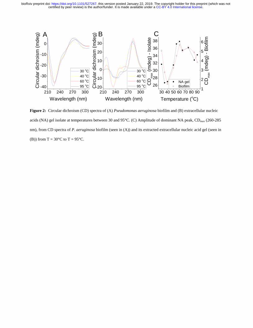
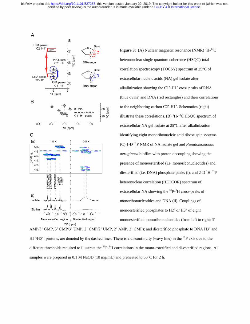
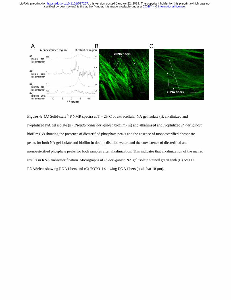
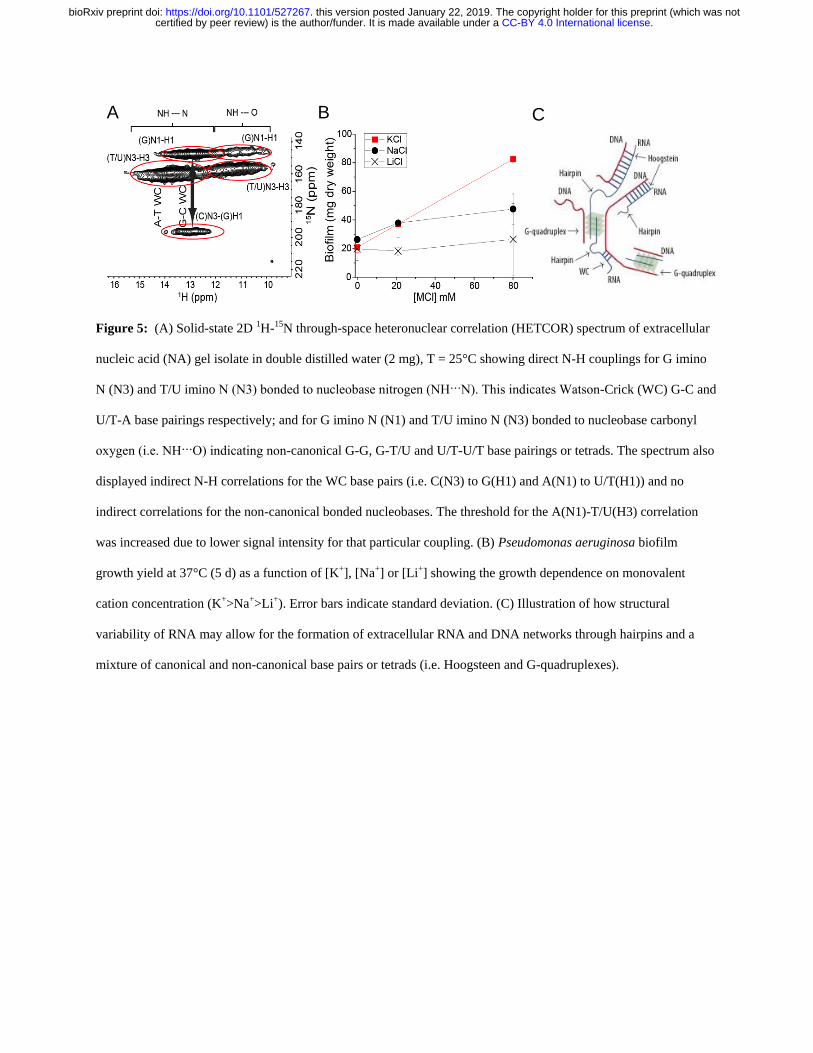
1
Supplementary Information for 5
The biofilm matrix scaffold of Pseudomonas species consists of non-
canonically base paired extracellular DNA and RNA
Thomas Seviour1
Fernaldo Richtia Winnerdy2 Wong Lan Li
1 Xiangyan Shi
2 Sudarsan
Mugunthan1 Gurjeet Singh Kohli
1 Heather M Shewan
3 Jason R Stokes
3 Scott A Rice
14 Anh
Tuacircn Phan2 Staffan Kjelleberg
1 5 6 10
1Singapore Centre for Environmental Life Sciences Engineering Nanyang Technological
University 637551 Singapore 2School of Physical and Mathematical Sciences Nanyang
Technological University 637371 Singapore 3School of Chemical Engineering The University
of Queensland 4072 Brisbane Australia 4
The iThree Institute The University of Technology 15
Sydney Sydney 2007 Australia 5 School of Biological Sciences Nanyang Technological
University 637551 Singapore 6
Centre for Marine Bio-Innovation School of Biological Earth
and Environmental Sciences University of New South Wales Sydney 2052 Australia
20
Correspondence to twseviourntuedusg
This PDF file includes
25
Supplementary Figs 1 to 19
Supplementary Tables 1-2
30
35
CC-BY 40 International licensecertified by peer review) is the authorfunder It is made available under aThe copyright holder for this preprint (which was notthis version posted January 22 2019 httpsdoiorg101101527267doi bioRxiv preprint
2
Supplementary Fig 1 (A) Shear stress and (B) normal stress differences (N1 ndashN2) as a function of shear rate for P
aeruginosa biofilm wild type PDO300 ΔPsl ΔPel pronase digested RNase A digested and DNase I digested
wild type biofilm dissolved in 1-ethyl-3-methylimidazolium acetate (40 mgmL) at 25 degC 100 microm rheometer
measurement gap shear stress sweep from 10 to 1000 Pa (N1 ndashN2) is not described for DNase I digested biofilm as 5
its normal force (FN) is less than the resolution of the rheometer (ie 01 N) and is set to zero for calculating (N1 ndash
N2) Lines indicate power-law fits to the data The power law dependences of shear stress on shear rate (m) (see
Table S1) indicate Newtonian-like rheological properties
10
A B
CC-BY 40 International licensecertified by peer review) is the authorfunder It is made available under aThe copyright holder for this preprint (which was notthis version posted January 22 2019 httpsdoiorg101101527267doi bioRxiv preprint
3
Supplementary Fig 2 Storage modulus (Grsquo) loss modulus (Grsquorsquo) and complex viscosity (η) of P aeruginosa wild
type biofilm in amplitude sweep at 25degC 50 microm gap 01 s-1 frequency showing that the biofilm behaves like a gel
at low amplitude (ie Grsquo gt Grsquorsquo) Note Grsquo is not measurable in the DNase I treated biofilm 5
10
01 1 10 100
001
01
1 G
G
G and G
P
a
1
10
100
P
as
CC-BY 40 International licensecertified by peer review) is the authorfunder It is made available under aThe copyright holder for this preprint (which was notthis version posted January 22 2019 httpsdoiorg101101527267doi bioRxiv preprint
4
Supplementary Fig 3
31P NMR spectrum of H3PO4 solution in EMIM-Ac ( upper) lyophilized P aeruginosa
wild type biofilm in EMIM-Ac (10 mgmL-1) ( second upper) asolectin standard in EMIM-Ac ( second lower) 5
and SDS and lysozyme-treated P aeruginosa wild type planktonic cells ( lower) at 25degC showing phospholipid
peaks for asolectin in EMIM-Ac for lysed P aeruginosa cells (SDS lysozyme) in water and the absence of
phospholipid peaks in the spectrum of P aeruginosa treated with EMIM-Ac
CC-BY 40 International licensecertified by peer review) is the authorfunder It is made available under aThe copyright holder for this preprint (which was notthis version posted January 22 2019 httpsdoiorg101101527267doi bioRxiv preprint
5
Supplementary Fig 4 Recovery yield of calf thymus DNA lipase and cytochrome c standards following EMIM-
Ac solubilization and recovery with perchloric acid (upper) followed by ethanol (lower) Error bars indicate standard
deviation
5
DNA Lipase Cyt C0
10
20
30
40
50
60
Ethanol Precipitate
Re
co
ve
ry y
ield
(
ww
)Perchloric Precipitate
DNA Lipase Cyt C0
10
20
30
40
50
60
70
Sample
CC-BY 40 International licensecertified by peer review) is the authorfunder It is made available under aThe copyright holder for this preprint (which was notthis version posted January 22 2019 httpsdoiorg101101527267doi bioRxiv preprint
6
Supplementary Fig 5 Storage modulus (Grsquo) loss modulus (Grsquorsquo) and complex viscosity (η) of P aeruginosa wild
type biofilm extracellular nucleic acid gel isolate in amplitude sweep at 25degC 50 microm gap 01 s-1 frequency showing
that the biofilm behaves like a gel at low amplitude (ie Grsquo gt Grsquorsquo) 5
01 10
1
10
100
1000 G
G
GG
(P
a)
1000
10000
100000
1000000
(
Pas
)
CC-BY 40 International licensecertified by peer review) is the authorfunder It is made available under aThe copyright holder for this preprint (which was notthis version posted January 22 2019 httpsdoiorg101101527267doi bioRxiv preprint
7
Supplementary Fig 6 Agarose electrophoretic gel showing P aeruginosa eDNA (Lanes 2 and 3) and digested
with DNase (Lane 4) Lane 1 is the GeneRuler 1 kbp ladder 5
Recovered
fractions
DNase I
treated
bp
10000
1000
250
1 2 3 4
CC-BY 40 International licensecertified by peer review) is the authorfunder It is made available under aThe copyright holder for this preprint (which was notthis version posted January 22 2019 httpsdoiorg101101527267doi bioRxiv preprint
8
5
10
15
20
Supplementary Fig 7 (A) Photograph of P aeruginosa biofilm extracellular polymeric substances recovered by
ethanol precipitation (70 vv) from 1-ethyl-3-methylimidazolium dialyzed against double distilled water and then 25
pelleted by centrifugation (10000 g 4degC) after it was dissolved (20 mgmL-1) and eDNA first removed by
perchloric acid precipitation (5 vv) showing that gelation was not observed once the nucleic acids were
removed (B) Photograph of calf thymus DNA recovered from 1-ethyl-3-methylimidazolium after dissolution (6
mgmL-1) perchloric acid precipitation and dialysis against double distilled water showing that gelation is not a
universal feature of all DNA following dissolution in 1-ethyl-3-methylimidazolium acetate and perchloric acid 30
precipitation
A B
CC-BY 40 International licensecertified by peer review) is the authorfunder It is made available under aThe copyright holder for this preprint (which was notthis version posted January 22 2019 httpsdoiorg101101527267doi bioRxiv preprint
9
5
10
15
20
Supplementary Fig 8 1H-13C HSQC-TOCSY and 1-D 1H NMR spectra of P aeruginosa biofilm extracellular NA
gel isolate following alkalinization (01 M NaOD 10 mgmL-1 55degC 2 h) at 25degC showing a distribution of 1H-13C
HSQC-TOCSY cross peaks that is consistent with the presence of nucleic acids and the absence of proteins and
hexose-based sugars
25
13
C
1
H
CC-BY 40 International licensecertified by peer review) is the authorfunder It is made available under aThe copyright holder for this preprint (which was notthis version posted January 22 2019 httpsdoiorg101101527267doi bioRxiv preprint
10
Supplementary Fig 9
1H-1H COSY NMR spectrum of P aeruginosa biofilm extracellular NA gel isolate
following alkalinization (01 M NaOD 10 mgmL-1 55degC 2 h)
5
CC-BY 40 International licensecertified by peer review) is the authorfunder It is made available under aThe copyright holder for this preprint (which was notthis version posted January 22 2019 httpsdoiorg101101527267doi bioRxiv preprint
11
Supplementary Fig 10 1H-13C HMBC NMR spectrum of P aeruginosa biofilm extracellular NA gel isolate at
25degC following alkalinization (01 M NaOD 10 mgmL-1 55degC 2 h)
5
CC-BY 40 International licensecertified by peer review) is the authorfunder It is made available under aThe copyright holder for this preprint (which was notthis version posted January 22 2019 httpsdoiorg101101527267doi bioRxiv preprint
12
Supplementary Fig 11 1H NMR spectra of P aeruginosa biofilm NA gel isolate in D2O at 25degC
10 8 6 4 2 0
1H (ppm)
CC-BY 40 International licensecertified by peer review) is the authorfunder It is made available under aThe copyright holder for this preprint (which was notthis version posted January 22 2019 httpsdoiorg101101527267doi bioRxiv preprint
13
5
Supplementary Fig 12 31P NMR spectrum of RNA standard (torula yeast) at 25degC following alkalinization (01 M
NaOD 10 mgmL-1 55degC 2 h) (A) and in D2O following EMIM-Ac solubilisation and perchloric acid recovery (10
mgmL-1) (B) demonstrating that alkalinization only leads to RNA transesterification
000E+000
100E+008
200E+008
300E+008
400E+008
500E+008
Inte
nsity (
au
)
5 4 3 2 1 0 -1 -2 -3-600E+008
-400E+008
-200E+008
000E+000
200E+008 B
Inte
nsity (
au
)
31P (ppm)
A
CC-BY 40 International licensecertified by peer review) is the authorfunder It is made available under aThe copyright holder for this preprint (which was notthis version posted January 22 2019 httpsdoiorg101101527267doi bioRxiv preprint
14
5
10
15
20
25
Supplementary Fig 13 Micrograph of P aeruginosa NA gel isolate digested with DNase I and stained green with
SYTO RNASelect showing that SYTO RNASelect is binding to extracellular RNA (scale bar 10 microm) 30
CC-BY 40 International licensecertified by peer review) is the authorfunder It is made available under aThe copyright holder for this preprint (which was notthis version posted January 22 2019 httpsdoiorg101101527267doi bioRxiv preprint
15
Supplementary Fig 14 Storage modulus (Grsquo) loss modulus (Grsquorsquo) and complex viscosity (η) of P aeruginosa
wild type biofilm extracellular nucleic acid gel isolate in amplitude sweep at 25degC 50 microm gap 01 s-1 frequency
showing that the biofilm behaves like a gel at low amplitude (ie Grsquo gt Grsquorsquo) even after digestion with RNase III (A)
and RNase H (B) 5
001 01 1 10 100
001
01
1
G
G
GG
P
a
10
100
(m
Pas
)
001 01 1 10 100
001
01
1
GG
P
a
10
100
B
(m
Pas
)
A
CC-BY 40 International licensecertified by peer review) is the authorfunder It is made available under aThe copyright holder for this preprint (which was notthis version posted January 22 2019 httpsdoiorg101101527267doi bioRxiv preprint
16
Supplementary Fig 15 Agarose gel loaded with chromosomal DNA extracted from P aeruginosa pre-culture
planktonic cells (upper) and extracellular NA gel isolate (lower) (A) Gene coverage of P aeruginosa biofilm
chromosomal (upper) and extracellular (lower) DNA normalized against rpoB numbers The red oval denotes the 5
peak resulting from bacteriophage Pf1 genes (B)
115866E6 239371E6 364123E6 486767E6 608308E6
0
2
4
6
8
10
Extracellular DNA
Mea
n g
en
e c
overa
ge
Chromosomal DNA
115866E6 239371E6 364123E6 486767E6 608308E6
0
2
4
6
8
10
Region
Chromosomal DNA
Extracellular NA
A B
CC-BY 40 International licensecertified by peer review) is the authorfunder It is made available under aThe copyright holder for this preprint (which was notthis version posted January 22 2019 httpsdoiorg101101527267doi bioRxiv preprint
17
Supplementary Fig 16 (A) Normal stress differences (N1 ndashN2) and (B) shear stress as a function of shear rate for
Pseudomonas biofilms P putida P protogens and P aeruginosa ΔPf4 dissolved in 1-ethyl-3-methylimidazolium
acetate (40 mgmL-1) at 25 degC Lines indicate FENE-P fits to the data 5
A B
CC-BY 40 International licensecertified by peer review) is the authorfunder It is made available under aThe copyright holder for this preprint (which was notthis version posted January 22 2019 httpsdoiorg101101527267doi bioRxiv preprint
18
Supplementary Fig 17 Overnight growth curve for P aeruginosa cells grown in lithium and potassium enhanced
modified M9 media showing that potassium does not enhance planktonic growth over lithium
5
CC-BY 40 International licensecertified by peer review) is the authorfunder It is made available under aThe copyright holder for this preprint (which was notthis version posted January 22 2019 httpsdoiorg101101527267doi bioRxiv preprint
19
5
10
15
Supplementary Fig 18 Photograph of nucleic acid gel extracted from (A) P protegens and (B) P putida
A B
CC-BY 40 International licensecertified by peer review) is the authorfunder It is made available under aThe copyright holder for this preprint (which was notthis version posted January 22 2019 httpsdoiorg101101527267doi bioRxiv preprint
20
5
10
15
20
Supplementary Fig 19 Photographs of P aeruginosa biofilm after 5-d static growth in lysogeny broth medium at
37degC in 2 L Erlenmeyer flask (A) before centrifugation and (B) in 50 mL centrifuge tube after centrifugation
showing the separation of biofilm from supernatant 25
Biofilm
Supernatant
A B
CC-BY 40 International licensecertified by peer review) is the authorfunder It is made available under aThe copyright holder for this preprint (which was notthis version posted January 22 2019 httpsdoiorg101101527267doi bioRxiv preprint
21
Supplementary Table 1 Power law exponent (ie n in 1198731 = 119870119899 and m in 120590 = 119870120590119898) of P aeruginosa biofilms
following dissolution in EMIM-Ac m values approaching unity indicate a Newtonian-like fluid property Viscosity 5
is slightly shear thinning (m = 08 to 09) for all samples except RNase DNase and EMIM-Ac which are Newtonian
like (m ge 093) which would be expected from dilute polymer solutions in viscous fluids (ie Boger fluids)
120590 = 119870120590119898 1198731 = 1198701198731
119899
Sample K m KN1 n
Ave Std Dev Ave Std Dev Ave Std Dev Ave Std Dev
P
aeruginosa
wild type 063
008
084 001 144 045 136 0
Psl 126 035 076 004 781 518 121 015
Pel 035 0 089 0 058 015 134 002
Pronase 055 003 089 0 018 011 164 008
RNase 034 001 093 0 211 009 091 001
P putida 132 001 091 001 056 016 148 008
PDO300 073 035 089 001 233 059 110 003
P protegens 114 001 090 001 275 130 110 011
Pf4 057 002 087 001 159 125 133 024
DNase 020 001 099 001 - -
EMIM-Ac 013 0 098 0 - -
10
Supplementary Table 2 Fitting parameters for the FENE-P model including 1 = relaxation time b = a measure of
the relative extensibility of the model spring s = solvent viscosity p = polymer contribution to the
viscosity Molecular extensibility and relaxation times as predicted by FENE-P decrease in accordance with
elasticity
Average Std Deviation
Sample b 1 p s b 1 p s
P
aeruginosa
wild type 22485 0322 0260 0145
6431 0066 0040 00026
Psl 35115 0676 0381 0161 10009 0219 0113 00114
Pel 20008 0227 0110 0133 6378 0056 0000 00025
Pronase 18718 0103 0197 0167 6931 0047 0015 00339
RNase 3992 0102 0095 0183 176 0008 0003 00077
PDO300 9094 0453 0138 0147 1342 0023 0026 00007
DNase 1357 0005 0020 0129 1068 0001 0011 00112
Pf4 8654 0230 0243 0166 1299 0034 0022 00072
P protegens 1372 0175 0501 0414 192 0028 0025 00013
P putida 3369 0071 0544 0440 535 0002 0018 00199 15
CC-BY 40 International licensecertified by peer review) is the authorfunder It is made available under aThe copyright holder for this preprint (which was notthis version posted January 22 2019 httpsdoiorg101101527267doi bioRxiv preprint

2
Supplementary Fig 1 (A) Shear stress and (B) normal stress differences (N1 ndashN2) as a function of shear rate for P
aeruginosa biofilm wild type PDO300 ΔPsl ΔPel pronase digested RNase A digested and DNase I digested
wild type biofilm dissolved in 1-ethyl-3-methylimidazolium acetate (40 mgmL) at 25 degC 100 microm rheometer
measurement gap shear stress sweep from 10 to 1000 Pa (N1 ndashN2) is not described for DNase I digested biofilm as 5
its normal force (FN) is less than the resolution of the rheometer (ie 01 N) and is set to zero for calculating (N1 ndash
N2) Lines indicate power-law fits to the data The power law dependences of shear stress on shear rate (m) (see
Table S1) indicate Newtonian-like rheological properties
10
A B
CC-BY 40 International licensecertified by peer review) is the authorfunder It is made available under aThe copyright holder for this preprint (which was notthis version posted January 22 2019 httpsdoiorg101101527267doi bioRxiv preprint
3
Supplementary Fig 2 Storage modulus (Grsquo) loss modulus (Grsquorsquo) and complex viscosity (η) of P aeruginosa wild
type biofilm in amplitude sweep at 25degC 50 microm gap 01 s-1 frequency showing that the biofilm behaves like a gel
at low amplitude (ie Grsquo gt Grsquorsquo) Note Grsquo is not measurable in the DNase I treated biofilm 5
10
01 1 10 100
001
01
1 G
G
G and G
P
a
1
10
100
P
as
CC-BY 40 International licensecertified by peer review) is the authorfunder It is made available under aThe copyright holder for this preprint (which was notthis version posted January 22 2019 httpsdoiorg101101527267doi bioRxiv preprint
4
Supplementary Fig 3
31P NMR spectrum of H3PO4 solution in EMIM-Ac ( upper) lyophilized P aeruginosa
wild type biofilm in EMIM-Ac (10 mgmL-1) ( second upper) asolectin standard in EMIM-Ac ( second lower) 5
and SDS and lysozyme-treated P aeruginosa wild type planktonic cells ( lower) at 25degC showing phospholipid
peaks for asolectin in EMIM-Ac for lysed P aeruginosa cells (SDS lysozyme) in water and the absence of
phospholipid peaks in the spectrum of P aeruginosa treated with EMIM-Ac
CC-BY 40 International licensecertified by peer review) is the authorfunder It is made available under aThe copyright holder for this preprint (which was notthis version posted January 22 2019 httpsdoiorg101101527267doi bioRxiv preprint
5
Supplementary Fig 4 Recovery yield of calf thymus DNA lipase and cytochrome c standards following EMIM-
Ac solubilization and recovery with perchloric acid (upper) followed by ethanol (lower) Error bars indicate standard
deviation
5
DNA Lipase Cyt C0
10
20
30
40
50
60
Ethanol Precipitate
Re
co
ve
ry y
ield
(
ww
)Perchloric Precipitate
DNA Lipase Cyt C0
10
20
30
40
50
60
70
Sample
CC-BY 40 International licensecertified by peer review) is the authorfunder It is made available under aThe copyright holder for this preprint (which was notthis version posted January 22 2019 httpsdoiorg101101527267doi bioRxiv preprint
6
Supplementary Fig 5 Storage modulus (Grsquo) loss modulus (Grsquorsquo) and complex viscosity (η) of P aeruginosa wild
type biofilm extracellular nucleic acid gel isolate in amplitude sweep at 25degC 50 microm gap 01 s-1 frequency showing
that the biofilm behaves like a gel at low amplitude (ie Grsquo gt Grsquorsquo) 5
01 10
1
10
100
1000 G
G
GG
(P
a)
1000
10000
100000
1000000
(
Pas
)
CC-BY 40 International licensecertified by peer review) is the authorfunder It is made available under aThe copyright holder for this preprint (which was notthis version posted January 22 2019 httpsdoiorg101101527267doi bioRxiv preprint
7
Supplementary Fig 6 Agarose electrophoretic gel showing P aeruginosa eDNA (Lanes 2 and 3) and digested
with DNase (Lane 4) Lane 1 is the GeneRuler 1 kbp ladder 5
Recovered
fractions
DNase I
treated
bp
10000
1000
250
1 2 3 4
CC-BY 40 International licensecertified by peer review) is the authorfunder It is made available under aThe copyright holder for this preprint (which was notthis version posted January 22 2019 httpsdoiorg101101527267doi bioRxiv preprint
8
5
10
15
20
Supplementary Fig 7 (A) Photograph of P aeruginosa biofilm extracellular polymeric substances recovered by
ethanol precipitation (70 vv) from 1-ethyl-3-methylimidazolium dialyzed against double distilled water and then 25
pelleted by centrifugation (10000 g 4degC) after it was dissolved (20 mgmL-1) and eDNA first removed by
perchloric acid precipitation (5 vv) showing that gelation was not observed once the nucleic acids were
removed (B) Photograph of calf thymus DNA recovered from 1-ethyl-3-methylimidazolium after dissolution (6
mgmL-1) perchloric acid precipitation and dialysis against double distilled water showing that gelation is not a
universal feature of all DNA following dissolution in 1-ethyl-3-methylimidazolium acetate and perchloric acid 30
precipitation
A B
CC-BY 40 International licensecertified by peer review) is the authorfunder It is made available under aThe copyright holder for this preprint (which was notthis version posted January 22 2019 httpsdoiorg101101527267doi bioRxiv preprint
9
5
10
15
20
Supplementary Fig 8 1H-13C HSQC-TOCSY and 1-D 1H NMR spectra of P aeruginosa biofilm extracellular NA
gel isolate following alkalinization (01 M NaOD 10 mgmL-1 55degC 2 h) at 25degC showing a distribution of 1H-13C
HSQC-TOCSY cross peaks that is consistent with the presence of nucleic acids and the absence of proteins and
hexose-based sugars
25
13
C
1
H
CC-BY 40 International licensecertified by peer review) is the authorfunder It is made available under aThe copyright holder for this preprint (which was notthis version posted January 22 2019 httpsdoiorg101101527267doi bioRxiv preprint
10
Supplementary Fig 9
1H-1H COSY NMR spectrum of P aeruginosa biofilm extracellular NA gel isolate
following alkalinization (01 M NaOD 10 mgmL-1 55degC 2 h)
5
CC-BY 40 International licensecertified by peer review) is the authorfunder It is made available under aThe copyright holder for this preprint (which was notthis version posted January 22 2019 httpsdoiorg101101527267doi bioRxiv preprint
11
Supplementary Fig 10 1H-13C HMBC NMR spectrum of P aeruginosa biofilm extracellular NA gel isolate at
25degC following alkalinization (01 M NaOD 10 mgmL-1 55degC 2 h)
5
CC-BY 40 International licensecertified by peer review) is the authorfunder It is made available under aThe copyright holder for this preprint (which was notthis version posted January 22 2019 httpsdoiorg101101527267doi bioRxiv preprint
12
Supplementary Fig 11 1H NMR spectra of P aeruginosa biofilm NA gel isolate in D2O at 25degC
10 8 6 4 2 0
1H (ppm)
CC-BY 40 International licensecertified by peer review) is the authorfunder It is made available under aThe copyright holder for this preprint (which was notthis version posted January 22 2019 httpsdoiorg101101527267doi bioRxiv preprint
13
5
Supplementary Fig 12 31P NMR spectrum of RNA standard (torula yeast) at 25degC following alkalinization (01 M
NaOD 10 mgmL-1 55degC 2 h) (A) and in D2O following EMIM-Ac solubilisation and perchloric acid recovery (10
mgmL-1) (B) demonstrating that alkalinization only leads to RNA transesterification
000E+000
100E+008
200E+008
300E+008
400E+008
500E+008
Inte
nsity (
au
)
5 4 3 2 1 0 -1 -2 -3-600E+008
-400E+008
-200E+008
000E+000
200E+008 B
Inte
nsity (
au
)
31P (ppm)
A
CC-BY 40 International licensecertified by peer review) is the authorfunder It is made available under aThe copyright holder for this preprint (which was notthis version posted January 22 2019 httpsdoiorg101101527267doi bioRxiv preprint
14
5
10
15
20
25
Supplementary Fig 13 Micrograph of P aeruginosa NA gel isolate digested with DNase I and stained green with
SYTO RNASelect showing that SYTO RNASelect is binding to extracellular RNA (scale bar 10 microm) 30
CC-BY 40 International licensecertified by peer review) is the authorfunder It is made available under aThe copyright holder for this preprint (which was notthis version posted January 22 2019 httpsdoiorg101101527267doi bioRxiv preprint
15
Supplementary Fig 14 Storage modulus (Grsquo) loss modulus (Grsquorsquo) and complex viscosity (η) of P aeruginosa
wild type biofilm extracellular nucleic acid gel isolate in amplitude sweep at 25degC 50 microm gap 01 s-1 frequency
showing that the biofilm behaves like a gel at low amplitude (ie Grsquo gt Grsquorsquo) even after digestion with RNase III (A)
and RNase H (B) 5
001 01 1 10 100
001
01
1
G
G
GG
P
a
10
100
(m
Pas
)
001 01 1 10 100
001
01
1
GG
P
a
10
100
B
(m
Pas
)
A
CC-BY 40 International licensecertified by peer review) is the authorfunder It is made available under aThe copyright holder for this preprint (which was notthis version posted January 22 2019 httpsdoiorg101101527267doi bioRxiv preprint
16
Supplementary Fig 15 Agarose gel loaded with chromosomal DNA extracted from P aeruginosa pre-culture
planktonic cells (upper) and extracellular NA gel isolate (lower) (A) Gene coverage of P aeruginosa biofilm
chromosomal (upper) and extracellular (lower) DNA normalized against rpoB numbers The red oval denotes the 5
peak resulting from bacteriophage Pf1 genes (B)
115866E6 239371E6 364123E6 486767E6 608308E6
0
2
4
6
8
10
Extracellular DNA
Mea
n g
en
e c
overa
ge
Chromosomal DNA
115866E6 239371E6 364123E6 486767E6 608308E6
0
2
4
6
8
10
Region
Chromosomal DNA
Extracellular NA
A B
CC-BY 40 International licensecertified by peer review) is the authorfunder It is made available under aThe copyright holder for this preprint (which was notthis version posted January 22 2019 httpsdoiorg101101527267doi bioRxiv preprint
17
Supplementary Fig 16 (A) Normal stress differences (N1 ndashN2) and (B) shear stress as a function of shear rate for
Pseudomonas biofilms P putida P protogens and P aeruginosa ΔPf4 dissolved in 1-ethyl-3-methylimidazolium
acetate (40 mgmL-1) at 25 degC Lines indicate FENE-P fits to the data 5
A B
CC-BY 40 International licensecertified by peer review) is the authorfunder It is made available under aThe copyright holder for this preprint (which was notthis version posted January 22 2019 httpsdoiorg101101527267doi bioRxiv preprint
18
Supplementary Fig 17 Overnight growth curve for P aeruginosa cells grown in lithium and potassium enhanced
modified M9 media showing that potassium does not enhance planktonic growth over lithium
5
CC-BY 40 International licensecertified by peer review) is the authorfunder It is made available under aThe copyright holder for this preprint (which was notthis version posted January 22 2019 httpsdoiorg101101527267doi bioRxiv preprint
19
5
10
15
Supplementary Fig 18 Photograph of nucleic acid gel extracted from (A) P protegens and (B) P putida
A B
CC-BY 40 International licensecertified by peer review) is the authorfunder It is made available under aThe copyright holder for this preprint (which was notthis version posted January 22 2019 httpsdoiorg101101527267doi bioRxiv preprint
20
5
10
15
20
Supplementary Fig 19 Photographs of P aeruginosa biofilm after 5-d static growth in lysogeny broth medium at
37degC in 2 L Erlenmeyer flask (A) before centrifugation and (B) in 50 mL centrifuge tube after centrifugation
showing the separation of biofilm from supernatant 25
Biofilm
Supernatant
A B
CC-BY 40 International licensecertified by peer review) is the authorfunder It is made available under aThe copyright holder for this preprint (which was notthis version posted January 22 2019 httpsdoiorg101101527267doi bioRxiv preprint
21
Supplementary Table 1 Power law exponent (ie n in 1198731 = 119870119899 and m in 120590 = 119870120590119898) of P aeruginosa biofilms
following dissolution in EMIM-Ac m values approaching unity indicate a Newtonian-like fluid property Viscosity 5
is slightly shear thinning (m = 08 to 09) for all samples except RNase DNase and EMIM-Ac which are Newtonian
like (m ge 093) which would be expected from dilute polymer solutions in viscous fluids (ie Boger fluids)
120590 = 119870120590119898 1198731 = 1198701198731
119899
Sample K m KN1 n
Ave Std Dev Ave Std Dev Ave Std Dev Ave Std Dev
P
aeruginosa
wild type 063
008
084 001 144 045 136 0
Psl 126 035 076 004 781 518 121 015
Pel 035 0 089 0 058 015 134 002
Pronase 055 003 089 0 018 011 164 008
RNase 034 001 093 0 211 009 091 001
P putida 132 001 091 001 056 016 148 008
PDO300 073 035 089 001 233 059 110 003
P protegens 114 001 090 001 275 130 110 011
Pf4 057 002 087 001 159 125 133 024
DNase 020 001 099 001 - -
EMIM-Ac 013 0 098 0 - -
10
Supplementary Table 2 Fitting parameters for the FENE-P model including 1 = relaxation time b = a measure of
the relative extensibility of the model spring s = solvent viscosity p = polymer contribution to the
viscosity Molecular extensibility and relaxation times as predicted by FENE-P decrease in accordance with
elasticity
Average Std Deviation
Sample b 1 p s b 1 p s
P
aeruginosa
wild type 22485 0322 0260 0145
6431 0066 0040 00026
Psl 35115 0676 0381 0161 10009 0219 0113 00114
Pel 20008 0227 0110 0133 6378 0056 0000 00025
Pronase 18718 0103 0197 0167 6931 0047 0015 00339
RNase 3992 0102 0095 0183 176 0008 0003 00077
PDO300 9094 0453 0138 0147 1342 0023 0026 00007
DNase 1357 0005 0020 0129 1068 0001 0011 00112
Pf4 8654 0230 0243 0166 1299 0034 0022 00072
P protegens 1372 0175 0501 0414 192 0028 0025 00013
P putida 3369 0071 0544 0440 535 0002 0018 00199 15
CC-BY 40 International licensecertified by peer review) is the authorfunder It is made available under aThe copyright holder for this preprint (which was notthis version posted January 22 2019 httpsdoiorg101101527267doi bioRxiv preprint
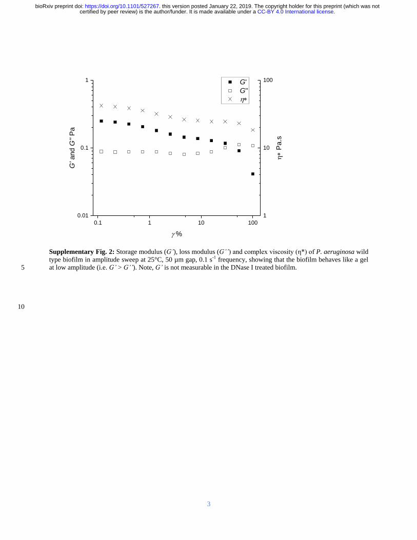
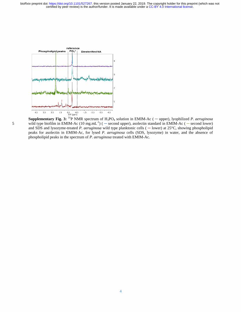
4
Supplementary Fig 3
31P NMR spectrum of H3PO4 solution in EMIM-Ac ( upper) lyophilized P aeruginosa
wild type biofilm in EMIM-Ac (10 mgmL-1) ( second upper) asolectin standard in EMIM-Ac ( second lower) 5
and SDS and lysozyme-treated P aeruginosa wild type planktonic cells ( lower) at 25degC showing phospholipid
peaks for asolectin in EMIM-Ac for lysed P aeruginosa cells (SDS lysozyme) in water and the absence of
phospholipid peaks in the spectrum of P aeruginosa treated with EMIM-Ac
CC-BY 40 International licensecertified by peer review) is the authorfunder It is made available under aThe copyright holder for this preprint (which was notthis version posted January 22 2019 httpsdoiorg101101527267doi bioRxiv preprint
5
Supplementary Fig 4 Recovery yield of calf thymus DNA lipase and cytochrome c standards following EMIM-
Ac solubilization and recovery with perchloric acid (upper) followed by ethanol (lower) Error bars indicate standard
deviation
5
DNA Lipase Cyt C0
10
20
30
40
50
60
Ethanol Precipitate
Re
co
ve
ry y
ield
(
ww
)Perchloric Precipitate
DNA Lipase Cyt C0
10
20
30
40
50
60
70
Sample
CC-BY 40 International licensecertified by peer review) is the authorfunder It is made available under aThe copyright holder for this preprint (which was notthis version posted January 22 2019 httpsdoiorg101101527267doi bioRxiv preprint
6
Supplementary Fig 5 Storage modulus (Grsquo) loss modulus (Grsquorsquo) and complex viscosity (η) of P aeruginosa wild
type biofilm extracellular nucleic acid gel isolate in amplitude sweep at 25degC 50 microm gap 01 s-1 frequency showing
that the biofilm behaves like a gel at low amplitude (ie Grsquo gt Grsquorsquo) 5
01 10
1
10
100
1000 G
G
GG
(P
a)
1000
10000
100000
1000000
(
Pas
)
CC-BY 40 International licensecertified by peer review) is the authorfunder It is made available under aThe copyright holder for this preprint (which was notthis version posted January 22 2019 httpsdoiorg101101527267doi bioRxiv preprint
7
Supplementary Fig 6 Agarose electrophoretic gel showing P aeruginosa eDNA (Lanes 2 and 3) and digested
with DNase (Lane 4) Lane 1 is the GeneRuler 1 kbp ladder 5
Recovered
fractions
DNase I
treated
bp
10000
1000
250
1 2 3 4
CC-BY 40 International licensecertified by peer review) is the authorfunder It is made available under aThe copyright holder for this preprint (which was notthis version posted January 22 2019 httpsdoiorg101101527267doi bioRxiv preprint
8
5
10
15
20
Supplementary Fig 7 (A) Photograph of P aeruginosa biofilm extracellular polymeric substances recovered by
ethanol precipitation (70 vv) from 1-ethyl-3-methylimidazolium dialyzed against double distilled water and then 25
pelleted by centrifugation (10000 g 4degC) after it was dissolved (20 mgmL-1) and eDNA first removed by
perchloric acid precipitation (5 vv) showing that gelation was not observed once the nucleic acids were
removed (B) Photograph of calf thymus DNA recovered from 1-ethyl-3-methylimidazolium after dissolution (6
mgmL-1) perchloric acid precipitation and dialysis against double distilled water showing that gelation is not a
universal feature of all DNA following dissolution in 1-ethyl-3-methylimidazolium acetate and perchloric acid 30
precipitation
A B
CC-BY 40 International licensecertified by peer review) is the authorfunder It is made available under aThe copyright holder for this preprint (which was notthis version posted January 22 2019 httpsdoiorg101101527267doi bioRxiv preprint
9
5
10
15
20
Supplementary Fig 8 1H-13C HSQC-TOCSY and 1-D 1H NMR spectra of P aeruginosa biofilm extracellular NA
gel isolate following alkalinization (01 M NaOD 10 mgmL-1 55degC 2 h) at 25degC showing a distribution of 1H-13C
HSQC-TOCSY cross peaks that is consistent with the presence of nucleic acids and the absence of proteins and
hexose-based sugars
25
13
C
1
H
CC-BY 40 International licensecertified by peer review) is the authorfunder It is made available under aThe copyright holder for this preprint (which was notthis version posted January 22 2019 httpsdoiorg101101527267doi bioRxiv preprint
10
Supplementary Fig 9
1H-1H COSY NMR spectrum of P aeruginosa biofilm extracellular NA gel isolate
following alkalinization (01 M NaOD 10 mgmL-1 55degC 2 h)
5
CC-BY 40 International licensecertified by peer review) is the authorfunder It is made available under aThe copyright holder for this preprint (which was notthis version posted January 22 2019 httpsdoiorg101101527267doi bioRxiv preprint
11
Supplementary Fig 10 1H-13C HMBC NMR spectrum of P aeruginosa biofilm extracellular NA gel isolate at
25degC following alkalinization (01 M NaOD 10 mgmL-1 55degC 2 h)
5
CC-BY 40 International licensecertified by peer review) is the authorfunder It is made available under aThe copyright holder for this preprint (which was notthis version posted January 22 2019 httpsdoiorg101101527267doi bioRxiv preprint
12
Supplementary Fig 11 1H NMR spectra of P aeruginosa biofilm NA gel isolate in D2O at 25degC
10 8 6 4 2 0
1H (ppm)
CC-BY 40 International licensecertified by peer review) is the authorfunder It is made available under aThe copyright holder for this preprint (which was notthis version posted January 22 2019 httpsdoiorg101101527267doi bioRxiv preprint
13
5
Supplementary Fig 12 31P NMR spectrum of RNA standard (torula yeast) at 25degC following alkalinization (01 M
NaOD 10 mgmL-1 55degC 2 h) (A) and in D2O following EMIM-Ac solubilisation and perchloric acid recovery (10
mgmL-1) (B) demonstrating that alkalinization only leads to RNA transesterification
000E+000
100E+008
200E+008
300E+008
400E+008
500E+008
Inte
nsity (
au
)
5 4 3 2 1 0 -1 -2 -3-600E+008
-400E+008
-200E+008
000E+000
200E+008 B
Inte
nsity (
au
)
31P (ppm)
A
CC-BY 40 International licensecertified by peer review) is the authorfunder It is made available under aThe copyright holder for this preprint (which was notthis version posted January 22 2019 httpsdoiorg101101527267doi bioRxiv preprint
14
5
10
15
20
25
Supplementary Fig 13 Micrograph of P aeruginosa NA gel isolate digested with DNase I and stained green with
SYTO RNASelect showing that SYTO RNASelect is binding to extracellular RNA (scale bar 10 microm) 30
CC-BY 40 International licensecertified by peer review) is the authorfunder It is made available under aThe copyright holder for this preprint (which was notthis version posted January 22 2019 httpsdoiorg101101527267doi bioRxiv preprint
15
Supplementary Fig 14 Storage modulus (Grsquo) loss modulus (Grsquorsquo) and complex viscosity (η) of P aeruginosa
wild type biofilm extracellular nucleic acid gel isolate in amplitude sweep at 25degC 50 microm gap 01 s-1 frequency
showing that the biofilm behaves like a gel at low amplitude (ie Grsquo gt Grsquorsquo) even after digestion with RNase III (A)
and RNase H (B) 5
001 01 1 10 100
001
01
1
G
G
GG
P
a
10
100
(m
Pas
)
001 01 1 10 100
001
01
1
GG
P
a
10
100
B
(m
Pas
)
A
CC-BY 40 International licensecertified by peer review) is the authorfunder It is made available under aThe copyright holder for this preprint (which was notthis version posted January 22 2019 httpsdoiorg101101527267doi bioRxiv preprint
16
Supplementary Fig 15 Agarose gel loaded with chromosomal DNA extracted from P aeruginosa pre-culture
planktonic cells (upper) and extracellular NA gel isolate (lower) (A) Gene coverage of P aeruginosa biofilm
chromosomal (upper) and extracellular (lower) DNA normalized against rpoB numbers The red oval denotes the 5
peak resulting from bacteriophage Pf1 genes (B)
115866E6 239371E6 364123E6 486767E6 608308E6
0
2
4
6
8
10
Extracellular DNA
Mea
n g
en
e c
overa
ge
Chromosomal DNA
115866E6 239371E6 364123E6 486767E6 608308E6
0
2
4
6
8
10
Region
Chromosomal DNA
Extracellular NA
A B
CC-BY 40 International licensecertified by peer review) is the authorfunder It is made available under aThe copyright holder for this preprint (which was notthis version posted January 22 2019 httpsdoiorg101101527267doi bioRxiv preprint
17
Supplementary Fig 16 (A) Normal stress differences (N1 ndashN2) and (B) shear stress as a function of shear rate for
Pseudomonas biofilms P putida P protogens and P aeruginosa ΔPf4 dissolved in 1-ethyl-3-methylimidazolium
acetate (40 mgmL-1) at 25 degC Lines indicate FENE-P fits to the data 5
A B
CC-BY 40 International licensecertified by peer review) is the authorfunder It is made available under aThe copyright holder for this preprint (which was notthis version posted January 22 2019 httpsdoiorg101101527267doi bioRxiv preprint
18
Supplementary Fig 17 Overnight growth curve for P aeruginosa cells grown in lithium and potassium enhanced
modified M9 media showing that potassium does not enhance planktonic growth over lithium
5
CC-BY 40 International licensecertified by peer review) is the authorfunder It is made available under aThe copyright holder for this preprint (which was notthis version posted January 22 2019 httpsdoiorg101101527267doi bioRxiv preprint
19
5
10
15
Supplementary Fig 18 Photograph of nucleic acid gel extracted from (A) P protegens and (B) P putida
A B
CC-BY 40 International licensecertified by peer review) is the authorfunder It is made available under aThe copyright holder for this preprint (which was notthis version posted January 22 2019 httpsdoiorg101101527267doi bioRxiv preprint
20
5
10
15
20
Supplementary Fig 19 Photographs of P aeruginosa biofilm after 5-d static growth in lysogeny broth medium at
37degC in 2 L Erlenmeyer flask (A) before centrifugation and (B) in 50 mL centrifuge tube after centrifugation
showing the separation of biofilm from supernatant 25
Biofilm
Supernatant
A B
CC-BY 40 International licensecertified by peer review) is the authorfunder It is made available under aThe copyright holder for this preprint (which was notthis version posted January 22 2019 httpsdoiorg101101527267doi bioRxiv preprint
21
Supplementary Table 1 Power law exponent (ie n in 1198731 = 119870119899 and m in 120590 = 119870120590119898) of P aeruginosa biofilms
following dissolution in EMIM-Ac m values approaching unity indicate a Newtonian-like fluid property Viscosity 5
is slightly shear thinning (m = 08 to 09) for all samples except RNase DNase and EMIM-Ac which are Newtonian
like (m ge 093) which would be expected from dilute polymer solutions in viscous fluids (ie Boger fluids)
120590 = 119870120590119898 1198731 = 1198701198731
119899
Sample K m KN1 n
Ave Std Dev Ave Std Dev Ave Std Dev Ave Std Dev
P
aeruginosa
wild type 063
008
084 001 144 045 136 0
Psl 126 035 076 004 781 518 121 015
Pel 035 0 089 0 058 015 134 002
Pronase 055 003 089 0 018 011 164 008
RNase 034 001 093 0 211 009 091 001
P putida 132 001 091 001 056 016 148 008
PDO300 073 035 089 001 233 059 110 003
P protegens 114 001 090 001 275 130 110 011
Pf4 057 002 087 001 159 125 133 024
DNase 020 001 099 001 - -
EMIM-Ac 013 0 098 0 - -
10
Supplementary Table 2 Fitting parameters for the FENE-P model including 1 = relaxation time b = a measure of
the relative extensibility of the model spring s = solvent viscosity p = polymer contribution to the
viscosity Molecular extensibility and relaxation times as predicted by FENE-P decrease in accordance with
elasticity
Average Std Deviation
Sample b 1 p s b 1 p s
P
aeruginosa
wild type 22485 0322 0260 0145
6431 0066 0040 00026
Psl 35115 0676 0381 0161 10009 0219 0113 00114
Pel 20008 0227 0110 0133 6378 0056 0000 00025
Pronase 18718 0103 0197 0167 6931 0047 0015 00339
RNase 3992 0102 0095 0183 176 0008 0003 00077
PDO300 9094 0453 0138 0147 1342 0023 0026 00007
DNase 1357 0005 0020 0129 1068 0001 0011 00112
Pf4 8654 0230 0243 0166 1299 0034 0022 00072
P protegens 1372 0175 0501 0414 192 0028 0025 00013
P putida 3369 0071 0544 0440 535 0002 0018 00199 15
CC-BY 40 International licensecertified by peer review) is the authorfunder It is made available under aThe copyright holder for this preprint (which was notthis version posted January 22 2019 httpsdoiorg101101527267doi bioRxiv preprint

5
Supplementary Fig 4 Recovery yield of calf thymus DNA lipase and cytochrome c standards following EMIM-
Ac solubilization and recovery with perchloric acid (upper) followed by ethanol (lower) Error bars indicate standard
deviation
5
DNA Lipase Cyt C0
10
20
30
40
50
60
Ethanol Precipitate
Re
co
ve
ry y
ield
(
ww
)Perchloric Precipitate
DNA Lipase Cyt C0
10
20
30
40
50
60
70
Sample
CC-BY 40 International licensecertified by peer review) is the authorfunder It is made available under aThe copyright holder for this preprint (which was notthis version posted January 22 2019 httpsdoiorg101101527267doi bioRxiv preprint
6
Supplementary Fig 5 Storage modulus (Grsquo) loss modulus (Grsquorsquo) and complex viscosity (η) of P aeruginosa wild
type biofilm extracellular nucleic acid gel isolate in amplitude sweep at 25degC 50 microm gap 01 s-1 frequency showing
that the biofilm behaves like a gel at low amplitude (ie Grsquo gt Grsquorsquo) 5
01 10
1
10
100
1000 G
G
GG
(P
a)
1000
10000
100000
1000000
(
Pas
)
CC-BY 40 International licensecertified by peer review) is the authorfunder It is made available under aThe copyright holder for this preprint (which was notthis version posted January 22 2019 httpsdoiorg101101527267doi bioRxiv preprint
7
Supplementary Fig 6 Agarose electrophoretic gel showing P aeruginosa eDNA (Lanes 2 and 3) and digested
with DNase (Lane 4) Lane 1 is the GeneRuler 1 kbp ladder 5
Recovered
fractions
DNase I
treated
bp
10000
1000
250
1 2 3 4
CC-BY 40 International licensecertified by peer review) is the authorfunder It is made available under aThe copyright holder for this preprint (which was notthis version posted January 22 2019 httpsdoiorg101101527267doi bioRxiv preprint
8
5
10
15
20
Supplementary Fig 7 (A) Photograph of P aeruginosa biofilm extracellular polymeric substances recovered by
ethanol precipitation (70 vv) from 1-ethyl-3-methylimidazolium dialyzed against double distilled water and then 25
pelleted by centrifugation (10000 g 4degC) after it was dissolved (20 mgmL-1) and eDNA first removed by
perchloric acid precipitation (5 vv) showing that gelation was not observed once the nucleic acids were
removed (B) Photograph of calf thymus DNA recovered from 1-ethyl-3-methylimidazolium after dissolution (6
mgmL-1) perchloric acid precipitation and dialysis against double distilled water showing that gelation is not a
universal feature of all DNA following dissolution in 1-ethyl-3-methylimidazolium acetate and perchloric acid 30
precipitation
A B
CC-BY 40 International licensecertified by peer review) is the authorfunder It is made available under aThe copyright holder for this preprint (which was notthis version posted January 22 2019 httpsdoiorg101101527267doi bioRxiv preprint
9
5
10
15
20
Supplementary Fig 8 1H-13C HSQC-TOCSY and 1-D 1H NMR spectra of P aeruginosa biofilm extracellular NA
gel isolate following alkalinization (01 M NaOD 10 mgmL-1 55degC 2 h) at 25degC showing a distribution of 1H-13C
HSQC-TOCSY cross peaks that is consistent with the presence of nucleic acids and the absence of proteins and
hexose-based sugars
25
13
C
1
H
CC-BY 40 International licensecertified by peer review) is the authorfunder It is made available under aThe copyright holder for this preprint (which was notthis version posted January 22 2019 httpsdoiorg101101527267doi bioRxiv preprint
10
Supplementary Fig 9
1H-1H COSY NMR spectrum of P aeruginosa biofilm extracellular NA gel isolate
following alkalinization (01 M NaOD 10 mgmL-1 55degC 2 h)
5
CC-BY 40 International licensecertified by peer review) is the authorfunder It is made available under aThe copyright holder for this preprint (which was notthis version posted January 22 2019 httpsdoiorg101101527267doi bioRxiv preprint
11
Supplementary Fig 10 1H-13C HMBC NMR spectrum of P aeruginosa biofilm extracellular NA gel isolate at
25degC following alkalinization (01 M NaOD 10 mgmL-1 55degC 2 h)
5
CC-BY 40 International licensecertified by peer review) is the authorfunder It is made available under aThe copyright holder for this preprint (which was notthis version posted January 22 2019 httpsdoiorg101101527267doi bioRxiv preprint
12
Supplementary Fig 11 1H NMR spectra of P aeruginosa biofilm NA gel isolate in D2O at 25degC
10 8 6 4 2 0
1H (ppm)
CC-BY 40 International licensecertified by peer review) is the authorfunder It is made available under aThe copyright holder for this preprint (which was notthis version posted January 22 2019 httpsdoiorg101101527267doi bioRxiv preprint
13
5
Supplementary Fig 12 31P NMR spectrum of RNA standard (torula yeast) at 25degC following alkalinization (01 M
NaOD 10 mgmL-1 55degC 2 h) (A) and in D2O following EMIM-Ac solubilisation and perchloric acid recovery (10
mgmL-1) (B) demonstrating that alkalinization only leads to RNA transesterification
000E+000
100E+008
200E+008
300E+008
400E+008
500E+008
Inte
nsity (
au
)
5 4 3 2 1 0 -1 -2 -3-600E+008
-400E+008
-200E+008
000E+000
200E+008 B
Inte
nsity (
au
)
31P (ppm)
A
CC-BY 40 International licensecertified by peer review) is the authorfunder It is made available under aThe copyright holder for this preprint (which was notthis version posted January 22 2019 httpsdoiorg101101527267doi bioRxiv preprint
14
5
10
15
20
25
Supplementary Fig 13 Micrograph of P aeruginosa NA gel isolate digested with DNase I and stained green with
SYTO RNASelect showing that SYTO RNASelect is binding to extracellular RNA (scale bar 10 microm) 30
CC-BY 40 International licensecertified by peer review) is the authorfunder It is made available under aThe copyright holder for this preprint (which was notthis version posted January 22 2019 httpsdoiorg101101527267doi bioRxiv preprint
15
Supplementary Fig 14 Storage modulus (Grsquo) loss modulus (Grsquorsquo) and complex viscosity (η) of P aeruginosa
wild type biofilm extracellular nucleic acid gel isolate in amplitude sweep at 25degC 50 microm gap 01 s-1 frequency
showing that the biofilm behaves like a gel at low amplitude (ie Grsquo gt Grsquorsquo) even after digestion with RNase III (A)
and RNase H (B) 5
001 01 1 10 100
001
01
1
G
G
GG
P
a
10
100
(m
Pas
)
001 01 1 10 100
001
01
1
GG
P
a
10
100
B
(m
Pas
)
A
CC-BY 40 International licensecertified by peer review) is the authorfunder It is made available under aThe copyright holder for this preprint (which was notthis version posted January 22 2019 httpsdoiorg101101527267doi bioRxiv preprint
16
Supplementary Fig 15 Agarose gel loaded with chromosomal DNA extracted from P aeruginosa pre-culture
planktonic cells (upper) and extracellular NA gel isolate (lower) (A) Gene coverage of P aeruginosa biofilm
chromosomal (upper) and extracellular (lower) DNA normalized against rpoB numbers The red oval denotes the 5
peak resulting from bacteriophage Pf1 genes (B)
115866E6 239371E6 364123E6 486767E6 608308E6
0
2
4
6
8
10
Extracellular DNA
Mea
n g
en
e c
overa
ge
Chromosomal DNA
115866E6 239371E6 364123E6 486767E6 608308E6
0
2
4
6
8
10
Region
Chromosomal DNA
Extracellular NA
A B
CC-BY 40 International licensecertified by peer review) is the authorfunder It is made available under aThe copyright holder for this preprint (which was notthis version posted January 22 2019 httpsdoiorg101101527267doi bioRxiv preprint
17
Supplementary Fig 16 (A) Normal stress differences (N1 ndashN2) and (B) shear stress as a function of shear rate for
Pseudomonas biofilms P putida P protogens and P aeruginosa ΔPf4 dissolved in 1-ethyl-3-methylimidazolium
acetate (40 mgmL-1) at 25 degC Lines indicate FENE-P fits to the data 5
A B
CC-BY 40 International licensecertified by peer review) is the authorfunder It is made available under aThe copyright holder for this preprint (which was notthis version posted January 22 2019 httpsdoiorg101101527267doi bioRxiv preprint
18
Supplementary Fig 17 Overnight growth curve for P aeruginosa cells grown in lithium and potassium enhanced
modified M9 media showing that potassium does not enhance planktonic growth over lithium
5
CC-BY 40 International licensecertified by peer review) is the authorfunder It is made available under aThe copyright holder for this preprint (which was notthis version posted January 22 2019 httpsdoiorg101101527267doi bioRxiv preprint
19
5
10
15
Supplementary Fig 18 Photograph of nucleic acid gel extracted from (A) P protegens and (B) P putida
A B
CC-BY 40 International licensecertified by peer review) is the authorfunder It is made available under aThe copyright holder for this preprint (which was notthis version posted January 22 2019 httpsdoiorg101101527267doi bioRxiv preprint
20
5
10
15
20
Supplementary Fig 19 Photographs of P aeruginosa biofilm after 5-d static growth in lysogeny broth medium at
37degC in 2 L Erlenmeyer flask (A) before centrifugation and (B) in 50 mL centrifuge tube after centrifugation
showing the separation of biofilm from supernatant 25
Biofilm
Supernatant
A B
CC-BY 40 International licensecertified by peer review) is the authorfunder It is made available under aThe copyright holder for this preprint (which was notthis version posted January 22 2019 httpsdoiorg101101527267doi bioRxiv preprint
21
Supplementary Table 1 Power law exponent (ie n in 1198731 = 119870119899 and m in 120590 = 119870120590119898) of P aeruginosa biofilms
following dissolution in EMIM-Ac m values approaching unity indicate a Newtonian-like fluid property Viscosity 5
is slightly shear thinning (m = 08 to 09) for all samples except RNase DNase and EMIM-Ac which are Newtonian
like (m ge 093) which would be expected from dilute polymer solutions in viscous fluids (ie Boger fluids)
120590 = 119870120590119898 1198731 = 1198701198731
119899
Sample K m KN1 n
Ave Std Dev Ave Std Dev Ave Std Dev Ave Std Dev
P
aeruginosa
wild type 063
008
084 001 144 045 136 0
Psl 126 035 076 004 781 518 121 015
Pel 035 0 089 0 058 015 134 002
Pronase 055 003 089 0 018 011 164 008
RNase 034 001 093 0 211 009 091 001
P putida 132 001 091 001 056 016 148 008
PDO300 073 035 089 001 233 059 110 003
P protegens 114 001 090 001 275 130 110 011
Pf4 057 002 087 001 159 125 133 024
DNase 020 001 099 001 - -
EMIM-Ac 013 0 098 0 - -
10
Supplementary Table 2 Fitting parameters for the FENE-P model including 1 = relaxation time b = a measure of
the relative extensibility of the model spring s = solvent viscosity p = polymer contribution to the
viscosity Molecular extensibility and relaxation times as predicted by FENE-P decrease in accordance with
elasticity
Average Std Deviation
Sample b 1 p s b 1 p s
P
aeruginosa
wild type 22485 0322 0260 0145
6431 0066 0040 00026
Psl 35115 0676 0381 0161 10009 0219 0113 00114
Pel 20008 0227 0110 0133 6378 0056 0000 00025
Pronase 18718 0103 0197 0167 6931 0047 0015 00339
RNase 3992 0102 0095 0183 176 0008 0003 00077
PDO300 9094 0453 0138 0147 1342 0023 0026 00007
DNase 1357 0005 0020 0129 1068 0001 0011 00112
Pf4 8654 0230 0243 0166 1299 0034 0022 00072
P protegens 1372 0175 0501 0414 192 0028 0025 00013
P putida 3369 0071 0544 0440 535 0002 0018 00199 15
CC-BY 40 International licensecertified by peer review) is the authorfunder It is made available under aThe copyright holder for this preprint (which was notthis version posted January 22 2019 httpsdoiorg101101527267doi bioRxiv preprint

6
Supplementary Fig 5 Storage modulus (Grsquo) loss modulus (Grsquorsquo) and complex viscosity (η) of P aeruginosa wild
type biofilm extracellular nucleic acid gel isolate in amplitude sweep at 25degC 50 microm gap 01 s-1 frequency showing
that the biofilm behaves like a gel at low amplitude (ie Grsquo gt Grsquorsquo) 5
01 10
1
10
100
1000 G
G
GG
(P
a)
1000
10000
100000
1000000
(
Pas
)
CC-BY 40 International licensecertified by peer review) is the authorfunder It is made available under aThe copyright holder for this preprint (which was notthis version posted January 22 2019 httpsdoiorg101101527267doi bioRxiv preprint
7
Supplementary Fig 6 Agarose electrophoretic gel showing P aeruginosa eDNA (Lanes 2 and 3) and digested
with DNase (Lane 4) Lane 1 is the GeneRuler 1 kbp ladder 5
Recovered
fractions
DNase I
treated
bp
10000
1000
250
1 2 3 4
CC-BY 40 International licensecertified by peer review) is the authorfunder It is made available under aThe copyright holder for this preprint (which was notthis version posted January 22 2019 httpsdoiorg101101527267doi bioRxiv preprint
8
5
10
15
20
Supplementary Fig 7 (A) Photograph of P aeruginosa biofilm extracellular polymeric substances recovered by
ethanol precipitation (70 vv) from 1-ethyl-3-methylimidazolium dialyzed against double distilled water and then 25
pelleted by centrifugation (10000 g 4degC) after it was dissolved (20 mgmL-1) and eDNA first removed by
perchloric acid precipitation (5 vv) showing that gelation was not observed once the nucleic acids were
removed (B) Photograph of calf thymus DNA recovered from 1-ethyl-3-methylimidazolium after dissolution (6
mgmL-1) perchloric acid precipitation and dialysis against double distilled water showing that gelation is not a
universal feature of all DNA following dissolution in 1-ethyl-3-methylimidazolium acetate and perchloric acid 30
precipitation
A B
CC-BY 40 International licensecertified by peer review) is the authorfunder It is made available under aThe copyright holder for this preprint (which was notthis version posted January 22 2019 httpsdoiorg101101527267doi bioRxiv preprint
9
5
10
15
20
Supplementary Fig 8 1H-13C HSQC-TOCSY and 1-D 1H NMR spectra of P aeruginosa biofilm extracellular NA
gel isolate following alkalinization (01 M NaOD 10 mgmL-1 55degC 2 h) at 25degC showing a distribution of 1H-13C
HSQC-TOCSY cross peaks that is consistent with the presence of nucleic acids and the absence of proteins and
hexose-based sugars
25
13
C
1
H
CC-BY 40 International licensecertified by peer review) is the authorfunder It is made available under aThe copyright holder for this preprint (which was notthis version posted January 22 2019 httpsdoiorg101101527267doi bioRxiv preprint
10
Supplementary Fig 9
1H-1H COSY NMR spectrum of P aeruginosa biofilm extracellular NA gel isolate
following alkalinization (01 M NaOD 10 mgmL-1 55degC 2 h)
5
CC-BY 40 International licensecertified by peer review) is the authorfunder It is made available under aThe copyright holder for this preprint (which was notthis version posted January 22 2019 httpsdoiorg101101527267doi bioRxiv preprint
11
Supplementary Fig 10 1H-13C HMBC NMR spectrum of P aeruginosa biofilm extracellular NA gel isolate at
25degC following alkalinization (01 M NaOD 10 mgmL-1 55degC 2 h)
5
CC-BY 40 International licensecertified by peer review) is the authorfunder It is made available under aThe copyright holder for this preprint (which was notthis version posted January 22 2019 httpsdoiorg101101527267doi bioRxiv preprint
12
Supplementary Fig 11 1H NMR spectra of P aeruginosa biofilm NA gel isolate in D2O at 25degC
10 8 6 4 2 0
1H (ppm)
CC-BY 40 International licensecertified by peer review) is the authorfunder It is made available under aThe copyright holder for this preprint (which was notthis version posted January 22 2019 httpsdoiorg101101527267doi bioRxiv preprint
13
5
Supplementary Fig 12 31P NMR spectrum of RNA standard (torula yeast) at 25degC following alkalinization (01 M
NaOD 10 mgmL-1 55degC 2 h) (A) and in D2O following EMIM-Ac solubilisation and perchloric acid recovery (10
mgmL-1) (B) demonstrating that alkalinization only leads to RNA transesterification
000E+000
100E+008
200E+008
300E+008
400E+008
500E+008
Inte
nsity (
au
)
5 4 3 2 1 0 -1 -2 -3-600E+008
-400E+008
-200E+008
000E+000
200E+008 B
Inte
nsity (
au
)
31P (ppm)
A
CC-BY 40 International licensecertified by peer review) is the authorfunder It is made available under aThe copyright holder for this preprint (which was notthis version posted January 22 2019 httpsdoiorg101101527267doi bioRxiv preprint
14
5
10
15
20
25
Supplementary Fig 13 Micrograph of P aeruginosa NA gel isolate digested with DNase I and stained green with
SYTO RNASelect showing that SYTO RNASelect is binding to extracellular RNA (scale bar 10 microm) 30
CC-BY 40 International licensecertified by peer review) is the authorfunder It is made available under aThe copyright holder for this preprint (which was notthis version posted January 22 2019 httpsdoiorg101101527267doi bioRxiv preprint
15
Supplementary Fig 14 Storage modulus (Grsquo) loss modulus (Grsquorsquo) and complex viscosity (η) of P aeruginosa
wild type biofilm extracellular nucleic acid gel isolate in amplitude sweep at 25degC 50 microm gap 01 s-1 frequency
showing that the biofilm behaves like a gel at low amplitude (ie Grsquo gt Grsquorsquo) even after digestion with RNase III (A)
and RNase H (B) 5
001 01 1 10 100
001
01
1
G
G
GG
P
a
10
100
(m
Pas
)
001 01 1 10 100
001
01
1
GG
P
a
10
100
B
(m
Pas
)
A
CC-BY 40 International licensecertified by peer review) is the authorfunder It is made available under aThe copyright holder for this preprint (which was notthis version posted January 22 2019 httpsdoiorg101101527267doi bioRxiv preprint
16
Supplementary Fig 15 Agarose gel loaded with chromosomal DNA extracted from P aeruginosa pre-culture
planktonic cells (upper) and extracellular NA gel isolate (lower) (A) Gene coverage of P aeruginosa biofilm
chromosomal (upper) and extracellular (lower) DNA normalized against rpoB numbers The red oval denotes the 5
peak resulting from bacteriophage Pf1 genes (B)
115866E6 239371E6 364123E6 486767E6 608308E6
0
2
4
6
8
10
Extracellular DNA
Mea
n g
en
e c
overa
ge
Chromosomal DNA
115866E6 239371E6 364123E6 486767E6 608308E6
0
2
4
6
8
10
Region
Chromosomal DNA
Extracellular NA
A B
CC-BY 40 International licensecertified by peer review) is the authorfunder It is made available under aThe copyright holder for this preprint (which was notthis version posted January 22 2019 httpsdoiorg101101527267doi bioRxiv preprint
17
Supplementary Fig 16 (A) Normal stress differences (N1 ndashN2) and (B) shear stress as a function of shear rate for
Pseudomonas biofilms P putida P protogens and P aeruginosa ΔPf4 dissolved in 1-ethyl-3-methylimidazolium
acetate (40 mgmL-1) at 25 degC Lines indicate FENE-P fits to the data 5
A B
CC-BY 40 International licensecertified by peer review) is the authorfunder It is made available under aThe copyright holder for this preprint (which was notthis version posted January 22 2019 httpsdoiorg101101527267doi bioRxiv preprint
18
Supplementary Fig 17 Overnight growth curve for P aeruginosa cells grown in lithium and potassium enhanced
modified M9 media showing that potassium does not enhance planktonic growth over lithium
5
CC-BY 40 International licensecertified by peer review) is the authorfunder It is made available under aThe copyright holder for this preprint (which was notthis version posted January 22 2019 httpsdoiorg101101527267doi bioRxiv preprint
19
5
10
15
Supplementary Fig 18 Photograph of nucleic acid gel extracted from (A) P protegens and (B) P putida
A B
CC-BY 40 International licensecertified by peer review) is the authorfunder It is made available under aThe copyright holder for this preprint (which was notthis version posted January 22 2019 httpsdoiorg101101527267doi bioRxiv preprint
20
5
10
15
20
Supplementary Fig 19 Photographs of P aeruginosa biofilm after 5-d static growth in lysogeny broth medium at
37degC in 2 L Erlenmeyer flask (A) before centrifugation and (B) in 50 mL centrifuge tube after centrifugation
showing the separation of biofilm from supernatant 25
Biofilm
Supernatant
A B
CC-BY 40 International licensecertified by peer review) is the authorfunder It is made available under aThe copyright holder for this preprint (which was notthis version posted January 22 2019 httpsdoiorg101101527267doi bioRxiv preprint
21
Supplementary Table 1 Power law exponent (ie n in 1198731 = 119870119899 and m in 120590 = 119870120590119898) of P aeruginosa biofilms
following dissolution in EMIM-Ac m values approaching unity indicate a Newtonian-like fluid property Viscosity 5
is slightly shear thinning (m = 08 to 09) for all samples except RNase DNase and EMIM-Ac which are Newtonian
like (m ge 093) which would be expected from dilute polymer solutions in viscous fluids (ie Boger fluids)
120590 = 119870120590119898 1198731 = 1198701198731
119899
Sample K m KN1 n
Ave Std Dev Ave Std Dev Ave Std Dev Ave Std Dev
P
aeruginosa
wild type 063
008
084 001 144 045 136 0
Psl 126 035 076 004 781 518 121 015
Pel 035 0 089 0 058 015 134 002
Pronase 055 003 089 0 018 011 164 008
RNase 034 001 093 0 211 009 091 001
P putida 132 001 091 001 056 016 148 008
PDO300 073 035 089 001 233 059 110 003
P protegens 114 001 090 001 275 130 110 011
Pf4 057 002 087 001 159 125 133 024
DNase 020 001 099 001 - -
EMIM-Ac 013 0 098 0 - -
10
Supplementary Table 2 Fitting parameters for the FENE-P model including 1 = relaxation time b = a measure of
the relative extensibility of the model spring s = solvent viscosity p = polymer contribution to the
viscosity Molecular extensibility and relaxation times as predicted by FENE-P decrease in accordance with
elasticity
Average Std Deviation
Sample b 1 p s b 1 p s
P
aeruginosa
wild type 22485 0322 0260 0145
6431 0066 0040 00026
Psl 35115 0676 0381 0161 10009 0219 0113 00114
Pel 20008 0227 0110 0133 6378 0056 0000 00025
Pronase 18718 0103 0197 0167 6931 0047 0015 00339
RNase 3992 0102 0095 0183 176 0008 0003 00077
PDO300 9094 0453 0138 0147 1342 0023 0026 00007
DNase 1357 0005 0020 0129 1068 0001 0011 00112
Pf4 8654 0230 0243 0166 1299 0034 0022 00072
P protegens 1372 0175 0501 0414 192 0028 0025 00013
P putida 3369 0071 0544 0440 535 0002 0018 00199 15
CC-BY 40 International licensecertified by peer review) is the authorfunder It is made available under aThe copyright holder for this preprint (which was notthis version posted January 22 2019 httpsdoiorg101101527267doi bioRxiv preprint
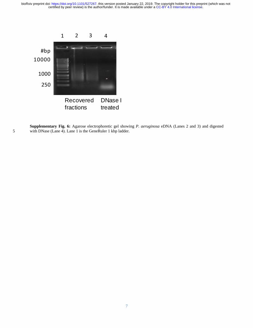
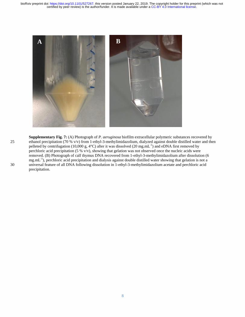
8
5
10
15
20
Supplementary Fig 7 (A) Photograph of P aeruginosa biofilm extracellular polymeric substances recovered by
ethanol precipitation (70 vv) from 1-ethyl-3-methylimidazolium dialyzed against double distilled water and then 25
pelleted by centrifugation (10000 g 4degC) after it was dissolved (20 mgmL-1) and eDNA first removed by
perchloric acid precipitation (5 vv) showing that gelation was not observed once the nucleic acids were
removed (B) Photograph of calf thymus DNA recovered from 1-ethyl-3-methylimidazolium after dissolution (6
mgmL-1) perchloric acid precipitation and dialysis against double distilled water showing that gelation is not a
universal feature of all DNA following dissolution in 1-ethyl-3-methylimidazolium acetate and perchloric acid 30
precipitation
A B
CC-BY 40 International licensecertified by peer review) is the authorfunder It is made available under aThe copyright holder for this preprint (which was notthis version posted January 22 2019 httpsdoiorg101101527267doi bioRxiv preprint
9
5
10
15
20
Supplementary Fig 8 1H-13C HSQC-TOCSY and 1-D 1H NMR spectra of P aeruginosa biofilm extracellular NA
gel isolate following alkalinization (01 M NaOD 10 mgmL-1 55degC 2 h) at 25degC showing a distribution of 1H-13C
HSQC-TOCSY cross peaks that is consistent with the presence of nucleic acids and the absence of proteins and
hexose-based sugars
25
13
C
1
H
CC-BY 40 International licensecertified by peer review) is the authorfunder It is made available under aThe copyright holder for this preprint (which was notthis version posted January 22 2019 httpsdoiorg101101527267doi bioRxiv preprint
10
Supplementary Fig 9
1H-1H COSY NMR spectrum of P aeruginosa biofilm extracellular NA gel isolate
following alkalinization (01 M NaOD 10 mgmL-1 55degC 2 h)
5
CC-BY 40 International licensecertified by peer review) is the authorfunder It is made available under aThe copyright holder for this preprint (which was notthis version posted January 22 2019 httpsdoiorg101101527267doi bioRxiv preprint
11
Supplementary Fig 10 1H-13C HMBC NMR spectrum of P aeruginosa biofilm extracellular NA gel isolate at
25degC following alkalinization (01 M NaOD 10 mgmL-1 55degC 2 h)
5
CC-BY 40 International licensecertified by peer review) is the authorfunder It is made available under aThe copyright holder for this preprint (which was notthis version posted January 22 2019 httpsdoiorg101101527267doi bioRxiv preprint
12
Supplementary Fig 11 1H NMR spectra of P aeruginosa biofilm NA gel isolate in D2O at 25degC
10 8 6 4 2 0
1H (ppm)
CC-BY 40 International licensecertified by peer review) is the authorfunder It is made available under aThe copyright holder for this preprint (which was notthis version posted January 22 2019 httpsdoiorg101101527267doi bioRxiv preprint
13
5
Supplementary Fig 12 31P NMR spectrum of RNA standard (torula yeast) at 25degC following alkalinization (01 M
NaOD 10 mgmL-1 55degC 2 h) (A) and in D2O following EMIM-Ac solubilisation and perchloric acid recovery (10
mgmL-1) (B) demonstrating that alkalinization only leads to RNA transesterification
000E+000
100E+008
200E+008
300E+008
400E+008
500E+008
Inte
nsity (
au
)
5 4 3 2 1 0 -1 -2 -3-600E+008
-400E+008
-200E+008
000E+000
200E+008 B
Inte
nsity (
au
)
31P (ppm)
A
CC-BY 40 International licensecertified by peer review) is the authorfunder It is made available under aThe copyright holder for this preprint (which was notthis version posted January 22 2019 httpsdoiorg101101527267doi bioRxiv preprint
14
5
10
15
20
25
Supplementary Fig 13 Micrograph of P aeruginosa NA gel isolate digested with DNase I and stained green with
SYTO RNASelect showing that SYTO RNASelect is binding to extracellular RNA (scale bar 10 microm) 30
CC-BY 40 International licensecertified by peer review) is the authorfunder It is made available under aThe copyright holder for this preprint (which was notthis version posted January 22 2019 httpsdoiorg101101527267doi bioRxiv preprint
15
Supplementary Fig 14 Storage modulus (Grsquo) loss modulus (Grsquorsquo) and complex viscosity (η) of P aeruginosa
wild type biofilm extracellular nucleic acid gel isolate in amplitude sweep at 25degC 50 microm gap 01 s-1 frequency
showing that the biofilm behaves like a gel at low amplitude (ie Grsquo gt Grsquorsquo) even after digestion with RNase III (A)
and RNase H (B) 5
001 01 1 10 100
001
01
1
G
G
GG
P
a
10
100
(m
Pas
)
001 01 1 10 100
001
01
1
GG
P
a
10
100
B
(m
Pas
)
A
CC-BY 40 International licensecertified by peer review) is the authorfunder It is made available under aThe copyright holder for this preprint (which was notthis version posted January 22 2019 httpsdoiorg101101527267doi bioRxiv preprint
16
Supplementary Fig 15 Agarose gel loaded with chromosomal DNA extracted from P aeruginosa pre-culture
planktonic cells (upper) and extracellular NA gel isolate (lower) (A) Gene coverage of P aeruginosa biofilm
chromosomal (upper) and extracellular (lower) DNA normalized against rpoB numbers The red oval denotes the 5
peak resulting from bacteriophage Pf1 genes (B)
115866E6 239371E6 364123E6 486767E6 608308E6
0
2
4
6
8
10
Extracellular DNA
Mea
n g
en
e c
overa
ge
Chromosomal DNA
115866E6 239371E6 364123E6 486767E6 608308E6
0
2
4
6
8
10
Region
Chromosomal DNA
Extracellular NA
A B
CC-BY 40 International licensecertified by peer review) is the authorfunder It is made available under aThe copyright holder for this preprint (which was notthis version posted January 22 2019 httpsdoiorg101101527267doi bioRxiv preprint
17
Supplementary Fig 16 (A) Normal stress differences (N1 ndashN2) and (B) shear stress as a function of shear rate for
Pseudomonas biofilms P putida P protogens and P aeruginosa ΔPf4 dissolved in 1-ethyl-3-methylimidazolium
acetate (40 mgmL-1) at 25 degC Lines indicate FENE-P fits to the data 5
A B
CC-BY 40 International licensecertified by peer review) is the authorfunder It is made available under aThe copyright holder for this preprint (which was notthis version posted January 22 2019 httpsdoiorg101101527267doi bioRxiv preprint
18
Supplementary Fig 17 Overnight growth curve for P aeruginosa cells grown in lithium and potassium enhanced
modified M9 media showing that potassium does not enhance planktonic growth over lithium
5
CC-BY 40 International licensecertified by peer review) is the authorfunder It is made available under aThe copyright holder for this preprint (which was notthis version posted January 22 2019 httpsdoiorg101101527267doi bioRxiv preprint
19
5
10
15
Supplementary Fig 18 Photograph of nucleic acid gel extracted from (A) P protegens and (B) P putida
A B
CC-BY 40 International licensecertified by peer review) is the authorfunder It is made available under aThe copyright holder for this preprint (which was notthis version posted January 22 2019 httpsdoiorg101101527267doi bioRxiv preprint
20
5
10
15
20
Supplementary Fig 19 Photographs of P aeruginosa biofilm after 5-d static growth in lysogeny broth medium at
37degC in 2 L Erlenmeyer flask (A) before centrifugation and (B) in 50 mL centrifuge tube after centrifugation
showing the separation of biofilm from supernatant 25
Biofilm
Supernatant
A B
CC-BY 40 International licensecertified by peer review) is the authorfunder It is made available under aThe copyright holder for this preprint (which was notthis version posted January 22 2019 httpsdoiorg101101527267doi bioRxiv preprint
21
Supplementary Table 1 Power law exponent (ie n in 1198731 = 119870119899 and m in 120590 = 119870120590119898) of P aeruginosa biofilms
following dissolution in EMIM-Ac m values approaching unity indicate a Newtonian-like fluid property Viscosity 5
is slightly shear thinning (m = 08 to 09) for all samples except RNase DNase and EMIM-Ac which are Newtonian
like (m ge 093) which would be expected from dilute polymer solutions in viscous fluids (ie Boger fluids)
120590 = 119870120590119898 1198731 = 1198701198731
119899
Sample K m KN1 n
Ave Std Dev Ave Std Dev Ave Std Dev Ave Std Dev
P
aeruginosa
wild type 063
008
084 001 144 045 136 0
Psl 126 035 076 004 781 518 121 015
Pel 035 0 089 0 058 015 134 002
Pronase 055 003 089 0 018 011 164 008
RNase 034 001 093 0 211 009 091 001
P putida 132 001 091 001 056 016 148 008
PDO300 073 035 089 001 233 059 110 003
P protegens 114 001 090 001 275 130 110 011
Pf4 057 002 087 001 159 125 133 024
DNase 020 001 099 001 - -
EMIM-Ac 013 0 098 0 - -
10
Supplementary Table 2 Fitting parameters for the FENE-P model including 1 = relaxation time b = a measure of
the relative extensibility of the model spring s = solvent viscosity p = polymer contribution to the
viscosity Molecular extensibility and relaxation times as predicted by FENE-P decrease in accordance with
elasticity
Average Std Deviation
Sample b 1 p s b 1 p s
P
aeruginosa
wild type 22485 0322 0260 0145
6431 0066 0040 00026
Psl 35115 0676 0381 0161 10009 0219 0113 00114
Pel 20008 0227 0110 0133 6378 0056 0000 00025
Pronase 18718 0103 0197 0167 6931 0047 0015 00339
RNase 3992 0102 0095 0183 176 0008 0003 00077
PDO300 9094 0453 0138 0147 1342 0023 0026 00007
DNase 1357 0005 0020 0129 1068 0001 0011 00112
Pf4 8654 0230 0243 0166 1299 0034 0022 00072
P protegens 1372 0175 0501 0414 192 0028 0025 00013
P putida 3369 0071 0544 0440 535 0002 0018 00199 15
CC-BY 40 International licensecertified by peer review) is the authorfunder It is made available under aThe copyright holder for this preprint (which was notthis version posted January 22 2019 httpsdoiorg101101527267doi bioRxiv preprint
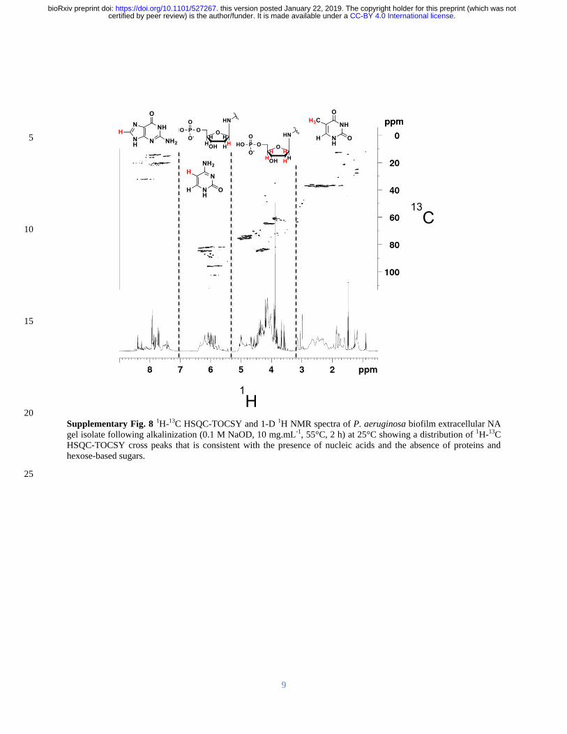
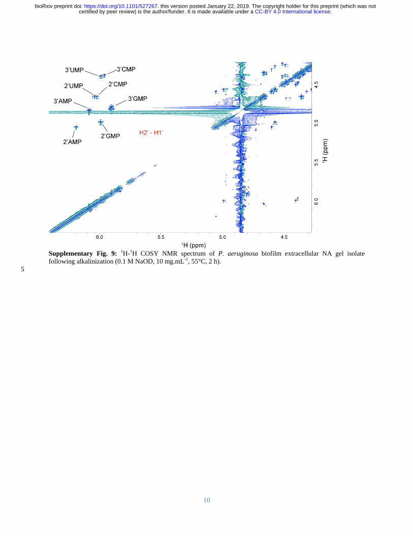
10
Supplementary Fig 9
1H-1H COSY NMR spectrum of P aeruginosa biofilm extracellular NA gel isolate
following alkalinization (01 M NaOD 10 mgmL-1 55degC 2 h)
5
CC-BY 40 International licensecertified by peer review) is the authorfunder It is made available under aThe copyright holder for this preprint (which was notthis version posted January 22 2019 httpsdoiorg101101527267doi bioRxiv preprint
11
Supplementary Fig 10 1H-13C HMBC NMR spectrum of P aeruginosa biofilm extracellular NA gel isolate at
25degC following alkalinization (01 M NaOD 10 mgmL-1 55degC 2 h)
5
CC-BY 40 International licensecertified by peer review) is the authorfunder It is made available under aThe copyright holder for this preprint (which was notthis version posted January 22 2019 httpsdoiorg101101527267doi bioRxiv preprint
12
Supplementary Fig 11 1H NMR spectra of P aeruginosa biofilm NA gel isolate in D2O at 25degC
10 8 6 4 2 0
1H (ppm)
CC-BY 40 International licensecertified by peer review) is the authorfunder It is made available under aThe copyright holder for this preprint (which was notthis version posted January 22 2019 httpsdoiorg101101527267doi bioRxiv preprint
13
5
Supplementary Fig 12 31P NMR spectrum of RNA standard (torula yeast) at 25degC following alkalinization (01 M
NaOD 10 mgmL-1 55degC 2 h) (A) and in D2O following EMIM-Ac solubilisation and perchloric acid recovery (10
mgmL-1) (B) demonstrating that alkalinization only leads to RNA transesterification
000E+000
100E+008
200E+008
300E+008
400E+008
500E+008
Inte
nsity (
au
)
5 4 3 2 1 0 -1 -2 -3-600E+008
-400E+008
-200E+008
000E+000
200E+008 B
Inte
nsity (
au
)
31P (ppm)
A
CC-BY 40 International licensecertified by peer review) is the authorfunder It is made available under aThe copyright holder for this preprint (which was notthis version posted January 22 2019 httpsdoiorg101101527267doi bioRxiv preprint
14
5
10
15
20
25
Supplementary Fig 13 Micrograph of P aeruginosa NA gel isolate digested with DNase I and stained green with
SYTO RNASelect showing that SYTO RNASelect is binding to extracellular RNA (scale bar 10 microm) 30
CC-BY 40 International licensecertified by peer review) is the authorfunder It is made available under aThe copyright holder for this preprint (which was notthis version posted January 22 2019 httpsdoiorg101101527267doi bioRxiv preprint
15
Supplementary Fig 14 Storage modulus (Grsquo) loss modulus (Grsquorsquo) and complex viscosity (η) of P aeruginosa
wild type biofilm extracellular nucleic acid gel isolate in amplitude sweep at 25degC 50 microm gap 01 s-1 frequency
showing that the biofilm behaves like a gel at low amplitude (ie Grsquo gt Grsquorsquo) even after digestion with RNase III (A)
and RNase H (B) 5
001 01 1 10 100
001
01
1
G
G
GG
P
a
10
100
(m
Pas
)
001 01 1 10 100
001
01
1
GG
P
a
10
100
B
(m
Pas
)
A
CC-BY 40 International licensecertified by peer review) is the authorfunder It is made available under aThe copyright holder for this preprint (which was notthis version posted January 22 2019 httpsdoiorg101101527267doi bioRxiv preprint
16
Supplementary Fig 15 Agarose gel loaded with chromosomal DNA extracted from P aeruginosa pre-culture
planktonic cells (upper) and extracellular NA gel isolate (lower) (A) Gene coverage of P aeruginosa biofilm
chromosomal (upper) and extracellular (lower) DNA normalized against rpoB numbers The red oval denotes the 5
peak resulting from bacteriophage Pf1 genes (B)
115866E6 239371E6 364123E6 486767E6 608308E6
0
2
4
6
8
10
Extracellular DNA
Mea
n g
en
e c
overa
ge
Chromosomal DNA
115866E6 239371E6 364123E6 486767E6 608308E6
0
2
4
6
8
10
Region
Chromosomal DNA
Extracellular NA
A B
CC-BY 40 International licensecertified by peer review) is the authorfunder It is made available under aThe copyright holder for this preprint (which was notthis version posted January 22 2019 httpsdoiorg101101527267doi bioRxiv preprint
17
Supplementary Fig 16 (A) Normal stress differences (N1 ndashN2) and (B) shear stress as a function of shear rate for
Pseudomonas biofilms P putida P protogens and P aeruginosa ΔPf4 dissolved in 1-ethyl-3-methylimidazolium
acetate (40 mgmL-1) at 25 degC Lines indicate FENE-P fits to the data 5
A B
CC-BY 40 International licensecertified by peer review) is the authorfunder It is made available under aThe copyright holder for this preprint (which was notthis version posted January 22 2019 httpsdoiorg101101527267doi bioRxiv preprint
18
Supplementary Fig 17 Overnight growth curve for P aeruginosa cells grown in lithium and potassium enhanced
modified M9 media showing that potassium does not enhance planktonic growth over lithium
5
CC-BY 40 International licensecertified by peer review) is the authorfunder It is made available under aThe copyright holder for this preprint (which was notthis version posted January 22 2019 httpsdoiorg101101527267doi bioRxiv preprint
19
5
10
15
Supplementary Fig 18 Photograph of nucleic acid gel extracted from (A) P protegens and (B) P putida
A B
CC-BY 40 International licensecertified by peer review) is the authorfunder It is made available under aThe copyright holder for this preprint (which was notthis version posted January 22 2019 httpsdoiorg101101527267doi bioRxiv preprint
20
5
10
15
20
Supplementary Fig 19 Photographs of P aeruginosa biofilm after 5-d static growth in lysogeny broth medium at
37degC in 2 L Erlenmeyer flask (A) before centrifugation and (B) in 50 mL centrifuge tube after centrifugation
showing the separation of biofilm from supernatant 25
Biofilm
Supernatant
A B
CC-BY 40 International licensecertified by peer review) is the authorfunder It is made available under aThe copyright holder for this preprint (which was notthis version posted January 22 2019 httpsdoiorg101101527267doi bioRxiv preprint
21
Supplementary Table 1 Power law exponent (ie n in 1198731 = 119870119899 and m in 120590 = 119870120590119898) of P aeruginosa biofilms
following dissolution in EMIM-Ac m values approaching unity indicate a Newtonian-like fluid property Viscosity 5
is slightly shear thinning (m = 08 to 09) for all samples except RNase DNase and EMIM-Ac which are Newtonian
like (m ge 093) which would be expected from dilute polymer solutions in viscous fluids (ie Boger fluids)
120590 = 119870120590119898 1198731 = 1198701198731
119899
Sample K m KN1 n
Ave Std Dev Ave Std Dev Ave Std Dev Ave Std Dev
P
aeruginosa
wild type 063
008
084 001 144 045 136 0
Psl 126 035 076 004 781 518 121 015
Pel 035 0 089 0 058 015 134 002
Pronase 055 003 089 0 018 011 164 008
RNase 034 001 093 0 211 009 091 001
P putida 132 001 091 001 056 016 148 008
PDO300 073 035 089 001 233 059 110 003
P protegens 114 001 090 001 275 130 110 011
Pf4 057 002 087 001 159 125 133 024
DNase 020 001 099 001 - -
EMIM-Ac 013 0 098 0 - -
10
Supplementary Table 2 Fitting parameters for the FENE-P model including 1 = relaxation time b = a measure of
the relative extensibility of the model spring s = solvent viscosity p = polymer contribution to the
viscosity Molecular extensibility and relaxation times as predicted by FENE-P decrease in accordance with
elasticity
Average Std Deviation
Sample b 1 p s b 1 p s
P
aeruginosa
wild type 22485 0322 0260 0145
6431 0066 0040 00026
Psl 35115 0676 0381 0161 10009 0219 0113 00114
Pel 20008 0227 0110 0133 6378 0056 0000 00025
Pronase 18718 0103 0197 0167 6931 0047 0015 00339
RNase 3992 0102 0095 0183 176 0008 0003 00077
PDO300 9094 0453 0138 0147 1342 0023 0026 00007
DNase 1357 0005 0020 0129 1068 0001 0011 00112
Pf4 8654 0230 0243 0166 1299 0034 0022 00072
P protegens 1372 0175 0501 0414 192 0028 0025 00013
P putida 3369 0071 0544 0440 535 0002 0018 00199 15
CC-BY 40 International licensecertified by peer review) is the authorfunder It is made available under aThe copyright holder for this preprint (which was notthis version posted January 22 2019 httpsdoiorg101101527267doi bioRxiv preprint
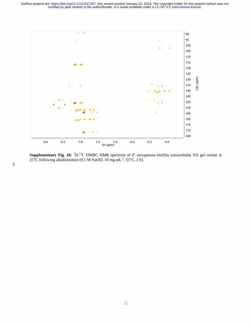
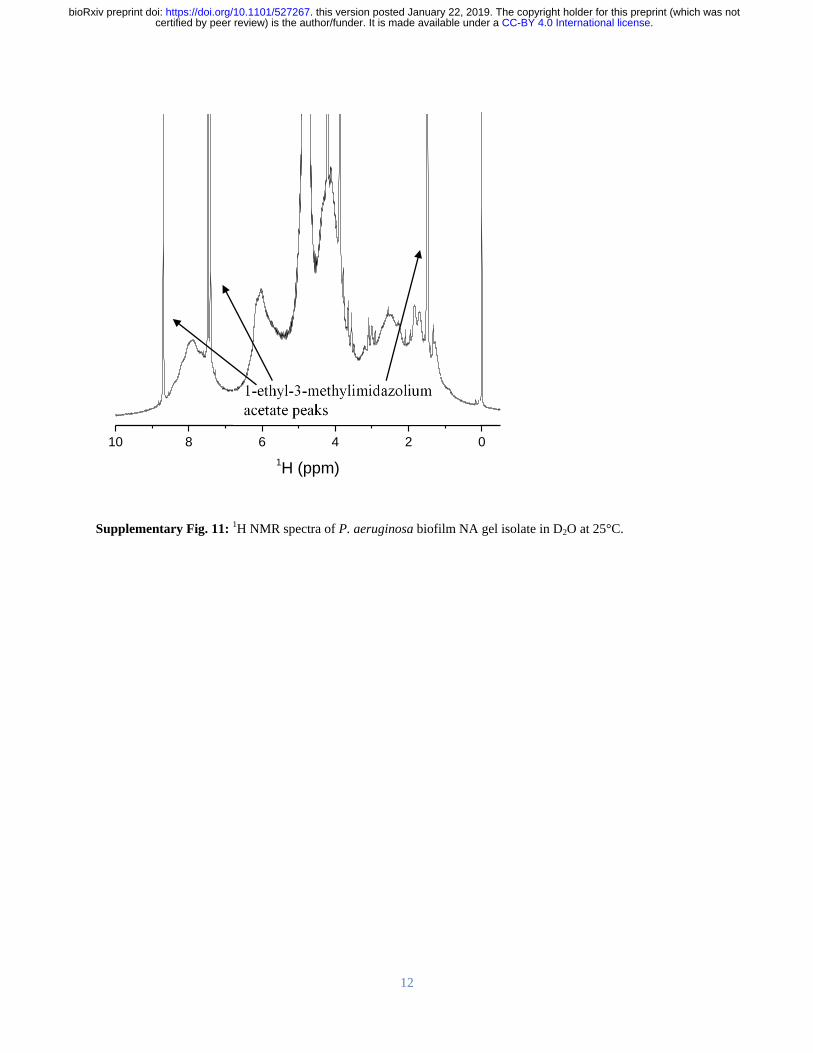
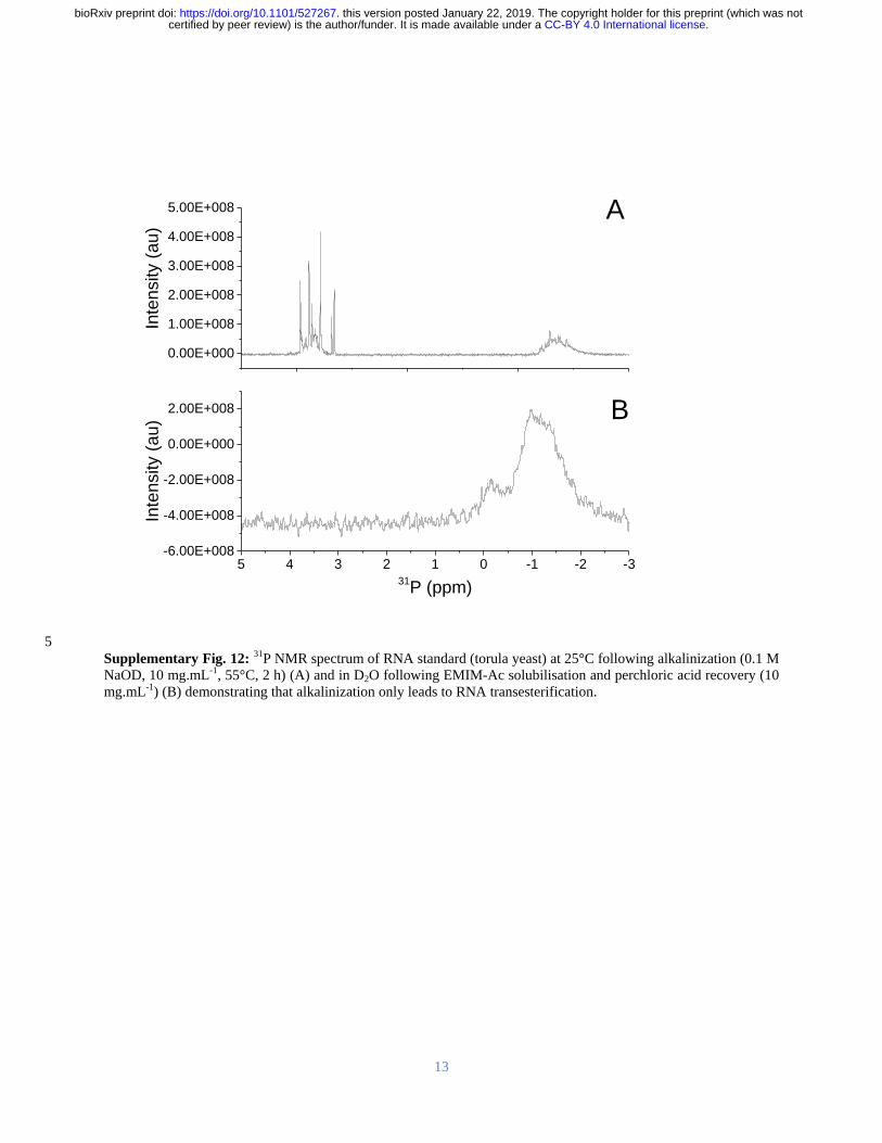
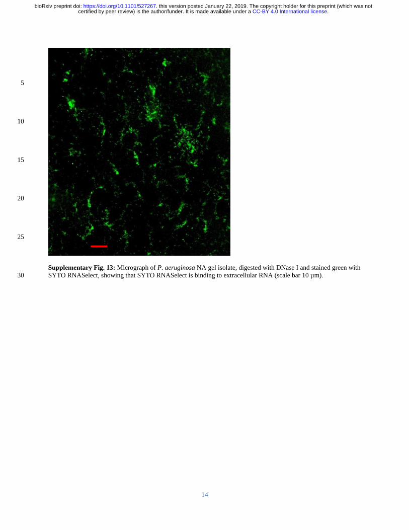
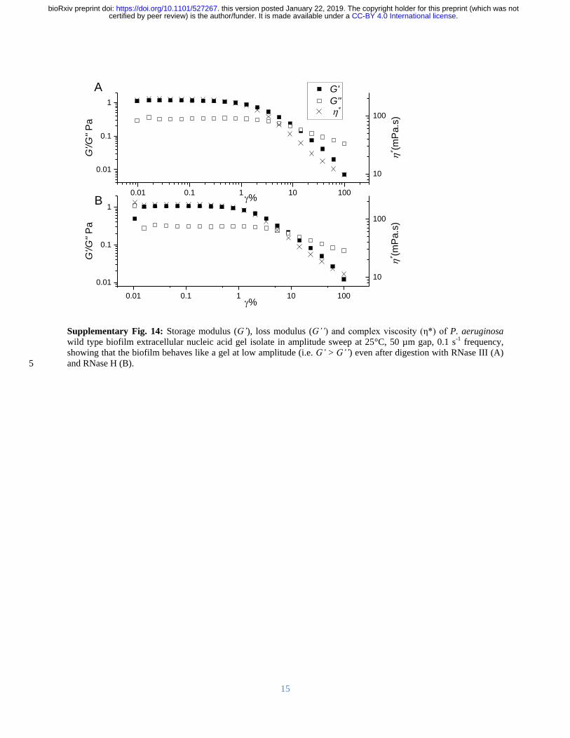
15
Supplementary Fig 14 Storage modulus (Grsquo) loss modulus (Grsquorsquo) and complex viscosity (η) of P aeruginosa
wild type biofilm extracellular nucleic acid gel isolate in amplitude sweep at 25degC 50 microm gap 01 s-1 frequency
showing that the biofilm behaves like a gel at low amplitude (ie Grsquo gt Grsquorsquo) even after digestion with RNase III (A)
and RNase H (B) 5
001 01 1 10 100
001
01
1
G
G
GG
P
a
10
100
(m
Pas
)
001 01 1 10 100
001
01
1
GG
P
a
10
100
B
(m
Pas
)
A
CC-BY 40 International licensecertified by peer review) is the authorfunder It is made available under aThe copyright holder for this preprint (which was notthis version posted January 22 2019 httpsdoiorg101101527267doi bioRxiv preprint
16
Supplementary Fig 15 Agarose gel loaded with chromosomal DNA extracted from P aeruginosa pre-culture
planktonic cells (upper) and extracellular NA gel isolate (lower) (A) Gene coverage of P aeruginosa biofilm
chromosomal (upper) and extracellular (lower) DNA normalized against rpoB numbers The red oval denotes the 5
peak resulting from bacteriophage Pf1 genes (B)
115866E6 239371E6 364123E6 486767E6 608308E6
0
2
4
6
8
10
Extracellular DNA
Mea
n g
en
e c
overa
ge
Chromosomal DNA
115866E6 239371E6 364123E6 486767E6 608308E6
0
2
4
6
8
10
Region
Chromosomal DNA
Extracellular NA
A B
CC-BY 40 International licensecertified by peer review) is the authorfunder It is made available under aThe copyright holder for this preprint (which was notthis version posted January 22 2019 httpsdoiorg101101527267doi bioRxiv preprint
17
Supplementary Fig 16 (A) Normal stress differences (N1 ndashN2) and (B) shear stress as a function of shear rate for
Pseudomonas biofilms P putida P protogens and P aeruginosa ΔPf4 dissolved in 1-ethyl-3-methylimidazolium
acetate (40 mgmL-1) at 25 degC Lines indicate FENE-P fits to the data 5
A B
CC-BY 40 International licensecertified by peer review) is the authorfunder It is made available under aThe copyright holder for this preprint (which was notthis version posted January 22 2019 httpsdoiorg101101527267doi bioRxiv preprint
18
Supplementary Fig 17 Overnight growth curve for P aeruginosa cells grown in lithium and potassium enhanced
modified M9 media showing that potassium does not enhance planktonic growth over lithium
5
CC-BY 40 International licensecertified by peer review) is the authorfunder It is made available under aThe copyright holder for this preprint (which was notthis version posted January 22 2019 httpsdoiorg101101527267doi bioRxiv preprint
19
5
10
15
Supplementary Fig 18 Photograph of nucleic acid gel extracted from (A) P protegens and (B) P putida
A B
CC-BY 40 International licensecertified by peer review) is the authorfunder It is made available under aThe copyright holder for this preprint (which was notthis version posted January 22 2019 httpsdoiorg101101527267doi bioRxiv preprint
20
5
10
15
20
Supplementary Fig 19 Photographs of P aeruginosa biofilm after 5-d static growth in lysogeny broth medium at
37degC in 2 L Erlenmeyer flask (A) before centrifugation and (B) in 50 mL centrifuge tube after centrifugation
showing the separation of biofilm from supernatant 25
Biofilm
Supernatant
A B
CC-BY 40 International licensecertified by peer review) is the authorfunder It is made available under aThe copyright holder for this preprint (which was notthis version posted January 22 2019 httpsdoiorg101101527267doi bioRxiv preprint
21
Supplementary Table 1 Power law exponent (ie n in 1198731 = 119870119899 and m in 120590 = 119870120590119898) of P aeruginosa biofilms
following dissolution in EMIM-Ac m values approaching unity indicate a Newtonian-like fluid property Viscosity 5
is slightly shear thinning (m = 08 to 09) for all samples except RNase DNase and EMIM-Ac which are Newtonian
like (m ge 093) which would be expected from dilute polymer solutions in viscous fluids (ie Boger fluids)
120590 = 119870120590119898 1198731 = 1198701198731
119899
Sample K m KN1 n
Ave Std Dev Ave Std Dev Ave Std Dev Ave Std Dev
P
aeruginosa
wild type 063
008
084 001 144 045 136 0
Psl 126 035 076 004 781 518 121 015
Pel 035 0 089 0 058 015 134 002
Pronase 055 003 089 0 018 011 164 008
RNase 034 001 093 0 211 009 091 001
P putida 132 001 091 001 056 016 148 008
PDO300 073 035 089 001 233 059 110 003
P protegens 114 001 090 001 275 130 110 011
Pf4 057 002 087 001 159 125 133 024
DNase 020 001 099 001 - -
EMIM-Ac 013 0 098 0 - -
10
Supplementary Table 2 Fitting parameters for the FENE-P model including 1 = relaxation time b = a measure of
the relative extensibility of the model spring s = solvent viscosity p = polymer contribution to the
viscosity Molecular extensibility and relaxation times as predicted by FENE-P decrease in accordance with
elasticity
Average Std Deviation
Sample b 1 p s b 1 p s
P
aeruginosa
wild type 22485 0322 0260 0145
6431 0066 0040 00026
Psl 35115 0676 0381 0161 10009 0219 0113 00114
Pel 20008 0227 0110 0133 6378 0056 0000 00025
Pronase 18718 0103 0197 0167 6931 0047 0015 00339
RNase 3992 0102 0095 0183 176 0008 0003 00077
PDO300 9094 0453 0138 0147 1342 0023 0026 00007
DNase 1357 0005 0020 0129 1068 0001 0011 00112
Pf4 8654 0230 0243 0166 1299 0034 0022 00072
P protegens 1372 0175 0501 0414 192 0028 0025 00013
P putida 3369 0071 0544 0440 535 0002 0018 00199 15
CC-BY 40 International licensecertified by peer review) is the authorfunder It is made available under aThe copyright holder for this preprint (which was notthis version posted January 22 2019 httpsdoiorg101101527267doi bioRxiv preprint
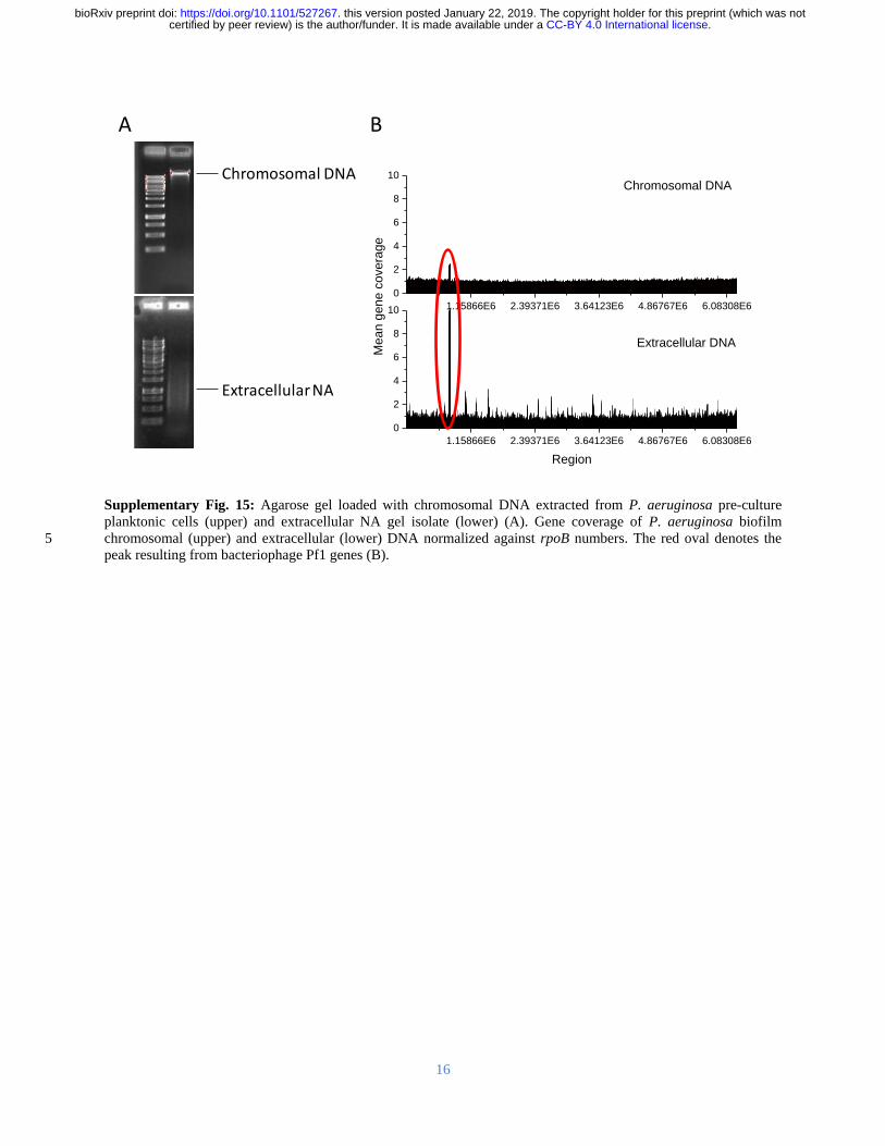
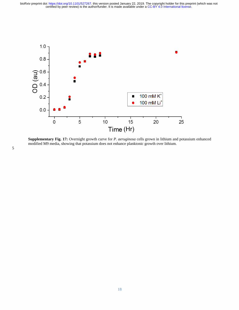

20
5
10
15
20
Supplementary Fig 19 Photographs of P aeruginosa biofilm after 5-d static growth in lysogeny broth medium at
37degC in 2 L Erlenmeyer flask (A) before centrifugation and (B) in 50 mL centrifuge tube after centrifugation
showing the separation of biofilm from supernatant 25
Biofilm
Supernatant
A B
CC-BY 40 International licensecertified by peer review) is the authorfunder It is made available under aThe copyright holder for this preprint (which was notthis version posted January 22 2019 httpsdoiorg101101527267doi bioRxiv preprint
21
Supplementary Table 1 Power law exponent (ie n in 1198731 = 119870119899 and m in 120590 = 119870120590119898) of P aeruginosa biofilms
following dissolution in EMIM-Ac m values approaching unity indicate a Newtonian-like fluid property Viscosity 5
is slightly shear thinning (m = 08 to 09) for all samples except RNase DNase and EMIM-Ac which are Newtonian
like (m ge 093) which would be expected from dilute polymer solutions in viscous fluids (ie Boger fluids)
120590 = 119870120590119898 1198731 = 1198701198731
119899
Sample K m KN1 n
Ave Std Dev Ave Std Dev Ave Std Dev Ave Std Dev
P
aeruginosa
wild type 063
008
084 001 144 045 136 0
Psl 126 035 076 004 781 518 121 015
Pel 035 0 089 0 058 015 134 002
Pronase 055 003 089 0 018 011 164 008
RNase 034 001 093 0 211 009 091 001
P putida 132 001 091 001 056 016 148 008
PDO300 073 035 089 001 233 059 110 003
P protegens 114 001 090 001 275 130 110 011
Pf4 057 002 087 001 159 125 133 024
DNase 020 001 099 001 - -
EMIM-Ac 013 0 098 0 - -
10
Supplementary Table 2 Fitting parameters for the FENE-P model including 1 = relaxation time b = a measure of
the relative extensibility of the model spring s = solvent viscosity p = polymer contribution to the
viscosity Molecular extensibility and relaxation times as predicted by FENE-P decrease in accordance with
elasticity
Average Std Deviation
Sample b 1 p s b 1 p s
P
aeruginosa
wild type 22485 0322 0260 0145
6431 0066 0040 00026
Psl 35115 0676 0381 0161 10009 0219 0113 00114
Pel 20008 0227 0110 0133 6378 0056 0000 00025
Pronase 18718 0103 0197 0167 6931 0047 0015 00339
RNase 3992 0102 0095 0183 176 0008 0003 00077
PDO300 9094 0453 0138 0147 1342 0023 0026 00007
DNase 1357 0005 0020 0129 1068 0001 0011 00112
Pf4 8654 0230 0243 0166 1299 0034 0022 00072
P protegens 1372 0175 0501 0414 192 0028 0025 00013
P putida 3369 0071 0544 0440 535 0002 0018 00199 15
CC-BY 40 International licensecertified by peer review) is the authorfunder It is made available under aThe copyright holder for this preprint (which was notthis version posted January 22 2019 httpsdoiorg101101527267doi bioRxiv preprint