Embed Size (px)
Citation preview
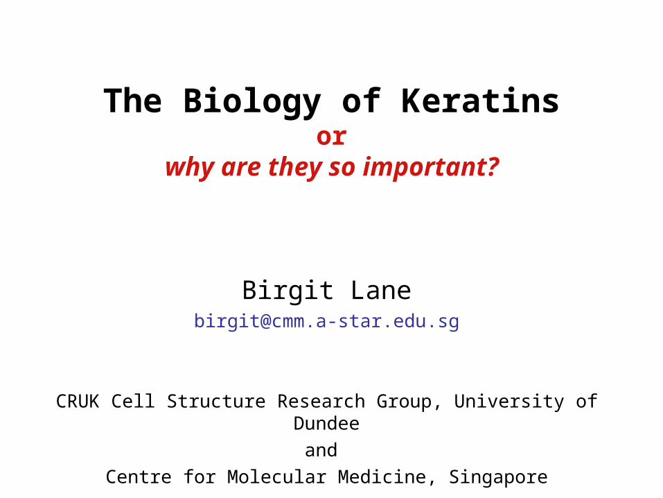
The Biology of Keratinsor
why are they so important?
Birgit [email protected]
CRUK Cell Structure Research Group, University of Dundee
and
Centre for Molecular Medicine, Singapore
immunofluorescence
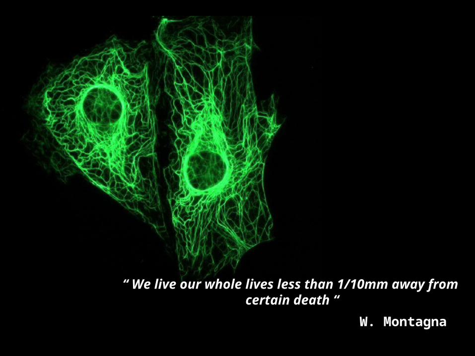
“ We live our whole lives less than 1/10mm away from certain death “
W. Montagna
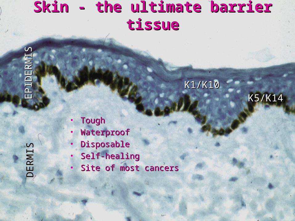
• Tough Tough • Waterproof Waterproof • DisposableDisposable• Self-healingSelf-healing • Site of most cancersSite of most cancers
Skin - the ultimate barrier tissueSkin - the ultimate barrier tissue
K5/K14K5/K14K1/K10K1/K10
EPID
ER
MIS
EPID
ER
MIS
DER
MIS
DER
MIS

QuickTime™ and aCinepak decompressor
are needed to see this picture.
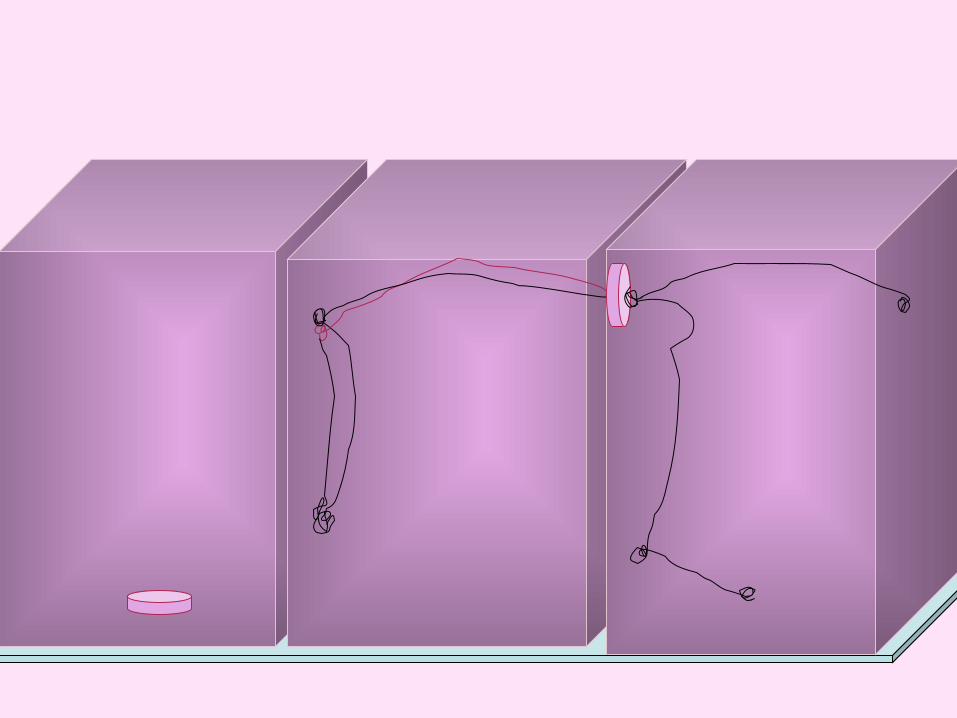
Scratch wound assays
movie: Sharon Jenner
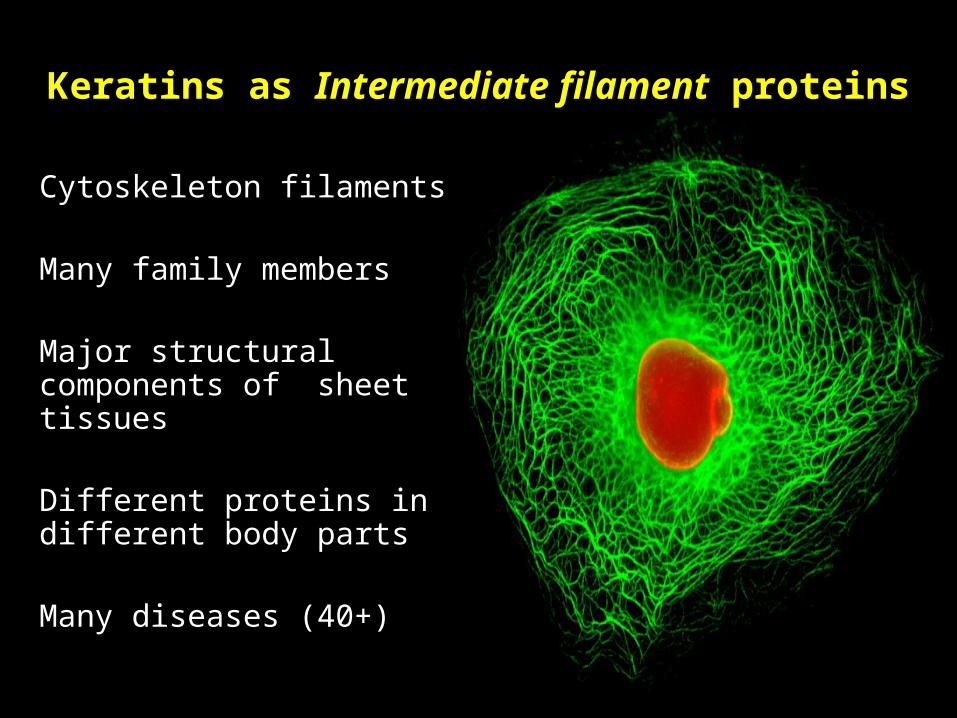
Keratins as Intermediate filament proteins
Cytoskeleton filaments
Many family members
Major structural components of sheet tissues
Different proteins in different body parts
Many diseases (40+)
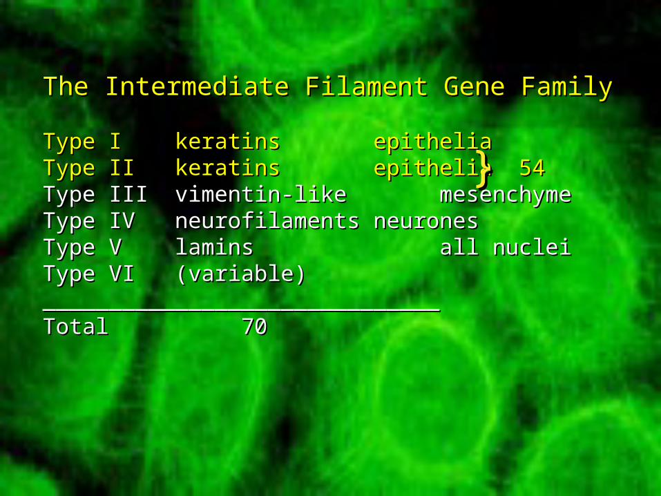
The Intermediate Filament Gene FamilyThe Intermediate Filament Gene Family
Type IType I keratinskeratins epitheliaepitheliaType IIType II keratinskeratins epitheliaepithelia 54 54Type IIIType III vimentin-likevimentin-like mesenchymemesenchymeType IVType IV neurofilamentsneurofilaments neuronesneuronesType VType V laminslamins all nucleiall nucleiType VIType VI (variable)(variable)____________________________________________________________TotalTotal 7070
}}

Structure of intermediate filaments
after Herrmann and Aebi
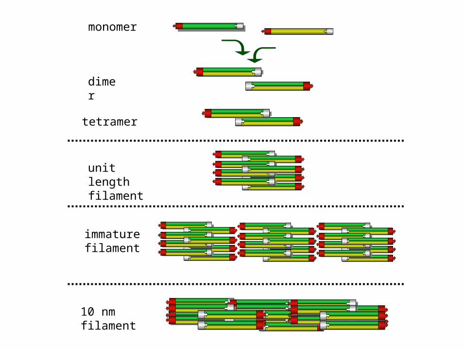
monomer
dimer
tetramer
unit length filament
10 nm filament
immaturefilament

stra
tifi
ed (
bar
rier
)s i
mp
le
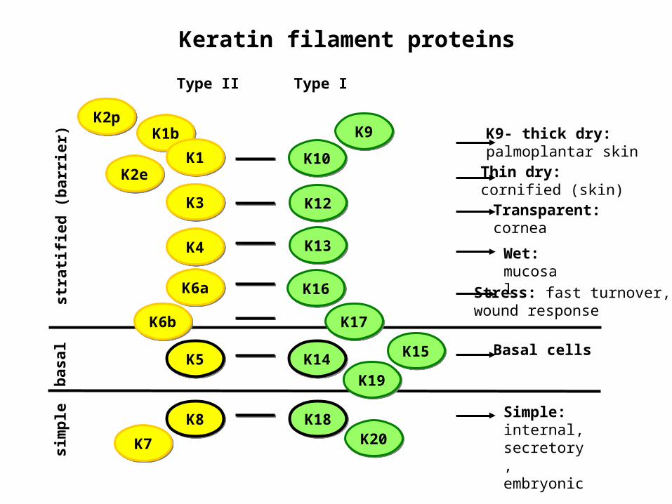
Basal cells
Transparent: cornea
Wet: mucosal
Thin dry: cornified (skin)
Stress: fast turnover, wound response
K9- thick dry: palmoplantar skin
Simple: internal,secretory,embryonic
Type II Type I
K2eK2e
K9K9K2pK2p
K10K10
K12K12K3K3
K18K18K8K8
K19K19K14K14K5K5 K15K15
K13K13K4K4
K7K7 K20K20
K16K16K6aK6a
K17K17K6bK6b
K1bK1b
K1K1
bas
alKeratin filament proteins
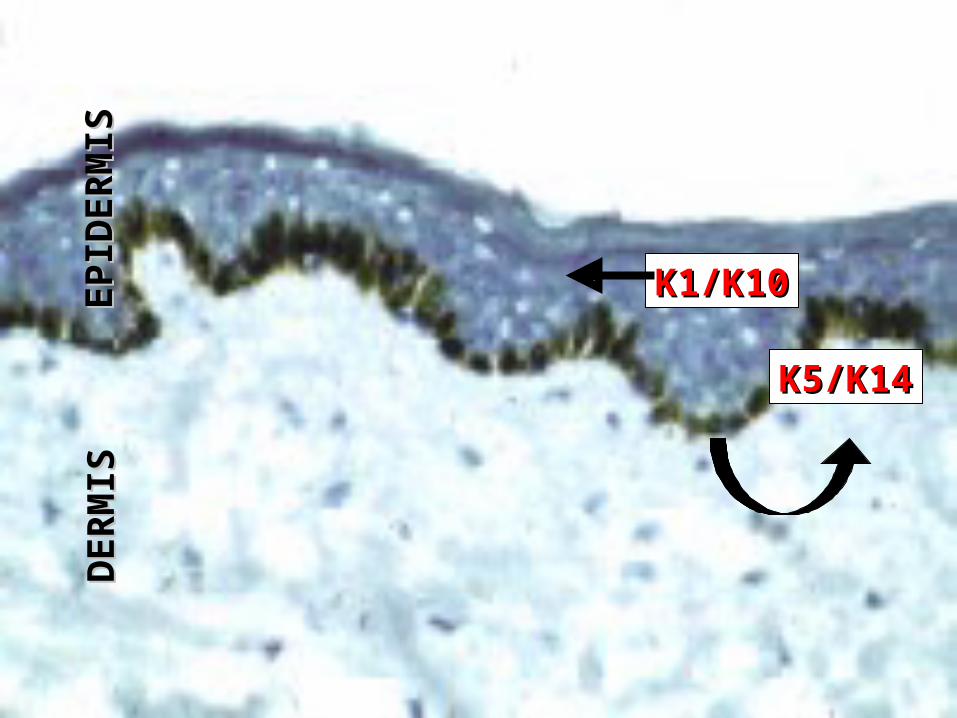
K5/K14K5/K14
K1/K10K1/K10
EPIDERMIS
EPIDERMIS
DERMIS
DERMIS
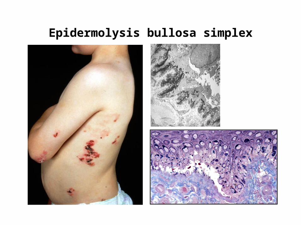
Epidermolysis bullosa simplex

stra
tifi
ed (
bar
rier
)s i
mp
le
Basal cells
Transparent: cornea
Wet: mucosal
Thin dry: cornified (skin)
Stress: fast turnover, wound response
K9- thick dry: palmoplantar skin
Simple: internal,secretory,embryonic
Type II Type I
K2eK2e
K9K9K2pK2p
K10K10
K12K12K3K3
K18K18K8K8
K19K19K14K14K5K5 K15K15
K13K13K4K4
K7K7 K20K20
K16K16K6aK6a
K17K17K6bK6b
K1bK1b
K1K1
bas
alKeratin filament proteins
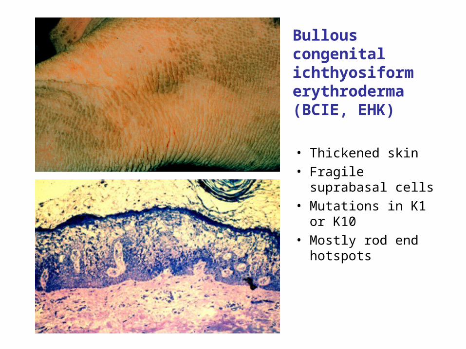
Bullous congenital ichthyosiform erythroderma (BCIE, EHK)
• Thickened skin• Fragile suprabasal cells• Mutations in K1 or K10• Mostly rod end hotspots

stra
tifi
ed (
bar
rier
)s i
mp
le
Basal cells
Transparent: cornea
Wet: mucosal
Thin dry: cornified (skin)
Stress: fast turnover, wound response
K9- thick dry: palmoplantar skin
Simple: internal,secretory,embryonic
Type II Type I
K2eK2e
K9K9K2pK2p
K10K10
K12K12K3K3
K18K18K8K8
K19K19K14K14K5K5 K15K15
K13K13K4K4
K7K7 K20K20
K16K16K6aK6a
K17K17K6bK6b
K1bK1b
K1K1
bas
alKeratin filament proteins
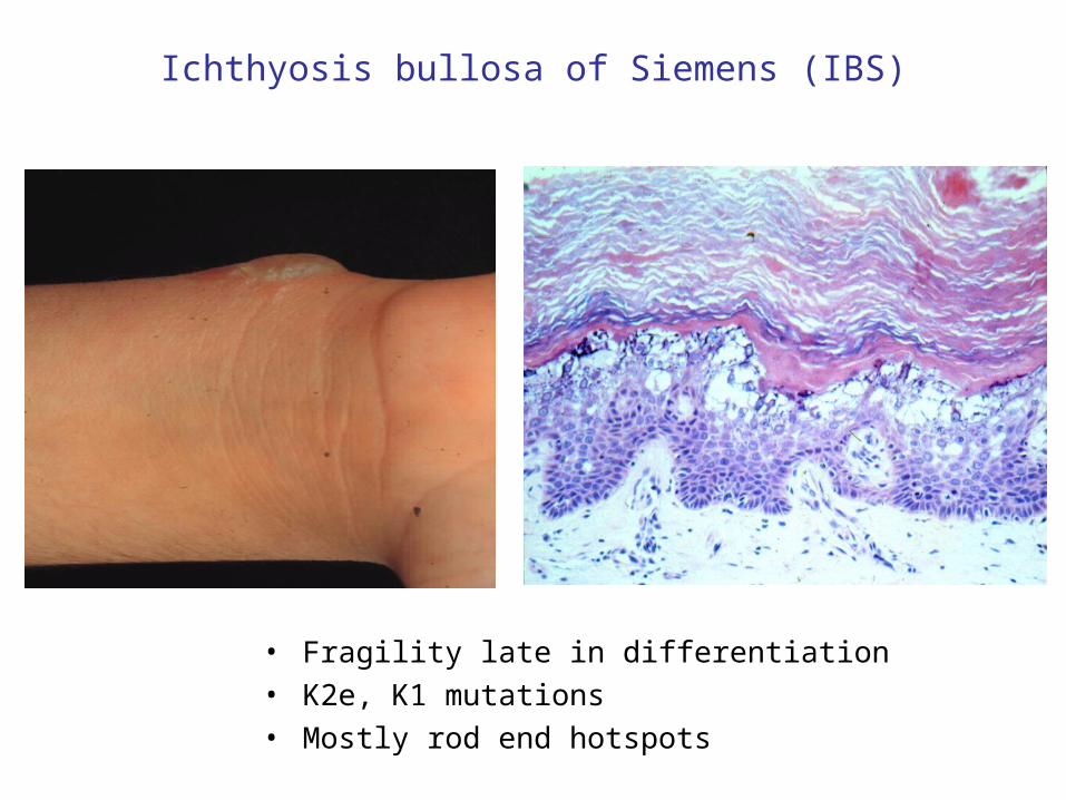
• Fragility late in differentiation• K2e, K1 mutations• Mostly rod end hotspots
Ichthyosis bullosa of Siemens (IBS)
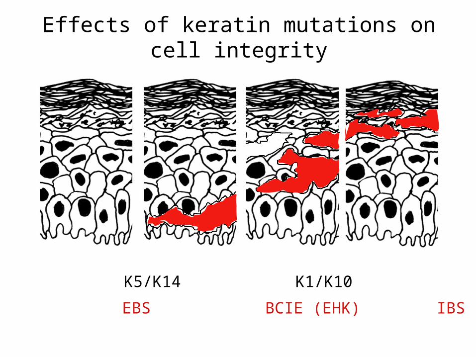
None K5/K14 K1/K10 K2e
Effects of keratin mutations on cell integrity
EBS BCIE (EHK) IBS
stra
tifi
ed (
bar
rier
)s i
mp
le
Basal cells
Transparent: cornea
Wet: mucosal
Thin dry: cornified (skin)
Stress: fast turnover, wound response
K9- thick dry: palmoplantar skin
Simple: internal,secretory,embryonic
Type II Type I
K2eK2e
K9K9K2pK2p
K10K10
K12K12K3K3
K18K18K8K8
K19K19K14K14K5K5 K15K15
K13K13K4K4
K7K7 K20K20
K16K16K6aK6a
K17K17K6bK6b
K1bK1b
K1K1
bas
alKeratin filament proteins
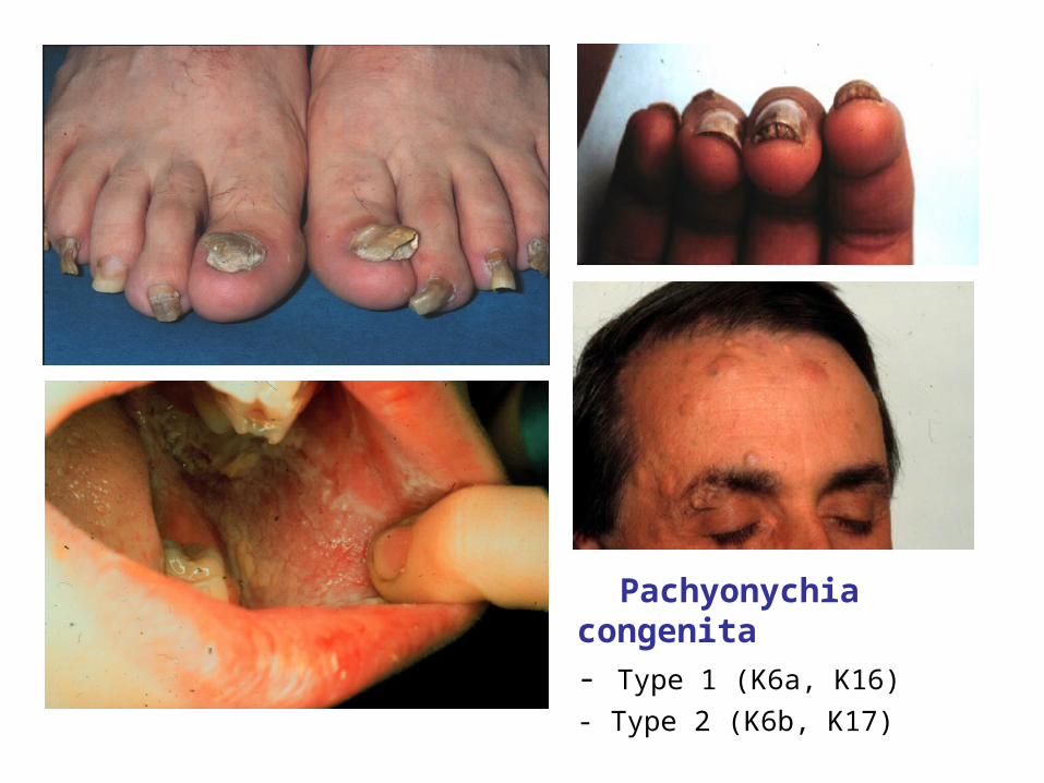
Pachyonychia congenita
- Type 1 (K6a, K16)
- Type 2 (K6b, K17)

K6a/K16, K6b/K17 - the stress response keratins
K6a, K16
- PC-1
K6b, K17
- PC-2
• major keratins in “wet” skin surfaces; outer hair follicle sheath; nail bed.
• NOT normally in thin “dry”skin but more in thick skin
• INDUCED by trauma, wounding• Often expressed in tissue cultured cells
• Small amounts in “wet” epithelia. Lots in deep hair follicle sheath; some basal cells; nail bed. Specialized and unspecialized cells.
• NOT normally in thin “dry” skin.• INDUCED by trauma, wounding, inflammation• Often expressed in tissue cultured cells
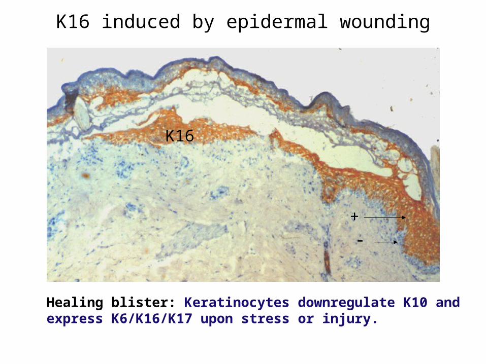
K16 induced by epidermal wounding
Healing blister: Keratinocytes downregulate K10 and express K6/K16/K17 upon stress or injury.
+-
K16
epidermis
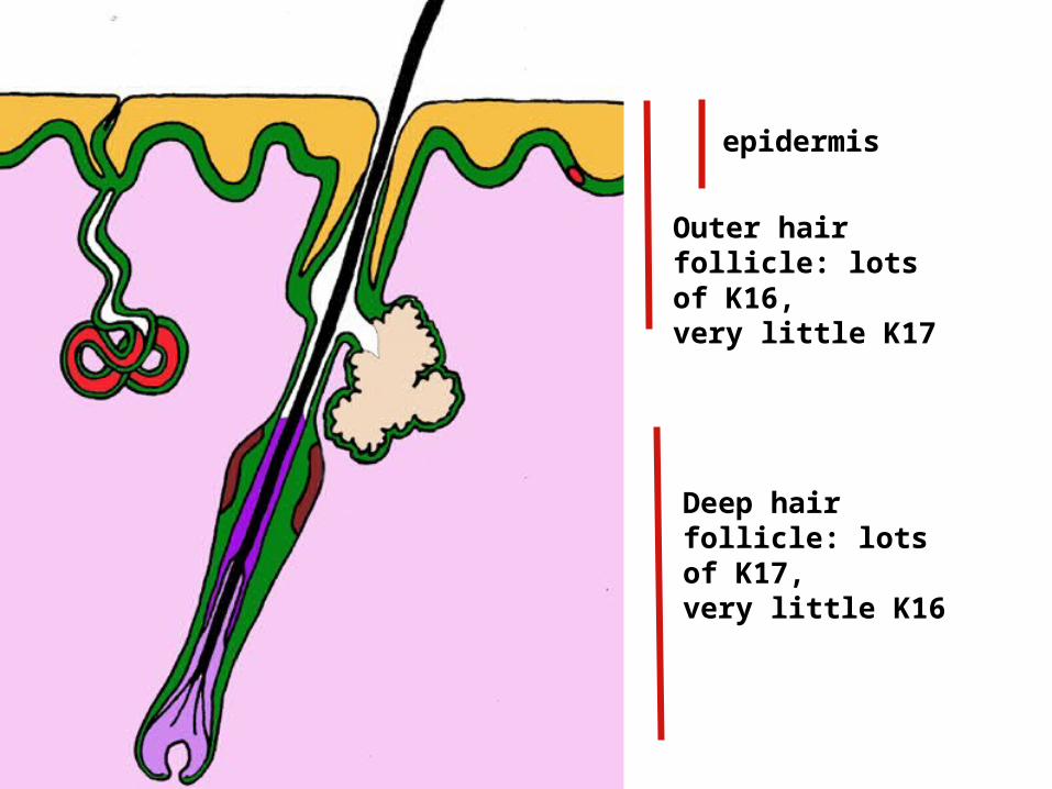
Outer hair follicle: lots of K16, very little K17
Deep hair follicle: lots of K17, very little K16
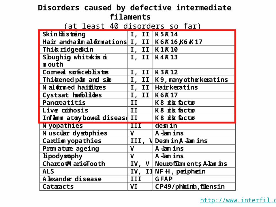
Disorders caused by defective intermediate filaments (at least 40 disorders so far)
Skin blistering I, II K5/K14 Hair and nail malformations I, II K6/K16, K6/K17 Thick ridged skin I, II K1/K10 Sloughing white skin in mouth
I, II K4/K13
Corneal surface blisters I, II K3/K12 Thickened palm and sole I, II K9, many other keratins Malformed hair fibres I, II Hair keratins Cysts at hair follicles I, II K6/K17 Pancreatitis II K8 risk factor Liver cirrhosis II K8 risk factor Inflammatory bowel disease II K8 risk factor Myopathies III desmin Muscular dystrophies V A-lamins Cardiomyopathies III, V Desmin, A-lamins Premature ageing V A-lamins lipodystrophy V A-lamins Charcot-Marie-Tooth IV, V Neurofilaments, A-lamins ALS IV, III NF-H, peripherin Alexander disease III GFAP Cataracts VI CP49/phakinin, filensin
http://www.interfil.org


![Differentiation-Dependent Expression of Keratins in Human Oral … · 2017. 2. 1. · [ 16, 17]. The expression of specific keratins appears to depend on the type of tissue, as well](https://img.pdfslide.net/doc/110x75/5ff979cead588c6cd35f8d9b/differentiation-dependent-expression-of-keratins-in-human-oral-2017-2-1-16.jpg)