Embed Size (px)
Citation preview
Dr. Rene V. YatDr. Rene V. Yat
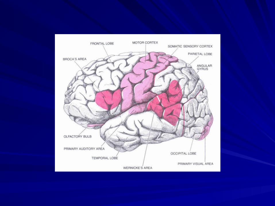
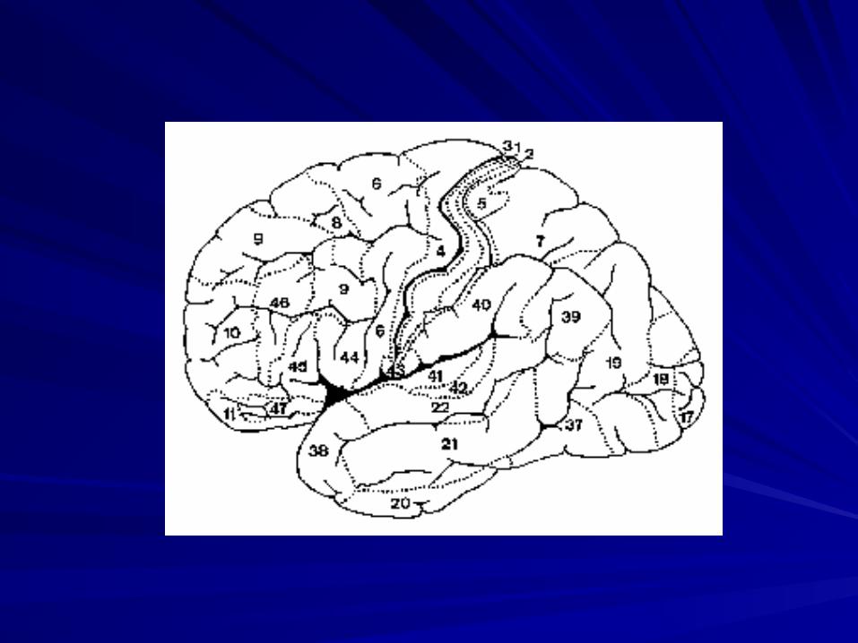
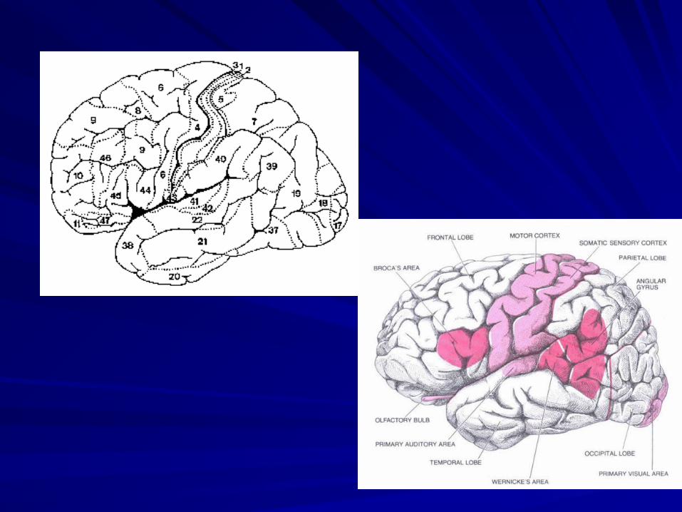
The Brain and Human The Brain and Human BehaviorBehavior

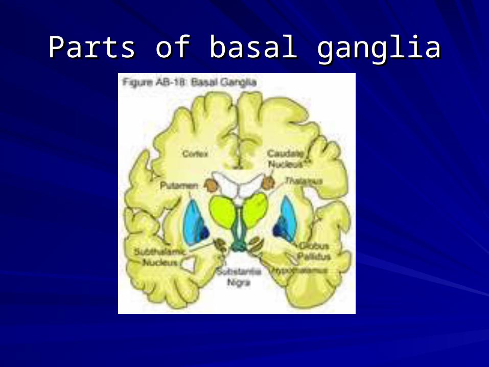
Parts of basal gangliaParts of basal ganglia
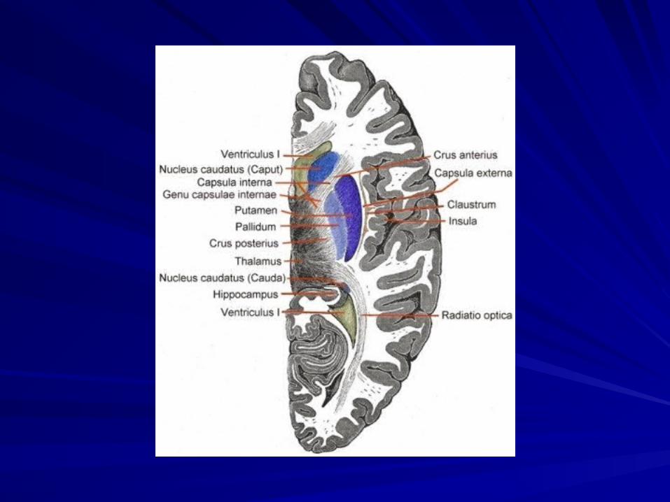
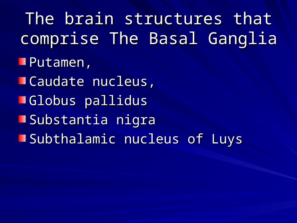
The brain structures that comprise The brain structures that comprise The Basal Ganglia The Basal Ganglia
Putamen, Putamen,
Caudate nucleus, Caudate nucleus,
Globus pallidus Globus pallidus
Substantia nigra Substantia nigra
Subthalamic nucleus of Luys Subthalamic nucleus of Luys
Conditions that cause basal ganglia Conditions that cause basal ganglia dysfunctiondysfunction
Drug overdoseDrug overdoseHead injuryHead injuryInfectionInfectionLiver diseaseLiver diseaseMetabolic processesMetabolic processesMultiple sclerosisMultiple sclerosisStrokeStrokeTumoursTumoursSide effects of medicationsSide effects of medications
Brain Disorders associated with Brain Disorders associated with Basal ganglia dysfunctionBasal ganglia dysfunction
Dystonias Dystonias
Huntington’s diseaseHuntington’s disease
Parkinson’s diseaseParkinson’s disease
Supranuclear PalsySupranuclear Palsy
Wilson’s diseaseWilson’s disease
The dopamine pathways in schizophrenia
In schizophrenia there is an increase in dopamine transmission between the substantia nigra to the caudate nucleus-putamen (neostriatum) compared with normal. While in the other major dopaminergic pathways — to the mesolimbic forebrain and the tubero-infundibular system — dopamine transmission is reduced. The dopamine hypothesis of schizophrenia proposes that increased levels of dopamine or dopamine receptors in the dorsal and or ventral striatum underlie the disorder.
The glutamate pathways in a brain affected by schizophrenia
In the normal brain the prominent glutaminergic pathways are: the cortico-cortical pathways; the pathways between the thalamus and the cortex; and the extrapyramidal pathway (the projections between the cortex and striatum). Other glutamate projections exist between the cortex, substantia nigra, subthalamic nucleus and pallidum. The glutaminergic pathways are hypoactive in the brains of people diagnosed with schizophrenia and this is thought to cause the confusion and psychosis associated with the disorder.
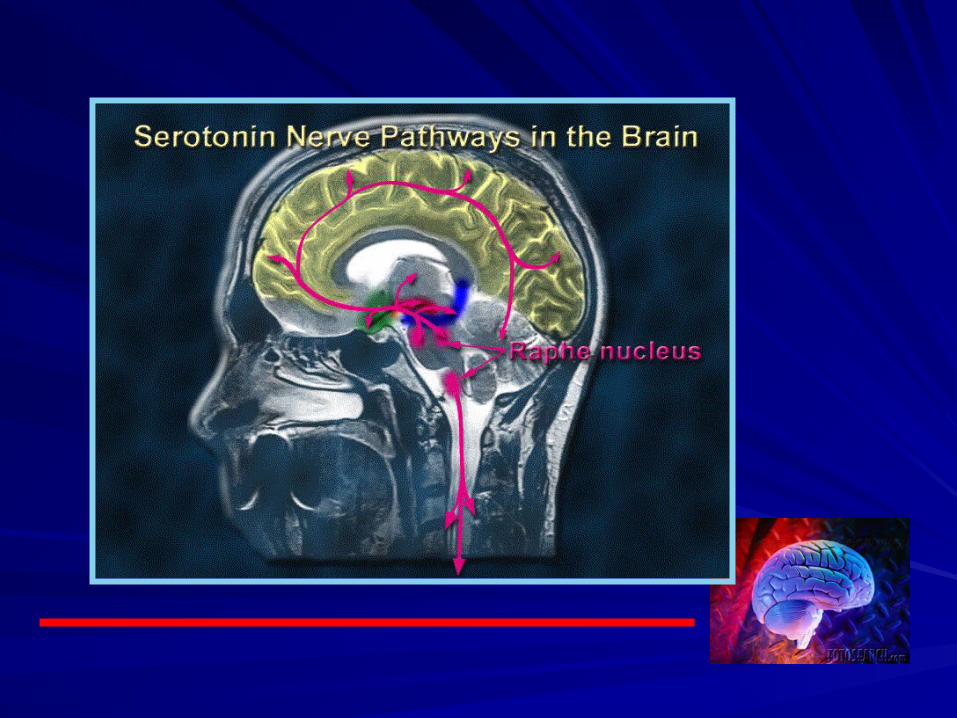
The serotonergic pathway showing the effects of schizophrenia
The two key serotonergic pathways in schizophrenia are the projections from the dorsal raphe nuclei into the substantia nigra and the projections from the rostral raphe nuclei ascending into the cerebral cortex, limbic regions and basal ganglia. The up-regulation of these pathways leads to hypofunction of the dopaminergic system, and this effect may be responsible for the negative symptoms of schizophrenia. The serotonergic nuclei in the brainstem that give rise to descending serotonergic axons remain unaffected in schizophrenia.
NEUROTRANSMITTERSNEUROTRANSMITTERS
Biogenic aminesBiogenic amines
Amino acidsAmino acids
PeptidesPeptides
NEUROTRANSMITTERSNEUROTRANSMITTERS
Biogenic aminesBiogenic amines
DopamineDopamine
NorepinephrineNorepinephrine
EpinephrineEpinephrine
SerotoninSerotonin
HistamineHistamine
AcetylcholineAcetylcholine
NEUROTRANSMITTERSNEUROTRANSMITTERS
Amino AcidsAmino Acids Amino acids are the most abundant Amino acids are the most abundant
neurotransmitters in the brain. Nichols suggested: neurotransmitters in the brain. Nichols suggested: “amino acids synapses exceed those of all the “amino acids synapses exceed those of all the other neurotransmitters combined…amino acids other neurotransmitters combined…amino acids are responsible for almost all the fast signaling are responsible for almost all the fast signaling between neurons, leaving predominantly between neurons, leaving predominantly modulatory roles for the other transmitters.” modulatory roles for the other transmitters.”
Amino acidAmino acidNEUROTRANSMITTERSNEUROTRANSMITTERSThe second neurotransmitter family is composed The second neurotransmitter family is composed of of amino acidsamino acids, organic compounds containing , organic compounds containing both an amino group (NH2) and a carboxylic acid both an amino group (NH2) and a carboxylic acid group (COOH). Amino acids that serve as group (COOH). Amino acids that serve as neurotransmitters include glycine, glutamic and neurotransmitters include glycine, glutamic and aspartic acids, and gamma-amino butyric acid aspartic acids, and gamma-amino butyric acid (GABA). Glutamic acid and GABA are the most (GABA). Glutamic acid and GABA are the most abundant neurotransmitters within the central abundant neurotransmitters within the central nervous system, and especially in the cerebral nervous system, and especially in the cerebral cortex, which is largely responsible for such higher cortex, which is largely responsible for such higher brain functions as thought and interpreting brain functions as thought and interpreting sensations sensations
NEUROTRANSMITTERSNEUROTRANSMITTERS
Amino AcidsAmino AcidsGlutamateGlutamate
GABAGABA
GlycineGlycine
L-ArginineL-Arginine
NEUROTRANSMITTERSNEUROTRANSMITTERS
Amino AcidsAmino AcidsGlutamateGlutamate is the major excitatory neurotransmitter is the major excitatory neurotransmitter and is distributed in all regions of the brain. and is distributed in all regions of the brain. Aspartate is closely related to glutamate and the Aspartate is closely related to glutamate and the two amino acids are often found together at axon two amino acids are often found together at axon terminals. Neurons synthesize glutamate and terminals. Neurons synthesize glutamate and aspartate and are independent of dietary supply. aspartate and are independent of dietary supply.
NEUROTRANSMITTERSNEUROTRANSMITTERS
Amino AcidsAmino AcidsGamma amino butyric acid (GABA)Gamma amino butyric acid (GABA) is the major is the major inhibitory neurotransmitter in the brain, derived inhibitory neurotransmitter in the brain, derived from glucose, which is transaminated in the Kreb’s from glucose, which is transaminated in the Kreb’s cycle to glutamine and then converted to GABA by cycle to glutamine and then converted to GABA by the enzyme, glutamic acid decarboxylase. The the enzyme, glutamic acid decarboxylase. The production of GABA appears to be independent of production of GABA appears to be independent of the dietary supply of glutamine but requires dietary the dietary supply of glutamine but requires dietary pyridoxine pyridoxine
NEUROTRANSMITTERSNEUROTRANSMITTERS
Amino AcidsAmino AcidsGlycine Glycine is an inhibitory neurotransmitter found is an inhibitory neurotransmitter found mostly in the brain stem and spinal cord. A major mostly in the brain stem and spinal cord. A major discovery that adds complexity to the already discovery that adds complexity to the already confusing story of neurotransmitters is that glycine confusing story of neurotransmitters is that glycine acts as a co-transmitter in excitatory NMDA acts as a co-transmitter in excitatory NMDA synapses. synapses.
NEUROTRANSMITTERSNEUROTRANSMITTERS
Amino AcidsAmino AcidsL-Arginine L-Arginine is the precursor of endogenous nitric oxide (NO), is the precursor of endogenous nitric oxide (NO), which is a vasodilator acting via the intracellular second-which is a vasodilator acting via the intracellular second-messenger cGMP. In healthy humans, L-arginine induces messenger cGMP. In healthy humans, L-arginine induces peripheral vasodilation and inhibits platelet aggregation due peripheral vasodilation and inhibits platelet aggregation due to an increased NO production. Prostaglandin E1 (PGE1) to an increased NO production. Prostaglandin E1 (PGE1) induces peripheral vasodilation via stimulating prostacyclin induces peripheral vasodilation via stimulating prostacyclin receptors. receptors. A mixture of branch-chain amino acids, leucine, valine and A mixture of branch-chain amino acids, leucine, valine and isoleucine will reduce tardive dyskinesia and movement isoleucine will reduce tardive dyskinesia and movement disorder that is caused by anti-schizophrenic drugs. Tarvil, disorder that is caused by anti-schizophrenic drugs. Tarvil, has been marketed in the USA that delivers 6.0 grams of the has been marketed in the USA that delivers 6.0 grams of the 3 amino acids per packet. A dose of 6 gm three times a day 3 amino acids per packet. A dose of 6 gm three times a day has been recommended. has been recommended.
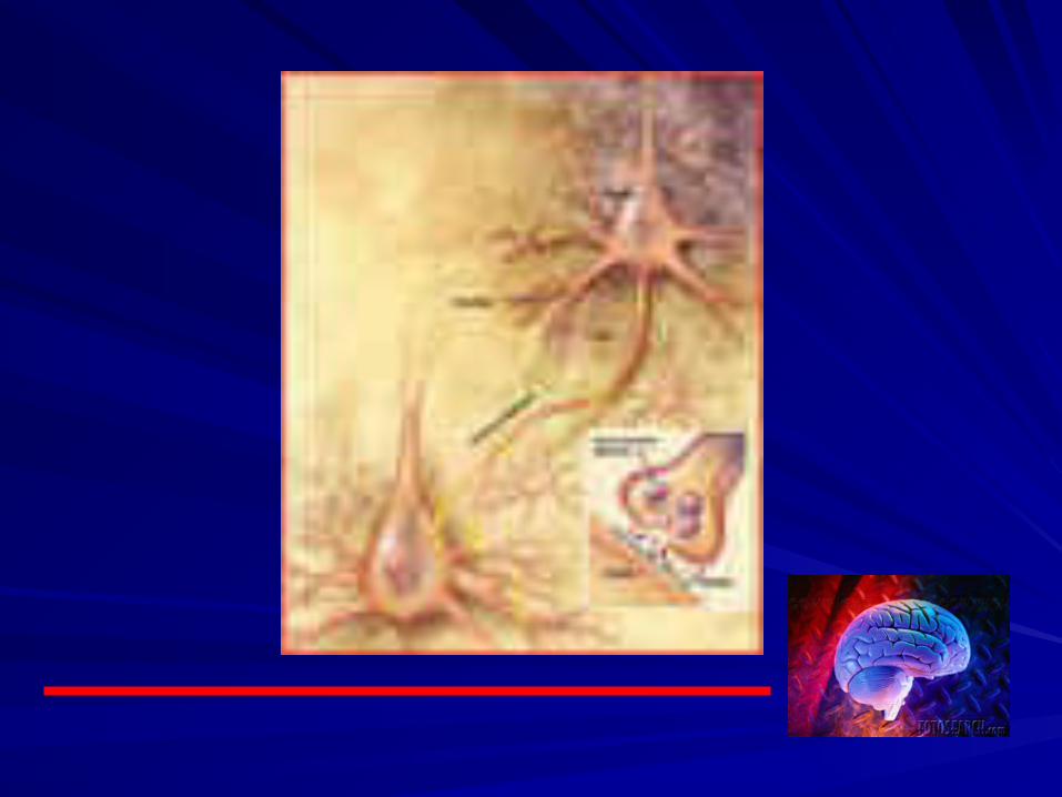
NeuropeptidesFunction of Neuropeptides: There are cells in the brain that produce various neuropeptides, and these neuropeptides do just about everything. They can be either pro-inflammatory or anti-inflammatory, with anti-inflammatory being preferred. They are responsible for many functions: They control our mood, energy levels, pain and pleasure reception, body weight, and ability to solve problems; they also form memories and regulate our immune system. These active little messengers in the brain actually turn on cellular function in the skin.
Characteristics of Neuropeptides: Peptides are compounds consisting of two or more amino acids (the building blocks of proteins), chained together by what is called a peptide bond. Neuropeptides are peptides released by neurons (brain cells) as intercellular messengers. Some neuropeptides function as neurotransmitters, and others function as hormones. Peptides and neuropeptides, like many substances in our bodies (think cholesterol) can work both for and against us. Anti-inflammatory neuropeptides work for us to reduce inflamation fo the skin.
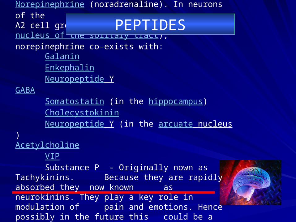
Transmitter names are shown in bold.Norepinephrine (noradrenaline). In neurons of the A2 cell group in the nucleus of the solitary tract), norepinephrine co-exists with:
Galanin Enkephalin Neuropeptide Y
GABASomatostatin (in the hippocampus) Cholecystokinin Neuropeptide Y (in the arcuate nucleus)
AcetylcholineVIP Substance P - Originally nown as Tachykinins.
Because they are rapidly absorbed they now known as neurokinins. They play a key role in modulation of pain and emotions. Hence possibly in the future this could be a source of new antidepresants
PEPTIDES
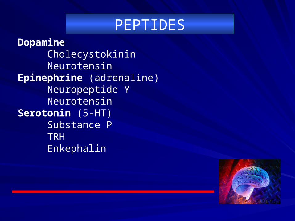
DopamineCholecystokinin Neurotensin
Epinephrine (adrenaline)Neuropeptide Y Neurotensin
Serotonin (5-HT)Substance P TRH Enkephalin
PEPTIDES
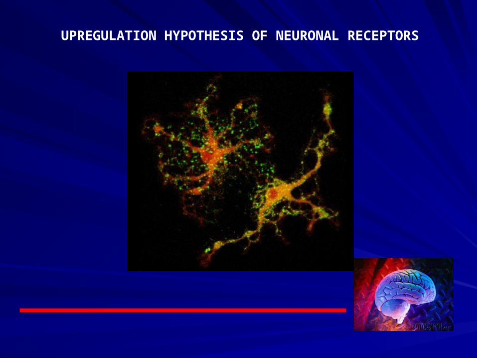
UPREGULATION HYPOTHESIS OF NEURONAL RECEPTORS
Supersensitivity is a compensatory response of the postsynaptic neuron when it receives too little stimulation. The neuron tries to make up for a lack of stimulation by increasing receptor responsiveness. Over time, the postsynaptic neuron may also compensate for lack of stimulation by synthesizing additional receptor sites. This process is known as up-regulation.
Upregulation theory
By increasing the amount of neurotransmitter in the cleft, you can normalize responsiveness. Increased neurotransmitter increases stimulation of receptor sites, which prompts the postsynaptic neuron to compensate by decreasing receptor sensitivity, a process known as desensitization
DESENSITIZATION THEORY
The postsynaptic neuron is also thought to compensate for increasing stimulation by decreasing the number of receptor sites, a process known as down-regulation.
Downregulation hypothesis

““Prolonged sitting can cause Prolonged sitting can cause ischial bursitis”.ischial bursitis”.

““To study medicine without books and To study medicine without books and mentors is like a shaman who professes mentors is like a shaman who professes who know everything but deep inside he who know everything but deep inside he knows nothing at all.knows nothing at all. You can shake, rattle, and roll. But at the You can shake, rattle, and roll. But at the end of the day, you wish that you have end of the day, you wish that you have studied hard for the life you are handling in studied hard for the life you are handling in front of you is not a guinea pig at all”.front of you is not a guinea pig at all”.

END OF THE LECTUREEND OF THE LECTURE