Embed Size (px)
Citation preview
the cellbiology 1
• How we investigate cells
• Cell types and size
• A tour of the cell
• The nucleus
• The endomembrane system
• Specialized organelles
How we investigate cells• Microscopy
– Light microscope (Hooke 1665)• Magnification x1000• Limited by resolution of light energy (0.2 µm)
– Electron microscope (since 1950s)• Uses beam of electrons to resolve 0.2nm)
– Transmission electron microscope (TEM) examines internal structure
– Scanning electron microscope (SEM) examines surface features (3d)
• Other techniques include cell fractionation (via centrifuge)
Cell types and sizes• First cells observed in 1665 by Hooke
were cork cells
• Further work by Leeuwenhoek, Schleiden and Schwann led to proposal of cell theory, which states– Organisms are composed of one or more
cells– Cells are the smallest unit of life– Cells arise only by division of previously
existing cells
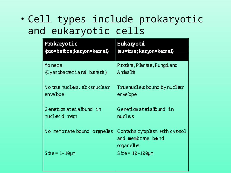
• Cell types include prokaryotic and eukaryotic cells
Prokaryotic(pro=before; karyon=kernel)
Eukaryotic(eu=true; karyon=kernel)
Monera
(Cyanobacteria and bacteria)
Protista, Plantae, Fungi, and
Animalia
No true nucleus, lacks nuclear
envelope
True nucleus bound by nuclear
envelope
Genetic material found in
nucleoid region
Genetic material found in
nucleus
No membrane bound organelles Contains cytoplasm with cytosol
and membrane bound
organelles
Size = 1–10µm Size = 10–100µm
The importance of surface area:volume ratio
• Let amount of metabolism that occurs within a cell be a function of cell volume
• Let rate of metabolism (which is a function of supply of reactants, and removal of products) be a function of surface area
• Therefore,surface area:volume ratio (SA:Vol) dictates the efficiency of a cell– Small cells have high SA:Vol, and are therefore efficient– Large cells tend towards a low SA:Vol ratio, and are
therefore inefficient
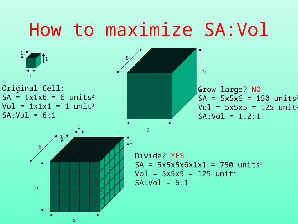
How to maximize SA:Vol
Original Cell:SA = 1x1x6 = 6 units2
Vol = 1x1x1 = 1 unit3
SA:Vol = 6:1
11
15
5
5
Grow large? NOSA = 5x5x6 = 150 units2
Vol = 5x5x5 = 125 unit3
SA:Vol = 1.2:1
11
1
5
5
5
Divide? YESSA = 5x5x5x6x1x1 = 750 units2
Vol = 5x5x5 = 125 unit3
SA:Vol = 6:1
SA:Vol – the limiting factor in cell size
• Prokaryotes, because of their smaller size, have optimum SA:Vol
• Single-celled eukaryotes which are larger cells compensate by an internal membrane system that partitions cells into compartments– Folded surface maximizes surface area for reaction– Membranes incorporate some enzymes that participate
in reactions– Compartments provide localized environment for
reactions
• For larger biomasses, eukaryotic strategy is instead to divide = multicellular organisms, with cell specialization
A tour of the cell• All cells have at least three components
– Nucleus or nucleoid region
– Plasma membrane
– Cytoplasm
• In addition, eukaryotic cells have membrane bound organelles, and endomembrane system
• Prokaryotic cells have an additional boundary - the cell wall, primarily a peptidoglycan matrix (review Prokaryotes, pp.84-85)
• Some eukaryotic cells (e.g., plants) also have a cell wall, which instead is cellulose based
Eukaryotes: the Nucleus• Most, but not all eukaryotic cells have a nucleus• Nucleus contains a majority of the genetic information
of the cell• Contains a mixture of DNA and proteins complexes
known as histones—this mixture is known as chromatin
• Enclosed by a nuclear envelope—a double bilipid layer membrane– Outside membrane is continuous with the cell’s endo-
membrane system– Inside membrane may be bound to a nuclear matrix
• Nucleolus is the site of rRNA production
The endomembrane system• Divides cell into compartments• Includes
– endoplasmic reticulum (ER)• Rough ER (with ribosomes)• Smooth ER (without ribosomes)
– Golgi apparatus• Golgi bodies
– (Lysosomes, vacuoles)
• Responsible for production of various macromolecules, including proteins and some lipids
The ER’s role in protein production• mRNA, transcribed from DNA, enters the cell’s
cytoplasm from the nucleus through a nuclear pore• mRNA binds with a ribosome, and migrates to the
surface of the ER• As polypeptide chain is assembled, it is passed into
the lumen of the ER• Polypeptide is passed along ER and budded off into
vesicle that travels to the cis face of a golgi body• Final assembly of molecule occurs in Golgi body,
vesicle budded off of trans face• Transport vesicle containing final assembled protein
travels to destination
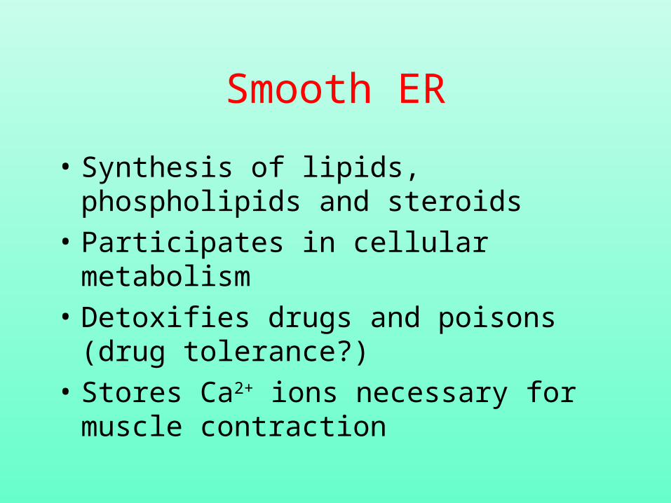
Smooth ER
• Synthesis of lipids, phospholipids and steroids
• Participates in cellular metabolism
• Detoxifies drugs and poisons (drug tolerance?)
• Stores Ca2+ ions necessary for muscle contraction
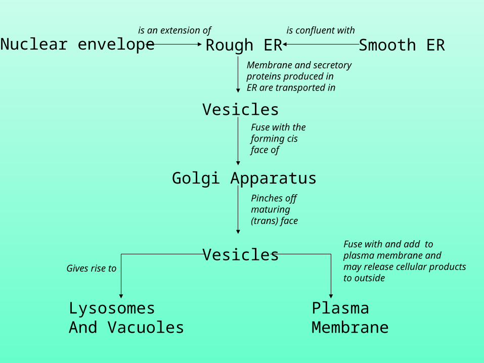
Rough ERNuclear envelope Smooth ER
Vesicles
Golgi Apparatus
Vesicles
LysosomesAnd Vacuoles
PlasmaMembrane
is an extension of is confluent with
Membrane and secretoryproteins produced inER are transported in
Fuse with theforming cis face of
Pinches off maturing (trans) face
Fuse with and add toplasma membrane andmay release cellular productsto outside
Gives rise to
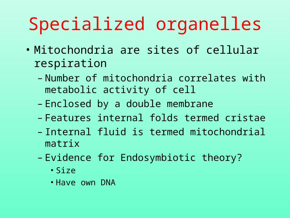
Specialized organelles
• Mitochondria are sites of cellular respiration– Number of mitochondria correlates with
metabolic activity of cell– Enclosed by a double membrane– Features internal folds termed cristae– Internal fluid is termed mitochondrial matrix– Evidence for Endosymbiotic theory?
• Size• Have own DNA
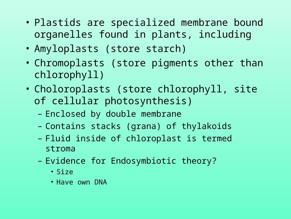
• Plastids are specialized membrane bound organelles found in plants, including
• Amyloplasts (store starch)• Chromoplasts (store pigments other than
chlorophyll)• Choloroplasts (store chlorophyll, site of
cellular photosynthesis)– Enclosed by double membrane– Contains stacks (grana) of thylakoids– Fluid inside of chloroplast is termed stroma– Evidence for Endosymbiotic theory?
• Size• Have own DNA
Cell walls• Some cells have an additional, structural
boundary outside of the plasma membrane• In plants, the cell wall is cellulose based• In bacteria, the cell wall is peptidoglycan
based– Gram positive and negative bacteria
• Structural rigidity of cell wall allows cells to take on more water than an animal cell could
The cytosol
• Contains cytoplasm and cytoskeleton• Cytoskeleton = network of fibers throughout
the cytoplasm that forms a dynamic framework for support and movement. Constructed from:– Microtubules (e.g., cilia, flagella, centrioles)– Intermediate filaments (e.g., the cellular scaffold– Microfilaments (actin: muscle contraction,
localized contractions of portions of cell)
The plasma membrane
• Just one example of a lipid bilayer membrane found in cells
• Controls passage of molecules in and out of the cell
• Like all membranes, represents a complex interactions of phospholipids, proteins and carbohydrates
Membrane theory
• Davson-Danielli model suggested phospholipid bilayer sandwiched between two layers of globular protein– Suggests a symmetrical geometry - in fact
not the case (inside and outside face)– Some membranes looked different, and had
different functions– Since most proteins are hydrophobic, woul
not be stable
The Fluid Mosaic Model• Proposed by Singer-Nicolson• Membrane is a mosaic of proteins
‘bobbing’ in a fluid phospholipid bilayer• Hydrophilic portions of phospholipid and
proteins are maximally exposed to aqueous interface, ensuring stability
• Most lipids and some proteins drift laterally across surface of membrane - fluidity is aided by addition of kinked hydrocarbon tails
Molecules found in the phospholipid bilayer matrix
• Integral proteins – Unilateral or transmembrane
• Peripheral proteins
• Carbohydrates (cell-cell recognition)– Oligosaccharides– Glycolipids– Glycoproteins
Permeability - the ultimate control of metabolism?
• Membranes display selective permeability– Solubility characteristics of the phospholipid bilayer
• Nonpolar molecules dissolve in membrane• Small polar molecules pass easily• Large polar molecules do not pass easily• Ions are usually pumped across
– Presence of specific integral transport proteins• Hydrophilic tunnel through membrane?• May bind to substance and move it across
Types of transport• Passive transport systems
– Diffusion - movement of molecules down a concentration gradient (high conc. to low conc.). Relies on intrinsic kinetic energy of molecules
– Osmosis (the diffusion of water) - the movement of water from a low concentration solute (hypertonic solution) to a high concentration solute (hypotonic solution)
– Facilitated diffusion. Uses transport proteins
• Active transport– Requires energy (ATP) to pump molecules against
concentration gradient
Large molecule transport across a membrane
• Exocytosis (out of) and endocytosis (into the cell)– Phagocytosis (engulfment of particles
using pseudopodia)– Pinocytosis (engulfment of fluid using
pseudopodia)– Receptor-mediated endocytosis
Cell Communication
• How do cells talk to each other? There are four general methods (Raven and Johnson, Figure 7.3:
• Direct contact
• Paracrine signalling (short-lived, local)
• Endocrine signalling (hormones)
• Synaptic signalling (neurotransmitters)
A general model of communication...
• Signal production• Signal Transmission• Signal Reception
How are signal received? Proteins play a key role - their 3-dimensionality provides specificity to signal types– Intracellular– Cell surface
Intracellular receptors• Lipid soluble or small molecule signals pass directly
through cell membrane to receptor• Receptors are almost always enzymes-addition of
chemical signal either inhibits or activates enzyme activity by altering shape of protein
• Effected enzymes may be in the cytoplasm or nucleus (DNA transcription)
• For example, Nitric Oxide activates the enzyme Guanylyl cyclase, which catalyzes the production of cyclic guanosine monophosphate, causing relaxation of smooth muscles around vessels, increasing blood flow
Cell Surface receptors
• Chemically gated ion channels
• Enzymic receptors
• G-protein linked receptors
Raven and Johnson Figure 7.6
Chemically gated ion channels
• Multipass protein whose core provides passage to ions if gate binds with signal
• Channels are specific to particular ions, depending on geometry
• Influx of ions (e.g., Ca2+, K+, Na+) has variable effects according to cell type
Enzymic receptors
• Binding of signal to exterior of trans-membrane protein reconfigure enzyme to be active on inside of cell
• In most cases the enzyme is a kinase, causing phosphorylation
G-Protein linked receptors
• Guanosine triphosphate (GTP)-binding protein
• Trans-membrane multipass protein (7 times) creates channel through membrane
• Addition of signal causes protein to bind GTP, activating it. GTP-protein complex then diffuses out to target
How does the receptor effect a change in the cell?
• Typically, the resultant change will effect secondary signal - commonly Ca2+ or cAMP (cyclic adenosine monophosphate)
• An amplification system causes the signal to proliferate throughout the cell
How to cells stick together
• Tight junctions• Anchoring junctions (cadherin-mediated
and integrin mediated links)– Hemidesmosome– Desmosome
• Communicating junctions– Gap junction– Plasmodesmata